adipose stem cells and their paracrine factors are
TRANSCRIPT
RESEARCH Open Access
Adipose stem cells and their paracrinefactors are therapeutic for early retinalcomplications of diabetes in the Ins2Akita
mouseSally L. Elshaer1,3, William Evans1, Mickey Pentecost4, Raji Lenin1, Ramesh Periasamy1, Kumar Abhiram Jha1,Shanta Alli1, Jordy Gentry1, Samuel M. Thomas1, Nicolas Sohl4 and Rajashekhar Gangaraju1,2*
Abstract
Background: Early-stage diabetic retinopathy (DR) is characterized by neurovascular defects. In this study, wehypothesized that human adipose-derived stem cells (ASCs) positive for the pericyte marker CD140b, or theirsecreted paracrine factors, therapeutically rescue early-stage DR features in an Ins2Akita mouse model.
Methods: Ins2Akita mice at 24 weeks of age received intravitreal injections of CD140b-positive ASCs (1000 cells/1 μL) or20× conditioned media from cytokine-primed ASCs (ASC-CM, 1 μL). Age-matched wildtype mice that received salineserved as controls. Visual function experiments and histological analyses were performed 3 weeks post intravitrealinjection. Biochemical and molecular analyses assessed the ASC-CM composition and its biological effects.
Results: Three weeks post-injection, Ins2Akita mice that received ASCs had ameliorated decreased b-wave amplitudesand vascular leakage but failed to improve visual acuity, whereas Ins2Akita mice that received ASC-CM demonstratedamelioration of all aforementioned visual deficits. The ASC-CM group demonstrated partial amelioration of retinal GFAPimmunoreactivity and DR-related gene expression but the ASC group did not. While Ins2Akita mice that received ASCsexhibited occasional (1 in 8) hemorrhagic retinas, mice that received ASC-CM had no adverse complications. In vitro,ASC-CM protected against TNFα-induced retinal endothelial permeability as measured by transendothelial electricalresistance. Biochemical and molecular analyses demonstrated several anti-inflammatory proteins including TSG-6 beinghighly expressed in cytokine-primed ASC-CM.
Conclusions: ASCs or their secreted factors mitigate retinal complications of diabetes in the Ins2Akita model. Furtherinvestigation is warranted to determine whether ASCs or their secreted factors are safe and effective therapeuticmodalities long-term as current locally delivered therapies fail to effectively mitigate the progression of early-stage DR.Nonetheless, our study sheds new light on the therapeutic mechanisms of adult stem cells, with implications forassessing relative risks/benefits of experimental regenerative therapies for vision loss.
Keywords: NPDR, Adult stem cells, MSC, Vascular permeability, TSG-6, CD140b
* Correspondence: [email protected], University of Tennessee Health Science Center, 930Madison Ave, Suite#768, Memphis, TN 38163, USA2Anatomy and Neurobiology, University of Tennessee Health Science Center,Memphis, TN 38163, USAFull list of author information is available at the end of the article
© The Author(s). 2018 Open Access This article is distributed under the terms of the Creative Commons Attribution 4.0International License (http://creativecommons.org/licenses/by/4.0/), which permits unrestricted use, distribution, andreproduction in any medium, provided you give appropriate credit to the original author(s) and the source, provide a link tothe Creative Commons license, and indicate if changes were made. The Creative Commons Public Domain Dedication waiver(http://creativecommons.org/publicdomain/zero/1.0/) applies to the data made available in this article, unless otherwise stated.
Elshaer et al. Stem Cell Research & Therapy (2018) 9:322 https://doi.org/10.1186/s13287-018-1059-y
BackgroundDiabetic retinopathy (DR) is one of the most commoncauses of sight-threatening disability worldwide [1, 2].Patients with type 1 diabetes may show evidence of ret-inopathy as early as 5 years after the onset of diabetes,with most patients show varying degrees of retinopathywithin 20 years. [3, 4]. Type 2 diabetic patients may ex-perience background retinopathy (non-proliferative DR,NPDR) at the time of diagnosis, consistent with the longduration of subclinical hyperglycemia, and more than60% of patients will experience retinopathy after 20 yearsof diabetes onset, with 8% developing vision-threateningcomplications [4, 5]. Progressive retinal vascular dys-function in patients with DR may advance into diabeticmacular edema (DME) or proliferative DR (PDR), lead-ing to visual loss and ultimately blindness. According tothe Wisconsin Epidemiologic Study of Diabetic Retinop-athy, around 20% of type 1 and 14~25% of type 2 dia-betic patients develop DME [6, 7].The three predominant localized treatments for DR are
laser surgery, vitrectomy surgery, and pharmacotherapy.Laser surgery, which had long been the gold standard fortreatment of PDR and DME, works by targeting the latevasoproliferative stage of the disease, eliminating abnor-mal blood vessels that form neovascularization byintentionally destroying tissue in retinal periphery. TheEarly Treatment of Diabetic Retinopathy Study (ETDRS)demonstrated that focal macular photocoagulation lasersurgery reduces the risk of moderate vision loss from clin-ically significant macular edema by half [8]. Moreover,both the Diabetic Retinopathy Study and ETDRS demon-strated that panretinal argon laser photocoagulation of theperipheral retina can halt the progression of severe NPDRor PDR by ablating ischemic retinal tissue that secretespro-angiogenic mediators such as vascular endothelialgrowth factor (VEGF) [9]. Pars plana vitrectomy surgeryhas been used to treat recalcitrant cases of DME or macu-lar traction secondary to PDR, but in the case of DME,visual acuity gains were not found to be significantly bet-ter than with laser [10, 11]. Intraocular pharmacotherapyinvolving intravitreal injection of anti-VEGF agents and/orcorticosteroids has also shown promising results in stabil-izing DME and reducing retinal thickness [12–17] as wellas in improving visual acuity in PDR [18, 19]. Currentlyavailable local treatments for DR are not intended for theearly stages of DR but rather for more advanced processeslike DME or PDR that may have produced irreversible vi-sion loss prior to treatment [20, 21]. Consequently, thereis a pressing need for the development of disease-modify-ing therapeutics that can mitigate or halt the progressionof NPDR by mediating inflammation and oxidative stressprior to the development of DME or PDR by targetingmultiple complex pathways of the retinal neurovascularunit leading to DR [22].
Recent advances highlight the feasibility of cell-basedtherapies for the preservation and regeneration of theretinal neurovascular unit [23]. We have previouslyshown that intravitreal injection of adipose-derived stemcells (ASCs) in diabetic rats imparts neurovascular bene-fits [24] which were also confirmed by other independ-ent investigators [25, 26]. ASCs are easily obtainedmultipotent cells with the potential to treat a range ofdegenerative conditions [27, 28]. Moreover, ASCs areconsidered an attractive option to treat vasculardestabilization and pericyte dropout seen in early DR be-cause pericytes and ASCs are mesenchymal in originand because a subset of ASCs share phenotypic cell sur-face markers with pericytes including NG2, PDGFR α(CD140a) and β (CD140b), and N-Cadherin [29].Pericyte-like ASCs may be able to preserve or even re-place lost pericytes, as pericyte-like progenitors frommesenchymal stem cells (MSCs) are able to engraft at aperivascular location [30, 31]. Yet, clinical translation ofASCs may require highly purified cells with ahomogenous phenotype since ASCs are naturally hetero-geneous [27]. Moreover, a recent human trial using theunrefined stromal vascular fraction containing ASCsamong other cell types resulted in retinal detachmentand visual loss in patients with age-related macular de-generation [32, 33]. Therefore, in this study, we hypothe-sized that the subpopulation of CD140b-positive ASCscould therapeutically rescue early-stage clinical DR fea-tures in the Ins2Akita mouse.Poor cell retention and viability in the hostile
pro-inflammatory environment of the injured target tis-sue is a known challenge for cell-based regenerativemedicine. On the other hand, paracrine factors producedby ASCs, including extracellular vesicles and proteins,have demonstrated efficacy in favorably modulating in-flammatory conditions [34–37], and removal of the cellsfrom a final therapeutic product could reduce supplychain challenges and costs typically associated with de-livering cell-based products that need to be cryopre-served. A cell-free biologic may also allow for greaterquality control and more precise dosing. Therefore, as analternative to cell therapy, we also investigated the thera-peutic potential of the conditioned media produced byCD140b-positive ASCs. Since both ASCs themselves wouldencounter an inflammatory milieu in the diabetic eye, andcytokine priming can enhance the anti-inflammatory andimmunomodulatory properties of ASCs, we decided toutilize concentrated conditioned media from ASCs primedby inflammatory cytokines [38].Using the well-established Ins2Akita mouse model of
NPDR [39] that was successfully used in stem celltransplantation studies [40], we demonstrate that onesingle intravitreal injection of ASCs and/or ASC-CMtherapeutically benefit the retina by modulating the
Elshaer et al. Stem Cell Research & Therapy (2018) 9:322 Page 2 of 18

neurovascular system, leading to improved vision. Bothmolecular and histological analyses further confirm thebeneficial effects of the ASCs and ASC-CM in vivo.Finally, cytokine-primed ASC-CM produces differentialexpression of chemokines and angiogenic proteinscompared to unstimulated cells with significantbiological activity to rescue endothelial barrier integrityin vitro.
MethodsAdipose-derived stem cell culture and FACS sorting ofCD140b-positive cellsHuman adipose-derived stem cells used in animal stud-ies were purchased from Lonza, Walkersville, MD (Cat#PT-5006, Lot#0000543947). ASCs were routinely cul-tured in EGM2-MV media (Lonza) and found to expressMSC markers including CD105 and negative for CD31as described previously [29, 41]. About 2 × 10⁶ ASCswere cultured in EGM2-MV media until 80% confluencyreached in four to six T150 flasks (TPP Techno PlasticProducts AG, Switzerland). Cells were washed with PBSand dissociated with 2 mL TrypLE Express (ThermoFisher Scientific) pelleted down (300 g/5 min) and labeledfor CD140b by incubation with CD140b-phycoerythrin(PE) antibody (MA1-10102, Thermo Fisher Scientific;20 μL/106 cells); isotype control IgG-PE (SA1-12186,Thermo Fisher Scientific; 20 μL/106 cells) and PBS wereused for unstained gating controls. Cells were incubatedin the dark for 45–60 min with slow shaking on an orbitalshaker at 4 °C. Excess unbound antibody was removedthen the cells were processed for CD140b-positive cellsorting (BD FacsAria II, BD Biosciences). The sorted cellswere collected into tubes containing sterile EGM2-MVmedia. ASCs enriched with CD140b, as confirmed byFACS analysis, were counted by Trypan Blue exclusionmethod using a hemocytometer, aliquoted at 2 × 10⁵ cells/ml and placed in liquid nitrogen vapor cryogenic storagefor further experiments.
Preparation of ASC-conditioned medium (ASC-CM)ASC-CM was prepared from CD140b-enriched ASCs inculture. About 1 × 10⁶ cells at passage 5 were cultured inT75 flasks with 10 ml EGMV2-MV media (Lonza) at37 °C, 5% CO2. At 80% confluency (in 3–5 days), cellswere primed with cytokines for 24 h and TNF-α (20 ng/mL) and IFNγ (10 ng/mL) in basal EBM2 media (Lonza).After 24 h of treatment, the media containing cytokineswas removed, cells were washed with DPBS, and freshserum-free EBM2 media was added. After a further 24 h,cell-free supernatant was collected, filtered with 0.2-μmsyringe filter and concentrated by 20× using 3KDa cutoffAmicon® Ultra Centrifugal Filters (Millipore Sigma). Theresulting concentrated primed ASC-CM was then ali-quoted and frozen at − 80 °C until further analysis.
ASC-CM prepared without cytokine stimulation wasprocessed similarly and used for further analysis.
Human cytokine antibody arrayHuman cytokine antibody arrays were analyzed as previouslydescribed with slight modifications [41]. Human cytokinesand angiogenic factors were assessed in the conditionedmedia from unstimulated or cytokine-stimulated ASCs(Lonza, Cat# PT-5006, Lot#0000535975; 88% positive forCD140b) using membrane-based Human Cytokine Anti-body Array C5 Kit (RayBiotech, Code: AAH-CYT-5-8) andHuman Angiogenesis Array C1 (RayBiotech, Code:AAH-ANG-1-8) according to manufacturer instructionswith modifications for IRDye Streptavidin detection and im-aging on a LI-COR Odyssey infrared imaging system(LI-COR, Lincoln, NE, USA). These multiplexed arraysallow for the detection of multiple analytes in a sample sinceeach printed spot corresponds to an antigen-specificantibody pair and the integrated fluorescence intensity ofthe spot is proportional to the relative concentration of theantigen/analyte in the sample. Duplicate membrane arraysincubated with basal media were used for backgroundsubtraction of non-specific signals. Ratiometric comparisonsof individual analytes were determined from thebackground-subtracted integrated intensities of the spots ontriplicate arrays incubated with unprimed ASC-CM orprimed ASC-CM. All compared arrays were processed andimaged at once, and the integrated intensity of each spotwas determined using LI-COR Odyssey software.
Retinal trans-endothelial electrical resistance in vitroMeasurements of trans-endothelial electrical resistance(TER) were performed using an electric cell-substrateimpedance sensing (ECIS) device (ECIS 1600R; AppliedBiophysics, Troy, NY), as described by us previously[42]. Human retinal endothelial cells (HREC; Cell Sys-tems, Inc.) were seeded at a density of 5 × 10⁵ cells/mLon gold electrodes (8W10E+; Applied Biophysics, Inc.)and grown for 16 h until maximum resistance wasattained (~ 1200 Ω). Cells were treated with high glucosemedia (HG; 30 mM) and TNF-α (1 ng/mL; Sino Bio-logical Inc.) with and without the cytokine-primed andun-primed ASC-CM (1 μL/well), and changes in resist-ance were monitored for up to 20 h. Mannitol (30 mM)was used as an osmolality control. Resistance values formultiple wells, at 4000 Hz, were normalized to an identi-cal starting resistance value and averaged and presentedas normalized resistance over time.
Animals and intravitreal injectionsIns2Akita heterozygote male mice and age-matchedC57BL/6J (WT) male mice were purchased from JacksonLaboratory (Bar Harbor, ME). Mice were housed in ani-mal facilities with 12 h of light-dark schedule in
Elshaer et al. Stem Cell Research & Therapy (2018) 9:322 Page 3 of 18

pathogen-free microisolator cages with access to foodand water ad libitum. All animal experiments were per-formed at 24 weeks of age. Ins2Akita mice were subdi-vided into three groups based on intravitreal injection ofdifferent treatments; the first group received sterile sa-line injection (Akita-Saline; 1 μL/eye), the second groupreceived ASCs (Akita-ASC; 1000 cells/1 μL/eye), and thethird group received cytokine-primed concentratedASC-CM (Akita-ASC-CM; 1 μL/eye) (flowchart and ex-perimental design, Fig. 1). Age-matched WT animals thatreceived intravitreal saline injection served as controls.Animal experiments were performed in a total of fourbatches with all groups and used different batches for vari-ous analyses. Retinal function was assessed in animalsafter 2–3 weeks post intravitreal injections followed by eu-thanasia to perform histological and molecular analysesexcept for one batch that ended day-3 post intravitreal in-jection for early gene expression analysis (Fig. 1). Randomblood glucose measurements were obtained to confirmdiabetes in Ins2Akita mice (Additional file 1: Figure S1).
Flash electroretinography (ERG)To assess retinal function, dark-adapted flash ERG(Diagnosys Espion E2 ERG system, Diagnosys LLC, Low-ell, MA) scotopic intensity response series was per-formed in anesthetized mice before and after intravitrealinjections. ERG was performed on each mouse sequen-tially beginning with the lowest excitation with graduallyincreasing intensity (0.0025, 0.025, 0.25, 2.5, and25 cd.s.m2). Each light intensity was repeated four to fivetimes, with an inter-stimulus interval ranging from 20 sfor dim flashes to 1 min for the brightest flashes. Threeto five ERG traces at each flash luminance were aver-aged, and the amplitude of the a-wave was measured
from the pre-stimulus baseline to the a-wave troughwhile b-wave was measured from a-wave trough to thepeak of the first visible b-wave.
Optokinetic measurementsAwake mice were placed on a platform inside the OptoMo-try virtual reality optokinetic reflex nystagmus (OKN) sys-tem to quantify the visual acuity and contrast sensitivitythresholds (OptoMotry, CerebralMechanics, Lethbridge,Alberta, Canada) as described previously [43]. According topublished methodologies [44], a step-wise paradigm definedby OptoMotry software was used with the screens of con-trasting bars of light not visible to the investigator and theinvestigator was blinded to the groups. Acuity testing wasperformed at 100% contrast with varying spatial frequencythreshold (i.e., white versus black stripes), while contrastsensitivity testing was performed at a fixed spatial frequencythreshold (0.042 c/d).
Fluorescein angiography and retinal fundus imagingFluorescein angiography (FA) was performed as de-scribed previously [45] with slight modifications in anes-thetized mice from all study groups. Micron IV retinalimaging microscope (Phoenix Research Labs, Pleasan-ton, CA) lens was appended to the cornea, and videoimaging was performed after intraperitoneal injection of75 μL of 1% sodium fluorescein (AK-Fluor®, Akorn Ani-mal Health, Lake Forest, IL). Images were captured be-tween 1 and 4 min in the left eye in both brightfield andgreen fluorescence filter settings.
Optical coherence tomography (OCT)OCT was performed as described previously by ourgroup [46] with slight modifications in anesthetized mice
Fig. 1 Experimental time line. About 24-week-old wildtype (WT) and age-matched Ins2Akita mice were used in the study. Four independentexperiments were performed with the same donor-derived cells and ASC-CM. Live retinal function experiments were performed after 2–3 weeksof intravitreal injection followed by euthanasia and molecular and histological analyses except for one batch that ended 3 days after stem celltransplantation for early gene expression analysis
Elshaer et al. Stem Cell Research & Therapy (2018) 9:322 Page 4 of 18

from all study groups. Animals were placed on a plat-form attached to a modified microscope stage thatallowed movement in the X, Y, and Z planes as well asrotation around the animal’s rostral-caudal axis. OCTwas completed using the Micron IV Image-Guided OCTsystem for rodents (Phoenix Research Labs). OCT imagegathering was performed with Reveal Micron OCT soft-ware, and retinal thickness was measured in a blindedfashion around the optic disk (1620 μm length) usingthe Insight segmentation software (Phoenix ResearchLabs) by manually creating layers that cover the ganglioncell layer (GCL) to outer nuclear layer (ONL).
Vascular permeabilityVascular permeability in mice was assessed by the albuminextravasation assay method as described by others and uspreviously with slight modifications [24, 47–49]. Briefly, atthe end of the study, mice were anesthetized with isoflur-ane and received tail vein injection of FITC-BSA (100 mg/kg, Sigma-Aldrich). About 1 h after injection, mice wereeuthanized, and eyes were enucleated and immersed in 4%paraformaldehyde for 1 h. After fixation, retinas were dis-sected and flat-mounted and images were captured at highresolution using 20× objective with Biotek Lionheart FXAutomated Microscope (Bio Tek Instruments Inc., Wi-nooski, VT) under GFP imaging filter cube for FITC-BSA.The total fluorescence intensity was quantified usingImageJ software (NIH.gov). The fluorescence values werethen normalized to the plasma level of FITC determinedby fluorimeter (Molecular Devices, Sunnyvale, CA).
GFAP immunohistochemistryEyes were enucleated at 3 weeks post-ASCs or ASC-CMinjections and fixed in 4% paraformaldehyde in PBS.GFAP immunohistochemical analysis was performed byan investigator blinded to the study groups. Briefly,8-μm paraffin-embedded retinal sections from near theoptic nerve head (ONH) were deparaffinized and incu-bated overnight with GFAP primary antibodies (ThermoFisher Scientific, 1:250) at 4 °C in a humidified chamber.Next day, sections were washed three times with 1× PBSand incubated with a 1:500 goat anti-mouse IgG conju-gated to AlexaFluor488, and DAPI (both Thermo FisherScientific) to stain nuclei for 1.5 h at room temperature,then washed with 1× PBS. For each slide, one sectionwas kept as a negative control without primary antibody.Digital images were captured from regions intermediateto the ONH and the ora serrata from three retinal sec-tions approximately 20–100 μm apart using a Zeiss 710laser scanning confocal microscope (Carl Zeiss Promen-ade, Germany) and quantification of pixel intensities ofantigen was computed using ImageJ analysis software.
Histological evaluationEyes were enucleated at 3 weeks post-ASCs or ASC-CMinjections and fixed in 4% paraformaldehyde in PBS, pH7.4. To evaluate histological changes, 8 mm paraffin em-bedded retinal sections from near the optic nerve head(ONH) were deparaffinized and stained withhematoxylin and eosin. Sections were mounted in Per-mount mounting medium and digital images were cap-tured using a 20X objective on a Nikon Optiphot 2upright brightfield microscope.
Immunohistochemistry (IHC)IHC was performed to localize the human ASCs in theretina. Post euthanasia eyes from all groups were enucle-ated, lens and vitreous were removed by cutting throughcornea. Retinal eyecups were fixed in 4% paraformalde-hyde in 0.1M phosphate buffer (PB) for 4 h at 4°C. Fol-lowing this, eyecups were cryopreserved in 15-30%sucrose in 0.1M PB, embedded in OCT in a cryostat(Microm-HM 550, Thermo scientific) at -20°C, sectionedat 12 μm thickness along a dorsal to the ventral axis.Sections were placed on to L-poly lysine coated slideswashed three times with 0.1M phosphate buffer saline(PBS) and 0.01% Triton-X and immersed in 5% normalserum in 0.1M PBS for 1 h to block non-specific bindingsites. Retinal sections were then incubated in the pri-mary antibody against human histone (dilutions: 2 μg/ml, rabbit polyclonal, catalog number: ZO334, Dako) for48 h at 4°C. After three consecutive washes with 0.1MPBSTriton-X, sections were incubated in secondary anti-bodies (goat anti-rabbit IgG Alexa Fluor 546, dilution:2μg/ml, Thermo Fisher Scientific) for 4 h at roomtemperature. Sections were then washed, incubated withDAPI for nuclear staining and mounted (Lab VisionTMPermaFlourTM, Fisher scientific). Retinal sections wereexamined under a Zeiss LSM 710 laser scanning con-focal microscope with a 20X objective with suitable fil-ters. Tissue sections without exposure to the primaryantibody were used as negative controls for immuno-staining. Human ASCs cultured in a 24-well plate oncoverslips served as positive controls.
Gene expression analysisEyes were enucleated at 3 days or 3 weeks post-ASCs orASC-CM injections, and retinal tissues were snap frozen.Whole mouse retinal tissue was used to isolate RNAusing NucleoSpin® RNA Plus kit (Macherey-Nagel Inc.,Bethlehem, PA), following the manufacturer’s protocol.Subsequently, about 250 ng of total RNA from each tis-sue was converted to cDNA using SuperScript IIIfirst-strand synthesis supermix (Thermo Fisher Scien-tific). The resulting cDNA sample served as a templatefor real-time qPCR using TaqMan probes (Table 1) andaccompanying Master Mix (Applied Biosystems, Foster
Elshaer et al. Stem Cell Research & Therapy (2018) 9:322 Page 5 of 18

City, CA). PCR amplification was carried out usingQuantstudio3 (Applied Biosystems). The expressionlevels of gene transcripts were determined using 2−DDCt
and normalized to 18s rRNA as described by us previ-ously [50]. All mouse probes used in the study were veri-fied not to display off-target effects with human genetranscripts.
Statistical analysisThe data shown here is represented as a mean ± stand-ard error of the mean (SEM). Statistical significance wascalculated using unpaired Student’s t test or one-wayANOVA followed by Bonferroni correction for multiplecomparisons to detect significant interactions amongvarious groups. Significance for all tests was determinedat α = 0.05, GraphPad Prism, Ver.6.
ResultsParacrine activity of ASCs can support endothelial barrierintegrity in vitroIns2Akita mice are a non-obese model for diabetic com-plications. These mice develop hyperglycemia andhypoinsulinemia by 4 weeks of age [39]. Studies haveshown moderate pathological defects in the retinal neu-rovascular unit of Ins2Akita mice, including pericyte loss,microglial reactivity, and vascular leakiness, resemblingearly-stage DR [39]. The overarching goals of this studyare to determine whether the retinal pathologies ob-served in the Ins2Akita mouse (i) result in functional vis-ual deficits and whether (ii) ASCs and/or their paracrinesecretions could act therapeutically to preserve visualfunction (Fig. 1). To ensure relative homogeneity of thecell source and to select for ASCs that share cell surfacemarkers with pericytes, we sorted out CD140-positiveASCs (Additional file 2: Figure S2). We used those cellsand their paracrine factors in all functional experiments.
Mesenchymal stem cells are well known to respond toenvironmental stimuli with changes in the compositionof secreted proteins. It has been shown that MSCs takeon an anti-inflammatory phenotype when primed withinflammatory cytokines [38]. Considering that ASCswould encounter an inflammatory milieu in vivo, wewondered if priming the cells with cytokines prior tocollecting the paracrine factors in the conditioned mediacould enhance any apparent therapeutic activity. Usingmembrane-based antibody arrays for the multiplexedsemi-quantitative profiling of analytes and immunoblotanalyses of additional analytes, we confirmed that cytokinepriming of ASCs changes the composition of the secre-tome by upregulating the secretion of anti-inflammatoryproteins like TNF-stimulated gene 6 protein (TSG-6), im-mune modulatory proteins like indoleamine 2,3-dioxygen-ase (IDO), antioxidant proteins like superoxide dismutase2 (SOD2), and various cytokines, chemokines, and factorsthat are known to modulate immunity, angiogenesis, andneuroprotection including IL-6, IL-8, CCL2, CXCL9,CCL5, CXCL10, CXCL11, CCL7, and ANG-1 with no ap-parent effect on cell viability (Fig. 2a, b, Additional file 3:Figure S3). These results suggest that the conditionedmedia from unprimed and primed cells would have differ-ent biological effects.We next tested the ability of unprimed or primed
ASC-CM to preserve retinal barrier function in an in vitromodel of the retinal endothelial barrier by measuringtrans-endothelial electrical resistance (TER) (Fig. 2c, d).TNFα and glucose play major roles in the breakdown ofthe blood-retinal barrier in DR [51–53]. Human retinalendothelial cells (HREC) exposed to high glucose (HG),and TNF-α induced a sustained reduction in barrier integ-rity as evidenced by decreased TER at 20 h time point(TNF +HG, 0.4 ± 0.01; control, 1.0 ± 0.0, p < 0.001 Fig. 2d).On the other hand, these effects were partially rescued by
Table 1 TaqMan assay primer and probes for gene transcript analysis
Genes Assay ID Reference sequence Amplicon length
18S ribosomal RNA (18s) Mm04277571 NR_003278 115
Serine (or cysteine) peptidase inhibitor,member 1 (Serping1)
Mm00437835 NM_009776.3 103
Chemokine (C–C motif) ligand 2 (CCL2) Mm00441242 NM_011333.3 74
Laminin, alpha 5 (LAMA5) Mm01222029 NM_001081171.2 64
Endothelin 2 (EDN2) Mm00432983 NM_007902.2 74
Guanylate binding protein 2 (GBP2) Mm00494576 NM_010260.1 77
Lectin, galactose binding, soluble 9 (LGALS9) Mm00495295 NM_001159301.1 68
Tissue inhibitor of metalloproteinase 1 (TIMP1) Mm01341361 NM_001044384.1 100
Cholesterol 25-hydroxylase (CH25H) Mm00515486 NM_009890.1 126
Intercellular adhesion molecule 1 (ICAM-1) Mm01175876 NM_010493.2 94
Crystallin, beta B2 (CRYBB2) Mm02343649 BC085185.1 129
Cysteine and glycine-rich protein 3 (Csrp3) Mm00443379 NM_001198841.1 60
Elshaer et al. Stem Cell Research & Therapy (2018) 9:322 Page 6 of 18

treatment with either primed or un-primed ASC-CM,primed CM (0.59 ± 0.006, p < 0.001), and unprimed CM(0.48 ± 0.036, p < 0.05) as compared to TNF +HG-treatedcultures. Mannitol, used as an osmolality control, did notinfluence the TER measurements (Fig. 2c). These resultsindicate that ASCs can preserve endothelial barrier integ-rity via paracrine signaling. Since primed CM was superiorto unprimed CM by 23.2% (p < 0.05) (Fig. 2d), we decidedto use this conditioned media in addition to injection ofASCs in subsequent animal experiments, described belowand diagrammed in Fig. 1.
Intravitreal injection of ASCs and ASC-CM mitigatesvascular permeability in Ins2Akita miceRetinal vascular permeability has been reported in Ins2A-kita mice [39]. We did not observe profound differences
between Ins2Akita and WT mice by fluorescein angiog-raphy (Additional file 4: Figure S4), which was also re-ported by Ambati group [40]. To better analyze retinalvascular permeability, we performed tail vein injection ofFITC-BSA and then prepared fixed retinal flat mountsfor quantitative microscopy analyses. As shown in Fig. 3,Ins2Akita mice experienced enhanced vascular leakage ofFITC-BSA in retinal vascular beds as illustrated in repre-sentative retinal flat mounts (Fig. 3a). As shown inFig. 3b, FITC-BSA fluorescence in retinal flat mountsnormalized to total plasma fluorescence in respective an-imals suggested significant increase in vascular leakinessin Ins2Akita mice that received saline injection comparedto WT animals (Akita-saline, 1.9 × 10⁷ ± 1.13 × 10⁶; WT,1.5 × 107 ± 1.7 × 10⁶, integrated density/unit retinal area(μm2), p < 0.05). Interestingly, intravitreal injection of
Fig. 2 Cytokine priming enhances chemokine and angiogenic proteins in ASC-CM and rescues trans-endothelial resistance in vitro. a Assessment ofrelative protein expression using membrane antibody arrays to determine differential expression of chemokines and angiogenic proteins in unprimedand cytokine-primed ASC-CM. Data represent mean ± SD from one donor ASC performed in triplicates. *p < 0.05; **p < 0.01; ***p < 0.001 compared tounprimed ASC-CM. b Increased levels of IDO, TSG-6, SOD3, SOD2, and IL6 but not TIMP1 proteins in cytokine-primed ASC-CM as assessed byimmunoblot. c Representative ECIS tracings plotted as normalized resistance expressed as mean ± SEM from TNF + HG with various study groups. dQuantification of normalized resistance plotted as a reference to untreated control cells at 20 h. Data represented from two independent experimentsperformed in duplicates. ***p < 0.001 compared to Ctrl; #, p < 0.05, ###, p < 0.001 compared to TNF + HG-treated cultures
Elshaer et al. Stem Cell Research & Therapy (2018) 9:322 Page 7 of 18
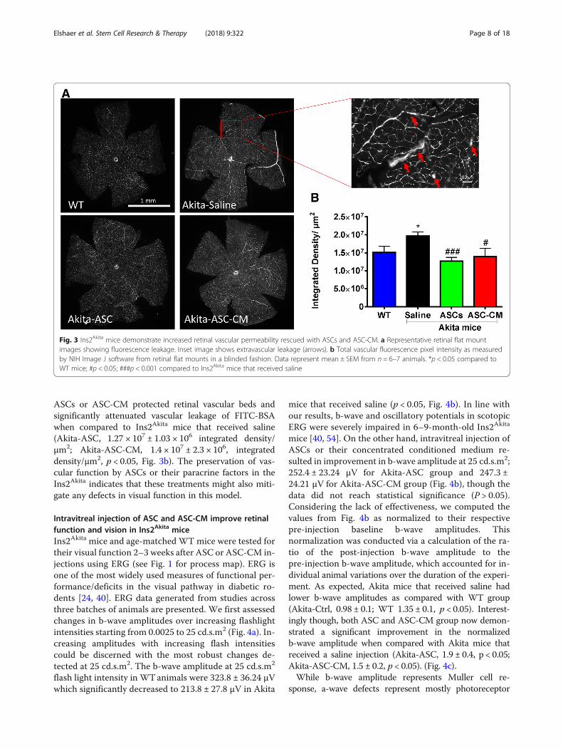
ASCs or ASC-CM protected retinal vascular beds andsignificantly attenuated vascular leakage of FITC-BSAwhen compared to Ins2Akita mice that received saline(Akita-ASC, 1.27 × 10⁷ ± 1.03 × 10⁶ integrated density/μm2; Akita-ASC-CM, 1.4 × 10⁷ ± 2.3 × 10⁶, integrateddensity/μm2, p < 0.05, Fig. 3b). The preservation of vas-cular function by ASCs or their paracrine factors in theIns2Akita indicates that these treatments might also miti-gate any defects in visual function in this model.
Intravitreal injection of ASC and ASC-CM improve retinalfunction and vision in Ins2Akita miceIns2Akita mice and age-matched WT mice were tested fortheir visual function 2–3 weeks after ASC or ASC-CM in-jections using ERG (see Fig. 1 for process map). ERG isone of the most widely used measures of functional per-formance/deficits in the visual pathway in diabetic ro-dents [24, 40]. ERG data generated from studies acrossthree batches of animals are presented. We first assessedchanges in b-wave amplitudes over increasing flashlightintensities starting from 0.0025 to 25 cd.s.m2 (Fig. 4a). In-creasing amplitudes with increasing flash intensitiescould be discerned with the most robust changes de-tected at 25 cd.s.m2. The b-wave amplitude at 25 cd.s.m2
flash light intensity in WTanimals were 323.8 ± 36.24 μVwhich significantly decreased to 213.8 ± 27.8 μV in Akita
mice that received saline (p < 0.05, Fig. 4b). In line withour results, b-wave and oscillatory potentials in scotopicERG were severely impaired in 6–9-month-old Ins2Akita
mice [40, 54]. On the other hand, intravitreal injection ofASCs or their concentrated conditioned medium re-sulted in improvement in b-wave amplitude at 25 cd.s.m2;252.4 ± 23.24 μV for Akita-ASC group and 247.3 ±24.21 μV for Akita-ASC-CM group (Fig. 4b), though thedata did not reach statistical significance (P > 0.05).Considering the lack of effectiveness, we computed thevalues from Fig. 4b as normalized to their respectivepre-injection baseline b-wave amplitudes. Thisnormalization was conducted via a calculation of the ra-tio of the post-injection b-wave amplitude to thepre-injection b-wave amplitude, which accounted for in-dividual animal variations over the duration of the experi-ment. As expected, Akita mice that received saline hadlower b-wave amplitudes as compared with WT group(Akita-Ctrl, 0.98 ± 0.1; WT 1.35 ± 0.1, p < 0.05). Interest-ingly though, both ASC and ASC-CM group now demon-strated a significant improvement in the normalizedb-wave amplitude when compared with Akita mice thatreceived a saline injection (Akita-ASC, 1.9 ± 0.4, p < 0.05;Akita-ASC-CM, 1.5 ± 0.2, p < 0.05). (Fig. 4c).While b-wave amplitude represents Muller cell re-
sponse, a-wave defects represent mostly photoreceptor
Fig. 3 Ins2Akita mice demonstrate increased retinal vascular permeability rescued with ASCs and ASC-CM. a Representative retinal flat mountimages showing fluorescence leakage. Inset image shows extravascular leakage (arrows). b Total vascular fluorescence pixel intensity as measuredby NIH Image J software from retinal flat mounts in a blinded fashion. Data represent mean ± SEM from n = 6–7 animals. *p < 0.05 compared toWT mice; #p < 0.05; ###p < 0.001 compared to Ins2Akita mice that received saline
Elshaer et al. Stem Cell Research & Therapy (2018) 9:322 Page 8 of 18

cells. Similar to b-wave, a-wave amplitudes were recordedat increasing light intensities starting from 0.0025 to25 cd.s.m2 (Additional file 5: Figure S5A), where most ro-bust changes were also observed at 25 cd.s.m2. While themean amplitudes in WT mice group were − 183.8 ±19.45 μV, it significantly decreased in Akita micegroup (− 125.5 ± 17.33 μV, p < 0.05, Additional file 5:Figure S5B). In contrast to b-wave amplitudes, in-jection of ASCs or ASC-CM failed to improvea-wave with values remained significantly lower thanthose of WT mice (Akita-ASC: − 144.7 ± 10.71 μV,p > 0.05 and Akita-ASC-CM: -142.0 ± 10.94 μV, p >0.05, Additional file 5: Figure S5B).Visual dysfunction in Ins2Akita mice has been shown to
manifest as a change in visual acuity and contrast sensitiv-ity defects through visual tracking behavior [40]. In thisstudy, we measured visual acuity and contrast sensitivity2–3 weeks post intravitreal injection of ASCs or ASC-CM.Studies described here were performed in one batch of an-imals. As expected, Ins2Akita mice demonstrated a signifi-cant reduction in visual acuity as compared to WT mice(WT-ctrl, 0.42 ± 0.004 c/d; Akita-Saline, 0.35 ± 0.007 c/d,p < 0.001, Fig. 4d). Interestingly, while Ins2Akita mice that
received ASC injection failed to improve visual acuity(0.36 ± 0.005, p = 0.17), Ins2Akita mice that receivedASC-CM demonstrated a significant alleviation in vis-ual acuity as compared to Ins2Akita mice that receivedsaline (Akita-ASC-CM, 0.37 ± 0.007, p < 0.05). Any de-crease in visual acuity may likely result in an increasein contrast sensitivity, and as expected, Ins2Akita micedemonstrated a significant increase in contrast neededto discriminate a fixed 0.042 c/d of acuity (WT-Ctrl, 19± 0.3%; Akita-Saline, 30.1 ± 1.98%, p < 0.001, Fig. 4e). Ina similar fashion to visual acuity, only Ins2Akita micethat received ASC-CM demonstrated a significant im-provement in contrast sensitivity as compared to Akitamice that received a saline injection (23.48 ± 1.73%, p <0.05). Although Ins2Akita group that received ASC in-jection also showed a decrease, the data did not reachstatistical significance (28.23 ± 1.28%, p = 0.226).
Intravitreal injection of ASCs and ASC-CM do notadversely affect retinal architectureWe used OCT to assess the retinal architecture. Asshown in Fig. 5, WT mice showed normal appearance ofretinal architecture (Fig. 5a). In line with published
Fig. 4 Intravitreal injection of ASC or ASC-CM improves retinal function and vision in Ins2Akita mice. a b-wave amplitude measurement in mice atvarious flash intensities. b b-wave amplitude at 25 cd.s.cm2 expressed as μV. Data represent combined mean ± SEM from n = 12–20 animals/group ofthe left eye only performed in three separate batches. *p < 0.05 compared to WT mice. c b-wave amplitude measurements in mice at 25 cd.s.cm2 flashintensity were plotted as a ratio of the post-injection b-wave amplitude to the pre-injection b-wave amplitude. Data represent combined mean ± SEMfrom an n = 8–12 animals/group of both eyes performed in two separate batches. *p < 0.05 compared to WT mice; #p < 0.05 compared to Ins2Akita
mice that received saline. d Visual acuity was measured by presenting black and white bars of varying spatial frequencies at 100% contrast, and thecontrast sensitivity was measured by changing the gradient that generates tracking at a fixed spatial frequency of 0.042 cycles per degree. Visual acuityin mice expressed as cycles/degree (c/d). e Contrast sensitivity in mice expressed as a percentage. OKN data represent both eyes combined mean ±SEM from n = 6–8 animals/group from a single batch. ***p < 0.001 compared to WT mice and #p < 0.05 compared to Ins2Akita mice that received saline
Elshaer et al. Stem Cell Research & Therapy (2018) 9:322 Page 9 of 18

literature [55], Ins2Akita mice that received saline did notshow any significant change in retinal architecture orretinal thickness around optic nerve head as detected byOCT (Fig. 5b, e). This near normal appearance of theretina was confirmed by H&E staining in some rep-resentative animals (Additional file 6: Figure S6A).
ASCs could occasionally be visualized in OCT in thevitreous on top of the retina (Additional file 6:Figure S6A–B). Interestingly, Ins2Akita mice that re-ceived ASC or ASC-CM did not result in any furtherdeterioration or change in retinal thickness aroundthe optic nerve head (Fig. 5c–e) except for onemouse out of eight that received ASCs demonstrated
Fig. 5 Ins2Akita mice demonstrate no apparent changes in OCT with andwithout ASCs and ASC-CM. Representative brightfield images showingb-scan location (arrow) with occasional bright spots (upper panels) in WT(a) and Ins2Akita mice with saline (b), ASCs (c), and ASC-CM (d) with theirvitreous, retinal layers, and choroidal layers clearly visible (lower panels). eCentral retinal thickness in the study groups. Data are representativefrom n= 3 (WT); 4–7 animals (other groups) both eyes included
Fig. 6 Ins2Akita mice show increased GFAP immunoreactivity partiallyrescued with ASC-CM. a Representative confocal images showing theGFAP immunostaining (green) counterstained with DAPI (blue) inwildtype animal (WT-Ctrl), Ins2Akita mice that received saline (Akita-Saline),Ins2Akita mice that received ASCs (Akita-ASC), and Ins2Akita mice thatreceived ASC-CM (Akita-ASC-CM). b Quantification of GFAP intensityexpressed as average mean integrated intensity measured by NIHImageJ. Data represent combined mean ± SEM from n= 4–7 eyes/groupperformed in one batch. *p< 0.02 compared to WT mice and # p= 0.06compared to Ins2Akita mice that received saline. Scale = 50 μm
Elshaer et al. Stem Cell Research & Therapy (2018) 9:322 Page 10 of 18

retinal tugging and hemorrhage (Fig. 5c andAdditional file 4: Figure S4, Additional file 6: FigureS6B). H&E staining for Ins2Akita mice that receivedASCs or ASC-CM did not reveal any retinal defectsseen in vivo perhaps suggesting transient nature ofthe observed retinal defects in this model or rare oc-currence (Additional file 6: Figure S6A).
Intravitreal injection of ASC-CM but not ASCs marginallyattenuates enhanced retinal expression of GFAP inIns2Akita miceUpregulation of GFAP expression in Müller glial cells isa hallmark of neuroinflammation and reactive gliosis[56]. In our studies, while GFAP immunoreactivity wasrestricted to GCL in WT mice, in Ins2Akita mice that
Fig. 7 Ins2Akita mice display differential gene expression with and without ASCs and ASC-CM. Gene transcripts associated with retinal inflammationand neurovascular tissue remodeling by TaqMan qPCR expressed as fold change normalized to internal control in the study groups 3 weeks postintravitreal injections. Data represented as the mean ± SEM from n = 6–8 animals/group normalized to WT mice. *p < 0.05 compared to WT mice; #p <0.05 compared to Ins2Akita mice that received saline; $ p < 0.05 compared to Ins2Akita mice that received ASCs
Elshaer et al. Stem Cell Research & Therapy (2018) 9:322 Page 11 of 18

received saline, GFAP immunoreactivity increased inGCL with thicker processes observed reaching to theouter retina (WT-Ctrl: 1.7 ± 0.4; Akita-Saline: 5.3 ± 1.0integrated density/equal area; p < 0.02 Fig. 6a, b). Ins2A-kita mice that received ASCs failed to show any signifi-cant reduction in GFAP expression (Akita-ASCs: 4.9 ±1.1; Akita-Saline: 5.3 ± 1.0 integrated density/equal area;p < 0.01, Fig. 6a, b). However, Ins2Akita mice that receivedASC-CM demonstrated an overall reduction in GFAPimmunoreactivity, yet, the data are only borderlinesignificant when an outlier is excluded from the data(Akita-ASC-CM: 3.0 ± 0.5; Akita-Saline: 5.3 ± 1.0 inte-grated density/equal area; p = 0.06, Fig. 6b).
ASCs and ASC-CM modulate retinal gene expression inIns2Akita miceAs shown in Fig. 7, Ins2Akita mice experienced the alteredexpression of genes associated with retinal inflammationand neurovascular tissue remodeling in response todiabetes-related stressors [57]. Compared to age-matchedWT mice, Ins2Akita mice that received saline showed in-creased gene transcripts of Ccl2, Ch25h, and Lama5 in theretinal lysates at 3 weeks post-injection (Ccl2: 2.7 ± 0.5,Ch25h: 2.3 ± 0.4, Lama5: 1.2 ± 0.1, as compared to WTmice: 1 ± 0.0, p < 0.05). Interestingly, Ins2Akita mice thatreceived intravitreal injections of ASCs showed a higherexpression profile of genes at 3 weeks including Ccl2,Edn2, Gbp2, Icam-1, and Timp1, where expression ofthese genes were significantly higher as compared to theAkita-Saline group (Ccl2: 19.6 ± 5.9 as compared toAkita-Saline, Edn2: 6.6 ± 1.8 as compared to 1.3 ± 0.3 forAkita-Saline, Gbp2: 8.9 ± 2.3 as compared to 1.2 ± 0.2 forAkita-Saline, Icam-1: 1.9 ± 0.4 as compared to 0.9 ± 0.2 forAkita-Saline, Timp1: 8.9 ± 3.3 as compared to 1.3 ± 0.2 forAkita-Saline, p < 0.05). On the other hand, Ins2Akita micethat received intravitreal injections of ASC-CM did notshow any further increase in gene expression as comparedto Ins2Akita mice that received saline (Ccl2: 4.3 ± 1.0,Ch25h: 2.3 ± 0.4, Gbp2: 1.1 ± 0.1, Icam-1: 1.2 ± 0.2, Lgals9:1.1 ± 0.2, Timp1: 1.7 ± 0.3 as compared to Akita-Saline,p > 0.05). Considering the variation in gene expression at3 weeks post intravitreal injection, specifically in Ins2Akita
mice that received ASCs, retinas at day 3 post injectionswere also assessed for gene expression (Additional file 7:Figure S7). At day 3 post-injection, Ins2Akita mice experi-enced similarly altered expression of genes associated withretinal inflammation and neurovascular tissue remodelingin response to diabetes-related stressors compared toage-matched WT mice (Ch25h: 4.2 ± 0.7, Crybb2: 7.1 ±2.6, Edn2: 2.3 ± 0.6, Gbp2: 1.3 ± 0.2, Icam1: 1.4 ± 0.2,Lgals9: 2.3 ± 0.4, Lama5: 1.4 ± 0.1, Serping: 2.2 ± 0.2,Timp1: 3.2 ± 0.7 as compared to WT mice: 1 ± 0.0, p <0.05). However, Ins2Akita mice that received intravitreal in-jections of ASCs or ASC-CM did not show any further
increase in the expression profile of genes, with the excep-tion of Edn2 (5.0 ± 0.8 for Akita-ASCs, 7.1 ± 1.1 forAkita-ASC-CM; p < 0.05).
DiscussionThe Ins2Akita model is superior to chemically inducedmodels of DR as it is highly reproducible, can facilitatelongitudinal studies of complications, and provides a rele-vant genetic background for testing new therapeutic strat-egies for early intervention in DR [39]. Accordingly, thismouse model was used previously to demonstrate thetherapeutic benefits of endothelial colony-forming cellsderived from cord blood [40]. The salient findings ofcurrent study are (1) both ASCs and ASC-CM therapeut-ically benefit the retina and improve vision in Ins2Akita
model of NPDR mostly through preserved neurovasculararchitecture, attenuated inflammation, and vascular leak-age, (2) immune competent Ins2Akita mice lack any appar-ent immune rejection of ASCs, supporting their allogeneictransplantation, and (3) ASC-CM is relatively more effect-ive than ASCs at the doses tested for early manifestationsof NPDR. Our study corroborates our previous findingsthat ASCs are able to rescue the neural retina fromhyperglycemia-induced degeneration observed in STZ-induced DR model [24] thereby establishing adult stemcells and their secretome as possible regenerative therap-ies for NPDR.Although the cell surface expression of platelet-derived
growth factor receptor (PDGFRβ, CD140b) is not a bonafide ASC marker [58], it is constitutively expressed inmost ASCs, regardless of passage number [59, 60] and in-creased with culture ranging from 40 to 70% [61]. Cd140bis thought to be a major cell surface marker defining peri-cytes [62], though the specific role of CD140b signaling inASCs in regulating the angiogenic potential of retinalendothelial cells is still not completely understood. Thepericytic ASCs are hypothesized to ameliorate the loss ofpericytes and consequent vascular permeability in DR[24]. Because ASCs produce cytoprotective factors, it isalso anticipated that they will also promote vascular andneurodegeneration repair in retinopathy [24]. Additionalwork from our lab has established a pivotal role forCD140b signaling in that ability of ASCs to modulate theangiogenic behavior of human retinal endothelial cells viadirect or paracrine signals [41]. For these reasons, wetested CD140b-positive ASCs and their conditionedmedium for their potential benefit in Ins2Akita model. Sur-prisingly, the CD140b-positive ASCs were not found in as-sociation with the retina by OCT analyses of live mice orby post-mortem histological analyses (Additional file 6:Figure S6 and Additional file 8: Figure S8). It is possiblethat the number of cells injected into the eye is far belowthe detection threshold of our assays or insufficient to
Elshaer et al. Stem Cell Research & Therapy (2018) 9:322 Page 12 of 18

integrate into the retina. The apparent lack of homing ofthese ASCs is consistent with previous observations thatMSCs are poorly retained and integrated into the retina[26, 63]. While it is interesting that one single intravitrealinjection of ASCs is sufficient to partially rescue visualfunction in the Ins2Akita model, the apparent superiorityof ASC-CM suggests that the observed therapeutic bene-fits of ASCs are largely paracrine mediated rather thancell-mediated.Here, we used ASC-CM from TNFα, and IFNγ-
stimulated ASCs. We have recently shown that cytokinepriming can enhance the anti-inflammatory propertiesof ASCs and mitigate visual deficits observed in a blastinjury model [64]. Cytokine-stimulated MSCs have beenshown to express extracellular superoxide dismutase(SOD3), which can aid neuroprotection by mitigatingoxidative stress [65]. We confirmed that SOD3 is upreg-ulated in the CM of TNFα and IFNγ-stimulated ASCs(Fig. 2). Surprisingly, we found that mitochondrial super-oxide dismutase (SOD2) is also present in the CM ofTNFα and IFNγ-stimulated ASCs. These SODs may helpmitigate hyperglycemia-induced oxidative stress seen inDR. TNFα and IL-1β are known to induce the expres-sion of the anti-inflammatory protein TNF-stimulatedgene 6 protein (TSG-6), which has shown therapeuticvalue in numerous animal models including ophthalmicdiseases [66–68]. Activation and proliferation of retinalmicroglial cells are linked to DR pathology [69]. TSG-6can suppress the expression of inflammatory gene tran-scripts in BV2 microglia activated with LPS [35]. We re-cently showed that TNFα and IFNγ synergize for TSG-6expression and that cytokine-stimulated ASC-CCM(cytokine-stimulated and concentrated CM from un-sorted ASCs) more potently suppresses nitric oxide pro-duction by LPS stimulated BV2 cells than ASC-CCMfrom unstimulated cells [64]. IDO is known to be upreg-ulated in IFNγ-stimulated MSCs and has been shown tolimit T cell function and promote immune tolerance [70,71]. Here, we confirmed that TSG-6 and IDO arepresent in the cytokine-stimulated ASC-CM.Although SODs, TSG-6, and IDO may contribute to
neuroprotection and suppression of inflammation in theretinas of Ins2Akita mice, we found many proteins that aredifferentially expressed in the CM of ASCs treated withcytokines. Using antibody arrays that can detect relativeamounts of cytokines, chemokines, and angiogenic fac-tors, we determined that TNFα and IFNγ also significantlyupregulated the secretion of IL-6, IL-8, CCL2, CXCL9,CCL5, CXCL10, CXCL11, CCL7, and ANGPT-1. Paradox-ically, the induction of interleukins and chemokines in theCM of stimulated ASCs may be beneficial to promotingregeneration of neurovascular tissue because the initial ro-bust recruitment of immune responders in the presenceof other ASC produced regulatory molecules may play an
important role in advancing the late stages of the immuneresponse leading to inflammation resolution and tissue re-pair. It has been hypothesized that MSC co-culture andMSC extracellular vesicle-treated macrophage populationsmay promote resolution of inflammation via reduction ofTh1 pathogenicity through Th17 conversion [72]. Othershave also demonstrated that the induction of regulatory Tcells by MSCs involves skewing monocytes towardM2-type macrophages [73, 74]. These findings indicatethat MSC-secreted factors coordinate with controlled re-cruitment of immune regulators with influence over theirphenotype and function at the site of injury while simul-taneously initiating the process of tissue remodeling. Al-though the tissue remodeling process involving ASCs iscomplex and involves many mediators, there is evidencein that stimulation of ASC-CM elicits a response patternaligned with controlled tissue remodeling. For example,ANG-1, a growth factor that stimulates there-endothelialization of blood vessels thereby counteract-ing permeability and other pro-inflammatory effects, wasincreased with stimulation, while ANG-2 levels remainunchanged [75]. Since these paracrine factors can displaypleiotropic and differential effects on immune modulation,chemoattraction, and tissue remodeling, it is not yet clearwhich protein alone or in combination contribute to theoverall therapeutic effect of the ASC-CM. Loss and gainof function studies of specific proteins within theASC-CM may identify the therapeutic roles and mecha-nisms of individual paracrine factors, though synergies be-tween factors may also exist. Detailed studies of secretomecomposition and molecular mechanisms of action mayallow for the development of a unique biologic that can beproduced reproducibly and at a scale that can meet FDArequirements for human clinical studies.Evidence compiled from clinical and basic research
supports the view that DR constitutes a change in theretinal neurovascular unit comprised of interactingneurons, glia, and vasculature [76]. Ins2Akita heterozy-gous mice experience early features of DR includingmicroglial activation, reduced GFAP expression in astro-cytes, and an increase in GFAP expression by Mullercells [39, 55]. In accordance with this observation, weshow a significant increase in GFAP expression in Ins2A-kita mice that was reduced by ASC-CM but not ASCs.This apparent difference between ASCs and ASC-CMwas also observed in retinal gene expression changes ob-served in this model, specifically, genes coding for pro-teins involved in infection and immune responses(Gbp2; Lgals9; Ch25h) and in intracellular signal trans-duction pathways activated by cytokines and chemo-kines, such as adhesion and tissue structure (Icam-1;Lama5), inflammation (Ccl2; Edn2; CD11b), and proteindegradation (Timp1). A recent study in an oxygen-inducedretinopathy model demonstrated a similar increase in genes
Elshaer et al. Stem Cell Research & Therapy (2018) 9:322 Page 13 of 18

associated with retinal inflammation and neurovascular tis-sue remodeling 5 days post intravitreal injection of ASCs.Taken in the context of similar therapeutic results in theIns2Akita mice, the targeted enhancement of genes regulat-ing retinal inflammation and tissue modulation may be re-quired to restore normal neurovascular tissue structure[77]. It is possible that the increase in some genes corre-lated with DR is a compensatory phenomenon by cellsresponding to ongoing stress with attempts at inflammationresolution and tissue regeneration. Therefore, the promo-tion of these genes may be an indication of a more robustwound healing response necessary for a therapeutic out-come that overcomes the deleterious effect of long-termchronic inflammation and oxidative stress. In support ofthis hypothesis, ASCs exposed to chronic levels of high glu-cose normalized high glucose challenged endothelial geneexpression levels compared to those of normal glucosemedium and also protected against oxidative stress [77]. Inline with these results, we showed ASCs exposed toexogenous TNFα, and IFNγ produces both inflamma-tory cytokines like IL-6 and anti-inflammatory TSG-6and antioxidant SODs. Thus, a dynamic and complexrepertoire of paracrine factors produced by ASCs mayaccount their unique ability to treat a variety ofpathological conditions across various tissues. Takentogether, our results suggest that preconditioning ofASCs likely benefit the outcomes in DR.Considering the variation in gene expression at 3 weeks
post intravitreal injections, retinas at day 3 post-injectionwere also assessed for gene expression. Our data at day 3demonstrated a significant upregulation of the genesCh25h, Edn2, Icam1, Timp1, Lama5, Lgals9, and Serpingin Ins2Akita mice that received saline (p < 0.05) with no fur-ther increase in most of these genes with both ASCs andASC-CM (Additional file 7: Figure S7). Interestingly, crys-tallin B2, a family of crystallins that are known to have arole in the adaptive mechanisms taking place in thediabetic retina [78], is increased twofold in Ins2Akita micethat received ASC-CM. Endothelin-2, a candidate bio-marker for controlled vascular modulation [79, 80], wassignificantly increased in Ins2Akita mice that received sa-line with a twofold further increase in mice that receivedASCs and ASC-CM. However, for any given gene, therelative differences in expression between Ins2Akita micethat received ASCs versus ASC-CM was more profoundat 3 weeks than at 3 days. We need more detailed tem-poral expression data and gain or loss of function studiesof these genes, as well as studies of gene expressionchanges in this model, confirmed at the protein level, tofully address the cause or effect relationship of these genetranscripts in the Ins2Akita model with ASCs andASC-CM.Previously, it was reported that Ins2Akita mice develop
retinal vascular permeability using the FITC-BSA tail
vein injection method [39]. We have confirmed suchleakage in the Ins2Akita model and show that both ASCsand ASC-CM can alleviate it. This study supports ourprevious observation that ASCs in an STZ-induced DRmodel suppress similar vascular leakiness [24]. Taken to-gether with the observation that ASC-CM demonstrateda reduction in TER in vitro, our data suggest thatASC-produced paracrine factors affect the integrity ofvascular endothelial cells in vivo. This observation isconsistent with studies showing that MSC-derived con-ditioned medium or extracellular vesicles can protectagainst endothelial permeability [81–83].Neural alterations of DR can be monitored in
real-time via visual function tests such as ERG andOKN. Our results in this study demonstrate significantlower b-wave amplitudes and visual acuity in Ins2Akita
mice suggesting retinal damage in this model. It is inter-esting to note that Ins2Akita mice that received ASCs andASC-CM alleviated ERG deficits while only Ins2Akita
mice that received ASC-CM group demonstrated im-proved OKN deficits. While the vision gains withASC-CM were modest, they indicate that ASC-CM hasthe ability to reverse the progression of chronic NPDRand initiate the restoration of neurovascular homeostasisin the presence of continued systemic disease. Althoughthe number of ASCs injected may not equate to theamount of ASC-CM injected into the eyes, the lack ofpositive response in the ASC group may suggest the per-sistence of disturbances of post-retinal connectionsdownstream of an accessory optic system that has beenlinked to OKN defects [84]. Considering the fact thatERG deficits are associated with retinal inflammation[85], and Ins2Akita mice that received ASC-CM had rela-tively decreased retinal inflammation, it may be possibleto suggest that ASC-CM may elicit its benefits by sup-pressing retinal neuronal inflammation.Previously, it was established that healthy, but not dia-
betic, mouse-derived ASCs were protective against vas-cular dropout in the Akimba diabetic mouse model [86].The same study failed to demonstrate any apparentbenefit with conditioned medium derived from mouseASCs [86]. We have similarly found a protective effectof healthy human ASCs in the Akita model and havealso established a therapeutic benefit with humanASC-CM. The differences between the two studies in-clude (1) inherent differences in the models due to dif-ferent genotypes, (2) different species of ASCs werestudied, (3) here neurovascular, rather than vascular,structures were studied, and (4) the ASC-CM generatedin our study is from human cells primed with a cytokinecocktail that might have produced beneficial proteins atthe required therapeutic dose.Our study is a step forward in the field to suggest an
allogenic ASC and cell-free ASC-CM therapy for NPDR.
Elshaer et al. Stem Cell Research & Therapy (2018) 9:322 Page 14 of 18

While ASC-CM appears to provide similar beneficialoutcomes compared to that of ASCs but without the in-creased risk of the occasional mass of cells overlying thephotosensitive retina, retinal hemorrhage and retinal de-tachments were observed in the group that receivedASCs. Compared to ASCs, the use of ASC-CM is alsowithout observable unwanted side effects, including sub-retinal fluid accumulation and/or retinal detachmentthat was observed in human subjects [33]. Although theuse of ASC-CM is promising, more studies are war-ranted to explore the long-term safety and effectivenessof such treatment further. If ASC-CM is indeed the bet-ter therapy, then it would be pertinent to determine thebest means of treating patients longitudinally. One ofthe benefits of ASC treatment is the presence of actualcells that allow for continued paracrine factor secretionfor some time after cells are delivered to the vitreous asdescribed in studies with ASC and other MSCs [87–91].With ASC-CM, we would need to explore whether regu-lar injections are a safer alternative compared to a po-tential depot implant of cells.We readily acknowledge some of the limitations of
this initial study. The ASCs and ASC-CM used in theIns2Akita mouse experiments come from theCD140b-sorted cells from a single human donor andmay, therefore, have unique or clonogenic properties.One single intravitreal injection in our studies mighthave produced only limited exposure to the thera-peutic. Note that we used human ASCs in the mousebecause these are the cells that would be tested inhuman clinical trials. However, suboptimal responsesacross species may exist if relevant human proteins ormacromolecules do not efficiently interact with mousetargets. In this study, ASCs demonstrated some riskincluding the occasional mass of cells overlying thephotosensitive retina, prolonged perturbation of neu-rovascular tissue remodeling gene expression, and ret-inal detachments. Moreover, the relative potency ofASCs versus concentrated ASC-CM is not compar-able, warranting additional studies with escalating andrepeated dosing. We have been able to study only20× concentrated ASC-CM, and future studies withlower or higher concentrations may be necessary tooptimize the dose response. Although ASC-CM pro-vided beneficial outcomes on visual function and vas-cular leakage without other observed safety concernsin the Ins2Akita model, additional pre-clinical studiesare required to establish long-term safety, tolerability,and effectiveness of such treatment. Finally, we choseto do intravitreal injections as it benefits from directdelivery of the ASCs into the eye close to the dam-aged retinal vasculature, in line with other cell ther-apies for DR. However, other routes of administrationrequire further exploration.
ConclusionsUsing a well-characterized Ins2Akita NPDR model, wedemonstrated that one single intravitreal injection ofASCs and/or ASC-CM therapeutically benefit the retinaby modulating the neurovascular system and improvingvision. Molecular and histological studies further confirmthe beneficial effects of the ASCs and ASC-CM in vivo.Future studies are needed to identify the specific proteinsinvolved in the observed beneficial effects. Long-termstudies will focus on the safety and efficacy of stem celltreatments, including the potential for rejection or thetherapeutic value of reinjection, as well as any relative ad-vantages of ASC-CM for manufacturing, quality, safety, oreffectiveness of ASC-derived therapies for DR.
Additional files
Additional file 1: Figure S1. Ins2Akita mice had markedly higher bloodglucose levels. Blood glucose concentration (mg/dL) in the study groups.Data represent mean ± SEM from n = 3–8 animals. *p < 0.05 compared toWT mice. (TIF 103 kb)
Additional file 2: Figure S2. Isolation and separation of CD140b-positive ASCs by FACS sorting. Representative flow cytometric histogramsof (A) unstained control (B) isotype antibody control and (C) the expres-sion of CD140b. ASCs were labeled with CD140b-PECy5 mAb and sortedby FACS. ASCs were gated based on forward (FSC-A), and side scatter(SSC-A) area to gate the live-viable cells. Single cells from viable cellpopulation were gated using side scatter height and width (SSC-W, SSC-H), forward scatter height (FSC-W, FSC-H). Further, using a histogram plot(x-axis: PEcy5 positive vs Y-axis: cell count), CD140b + ASC (right side gate)and CD140b- ASC (left side gate) were sorted. Data is from one donorwith 2 other donor with similar results. (TIF 2704 kb)
Additional file 3: Figure S3. Cytokine priming preserves cell viabilityand enhances chemokine and angiogenic proteins in ASC-CM. (A) Assess-ment of cell viability by MTT proliferation assay did not affect cell viabilitybefore and after priming with cytokines. Data represent Mean ± SD fromone donor performed in triplicates. Differential expression of all assessed(B) chemokines and (C) angiogenic proteins in primed versus un-primedASC-CM. (TIF 1333 kb)
Additional file 4: Figure S4. Ins2Akita mice demonstrate no apparentretinal vascular permeability with or without ASCs and ASC-CM. (A) Rep-resentative brightfield images showing no apparent architectural defectsexcept for some Ins2Akita mice that received ASCs injection demonstrat-ing hemorrhages (arrows). (B) Representative green fluorescence imagesshowing no apparent vascular leakiness in any study groups. Data is rep-resentative of n = 6–8 animals/group. (TIF 3396 kb)
Additional file 5: Figure S5. Ins2Akita mice show altered a-wave ampli-tudes significantly rescued with ASCs and ASC-CM. (A) a-wave amplitudemeasurement in mice at various flash intensities. (B). the a-wave ampli-tude at 25 cd.s.cm2 expressed as μV. Data represents combined Mean ±SEM from n = 12–20 animals/group of the left eye only performed in 3separate batches. *p < 0.05 compared to WT mice. (TIF 262 kb)
Additional file 6: Figure S6. Ins2Akita mice demonstrate no apparentchanges in histological and OCT defects with and without ASCs and ASC-CM. (A). Representative photomicrographs of H&E stainings from all studygroups. Scale = 50 μm. Data represents 5–7 animals/group from onebatch. (B). Representative brightfield images showing b-scan location(upper panels) with the retinal and choroidal layers (Lower panels). Noapparent structural defects except for occasional hemorrhages in Ins2Akita
mice that received ASCs injection noted. The central bright spot is a dustparticle trapped in the camera and should be considered as an artifact.Data represents 3–4 animals/group from one batch. (TIF 11174 kb)
Elshaer et al. Stem Cell Research & Therapy (2018) 9:322 Page 15 of 18

Additional file 7: Figure S7. Ins2Akita mice display differential geneexpression with and without ASCs and ASC-CM. Gene transcripts associ-ated with retinal inflammation and neurovascular tissue remodeling byTaqMan qPCR expressed as fold change normalized to internal control inthe study groups 3 days post intravitreal injections. Data represented asthe Mean ± SEM from n = 6–8 animals/group normalized to WT mice. *p< 0.05 compared to WT mice; # p < 0.05 compared to Ins2Akita mice thatreceived saline; $p < 0.05 compared to Ins2Akita mice that received ASCs.(TIF 238 kb)
Additional file 8: Figure S8. Incorporation of intravitreally deliveredASCs into host vasculature in Ins2Akita mice as assessed by human histoneIgG immunostaining. Representative confocal images demonstrated noimmunostaining for human IgG (thus no association of the ASCs with hostvessels or any structures within the retina) in wildtype mice (A) or Ins2Akita
mice that received ASCs (B). Positive immunostaining is shown with humanASCs cultured in vitro (C). Retinal sections incubated with no primary antibodydemonstrated immunostaining specificity of the antibody (D). Data shown arerepresentative of 3–5 animals/group. Scale = 50 μm. (TIF 3769 kb)
AcknowledgementsAuthors acknowledge the technical support from TJ Hollingsworth, Ph.D.(Neuroscience Institute, UTHSC). Authors acknowledge Lada Klaic, Ph.D. (CellCare Therapeutics Inc), Houman David Hemmati, M.D., Ph.D. (Levation PharmaLtd), and Jeffrey L. Edelman, Ph.D. (Ocular Drug Discovery and Development)for insightful and constructive editorial assistance and discussions.
FundingThis study was supported by grants from the National Eye Institute(EY023427), unrestricted funds from Research to Prevent Blindness to R.G.The funders played no role in the conduct of the study, collection of data,management of the study, analysis of data, interpretation of data, orpreparation of the manuscript.
Availability of data and materialsAll data generated and/or analyzed during this study are included in thispublished article. Data sharing not applicable to this article as no datasetswere generated or analyzed during the current study.
Authors’ contributionsSLE, WE, MP, RL, and RP contributed to the design, collection, and assemblyof data, data analysis and interpretation, and manuscript writing; KJ, SA, JG,and SMT contributed to the collection and assembly of data; NS contributedto the conception and design and manuscript writing; RG contributed to theconception and design, collection and assembly of data, data analysis andinterpretation, manuscript writing, and final approval of the manuscript. Allauthors read and approved the final manuscript.
Ethics approval and consent to participateStudies involving human adipose tissue were approved by UTHSCInstitutional Review Board in accordance with relevant guidelines andregulations following the tenets of the Declaration of Helsinki. InstitutionalIRB approved the study as an exempt since the study involved de-identifiedcell lines from commercial sources. Animal studies were approved by theInstitutional Animal Care and Use Committee, University of Tennessee HealthSciences Center (UTHSC), following the guidelines as per the Association forResearch in Vision and Ophthalmology (ARVO) Statement for the Use ofAnimals in Ophthalmic and Vision Research.
Consent for publicationNot applicable.
Competing interestsNS and RG are co-founders and hold equity in Cell Care Therapeutics Inc.,whose interest is in the use of adipose-derived stromal cells in visual disorders.MP is an employee of Cell Care Therapeutics Inc. with equity. None of the otherauthors declare any financial conflicts.
Publisher’s NoteSpringer Nature remains neutral with regard to jurisdictional claims inpublished maps and institutional affiliations.
Author details1Ophthalmology, University of Tennessee Health Science Center, 930Madison Ave, Suite#768, Memphis, TN 38163, USA. 2Anatomy andNeurobiology, University of Tennessee Health Science Center, Memphis, TN38163, USA. 3Pharmacology & Toxicology Department, College of Pharmacy,Mansoura University, Mansoura, Egypt. 4Cell Care Therapeutics, Inc., Monrovia,CA 91016, USA.
Received: 2 July 2018 Revised: 5 October 2018Accepted: 23 October 2018
References1. Klein R, Deng Y, Klein BE, Hyman L, Seddon J, Frank RN, Wallace RB, Hendrix
SL, Kuppermann BD, Langer RD, Kuller L, Brunner R, Johnson KC, ThomasAM, Haan M. Cardiovascular disease, its risk factors and treatment, and age-related macular degeneration: Women’s Health Initiative Sight Examancillary study. Am J Ophthalmol. 2007;143(3):473–83.
2. Klein R, Klein BE. The prevalence of age-related eye diseases and visualimpairment in aging: current estimates. Invest Ophthalmol Vis Sci. 2013;54(14):ORSF5–ORSF13.
3. Aiello LP, Gardner TW, King GL, Blankenship G, Cavallerano JD, Ferris FL 3rd,Klein R. Diabetic retinopathy. Diabetes Care. 1998;21(1):143–56.
4. Shah CA. Diabetic retinopathy: a comprehensive review. Indian J Med Sci.2008;62(12):500–19.
5. Kempen JH, O'Colmain BJ, Leske MC, Haffner SM, Klein R, Moss SE, TaylorHR, Hamman RF. The prevalence of diabetic retinopathy among adults inthe United States. Arch Ophthalmol. 2004;122(4):552–63.
6. Klein R, Klein BE, Moss SE, Davis MD, DeMets DL. The Wisconsinepidemiologic study of diabetic retinopathy. II. Prevalence and risk ofdiabetic retinopathy when age at diagnosis is less than 30 years. ArchOphthalmol. 1984;102(4):520–6.
7. Klein R, Klein BE, Moss SE, Davis MD, DeMets DL. The Wisconsinepidemiologic study of diabetic retinopathy. III. Prevalence and risk ofdiabetic retinopathy when age at diagnosis is 30 or more years. ArchOphthalmol. 1984;102(4):527–32.
8. Photocoagulation for diabetic macular edema. Early Treatment DiabeticRetinopathy Study report number 1. Early Treatment Diabetic RetinopathyStudy research group. Arch Ophthalmol. 1985;103(12):1796–806.
9. Preliminary report on effects of photocoagulation therapy. The DiabeticRetinopathy Study Research Group. Am J Ophthalmol.1976;81(4):383–96.
10. Jackson TL, Nicod E, Angelis A, Grimaccia F, Pringle E, Kanavos P. Pars planavitrectomy for diabetic macular edema: a systematic review, meta-analysis,and synthesis of safety literature. Retina. 2017;37(5):886–95.
11. Two-year course of visual acuity in severe proliferative diabetic retinopathywith conventional management. Diabetic Retinopathy Vitrectomy Study(DRVS) report #1. Ophthalmology. 1985;92(4):492–502.
12. Arevalo JF, Garcia-Amaris RA. Intravitreal bevacizumab for diabeticretinopathy. Curr Diabetes Rev. 2009;5(1):39–46.
13. Ozkiris A. Intravitreal bevacizumab (Avastin) for primary treatment ofdiabetic macular oedema. Eye (Lond). 2009;23(3):616–20.
14. Velez-Montoya R, Fromow-Guerra J, Burgos O, Landers MB 3rd, Morales-Caton V, Quiroz-Mercado H. The effect of unilateral intravitreal bevacizumab(avastin), in the treatment of diffuse bilateral diabetic macular edema: apilot study. Retina. 2009;29(1):20–6.
15. Haritoglou C, Kook D, Neubauer A, Wolf A, Priglinger S, Strauss R, GandorferA, Ulbig M, Kampik A. Intravitreal bevacizumab (Avastin) therapy forpersistent diffuse diabetic macular edema. Retina. 2006;26(9):999–1005.
16. Michaelides M, Kaines A, Hamilton RD, Fraser-Bell S, Rajendram R, Quhill F,Boos CJ, Xing W, Egan C, Peto T, Bunce C, Leslie RD, Hykin PG. Aprospective randomized trial of intravitreal bevacizumab or laser therapy inthe management of diabetic macular edema (BOLT study) 12-month data:report 2. Ophthalmology. 2010;117(6):1078–1086.e2.
17. Stewart MW, Flynn HW Jr, Schwartz SG, Scott IU. Extended durationstrategies for the pharmacologic treatment of diabetic retinopathy: currentstatus and future prospects. Expert Opin Drug Deliv. 2016;13(9):1277–87.
Elshaer et al. Stem Cell Research & Therapy (2018) 9:322 Page 16 of 18

18. Hattori T, Shimada H, Nakashizuka H, Mizutani Y, Mori R, Yuzawa M. Dose ofintravitreal bevacizumab (Avastin) used as preoperative adjunct therapy forproliferative diabetic retinopathy. Retina. 2010;30(5):761–4.
19. Mason JO 3rd, Nixon PA, White MF. Intravitreal injection of bevacizumab(Avastin) as adjunctive treatment of proliferative diabetic retinopathy. Am JOphthalmol. 2006;142(4):685–8.
20. Cheung N, Mitchell P, Wong TY. Diabetic retinopathy. Lancet. 2010;376(9735):124–36.
21. Lattanzio R, Cicinelli MV, Bandello F. Intravitreal steroids in diabetic macularedema. Dev Ophthalmol. 2017;60:78–90.
22. Robinson R, Barathi VA, Chaurasia SS, Wong TY, Kern TS. Update on animalmodels of diabetic retinopathy: from molecular approaches to mice andhigher mammals. Dis Model Mech. 2012;5(4):444–56.
23. Bhattacharya S, Gangaraju R, Chaum E. Recent advances in retinal stem celltherapy. Curr Mol Biol Rep. 2017;3(3):172–82.
24. Rajashekhar G, Ramadan A, Abburi C, Callaghan B, Traktuev DO, Evans-Molina C, Maturi R, Harris A, Kern TS, March KL. Regenerative therapeuticpotential of adipose stromal cells in early stage diabetic retinopathy. PLoSOne. 2014;9(1):e84671.
25. Mendel TA, Clabough EB, Kao DS, Demidova-Rice TN, Durham JT, Zotter BC,Seaman SA, Cronk SM, Rakoczy EP, Katz AJ, Herman IM, Peirce SM, Yates PA.Pericytes derived from adipose-derived stem cells protect against retinalvasculopathy. PLoS One. 2013;8(5):e65691.
26. Ezquer M, Urzua CA, Montecino S, Leal K, Conget P, Ezquer F. Intravitrealadministration of multipotent mesenchymal stromal cells triggers acytoprotective microenvironment in the retina of diabetic mice. Stem CellRes Ther. 2016;7:42.
27. Rajashekhar G. Mesenchymal stem cells: new players in retinopathy therapy.Front Endocrinol (Lausanne). 2014;5:59.
28. Frese L, Dijkman PE, Hoerstrup SP. Adipose tissue-derived stem cells inregenerative medicine. Transfus Med Hemother. 2016;43(4):268–74.
29. Traktuev DO, Merfeld-Clauss S, Li J, Kolonin M, Arap W, Pasqualini R,Johnstone BH, March KL. A population of multipotent CD34-positiveadipose stromal cells share pericyte and mesenchymal surface markers,reside in a periendothelial location, and stabilize endothelial networks. CircRes. 2008;102(1):77–85.
30. Chen CW, Okada M, Proto JD, Gao X, Sekiya N, Beckman SA, Corselli M,Crisan M, Saparov A, Tobita K, Peault B, Huard J. Human pericytes forischemic heart repair. Stem Cells. 2013;31(2):305–16.
31. Bouacida A, Rosset P, Trichet V, Guilloton F, Espagnolle N, Cordonier T,Heymann D, Layrolle P, Sensebe L, Deschaseaux F. Pericyte-like progenitorsshow high immaturity and engraftment potential as compared withmesenchymal stem cells. PLoS One. 2012;7(11):e48648.
32. Saraf SS, Cunningham MA, Kuriyan AE, Read SP, Rosenfeld PJ, Flynn HW Jr,Albini TA. Bilateral retinal detachments after intravitreal injection of adipose-derived ‘stem cells’ in a patient with exudative macular degeneration.Ophthalmic Surg Lasers Imaging Retina. 2017;48(9):772–5.
33. Kuriyan AE, Albini TA, Townsend JH, Rodriguez M, Pandya HK, Leonard RE2nd, Parrott MB, Rosenfeld PJ, Flynn HW Jr, Goldberg JL. Vision loss afterintravitreal injection of autologous “stem cells” for AMD. N Engl J Med.2017;376(11):1047–53.
34. Zhang R, Liu Y, Yan K, Chen L, Chen XR, Li P, Chen FF, Jiang XD. Anti-inflammatoryand immunomodulatory mechanisms of mesenchymal stem cell transplantationin experimental traumatic brain injury. J Neuroinflammation. 2013;10:106.
35. Liu Y, Zhang R, Yan K, Chen F, Huang W, Lv B, Sun C, Xu L, Li F, JiangX. Mesenchymal stem cells inhibit lipopolysaccharide-inducedinflammatory responses of BV2 microglial cells through TSG-6. JNeuroinflammation. 2014;11:135.
36. Xie J, Broxmeyer HE, Feng D, Schweitzer KS, Yi R, Cook TG, Chitteti BR,Barwinska D, Traktuev DO, Van Demark MJ, Justice MJ, Ou X, Srour EF,Prockop DJ, Petrache I, March KL. Human adipose-derived stem cellsameliorate cigarette smoke-induced murine myelosuppression via secretionof TSG-6. Stem Cells. 2015;33(2):468–78.
37. Choi H, Lee RH, Bazhanov N, Oh JY, Prockop DJ. Anti-inflammatory proteinTSG-6 secreted by activated MSCs attenuates zymosan-induced mouseperitonitis by decreasing TLR2/NF-kappaB signaling in residentmacrophages. Blood. 2011;118(2):330–8.
38. Harting MT, Srivastava AK, Zhaorigetu S, Bair H, Prabhakara KS, ToledanoFurman NE, Vykoukal JV, Ruppert KA, Cox CS Jr, Olson SD. Inflammation-stimulated mesenchymal stromal cell-derived extracellular vesicles attenuateinflammation. Stem Cells. 2018;36(1):79–90.
39. Barber AJ, Antonetti DA, Kern TS, Reiter CE, Soans RS, Krady JK, Levison SW,Gardner TW, Bronson SK. The Ins2Akita mouse as a model of early retinalcomplications in diabetes. Invest Ophthalmol Vis Sci. 2005;46(6):2210–8.
40. Cahoon JM, Rai RR, Carroll LS, Uehara H, Zhang X, O'Neil CL, Medina RJ, Das SK,Muddana SK, Olson PR, Nielson S, Walker K, Flood MM, Messenger WB, ArcherBJ, Barabas P, Krizaj D, Gibson CC, Li DY, Koh GY, Gao G, Stitt AW, Ambati BK.Intravitreal AAV2.COMP-Ang1 prevents neurovascular degeneration in amurine model of diabetic retinopathy. Diabetes. 2015;64(12):4247–59.
41. Periasamy R, Elshaer SL, Gangaraju R. CD140b (PDGFRβ) signaling in adipose-derived stem cells mediates angiogenic behavior of retinal endothelial cells.Regen Eng Transl Med. 2018; In Press (https://rdcu.be/2caQ).
42. Rajashekhar G, Shivanna M, Kompella UB, Wang Y, Srinivas SP. Role of MMP-9 in the breakdown of barrier integrity of the corneal endothelium inresponse to TNF-alpha. Exp Eye Res. 2014;122:77–85.
43. Prusky GT, Alam NM, Beekman S, Douglas RM. Rapid quantification of adultand developing mouse spatial vision using a virtual optomotor system.Invest Ophthalmol Vis Sci. 2004;45(12):4611–6.
44. Lenin R, Nagy PG, Alli S, Rao VR, Clauss MA, Kompella UB, Gangaraju R.Critical role of endoplasmic reticulum stress in chronic endothelialactivation-induced visual deficits in tie2-tumor necrosis factor mice. J CellBiochem. 2018;119(10):8460-71.
45. Walshe TE, Saint-Geniez M, Maharaj AS, Sekiyama E, Maldonado AE,D'Amore PA. TGF-beta is required for vascular barrier function,endothelial survival and homeostasis of the adult microvasculature.PLoS One. 2009;4(4):e5149.
46. Corson TW, Samuels BC, Wenzel AA, Geary AJ, Riley AA, McCarthy BP,Hanenberg H, Bailey BJ, Rogers PI, Pollok KE, Rajashekhar G, Territo PR.Multimodality imaging methods for assessing retinoblastoma orthotopicxenograft growth and development. PLoS One. 2014;9(6):e99036.
47. Li Q, Verma A, Han PY, Nakagawa T, Johnson RJ, Grant MB, Campbell-ThompsonM, Jarajapu YP, Lei B, Hauswirth WW. Diabetic eNOS-knockout mice developaccelerated retinopathy. Invest Ophthalmol Vis Sci. 2010;51(10):5240–6.
48. Cai J, Wu L, Qi X, Li Calzi S, Caballero S, Shaw L, Ruan Q, Grant MB, BoultonME. PEDF regulates vascular permeability by a gamma-secretase-mediatedpathway. PLoS One. 2011;6(6):e21164.
49. Zhang Q, Zhang ZM. Oxygen-induced retinopathy in mice with retinalphotoreceptor cell degeneration. Life Sci. 2014;102(1):28–35.
50. Rajashekhar G, Loganath A, Roy AC, Chong SS, Wong YC. Hypoxia up-regulated angiogenin and down-regulated vascular cell adhesion molecule-1 expression and secretion in human placental trophoblasts. J Soc GynecolInvestig. 2005;12(5):310–9.
51. Zhang Q, Jiang Y, Toutounchian JJ, Soderland C, Yates CR, Steinle JJ. Insulin-like growth factor binding protein-3 inhibits monocyte adhesion to retinalendothelial cells in high glucose conditions. Mol Vis. 2013;19:796–803.
52. Busik JV, Mohr S, Grant MB. Hyperglycemia-induced reactive oxygen speciestoxicity to endothelial cells is dependent on paracrine mediators. Diabetes.2008;57(7):1952–65.
53. Costa GN, Vindeirinho J, Cavadas C, Ambrosio AF, Santos PF. Contribution ofTNF receptor 1 to retinal neural cell death induced by elevated glucose.Mol Cell Neurosci. 2012;50(1):113–23.
54. Hombrebueno JR, Chen M, Penalva RG, Xu H. Loss of synaptic connectivity,particularly in second order neurons is a key feature of diabetic retinalneuropathy in the Ins2Akita mouse. PLoS One. 2014;9(5):e97970.
55. McLenachan S, Chen X, McMenamin PG, Rakoczy EP. Absence of clinicalcorrelates of diabetic retinopathy in the Ins2Akita retina. Clin ExpOphthalmol. 2013;41(6):582–92.
56. Iandiev I, Biedermann B, Bringmann A, Reichel MB, Reichenbach A, PannickeT. Atypical gliosis in Muller cells of the slowly degenerating RDS mutantmouse retina. Exp Eye Res. 2006;82(3):449–57.
57. Freeman WM, Bixler GV, Brucklacher RM, Walsh E, Kimball SR, Jefferson LS,Bronson SK. Transcriptomic comparison of the retina in two mouse modelsof diabetes. J Ocul Biol Dis Inform. 2009;2(4):202–13.
58. Bourin P, Bunnell BA, Casteilla L, Dominici M, Katz AJ, March KL, Redl H,Rubin JP, Yoshimura K, Gimble JM. Stromal cells from the adipose tissue-derived stromal vascular fraction and culture expanded adipose tissue-derived stromal/stem cells: a joint statement of the International Federationfor Adipose Therapeutics and Science (IFATS) and the International Societyfor Cellular Therapy (ISCT). Cytotherapy. 2013;15(6):641–8.
59. Ryu YJ, Cho TJ, Lee DS, Choi JY, Cho J. Phenotypic characterization and invivo localization of human adipose-derived mesenchymal stem cells. MolCell. 2013;35(6):557–64.
Elshaer et al. Stem Cell Research & Therapy (2018) 9:322 Page 17 of 18

60. Baek SJ, Kang SK, Ra JC. In vitro migration capacity of human adiposetissue-derived mesenchymal stem cells reflects their expression of receptorsfor chemokines and growth factors. Exp Mol Med. 2011;43(10):596–603.
61. Maumus M, Peyrafitte JA, D'Angelo R, Fournier-Wirth C, Bouloumie A,Casteilla L, Sengenes C, Bourin P. Native human adipose stromal cells:localization, morphology and phenotype. Int J Obes. 2011;35(9):1141–53.
62. Crisan M, Corselli M, Chen WC, Peault B. Perivascular cells for regenerativemedicine. J Cell Mol Med. 2012;16(12):2851–60.
63. Johnson TV, Bull ND, Hunt DP, Marina N, Tomarev SI, Martin KR.Neuroprotective effects of intravitreal mesenchymal stem celltransplantation in experimental glaucoma. Invest Ophthalmol Vis Sci. 2010;51(4):2051–9.
64. Jha KA, Pentecost M, Lenin R, Klaic L, Elshaer SL, Gentry J, Russell JM, BelandA, Reiner A, Jotterand V, Sohl N, Gangaraju R. Concentrated conditionedmedia from adipose tissue derived mesenchymal stem cells mitigates visualdeficits and retinal inflammation following mild traumatic brain injury. Int JMol Sci. 2018;19(7):E2016.
65. Kemp K, Gray E, Mallam E, Scolding N, Wilkins A. Inflammatory cytokineinduced regulation of superoxide dismutase 3 expression by humanmesenchymal stem cells. Stem Cell Rev. 2010;6(4):548–59.
66. Prockop DJ. Inflammation, fibrosis, and modulation of the process bymesenchymal stem/stromal cells. Matrix Biol. 2016;51:7–13.
67. Kim SJ, Lee HJ, Yun JH, Ko JH, Choi DY, Oh JY. Intravitreal TSG-6 suppresseslaser-induced choroidal neovascularization by inhibiting CCR2+ monocyterecruitment. Sci Rep. 2015;5:11872.
68. Tuo J, Cao X, Shen D, Wang Y, Zhang J, Oh JY, Prockop DJ, Chan CC. Anti-inflammatory recombinant TSG-6 stabilizes the progression of focal retinaldegeneration in a murine model. J Neuroinflammation. 2012;9:59.
69. Altmann C, Schmidt MHH. The role of microglia in diabetic retinopathy:inflammation, microvasculature defects and neurodegeneration. Int J MolSci. 2018;19(1):110.
70. Chinnadurai R, Rajan D, Qayed M, Arafat D, Garcia M, Liu Y, Kugathasan S,Anderson LJ, Gibson G, Galipeau J. Potency analysis of mesenchymalstromal cells using a combinatorial assay matrix approach. Cell Rep. 2018;22(9):2504–17.
71. Kim DS, Jang IK, Lee MW, Ko YJ, Lee D-H, Lee JW, Sung KW, Koo HH, YooKH. Enhanced immunosuppressive properties of human mesenchymal stemcells primed by interferon-γ. EBioMedicine. 2018;28:261–73.
72. Hyvärinen K, Holopainen M, Skirdenko V, Ruhanen H, Lehenkari P, KorhonenM, Käkelä R, Laitinen S, Kerkelä E. Mesenchymal stromal cells and theirextracellular vesicles enhance the anti-inflammatory phenotype ofregulatory macrophages by downregulating the production of interleukin(IL)-23 and IL-22. Front Immunol. 2018;9:771.
73. Chiossone L, Conte R, Spaggiari GM, Serra M, Romei C, Bellora F,Becchetti F, Andaloro A, Moretta L, Bottino C. Mesenchymal stromalcells induce peculiar alternatively activated macrophages capable ofdampening both innate and adaptive immune responses. Stem Cells.2016;34(7):1909–21.
74. Melief SM, Schrama E, Brugman MH, Tiemessen MM, Hoogduijn MJ, FibbeWE, Roelofs H. Multipotent stromal cells induce human regulatory T cellsthrough a novel pathway involving skewing of monocytes toward anti-inflammatory macrophages. Stem Cells. 2013;31(9):1980–91.
75. Takahashi M, Suzuki E, Kumano S, Oba S, Sato T, Nishimatsu H, Kimura K,Nagano T, Hirata Y. Angiopoietin-1 mediates adipose tissue-derived stemcell-induced inhibition of neointimal formation in rat femoral artery. Circ J.2013;77(6):1574–84.
76. Antonetti DA, Klein R, Gardner TW. Diabetic retinopathy. N Engl J Med.2012;366(13):1227–39.
77. Hajmousa G, Przybyt E, Pfister F, Paredes-Juarez GA, Moganti K, Busch S,Kuipers J, Klaassen I, van Luyn MJA, Krenning G, Hammes HP, Harmsen MC.Human adipose tissue-derived stromal cells act as functional pericytes inmice and suppress high-glucose-induced proinflammatory activation ofbovine retinal endothelial cells. Diabetologia. 2018;61(11):2371-85.
78. Heise EA, Marozas LM, Grafton SA, Green KM, Kirwin SJ, Fort PE. Strain-independent increases of crystallin proteins in the retina of type 1 diabeticrats. PLoS One. 2013;8(12):e82520.
79. Binz N, Rakoczy EP, Ali Rahman IS, Vagaja NN, Lai CM. Biomarkers fordiabetic retinopathy - could endothelin 2 be part of the answer? PLoS One.2016;11(8):e0160442.
80. Rattner A, Yu H, Williams J, Smallwood PM, Nathans J. Endothelin-2signaling in the neural retina promotes the endothelial tip cell state andinhibits angiogenesis. Proc Natl Acad Sci U S A. 2013;110(40):E3830–9.
81. Potter DR, Miyazawa BY, Gibb SL, Deng X, Togaratti PP, Croze RH, SrivastavaAK, Trivedi A, Matthay M, Holcomb JB, Schreiber MA, Pati S. Mesenchymalstem cell-derived extracellular vesicles attenuate pulmonary vascularpermeability and lung injury induced by hemorrhagic shock and trauma. JTrauma Acute Care Surg. 2018;84(2):245–56.
82. Yang Y, Chen QH, Liu AR, Xu XP, Han JB, Qiu HB. Synergism of MSC-secreted HGF and VEGF in stabilising endothelial barrier function uponlipopolysaccharide stimulation via the Rac1 pathway. Stem Cell Res Ther.2015;6:250.
83. Lu H, Poirier C, Cook T, Traktuev DO, Merfeld-Clauss S, Lease B, Petrache I,March KL, Bogatcheva NV. Conditioned media from adipose stromal cellslimit lipopolysaccharide-induced lung injury, endothelial hyperpermeabilityand apoptosis. J Transl Med. 2015;13:67.
84. Aung MH, Kim MK, Olson DE, Thule PM, Pardue MT. Early visual deficits instreptozotocin-induced diabetic long evans rats. Invest Ophthalmol Vis Sci.2013;54(2):1370–7.
85. Yeh P-T, Huang H-W, Yang C-M, Yang W-S, Yang C-H. Astaxanthin inhibitsexpression of retinal oxidative stress and inflammatory mediators inStreptozotocin-induced diabetic rats. PLoS One. 2016;11(1):e0146438.
86. Cronk SM, Kelly-Goss MR, Ray HC, Mendel TA, Hoehn KL, Bruce AC, Dey BK,Guendel AM, Tavakol DN, Herman IM, Peirce SM, Yates PA. Adipose-derivedstem cells from diabetic mice show impaired vascular stabilization in a murinemodel of diabetic retinopathy. Stem Cells Transl Med. 2015;4(5):459–67.
87. Mead B, Tomarev S. Bone marrow-derived mesenchymal stem cells-derivedexosomes promote survival of retinal ganglion cells through miRNA-dependent mechanisms. Stem Cells Transl Med. 2017;6(4):1273–85.
88. Mead B, Logan A, Berry M, Leadbeater W, Scheven BA. Paracrine-mediatedneuroprotection and neuritogenesis of axotomised retinal ganglion cells byhuman dental pulp stem cells: comparison with human bone marrow andadipose-derived mesenchymal stem cells. PLoS One. 2014;9(10):e109305.
89. Mead B, Hill LJ, Blanch RJ, Ward K, Logan A, Berry M, Leadbeater W, SchevenBA. Mesenchymal stromal cell-mediated neuroprotection and functionalpreservation of retinal ganglion cells in a rodent model of glaucoma.Cytotherapy. 2016;18(4):487–96.
90. Nakagami H, Maeda K, Morishita R, Iguchi S, Nishikawa T, Takami Y, Kikuchi Y,Saito Y, Tamai K, Ogihara T, Kaneda Y. Novel autologous cell therapy inischemic limb disease through growth factor secretion by cultured adiposetissue-derived stromal cells. Arterioscler Thromb Vasc Biol. 2005;25(12):2542–7.
91. Cho YJ, Song HS, Bhang S, Lee S, Kang BG, Lee JC, An J, Cha CI, Nam DH,Kim BS, Joo KM. Therapeutic effects of human adipose stem cell-conditioned medium on stroke. J Neurosci Res. 2012;90(9):1794–802.
Elshaer et al. Stem Cell Research & Therapy (2018) 9:322 Page 18 of 18