research article canine adipose derived mesenchymal stem ... · canine adipose derived mesenchymal...
TRANSCRIPT

Research ArticleCanine Adipose Derived Mesenchymal Stem CellsTranscriptome Composition Alterations: A Step towardsStandardizing Therapeutic
Nina KrešiT, Ivana ŠimiT, Ivana LojkiT, and Tomislav BedekoviT
Virology Department, Croatian Veterinary Institute, Savska Cesta 143, 10,000 Zagreb, Croatia
Correspondence should be addressed to Nina Kresic; [email protected]
Received 3 August 2016; Revised 19 December 2016; Accepted 4 January 2017; Published 26 January 2017
Academic Editor: Shimon Slavin
Copyright © 2017 Nina Kresic et al. This is an open access article distributed under the Creative Commons Attribution License,which permits unrestricted use, distribution, and reproduction in any medium, provided the original work is properly cited.
Although canine adipose derived stem cells (cASCs) morphology characteristics and differentiation ability are well documented,transcriptome alterations of undifferentiated cASCs during ex vivo cultivation remain unknown. Here we demonstrate, for thefirst time, the transcriptome composition of isolated cASCs in undifferentiated state originating from six donors. Transcriptomechanges were monitored during ex vivo cultivation between passage 3 (P3) and P5, which are mostly used in therapy. Influenceof donors’ age in given passage number on transcriptome composition was also investigated. Cultivation from P3 to P5 resultedin 16 differentially expressed genes with cooverexpression of pluripotency and self-renewal transcription factors genes SOX2 andPOU5F1 dominant in old donors’ cells. Furthermore, cASCs demonstrated upregulation of IL-6 in young and old donors’ cells. Inaddition, ex vivo cultivation of cASCs revealed well-known morphological alterations accompanied with decrease in expressionof CD90 and CD44 markers in P4 and higher monitored by flow cytometry and successful osteo- and chondrodifferentiationbut inefficient adipodifferentiation in P3. Our results revealed the impact of ex vivo cultivation on nature of cells. Correlation oftranscriptome changes with secretome composition is needed and its further impact on therapeutic potential of cASCs remains tobe evaluated in clinical trials.
1. Introduction
Mesenchymal stem cells (MSCs), their unique immunoreg-ulatory properties, and capacity for self-renewal, combinedwith multilineage differentiation, are some of the uniquefeatures that make them potent for therapeutic application[1, 2].
With mission to drive the translation of all cellular thera-pies for the benefit of patients International Society for Cellu-lar Therapy (ISCT) established minimum criteria for defini-tion of MSCs [3, 4]. However, the characterization of canineMSC is poorly defined compared to human MSC (hMSC)[5, 6]. Canines share many similar pathologies with humans;they represent perfect model for human conditions, muchbetter than artificially created diseases in laboratory animals[7]. Thus, research on canine adipose derived mesenchymalstem cells (cASCs) may provide insight into stem cell therapynot just for canines but for humans as well.
To generate sufficient number of cASCs for therapeuticapplication, which are required in large proportions, longterm culture is needed [8]. Simultaneously, in vitro expan-sion provokes continuous changes in the form of decayedproliferation rate, increased cell size, affected differentiationpotential, acquired chromosomal instabilities, and molecu-lar changes [9]. Although MSC populations become morehomogeneous with serial passaging [8], inevitably gene ex-pression changes (i.e., transcriptome changes) are occurringwhich might also have therapeutic consequences [9].
Driven by endeavor to light up cASCs properties exposedto harsh and intense conditions in cell culture flasks unlike“stem friendly niche” in residence tissue, present study for thefirst time brings cASCs transcriptome in time course. Here,we present new sight into peculiarities of cASCs regardingexpansion, differentiation, and immunophenotype character-istics, with special attention to level of change in expression of84 key genes, between passage 3 (P3) and P5, mostly used for
Hindawi Publishing CorporationStem Cells InternationalVolume 2017, Article ID 4176292, 12 pageshttps://doi.org/10.1155/2017/4176292

2 Stem Cells International
therapy. Furthermore, we investigated whether donors’ ageaffects baseline difference in gene expression.
Obtained results revealed peculiarities of culture-ex-panded cASCs, their transcriptome composition alterationswhich have potential to serve as valuable tool for predictionof cASC secretome. Better understanding of transcriptomecomposition during unavoidable stem cells cultivation maycontribute to correlation of clinic outcome with therapeuticinput.
2. Methods
2.1. Cultivation and Expansion of Canine Adipose DerivedStem Cells
2.1.1. Adipose Tissue Collection. Abdominal adipose tissuesamples from six female pet dogs (three young (8months–2,7years) and three aged (10–11,7 years)) of different breeds wereselected for this study. All donors were dehelmintizated andvaccinated prophylactically against rabies, distemper, canineparvovirus, canine adenovirus 2, canine parainfluenza, andLeptospira spp. and referred to surgery. The adipose tissue ofeach dog was collected as medical waste.
2.1.2. Isolation of cASCs. All collected samples were storedat 4∘C and processed within 8 h after sampling. Isolationof the cASCs was performed using a minimum 5 g ofabdominal adipose tissue. Samples (𝑛 = 6) were washed withsterile PBS (in house reagent) with addition of 1% antibiotic(penicillin/streptomycin, p/s, Sigma-Aldrich, USA), minced,and placed in 0,2% collagenase type I solution (ThermoFisherScientific, USA) for digestion during 50 minutes at 37∘C, 5%CO2, and 95% humidity, briefly stirring every 10 minutes.
Foetal bovine serum (10%) (FBS, ThermoFisher Scientific,USA) was added to digested tissue; suspension was filteredthrough cell strainer 70𝜇m (BD Bioscience, USA) andcentrifuged (Hettich Rotina 420, Germany) 5 minutes at2000 rpm (1400×g). Cell pellet was resuspended in 10mlDulbecco’s Modified Eagle’s Medium (DMEM) Low Glucose(ThermoFisher Scientific, USA) and centrifuged again atthe same conditions. Finally, pellet was resuspended inprewarmed 79% DMEM Low Glucose + 20% FBS + 1% p/s(basal media) and incubated at 37∘C, 5% CO
2, 95% humidity.
The media were changed 24 h later and all nonadherent cellswere removed. Confluent, adherent cells were designated P0.Passaging was performed in T75 cell culture flask (Nunc,ThermoFisher Scientific, USA) using basal media. Cells werecryopreservated in P2 in 90% FBS + 10% DMSO (Sigma-Aldrich, USA) at −80∘C using Nalgene Cryo −1∘C FreezingContainer and then placed in liquid nitrogen. After thawingcells were cultivated in T25 cell culture flasks (Nunc,ThermoScientific, USA). Passaging was performed at confluence of80% up to P7. The cASCs suspensions were culture negativefor bacteria and fungi and polymerase chain reaction (PCR)was negative forMycoplasma spp.
2.2. Differentiation Assay. To evaluate the “stemness” ofestablished cultures, cells in P3 after cryopreservation were
induced to differentiate toward trilineage (adipogenic,osteogenic, and chondrogenic). Protocols were performedaccording to manufacturer’s instructions (Miltenyi Biotec,Germany) with modifications. All trilineage differentiationtests were performed using 96-microwell plate (Nunc,Thermo Scientific, USA) by seeding 2.0 × 10⁴ cells per wellin basal media. After 48 h it was decanted and 300 𝜇l Stem-MACS AdipoDiff. Media for adipocytes, StemMACS Oste-Diff. Media for osteoblasts, and StemMACS ChondroDiff.Media for chondrocytes (Miltenyi Biotec, Germany) wereadded to particular wells except for control wells whichwere further cultivated in basal media. Differentiation andbasal media were changed every 48–72 hours and plateswere microscopically (Zeiss, Germany) examined (×10).Differentiation was performed during 15 days.
2.2.1. Detection of Adipocytes . Detection of adipocytes wasperformed by removing StemMACS AdipoDiff Media andwashing the cells twice with 300𝜇l of sterile PBS (in housereagent). Cells were fixed with 300 𝜇l of methanol (CarloErba, France) and incubated 5 minutes at room temperature(RT). Methanol was aspirated completely, cells were washedtwice with deionized H
2O, and 300𝜇l of Oil Red O (Sigma-
Aldrich,USA)was added to all wells. Plateswere incubated 20minutes at RT. Oil Red O was aspirated, cells were washed 2xwith deionized H
2O, and finally 100 𝜇l of deionized H
2Owas
added to keep cells moisture. Immediately after staining cellswere examined under microscope and pictures were takenwith camera (Zeiss, Germany). Red color stained cells wereconsidered to be positive.
2.2.2. Detection of Osteoblasts. Detection of osteoblasts wasperformed by removing StemMACS OsteoDiff. Media (Mil-tenyi Biotec, Germany) and washing the cells twice with300 𝜇l of sterile PBS (in house reagent). Cells were fixedby adding 300 𝜇l of ice cold methanol (Carlo Erba, France)and incubating 5 minutes at −20∘C. Methanol was aspiratedcompletely, cells were washed twice with deionisated H
2O,
and 300 𝜇l of SIGMAFAST BCIP/NBT substrate (Sigma-Aldrich, USA)was added to all wells. Plates were incubated 10minutes at RT. Substrate was aspirated, cells were washed 2xwith deionisated H
2O, and finally 100 𝜇l of deionisated H
2O
was added to keep cells moisture. Immediately after stainingstained cells were examined under microscope and pictureswere taken with camera. Purple stained cells were consideredto be positive.
2.2.3. Detection of Chondrocytes. Detection of chondrocyteswas performed by carefully removing StemMACS Chon-droDiff. Media (Miltenyi Biotec, Germany) without aspirat-ing spheroids and washing the spheroids twice with 300 𝜇lof sterile PBS (in house reagent). Spheroides were fixed byadding 300 𝜇l of neutral buffered formalin (10%) (in housereagent) and incubating 60 minutes at RT. Formalin wasaspirated and spheroids were washed twice with deionisatedH2O. Alcian staining solution (60ml 98–100% ethanol +
40ml acetic acid (98–100%) + 10mg Alcian Blue 8GX(Sigma-Aldrich, Germany)) (300 𝜇l) was added to carefully

Stem Cells International 3
Table 1: Antibody panel used for flow cytometry analysis.
Cell surface marker Antibody clone Species reactivity Host Clonality Source Expression on cASCsCD14 PE-Cy7 M5E2 Canine, human Mouse Monoclonal BD Pharmingen Negative markerCD44 FITC YKIX337.8 Canine Canine Monoclonal eBioscience Positive markerCD45 PE YKIX716.13 Canine Rat Monoclonal AbD Serotec Negative markerCD90 APC YKIX337.217 Canine Canine Monoclonal eBioscience Positive markerCD271 FITC ME20.4-1.H4 Canine Mouse Monoclonal Miltenyi Biotec Positive markerCD 29 FITC MEM-101A Human, canine, pig Mouse Monoclonal Antibodies-online.com Positive markerIgG2a PE-Cy7 G155–178 Mouse Monoclonal BD PharmingenIgG2a FITC eBR2a Rat Monoclonal eBioscienceIgG2b PE R35–38 Rat Monoclonal BD PharmingenIgG2b APC eB149/10H5 Rat Monoclonal eBioscienceCD: cluster of differentiation, Ig: immunoglobulin, APC: allophycocyanin, PE: Phycoerythrin, FITC: fluorescein isothiocyanate, PE-Cy7: R-Phycoerythrin-Cyanine 7.
cover spheroids and incubated overnight at RT in the dark.Alcian staining solution was removed and spheroids werewashed with destaining solution (120ml 98–100% ethanol +80ml acetic acid (98–100%)) 2x for 20 minutes. Destainingsolution was aspirated and 300 𝜇l PBS was added. Imme-diately after staining cartilage spheroids were observed forintensive dark-blue. Spheroids were carefully transferred tomicroscopic slides and pressed with cover slide. Preparationswere microscopically examined and pictures were taken withcamera.
2.3. Senescent Cells Detection. Detection of senescent cellswas performed using Senescence Detection Kit (Abcam,UK) which is designed to histochemically detect senescenceassociated (SA), beta-Gal activity in culture cells. The SA-beta-Gal is present only in senescent cells and is not found inpresenescent, quiescent, or immortal cells. Cells in passages 4,5, and 6 were seeded in 12-well plates. In this assay, themono-layered cells cultured in a 12-well plate (5 × 10⁴) overnightwere washed in phosphate-buffered saline (PBS, pH 7.4) andthen fixed for 15min with 0,5ml of fixative solution at roomtemperature. After washing in PBS, the fixed cells were incu-bated in the staining solutionsmix (staining solution, stainingsupplement, and 20mg/ml X-Gal in DMSO) with incubatorat 37∘C over night without supplying CO
2. The cells were
then examined under a stereomicroscope (Stereo Discovery,V20, CL1500 ECO, Zeiss) at 18x and 60x magnification forqualitative detection of SA-beta-Gal activity.
2.4. Cell Characterization by Flow Cytometry. All sampleswere measured on six color, two laser FACSVerse (BDBiosciences, USA), serial number Z6511540253. Target chan-nels were defined for all fluorochromes of settings usingcalibration bead particles (BD FACSuite CS&T ResearchBeads). Cells were analyzed in P1–P6, respectively. Mediawere decanted; cells were washed with 10ml DMEM LowGlucose (ThermoFisher Scientific, USA) and detached with3,3ml Accutase (eBioscience, USA). Cell Wash (BD Bio-sciences, USA) with 20% FBS was added for resuspension;cells were counted and split into 4 tubes (BD Biosciences,USA) each containing 1 × 10⁵ cells/mL and centrifuged
(235×g 5min). Supernatant was decanted and pellets werebriefly finger tapped. Staining was performed using commer-cially available fluorochrome-conjugated anti-canine mon-oclonal antibodies (Table 1) during 30 minutes at 4∘C inthe dark, washed with Cell Wash (BD Biosciences, USA),and centrifuged 5 minutes at 235×g. Finally, cells wereresuspended in 500 𝜇l of Cell Wash for flow cytometricanalysis.
Experimental settings were set up using unstained cells,single stain, appropriate isotype controls (Table 1), and FMOcontrols to establish the boundary between negative andpositive fluorescent regions. Further experiments were per-formed using unstained cells,mixture of isotype controls, andmixture of mentioned CDs and FMO for APC. Exclusionof nonviable cells (0-1%) was performed using Propid-ium Iodide staining solution (BD Bioscience, USA) for allanalyzed cell samples. Compensation has been performedautomatically. The same gating strategy has been used forall data files. The results for 10,000 acquired events wereexpressed as the percentage of cells falling above the negativeregion for MSCs positive markers. Results were analyzedusing FACSuite software.
2.5. Gene Expression Analysis. For the gene expression analy-sis real time PCR array method was chosen. To analyze genesexpressed in isolated and culture-expanded undifferentiatedcASCs (Figure 4) commercially available validated RT2 Pro-filer PCR Array Format R suitable for use with Rotor-Gene Q(Qiagen, Germany) was used. Array detects the expressionof 84 genes classified into four major groups of markers(stemness, MSC specific, associated with MSC, and MSCdifferentiation). This array includes SYBR green-optimizedprimer assays.
Change in level of relative gene expression between P3and P5 was assessed. The underlying criterion for choosingthe mentioned passages was microscopically identified alter-ations in phenotype of cultured cASCs as well as use of cellsin these passages in therapeutic purposes.
Total RNA was isolated using RNeasy Mini Kit (Qiagen,Germany) following manufacturer’s instructions. Integrity of

4 Stem Cells International
RNA was examined by 1% agarose gel electrophoresis; theRNA concentration and purity were determined by mea-suring the absorbance in a Nanophotometer P360 (Implen,Germany). Obtained results matched the criteria needed fordownstream application of RNA prescribed by array manu-facturer (A
260: A230
ratio greater than 1,7; A260
: A230
ratio 1,8–2,0; concentration determinate byA
260> 40𝜇g/ml). RT2 First
Strand Kit (Qiagen, Germany) was used for genomic DNAelimination and cDNA synthesis which served as template forRT2 Profiler PCR Array (Qiagen, Germany).
Cycling conditions and the starting amount of RNA wereused according tomanufacturer’s instructions. Obtained datawere analyzed using RT2 Profiler PCR Array Data Analysisversion 3.5 software available at http://pcrdataanalysis.sa-biosciences.com/pcr/arrayanalysis.php?target=upload. Thesoftware analyzes the data using ΔΔCt method and performsstatistical analysis of the data (based on Student’s 𝑡-test);differences between gene expression levels were consideredsignificant when 𝑝 < .05. Fold change cutoff of 2 was chosen.
To monitor expression changes through passages andconsidering donor age we created the following comparisonset (CS):
(CS1) P3 (6 donors) (control group) versus P5 (6 donors)(test group)
(CS2) P3 of young donors’ cells (𝑛 = 3) (control group)versus P5 of young donors’ cells (𝑛 = 3) (test group)
(CS3) P3 of old donors’ cells (𝑛 = 3) (control group) versusP5 of old donors’ cells (𝑛 = 3) (test group)
(CS4) P3 of young donors’ cells (𝑛 = 3) (control group)versus P3 of old donors’ cells (𝑛 = 3) (test group)
(CS5) P5 of young donors’ cells (𝑛 = 3) (control group)versus P5 of old donors’ cells (𝑛 = 3) (test group)
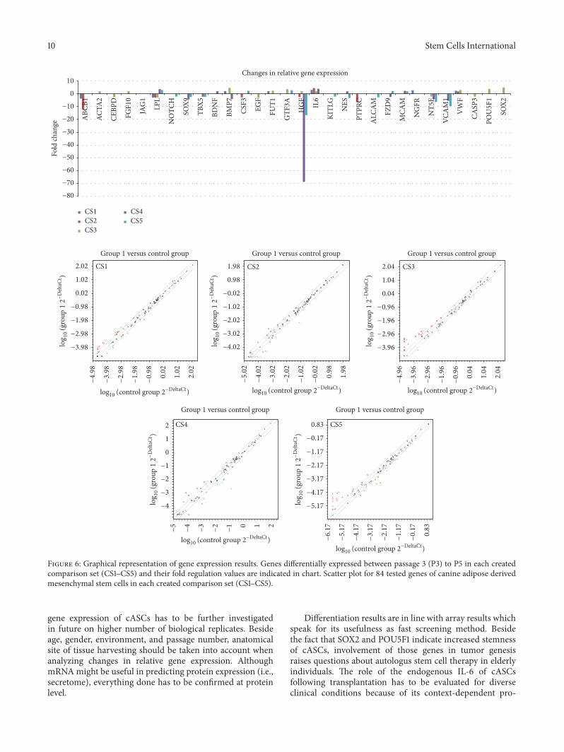
(CS1) presents the changes in relative expression levels of84 genes (Figure 3) of cASCs from 6 donors between P3 andP5. (CS1) is the most representative one since it includes sixbiological replicates in each control and test group. (CS2) and(CS3) are created by dividing donors in age groups: young(𝑛 = 3) and old (𝑛 = 3) to monitor transcriptome changesduring cultivation. (CS4) and (CS5) are made to comparelevels of expression in young and old donors’ cells in the samepassage.
2.5.1. Gene Filtering. Gene filtering was performed by select-ing genes with stable changes in expression (threshold cycle(Ct) value < 30) for further analysis and excluding geneswith relatively low or nondetected expression either in test orcontrol group (average Ct > 30 for one group and average Ct< 30 for another group) as suggested by software instructions.Although aware of possible biological importance of thoseexcluded genes, it would be dangerous to interpret those datawithout using more biological replicates for verification ofthose results.
Obtained results were further analyzed using GeneOntology.
2.6. Statistica. Further statistical analysis of results obtainedfor CSs was performed using software Stata 13 using Fisher’sexact test and 𝑝 value ≤ .05 was considered significant.
3. Results
3.1. Expansion and Differentiation Characteristics. Isolatedadherent fibroblast-like cASCs during expansion demon-strated enlargement already in P3, morphological alterationin P4 (cuboidal shape, roundness, and plate appearance werepresent in part of cells in flask), and ultimately proliferationarrest in P7 regardless of age of donor. Expansion charac-teristics observed microscopically were similar in all donors.Canine ASCs were refractory to adipogenic differentiationbut able to differentiate into osteoblasts (Figure 1(b)) andchondrocytes (Figures 1(c) and 1(d)).
3.2. Detection of Senescent Cells. SA-beta-Gal activity wasdetected already in P4 in minority of cells in all donors (bluestaining) (Figure 2). With each passage (5, 6) higher numberof cells showed marked SA beta-Gal activity.
3.3. Flow Cytometry Surface Marker Profile. Undifferentiatedcanine ASCs were analyzed by flow cytometry (Table 1) tomonitor the surface marker profile during their ex vivoexpansion. Each passage demonstrated a homogeneous pop-ulation of viable cells, with continuous expression of CD90,CD44, CD29 (>95% cells in population), and CD271 and theabsence of CD45 and CD14 (Figures 3 and 4) cell markersfrom P1 to P4 irrespective of the donor included in the study.Small proportion of cells expressed CD271 (up to 3,21%).Canine ASCs expressed low levels of autofluorescency in P1-P2 and it started to increase in P3 to the end of cultivationwhich was accompanied by increased median of the forwardand side scatter signal (Figure 2). However, expression ofCD90 and CD44 decreased in P5-P6 in all donors cells(Figure 4).
Examined clusters of differentiation together with stemcell specific genes for CD105, CD73, and CD166 were alsofound at mRNA level in P3 and P5 using array.The examinedmarkers showed stable expression level during that expansionperiod.
3.4. Gene Expression. Results (Table 2)were analyzed accord-ing to created comparison sets (CS) (Figures 5 and 6) asdescribed.
Obtained data revealed in total 7 differently expressedgenes in CS1. Comparing CS2 and CS3 it can be noted thatyoung donors’ cells in P5 demonstrated more downregulatedgenes (71,43%) than old donors’ cells in P5 (33,34%) whileresults for upregulated genes are opposite (66,66% for oldand 28,57% for young donors’ cells). Difference between CS2and CS3 was statistically significant (𝑝 ≤ .05). Comparingbaseline difference in gene expression in P3 and P5 in donorsof different age (CS4 and CS5) result was not statisticallysignificant.
Stemness genes (bFGF, INS, LIF, POU5F1, and SOX2)were expressed at similar levels in young and old donors,

Stem Cells International 5
(a) (b)
(c) (d)
Figure 1: Differentiation of canine adipose derived stem cells. (a) Canine adipose derived stem cells negative for alkaline phosphataseactivity (negative control for osteoblasts differentiation). (b) Osteoblast differentiation. Canine adipose derived stem cells stained for alkalinephosphatase activity with NBT substrate (purple). (c) Chondrodifferentiation of canine adipose derived stem cells in microwell plate stainedwith Alcian Staining Solution, negative and positive well. Positive well contains blue spheroid. (d) Chondrodifferentiation of canine adiposederived stem cells. Spheroid stained with Alcian Staining Solution.
(a) (b) (c)
Figure 2: Detection of senescent cells. Qualitative SA-𝛽-Gal assay of the most representative donors’ canine adipose derived stem cells inpassage 4. A local region of senescence cell is shown (b, c). Negative control cells (a).
except for CS3 where old donors’ cells in P3 expressed higherlevels of SOX2 and POU5F1.
To better understand cASC properties in biological andmolecular context we analyzed obtained data using GeneOntology. According to biological function changed geneswere classified into those responsible for apoptotic pro-cess, biological adhesion, biological regulation, cellular pro-cess, developmental process, immune system, localization,metabolic process, reproduction, and response to stimulus.
Their molecular function is related to binding, catalytic, andreceptor activity.
4. Discussion
Although the “gold rush” in using mesenchymal stem cells(MSCs) for therapeutic purposes beganwith high enthusiasmnumerous scientific issues remain to be resolved. Availableliterature offers a lot of information but still makes readership

6 Stem Cells International
0 102
103
104
105
0
102
103
104
105
CD45
PE-A
CD90
APC
-A
3/15, unst, 3p - P2 3/15, unst, 3p - P2 3/15, unst, 5p - P2 3/15, unst, 5p - P2
CD14 PE-Cy7-A CD44 FITC-A0 10
210
310
410
5
0
102
103
104
105
CD45
PE-A
CD14 PE-Cy7-A0 10
210
310
410
5
0
102
103
104
105
CD90
APC
-A
CD44 FITC-A0 10
210
310
410
5
0
102
103
104
105
P3 P4
P5
P3 P4
P5
(a) Unstained cASCs
3/15 izo - P2 3/15 izo - P2 3/15, izo - P2 3/15, izo - P2
0 102
103
104
105
0
102
103
104
105
CD45
PE-A
CD14 PE-Cy7-A0 10
210
310
410
5
0
102
103
104
105
CD45
PE-A
CD14 PE-Cy7-A
CD90
APC
-A
CD44 FITC-A0 10
210
310
410
5
0
102
103
104
105
CD90
APC
-A
CD44 FITC-A0 10
210
310
410
5
0
102
103
104
105
P3 P4
P5
P3 P4
P5
(b) Isotype control
3/15 mix - P2 3/15 mix - P2 3/15, mix - P2 3/15, mix - P2
0 102
103
104
105
0
102
103
104
105
CD45
PE-A
CD14 PE-Cy7-A0 10
210
310
410
5
0
102
103
104
105
CD45
PE-A
CD14 PE-Cy7-A
CD90
APC
-A
CD44 FITC-A0 10
210
310
410
5
0
102
103
104
105
CD90
APC
-A
CD44 FITC-A0 10
210
310
410
5
0
102
103
104
105
P3P4
P5
P3 P4
P5
(c) Fully stained cASCs
Figure 3: Flow cytometry results.Themost representative canine adipose derived stem cells from one donor are shown in passage 3 and 5.Thefigure shows P2 population after excluding dead cells and doublets. CD: cluster of differentiation, APC: allophycocyanin, PE: Phycoerythrin,FITC: fluorescein isothiocyanate, and PE-Cy7: R-Phycoerythrin-Cyanine 7.
pretty confused (no uniform characterization criteria forMSCs [10], little consensus about immunomodulation ofMSCs, secretome composition following infusion which isuncertain [11], and MSCs which are not always immunosup-pressive [12]).
In stem cell transplantation, canines have been used formore than 30 years. As a model they are more comparablewith humans than rodents. Clinical application of stemcells often means transplantation of cells with unknowncharacteristics [13]. It is important to identify the factorsthat are involved in the regulation of the expression andproduction of paracrine molecules in MSCs to achieve anoptimal therapeutic outcome [14]. Therefore, this study pro-vides comprehensive insight into cASCs from the aspects ofmorphology, differentiation, and immunophenotyping withspecial emphasis on transcriptome.
Morphological changes of cASCs in this study are inline with those described in the literature [15, 16]. However,senescent appearance, confirmed with SA-beta-Gal activity,was observed in P4 in part of flask seeded cells and P7represents the upper cultivation limit. Those findings can bea result of culturing conditions. When cells are explantedfrom an organism and placed in culture, they have to adapt
to abnormal concentrations of nutrients and growth factors,as well as the absence of surrounding cells and extracellularmatrix [16]. Early senescence in this case could be due tohigh seeding density (3 × 10⁴/cm2) and high percentageof FBS (20%) in basal media. Limited lifespan (up to onlyP7) is confirmed continuously on multiple donors (𝑛 =18, unpublished data) of different age and breeds in ourlaboratory conditions. Contributing factors to the limitedlifespan are the same as those responsible for senescence, butgenerally shorter lifespan of canine specieswhen compared tohumans should be also considered. Dogs lose telomeric DNAapproximately 10-fold faster than humans which is similarto the ratio of average life spans between these species [17].Elongation of life span can be achieved by culturing in serum-free medium supplemented with a number of defined growthfactors [18] or by culturing under physiological oxygenconditions [19] as described formouse embryonic fibroblasts.Morphological and immunophenotypic cells obtained fromall donors were similar which is in line with observationsmade by others [20].
For the differentiation test we have chosen human differ-entiationmedia because of their controlled origin and qualityand to ensure equal conditions for all steps of differentiation.

Stem Cells International 7
Table 2: Results of relative gene expression analysis. Summary of all results in different comparison set (CS).
GENE NAME CS1 CS2 CS3 CS4 CS5All donors Y P3/Y P5 O P3/O P5 Y P3/O P3 Y P5/O P5
ABCB1 −3,70↓ −12,09↓ −2,70↓ 3,16↑ACTA2 2,09↑CEBPD −2,32↓FGF10 2,23↑ −3,05↓ −2,50↓JAG1 2,93↑LPL −2,82↓ −2,82↓ −2,82↓ 3,75↑ 3,02↑NOTCH −2,05↓SOX9 −4,24↓ −2,45↓TBX5 −2,19↓ −2,3↓BDNF 2,10↑BMP7 2,10↑ 4,76↑ −3,98↓CSF3 −2,65↓ 2,38↑EGF −2,98↓FUT1 2,15↑ 2,55↑GTF3A 3,68↑ 2,84↑HGF −2,44↓ 2,10↑ −68,91↓ −16,6↓IL6 3,16↑ 4,59↑ 2,15↑ 4↑KITLG −2,18↓NES 2,09↑ −3,2↓PTPRC −6,52↓ALCAM −2,65↓FZD9 −2,36↓ 2,33↑MCAM 2,42↑ 2,08↑NGFR 2,81↑NT5E −2,06↓ −3,98↓ −6,29↓VCAM1 −5,37↓ −9,5↓VWF 2,48↑ 2,00↑ 3,08↑CASP3 −2,11↓POU5F1 3,97↑SOX2 5,07↑Total (%) 7 (100) 7 (100) 15 (100) 15 (100) 16 (100)Upregulated (%) 5 (71,42) 2 (28,57) 10 (66,66) 6 (40) 6 (37,5)Downregulated (%) 2 (28,57) 5 (71,42) 5 (33,33) 9 (60) 10 (62,5)Total (%) 2 (28,57) 2 (28,57) 4 (26,6) 6 (40) 6 (37,5)Differentiation markers 2 (28,57) 2 (28,57) 4 (26,6) 6 (40) 6 (37,5)MSCs associated markers (%) 3 (42,8) 4 (57,1) 6 (40) 5 (33,33) 5 (31,2)MSCs specific markers (%) 2 (28,57) 1 (14,28) 3 (20) 4 (26,6) 5 (31,2)Stemness markers (%) / / 2 (13,3) / /CS = comparison set, Y = young donor, O = old donor, and P = passage; numbers in italic and bold indicate fold regulation value.
Lack of adipogenic differentiation in cASCs was probablydue to applying human media as described by Neupaneet al. who suggest usage of canine adjusted media [21].Same authors also reported refraction of cells to osteogenicdifferentiation, but our results suggest the opposite. Dif-ferentiation results were further supported by upregulatedBMP7 and IL-6 and downregulated LPL mRNA. The BMPsignal induces osteoblastic differentiation, at the same timeinhibiting adipogenesis and myogenesis [22]. LPL expressionmay reflect the growth-arrest stage which is prerequisite
for adipocytes differentiation [23]. Upregulated BMP7 anddownregulated LPLmRNA strongly support cASCs behaviorduring differentiation process, absence of adipogenic andsuccessful osteogenic differentiation. Of further interest is thefact that pretreatment of MSCs with IL-6 inhibits adipogenicand chondrogenic differentiation [24]. Our results establishthe need to investigate what is the role of endogenous IL-6 oninhibition of adipogenesis and influence on chondrogenesis.
For a more in-deep characterization the immunopheno-typingwas performedwith flow cytometry.Wehave observed

8 Stem Cells International
0
20
40
60
80
100
120
0 1 2 3 4 5 6 7
CD90 APC and CD44 FITC
Perc
ent p
ositi
vity
0
0,2
0,4
0,6
0,8
1
0 2 4 6 8
Perc
ent p
ositi
vity
CD14 PE-Cy7 and CD45 PE
Donor 1Donor 2Donor 3
Donor 4Donor 5Donor 6
Figure 4: Flow cytometry results. Expression of cluster of differen-tiation (CD) during cultivation from passage 1 (P1) to P7.
stable expression of analyzedmarkers during cultivation timewhich is in compliance with previous reports [25] and nodifferences in marker expression were observed betweenyoung and old donors. We also examined the expression ofCD271 which has been proposed asmarker of primary choicefor tissue regeneration [26]. Expression values for CD271 incASCs were similar as in human ASCs [27]. To complementthis marker analysis based on detection using mAbs, whichare often commercially unavailable, we also characterizedcASCs at the mRNA level. Results of molecular analysisenabled us to see what genes those cells are expressingand which of them are changed during culturing from P3to P5 and to what magnitude. Molecular analysis providesexplanation and confirmation of the observed results (lackof adipogenic differentiation, immunophenotype properties,and stemness preservation).Therefore, these molecular tech-niques are valuable tool for fast screening of cells priorto application and for better understanding of therapeuticpower of cASCs.
By analyzing results of created comparison sets (CS)using Gene Ontology one can see that part of changedgenes in CS1 was involved in fundamental cellular functions
but our attention was dragged by upregulated interleukin-6 (IL-6) as controversial molecule. IL-6 expressed contin-uous upregulation in CS 1, 2, 3, 4 with fold regulationvalues higher in young (CS2) than in old donors (CS3).This may indicate that young donors’ cells are more potentpromoters of immunomodulation. It is well-known fact thatMSCs produce IL-6 whose biology is complex [28]. IL-6 is thought to be harmful because it can be key to themaintenance of chronic inflammation [29] but at the sametime it stimulates the secretion of anti-inflammatory IL-10 [12]. Through production of IL-6, MSCs prevent thedifferentiation ofmonocytes towards antigen-presenting cellsand skew differentiation towards an anti-inflammatory IL-10-producing cell type [30]. IL-10 was not produced by cASCsin present study which is in line with results of mentionedauthors [30] who report IL-10 production exclusively bymonocytes after exposure to MSCs-produced IL-6. cASCsfollowing infusion meet microenvironment for interactionwith immune cells and by IL-6 they could stimulate secretionof favorable IL-10. Based on the above-mentioned facts, webelieve that upregulated IL-6 in cASC during culturing mayindicate beneficial therapeutic effect. However, it should benoted that IL-6 and IL-10 are not the only cytokines involvedin these complex interactions. Paracrine mechanisms for thetherapeutic effects ofMSC are very complex and involve largenumber of growth factors, cytokines, signaling molecules,and related receptors with a broad range of biological func-tions which should be investigated in future.
Comparison of CS2 and CS3 enabled us to investigateinfluence of cASC cultivation on changes in relative geneexpression in different age groups. Interestingly, old donors’cells (CS3) exhibited 2,14-fold higher changed genes, mostlyupregulated, which could indicate more intense processeswithin those cells as response to cultivation conditions.Only two genes were in common, upregulated IL-6 anddownregulated LPL. Fold regulation of IL-6 was 2,13-foldhigher in young donors’ cells and fold regulation for LPLwas the same in both CS. Only old donors’ cells expressedupregulation of two stemness markers, SOX2 and POU5F1.These transcription factors for pluripotency and self-renewalare naturally expressed inMSCs at low levels in early passagesand gradually decrease as the passage number increases [31].The effect of cooverexpression of POU5F1 and SOX2 inhuman adipose tissue MSCs (hAT-MSC) has been inves-tigated. Those results show effectively enhanced mesoder-mal differentiation potency indicating increased stemnessof hAT-MSCs [31]. Seen upregulation of these genes speaksfor preserved stemness characteristics of old donors’ cellswhich raises hope from therapeutic perspective, but theirinvolvement in tumor genesis should always be kept in mind.It remains to view what characteristic will be predominantin older hosts since incidence of tumor formation increaseswith age [32, 33] and aging represents the single biggest riskfactor for most cancers [34–36]. Furthermore, geneticallyunmodified MSCs can undergo chromosomal abnormalitieseven at early passages and form malignant tumors whentransplanted in vivo. Careful monitoring of chromosomalstatus is warranted when in vitro expanded MSCs are usedfor cell therapy [37].

Stem Cells International 9
03
P5Y3
P503
P3Y2
P5Y3
P3Y2
P302
P301
P301
P502
P5Y1
P5Y1
P3
Magnitude of gene expression
Min Avg Max
IL10INSR
RPL13AGTF3A
CSF2IL6
CTNNB1GDF5
PROM1
IT GAXSOX2
IT GAVWNT3A
GDF7INS
TNFADIPOQ
TGFB3COL1A1
IFNGIL1B
FGDCENG
POU5F1IGF1
BGLAPTGFB1
SMURF1SMURF2ANXA5
VIMHNF1AKAT2B
CD44PIGSEGF
MMP2B2M
MITFPPARG
SLC17A5FASNFGF2
BMP4THY1
PDGFRBSMAD4VCAM1
VWFNGFRTBX5
KITLGSOX9HGF
NT5ELIF
NOTCH1HAT1
RHOARUNX2MCAM
CSF3FZD9JAG1PTK2
CEBPDERBB2
ANPEPNUDT6CASP3HPRT1
ACTBHDAC1
NESVEGFAIT GB1
KDRLPL
PTPRCBMP2
ABCB1FUT1
FGF10GDF15
ALCAMBDNFBMP7
ACTA2ICAM1
Figure 5: Clustergram presents 84 analyzed genes of cASCs of 6 donors (3 young donors (Y1, Y2, and Y3) in passage 3 (P3) and P5 and 3 olddonors (O1, O2, and O3) in P3 and P5). Passage 3 of cASCs was used as control group and P5 of cASCs represents test group. Nonsupervisedhierarchical clustering of the entire dataset displayed no age or passage number related clustering.
Comparison of CS4 and CS5 enabled us to investigatedifferences in transcriptome of cASC in the same passage butdifferent donors’ age group. Old donors’ cells in lower (P3)(CS4) and higher passage (P5) (CS5) differently expressed
genes mainly differentiation associated and downregulated.This together with upregulated stemness genes in CS3 speaksfor preserved undifferentiated state what is important fortherapy. How exactly donors’ age affects level of change in
10 Stem Cells International
−80
−70
−60
−50
−40
−30
−20
−10
0
10A
BCB1
ACTA
2
CEBP
D
FGF1
0
JAG
1
LPL
NO
TCH
SOX9
TBX5
BDN
F
BMP7
CSF3
EGF
FUT1
GTF
3A
HG
F
IL6
KITL
G
NES
PTPR
C
ALC
AM
FZD
9
MCA
M
NG
FR
NT5
E
VCA
M1
VW
F
CASP
3
POU
5F1
SOX2
Fold
chan
ge
Changes in relative gene expression
CS1
CS1
CS2
CS2
CS3
CS3
CS4
CS4
CS5
CS5
log10
(control group 2−DeltaCt)log
10(control group 2
−DeltaCt)
log10
(control group 2−DeltaCt)
log10
(control group 2−DeltaCt)
log 1
0(g
roup
12−
Delt
aCt )
log 1
0(g
roup
12−
Delt
aCt )
log 1
0(g
roup
12−
Delt
aCt )
log 1
0(g
roup
12−
Delt
aCt )
log 1
0(g
roup
12−
Delt
aCt )
Group 1 versus control groupGroup 1 versus control groupGroup 1 versus control group
Group 1 versus control group Group 1 versus control group
2.02
1.02
0.02
−0.98
−1.98
−2.98
−3.98
−4.98
−3.98
−2.98
−1.98
−0.98
0.02
1.02
2.02
1.98
0.98
−0.02
−1.02
−2.02
−3.02
−4.02
−5.02
−4.02
−3.02
−2.02
−1.02
−0.02
0.98
1.98
2.04
1.04
0.04
−0.96
−1.96
−2.96
−3.96
−4.96
−3.96
−2.96
−1.96
−0.96
0.04
1.04
2.04
2
1
0
−1
−2
−3
−4
−5
−4
−3
−2
−1 0 1 2
0.83
−0.17
−1.17
−2.17
−3.17
−4.17
−5.17
−6.17
−5.17
−4.17
−3.17
−2.17
−1.17
−0.17
0.83
log10
(control group 2−DeltaCt)
Figure 6: Graphical representation of gene expression results. Genes differentially expressed between passage 3 (P3) to P5 in each createdcomparison set (CS1–CS5) and their fold regulation values are indicated in chart. Scatter plot for 84 tested genes of canine adipose derivedmesenchymal stem cells in each created comparison set (CS1–CS5).
gene expression of cASCs has to be further investigatedin future on higher number of biological replicates. Besideage, gender, environment, and passage number, anatomicalsite of tissue harvesting should be taken into account whenanalyzing changes in relative gene expression. AlthoughmRNAmight be useful in predicting protein expression (i.e.,secretome), everything done has to be confirmed at proteinlevel.
Differentiation results are in line with array results whichspeak for its usefulness as fast screening method. Besidethe fact that SOX2 and POU5F1 indicate increased stemnessof cASCs, involvement of those genes in tumor genesisraises questions about autologus stem cell therapy in elderlyindividuals. The role of the endogenous IL-6 of cASCsfollowing transplantation has to be evaluated for diverseclinical conditions because of its context-dependent pro-

Stem Cells International 11
and anti-inflammatory properties. Transcriptomic resultsrevealed the need to monitor and determine the potentialinfluence of SOX2, POU5F1, IL-6, and other molecules ascASCs secretome components in the stem cell therapy.
Abbreviations
cASCs: Canine adipose derived stem cellsMSCs: Mesenchymal stem cellsISCT: International Society for Cell TherapyPBS: Phosphate-buffered salineFBS: Foetal bovine serumDMEM: Dulbecco’s Modified Eagle MediumDMSO: Dimethyl sulfoxideRT: Room temperatureCD: Cluster of differentiationIg: ImmunoglobulinAPC: AllophycocyaninPE: PhycoerythrinFITC: Fluorescein isothiocyanatePE-Cy7: R-Phycoerythrin-Cyanine 7P: PassageCS: Comparison setY: Young donorO: Old donor.
Consent
All materials used in this study were collected as medicalwaste after surgical procedure with owner consent at veteri-nary clinics.
Disclosure
Nina Kresic and Ivana Simic are co-first authors.
Competing Interests
The authors declare that they have no competing interests.
Acknowledgments
Theauthorswould like to thankCroatianVeterinary Institute,Croatia, for providing them with the possibility to performthis work. The authors thank Kreso Bendelja, Ph.D., ReljaBeck, Ph.D., Dragan Brnic, Ph.D., Miroslav Benic, Ph.D., andAlexandra Stolzing, Ph.D. for support during experiment.Also, they would like to thankMaura Ferrari, Ph.D., AnnalisaGhizzardi, and personnel of CSC, IZSLER, Italy, for introduc-ing them to stem cell field.
References
[1] I. Ullah, R. B. Subbarao, and G. J. Rho, “Human mesenchymalstem cells—current trends and future prospective,” BioscienceReports, vol. 35, Article ID e00191, 2015.
[2] C. V. Machado, P. D. S. Telles, and I. L. O. Nascimento, “Im-munological characteristics ofmesenchymal stem cells,”Revista
Brasileira de Hematologia e Hemoterapia, vol. 35, no. 1, pp. 62–67, 2013.
[3] Y. Kfoury and D. T. Scadden, “Mesenchymal cell contributionsto the stem cell niche,” Cell Stem Cell, vol. 16, no. 3, pp. 239–253,2015.
[4] M. Dominici, K. Le Blanc, I. Mueller et al., “Minimal crite-ria for defining multipotent mesenchymal stromal cells. TheInternational Society for Cellular Therapy position statement,”Cytotherapy, vol. 8, no. 4, pp. 315–317, 2006.
[5] R. Screven, E. Kenyon, M. J. Myers et al., “Immunophenotypeand gene expression profile of mesenchymal stem cells derivedfrom canine adipose tissue and bone marrow,” VeterinaryImmunology and Immunopathology, vol. 161, no. 1-2, pp. 21–31,2014.
[6] H. Takemitsu, D. Zhao, I. Yamamoto, Y. Harada, M. Michishita,and T. Arai, “Comparison of bone marrow and adipose tissue-derived canine mesenchymal stem cells,” BMC VeterinaryResearch, vol. 8, article no. 150, 2012.
[7] M. R. Schneider, E. Wolf, J. Braun, H.-J. Kolb, and H. Adler,“Canine embryo-derived stem cells and models for humandiseases,”HumanMolecular Genetics, vol. 17, no. 1, pp. R42–R47,2008.
[8] I. H. Bellayr, J. G. Catalano, S. Lababidi et al., “Gene markersof cellular aging in humanmultipotent stromal cells in culture,”Stem Cell Research andTherapy, vol. 5, no. 2, article 59, 2014.
[9] W. Wagner, S. Bork, G. Lepperdinger et al., “How to trackcellular aging of mesenchymal stromal cells?” Aging, vol. 2, no.4, pp. 224–230, 2010.
[10] E. de Bakker, B. Van Ryssen, C. De Schauwer, and E. Meyer,“Caninemesenchymal stem cells: state of the art, perspectives astherapy for dogs and as a model for man,” Veterinary Quarterly,vol. 33, no. 4, pp. 225–233, 2013.
[11] O. Levy, W. Zhao, L. J. Mortensen et al., “mRNA-engineeredmesenchymal stem cells for targeted delivery of interleukin-10to sites of inflammation,” Blood, vol. 122, no. 14, pp. e23–e32,2013.
[12] D. Kyurkchiev, “Secretion of immunoregulatory cytokines bymesenchymal stem cells,”World Journal of Stem Cells, vol. 6, no.5, p. 552, 2014.
[13] G. Q. Daley, “The promise and perils of stem cell therapeutics,”Cell Stem Cell, vol. 10, no. 6, pp. 740–749, 2012.
[14] R. C. Zhao, “Essentials ofmesenchymal stem cell biology and itsclinical translation,”Essentials ofMesenchymal StemCell Biologyand Its Clinical Translation, pp. 1–313, 2013.
[15] T. Martinello, I. Bronzini, L. Maccatrozzo et al., “Canineadipose-derived-mesenchymal stem cells do not lose stem fea-tures after a long-term cryopreservation,”Research inVeterinaryScience, vol. 91, no. 1, pp. 18–24, 2011.
[16] T. Kuilman, C. Michaloglou, W. J. Mooi, and D. S. Peeper, “Theessence of senescence,” Genes and Development, vol. 24, no. 22,pp. 2463–2479, 2010.
[17] L. J. Fick, G.H. Fick, Z. Li et al., “Telomere length correlates withlife span of dog breeds,”Cell Reports, vol. 2, no. 6, pp. 1530–1536,2012.
[18] D. T. Loo, J. I. Fuquay, C. L. Rawson, and D. W. Barnes,“Extended culture of mouse embryo cells without senescence:inhibition by serum,” Science, vol. 236, no. 4798, pp. 200–202,1987.
[19] S. Parrinello, E. Samper, A. Krtolica, J. Goldstein, S. Melov, andJ. Campisi, “Oxygen sensitivity severely limits the replicativelifespan of murine fibroblasts,” Nature Cell Biology, vol. 5, no.8, pp. 741–747, 2003.
12 Stem Cells International
[20] M. S. Choudhery, M. Badowski, A. Muise, J. Pierce, and D. T.Harris, “Donor age negatively impacts adipose tissue-derivedmesenchymal stem cell expansion and differentiation,” Journalof Translational Medicine, vol. 12, no. 1, article no. 8, 2014.
[21] M. Neupane, C.-C. Chang, M. Kiupel, and V. Yuzbasiyan-Gurkan, “Isolation and characterization of canine adipose-derived mesenchymal stem cells,” Tissue Engineering - Part A.,vol. 14, no. 6, pp. 1007–1015, 2008.
[22] Y.-T. Xiao, L.-X. Xiang, and J.-Z. Shao, “Bone morphogeneticprotein,” Biochemical and Biophysical Research Communica-tions, vol. 362, no. 3, pp. 550–553, 2007.
[23] J. Pairault and H. Green, “A study of the adipose conversion ofsuspended 3T3 cells by using glycerophosphate dehydrogenaseas differentiation marker,” Proceedings of the National Academyof Sciences of the United States of America, vol. 76, no. 10, pp.5138–5142, 1979.
[24] K. L. Pricola, N. Z. Kuhn, H. Haleem-Smith, Y. Song, andR. S. Tuan, “Interleukin-6 maintains bone marrow-derivedmesenchymal stem cell stemness by an ERK1/2-dependentmechanism,” Journal of Cellular Biochemistry, vol. 108, no. 3, pp.577–588, 2009.
[25] W. Wagner, P. Horn, M. Castoldi et al., “Replicative senescenceof mesenchymal stem cells: a continuous and organized pro-cess,” PLOS ONE, vol. 3, no. 5, Article ID e2213, 2008.
[26] I. Roato, D. Alotto, D. C. Belisario et al., “Adipose Derived-mesenchymal stem cells viability and differentiating features fororthopaedic reparative applications: banking of adipose tissue,”Stem Cells International, vol. 2016, Article ID 4968724, 11 pages,2016.
[27] F. J. Lv, R. S. Tuan, K. M. Cheung, and V. Y. Leung, “Concisereview: the surface markers and identity of human mesenchy-mal stem cells,” Stem Cells, vol. 32, no. 6, pp. 1408–1419, 2014.
[28] T. C. Barnes, M. E. Anderson, and R. J. Moots, “The many facesof interleukin-6: the role of IL-6 in inflammation, vasculopathy,and fibrosis in systemic sclerosis,” International Journal ofRheumatology, vol. 2011, Article ID 721608, 6 pages, 2011.
[29] C. A. Hunter and S. A. Jones, “IL-6 as a keystone cytokine inhealth and disease,” Nature Immunology, vol. 16, no. 5, pp. 448–457, 2015.
[30] S.M.Melief, S. B. Geutskens,W. E. Fibbe, andH. Roelofs, “Mul-tipotent stromal cells skew monocytes towards an anti-inflam-matory interleukin-10-producing phenotype by production ofinterleukin-6,”Haematologica, vol. 98, no. 6, pp. 888–895, 2013.
[31] S.-M. Han, S.-H. Han, Y.-R. Coh et al., “Enhanced proliferationand differentiation of Oct4- And Sox2-overexpressing humanadipose tissue mesenchymal stem cells,” Experimental andMolecular Medicine, vol. 46, no. 6, article e101, 2014.
[32] V. N. Anisimov, “The relationship between aging and car-cinogenesis: a critical appraisal,” Critical Reviews in Oncology/Hematology, vol. 45, no. 3, pp. 277–304, 2003.
[33] J. P. De Magalhaes, “How ageing processes influence cancer,”Nature Reviews Cancer, vol. 13, no. 5, pp. 357–365, 2013.
[34] B. D. Smith, G. L. Smith, A. Hurria, G. N. Hortobagyi, and T.A. Buchholz, “Future of cancer incidence in the United States:burdens upon an aging, changing nation,” Journal of ClinicalOncology, vol. 27, no. 17, pp. 2758–2765, 2009.
[35] R. Yancik, “Population aging and cancer: a cross-nationalconcern,” Cancer Journal, vol. 11, no. 6, pp. 437–441, 2005.
[36] P. D. Adams, H. Jasper, and K. L. Rudolph, “Aging-induced stemcell mutations as drivers for disease and cancer,” Cell Stem Cell,vol. 16, no. 6, pp. 601–612, 2015.
[37] J.-O. Jeong, J. W. Han, J.-M. Kim et al., “Malignant tumorformation after transplantation of short-term cultured bonemarrow mesenchymal stem cells in experimental myocardialinfarction and diabetic neuropathy,” Circulation Research, vol.108, no. 11, pp. 1340–1347, 2011.

Submit your manuscripts athttps://www.hindawi.com
Hindawi Publishing Corporationhttp://www.hindawi.com Volume 2014
Anatomy Research International
PeptidesInternational Journal of
Hindawi Publishing Corporationhttp://www.hindawi.com Volume 2014
Hindawi Publishing Corporation http://www.hindawi.com
International Journal of
Volume 2014
Zoology
Hindawi Publishing Corporationhttp://www.hindawi.com Volume 2014
Molecular Biology International
GenomicsInternational Journal of
Hindawi Publishing Corporationhttp://www.hindawi.com Volume 2014
The Scientific World JournalHindawi Publishing Corporation http://www.hindawi.com Volume 2014
Hindawi Publishing Corporationhttp://www.hindawi.com Volume 2014
BioinformaticsAdvances in
Marine BiologyJournal of
Hindawi Publishing Corporationhttp://www.hindawi.com Volume 2014
Hindawi Publishing Corporationhttp://www.hindawi.com Volume 2014
Signal TransductionJournal of
Hindawi Publishing Corporationhttp://www.hindawi.com Volume 2014
BioMed Research International
Evolutionary BiologyInternational Journal of
Hindawi Publishing Corporationhttp://www.hindawi.com Volume 2014
Hindawi Publishing Corporationhttp://www.hindawi.com Volume 2014
Biochemistry Research International
ArchaeaHindawi Publishing Corporationhttp://www.hindawi.com Volume 2014
Hindawi Publishing Corporationhttp://www.hindawi.com Volume 2014
Genetics Research International
Hindawi Publishing Corporationhttp://www.hindawi.com Volume 2014
Advances in
Virolog y
Hindawi Publishing Corporationhttp://www.hindawi.com
Nucleic AcidsJournal of
Volume 2014
Stem CellsInternational
Hindawi Publishing Corporationhttp://www.hindawi.com Volume 2014
Hindawi Publishing Corporationhttp://www.hindawi.com Volume 2014
Enzyme Research
Hindawi Publishing Corporationhttp://www.hindawi.com Volume 2014
International Journal of
Microbiology