adenosine thiamine triphosphate and adenosine thiamine...
TRANSCRIPT
52
ISSN 2409-4943. Ukr. Biochem. J., 2018, Vol. 90, N 4
UDC 577.164.11:577.152.3
Adenosine thiAmine triphosphAteAnd Adenosine thiAmine triphosphAte hydrolAse
Activity in AnimAl tissues
A. F. MAkArchIkoV1,2, T. V. SArokA3, T. G. kUdyrkA1,2, I. k. kolAS1,2,T. A. lUchko2, I. M. rUSINA1,2, V. A. GUrINoVIch2
1Grodno State Agrarian University, Belarus;2Institute of Biochemistry of Biologically Active compounds,
National Academy of Sciences of Belarus;3yanka kupala State University of Grodno, Belarus;
e-mail: [email protected]
Adenosine thiamine triphosphate (AThTP), a vitamin B1 containing nucleotide with unknown biochemi-cal role, was found previously to be present in various biological objects including bacteria, yeast, some hu-man, rat and mouse tissues, as well as plant roots. In this study we quantify AThTP in mouse, rat, bovine and chicken tissues. We also show that in animal tissues the hydrolysis of AThTP is catalyzed by a membrane-bound enzyme seemingly of microsomal origin as established for rat liver, which exhibits an alkaline ph optimum of 8.0-8.5 and requires no Mg2+ ions for activity. In liver homogenates, AThTP hydrolase obeys Michaelis-Menten kinetics with apparent km values of 84.4 ± 9.4 and 54.6 ± 13.1 µМ as estimated from the hanes plots for rat and chicken enzymes, respectively. The hydrolysis of AThTP has been found to occur in all samples examined from rat, chicken and bovine tissues, with liver and kidney being the most abundant in enzyme activity. In rat liver, the activity of AThTP hydrolase depends on the age of animals.
k e y w o r d s: vitamin B1, adenosine thiamine triphosphate, adenosine thiamine triphosphate hydrolase, ani-mal tissues.
© 2018 Makarchikov A. F. et al. This is an open-access article distributed under the terms of the Creative Commons Attribution License, which permits unrestricted use, distribution, and reproduction in any medium, provided the original author and source are credited.
Vitamin B1 in the form of thiamine diphos-phate (ThDP) plays an important role in the vital activity of the cell, fulfilling catalytic
functions. Currently, more than 30 ThDP-depen-dent enzymes have been known, five of them are encoded by animal genomes. Pyruvate dehydro-genase (EC 1.2.4.1), oxoglutarate dehydrogenase (EC 1.2.4.2) as well as 3-methyl-2-oxobutanoat dehy-drogenase (EC 1.2.4.4) are components of mitochon-drial dehydrogenase complexes involved in carbo-hydrate, amino acid and energy metabolism [1]. The metabolic role of transketolase (EC 2.2.1.1), which catalyzes a two-carbon moiety transfer from ketose to aldose sugars, is to provide a reversible link be-tween glycolysis and pentose phosphate pathway [2]. One more ThDP-dependent enzyme, 2-hydroxyacil-CoA lyase (EC 4.1.2.n2), is involved in the peroxi-somal metabolism of 3-methyl-branched fatty acids
and 2-hydroxy straight-chain fatty acids [3]. Along-side with ThDP, small amounts of thiamine, thia-mine monophosphate (ThMP) and thiamine triphos-phate (ThTP) are also present in animal tissues; the biological significance of these phosphate derivatives have not been yet established [4].
In 2007, a new nucleotide, adenosine thiamine triphosphate (AThTP), was revealed and identified in bacteria, some plant and animal tissues [5]. Though biochemical role for AThTP has not been ascertained to date, experiments with E. coli may indicate this compound to be involved in the molecular mecha-nisms of short-term adaptation through signaling/regulatory function. For instance, the concentration of AThTP, normally contained in a bacterial cell only in tiny amounts, was found to increase many times under carbon starvation. A similar picture was observed when the energy metabolism of the cell
doi: https://doi.org/10.15407/ubj90.04.052
53
was disturbed with uncoupling agents as well as the inhibitors of glycolysis and respiratory chain [6]. In accordance to Tanaka et al. [7], in vitro AThTP in-hibits the activity of poly(ADP-ribose) polymerase 1, a nuclear enzyme playing the central role in cellular responses to various stress factors.
At present, the enzymes involved in AThTP metabolism are not identified. It is only known that the synthesis of AThTP in E. coli is catalyzed from ThDP and ADP(ATP) by a soluble Mg2+-dependent protein with molecular mass of 355 kDa [8]. An en-zyme capable of AThTP hydrolyzing was also found to be localized in the bacterial membrane [5].
This work is aimed at determining AThTP and characterizing some properties of the enzyme which catalyzes the hydrolysis of AThTP in animal tissues.
materials and methods
Alcohol dehydrogenase (ADH, EC 1.1.1.1), AMP, ThDP, NAD+, NADH, Tris base, trichloro-acetic acid (TCA), bovine serum albumin, glucose-6-phosphate, SP-Sephadex C-25 and DEAE-Se-phadex A-25 were purchased from Sigma-Aldrich (St. Louis, Mo., USA), pyridine, N,N’-dicyclohexyl-carbodiimide (DCC) and tetra-n-butylammonium hydrogen sulfate were from Acros Organics (New Jersey, USA), tetrahydrofuran was from Fisher Scien tific (Loughborough, UK), all other chemicals were of analytical grade from Reakhim (Moscow, Russia).
Apoenzyme of pyruvate decarboxylase (PDC, EC 4.1.1.1) was prepared from brewer’s yeast Sac-charomyces carlsbergensis as described by Cherni-kevich et al. [9].
AThTP was synthesized by condensing ThDP and AMP in pyridine in the presence of DCC [5]. Briefly, the procedure of AThTP preparation was as follows. The reaction mixture containing 1.38 mg/ml of ThDP, 1.26 mg/ml of AMP and 86.2 mg/ml of DCC was incubated for 3 h at 50 °C, after that 3 volumes of absolute ethanol were added. To purify the product the precipitate formed was dissolved in 5 mM sodium acetate, pH 3.8, and passed through a column of SP-Sephadex C-25 in the same buffer. The fractions containing AThTP were combined and applied to a DEAE-Sephadex A-25 column equili-brated with 10 mM Tris-HCl, pH 7.2. The elution was carried out with a linear gradient of increasing NaCl concentration from 0 to 0.3 M. Then the AThTP fractions were concentrated by evaporation and precipitated with absolute ethanol. Finally, the
precipitate was dissolved in 5 mM sodium acetate buffer, pH 3.8, and chromatographed repeatedly on a SP-Sephadex C-25 column. The purity of the prepa-ration obtained was 99%.
Male Wistar rats (180-190 g) and mice (23-25 g) were from the institutional animal quarters. All experiments were performed in accordance with Directive 2010/63/EU of the European Parliament and of the Council on the protection of animals used for scientific purposes. The samples of bovine tis-sues were obtained from the local slaughterhouse. Chicken tissue samples were from 43-day old free-range Leghorn chicks grown on a courtyard. After killing animals the tissues were immediately frozen in liquid nitrogen and stored at –80 °C until use.
For enzyme assays, frozen tissues were homoge nized in 5 volumes of 50 mM Tris-HCl buffer, pH 7.5, containing 0.15 M KCl and 0.2 mM EDTA, in a glass/glass Potter-Elvehjem homogenizer at 4 °C. To prepare extracts the homogenates were centrifuged at 20 000 g for 60 min.
All operations on isolating subcellular fractions from rat liver were performed at 0-4 °C. A sample of fresh tissue was forced through a 1 mm pore press and homogenized by 10 strokes in a teflon/glass Potter-Elvehjem device at 600 rpm in 10 volu mes of 10 mM Tris-HCl buffer, pH 7.4, containing 0.25 M sucrose, 0.5 mM EDTA and 0.1 mM MgCl2. The homogenate was filtered through four layers of wet gauze and centrifuged at 600 g for 10 min. The pellet was suspended in 10 volumes of the initial buffer and centrifuged (600 g, 10 min) again to yield a crude nuclear fraction. To prepare mitochondria the post-nuclear supernatants were combined and subjected to further centrifugation at 12 000 g for 10 min. The particles were washed once with the same medium giving a crude mitochondrial fraction. Then the post-mitochondrial supernatants were pooled and adjusted to pH 7.5 with 0.5 M Tris-HCl, pH 8.0, fol-lowed by adding CaCl2 to a final concentration of 15 mM. After 2 hours, the suspension was centri-fuged (14 000 g, 10 min) to obtain microsomal and cytosol fractions. Finally, the particular fractions were suspended in 50 mM Tris-HCl buffer, pH 7.3, containing 0.15 M KCl, 0.2 mM EDTA, and frozen at –80 °C along with the cytosol for storage.
The activities of marker enzymes (lactate dehydrogenase (LDH, EC 1.1.1.27), succinate dehydrogenase (SDH, EC 1.3.5.1) and glucose-6-phosphatase (G6Pase, EC 3.1.3.9)) as well as the quantity of DNA in the subcellular fractions were assayed by conventional methods.
A. F. Makarchikov, T. V. Saroka, T. G. kudyrka et al.
54
ISSN 2409-4943. Ukr. Biochem. J., 2018, Vol. 90, N 4
The standard mixture for AThTP hydrolase activity determination contained 50 mM sodium phosphate buffer, pH 8.0, 0.05 mM substrate and an aliquot of enzyme solution (1-10 microliters of tis-sue homogenate) in a total volume of 0.1 ml. Kinetic experiments required some alterations in the assay with respect to pH or substrate concentration; it is indicated as necessary. The reaction was carried out for 10-30 min at 37 °C, depending on tissue and homogenate amount, to measure the initial velocity. Linearity in product accumulation was observed at given time intervals. After the reaction was stopped by boiling for 1 min, the mixture was diluted five-fold with 20 mM sodium phosphate buffer, pH 6.8, and centrifuged (3 000 g, 10 min) to remove a pro-tein precipitate. The amount of ThDP produced was measured by enzymatic method [9]. For this purpose, a 0.05-ml aliquot of the supernatant was incubated for 30 min at 25 °C with yeast apoPDC to reconsti-tute an active enzyme. The activity of holoPDC was then determined on decreasing NADH adsorption at 340 nm in a coupled reaction with ADH. To quantify ThDP a calibration graph was built using a chroma-tographically pure standard.
A unit of enzyme activity (U) was defined as the amount that catalyzes the formation of 1 µmole of ThDP in one minute.
The content of AThTP in animal tissues was evaluated by ion-pair reversed-phase HPLC [10]. Frozen tissue samples were homogenized in 5 volu-mes of 12% TCA in a glass/glass Potter-Elvehjem homogenizer at 0-4 °C and centrifuged (20 000 g, 15 min, 4 °C). To extract the acid an aliquot (0.2 ml) of the supernatant was treated with diethyl ester (3 times 0.6 ml each). Then the solution was placed into Agilent 1200 autosampler where an aliquot of 13 µl was mixed with 8 µl of 4.3 mM K3[Fe(CN)6] in 15% KOH to produce fluorescent thiochrome deriva-tives of thiamine compounds and injected into the chromatographic system. The separation was per-formed at a flow rate of 0.5 ml/min on a PRP-1 col-umn (4.1×150 mm, Hamilton Co, Reno, NV, USA), equipped with a PRP-X100 guard cartridge, in 50 mM KH2PO4 buffer, pH 8.8, containing 25 mM tet-ra-n-butylammonium hydrogen sulfate and 4% tet-rahydrofuran. Thiochrome derivatives were quan-tified using a fluorometric detector (λex = 365 nm, λem = 433 nm).
The identification of fluorescing products of AThTP hydrolase was done with HPLC as described above after the reaction was stopped by addition of 0.1 ml of 12% TCA and the mixture was centrifuged
(12 000 g, 10 min, 4 °C) to remove precipitated pro-teins.
Protein was determined by the method of Pe-terson [11] with bovine serum albumin as a standard. Statistical calculations were made using a Graph-PadPrism 5.0 program.
results and discussion
Occurrence and quantification of AThTP in bovine and chicken tissues. AThTP was originally discovered and identified in E. coli where it is ac-cumulated in high amounts during carbon starva-tion [5]. This nucleotide seems to be also present in yeast, plant roots and some human, rat and mouse tissues, based on chromatographic peaks appearance corresponding to the retention time (tR) of an authen-tic standard [5, 12, 13]. In this work we studied by HPLC the occurrence of AThTP in tissues of two species of farm animals, namely the cattle (Bos taurus taurus) and the chicken (Gallus gallus). To compare with literature data the content of AThTP in some tissues of rats (rattus norvegicus) and mice (Mus musculus) was determined as well.
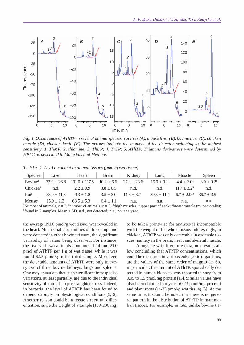
Judging by the chromatographic peaks, the bio-synthesis of AThTP takes place in all bovine tissues examined. However, we were not able to detect this compound in any sample of chicken liver or kidney, much like it was reported previously [12] for another bird species, the quail (coturnix coturnix). It is to be noted that AThTP was also not found in human liver biopsies [13]. On the other hand, small but clearly distinguishable peaks with tR corresponding to AThTP could be observed on chromatograms of ex-tracts from chicken heart, brain and skeletal muscle, while no AThTP was detectable in any quail tissue [12]. Fig. 1 shows examples of chromatograms for chicken brain and breast muscle as well as mamma-lian livers. The results obtained on AThTP quantifi-cation in animal tissues are summarized in Table 1.
According to our data, the concentration of AThTP in rats varies from 3.5 to 89.3 pmol/g wet tissue, the lung being the most abundant tissue followed by the spleen and liver. In mice, AThTP levels were found to be within the same range (6.4-68.5 pmol/g wet tissue). As a whole, these data are in a good agreement with the results of studies where the content of AThTP was expressed per 1 mg of protein [5, 12], especially in view of incomplete al-kaline extraction of proteins from TCA precipitates. As can be seen from Table 1, among the bovine tis-sues examined the highest AThTP concentration, on
55
the average 191.0 pmol/g wet tissue, was revealed in the heart. Much smaller quantities of this compound were detected in other bovine tissues, the significant variability of values being observed. For instance, the livers of two animals contained 12.4 and 21.0 pmol of AThTP per 1 g of wet tissue, while it was found 62.5 pmol/g in the third sample. Moreover, the detectable amounts of AThTP were only in eve-ry two of three bovine kidneys, lungs and spleens. One may speculate that such significant intraspecies variations, at least partially, are due to the individual sensitivity of animals to pre-slaughter stress. Indeed, in bacteria, the level of AThTP has been found to depend strongly on physiological conditions [5, 6]. Another reason could be a tissue structural differ-entiation, since the weight of a sample (100-200 mg)
to be taken pointwise for analysis is incompatible with the weight of the whole tissue. Interestingly, in chicken, AThTP was only detectable in excitable tis-sues, namely in the brain, heart and skeletal muscle.
Alongside with literature data, our results al-low concluding that AThTP concentrations, which could be measured in various eukaryotic organisms , are the values of the same order of magnitude. So, in particular, the amount of AThTP, sporadically de-tected in human biopsies, was reported to vary from 0.05 to 1.5 pmol/mg protein [13]. Similar values have also been obtained for yeast (0.23 pmol/mg protein) and plant roots (14-33 pmol/g wet tissue) [5]. At the same time, it should be noted that there is no gene-ral pattern in the distribution of AThTP in mamma-lian tissues. For example, in rats, unlike bovine tis-
Fig. 1. occurrence of AThTP in several animal species: rat liver (A), mouse liver (B), bovine liver (C), chicken muscle (D), chicken brain (E). The arrows indicate the moment of the detector switching to the highest sensitivity. 1, ThMP; 2, thiamine; 3, ThdP; 4, ThTP; 5, AThTP. Thiamine derivatives were determined by hPlc as described in Materials and Methods
Fluo
resc
ence
Time, min
-25
-50
-75
-100
-125
-150
0
25
0 8 16 0 8 16 0 8 16 0 8 16 0 8 16
-100
-80
-60
-40
-20
20
0
A B C D3
1 212
2
1 2
3
4
3
5
5
4
5
5
4
3
4
5
1
3
1 2
E4
15
10
5
0
-5
-10
-15
0
10
20
30
40140
120
100
0
40
60
80
20
T a b l e 1. AThTP content in animal tissues (pmol/g wet tissue)
1Number of animals, n = 3; 2number of animals, n = 9; 3thigh muscles; 4upper part of neck; 5breast muscle (m. pectoralis); 6found in 2 samples; Mean ± SD; n.d., not detected; n.a., not analyzed
Species Liver Heart Brain Kidney Lung Muscle SpleenBovine1 32.0 ± 26.8 191.0 ± 117.8 10.2 ± 6.6 27.3 ± 23.66 15.9 ± 0.16 4.4 ± 2.04 3.0 ± 0.26
Chicken1 n.d. 2.2 ± 0.9 3.8 ± 0.5 n.d. n.d. 11.7 ± 3.25 n.d.Rat1 33.9 ± 11.8 9.3 ± 1.0 3.5 ± 3.0 14.3 ± 3.7 89.3 ± 11.4 6.7 ± 2.03,6 36.7 ± 3.5Mouse2 15.9 ± 2.2 68.5 ± 5.3 6.4 ± 1.1 n.a. n.a. n.a. n.a.
A. F. Makarchikov, T. V. Saroka, T. G. kudyrka et al.
56
ISSN 2409-4943. Ukr. Biochem. J., 2018, Vol. 90, N 4
sues, the lung was found to be the highest in AThTP content (89.3 ± 11.4 pmol/g wet tissue), whereas the level of this substance in rodent heart was only 9.3 ± 1.0 pmol/g wet tissue (Table 1). Thus, the data available indicate the existence of interspecies fea-tures as regards to AThTP content in mammalian tis-sues. It is not clear yet whether these features reflect the physiology of species or they are to be attribu-ted to some external factors or technical reasons. Further studies concerning AThTP distribution and quantification in living organisms might be a step on the way of elucidating its biochemical functions.
Identification of fluorescent products of AThTP hydrolase. Commonly, the quantitative determina-tion of enzyme activity relies on the measurement of initial reaction rates, with either the disappearan ce of a substrate or increasing a product concentration being traced. Since the initial reaction rate can usu-ally be measured only in the interval of time until the conversion of substrate reaches 20% of the maxi-mum possible, in view of measurements precision it is preferably to determine product concentrations if an appropriate analytical method is available.
It was shown earlier that at pH 7.2 the cleavage of AThTP in E. coli is catalyzed by a membrane-bound enzyme, the fluorescent products of the reac-tion being ThDP and ThMP. The appearance of the last was supposed to be due to ThDP hydrolysis by endogenous enzymes, which has also to be respon-sible for the degradation of a nucleotide product of AThTP hydrolase reaction [5]. In such a case, when alongside with the enzyme under study the system contains some other proteins capable of further transformation of the reaction products, the only way to determine the activity of this enzyme is to trace the disappearance of its substrate. If AThTP hydrolase is to be assayed with this approach, the separation of constituents of the final reaction mix-ture should be performed. Indeed, the fluorescence of thiochrome derivative of AThTP is essentially undistinguishab le qualitatively from thiochrome and thiochrome phosphates [12]. As to the absorban-ce spectrum of AThTP, it overlaps with those for thiamine derivatives (unpublished data). In princi-ple, the problem of separation and quantification of thiamine compounds could be resolved using HPLC [5]. On the other hand, there is a possibility to de-termine AThTP hydrolase activity without exploi-ting any separation technics. This implies an enzy-matic quantification of ThDP through its biospecific binding to apo form of a ThDP -dependent enzyme,
for example, PDC followed by assaying the activity of reconstituted holoprotein. In respect of PDC, the enzyme from yeast Saccharomyces carlsbergensis was successfully used before as an analytical tool in studying thiamine metabolism, including the as-says of thiamine pyrophosphokinase (EC 2.7.6.2) and thiamine triphosphatase (EC 2.7.6.2) [14-16]. It is evident that two circumstances should be taken into account restric ting the suitability of enzyme detection of ThDP for AThTP hydrolase assay. To begin with, ThDP should be the only thiamine derivative as a reaction product, i.e. no its further conversion occurs by the enzyme. Moreover, the activities of phosphatases, which might be present in tissue extract/homogenate and capable of ThDP hydrolyzing, should be avoided. If the first clause fits, these unwanted activities, in principle, may be excluded (minimized) by compo sing the reaction medium (pH, ionic strength, buffer species, addi-tion of specific phosphatase inhibitors). Addressing these issues, we performed a series of experiments to identify fluorescent products in the reaction mixture after AThTP being incubated with aliquots of rat and chicken liver homogenates. The liver was cho-sen as the object of the study because it seems from literature data [17, 18] to have the highest ThDPase activity .
At first, we explored the effect of Mg2+ ions on AThTP hydrolysis in rat liver homogenate at pH 8.0 in two buffer systems, sodium phosphate and Tris-HCl. Fig. 2 represents the chromatograms of incu-bation mixtures in phosphate buffer system after stopping the reaction. As can be seen in Fig. 2, B, in the absence of Mg2+ the only reaction product to contain the thiamine moiety was ThDP. The ad-dition of Mg2+ into the incubation medium caused the appearance of small amounts of ThMP (about 0.9% of ThDP formed) (Fig. 2, c), seemingly, due to ThDP hydroly sis by phosphatases which are present in the liver homogenate. Hitherto, there is no proof for a specific ThDPase to exist in biological objects. Nevertheless, it has been known that mammalian cells express two nucleoside diphosphatases (L-type NDPase and B-type NDPase, EC 3.6.1.6) capable of ThDP hydrolyzing at neutral and alkaline pHs [19, 20]. Both of them require Mg2+ for acti vity. This may explain, at least partially, the lack of ThMP in the reac tion mixture containing no Mg2+ ions. Be-sides the NDPases above, ThDP was shown to serve as a substrate for alkaline phosphatase (ALKP, EC 3.1.3.1) [17, 21]. However, ALKP should not con-

57
tribute significantly to the further dephosphoryla-tion of ThDP formed in AThTP hydrolyzing, since ThDPase activity of ALKP is Mg2+ indepen dent [21]. On the other hand, the percentage of ThMP in the final reaction mixture without Mg2+ was 7.6% of the amount of ThDP, if AThTP hydrolase reaction was carried out in Tris-HCl buffer. In this case, the addition of Mg2+ increased the extent of ThDP hy-drolysis to 14.6% (data not shown). Thus, the factor of great importance preventing ThDP hydrolysis in Na-phosphate buffer system, as might be expected, is the high concentration (50 mM) of phosphate ions which inhibit phosphatase activities. Just like in rats, the chicken liver contains an enzyme catalyzing the hydrolysis of AThTP with the production of ThDP (Fig. 2, d, E). No ThTP was observed to appear in the reaction mixture in either case, excepting as a tiny admixture (less than 1%) which sometimes may be present in AThTP preparations (Fig. 2, d, E).
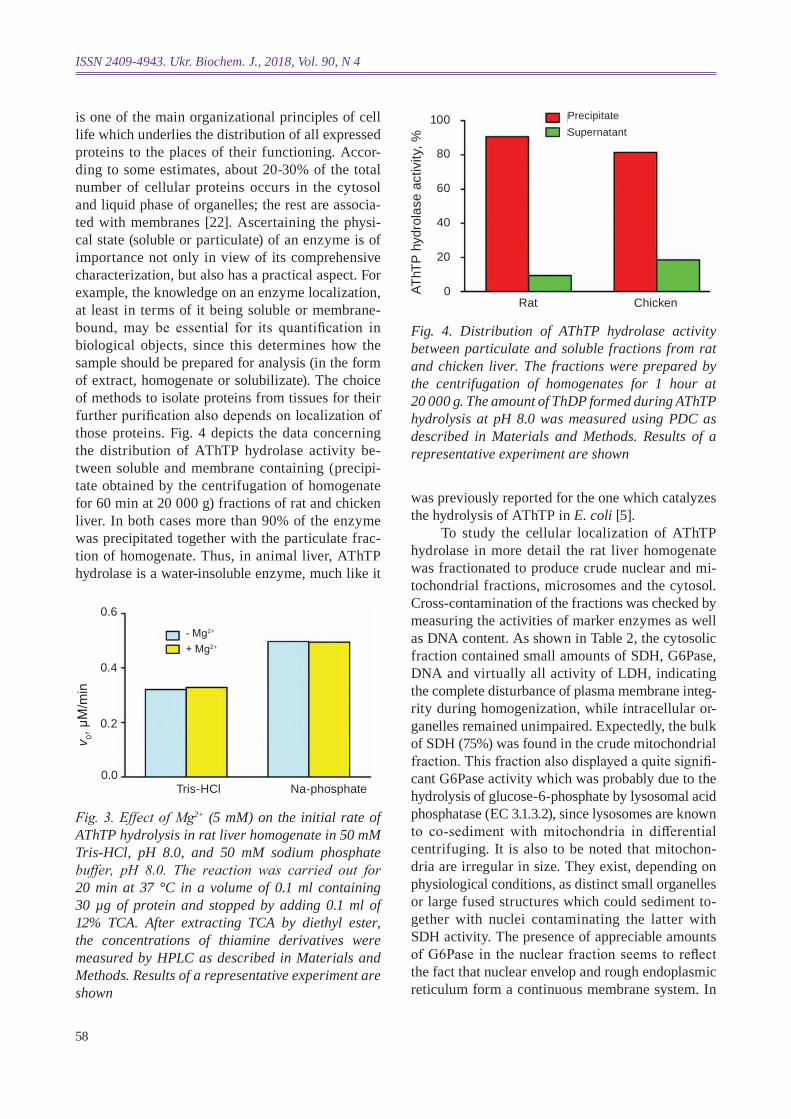
The results of measuring the rate of AThTP hydrolysis in rat liver homogenates by HPLC, in both Na-phosphate and Tris-HCl buffer systems in the presence or without Mg2+ ions, are illustrated in Fig. 3. As can be seen from the diagram, Mg2+ had no effect on the reaction rate in either buffer solution, in phosphate buffer the rate of AThTP hydrolysis being 53% higher than in Tris-HCl. Thus, AThTP hydrolase was proven to be a metal-independent pro-tein. It is not excluded that phosphate ions, exerting an inhibitory effect on liver phosphatases, activate AThTP hydrolase itself, though the enzyme inhibi-tion with Tris-HCl might also be the case.
All the further assays of AThTP hydrolase activity were done on the rate of ThDP formation which was quantified by enzymatic method using apo PDC [9].
localization of AThTP hydrolase in rat and chicken liver. The compartmentation of metabolism
Fig. 2. Chromatographic separation of fluorescent constituents of AThTP hydrolase reaction medium. (A) composition of the reaction mixture for rat liver homogenate at zero time. (B) Incubation of AThTP with rat liver homogenate was carried out in 50 mM Na-phosphate buffer, pH 8.0, with no Mg2+ added. (C) Incuba-tion of AThTP with rat liver homogenate was carried out in 50 mM Na-phosphate buffer, pH 8.0, with 5 mM Mg2+ added. (D) composition of the reaction mixture for chicken liver homogenate at zero time. (E) Incuba-tion of AThTP with chicken liver homogenate was carried out in 50 mM Na-phosphate buffer, pH 8.0, without adding Mg2+ ions. 1, ThMP; 2, ThdP; 3, AThTP
Fluo
resc
ence
Time, min
30
25
20
15
10
5
35
40
0 10 20 0 10 20 0 10 20 0 8 16 0 8 16
10
30A B C D3
2
2
1
3 3
1
3
2
2
E
25
20
15
10
5
0
5
10
15
20 14
12
10
0
4
6
8
2
25
15
20
5
303
2
16
A. F. Makarchikov, T. V. Saroka, T. G. kudyrka et al.
58
ISSN 2409-4943. Ukr. Biochem. J., 2018, Vol. 90, N 4
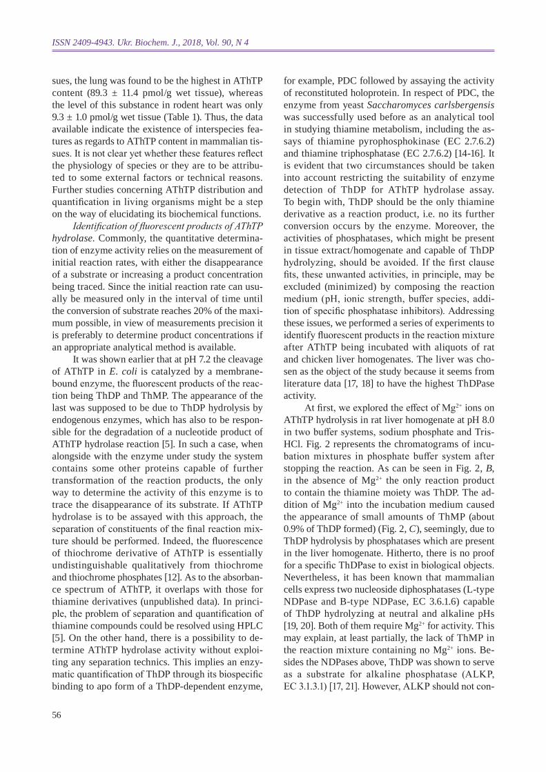
is one of the main organizational principles of cell life which underlies the distribution of all expressed proteins to the places of their functioning. Accor-ding to some estimates, about 20-30% of the total number of cellular proteins occurs in the cytosol and liquid phase of organelles; the rest are associa-ted with membranes [22]. Ascertaining the physi-cal state (solub le or particulate) of an enzyme is of importance not only in view of its comprehensive characterization, but also has a practical aspect. For example, the knowledge on an enzyme localization, at least in terms of it being soluble or membrane-bound, may be essential for its quantification in biological objects, since this determines how the sample should be prepared for analysis (in the form of extract, homogenate or solubilizate). The choice of methods to isolate proteins from tissues for their further purification also depends on localization of those proteins. Fig. 4 depicts the data concerning the distribution of AThTP hydrolase activity be-tween soluble and membrane containing (precipi-tate obtained by the centrifugation of homogenate for 60 min at 20 000 g) fractions of rat and chicken liver . In both cases more than 90% of the enzyme was precipitated together with the particulate frac-tion of homogenate. Thus, in animal liver, AThTP hydrolase is a water-insoluble enzyme, much like it
Fig. 3. Effect of Mg2+ (5 mM) on the initial rate of AThTP hydrolysis in rat liver homogenate in 50 mM Tris-hcl, ph 8.0, and 50 mM sodium phosphate buffer , pH 8.0. The reaction was carried out for 20 min at 37 °c in a volume of 0.1 ml containing 30 µg of protein and stopped by adding 0.1 ml of 12% TcA. After extracting TcA by diethyl ester, the concentrations of thiamine derivatives were measured by hPlc as described in Materials and Methods. results of a representative experiment are shown
Fig. 4. distribution of AThTP hydrolase activity between particulate and soluble fractions from rat and chicken liver. The fractions were prepared by the centrifugation of homogenates for 1 hour at 20 000 g. The amount of ThdP formed during AThTP hydrolysis at ph 8.0 was measured using Pdc as described in Materials and Methods. results of a representative experiment are shown
v 0, μM
/min
0.2
0.0
0.4
0.6
Tris-HCl Na-phosphate
- Mg2+
+ Mg2+
AThT
P hy
drol
ase
activ
ity, %
40
20
60
80
Rat Chicken
SupernatantPrecipitate
0
100
was previously reported for the one which catalyzes the hydrolysis of AThTP in E. coli [5].
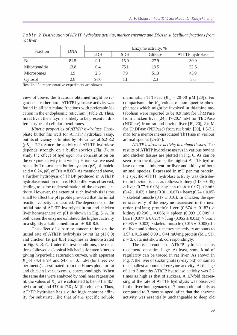
To study the cellular localization of AThTP hydrolase in more detail the rat liver homogenate was fractionated to produce crude nuclear and mi-tochondrial fractions, microsomes and the cytosol. Cross-contamination of the fractions was checked by measuring the activities of marker enzymes as well as DNA content. As shown in Table 2, the cytosolic fraction contained small amounts of SDH, G6Pase, DNA and virtually all activity of LDH, indicating the complete disturbance of plasma membrane integ-rity during homogenization, while intracellular or-ganelles remained unimpaired. Expectedly, the bulk of SDH (75%) was found in the crude mitochondrial fraction. This fraction also displayed a quite signifi-cant G6Pase activity which was probably due to the hydrolysis of glucose-6-phosphate by lysosomal acid phosphatase (EC 3.1.3.2), since lysosomes are known to co-sediment with mitochondria in differential centrifuging. It is also to be noted that mitochon-dria are irregular in size. They exist, depending on physio logical conditions, as distinct small organelles or large fused structures which could sediment to-gether with nuclei contaminating the latter with SDH activi ty. The presence of appreciable amounts of G6Pase in the nuclear fraction seems to reflect the fact that nuclear envelop and rough endoplasmic reticulum form a continuous membrane system. In
59
view of above, the fractions obtained might be re-garded as rather pure. AThTP hydrolase activity was found in all particulate fractions with preferable lo-cation in the endoplasmic reticulum (Table 2). Thus, in rat liver, the enzyme is likely to be present in dif-ferent types of cellular membranes.
kinetic properties of AThTP hydrolase. Phos-phate buffer fits well for AThTP hydrolase assay, but its efficiency is limited by pH values of 6.2-8.2 (pka = 7.2). Since the activity of AThTP hydrolase depends strongly on a buffer species (Fig. 3), to study the effect of hydrogen ion concentration on the enzyme activity in a wider pH interval we used basically Tris-maleate buffer system (pka of maleic acid = 6.24, pka of Tris = 8.08). As mentioned above, a further hydrolysis of ThDP produced in AThTP hydrolase reaction in Tris-HCl buffer was observed, leading to some underestimation of the enzyme ac-tivity. However, the extent of such hydrolysis is too small to affect the pH profile provided that the initial reaction velocity is measured. The dependence of the initial rate of AThTP hydrolysis in rat and chicken liver homogenates on pH is shown in Fig. 5, A. In both cases the enzyme exhibited the highest activity in a slightly alkaline medium at pH 8.0-8.5.
The effect of substrate concentration on the initial rate of AThTP hydrolysis by rat (at pH 8.0) and chicken (at pH 8.5) enzymes is demonstrated in Fig. 5, B, c. Under the test conditions, the reac-tions followed a classical Michaelis-Menten kinetics giving hyperbolic saturation curves, with apparent km of 84.4 ± 9.4 and 54.6 ± 13.1 µМ (for three ex-periments) as estimated from the Hanes plots for rat and chicken liver enzymes, correspondingly. When the same data were analyzed by nonlinear regression fit, the values of km were calculated to be 63.1 ± 10.1 µМ (for rat) and 43.0 ± 17.8 µМ (for chicken). Thus, AThTP hydrolase has a quite high apparent affin-ity for substrate, like that of the specific soluble
T a b l e 2. distribution of AThTP hydrolase activity, marker enzymes and dNA in subcellular fractions from rat liver
Results of a representative experiment are shown
Fraction DNAEnzyme activity, %
LDH SDH G6Pase AThTP hydrolaseNuclei 81.5 0.1 15.9 27.9 30.0Mitochondria 13.8 0.4 75.1 18.5 22.5Microsomes 1.9 2.5 7.9 51.3 43.9Cytosol 2.8 97.0 1.1 2.3 3.6
mammalian ThTPase (km = 20-50 µМ [23]). For comparison, the km values of non-specific phos-phatases which might be involved in thiamine me-tabolism were reported to be 0.9 mM for ThMPase from chicken liver [24], 17-20.7 mM for ThDPase (NDPase) from rat and bovine liver [19, 20], 2 mM for ThDPase (NDPase) from rat brain [20], 1.5-2.2 mM for a membrane-associated ThTPase in various animal species [25-27].
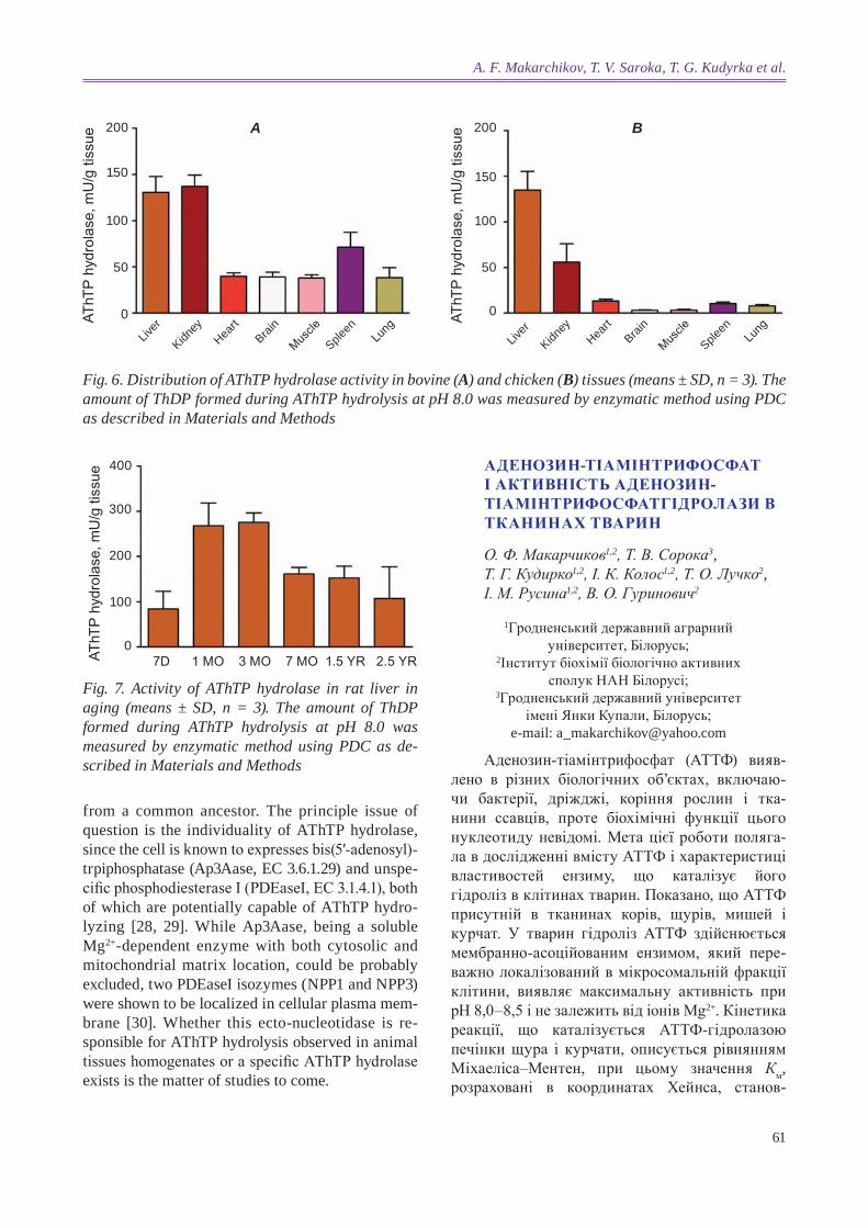
AThTP hydrolase activity in animal tissues. The results of AThTP hydrolase assays in various bovine and chicken tissues are plotted in Fig. 6. As can be seen from the diagrams, the highest AThTP hydro-lase content is inherent for liver and kidney of both animal species. Expressed in mU per mg protein, the specific AThTP hydrolase activity was distribu-ted in bovine tissues as follows: kidney (1.13 ± 0.19) > liver (0.77 ± 0.06) > spleen (0.46 ± 0.07) ≈ brain (0.42 ± 0.03) > lung (0.31 ± 0.07) > heart (0.24 ± 0.05) > skeletal muscle (0.17 ± 0.01). In chicken , the spe-cific activity of the enzyme decreased in the next order (mU/mg protein): liver (0.674 ± 0.187) > kidney (0.296 ± 0.066) > spleen (0.093 ±0.009) > heart (0.077 ± 0.027) > lung (0.055 ± 0.013) > brain (0.035 ± 0.003) > skele tal muscle (0.015 ± 0.005). In rat liver and kidney, the enzyme activity amounts to 1.57 ± 0.15 and 0.99 ± 0.41 mU/mg protein (M ± SD, n = 3, data not shown), correspondingly.
The tissue content of AThTP hydrolase seems to depend on animal age. At least, some kind of regu larity can be traced in rat liver. As shown in Fig. 7, the liver of sucking rats (7-day old) contained the smallest amounts of enzyme activity. At the age of 1 to 3 months AThTP hydrolase activity was 3.2 times as high as that of suckers. A 1.7-fold decrea-sing of the rate of AThTP hydrolysis was observed in the liver homogenates of 7-month old animals as compared to 3 months aged, after that the enzyme activity was essentially unchangeable to deep old
A. F. Makarchikov, T. V. Saroka, T. G. kudyrka et al.

60
ISSN 2409-4943. Ukr. Biochem. J., 2018, Vol. 90, N 4
Fig. 5. kinetic properties of rat and chicken liver AThTP hydrolase. (A) Effect of pH on the initial rate of AThTP hydrolysis in chicken and rat liver homogenates. The buffers (25 mM final concentration) used were: acetate (ph 5.0), Tris-maleate (ph 5.5-9.0), glycine (ph 9.5). The reaction was carried out for 10 min with 60 µg of protein. The concentration of ThdP produced was determined by enzymatic procedure as described in Materials and Methods. (B) Effect of substrate concentration on the initial rate of AThTP hydrolysis in chicken liver homogenate at ph 8.5. The reaction proceeded for 10 min with 120 µg of protein. (C) Effect of substrate concentration on the initial rate of AThTP hydrolysis in rat liver homogenate at ph 8.0. The reaction time was 10 min with 60 µg of protein in the reaction mixture. Insets: The hanes plots of the data. results of a representative experiment in each graph are shown
v 0, μM
/min
0.2
0.0
0.4
0.6
pH
RatChicken
4 5 6 7 8 9 10
0.8 A
v 0, μM
/min
0.2
0.0
0.4
0.6
[AThTP], μM0 10 20 30 40 50
B
[AThTP], μM-50 -25 0 25 50
80
60
20
40S/v
0, m
in
C1.0
v 0, μM
/min
0.2
0.0
0.4
0.6
[AThTP], μM0 20 40 60 80 100
[AThTP], μM-100 -50 0 50 100
150
100
50S/v
0, m
in
0.8
age (2.5 years). The significance of the differences was established by ANOVA (p = 0.0004) followed by Turkey’s test for post hoc comparisons among the groups. It is worth mentioning the experimental groups were formed from rats which inhabited the institutional animal house simultaneously.
At present, no literature data are available on the biosynthesis of AThTP in animals. Yet, the ex-tremely low activity of ThDP adenylyltransferase (EC 2.7.7.B3) in E. coli [8] and rat lung (unpublished data) implies the crucial tool to control the concen-tration of this compound in the cell might be the rate of its hydrolysis. The analysis of our data, however, indicate no correlation, either direct or inverse, to
occur between the tissue AThTP content (Table 1) and AThTP hydrolase activity (Fig. 6). A possible reason of such a discrepancy could be the existence of kinetic regulatory mechanisms which are ceased due to the cell integrity disruption as well as dilution during homogenate preparing and enzyme assay.
In conclusion, our results suggest considerab le differences, intra- and interspecies, in AThTP con-tent as well as AThTP hydrolase activity in animal tissues. As regards to the enzymes catalyzing the hydrolysis of AThTP in rat and chicken liver, they resemble each other in having alkaline pH optima, simi lar values of km, and also being metal-inde-pendent. This may imply their evolutional origin
61
Fig. 6. distribution of AThTP hydrolase activity in bovine (A) and chicken (B) tissues (means ± Sd, n = 3). The amount of ThdP formed during AThTP hydrolysis at ph 8.0 was measured by enzymatic method using Pdc as described in Materials and Methods
AThT
P hy
drol
ase,
mU
/g ti
ssue
50
100
150
Liver
0
200 A
Kidney
Heart
Brain
Muscle
Spleen
Lung
AThT
P hy
drol
ase,
mU
/g ti
ssue
50
100
150
Liver
0
200 B
Kidney
Heart
Brain
Muscle
Spleen
Lung
Fig. 7. Activity of AThTP hydrolase in rat liver in aging (means ± Sd, n = 3). The amount of ThdP formed during AThTP hydrolysis at ph 8.0 was measured by enzymatic method using Pdc as de-scribed in Materials and Methods
AThT
P hy
drol
ase,
mU
/g ti
ssue
100
200
300
0
400
7D 1 MO 3 MO 7 MO 1.5 YR 2.5 YR
from a common ancestor. The principle issue of question is the individuality of AThTP hydrolase, since the cell is known to expresses bis(5′-adenosyl)-trpiphosphatase (Ap3Aase, EC 3.6.1.29) and unspe-cific phosphodiesterase I (PDEaseI, EC 3.1.4.1), both of which are potentially capable of AThTP hydro-lyzing [28, 29]. While Ap3Aase, being a soluble Mg2+-dependent enzyme with both cytosolic and mitochondrial matrix location, could be probably excluded, two PDEaseI isozymes (NPP1 and NPP3) were shown to be localized in cellular plasma mem-brane [30]. Whether this ecto-nucleotidase is re-sponsible for AThTP hydrolysis observed in animal tissues homogenates or a specific AThTP hydrolase exists is the matter of studies to come.
Аденозин-тіАмінтрифосфАт i Активнiсть Аденозин-тіАмінтрифосфАтгідролАзи в ткАнинАх твАрин
О. Ф. Макарчиков1,2, Т. В. Сорока3, Т. Г. Кудирко1,2, І. К. Колос1,2, Т. О. Лучко2, I. М. Русина1,2, В. О. Гуринович2
1Гродненський державний аграрний університет, Білорусь;
2Інститут біохімії біологічно активних сполук НАН Білорусі;
3Гродненський державний університет імені Янки Купали, Білорусь;
e-mail: [email protected]
Аденозин-тіамінтрифосфат (АТТФ) вияв-лено в різних біологічних об’єктах, включаю-чи бактерії, дріжджі, коріння рослин і тка-нини ссавців, проте біохімічні функції цього нуклео тиду невідомі. Мета цієї роботи поляга-ла в дослідженні вмісту АТТФ і характеристиці властивостей ензиму, що каталізує його гідроліз в клітинах тварин. Показано, що АТТФ присутній в тканинах корiв, щурів, мишей і курчат. У тварин гідроліз АТТФ здійснюється мембранно-асоційованим ензимом, який пере-важно локалізований в мікросомальнiй фракції клітини, виявляє максимальну активність при рН 8,0–8,5 і не залежить від іонів Mg2+. Кінетика реакції, що каталізується АТТФ-гідролазою печінки щура і курчати, описується рівнянням Міхаеліса–Ментен, при цьому значення Км, розраховані в координатах Хейнса, станов-
A. F. Makarchikov, T. V. Saroka, T. G. kudyrka et al.

62
ISSN 2409-4943. Ukr. Biochem. J., 2018, Vol. 90, N 4
лять відповідно 84,4 ± 9,4 і 54,6 ± 13,1 мкМ. Серед зразків тканин, що досліджувались най-вищу АТТФ-гідролазну активність визначили в печінці і нирках. У печінці щурів швидкість гідролізу АТТФ залежила від віку тварин.
К л ю ч о в і с л о в а: вітамін В1, аденозин-тіамінтрифосфат, аденозин-тіамінтрифосфат-гідролаза, тканини тварин.
Аденозин-тиАминтрифосфАт и Активнсть Аденозин-тиАминтрифосфАт-гидролАзы в ткАнях животных
А. Ф. Макарчиков1,2, Т. В. Сорока3, Т. Г. Кудырко1,2, И. К. Колос1,2, Т. А. Лучко2, И. М. Русина1,2, В. А. Гуринович2
1Гродненский государственный аграрный университет, Беларусь;
2Институт биохимии биологически активных соединений НАН Беларуси;
3Гродненский государственный университет имени Янки Купалы, Беларусь;
e-mail: [email protected]
Аденозин-тиаминтрифосфат (АТТФ) об-наружен в различных биологических объектах, включая бактерии, дрожжи, корни растений и ткани млекопитающих, однако биохимические функции этого нуклеотида неизвестны. Цель данной работы заключалась в исследовании со-держания АТТФ и характеристике свойств эн-зима, катализирующего его гидролиз в клетках животных. Показано, что АТТФ присутствует в тканях быка, крысы, мыши и цыпленка. У жи-вотных гидролиз АТТФ осуществляется мем-бранно-ассоциированным энзимом, который преимущественно локализован в микросомаль-ной фракции клетки, проявляет максимальную активность при рН 8,0-8,5 и не зависит от ионов Mg2+. Кинетика реакции, катализируемой АТТФ-гидролазой печени крысы и цыпленка описы-вается уравнением Михаэлиса–Ментен, при этом значения Км, рассчитанные в координатах Хейнса, составляют соответственно 84,4 ± 9,4 и 54,6 ± 13,1 мкМ. Среди образцов исследованных тканей самой высокой АТТФ-гидролазной ак-тивностью обладали печень и почки. В печени крыс скорость гидролиза АТТФ зависила от воз-раста животных.
К л ю ч е в ы е с л о ва: витамин В1, аденозин-тиаминтрифосфат, аденозин-тиамин-трифосфат-гидролаза, ткани животных.
references
1. Metzler DE. Biochemistry. The Chemical Reactions of Living Cells. Harcourt/Academic Press. 2nd ed. 2001, Vol 1, 937 p.
2. Zhao J, Zhong CJ. A review on research progress of transketolase. Neurosci Bull. 2009; 25(2): 94-99.
3. Casteels M, Sniekers M, Fraccascia P, Man-naerts GP, Van Veldhoven PP. The role of 2-hydroxyacyl-CoA lyase, a thiamin pyrophosphate-dependent enzyme, in the peroxisomal metabolism of 3-methyl-branched fatty acids and 2-hydroxy straight-chain fatty acids. Biochem Soc Trans. 2007; 35(Pt 5): 876-880.
4. Makarchikov AF. Vitamin B1: metabolism and functions. Biochemistry (Moscow) Suppl Ser B: Biomed chem. 2009; 3(2): 116-128.
5. Bettendorff L, Wirtzfeld B, Makarchikov AF, Mazzucchelli G, Frédérich M, Gigliobianco T, Gangolf M, De Pauw E, Angenot L, Wins P. Discovery of a natural thiamine adenine nucleotide. Nat chem Biol. 2007; 3(4): 211-212.
6. Gigliobianco T, Lakaye B, Wins P, El Moualij B, Zorzi W, Bettendorff L. Adenosine thiamine triphosphate accumulates in Escherichia coli cells in response to specific conditions of metabolic stress. BMc Microbiol. 2010; 10: 148.
7. Tanaka T, Yamamoto D, Sato T, Tanaka S, Usui K, Manabe M, Aoki Y, Iwashima Y, Saito Y, Mino Y, Deguchi H. Adenosine thiamine triphosphate (AThTP) inhibits poly(ADP-ribose) polymerase-1 (PARP-1) activity. J Nutr Sci Vitaminol (Tokyo). 2011; 57(2): 192-196.
8. Makarchikov AF, Brans A, Bettendorff L. Thiamine diphosphate adenylyl transferase from E. coli: functional characterization of the enzyme synthesizing adenosine thiamine triphosphate. BMc Biochem. 2007; 8: 17.
9. Chernikevich IP, Gritsenko EA, Makarchikov AF, Voskoboev AI. Fermentation micromethod for the quantitative determination of thiamine diphosphate in biological fluids. Prikl Biokhim Mikrobiol. 1991; 27(5): 762-771. (In Russian).
10. Bettendorff L, Peeters M, Jouan C, Wins P, Schoffeniels E. Determination of thiamin and its phosphate esters in cultured neurons and
63
astrocytes using an ion-pair reversed-phase high-performance liquid chromatographic method. Anal Biochem. 1991; 198(1): 52-59.
11. Peterson GL. A simplification of the protein assay method of Lowry et al. which is more generally applicable. Anal Biochem. 1977; 83(2): 346-356.
12. Frédérich M, Delvaux D, Gigliobianco T, Gangolf M, Dive G, Mazzucchelli G, Elias B, De Pauw E, Angenot L, Wins P, Bettendorff L. Thiaminylated adenine nucleotides. Chemical synthesis, structural characterization and natural occurrence. FEBS J. 2009; 276(12): 3256-3268.
13. Gangolf M, Czerniecki J, Radermecker M, Detry O, Nisolle M, Jouan C, Martin D, Chantraine F, Lakaye B, Wins P, Grisar T, Bettendorff L. Thiamine status in humans and content of phosphorylated thiamine derivatives in biopsies and cultured cells. PloS one. 2010; 5(10): e13616.
14. Parkhomenko YuM, Pavlova AS, Mejenskaya OA, Stepanenko SP, Chekhivska LI. Thiamine diphosphate synthesis and redox state indicator in rat brain during of B1 hypovitaminosis. Ukr Biochem J. 2017; 89(5): 84-95.
15. Voskoboyev AI, Chernikeevich IP. Biosynthesis, degradation and transport of thiamine phosphoric esters. Minsk: Nauka and Tekhnika, 1987. 200 p. (In Russian).
16. Makarchikov AF, Chernikevich IP. Purification and characterization of thiamine triphosphatase from bovine brain. Biochim Biophys Acta. 1992; 1117(3): 326-332.
17. Ogawa K, Sakai M, Inomata K. Recent findings on ultracytochemistry of thiamin phosphatases. Ann N y Acad Sci. 1982; 378(1): 188-214.
18. Makarchikov AF, Luchko TA, Rusina IM, Gulyai IE, Makar EA. Studies on enzymes of thiamine mono- and diphosphate hydrolysis in the membrane preparations from bovine tissues. Proc Natl Acad Sci Belarus, Biol Series. 2009; 2: 62-66. (In Russian).
19. Yamazaki M, Hayaishi O. Allosteric properties of nucleoside diphosphatase and its identity with thiamine pyrophosphatase. J Biol chem. 1968; 243(11): 2934-2942.
20. Sano S, Matsuda Y, Miyamoto S, Nakagawa H. Thiamine pyrophosphatase and nucleoside diphosphatase in rat brain. Biochem Biophys res commun. 1984; 118(1): 292-298.
21. Eaton RH, Moss DW. Organic pyrophosphates as substrates for human alkaline phosphatases. Biochem J. 1967; 105(3): 1307-1312.
22. Stevens TJ, Arkin IT. Do more complex orga-nisms have a greater proportion of membrane proteins in their genomes? Proteins. 2000; 39(4): 417-420.
23. Makarchikov AF, Lakaye B, Gulyai IE, Czerniecki J, Coumans B, Wins P, Grisar T, Bettendorff L. Thiamine triphosphate and thiamine triphosphatase activities: from bacteria to mammals. cell Mol life Sci. 2003; 60(7): 1477-1488.
24. Kolas IK, Makarchikov AF. Copurification of chicken liver soluble thiamine monophosphatase and low molecular weight acid phosphatase. Ukr Biochem J. 2017; 89(6): 13-21.
25. Barchi RL, Braun PE. A membrane-associated thiamine triphosphatase from rat brain. Properties of the enzyme. J Biol chem. 1972; 247(23): 7668-7673.
26. Bettendorff L, Michel-Cahay C, Grandfils C, De Rycker C, Schoffeniels E. Thiamine triphosphate and membrane-associated thiamine phosphatases in the electric organ of Electrophorus electricus. J Neurochem. 1987; 49(2): 495-502.
27. Kolas IK, Makarchikov AF. Properties of chicken liver membrane-associated thiamine triphosphatase. Ukr Biochem J. 2015; 87(3): 37-46. (In Russian).
28. Guranowski A, Wojdyła AM, Pietrowska-Borek M, Bieganowski P, Khurs EN, Cliff MJ, Blackburn GM, Błaziak D, Stec WJ. Fhit proteins can also recognize substrates other than dinucleoside polyphosphates. FEBS lett. 2008; 582(20): 3152-3158.
29. Cameselle JC, Costas MJ, Günther Sillero MA, Sillero A. Two low km hydrolytic activities on dinucleoside 5',5"'-P1,P4-tetraphosphates in rat liver. Characterization as the specific dinucleoside tetraphosphatase and a phosphodiesterase I-like enzyme. J Biol chem. 1984; 259(5): 2879-2885.
30. Zimmermann H, Zebisch M, Sträter N. Cellular function and molecular structure of ecto-nucleotidases. Purinergic Signal. 2012; 8(3): 437-502.
Received 23.03.2018
A. F. Makarchikov, T. V. Saroka, T. G. kudyrka et al.