acute tumor lysis syndrome in hodgkin disease
TRANSCRIPT
Med Pediatr Oncol 2002;39:69–70
Letter to the Editor: Acute Tumor Lysis Syndromein Hodgkin Disease
To the Editor: Acute tumor lysis syndrome (ATLS)frequently complicates the clinical course of lymphopro-liferative malignancies. In these patients it can occur withminimal therapy or even spontaneously [1,2]. ATLS hasalso rarely been reported in non-hematopoietic cancers[3,4], but we are unaware of it having been reported inHodgkin disease (HD) in children. Our experience withsuch a patient highlights this unusual complication.
A boy presented at the age of 4 years with stage IIIHodgkin disease. He had hepatosplenomegaly and chestX-ray films showed massive hilar lymphadenopathy.The LDH was elevated at 2,338 U/l. CT scan revealedextensive retroperitoneal and intraperitoneal lymphade-nopathy with bilateral hydronephrosis.
The diagnosis was initially unclear, and in view of theconsiderable tumor load, hyperhydration and allopurinolwere commenced. General anaesthesia for biopsy pur-poses was considered too risky and vincristine (1.5 mg/m2) and prednisolone (40 mg/m2) were given empirically.There was no response to treatment and 72 hr followingadmission he developed mild inspiratory stridor withdecreased air entry on the right side. Radiologically therewas further narrowing of the right main bronchus. Heunderwent emergency radiotherapy (4 Gy as 2 Gy/day foreach of two fractions) to a limited field around the carina,and his airway improved within 24 hr.
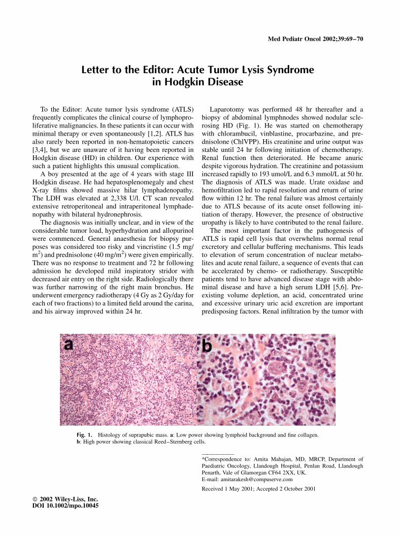
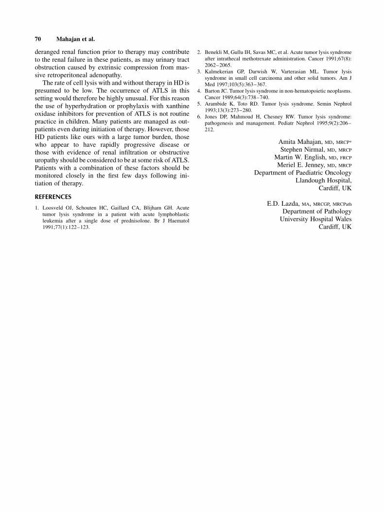
Laparotomy was performed 48 hr thereafter and abiopsy of abdominal lymphnodes showed nodular scle-rosing HD (Fig. 1). He was started on chemotherapywith chlorambucil, vinblastine, procarbazine, and pre-dnisolone (ChlVPP). His creatinine and urine output wasstable until 24 hr following initiation of chemotherapy.Renal function then deteriorated. He became anuricdespite vigorous hydration. The creatinine and potassiumincreased rapidly to 193 umol/L and 6.3 mmol/L at 50 hr.The diagnosis of ATLS was made. Urate oxidase andhemofiltration led to rapid resolution and return of urineflow within 12 hr. The renal failure was almost certainlydue to ATLS because of its acute onset following ini-tiation of therapy. However, the presence of obstructiveuropathy is likely to have contributed to the renal failure.
The most important factor in the pathogenesis ofATLS is rapid cell lysis that overwhelms normal renalexcretory and cellular buffering mechanisms. This leadsto elevation of serum concentration of nuclear metabo-lites and acute renal failure, a sequence of events that canbe accelerated by chemo- or radiotherapy. Susceptiblepatients tend to have advanced disease stage with abdo-minal disease and have a high serum LDH [5,6]. Pre-existing volume depletion, an acid, concentrated urineand excessive urinary uric acid excretion are importantpredisposing factors. Renal infiltration by the tumor with
Fig. 1. Histology of suprapubic mass. a: Low power showing lymphoid background and fine collagen.b: High power showing classical Reed–Sternberg cells.
——————*Correspondence to: Amita Mahajan, MD, MRCP, Department ofPaediatric Oncology, Llandough Hospital, Penlan Road, LlandoughPenarth, Vale of Glamorgan CF64 2XX, UK.E-mail: [email protected]
Received 1 May 2001; Accepted 2 October 2001
� 2002 Wiley-Liss, Inc.DOI 10.1002/mpo.10045
deranged renal function prior to therapy may contributeto the renal failure in these patients, as may urinary tractobstruction caused by extrinsic compression from mas-sive retroperitoneal adenopathy.
The rate of cell lysis with and without therapy in HD ispresumed to be low. The occurrence of ATLS in thissetting would therefore be highly unusual. For this reasonthe use of hyperhydration or prophylaxis with xanthineoxidase inhibitors for prevention of ATLS is not routinepractice in children. Many patients are managed as out-patients even during initiation of therapy. However, thoseHD patients like ours with a large tumor burden, thosewho appear to have rapidly progressive disease orthose with evidence of renal infiltration or obstructiveuropathy should be considered to be at some risk of ATLS.Patients with a combination of these factors should bemonitored closely in the first few days following ini-tiation of therapy.
REFERENCES
1. Loosveld OJ, Schouten HC, Gaillard CA, Blijham GH. Acutetumor lysis syndrome in a patient with acute lymphoblasticleukemia after a single dose of prednisolone. Br J Haematol1991;77(1):122–123.
2. Benekli M, Gullu IH, Savas MC, et al. Acute tumor lysis syndromeafter intrathecal methotrexate administration. Cancer 1991;67(8):2062–2065.
3. Kalmekerian GP, Darwish W, Varterasian ML. Tumor lysissyndrome in small cell carcinoma and other solid tumors. Am JMed 1997;103(5):363–367.
4. Barton JC. Tumor lysis syndrome in non-hematopoietic neoplasms.Cancer 1989;64(3):738–740.
5. Arambide K, Toto RD. Tumor lysis syndrome. Semin Nephrol1993;13(3):273–280.
6. Jones DP, Mahmoud H, Chesney RW. Tumor lysis syndrome:pathogenesis and management. Pediatr Nephrol 1995;9(2):206–212.
Amita Mahajan, MD, MRCP*
Stephen Nirmal, MD, MRCP
Martin W. English, MD, FRCPMeriel E. Jenney, MD, MRCP
Department of Paediatric OncologyLlandough Hospital,
Cardiff, UK
E.D. Lazda, MA, MRCGP, MRCPath
Department of PathologyUniversity Hospital Wales
Cardiff, UK
70 Mahajan et al.