acute nontraumatic muscle weakness · acute nontraumatic muscle weakness jangra et al. 237 journal...
TRANSCRIPT

Acute Nontraumatic Muscle Weakness Jangra et al.236 Review Article
Acute Nontraumatic Muscle WeaknessKiran Jangra1 Hemant Bhagat1 Aastha Takkar2
1Department of Anaesthesia and Intensive Care, Postgraduate Institute of Medical Education and Research (PGIMER), Chandigarh, India
2Department of Neurology, Postgraduate Institute of Medical Education and Research (PGIMER), Chandigarh, India
receivedFebruary 24, 2019accepted after revisionJune 19, 2019published onlineSeptember 11, 2019
Address for correspondence Kiran Jangra, DM, Department of Anaesthesia and Intensive Care, Postgraduate Institute of Medical Education and Research (PGIMER), 4th Floor, Nehru Hospital, Chandigarh 160012, India (e-mail: [email protected]).
Acute nontraumatic weakness (ANTW) is defined as acute onset of weakness in any part of the body. The weakness occurs due to interruption at any point along the motor pathway. The motor pathway originates from upper motor neuron cells in the cerebral cortex and traverses through the brainstem till lower motor neurons in the spinal cord. The axon of a lower motor neuron is known as the peripheral motor nerve that synapses with muscle. ANTW is of varied etiology and presentation that may be immediately life-threatening if respiratory muscles or autonomic nervous system is involved. Involvement of respiratory muscles may be associated with respiratory failure that may require mechanical ventilation. The weakness may be localized to one limb or generalized involving several muscle groups. When bulbar muscles are involved, weakness leads to problem in swallowing and coughing that endangers the patient's airway. Similarly, the course of the disease also varies, and these patients may worsen rapidly. Hence, a comprehensive history, systematic evaluation, and a detailed neuro-logical examination are performed to localize the disorder. There are specific clinical features peculiar to the particular location of the lesion in the body. Hence, it is pos-sible to anatomically localize these lesions based on the clinical features. Initial labo-ratory tests and appropriate neuroimaging should be obtained as indicated by history and examination. The time-sensitive emergencies should be addressed immediately, as the delay in management may lead to either permanent neurological damage or may worsen the overall outcome in such conditions. The initial management should always include care of airway, breathing, and circulation (ABC). The imaging should be obtained only after initial stabilization of ABC. The definitive treatment should be done as per the etiology.
Keywords ► acute weakness ► neuromuscular weakness ► nontraumatic weakness ► respiratory failure
DOI https://doi.org/ 10.1055/s-0039-1696061 ISSN 2348-0548.
Copyright ©2019 Indian Society of Neuroanaesthesiology and Critical Care
IntroductionAcute nontraumatic weakness (ANTW) is defined as the sud-den onset of paralysis/weakness in any part of the body. The motor functions are controlled by the motor pathway involv-ing upper and lower motor neurons (►Fig. 1). The upper motor neurons (UMNs) arise from the pyramidal cells of the neocortex and pass through the posterior limb of the internal capsule to enter the crus of the midbrain.1 The motor tracts then pass through the pons and medulla as the corticospi-nal tract, which are also known as the pyramidal tracts.1 The corticospinal tract divides as the lateral corticospinal tract
(decussates at pyramids) and the anterior corticospinal tract (crosses at corresponding spinal cord) as these pass down in the spinal cord.1,2 Approximately 90% of motor fibers form the lateral tract, while the rest (~10%) form the anterior tract. The lateral corticospinal tracts control the opposite side of the body, while the anterior corticospinal tract neurons con-trol the same side of the body and trunk muscles.1 Axons from UMNs synapse with the interneurons in the spinal cord, and occasionally directly with the lower motor neurons.2 The lower motor neuron is located in the spinal cord, and its axon projects out of the spinal cord and controls the movement of muscles.1 If there is a disease involving any part of the motor
J Neuroanaesthesiol Crit Care 2019;6:236–256
Abstract
THIEME
Published online: 2019-09-11

237Acute Nontraumatic Muscle Weakness Jangra et al.
Journal of Neuroanaesthesiology and Critical Care Vol. 6 No. 3/2019
pathway, patients will develop weakness. ►Figure 1 depicts the motor pathway.
The etiology of ANTW varies from immediately life-threat-ening conditions to minor disorders as shown in ►Fig. 2.3 The weakness may be localized to one limb or may become generalized involving the respiratory and bulbar muscles. In the latter scenario, protection of airway and care of breathing become the priority. In a few cases, weakness is associated with autonomic disturbances and may lead to hemodynamic instability. Hence, the management of ANTW should include simultaneous resuscitation (care of airway, breathing, and circulation [ABC]) and evaluation of underlying disease pathology. ANTW is one of the few neurological conditions where delaying the diagnosis and initiation of treatment directly affects the outcome. With a thorough history and clinical examination, we should be able to localize the lesion in many patients or should be able to narrow down the dif-ferential diagnosis list.
Here in this review, we discuss the systematic approach to the management of patients with ANTW. Traumatic and chronic weaknesses are beyond the scope of this review.
Initial EvaluationThe initial evaluation should include the assessment of the patient’s ability to protect the ABC and appropriate measures should be taken to optimize ABC before the neurological examination.4,5 Initial neurological examination should be done quickly to rule out time-critical emergencies including
Fig. 1 Diagrammatic representation of the motor tract. It originates from pyramidal cells (motor neuron) of the cerebral cortex as the upper motor neurons (UMNs) and synapse in the spinal cord with lower motor neurons (LMNs). UMN is colored as green, internuncial neuron as red, and LMN as black.
Fig. 2 Diagrammatic representation of various etiologies of acute motor weakness along the motor tract. LMN, lower motor neuron; UMN, upper motor neuron.
acute ischemic stroke, acute spinal cord compression, sta-tus epilepticus, dyselectrolytemia, hypoglycemia, and toxin. Various assessment tools are available to assess the neurolog-ical status of the patient including the Glasgow Coma Scale, FOUR score (includes eye response, motor response, brain-stem response, and respiration), and prehospital stroke scale score. Early recognition and activation of the stroke code sys-tem are necessary for optimum outcome in these patients. The acute spinal cord compression may present with flac-cid paralysis below the level of insult, and a sensory deficit limited to the involved segment. There may be certain syn-dromes having their own specific features such as acute cauda equina syndrome that presents with lower severe back pain, sciatica, perineal hypoesthesia, bowel and bladder inconti-nence, and limb weakness with decreased reflexes.3 If toxin exposure is suspected, scene safety should be ensured, and history related to amount and type toxin should be elicited. Initial biochemistry must include blood glucose, electrolytes (sodium, potassium, calcium, magnesium, and phosphorus), kidney and liver function tests, blood coagulation test, and complete blood counts. Relevant imaging should be obtained

238
Journal of Neuroanaesthesiology and Critical Care Vol. 6 No. 3/2019
Acute Nontraumatic Muscle Weakness Jangra et al.
as indicated by history and examination. A detailed motor and sensory examination should be done to locate the lesion anatomically by characteristics of the weakness.
Assessment of Airway and VentilationNeurologically ill patients need airway protection and ven-tilation if their airway is at risk of aspiration or breathing is inadequate. There are various causes of the airway and respi-ratory compromise including pharyngeal muscle weakness, leading to the upper airway obstruction and increased risk of aspiration, and respiratory muscle weakness leading to respi-ratory failure.6 Pulmonary gas exchange is usually preserved but may be affected due to atelectasis. Noninvasive ventila-tion may be tried if the airway reflexes are intact, but respi-ratory failure occurs due to respiratory muscle weakness. Patients should be monitored regularly as patient’s clinical condition may deteriorate rapidly.
Besides the patient’s physiology, the plan to intubate is also influenced by the clinical environment and the antici-pated course of the disease. If the patient is comatose and needs to be transported to a higher center, for imaging, or other invasive intervention, it would be most appropriate to secure the airway with endotracheal intubation. The patients who are expected to deteriorate in due course of time may need intubation, such as rapidly progressing Guillain–Berré syndrome (GBS).7,8 On the other hand, if the patient has a known illness, which is stable and expected to improve, can be managed by noninvasive ventilation.
Various predictors for the need of intubation are enumer-ated in ►Table 1. A combination of clinical signs and objec-tive parameters should be used to predict the need for intu-bation rather than a single parameter alone. Rapid sequence induction is the preferred modality of emergency intubation in the neurologically ill patients who are at risk of aspira-tion.9-12 Pharmacological agents must be carefully chosen as these patients may be at risk of succinylcholine-induced hyperkalemia (e.g., GBS) or resistant to it (e.g., myasthenia gravis).13,14 The patients may be highly sensitive to the sed-ative-hypnotic agents due to associated autonomic nervous system disturbance.
After intubation, the goals of mechanical ventilation are to normalize oxygenation using the lowest possible inspired oxygen to achieve a hemoglobin saturation >94%, a system-ic pH of 7.3 to 7.4, and partial pressure of carbon dioxide or end-tidal carbon dioxide of 30 to 40 mm Hg, to reduce the work of breathing, and to prevent ventilator-induced lung injury.15 Once the patient’s ABC are stabilized, a detailed and comprehensive neurological examination is done to localize the lesion.
Clinical and Anatomical LocalizationThe cause of weakness can be localized anatomically based on detailed history and examination as the patterns of weak-ness and associated findings are often specific for each ana-tomical region. Then we can make a focused differential diag-nosis, and specific testing can be done to make an accurate
Table 1 Indications for intubation2,3
Clinical symptoms• Increasing generalized muscle weakness• Dysphagia• Dysphonia• Dyspnea on exertion and at rest• Unable to remove secretions from the throat
Subjective (clinical signs)• Tachypnea/hypopnea• Inadequate chest rise• Paradoxical respiratory pattern• Weak coughing ability• Unable to complete full sentences (gasping for air)• Inability to perform single-breath count: count from 1 to 20 in single exhalation (FVC 1.0 L is roughly equal to counting
from 1 to 10)• Use of accessory muscles (cervical/trapezius/nasal flaring)• Weakness of trapezius and neck muscles: inability to lift head from bed• Orthopnea• Tachycardia/hypertension (secondary to sympathetic stimulation due to hypoxia and hypercapnia)• Sweating
Objective• Loss of consciousness• Hypoxemia (<60 mm Hg)• PFT findings
–Vital capacity <1 L or 20 mL/kg, or 50% decrease in VC in a day–Maximum inspiratory pressure > −30 cm H2O–Maximum expiratory pressure < 40 cm H2O
• Hypercarbia
Abbreviations: FVC, forced vital capacity; PFT, pulmonary function tests; VC, vital capacity.
239Acute Nontraumatic Muscle Weakness Jangra et al.
Journal of Neuroanaesthesiology and Critical Care Vol. 6 No. 3/2019
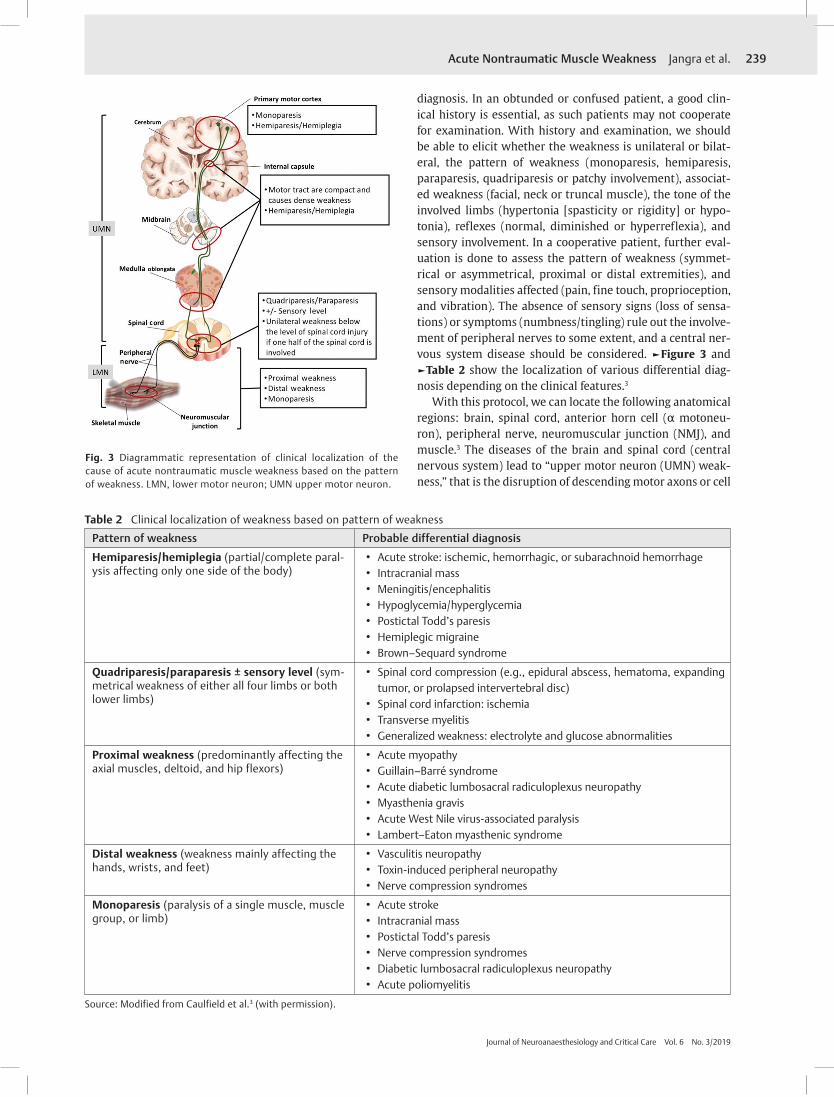
diagnosis. In an obtunded or confused patient, a good clin-ical history is essential, as such patients may not cooperate for examination. With history and examination, we should be able to elicit whether the weakness is unilateral or bilat-eral, the pattern of weakness (monoparesis, hemiparesis, paraparesis, quadriparesis or patchy involvement), associat-ed weakness (facial, neck or truncal muscle), the tone of the involved limbs (hypertonia [spasticity or rigidity] or hypo-tonia), reflexes (normal, diminished or hyperreflexia), and sensory involvement. In a cooperative patient, further eval-uation is done to assess the pattern of weakness (symmet-rical or asymmetrical, proximal or distal extremities), and sensory modalities affected (pain, fine touch, proprioception, and vibration). The absence of sensory signs (loss of sensa-tions) or symptoms (numbness/tingling) rule out the involve-ment of peripheral nerves to some extent, and a central ner-vous system disease should be considered. ►Figure 3 and ►Table 2 show the localization of various differential diag-nosis depending on the clinical features.3
With this protocol, we can locate the following anatomical regions: brain, spinal cord, anterior horn cell (α motoneu-ron), peripheral nerve, neuromuscular junction (NMJ), and muscle.3 The diseases of the brain and spinal cord (central nervous system) lead to “upper motor neuron (UMN) weak-ness,” that is the disruption of descending motor axons or cell
Table 2 Clinical localization of weakness based on pattern of weakness
Pattern of weakness Probable differential diagnosis
Hemiparesis/hemiplegia (partial/complete paral-ysis affecting only one side of the body)
• Acute stroke: ischemic, hemorrhagic, or subarachnoid hemorrhage • Intracranial mass • Meningitis/encephalitis • Hypoglycemia/hyperglycemia • Postictal Todd’s paresis • Hemiplegic migraine • Brown–Sequard syndrome
Quadriparesis/paraparesis ± sensory level (sym-metrical weakness of either all four limbs or both lower limbs)
• Spinal cord compression (e.g., epidural abscess, hematoma, expanding tumor, or prolapsed intervertebral disc)
• Spinal cord infarction: ischemia • Transverse myelitis • Generalized weakness: electrolyte and glucose abnormalities
Proximal weakness (predominantly affecting the axial muscles, deltoid, and hip flexors)
• Acute myopathy • Guillain–Barré syndrome • Acute diabetic lumbosacral radiculoplexus neuropathy • Myasthenia gravis • Acute West Nile virus-associated paralysis • Lambert–Eaton myasthenic syndrome
Distal weakness (weakness mainly affecting the hands, wrists, and feet)
• Vasculitis neuropathy • Toxin-induced peripheral neuropathy • Nerve compression syndromes
Monoparesis (paralysis of a single muscle, muscle group, or limb)
• Acute stroke • Intracranial mass • Postictal Todd’s paresis • Nerve compression syndromes • Diabetic lumbosacral radiculoplexus neuropathy • Acute poliomyelitis
Source: Modified from Caulfield et al.3 (with permission).
Fig. 3 Diagrammatic representation of clinical localization of the cause of acute nontraumatic muscle weakness based on the pattern of weakness. LMN, lower motor neuron; UMN upper motor neuron.

240
Journal of Neuroanaesthesiology and Critical Care Vol. 6 No. 3/2019
Acute Nontraumatic Muscle Weakness Jangra et al.
bodies that innervate the lower motor neurons located in the anterior horn cells of the spinal cord. Lower motor neurons type of weakness is caused by lesions of the anterior horn cells, peripheral nerve, and NMJ. UMN lesions are usually characterized by increased muscle tone, hyperreflexia, and a positive Babinski sign (great toe extension and fanning of fingers). LMN lesions, in contrast, cause flaccidity, areflexic weakness, and fasciculations (involuntary contractions or twitching of muscle fibers). During the acute phase of weak-ness, the UMN lesions may mimic the LMN lesions and may present with flaccid paralysis, normal or decreased tone, unreliable reflexes, and absent atrophy and fasciculations (occurs after a longer duration of paralysis).16,17
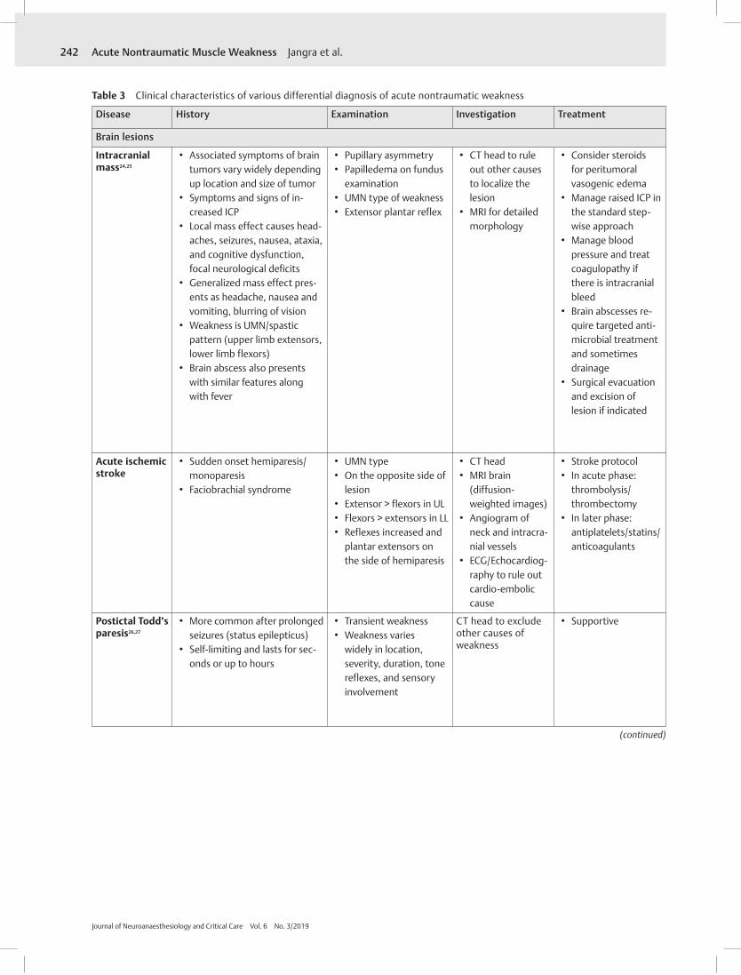
The characteristic features, history, clinical examination diagnosis, and treatment of various causes are represented in ►Fig. 4.
Approach of a Patient with ANTWIrrespective of clinical presentation, the initial management always includes care of ABC. Along with checking the vitals (pulse rate, blood pressure, and temperature), blood sugar should be checked in all patients presenting with weakness. After that, a detailed history and neurological examination are done to make the initial working diagnosis and differential diag-nosis. The algorithm suggested by ENLS is shown in ►Fig. 5.3
The diagnostic tests and definitive management vary greatly from one patient to another and may range from an emergent stroke imaging to elective nerve/muscle biopsy for specific illnesses. Various diagnostic modalities and treat-ment options for major differential diagnosis are enumerat-ed in ►Table 3. After making an initial working diagnosis and differential diagnosis, the patients are further evaluated by various investigations including initial laboratory tests such as glucose, electrolytes (sodium, calcium, magnesium, potas-sium, and phosphorous), blood urea nitrogen, creatinine, liv-er function tests, coagulation profile, complete blood counts, and arterial blood gas analysis. If history and examination suggest, certain specific tests may be performed such as thy-roid function tests, creatine phosphokinase or CK, erythrocyte sedimentation rate, parathyroid hormone, gamma-glutam-yl transferase, serum toxicology, and drug level. Once the patient’s ABC are optimized, the relevant imaging is obtained (computed tomography/magnetic resonance imaging [CT/MRI]). Nerve conduction studies, electromyography, a biop-sy of nerve and muscle, and lumbar puncture are to be done if indicated. Patients should be periodically screened for air-way and ventilation as these may be involved as the disease progresses. If the examination findings, laboratory tests, and imaging are all within normal limits, then we should consider functional causes such as malingering, conversion disorder, anxiety disorders, fibromyalgia, and chronic fatigue syndrome.
Psychiatric IllnessesConversion disorders are a quite common cause of ANTW and constitute around 5 to 14% of the cases presenting in
the emergency department.18 The conversion symptoms may represent a form of communication where patients are not able to express their emotions otherwise, or they may intend to gain attention and rewards from others. The con-version symptoms may originate from a stressful environ-ment where an idea or psychological conflict is converted into somatic symptoms. A detailed psychodynamic assess-ment helps in making the diagnosis. In psychiatric illness-es presenting as ANTW, the history, clinical examination, initial laboratory tests, and imaging all are inconclusive for any physical illness. Usually, there is a temporal associa-tion with psychosocial stressors, and symptom substitution is frequently present. On examination, there is a “La belle indifference” (the person is unconcerned with symptoms) and distribution of weakness does not follow any anatomi-cal pattern. Various investigations, such as MRI/CT and EEG, should be done to rule out organic lesions. Visual-evoked potentials and brainstem auditory-evoked responses should be done to rule out malingering/compensation neurosis if affective or emotional disturbances are found on clinical examination.
Fig. 4 Diagrammatic representation of anatomical localization of the cause of acute nontraumatic muscle weakness at various levels along the motor tract. LMN, lower motor neuron; UMN upper motor neuron.

241Acute Nontraumatic Muscle Weakness Jangra et al.
Journal of Neuroanaesthesiology and Critical Care Vol. 6 No. 3/2019
Fig. 5 Algorithm for the management of acute nontraumatic weakness. ANTW, acute nontraumatic weakness; ALS, amyotrophic lateral sclerosis; CNS, central nervous system; UMN, upper motor neuron. Source: Adapted with permission from Caulfield et al.3
242
Journal of Neuroanaesthesiology and Critical Care Vol. 6 No. 3/2019
Acute Nontraumatic Muscle Weakness Jangra et al.
Table 3 Clinical characteristics of various differential diagnosis of acute nontraumatic weakness
Disease History Examination Investigation Treatment
Brain lesions
Intracranial mass24,25
• Associated symptoms of brain tumors vary widely depending up location and size of tumor
• Symptoms and signs of in-creased ICP
• Local mass effect causes head-aches, seizures, nausea, ataxia, and cognitive dysfunction, focal neurological deficits
• Generalized mass effect pres-ents as headache, nausea and vomiting, blurring of vision
• Weakness is UMN/spastic pattern (upper limb extensors, lower limb flexors)
• Brain abscess also presents with similar features along with fever
• Pupillary asymmetry • Papilledema on fundus
examination • UMN type of weakness • Extensor plantar reflex
• CT head to rule out other causes to localize the lesion
• MRI for detailed morphology
• Consider steroids for peritumoral vasogenic edema
• Manage raised ICP in the standard step-wise approach
• Manage blood pressure and treat coagulopathy if there is intracranial bleed
• Brain abscesses re-quire targeted anti-microbial treatment and sometimes drainage
• Surgical evacuation and excision of lesion if indicated
Acute ischemic stroke
• Sudden onset hemiparesis/monoparesis
• Faciobrachial syndrome
• UMN type • On the opposite side of
lesion • Extensor > flexors in UL • Flexors > extensors in LL • Reflexes increased and
plantar extensors on the side of hemiparesis
• CT head • MRI brain
(diffusion- weighted images)
• Angiogram of neck and intracra-nial vessels
• ECG/Echocardiog-raphy to rule out cardio-embolic cause
• Stroke protocol • In acute phase:
thrombolysis/thrombectomy
• In later phase: antiplatelets/statins/anticoagulants
Postictal Todd’s paresis26,27
• More common after prolonged seizures (status epilepticus)
• Self-limiting and lasts for sec-onds or up to hours
• Transient weakness • Weakness varies
widely in location, severity, duration, tone reflexes, and sensory involvement
CT head to exclude other causes of weakness
• Supportive
(continued)

243Acute Nontraumatic Muscle Weakness Jangra et al.
Journal of Neuroanaesthesiology and Critical Care Vol. 6 No. 3/2019
Table 3 (Continued)
Disease History Examination Investigation TreatmentHypertensive encephalopa-thy28,29
• Long standing, poorly con-trolled hypertension
• Poor compliance with antihy-pertensive agents,
• Headaches, confusion, visual disturbances, nausea, and vomiting
• Severe, sustained hypertension
• Transient, migratory neurological non-focal deficits, ranging from nystagmus to weak-ness, and an altered mental status, ranging from confusion to coma
• Funduscopic may reveal f/s/o HTN retinopathy—papilledema, hemor-rhage, exudates, and cotton wool spots
• CT head • Urine toxicology
screen • Coagulation
profile
• Invasive BP monitoring
• IV antihypertensive agent
• Aim to reduce initial MAP by no more than 25%
• Avoid lowering BP too much, too quickly, as it may lead to cerebral ischemia
Hemiplegic migraine30,31
• Start in the first or second decade of life as sporadic or familial
• Most patients also have attacks of migraine with typi-cal aura without weakness
• Aura consists of a fully revers-ible motor weakness
• Weakness may resolve before the headache starts or may persist for days
• May be accompanied by ipsi-lateral numbness or tingling, with or without a speech disturbance
• In familial hemiplegic migraine (FHM), there is positive family history in at least one first- or second- degree relative
• Neurological examina-tion assessing for other causes of hemiplegia
• The short time course and full reversal spontaneously
• Diagnosis of exclusion
• CT or MRI to exclude other etiologies
• Angiography to rule out transient ischemic attacks and vascular abnormality
• SPECT scan may show hypoperfu-sion during the aura phase
• Genetic testing is available for FHM
• Early neurologist involvement
• Antiemetics, nonste-roidal anti-inflam-matory drugs, and nonnarcotic pain relievers
• Prophylactic treat-ment may include lamotrigine and acetazolamide
(continued)

244
Journal of Neuroanaesthesiology and Critical Care Vol. 6 No. 3/2019
Acute Nontraumatic Muscle Weakness Jangra et al.
Table 3 (Continued)
Disease History Examination Investigation TreatmentSpinal cord lesionsSpinal cord infarction32
• Acute quadriparesis or para-paresis with a sensory level corresponding with level of cord infarct
• No history of trauma or infection
• 60% of patients present with pain that localizes to the level of injury
• May be associated with aortic surgery or procedures such as celiac ganglion ablation
• May be having risk factors leading to hypercoagulable states
• May present with ante-rior or posterior spinal artery syndrome (A/PSAS) depending upon the portion of spinal cord involvement
• ASAS: loss of motor power, usually bilateral weakness, occasionally unilateral
Initially flaccid paralysis and loss of deep tendon reflexes
Loss of pain/temperature sensation
• PSAS: loss of proprio-ception and vibratory sense below the level of the injury
Total anesthesia at the level of injury
• Other variants possible
• MRI: Ischemic lesion matching an arterial territo-ry of the cord
• Spinal an-giogram: as suggested from MRI to rule out malformations
• Other investiga-tions to rule out hypercoagulable state: prothrom-botic and vasculi-tis screen
Toxicology screen
Electrocardiography
Echocardiography
Duplex ultrasonog-raphy of the cervical arteries
24-hour Holter electrocardiography
• Supportive treat-ment only
• Corticosteroids are currently not recommended
• Consider antiplatelet agents in patients with underlying vas-cular risk factors
• Intervention di-rected toward the underlying lesion
Aortic dissection33–35
• Severe, sharp or “tearing” posterior chest or back pain
• May be associated with an acute neurological deficit
• Neurological features may include hemiplegia, monople-gia, and paraplegia
• One-third experiences neurological deficits18
• 10% of type A dissec-tions may present with only neurological manifestations
• Weak or absent pulse (15.1%) (carotid, brachi-al, or femoral)17
• Associated features may include acute myocardial infarction, cardiac tamponade, hemothorax, hypoten-sion, pain, abdominal pain, back or flank pain, renal failure, or Horner’s syndrome
• ECG to exclude myocardial infarction
• CXR for medias-tinum widening and hemothorax
• Bedside echocar-diogram trans-esophageal or transthoracic
• CT aortogram • CT head
• Reduce systolic blood pressure and heart rate using IV β blocker; consider a nitroprusside infusion; avoid hydralazine
• Surgical intervention as soon as possible and if indicated
(continued)

245Acute Nontraumatic Muscle Weakness Jangra et al.
Journal of Neuroanaesthesiology and Critical Care Vol. 6 No. 3/2019
Table 3 (Continued)
Disease History Examination Investigation TreatmentBrown–Sequard syndrome36,37
• Sudden onset hemiplegia with contralateral loss of pain and temperature
• Ipsilateral weakness • Ipsilateral loss of
proprioception and vibratory sensation
• Contralateral loss of pain and temperature sensation
• Urinary bladder and bowel involvement
• MRI • CT myelog-
raphy if MRI contraindicated
• Immobilization • Surgery with spinal
cord decompression
Transverse myelitis38
• Isolated spinal cord dysfunc-tion over hours or days
• Weakness and sensory distur-bance below the level of the lesion
• Back pain with bladder and bowel dysfunction is common
• No evidence of compressive lesion
• Segmental spinal cord injury caused by acute inflammation
• Thoracic cord most commonly involved
• 50% have preceding viral infection
• Evidence of myelop-athy, with weakness and sensory symptoms corresponding to a specific dermatomal and myotomal level
• Increased or decreased sensation with pares-thesia may be present
• Urinary bladder and bowel involvement
MRI is diagnostic • IV methylprednisolone
• IVIG • Plasma exchange
Amyotrophic lateral sclero-sis (ALS)39–42
• Progressive weakness which may start in a limb
• May manifest by slurred speech and dysphagia
• A small percentage may have respiratory involvement initially
• Other neurological symptoms: changes in mental function (e.g., dementia, pseudobulbar affect
• Absence of alternative diagnosis
• A mixture of UMN signs and LMN signs
• The sensory examina-tion is usually normal
• Spares urinary bladder/ bowel
• Nerve conduction studies
• Electromyography (EMG)
• MRI (to exclude other intracranial lesions)
• To exclude other diagnoses: an-ti-GM1 antibody (multifocal motor neuropathy), SPEP (multiple myelo-ma, lymphoma), heavy metals,HIV, Lyme anti-body, myasthenia gravis
• Lumbar puncture: HIV, Lyme disease or chronic
Inflammatory demyelinating
• Supportive care
(continued)

246
Journal of Neuroanaesthesiology and Critical Care Vol. 6 No. 3/2019
Acute Nontraumatic Muscle Weakness Jangra et al.
Table 3 (Continued)
Disease History Examination Investigation TreatmentPeripheral nerve lesionsGuillain–Barré syndrome7,8,42–45
• Precedental history of mild respiratory or gastrointestinal illness (2–4 weeks prior)
• Typically, symmetrical as-cending paralysis with limb paresthesia is common (80%) and pain
• Dysautonomia occurs in 70% • Upper limb/facial weakness
(10%) • Respiratory failure (~10%) • Oculomotor weakness (15%)
• Symmetrical ascending paralysis
• Absent deep tendon reflexes
• Miller Fisher syndrome variant presents with ophthalmoplegia, atax-ia, and areflexia
• In acute motor axonal neuropathy variant, sensation is preserved
• Acute motor and sen-sory axonal neuropathy has more sensory symptoms
• Other rarer variants may exist40
• CSF analysis: elevated protein, normal cell count
• Electromyography • Nerve conduction
studies • Glycolipid anti-
bodies may be present in some variants
• Supportive care • Plasma exchange • IVIG • No benefit for
corticosteroids41
Vasculitic neuropathy46,47
• May be part of systemic vascu-litis or a nonsystemic vasculitic neuropathy
• Asymmetric or multifocal pain-ful sensorimotor neuropathy
• May present as mononeuritis multiplex or a sensorimotor neuropathy
• Sensory symptoms of pain, burning, or paresthesias precede and virtually always present
• Weakness of muscles supplied by the affected nerve
• Constitutional symptoms, in-cluding weight loss, anorexia, fatigue, arthralgia, myalgia, and fever, occur in approxi-mately two-thirds of patients
• Flaccid asymmetric paresis with sensory ab-normalities in variable distributions
• Lower limbs are more commonly involved
• Distal involvement is more frequent than proximal
• Cranial nerve (facial) may be involved in 8% of patients
• Vasculitic screen: – Erythrocyte sedimentation rate
– Antinuclear antibodies
– Extractable nu-clear antigens
– Rheumatoid factor
– Antineutrophil – cytoplasmic an-tibodies Serum complement
– Serum immu-nofixation/ immunoelectro-phoresis
– Quantitative immuno-globulins Cryoglobulins
• Hepatitis B/C antigen andantibody
• Nerve conduction studies
• EMG • Nerve and muscle
biopsy
• Combination thera-py with steroids and cyclophosphamide
• Treat neuropathic pain with agents such as
– Pregabalin – Gabapentin – Amitriptyline – Nortriptyline – Carbamazepine
(continued)

247Acute Nontraumatic Muscle Weakness Jangra et al.
Journal of Neuroanaesthesiology and Critical Care Vol. 6 No. 3/2019
Table 3 (Continued)
Disease History Examination Investigation TreatmentToxin-induced peripheral neuropathy48
(alcohol, amiodarone, chlorampheni-col, disulfiram, isoniazid, lithium, met-ronidazole, nitrofurantoin, nitrous oxide, thalidomide, vincristine, thallium, etc.)
• Many drugs and industrial chemicals may cause distal axonopathy
• Dose, duration of expo-sure, and host factors affect outcome
• Presentation with pain, pares-thesia, and hypoesthesia in the feet and distal weakness and gait disturbance
• Autonomic dysfunction may be present
• Sensory changes in glove and stocking distribution
• Distal weakness that progresses proximally
• Hyporeflexia, symmet-rical loss of ankle jerks first
• CNS may be involved
• EMG (electromy-ography)
• Nerve conduction study
• Serum levels for suspected toxin
• Consider nerve/muscle biopsies
• Prevent ongoing exposure
• Supportive care
Heavy metal toxicity49–51
• Peripheral neuropathies may occur within a few hours to days of acute high dose expo-sure, especially lead, arsenic, and thallium47
• Common presentation: nausea, persistent vomiting, diarrhea, and abdominal pain, with encephalopathy, cardiomyopathy, dysrhyth-mias, acute kidney injury, and metabolic acidosis
• Lead neuropathy initial-ly affects motor fibers in radial and peroneal distributions
• Mees lines (horizontal hypopigmented lines across all nails)
• Evidence of anemia and other major organ failures
• CBC (anemia) with blood film analysis for basophilic stip-pling (lead/arsenic toxicity),
• Kidney and liver function tests and coagulation studies
• Serum and urine metal levels of suspected metal
• Stop further exposure
• Consult toxicologist/poison center
• Symptomatic treatment
• Consider chelation therapy
Nerve com-pression syndromes52,53
(median nerve at wrist, ulnar nerve at elbow and wrist, radial nerve in prox-imal forearm, scapular nerve, lateral femo-ral cutaneous nerve, common peroneal nerve, tibial nerve, and lower brachial plexus)
• History of acute or prolonged neural pressure
• History depends on the region involved
• Pain and paresthesias typically precede hypoesthesia and weakness/atrophy
• May be caused by systemic conditions such as pregnancy, obesity, hypothyroidism, and diabetes
• Local causes such as prolapsed intervertebral disc produces symptoms in the affected dermatome and myotome
• Weakness in the mus-cles supplied by the affected nerve
• Flaccid, hypotonic, hyporeflexic paralysis
• Sensory symptoms include pain, paresthe-sias, and hypoesthesia
• Wasting and atrophy if long standing
• Skin changes include dry, thin, hairless skin; ridged, thickened, cracked nails; and re-current skin ulceration
• Nerve conduction studies
• MRI • EMG
• Treat or remove precipitants
• Decompressive sur-gery if conservative management fails
(continued)
248
Journal of Neuroanaesthesiology and Critical Care Vol. 6 No. 3/2019
Acute Nontraumatic Muscle Weakness Jangra et al.
Table 3 (Continued)
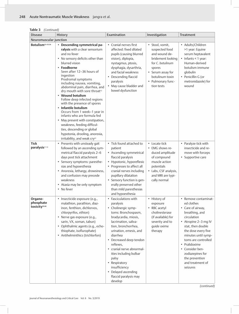
Disease History Examination Investigation TreatmentNeuromuscular junctionBotulism53–55,56 • Descending symmetrical pa-
ralysis with a clear sensorium and no fever
• No sensory deficits other than blurred vision
• FoodborneSeen after 12–36 hours of ingestionProdromal symptoms including nausea, vomiting, abdominal pain, diarrhea, and dry mouth with sore throat42
• Wound botulismFollow deep infected regions with the presence of spores
• Infantile botulismOccurs from 1 week–1 year in infants who are formula fed
• May present with constipation, weakness, feeding difficul-ties, descending or global hypotonia, drooling, anorexia, irritability, and weak cry43
• Cranial nerves first affected: fixed dilated pupils (causing blurred vision), diplopia, nystagmus, ptosis, dysphagia, dysarthria, and facial weakness
• Descending flaccid paralysis
• May cause bladder and bowel dysfunction
• Stool, vomit, suspected food and wound de-bridement looking for C. botulinum spores
• Serum assay for botulinum toxin
• Pulmonary func-tion tests
• Adults/Children >1 year: Equine serum heptavalent
• Infants < 1 year: Human-derived botulism immune globulin
• Penicillin G (or metronidazole) for wound
Tick paralysis57,58
• Presents with unsteady gait followed by an ascending sym-metrical flaccid paralysis 2–6 days post tick attachment
• Sensory symptoms: paresthe-sias and hypoesthesia
• Anorexia, lethargy, drowsiness, and confusion may precede weakness
• Ataxia may be only symptom • No fever
• Tick found attached to patient
• Ascending symmetrical flaccid paralysis
• Hypotonic, hyporeflexic • Progresses to affect all
cranial nerves including pupillary dilatation
• Sensory function is gen-erally preserved other than mild paresthesias and hypoesthesia
• Locate tick • EMG shows re-
duced amplitude of compound muscle action potentials
• Labs, CSF analysis, and MRI are typi-cally normal
• Paralyze tick with insecticide and re-move with forceps
• Supportive care
Organo-phosphate toxicity59,60
• Insecticide exposure (e.g., malathion, parathion, diaz-inon, fenthion, dichlorvos, chlorpyrifos, ethion)
• Nerve gas exposure (e.g., sarin, VX, soman, tabun)
• Ophthalmic agents (e.g., echo-thiophate, isoflurophate)
• Antihelminthics (trichlorfon)
• Fasciculations with paralysis
• Cholinergic symp-toms: Bronchospasm, bradycardia, miosis, lacrimation, saliva-tion, bronchorrhea, urination, emesis, and diarrhea
• Decreased deep tendon reflexes,
• cranial nerve abnormal-ities including bulbar palsy
• Respiratory insufficiency
• Delayed ascending flaccid paralysis may develop
• History of exposure
• RBC acetyl cholinesterase (if available) for severity and to guide oxime therapy
• Remove contaminat-ed clothes
• Care of airway, breathing, and circulation
• Atropine 2–3 mg IV stat, then double the dose every five minutes until symp-toms are controlled
• Pralidoxime • Consider ben-
zodiazepines for the prevention and treatment of seizures
(continued)

249Acute Nontraumatic Muscle Weakness Jangra et al.
Journal of Neuroanaesthesiology and Critical Care Vol. 6 No. 3/2019
Table 3 (Continued)
Disease History Examination Investigation TreatmentMyasthenia gravis61,62
• History of myasthenia gravis • Acute decompensation
(myasthenic crisis) may be spontaneous or precipitated by infection, surgery, or taper-ing of immunosuppression, certain antibiotics and other precipitating factors
• Excessive treatment with cholinesterase inhibitors may paradoxically cause weakness (Cholinergic crisis)
• 85% of patients have involvement of the eyelids and extra-ocular muscles, resulting in ptosis and/or diplopia23
• Fatiguability • Flaccid muscles
weakness • Central muscles are
predominantly involved such as bulbar muscles
• Neck and proximal limb weakness may occur
• Respiratory failure occurs in 1%
• Weakness increases after exertion
• Ice pack test (e.g., ice on affected eyelid improves ptosis)
• ACh receptor anti-bodies if diagnosis uncertain
• Pulmonary func-tion tests
• Consider arterial blood gas
• Consider CT chest (thymoma may affect breathing)
• For acute decom-pensation, admit to ICU
• Airway and ventila-tion should be as-sessed and managed with either non-in-vasive ventilation or intubation
• Withdraw anti-cholinesterase medications
• Plasmapheresis or IVIG
• High-dose steroids • Consider other im-
munosuppressantsLambert–Eaton myasthenic syndrome (LEMS)63,64
• In 40% of patients, small cell lung cancer is present
• Progressive proximal lower limb weakness
• Ptosis, diplopia, and dysarthria as cranial nerves become involved, (less common than myasthenia gravis)
• Autonomic dysfunction • Exacerbated by heat or fever
and certain drugs
• Proximal muscle weak-ness, lower limbs more than upper
• Depressed tendon reflexes
• Post-tetanic potentiation
• Sensation preserved • Respiratory failure rare
• Voltage gated calcium channel antibodies
• AChR antibodies • Repetitive nerve
stimulation • EMG • Look for ma-
lignancy with imaging/Bronchoscopy
• Confirm diagnosis and distinguish from myasthenia gravis before starting treatment
• Supportive treatment
• Treat underlying malignancy
• Consider 3,4-diaminopyridine
• IVIG • Plasma exchange
MuscleDermatomyo-sitis65
• May present with skin and/or muscle involvement
• Proximal muscle weakness • Characteristic rash • Systemic symptoms include
arthralgia, arthritis, dyspnea, dysphagia, arrhythmia, and dysphonia
• Heliotrope rash (blue-purple discol-oration on the upper eyelids)
• Raised, violaceous, scaly eruption on the knuckles (Gottron’s papules)
• Proximal symmetrical muscle weakness
• Muscle pain and tenderness
• Normal sensation and tendon reflexes
• Joint swelling (particu-larly of the hand) may occur occasionally in some patients
• Elevated CK, aldolase, lactate dehydroge-nase, or alanine aminotransferase
• Auto-antibody serology
• Skin biopsy • Muscle biopsy • NCS/EMG
• Corticosteroids • Consider immu-
nosuppressive or cytotoxic steroid sparing agents
• IVIG in refractory cases
(continued)

250
Journal of Neuroanaesthesiology and Critical Care Vol. 6 No. 3/2019
Acute Nontraumatic Muscle Weakness Jangra et al.
Table 3 (Continued)
Disease History Examination Investigation TreatmentGeneralized weakness due to systemic causesHyperglyce-mia66,67
• History of diabetes • Possible precipitating events
(e.g., infection, myocardial infarction, surgery, critical illness)
• Neurological symptoms primarily occur when plasma osmolality is greater than 320 mOsmol/L
• Neurological symptoms may include hemiparesis, focal motor deficits, decreased con-sciousness, and seizures
• May have polyuria, polydipsia, and weight loss for several days before presentation
• Level of consciousness may be reduced
• Focal motor and sen-sory deficits including aphasia, hyperreflexia, hemianopia, and brain-stem dysfunction
• Other findings asso-ciated with DKA or HHS include evidence of volume depletion, hyperventilation and abdominal pain
• Serum glucose • Plasma osmolality • Serum electro-
lytes (with anion gap), urea, and creatinine
• Urinalysis, and urine/ serum ke-tones by dipstick
• Blood gas • Electrocardiogram • CT head to ex-
clude other causes
• Fluid replacement to correct hy-pervolemia and hyperosmolality
• Insulin infusion • Close monitoring
of urine output and electrolytes (potas-sium, magnesium, and phosphate)
• Treat precipitating cause
Hypoglyce-mia (serum glucose<3 mmol/L; <50 mg/dL)68
• Diabetes • Insulin regimen • Oral hypoglycemics • Alcohol • Sepsis • Liver disease • Hypocortisolemia
• Decreased consciousness
• Many forms of focal neurological deficit possible, which may mimic
• Dysphoria • Seizures stroke • Tremor, palpitations,
anxiety, sweating, hun-ger, and paresthesia
• Blood glucose level
• CT head to rule out intracranial causes
• IV dextrose • Oral if patient is
conscious • Alternatively, 1 mg
glucagon IM or IV
Hyponatremia, hypernatre-mia69,70
• Hyponatremia: diuretic overdose, hypervolemia, CHF, cirrhosis, SIADH, cerebral salt wasting and water intoxication
• Hypernatremia: dehy-dration, pituitary insuffi-ciency, iatrogenic sodium supplementation
• Lethargy and confusion are most common followed by seizures and coma in both extremes
• Depressed level of con-sciousness or delirium
• Serum sodium levels
• Hyponatremia: fluid restriction, stop diuretics, avoid rapid correction
• Hypernatremia: IV fluids if hypovole-mic, prefer hypo-tonic solutions (5D, 0.45% NS), avoid rapid correction
• if urine specif-ic gravity is low (pituitary insuffi-ciency): administer desmopressin
Hypermagne-semia71
• Typically follows excessive magnesium administration (e.g., management of pre-ec-lampsia) in context of renal impairment
• Lethargy and confusion are most common neurologic manifestations followed by generalized weakness, and respiratory failure
• Hyporeflexia: (early sign) loss of deep ten-don reflexes
• Flaccid tetraparesis involving all muscle groups
• Lethargy, confusion
Serum magnesium levels
• Stop magnesium administration
• IV calcium gluconate/chloride
• IV fluids • Consider dialysis
(continued)

251Acute Nontraumatic Muscle Weakness Jangra et al.
Journal of Neuroanaesthesiology and Critical Care Vol. 6 No. 3/2019
Table 3 (Continued)
Disease History Examination Investigation TreatmentHypophospha-temia72,73
• Causes of hypophosphatemia include:
– Intracellular shift: refeed-ing syndrome, respiratory alkalosis, diabetic ketoaci-dosis, rapidly growing ma-lignancies, osmotic diuresis, malabsorption, renal tubular acidosis
– Increased urinary excre-tion: primary or secondary hyperparathyroidism, osmot-ic diuresis, renal tubular aci-dosis, transplanted kidneys, Fanconi syndrome, etc.
– Decreased intestinal absorption: diarrhea, mal-absorption syndromes,
phosphate binders – Decreased dietary intake: anorexia nervosa or chronic alcoholism, Hypothermia
• Painful proximal myopathy • Other symptoms: changes
in mental function, seizures, neuropathies, arrhythmias, skeletal muscle weakness, respiratory failure, rhabdomy-olysis, leucocyte dysfunction, sepsis, and sudden death
• Proximal muscle weak-ness is common
• Any muscle group may be involved in various combinations, ranging from ophthalmoplegia to proximal myopathy to dysphagia or ileus
• Weakness may be so profound as to mimic Guillain–Barre syndrome
• Neurological features: Confusion, seizures, and coma
• Cardiac contractility may be impaired lead-ing to global myocardial depression
• Serum phosphate • Hypercalcemia or
Hypomagnese-mia is commonly associated
• Other electrolytes • Rhabdomyolysis
screen
• Correct precipitant • Replace total body
phosphate with careful IV sodi-um or potassium phosphate
Periodic paral-ysis (PP)74
• Repeated episodes of flaccid muscle weakness occurring at irregular intervals with normal strength between episodes
• Usually hereditary • Various types of periodic paral-
ysis exist, including: – Hyperkalemic PP – Hypokalemic PP – Paramyotonia congenita – Thyrotoxic PP – Andersen-Tawil syndrome
• Look for precipitating factor (e.g., post exercise, fasting, cold alcohol, stress, and dura-tion of episode)
• All forms usually exhibit:
– Interictal lid lag and eyelid myotonia
– Normal sensation – Fixed proximal weakness
– Diminished reflexes during episode
– Normal power in be-tween the episodes
• Serum potassium • Elevated creatine
kinase (CK) • Potassium: creati-
nine ratio • Blood gas analysis
for evidence of concomitant metabolic acidosis or alkalosis
• ECG • EMG • Nerve conduction
studies
• Hyperkalemic PP: – High carbohydrate food
– Thiazide or acetazolamide
• Hypokalemic PP: – Potassium supplementation
– Acetazolamide • Thyrotoxic PP:
– Beta blockers – Treat thyrotoxicosis
• Andersen–Tawil syndrome:
• Acetazolamide(continued)

252
Journal of Neuroanaesthesiology and Critical Care Vol. 6 No. 3/2019
Acute Nontraumatic Muscle Weakness Jangra et al.
Table 3 (Continued)
Disease History Examination Investigation TreatmentMiscellaneousEnvenom-ation75,76
• Snake bite16
• Scorpion sting • Marine envenomation • Ingestion of puffer fish
• Snake bites16:Cardiovascular: hypo-tension, shock, arrest
– Neurological: paral-ysisptosis, diplopia, bulbar palsy, dys-arthria; respiratory muscle paralysisCoagulopathy: intra-cranial hemorrhage, bleeding from bite site, ecchymoses, bleeding gums, hemarthrosesRhabdomyolysis: tender muscles
• Scorpion sting: cranial nerve and somatic skeletal neuromuscular dysfunction, with pain and paresthesia
• Blue-ringed octopus and puffer fish enven-omation: descending symmetrical flaccid paralysis with clear sensorium, nausea, and vomiting, blurred vision, ataxia, respirato-ry failure
• Stonefish envenom-ation: weakness in the affected limb, severe pain, shock
• Serial bedside pul-monary function tests if descending paralysis
• Other investiga-tions as CBC, LFTs, CK, whole blood clotting time, co-agulation, screen, D-dimer, fibrin-ogen levels, uri-nalysis for blood (myoglobin),
• Head-CT if de-creased GCS
• Use venom de-tection kit for bite swab and urine
• Supportive care of airway, breathing, and circulation
• Pressure immobiliza-tion bandage
• Specific antivenom
Locked-in syndrome77
• Sudden onset tetraplegia, facial weakness, and horizontal gaze palsy
• Causes ischemic stroke (most common), central pontine myelinolysis, encephalitis, or tumor
• Flaccid symmetrical tetraparesis
• Consciousness preserved or may be affected initially but returns to normal
• Voluntary vertical eye and eyelid movements preserved
• Hearing, vision, pupillary reflexes, and sensation all normal
• CT brain with spiral CT angiography35
• MRI/MRA
Follow acute stroke protocol
(continued)

253Acute Nontraumatic Muscle Weakness Jangra et al.
Journal of Neuroanaesthesiology and Critical Care Vol. 6 No. 3/2019
Table 3 (Continued)
Disease History Examination Investigation TreatmentAcute porphyria78
• Abdominal pain: may begin in chest or back and move to abdomen
• Gastrointestinal symptoms such as vomiting, diarrhea, and constipation are common
• Psychiatric symptoms • Acute weakness (early or late) • May develop seizures • Certain medications are
known to exacerbate
• Muscle weakness usually begins proxi-mally and more often in upper limbs
• Symmetrical hypotonia • Hyporeflexic • Flaccid paralysis • No rash unlike other
forms of porphyria • Tachycardia and hyper-
tension may be present
• Hyponatremia • Urine: dark/
reddish • Urine analy-
sis: increased porphobilinogen
• IV hemin • Manage
hyponatremia • Consider antiepilep-
tic drugs • Supportive
management
Diabetic lumbosacral radiculoplexus neuropathy79
• Diabetes mellitus with proxi-mal weakness
• Asymmetrical pain in the hip, buttock, or thigh
• Associated with poor glycemic control
• Patients without distal sym-metrical polyneuropathy most often have sudden, unilateral onset
• Occasionally may be initial pre-sentation of diabetes mellitus
• Proximal lower limb muscle weakness and wasting
• Minimal sensory loss is observed
• Knee-jerk reflex is absent, with commonly preserved ankle jerks
• Ankle jerks may also be absent, with underly-ing distal symmetrical polyneuropathy
• Fasting blood glu-cose and glycated hemoglobin
• Imaging of lum-bo-sacral spine to exclude other causes
• EMG • Nerve conduction
studies
• Optimize glycemic control
• Physical and occupa-tional therapy
Psychiatric illness
• No history suggestive of any physical illness
• Temporal associations with psychosocial stressors
• Symptom substitution fre-quently present
• Primary psychological or per-sonal gain present
• La belle indifference present
• Distribution does not follow anatomical pattern
• Presence of affective or emotional disturbanc-es on mental status examination
• Relevant investi-gations to rule out organic lesions like (MRI/CT, EEG).
• Visual-evoked potentials and brainstem auditory evoked responses to rule out malingering/compensation neurosis
• Minimize and stop further investigations
• Decrease secondary gains
• Increase functioning • Refer for special-
ist psychiatric interventions
Source: Adapted with permission from Caulfield et al.3
Abbreviations: AChR, acetylcholine receptor; ASAS, anterior spinal artery syndrome; BP, blood pressure; CBC, complete blood count; CHF, congestive heart failure; CNS, central nervous system; CSF, cerebrospinal fluid; CT, computed tomography; DKA, diabetes keto-acidosis; ECG, electrocardiography; EEG, electroencephalogram; EMG, electromyography; GCS, Glasgow Coma Scale; HHC, hyperos-molar coma; HIV, human immuno-deficiency virus; HTN, hypertension; ICP, intracranial pressure; IV, intravenous; IVIG, intravenous immunoglobulin; LL, lower limb; LMN, lower motor neuron; MRA, magnetic resonance angiography; MRI, magnetic resonance im-aging; NCS, nerve conduction study; PSAS, posterior spinal artery; SIAD, syndrome of inappropriate antidiuretic hormone secretion; SPECT, single-photon emission computed tomography; UL, Upper limb; UMN, upper motor neuron.
254
Journal of Neuroanaesthesiology and Critical Care Vol. 6 No. 3/2019
Acute Nontraumatic Muscle Weakness Jangra et al.
Special Consideration in Pediatric PatientsThe basic principles of assessment of the airway/ventilation and localization are the same as in adults. The major differ-ences are in the presentation, and the common etiologies leading to weakness are highlighted here. In children, the presenting symptoms may be mutable such as irritability, agitation, restlessness, refusal to walk, frequent awakening from sleep, willingness to be held frequently, or regression of milestones. The history should focus on the evaluation of various risk factors such as congenital heart diseases, sickle cell anemia, and prothrombotic states. The examination of reflexes, signs of bulbar weakness, and assessment of senso-ry level are as critical as in adults, but it may be difficult in very young children. In children, it is difficult to distinguish the various causes of difficulty in walking such as weakness, pain, and ataxia.
The common causes of ANTW in children include Todd’s paresis, acute demyelinating encephalomyelitis, acute trans-verse myelitis, GBS, and myasthenia gravis.19-23 Stroke is a rare presentation in children but may occur in various conditions including sickle cell, congenital heart disease, prothrombotic disorder, and Moyamoya disease. The aortic dissection is quite common ischemic spinal cord injury in children lead-ing to spinal artery infarcts. If reflexes are intact, then con-sider transverse myelitis, Todd’s paresis, myasthenia gravis or stroke, while in patients with reduced or absent reflexes, consider early transverse myelitis with spinal shock or GBS. Imaging modalities and laboratory tests should be directed as per the differential diagnosis.
Referral to a Higher CenterHealthcare providers should provide the following details including patient’s age, history of present illness, complete details of patient’s initial assessment (ABC), salient history, examination findings, laboratory reports, imaging results, and treatment provided. The further plan about a pending investigations, list of potential considerations, and manage-ment (if the diagnosis of acute weakness is known) should also be provided.
ConclusionAcute nontraumatic muscle weakness occurs due to a lesion in the motor tract anywhere from pyramidal cells to periph-eral muscles. These may prove to be life threatening if airway, breathing, or circulation is affected. Care of ABC takes prior-ity over managing and localizing the weakness. We should focus on a detailed history and examination to localize the lesion quickly, make an initial working diagnosis, and screen the patients for time-sensitive emergencies. The laboratory tests and neurological imaging are done to make the diag-nosis. A systematic algorithm/protocol should be followed so that we do not miss any important cause of weakness.
Conflict of InterestNone declared.
References
1 Gerard T, Bryan D, Principles of Anatomy & Physiology. 14th ed. New Jersey: John Wiley & Sons, Inc; 2014:406, 502, 541
2 Gillian P, Christopher R, Human Physiology: The Basis of Med-icine. 3rd ed. Oxford: Oxford University Press; 2006;151–153
3 Caulfield AF, Flower O, Pineda JA, Uddin S. Emergency neuro-logical life support: acute non-traumatic weakness. Neurocrit Care 2017;27(Suppl 1):29–50
4 Mehta S. Neuromuscular disease causing acute respiratory fail-ure. Respir Care 2006;51(9):1016–1021, discussion 1021–1023
5 Lawn ND, Fletcher DD, Henderson RD, Wolter TD, Wijdicks EF. Anticipating mechanical ventilation in Guillain-Barré syn-drome. Arch Neurol 2001;58(6):893–898
6 Coplin WM, Pierson DJ, Cooley KD, Newell DW, Rubenfeld GD. Implications of extubation delay in brain-injured patients meeting standard weaning criteria. Am J Respir Crit Care Med 2000;161(5):1530–1536
7 Ropper AH. The Guillain-Barré syndrome. N Engl J Med 1992;326(17):1130–1136
8 Seneviratne U. Guillain-Barré syndrome. Postgrad Med J 2000;76(902):774–782
9 Sagarin MJ, Barton ED, Chng YM, Walls RM; National Emergen-cy Airway Registry Investigators. Airway management by US and Canadian emergency medicine residents: a multicenter analysis of more than 6,000 endotracheal intubation attempts. Ann Emerg Med 2005;46(4):328–336
10 Li J, Murphy-Lavoie H, Bugas C. Martinez J, Preston C. Compli-cations of emergency intubation with and without paralysis. Am. J Emerg Med 1999;17(2):141–143
11 Sakles JC, Laurin EG, Rantapaa AA, Panacek EA. Airway manage-ment in the emergency department: a one-year study of 610 tracheal intubations. Ann Emerg Med 1998;31(3):325–332
12 Walls RM. Rapid-sequence intubation in head trauma. Ann Emerg Med 1993;22(6):1008–1013
13 Abel M, Eisenkraft JB. Anesthetic implications of myasthenia gravis. Mt Sinai J Med 2002;69(1)(2):31–37
14 Orebaugh SL. Succinylcholine: adverse effects and alternatives in emergency medicine. Am. J Emerg Med 1999;17(7):715–721
15 Rajajee V, Riggs B, Seder DB. Emergency neurological life support: airway, ventilation, and sedation. Neurocrit Care 2017;27(Suppl 1):4–28
16 Jan MM, Al-Buhairi AR, Baeesa SS. Concise outline of the ner-vous system examination for the generalist. Neurosciences (Riyadh) 2001;6(1):16–22
17 Florman JE, Duffau H, Rughani AI. Lower motor neuron findings after upper motor neuron injury: insights from postoperative supplementary motor area syndrome. Front Hum Neurosci 2013;7:85
18 Folks DG, Ford CV, Regan WM. Conversion symptoms in a gener-al hospital. Psychosomatics 1984;25(4):285–289, 291, 294–295
19 Zuccoli G, Panigrahy A, Bailey A, Fitz C. Redefining the Guil-lain-Barré spectrum in children: neuroimaging findings of cra-nial nerve involvement. Am J Neuroradiol 2011;32(4):639–642
20 Pidcock FS, Krishnan C, Crawford TO, Salorio CF, Trovato M, Kerr DA. Acute transverse myelitis in childhood: center-based analysis of 47 cases. Neurology 2007;68(18):1474–1480
21 Chen L, Li J, Guo Z, Liao S, Jiang L. Prognostic indicators of acute transverse myelitis in 39 children. Pediatr Neurol 2013;49(6):397–400
22 Wolf VL, Lupo PJ, Lotze TE. Pediatric acute transverse myeli-tis overview and differential diagnosis. J Child Neurol 2012;27(11):1426–1436
23 Bernard TJ, Rivkin MJ, Scholz K, et al; Thrombolysis in Pediatric Stroke Study. Emergence of the primary pediatric stroke cen-ter: impact of the thrombolysis in pediatric stroke trial. Stroke 2014;45(7):2018–2023
24 DeAngelis LM. Brain tumors. N Engl J Med 2001;344(2):114–123

255Acute Nontraumatic Muscle Weakness Jangra et al.
Journal of Neuroanaesthesiology and Critical Care Vol. 6 No. 3/2019
25 Muzumdar D, Jhawar S, Goel A. Brain abscess: an overview. Int J Surg 2011;9(2):136–144
26 Gallmetzer P, Leutmezer F, Serles W. Assem-Hilger E, Spatt J, Baumgartner C. Postictal paresis in focal epilepsies—inci-dence, duration, and causes: a video-EEG monitoring study. Neurology 2004;62(12):2160–2164
27 Rolak LA, Rutecki P, Ashizawa T, Harati Y. Clinical features of Todd’s post-epileptic paralysis. J Neurol Neurosurg Psychiatry 1992;55(1):63–64
28 Aggarwal M, Khan IA. Hypertensive crisis: hypertensive emer-gencies and urgencies. Cardiol Clin 2006;24(1):135–146
29 Vaughan CJ, Delanty N. Hypertensive emergencies. Lancet 2000;356(9227):411–417
30 Headache Classification Subcommittee of the International Headache, Society. The International Classification of Head-ache Disorders: 2nd edition. Cephalalgia 200;424(Suppl 1): 9–160
31 Russell MB, Ducros A. Sporadic and familial hemiplegic migraine: pathophysiological mechanisms, clinical char-acteristics, diagnosis, and management. Lancet Neurol 2011;10(5):457–470
32 Novy J, Carruzzo A, Maeder P, Bogousslavsky J. Spinal cord ischemia: clinical and imaging patterns, pathogenesis, and outcomes in 27 patients. Arch Neurol 2006;63(8):1113–1120
33 Hagan PG, Nienaber CA, Isselbacher EM, et al. The Internation-al Registry of Acute Aortic Dissection (IRAD): new insights into an old disease. JAMA 2000;283(7):897–903
34 Gaul C, Dietrich W, Friedrich I, Sirch J, Erbguth FJ. Neu-rological symptoms in type A aortic dissections. Stroke 2007;38(2):292–297
35 Tsai TT, Nienaber CA, Eagle KA. Acute aortic syndromes. Circu-lation 2005;112(24):3802–3813
36 Sayer FT, Vitali AM, Low HL, Paquette S, Honey CR. Brown-Sèquard syndrome produced by C3-C4 cervical disc herniation: a case report and review of the literature. Spine 2008;33(9):E279–E282
37 Antich PA, Sanjuan AC, Girvent FM, Simó JD. High cervi-cal disc herniation and Brown-Sequard syndrome. A case report and review of the literature. J Bone Joint Surg Br 1999;81(3):462–463
38 Brinar VV, Habek M, Brinar M, Malojcić B, Boban M. The dif-ferential diagnosis of acute transverse myelitis. Clin Neurol Neurosurg 2006;108(3):278–283
39 Rowland LP, Shneider NA. Amyotrophic lateral sclerosis. N Engl J Med 2001;344(22):1688–1700
40 Kiernan MC, Vucic S, Cheah BC, et al. Amyotrophic lateral scle-rosis. Lancet 2011;377(9769):942–955
41 Miller RG, Jackson CE, Kasarskis EJ, et al; Quality Standards Sub-committee of the American Academy of Neurology. Practice parameter update: the care of the patient with amyotrophic later-al sclerosis: multidisciplinary care, symptom management, and cognitive/behavioral impairment (an evidence-based review): report of the Quality Standards Subcommittee of the American Academy of Neurology. Neurology 2009;73(15):1227–1233
42 Meena AK, Khadilkar SV, Murthy JM. Treatment guide-lines for Guillain-Barré Syndrome. Ann Indian Acad Neurol 2011;14(Suppl 1):S73–S81
43 Alshekhlee A, Hussain Z, Sultan B, Katirji B. Guillain- Barré syndrome: incidence and mortality rates in US hospitals. Neurology 2008;70(18):1608–1613
44 Ropper AH. Further regional variants of acute immune polyneuropathy. Bifacial weakness or sixth nerve paresis with paresthesias, lumbar polyradiculopathy, and ataxia with pharyngeal-cervical-brachial weakness. Arch Neurol 1994;51(7):671–675
45 Hughes RA, Swan AV, van Koningsveld R, van Doorn PA. Cor-ticosteroids for Guillain-Barré syndrome. Cochrane Database Syst Rev 2006;19(2):CD001446
46 Davies L, Spies JM, Pollard JD, McLeod JG. Vasculitis confined to peripheral nerves. Brain 1996;119(Pt 5):1441–1448
47 Mathew L, Talbot K, Love S, Puvanarajah S, Donaghy M. Treat-ment of vasculitic peripheral neuropathy: a retrospective analysis of outcome. QJM 2007;100(1):41–51
48 London Z, Albers JW. Toxic neuropathies associated with pharmaceutic and industrial agents. Neurol Clin 2007;25(1):257–276
49 Graeme KA, Pollack CV, Jr. Heavy metal toxicity, part I: arsenic and mercury. J Emerg Med 1998;16(1):45–56
50 Graeme KA, Pollack CV, Jr. Heavy metal toxicity, part II: lead and metal fume fever. J Emerg Med 1998;16(2):171–177
51 Marx A, Glass JD, Sutter RW. Differential diagnosis of acute flaccid paralysis and its role in poliomyelitis surveillance. Epidemiol Rev 2000;22(2):298–316
52 Fox IK, Mackinnon SE. Adult peripheral nerve disorders: nerve entrapment, repair, transfer, and brachial plexus disorders. Plast Reconstr Surg 2011;127(5):105
53 Neal S, Fields KB. Peripheral nerve entrapment and injury in the upper extremity. Am Fam Physician 2010;81(2):147–155
54 Varma JK, Katsitadze G, Moiscrafishvili M, et al. Signs and symptoms predictive of death in patients with foodborne botulism—Republic of Georgia, 1980-2002. Clin Infect Dis 2004;39(3):357–362
55 Long SS. Infant botulism. Pediatr Infect Dis J 2001;20(7): 707–709
56 Chalk C, Benstead TJ, Keezer M. Medical treatment for botu-lism. Cochrane Database Syst Rev 2011 ;16(3):CD008123
57 Grattan-Smith PJ, Morris JG, Johnston HM, et al. Clini-cal and neurophysiological features of tick paralysis. Brain 1997;120(Pt 11):1975–1987
58 Edlow JA, McGillicuddy DC. Tick paralysis. Infect Dis Clin North Am 2008;22(3):397–413, vii
59 Eddleston M, Buckley NA, Eyer P, Dawson AH. Management of acute organophosphorus pesticide poisoning. Lancet 2008;371(9612):597–607
60 Roberts DM, Aaron CK. Management of acute organophospho-rus pesticide poisoning. BMJ 2007;334(7594):629–634
61 Grob D, Brunner N, Namba T, Pagala M. Lifetime course of myasthenia gravis. Muscle Nerve 2008;37(2):141–149
62 Meriggioli MN, Sanders DB. Advances in the diagnosis of neuromuscular junction disorders. Am J Phys Med Rehabil 2005;84(8):627–638
63 Wirtz PW, Smallegange TM, Wintzen AR, Verschuuren JJ. Dif-ferences in clinical features between the Lambert-Eaton myas-thenic syndrome with and without cancer: an analysis of 227 published cases. Clin Neurol Neurosurg 2002;104(4):359–363
64 Keogh M, Sedehizadeh S, Maddison P. Treatment for Lambert- Eaton myasthenic syndrome. Cochrane Database Syst Rev 2011
65 Iorizzo LJ, III. Jorizzo JL. The treatment and prognosis of dermatomyositis: an updated review. J Am Acad Dermatol 2008;59(1):99–112
66 Guisado R, Arieff AI. Neurologic manifestations of diabetic comas: correlation with biochemical alterations in the brain. Metabolism 1975;24(5):665–679
67 Kitabchi AE, Umpierrez GE, Miles JM, Fisher JN. Hypergly-cemic crises in adult patients with diabetes. Diabetes Care 2009;32(7):1335–1343
68 DeRosa MA, Cryer PE. Hypoglycemia and the sympathoadrenal system: neurogenic symptoms are largely the result of sympa-thetic neural, rather than adrenomedullary, activation. Am J Physiol Endocrinol Metab 2004;287(1):E32–E41
69 Reynolds RM, Padfield PL, Seckl JR. Disorders of sodium bal-ance. BMJ 2006;332(7543):702–705
70 Adrogué HJ, Madias NE. Hypernatremia. N Engl J Med 2000;342(20):1493–1499
71 Riggs JE. Neurologic manifestations of electrolyte disturbances. Neurol Clin 2002;20(1):227–239
256
Journal of Neuroanaesthesiology and Critical Care Vol. 6 No. 3/2019
Acute Nontraumatic Muscle Weakness Jangra et al.
72 Ravid M, Robson M. Proximal myopathy caused by latrogenic phosphate depletion. JAMA 1976;236(12):1380–1381
73 Sebastian S, Clarence D, Newson C. Severe hypophospha-taemia mimicking Guillain-Barré syndrome. Anaesthesia 2008;63(8):873–875
74 Venance SL, Cannon SC, Fialho D, et al; CINCH investigators. The primary periodic paralyses: diagnosis, pathogenesis and treatment. Brain 2006;129(Pt 1) :8–17
75 White J. Venomous animals: clinical toxinology. EXS 2010;100:233–291
76 Warrell DA. Snake bite. Lancet 2010;375(9708):77–8877 Bruno MA, Pellas F, Schnakers C, et al. [Blink and you live: the
locked-in syndrome] Rev Neurol (Paris) 2008;164(4):322–33578 Anderson KE, Bloomer JR, Bonkovsky HL, et al. Recommenda-
tions for the diagnosis and treatment of the acute porphyrias. Ann Intern Med 2005;142(6):439–450
79 Tracy JA, Dyck PJ. The spectrum of diabetic neuropathies. Phys wwMed Rehabil Clin N Am 2008;19(1):1–26, v