evaluation of weakness localization to muscle... · 2019-10-11 · often presents as a...
TRANSCRIPT
1
Samantha LoRusso, MDAssistant Professor-Clinical
Department of NeurologyThe Ohio State University Wexner Medical Center
Evaluation of WeaknessPart 1: Upper motor neuron weakness
LocalizationLocalization• Central Nervous System
Brain Spinal cord
• Peripheral Nervous System Cranial nerves Spinal nerves and dorsal root
ganglia Motor neuron Plexus Peripheral Nerves Muscle Neuromuscular junction
Upper motor neuron signs
Lower motor neuron signs
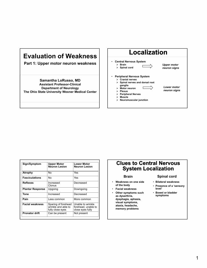
Sign/Symptom Upper Motor Neuron Lesion
Lower Motor Neuron Lesion
Atrophy No Yes
Fasciculations No Yes
Reflexes IncreasedClonus
Decreased
Plantar Response Upgoing Downgoing
Tone Increased Decreased
Pain Less common More common
Facial weakness Sparing of foreheadwrinkle and able to fully close eyes
Unable to wrinkle forehead, unable to close eyes fully
Pronator drift Can be present Not present
Clues to Central Nervous System Localization
Clues to Central Nervous System Localization
• Weakness on one side of the body
• Facial weakness
• Other symptoms such as dysarthria, dysphagia, aphasia, visual symptoms, ataxia, headache, memory problems
• Bilateral weakness
• Presence of a ‘sensory level’
• Bowel or bladder symptoms
Brain Spinal cord

2
Exceptions to the rulesExceptions to the rulesBrain lesions that cause bilateral weakness
Multiple lesions on both sides of the brain Bilateral watershed infarcts, bilateral brainstem
lesions, bilateral medial frontal lesions Also note that ‘crossed’ findings (symptoms on one
side of the face and on the opposite side of the body) are classic for a brainstem lesion
OpenStax College (CC BY 3.0)
Exceptions to the RuleExceptions to the RuleSpinal cord lesions that cause unilateral weakness or facial symptoms
Any lesion only affecting one half of the spinal cord Often presents as a Brown-Sequard syndrome:
weakness and reduced vibration and proprioception ipsilateral to the lesion and reduced pinprick sensation contralateral to the lesion.
High cervical lesions may involve the spinal trigeminal nucleus and can cause decreased facial sensation but NOT weakness.
Niels Olson (CC BY-SA 3.0)
Case Presentation 1Case Presentation 164 year old man who says that 2 weeks ago he was sitting, drinking a beer and watching TV and noticed that his right arm felt weak. He then went to bed and by the next morning was unable to move his hand at all so he went to the emergency room. In the emergency room, he reports that they performed a CT of the brain which was unremarkable so he was sent home. Since then it has gotten a little better– now he is able to grip a little bit. Denies numbness and pain. Denies other symptoms.
ExaminationExamination• Mental Status: normal.• Cranial nerves: normal. • Motor exam: 5/5 strength throughout except for
4+/5 strength in his proximal right arm and 2/5 strength in his right hand. There is right pronator drift. Bulk and tone are normal.
• Reflexes: 2+ in the right biceps, otherwise 1+ on the right and left arms. 0 in the knees and ankles.
• Sensation: Symmetric to pinprick. Decrease in vibration in the toes.
• Coordination: normal• Gait: normal
3
Work-upWork-up
• MRI Brain without contrast: a small acute ischemic cortical infarct is noted in the left precentral gyrus.
• Followed by a stroke work-up and management of risk factors.
Clues to Stroke DiagnosisClues to Stroke Diagnosis• History: sudden onset of symptoms,
painless, PMHx
• Exam: Relatively increased reflexes in the area of weakness, pronator drift and the fact that the weakness does not follow a clear nerve root or nerve distribution suggests an upper motor neuron process.
• Upper motor neuron findings on exam with weakness on only one side of the body suggests the Brain
Why was the CT of the brain negative?Why was the CT of the brain negative?
• The stroke was small
• The CT scan was done soon after symptom onset– strokes become more clear on CT 6-12 hours after symptom onset
• Also note that the CT does a very poor job of imaging the brainstem (not relevant in this case)
Case Presentation 2Case Presentation 252 year old man who woke up one day around June 2016 and noticed that he had a difficult time walking, specifically because of right leg weakness. He also noted some right foot numbness around November 2018. He thinks that his problem has been slowly getting worse over time. He said that he will trip and has to drag his right leg. He also has to drive with two feet because he can’t move his right leg from one pedal to the other. Denies bowel/bladder problems. No pain.
4
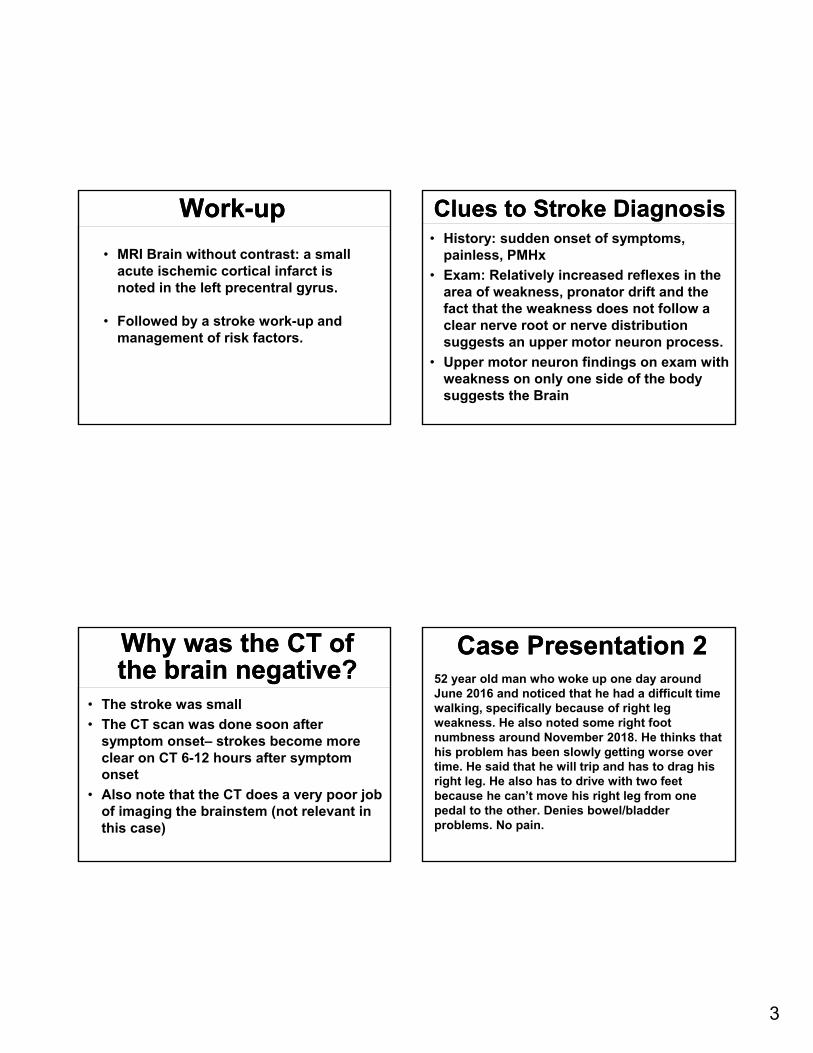
ExaminationExamination• Mental status and Cranial nerves normal• Motor: 5/5 strength in both upper extremities and
in the left lower extremity. In the right lower extremity he had 2/5 hip flexion, 4/5 knee extension, 2/5 knee flexion, 4/5 dorsiflexion and 4+/5 plantar flexion.
• Reflexes:
Right Left
Biceps 3 2
Triceps 1 1
Brachioradialis 2 1
Patella 3 2
Achilles 2 1
Plantar response Upgoing Mute
Examination Examination • Sensory: Decreased in the right leg to pinprick
and vibration compared to the left otherwise normal.
• Coordination: normal
• Gait: hemiplegic in the right lower extremity
‒ View Video Demonstration from link on webcast downloads for this webcast.
Previous testing done prior to referral
Previous testing done prior to referral
• MRI Hip: mild bilateral hip joint osteoarthritis
• EMG/NCS: Normal
• MRI Lumbar spine without contrast: mild disc bulging diffusely. Mild to moderate foraminal narrowing throughout.
• CT of the brain unremarkable
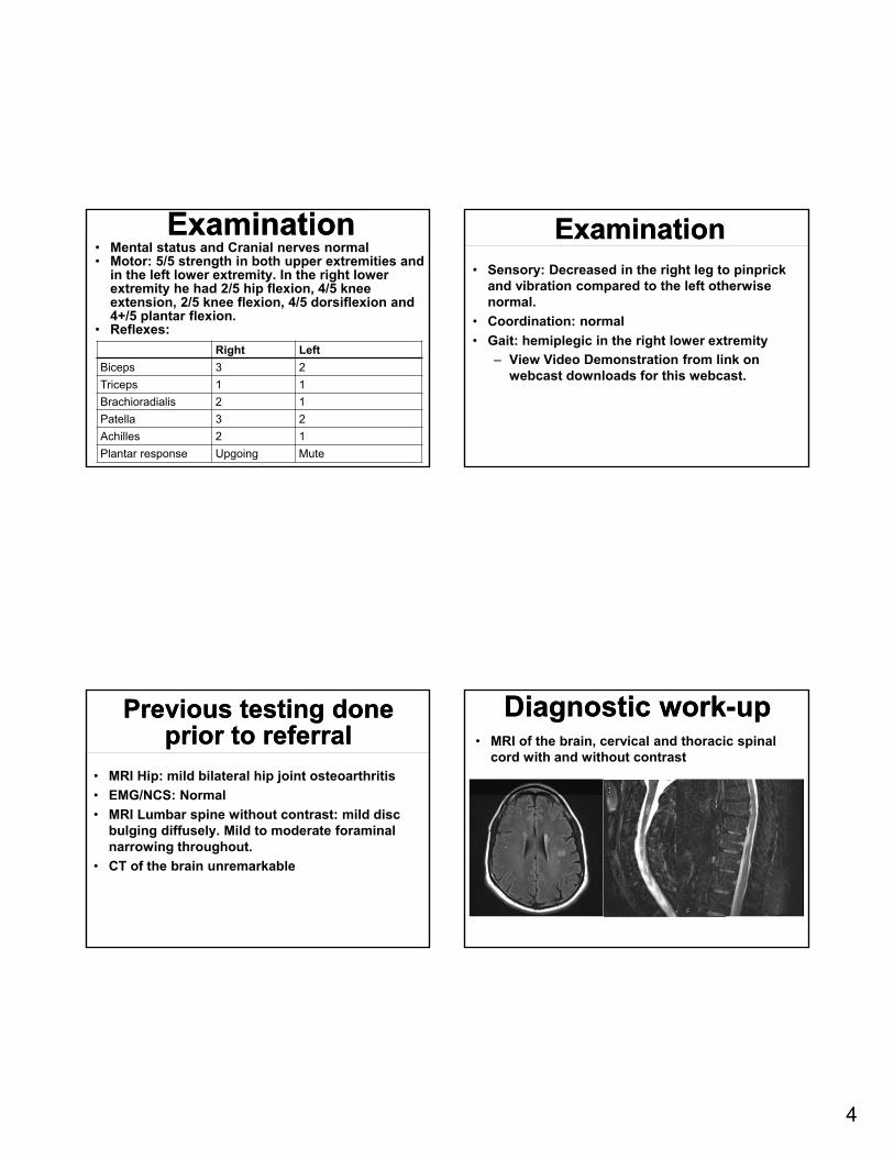
Diagnostic work-upDiagnostic work-up• MRI of the brain, cervical and thoracic spinal
cord with and without contrast

5
• Diagnosis: Primary progressive multiple sclerosis
Clues to localization Clues to localization • Clear upper motor neuron signs and
painless Would not necessarily expect
EMG/NCS or MRI of the lumbar spine to be abnormal
Why would you not expect MRI of the lumbar spine to be abnormal?Why would you not expect MRI of the lumbar spine to be abnormal?
• MRI of the lumbar spine only images the bottom of the spinal cord and the cauda equina
Case Presentation 3Case Presentation 371 year old man presents with 2 years of worsening gait problems. He reports that his legs felt “wobbly” as if they were going to buckle while walking. When asked about numbness or tingling, he said that he had noticed some numbness in his hands over the same time period. Denied any other symptoms.

6
ExaminationExamination• Mental Status: normal
• Cranial nerves: normal
• Motor exam: Normal with the exception of 4+/5 hip flexor weakness bilaterally. Tone mildly increased in the legs.
• Reflexes: 3+ throughout with bilateral upgoingtoes
• Sensation: decreased vibratory and pinprick sensation in his hands and feet without a clear sensory level
• Coordination: normal
• Gait: normal
Work-upWork-up• MRI Cervical and thoracic spine without contrast
• Other myelopathy labs such as vitamin B12, Copper, Vitamin E
Clues to localization and diagnosisClues to localization and diagnosis• Bilateral weakness with upper motor neuron signs so
most likely spinal cord localization
• A thoracic lesion may be suggested by the lack of arm weakness BUT The patient had sensory changes in his hands that
would not be explained by a thoracic lesion Cervical spondylotic myelopathy is more common! It
is the most common cause of spinal cord dysfunction worldwide in patients older than 55 years old.(Nouri et al., 2015)
It is common for cervical lesions like this to cause more symptoms in the lower extremities than in the upper extremities (Stino et al., 2018)
Imaging in structural spine disease
Imaging in structural spine disease
• MRIs are superior to x-rays and CT scans for imaging of the spinal cord and the nerve roots.
• If a patient cannot get an MRI, but structural spine disease is suspected then a CT myelogram should be performed.
• If significant structural spinal cord abnormalities are found then the patient should be referred to a neurosurgeon

7
Don’t forgetDon’t forget• Localize—does the patient have an ‘upper motor
neuron’ exam?
• Bilateral lower extremity weakness with clear upper motor neuron signs should prompt evaluation of the spinal cord
• Unilateral weakness should prompt imaging of the brain
• Almost always appropriate to refer to neurology when weakness and upper motor neuron findings on exam, but would always start with imaging of the CNS
• If there is a compressive lesion then referral to neurosurgery
ReferencesReferences• Blumenfeld H. Neuroanatomy through clinical cases.
2002.
• https://en.wikipedia.org/wiki/Cortical_homunculus
• https://en.wikipedia.org/wiki/Brown-S%C3%A9quard_syndrome
• Nouri A, Tetreault L, Singh A, et al. Degenerative cervical myelopathy: epidemiology, genetics, and pathogenesis. Spine 2015;40(12):E675-E693.
• Stino AM & LoRusso SJ. Myelopathies due to structural cervical and thoracic disease. Continuum 2018 Apr:24(2, Spinal Cord Disorders): 567-583.
Bakri Elsheikh, MBBS, FRCP, FAANAssociate Professor-Clinical
Director of the EMG LaboratoryDepartment of Neurology
The Ohio State University Wexner Medical Center
Evaluation of WeaknessPart 2: Lower motor neuron weakness
Localization!Lower motor neuron
Localization!Lower motor neuron
• Peripheral Nervous System Cranial nerves Spinal nerves and dorsal
root ganglia Motor neuron Plexus Peripheral Nerves Muscle Neuromuscular junction

8
Objective weakness
Generalized
Symmetric
Proximal
Myopathy
NMJ disorders
Inflammatory neuropathy (GBS/CIDP)
MND (SMA)
Distal
Neuropathy
Distal myopathy
Asymmetric
ALS
Myotonic Dystrophy
FSHD
IBM
Focal
Mono-neuropathy
Plexopathy
Radiculopathy
Focal variants of generalized
disease
Multifocal
Vasculiticneuropathy
MMN
HNPP
Pattern Recognition Symmetric Proximal Predominant Weakness Pattern
Symmetric Proximal Predominant Weakness Pattern • Sensory examination –normal
‒ Myopathy ‒ NMJ Disorder ‒ MND- SMA
• Sensory examination –abnormal ‒ GBS‒ CIDP
• Remember to examine muscles against gravity‒ Hip abductors ‒ Axial muscles
• Clues ‒ Cranial (ocular, facial,
pharyngeal) ‒ Calf hypertrophy ‒ Scapular winging ‒ Scoliosis‒ Rash
“Good history and examination ..can not be replaced”
“Good history and examination ..can not be replaced”
• Age at symptom onset‒ Birth vs. childhood vs.
adulthood‒ Mild childhood sx. are
usually missed
• Evolution of symptoms ‒ Acute/sub-acute vs.
Chronic‒ Static vs. Episodic
• PMH‒ Thyroid, parathyroid, adrenal,
GH, cancer, HIV, DM, Kidney disease
‒ Cardiac, pulmonary, musculoskeletal
• FH‒ X-linked, AD, AR, maternal
transmission
• SH‒ Smoking >>paraneoplastic
• Meds‒ Statins, amiodarone,
chloroquine, Colchicine, prednisone
VignetteVignette• 45 year old female
‒ 2/12 h/o weakness‒ Difficulty going up
steps‒ Facial and knuckles
rash ‒ Swelling around the
eyes‒ Difficulty swallowing ‒ Recent h/o ovarian
cancer ‒ CK normal
Dermatomyositis

9
TestsTests• CK
– High CK group (52-520 U/L)• Black men
– Intermediate CK group (25-345 U/L)
• Black women • Non-black men
– Low CK group (25-145 U/L)• Non-black women
• CK in normal in 10% Dermatomyositis pts
• EMG
Myopathic MUPs
Mammen, A. L. (2010), Dermatomyositis and polymyositis. Annals of the New York Academy of Sciences, 1184: 134-153. doi:10.1111/j.1749-6632.2009.05119.x
• Muscle MRI and USTestsTests
• ANA • ESR • TSH• Auto-antibodies
• Jo-1 20% IIM• Anti-SRP Myocarditis and
NM• Mi-2 15-20% DM• HMGCOR
• Muscle biopsy
• Malignancy screen Increased in dermatomyositis and NM
• Types of associated cancerBreast, ovary, lung, pancreas, non-Hodgkin’s, stomach, colorectal, melanomaNasopharyngeal (Asia)
Objective weakness
Generalized
Symmetric
Proximal
Myopathy
NMJ disorders
Inflammatory neuropathy (GBS/CIDP)
MND (SMA)
Distal
Neuropathy
Distal myopathy
Asymmetric
ALS
Myotonic Dystrophy
FSHD
IBM
Focal
Mono-neuropathy
Plexopathy
Radiculopathy
Focal variants of generalized
disease
Multifocal
Vasculiticneuropathy
MMN
HNPP
Myasthenia Gravis Myasthenia Gravis • Myasthenia Gravis
‒ Fatigable weakness‒ Ocular symptoms ‒ Bulbar • Facial muscle weakness• Dysphagia • Difficulty chewing
• Painless• Diagnosis
• Serology • AchR (Binding antibodies)• Musk• LRP4
• Pharmacological test • Edrophonium test• Ice Pack test
• Electrodiagnostic tests• RNS• SFEMG
• Radiology• CT chest (Thymoma)

10
Lambert Eaton Myasthenic Syndrome (LEMS)
Lambert Eaton Myasthenic Syndrome (LEMS)
Classic features •Weakness of proximal limb muscles
‒ Chronic fluctuating‒ Some improvement in power with brief exercise‒ Patients report myalgia and muscle stiffness
•Autonomic symptoms ‒ Dry mouth, erectile dysfunction, constipation ‒ Orthostatic intolerance, urination difficulty, dry eyes
•Absent or hypoactive reflexes on examination‒ Post exercise facilitation
Diagnosis -serology ‒ P/Q Ca++ antibodies‒ Ct Chest (Paraneoplastic disorder (2/3))
• 90% small cell lung cancer‒ EDX
Chronic Inflammatory Demyelinating Polyradiculoneuropathy (CIDP)
Chronic Inflammatory Demyelinating Polyradiculoneuropathy (CIDP)
• 38 year old AA female • PMH: Hodgkin’s lymphoma
1996 s/p MOPP/ABVD in remission
• Two months history of ‒ Fatigue; tiredness‒ Numbness and tingling
(feet-> legs to the thigh)‒ Progressive weakness
(Non-ambulatory)• Diagnosed with GBS and
received IVIG treatment with improvement
• Recurrence after one month‒ Improved after second
course of IVIG• Examination
‒ P>D weakness; Arms=Legs• DTR absent
‒ Distal sensory gradient• Pinprick & vibration
• Elevated CSF protein (>45mg/dl) 80-95%
• Cell count: Normal ‒ 10% have > 5 lymph/mm3
‒ AAN criteria: < 10 lymph/mm3
‒ Increased cell count • HIV, Lyme, lymphoma,
leukemia , Sarcoid• IgM, IgG, IgA monoclonal
gammopathy in about 25%
Objective weakness
Generalized
Symmetric
Proximal
Myopathy
NMJ disorders
Inflammatory neuropathy (GBS/CIDP)
MND (SMA)
Distal
Neuropathy
Distal myopathy
Asymmetric
ALS
Myotonic Dystrophy
FSHD
IBM
Focal
Mono-neuropathy
Plexopathy
Radiculopathy
Focal variants of generalized
disease
Multifocal
Vasculiticneuropathy
MMN
HNPP
Symmetric Distal Weakness Pattern Symmetric Distal Weakness Pattern
• Sensory examination –abnormal
‒ Peripheral neuropathy
• Sensory examination –normal
‒ Distal myopathy
‒ Motor neuropathies
• It is not usual for diabetic neuropathy to cause weakness or bilateral foot drop

11
VignetteVignette• 23 year old man
‒ Difficulty running and toe walking since age 3
‒ No arm weakness or sensory symptoms
‒ FH: Pos. with male to male transmission
‒ Weak ankle dorsiflexor and big toe extensor
‒ CK normal ‒ EMG myopathic
Distal myopathies
Objective weakness
Generalized
Symmetric
Proximal
Myopathy
NMJ disorders
Inflammatory neuropathy (GBS/CIDP)
MND (SMA)
Distal
Neuropathy
Distal myopathy
Asymmetric
ALS
Myotonic Dystrophy
FSHD
IBM
Focal
Mono-neuropathy
Plexopathy
Radiculopathy
Focal variants of generalized
disease
Multifocal
Vasculiticneuropathy
MMN
HNPP
• ALS - UMN plus LMN
• Weakness• Muscle atrophy • Cramps• Fasciculation's • Dysphagia• Dysarthria • Pseudobulbar affect
Amyotrophic Lateral Sclerosis-ALS
Amyotrophic Lateral Sclerosis-ALS
Myotonic Dystrophy 1 (DM1)Myotonic Dystrophy 1 (DM1)• 42 year old female • Stiffness of the hands x 5
years• Swallowing difficulty• Cataract surgery age 20• Pacemaker • Excess daytime sleepiness
• Most common adult muscular dystrophy
• AD inheritance• Single locus in
chromosome 19q13.3 • dystrophia myotonica
protein kinase (DMPK)• 3' untranslated region with
increase in trinucleotide CTG repeats
• Multisystem disease ‒ Cardiac conduction
defects …..Pacemaker‒ Cardiomyopathy‒ Hypersomnia‒ Cognitive Impairment‒ Gastrointestinal
symptoms ‒ Insulin insensitivity

12
FSHDFSHD• 32 y/o female • Facial weakness • Sleep eyes open• Can’t whistle • Difficulty raising arm above
shoulder • Shoulder pain • Pos. FH
• Autosomal dominant linked to 4q35
• Deletion of 3.3 kb repeated sequence (D4Z4)
• Symptoms begin < age 20 in ~ 80%
• Typically begins in face; subtle or absent~4%
• Shoulder weakness, pain presenting c/o in 80%
• ~20% asymptomatic at dx • 15% will require use of
wheel chair
Mimics‒ LGMD (Calpain)‒ Acid maltase deficiency ‒ Myofibrillar myopathy‒ Scapuloperoneal
dystrophy
Sporadic Inclusion Body Myositis (sIBM)Sporadic Inclusion Body Myositis (sIBM)• Commonest inflammatory
myopathy after age 50• Refractory PM • = IBM or Dystrophy • More common in men • Onset: Months-Years• Dysphagia ~30-60% • CK mild to moderate elevation• Not responsive to
immunosuppressive Rx
• 52 year old male • 3 years history of grip weakness
and walking difficulty • Recently trouble swallowing • Examination
‒ Asymmetric wrist and finger flexor weakness
‒ Bilateral quad (Knee extensor) weakness
• Facial weakness -mild
Objective weakness
Generalized
Symmetric
Proximal
Myopathy
NMJ disorders
Inflammatory neuropathy (GBS/CIDP)
MND (SMA)
Distal
Neuropathy
Distal myopathy
Asymmetric
ALS
Myotonic Dystrophy
FSHD
IBM
Focal
Mono-neuropathy
Plexopathy
Radiculopathy
Focal variants of generalized
disease
Multifocal
Vasculiticneuropathy
MMN
HNPP
Ptosis With or Without Ophthalmoplegia Pattern
Ptosis With or Without Ophthalmoplegia Pattern
• Ptosis alone‒ Myotonic dystrophy‒ Cong. Myopathy‒ Myofibrillar Myopathies
• Ptosis and Opthalmoplegia ‒ OPMD‒ Mitochondrial myopathy
Ex. CPEO‒ NMJ disorders Ex. MG
• 35 year old • Droopy eyelids • Progressive ophthalmoplegia• Proximal weakness• Short stature • Third degree AV block
Kearns-Sayre Syndrome

13
Neck Extensor WeaknessNeck Extensor Weakness• Dropped head syndrome
• DD: ALS, MG, Parkinson’s
• Examples
‒ INEM
‒ Inflammatory myopathy
‒ FSHD
‒ MD
‒ Congenital Myopathy
INEM
• 7th decade or older
• Weakness over days to Wks.
• Dull or burning neck pain
• Some report deltoid weakness
• EMG changes limited to cervical (mid to lower) and upper thoracic spine
• MRI fatty replacement and atrophy of the paraspinalmuscles.
Focal versus multifocal lesion Missing the forest for the trees!Focal versus multifocal lesion
Missing the forest for the trees! 20 year old college student One week history of wrist
drop No sensory symptoms No trauma Examination
Wrist and finger extension weakness
Elbow flexion weakness
No sensory deficit
Hereditary neuropathy with liability to pressure palsies (HNPP)
Autosomal dominant PMP22 gene deletion Recurrent and multiple focal
neuropathies Trivial compression
Commonly involved nerves Peroneal; radial; ulnar;
median
Is it vasculitic neuropathy? Is it diabetic lumbosacral
radiculopathy? Is it neuralgic amyotrophy?
Take Home Message Take Home Message • Detailed history and exam are fundamental steps
to reaching a specific diagnosis
• Pattern of weakness and presence or absence of sensory
changes help guide the diagnosis
• Normal CK does not exclude muscle disease• High CK is not necessary indicative of muscle
disease
• EMG is a valuable diagnostic tool for weakness