acute kidney injury - sth · use of drugs with nephrotoxic potential in the perioperative period...
TRANSCRIPT

Acute Kidney Injury

ThinkKidneys/Ipsos MORI 2014
ThinkKidneys/Ipsos MORI 2014

Have you heard of the Term Acute Kidney
Injury?
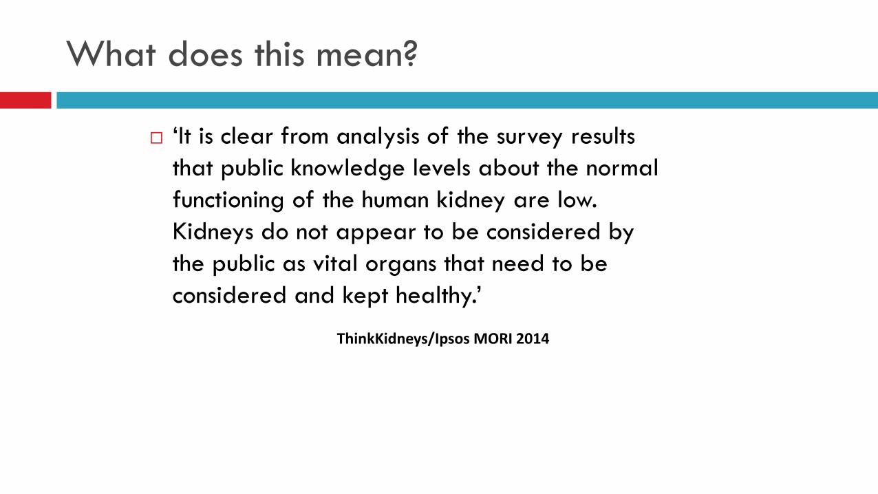
What does this mean?
‘It is clear from analysis of the survey results
that public knowledge levels about the normal
functioning of the human kidney are low.
Kidneys do not appear to be considered by
the public as vital organs that need to be
considered and kept healthy.’
ThinkKidneys/Ipsos MORI 2014

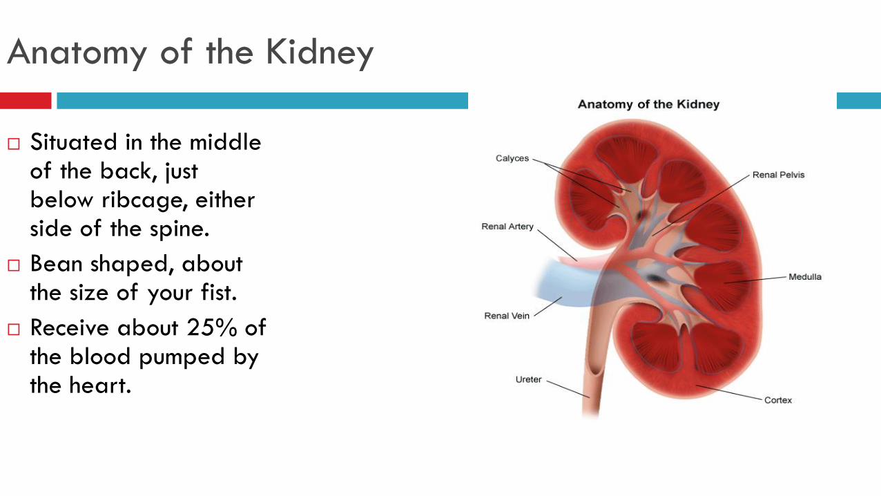
Anatomy of the Kidney
Situated in the middle of the back, just below ribcage, either side of the spine.
Bean shaped, about the size of your fist.
Receive about 25% of the blood pumped by the heart.

Nephron
Nephron is the basic unit of the kidney.
Approx. one million in each kidney.
Composed of a glomerulus, proximal tubule, loop of Henle, distal tubule and the collecting duct.
Filtration from glomerulus into renal tubules.
Reabsorption and secretion occurs within tubules.
Produces urine.
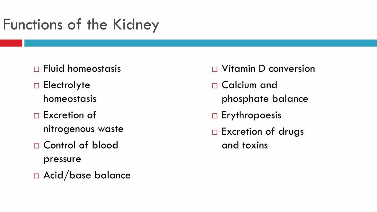
Functions of the Kidney
Fluid homeostasis
Electrolyte
homeostasis
Excretion of
nitrogenous waste
Control of blood
pressure
Acid/base balance
Vitamin D conversion
Calcium and
phosphate balance
Erythropoesis
Excretion of drugs
and toxins

What are AKI and CKD?
Chronic Kidney Disease (CKD) is the gradual decline of renal function over months or years
CKD defined using eGFR
eGFR should not be used as a means to detect AKI

Classification of CKD
Stages of Kidney disease
Stage Description GFR mL/min/1.73m2
1 Kidney damage with normal or ↑GFR ≥90
2 Kidney damage with mild ↓GFR 60-89
3A Moderate ↓GFR 45-59
3B Moderate ↓GFR 30-44
4 Severe ↓GFR 15-29
5 Kidney failure <15 or dialysis

AKI
Acute Kidney Injury (AKI) is now the universal term used to describe sudden deterioration of renal function, and it replaces the previous term know as Acute Renal Failure (ARF).
AKI defined using serum creatinine and urine output as indicators
Until 2004 there was no universal definition for AKI this meant early recognition of AKI was often missed.
Up to 30% of AKI cases are preventable

Facts and Figures
An average of 32 people a day die of AKI
Estimated to affect 20% of all emergency
admissions
AKI prolongs hospital stay by 2.5 times and costs
the NHS £434 million per year
AKI is an indicator of basic and safe care
200 times the number of people dying of MRSA,
die of AKI. (renaltsar.blogspot.co.uk)
Evidence suggests we are poor at picking up AKI
on admission.

Risk Factors in Acutely Ill patients
chronic kidney disease (adults with an estimated glomerular filtration rate [eGFR] less than 60 ml/min/1.73 m2 are at particular risk)
heart failure
liver disease
diabetes
history of acute kidney injury
oliguria (urine output less than 0.5 ml/kg/hour)
neurological or cognitive impairment or disability, which may mean limited access to fluids because of reliance on a carer
hypovolaemia
use of drugs with nephrotoxic potential (such as non-steroidal anti-inflammatory drugs
[NSAIDs], aminoglycosides, angiotensin-converting enzyme [ACE] inhibitors, angiotensin II receptor
antagonists [ARBs] and diuretics) within the past week, especially if hypovolaemic
use of iodinated contrast agents within the past week
symptoms or history of urological obstruction, or conditions that may lead to obstruction
sepsis
deteriorating early warning scores
Age 65 years or over
(NICE Guideline 169, 2013)

Surgery and AKI
Assess the risk of acute kidney injury in adults before surgery. Be aware that increased risk is associated with:
emergency surgery, especially when the patient has sepsis or hypovolaemia
intraperitoneal surgery
chronic kidney disease (adults with an eGFR less than 60 ml/min/1.73 m2 are at particular risk)
diabetes
heart failure
age 65 years or over
liver disease
use of drugs with nephrotoxic potential in the perioperative period (in particular, NSAIDs after surgery).
Use the risk assessment to inform a clinical management plan.
(NICE Guideline 169, 2013)

AKI and Contrast Agents
Before offering iodinated contrast agents to adults for emergency or non-emergency imaging, assess their risk of acute kidney injury. Be aware that increased risk is associated with:
chronic kidney disease (adults with an eGFR less than 40 ml/min/1.73 m2 are at particular risk)
diabetes but only with chronic kidney disease (adults with an eGFR less than 40 ml/min/1.73 m2 are at particular risk)
heart failure
renal transplant
age 75 years or over
hypovolaemia
increasing volume of contrast agent
intra-arterial administration of contrast agent.
Ensure that risk assessment does not delay emergency imaging.
(NICE Guideline 169, 2013)
CKD – Major Risk Factor
Pre-existing CKD has been identified as the
most consistent factor contributing to the
development of AKI in people recovering from
surgery, but other factors shown to be
important include age, diabetes and reduced
cardiac function.
The more risk factors a person has the greater
the risk of developing AKI.

PRIMARY CARE AT-RISK PATIENTS (THINK KIDNEYS
2015)
Diabetics – HHNS, particularly in Type 2 patients. Medications
CKD on anti-hypertensives. If acutely ill contact GP re. holding medications.
Dementia – Poor fluid intake. Medications
Heart failure – Medications, hypovolaemia
Elderly – most susceptible to AKI. Early detection and management vital.
Psychiatric patients – self neglect or unable to care for oneself, laxative or diuretic abuse, medications prescribed and recreational. Self harm.
Patients with Cancer
AKI risk can vary depending on type of cancer, treatment and other
risk factors.
Kidney cancer, myeloma, liver cancer, acute lymphoma or leukaemia
undergoing chemotherapy are at high risk of AKI.
AKI complication of bone marrow transplants
Medication-induced AKI
Older patients with traditional risk factors at a higher risk of AKI
Volume depletion – nausea, vomiting, diarrhoea, mucositis,
Hypercalcaemia induced diabetes insipidus, malignant ascites,
pleural effusionor neutropenic fever
Sepsis
Obstruction from prostate, bladder, kidney cancer or metastatic
abdominal or pelvic malignancies.

Assessing the Risk
Remain vigilant in identifying those most at risk.
Identify potential nephrotoxic interventions and
treatments
Identify renal insults – modifiable risk factors
Fluid status
Nephrotoxic medications
Iodinated contrast media
Sepsis
Risk factor/s + Insult = At Risk Patient

Identification
Reduced urine output:
< 0.5mls/kg/hr for 6 hours (half body weight)
Blood creatinine rise from baseline:
26mmols rise within 48 hours
> 50% rise from baseline: lowest value within 7 days, median value within 365 days

The Acute Kidney Injury Network Diagnostic Criteria for AKI
AKIN stage Serum Creatinine Criteria (SCr) Urine output criteria
1 Increase in SCr ≥ 26.4 µmol/L
Or
Increase in SCr ≥150 - 200%
(1.5-2 fold) from baseline
< 0.5 ml/kg/hr for > 6 hr
2 Increase in SCr > 200 – 300%
(>2-3 fold) from baseline
< 0.5 ml/kg/hr for > 12 hr
3 Increase in SCr > 300% (>3 fold) from baseline
Or
SCr ≥ 354 µmol/L with an acute rise of ≥ 44 µmol/L in ≤
24 hr
Or
Initiated on Renal Replacement Therapy (irrespective of
stage at time of initiation)
< 0.3 ml/kg/hr for 24 hr
Or
Anuria for 12 hr

Assessing urine output in hospital If urine output is less than the minimum required output of 0.5mls/kg/hr (oliguria) as per the
identifying AKI criteria, medical staff need to be informed
None Catheterised Catheterised
• Always consider the urine output even if
the patient is not catheterised.
• Explain to the patient the importance of
monitoring urine output. Provide container
to measure
• Record amount of incontinence; damp or
saturated, weigh the pad
• Bladder scan as a none invasive
intervention. Record findings
• Consider catheterising if patient shows
signs of deterioration
• Report reduced urine
output (oliguria) early so
that appropriate
management/treatments
can be implemented
Assessing urine output in the community
Question the patient and or relatives:
Have you passed urine today?
How often?
Did it seem a normal amount for you?
What colour was it?
Have you been drinking ok?
Other sources to gain information from:
Does the patient have regular carers (relatives and or professionals)?
Do external carers have notes for assessments and communication? Do you read them?
Do you communicate and advise carers or could you with regards to hydration and urine output?


Preventing AKI
Remain vigilant in identifying those most at risk.
Monitor – EWS, fluid input/output, daily weight, serum creatinine, U & E’s.
What systems are in place to respond to oliguria? (Urine ouput less than 0.5 mls/kg/hr)
Iodinated contrast agents – follow local protocols
Check local AKI protocols and guidelines.
Nursing care guideline
Care Bundle Checklist

Causes of AKI
Many causes of AKI
Identifying the cause can be divided into categories where the physiological insult occurs
pre renal
intra renal (intrinsic)
post renal
Acute Kidney Injury | Doctor | Patient UK

Pre renal AKI
Most common form of AKI, accounting for 50-65% of reported incidences of AKI.
For the kidneys to function normally the kidneys require 25% of the cardiac output and a mean arterial pressure between 65 to 110mmhg to maintain adequate renal blood flow.
The loss or reduction of blood supply to kidneys by circulatory volume depletion, inadequate cardiac function or obstruction of the arterial supply can impair the renal perfusion which then can lead to ischemia.
If left untreated / undiagnosed the damage can lead to intrinsic injury, as Acute Tubular Necrosis (ATN) will occur.

Intra Renal AKI
Second most common cause of AKI with 20–35% of reported cases.
The damage occurs within the structure of the kidney (renal parenchyma).
ATN is reported as being the most common cause of Intra renal AKI.
Medications can exacerbate hypovolaemia & hypotension, meds causing direct damage, toxins, systemic diseases, diseases of the kidney.
Even if the cause is found there are no guarantees that full recovery of function will return.

Post Renal AKI
Make up the last 15% of AKI incidences
Involves obstruction of the urinary flow causing backpressure, which inhibits the filtration.
AKI only occurs if the both kidneys are affected or a
person has only one functioning kidney or a solitary kidney.
Obstruction can occur from both within and outside the urinary system
Enlarged prostate, pelvic, abdominal masses, kidney stones

Common causes
Systemic infection e.g. UTI, Chest infection, sepsis
Dehydration.
Drugs e.g. Diuretics, ACE Inhibitors, Nephrotoxic medication, contrast media
Urinary tract obstruction.
Progression of underlying disease e.g. lupus (SLE), cardiac failure
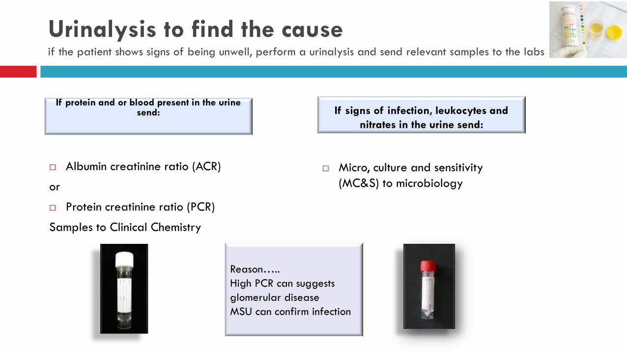
Urinalysis to find the cause if the patient shows signs of being unwell, perform a urinalysis and send relevant samples to the labs
If protein and or blood present in the urine send:
Albumin creatinine ratio (ACR)
or
Protein creatinine ratio (PCR)
Samples to Clinical Chemistry
If signs of infection, leukocytes and
nitrates in the urine send:
Micro, culture and sensitivity
(MC&S) to microbiology
Reason…..
High PCR can suggests
glomerular disease
MSU can confirm infection

Managing AKI
Check local policies
Fluid status
Hyperkalaemia
Maintain strict input/output charts. Catheterise if appropriate.
Urinalysis
Check for nephrotoxic medications
Daily U & E’s
Discuss any planned contrast imaging with senior medical staff.
Refer according to AKIN/RIFLE and/or local policy to nephrology service

Questions to ask, points to consider when assessing
urine output
When did you last pass urine? What colour was it? Did it seem a normal amount to you? Have you been drinking OK? If concerned measure the urine, weigh pads etc. if the patient has been incontinent. Is it enough for them? Consider bladder scan or catheter if not passed urine or patient becomes unwell. Do we need an accurate urine output to provide a full assessment of the patient? If patient has reduced or no urine output, why? Consider: Are they hydrated? Have they got a blockage?

Fluid Charts: Key parts of the chart that should always be completed
Patient Name
Patient Number
Ward Location
Date
Weight
Minimum Urine Output
Record input and total with each entry
Record output and total with each entry
Does the urine output meet the patients minimum urine output (0.5ml/kg/hr = half body weight in mls)
Overall output recorded for 24hours ‘+’ or ‘-’ (always take away the output from the input to get the
positive or negative calculation)
See the example shown below
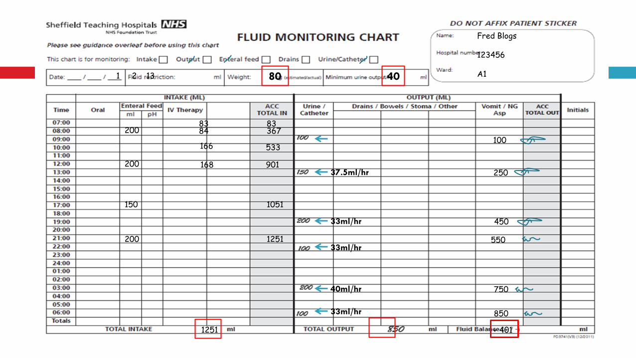
80 40
37.5ml/hr
33ml/hr
33ml/hr
40ml/hr
33ml/hr
200
200
150
200
83 84
166
168
83 367
533
901
1051
1251
1251 + 401
100
250
450
550
750
850
1 2 13
Fred Blogs 123456 A1

Creatinine levels
The AKI Lab Alert is generated from a national algorithm and is reported on the ICE system. If you see this
alert get the patient reviewed by medical staff! By clicking on the alert hyperlinks will let you view and print
the AKI Bundle

Management Plan for patients showing signs of deterioration,
at risk of or with AKI:
Screen for Sepsis and hypo perfusion
Toxins avoid/stop;
Review medication
Iodinated Radiological Contrast
Optimise B/P –assess volume status;
EWS monitoring – follow the EWS algorithm
Urine output monitoring (0.5ml/kg/hr = half body weight), consider catheter
IV fluids
Consider/hold antihypertensive
Prevent Harm
Identify cause of AKI – (pre, intrinsic, post renal), urinalysis
Treat complications – (Hyperkalaemia, acidosis, overload)
Daily U&Es/Renal Profile, additional checks following surgery or invasive procedures
Refer to the AKI nursing care guidance no. 20 for
patients at risk or with AKI
If your patient has an AKI diagnosed the AKI Care
Bundle Checklist must be completed by:
Medical staff and placed in the patients evaluation
notes.
Nurses can initiate the chart and complete the
parts that they have done – urinalysis, started strict
I & O.
A pharmacist can do a drug review while you wait
for the doctor to arrive
Care Bundle Example

Managing AKI in the Community
Recognising and responding to AKI for adults in Primary Care
Quick reference guide for community staff
Sick day guidance

Information
Advise on treatment options in both short and long term, particularly if patient will require long term dialysis
Discuss the risk of developing acute kidney injury, particularly the risk associated with conditions leading to dehydration (for example, diarrhoea and vomiting) and drugs with nephrotoxic potential (including over-the-counter NSAIDs), with people who are at risk of acute kidney injury, particularly those who have:
chronic kidney disease with an eGFR less than 60 ml/min/1.73 m2
neurological or cognitive impairment or disability, which may mean limited access to fluids because of reliance on a carer.
o Involve parents and carers in the discussion if appropriate.

Support and Developments
Support
Think Kidneys
NICE e-learning
London AKI Network
AKI competency framework
Local Developments
Local policies and guidelines
E-alerts from laboratories
Care bundles
Bedside teaching

Drug-induced AKI
Well recognised but frequency unknown Reversible
totally or partially
Be aware of common causative agents NSAIDs ACE inhibitors Aminoglycosides Contrast media
Can be dose dependant or idiosyncratic
toxicity

10%
80%
10%
Aetiology of drug induced AKI
Diuretics
ACE inhibitors
ARBs
NSAIDs
Ciclosporin
Tacrolimus

10%
80%
10%
Aetiology of drug induced AKI
Radio-contrast dye
Aminoglycosides
Penicillins
Rifampicin
Thiazides
Methotrexate
Lithium
Tetracyclines
Statins
Allopurinol
Cephalosporins

10%
80%
10%
Aetiology of drug induced AKI
Aciclovir
Sulfonamides

Drug-induced AKI: NSAIDs
Approximately, each year, 5% of people taking NSAIDs will develop AKI
Vasoconstriction of blood vessels
supplying kidney
Decreased renal blood flow
Decreased glomerular
filtration rate
Renal ischaemia
Avoid if at high risk of AKI
Low dose aspirin can continue for primary or secondary prevention of CVD

Drug-induced AKI: ACEI & ARB
Frequent cause of AKI
Vasodilation of renal blood vessels
Decreased glomerular filtration
Increase in serum creatinine of up to 30% is expected within first week of treatment
If more than 30% stop treatment

Drug-induced AKI: Statins
Associated with rhabdomyolysis
Rhabdomyolysis: muscle breakdown
Myoglobin, creatinine kinase, urate released into circulation
Can lead to AKI
direct toxicity of myoglobin
intravascular volume depletion

Drug-induced AKI: Gentamicin
Drug 100% excreted by kidney
Toxic to cells of proximal tubule
Risk factors
Dose/duration
CKD/AKI
Other nephrotoxic drugs
Age

Treatment of AKI
Treatment aims
Prevent further injury
Recovery of renal function to baseline
No specific treatment
Treat reversible causes where possible (e.g. sepsis, dehydration, stop nephrotoxics)
Monitor (U&Es, fluid balance, urine output)
Review medication

Impact of AKI on medicines
Absorption
Distribution Metabolism
Excretion

Dosing adjustments usually not necessary
Timing important: e.g. Phosphate binders and quinolone
antibiotics (e.g ciprofloxacin)
Absorption
Impact of AKI on medicines
Altered fluid balance and volume of distribution may alter drug kinetics
Distribution

Hepatic pathways generally unaffected
Kidneys- 99% is just secretion/excretion
Kidneys are site of metabolism for some drugs:
Metabolism
1. Vitamin D metabolism to active form Activated vitamin D (1-alfacalcidol) given to renal
patients
2. Insulin Reduced insulin requirements in AKI
Impact of AKI on medicines

Majority of drugs and their metabolites are excreted by the kidneys via urine
Elimination of the drug will be reduced in AKI
The amount of reduction will depend upon the severity of the renal impairment
Drugs can accumulate Prolonged effect
Risk of toxicity (non-renal)
Excessive side effects
Elimination
Impact of AKI on medicines

Drug choice in AKI – questions to consider
1. Is the drug nephrotoxic? Be aware of the commonly prescribed agents
Avoid and consider an alternative
2. Is the drug excreted unchanged by the kidney?
3. Can the medicine cause non-renal toxicity? Renally excreted drugs may accumulate
metformin lactic acidosis
methotrexate toxicity (neutropenia)

4. Does the drug have any active metabolites that are renally excreted?
Codeine is metabolised to morphine which can accumulate leading to opiate toxicity
Drug choice in AKI – questions to consider
If yes to any of the above.... Is a dosage adjustment needed?
Should the dosing interval be changed? Should an alternative be prescribed?

Useful Resources
BNF – not very helpful in specialist setting
Summary of Product Characteristics licensed dosage information
www.medicines.org.uk/emc
Renal drug handbook dosage information for different stages of kidney
disease
not always licensed
Careful – book may be out of date, can be accessed online
Dosage adjustments based on Creatinine Clearance