acute intracranial hypertension during pregnancy: special
TRANSCRIPT

Neurocrit Care (2022) 36:302–316https://doi.org/10.1007/s12028-021-01333-x
REVIEW ARTICLE
Acute Intracranial Hypertension During Pregnancy: Special Considerations and Management AdjustmentsDaniel Agustin Godoy1,2*, Chiara Robba3, Wellingson Silva Paiva4 and Alejandro A. Rabinstein5
© 2021 Springer Science+Business Media, LLC, part of Springer Nature and Neurocritical Care Society
Abstract
Pregnancy is associated with a number of pathophysiological changes (including modification of vascular resistance, increased vascular permeability, and coagulative disorders) that can lead to specific (eclampsia, preeclampsia) or not specific (intracranial hemorrhage) neurological complications. In addition to these disorders, pregnancy can affect numerous preexisting neurologic conditions, including epilepsy, brain tumors, and intracerebral bleeding from cer-ebral aneurysm or arteriovenous malformations. Intracranial complications related to pregnancy can expose patients to a high risk of intracranial hypertension (IHT). Unfortunately, at present, the therapeutic measures that are generally adopted for the control of elevated intracranial pressure (ICP) in the general population have not been examined in pregnant patients, and their efficacy and safety for the mother and the fetus is still unknown. In addition, no specific guidelines for the application of the staircase approach, including escalating treatments with increasing intensity of level, for the management of IHT exist for this population. Although some of basic measures can be considered safe even in pregnant patients (management of stable hemodynamic and respiratory function, optimization of systemic physiology), some other interventions, such as hyperventilation, osmotic therapy, hypothermia, barbiturates, and decompressive craniectomy, can lead to specific concerns for the safety of both mother and fetus. The aim of this review is to summarize the neurological pathophysiological changes occurring during pregnancy and explore the effects of the possible therapeutic interventions applied to the general population for the management of IHT during pregnancy, taking into consideration ethical and clinical concerns as well as the decision for the timing of treatment and delivery.
Keywords: Intracranial pressure, Cerebral perfusion pressure, Pregnancy, Intracranial hypertension, Cerebral autoregulation, Intracranial pressure monitoring
IntroductionIntracranial hypertension (IHT) may occur as a con-sequence of intracranial pathologies—such as cerebral edema, trauma, and intracranial hemorrhage with con-sequent loss of the usual balance between the different intracranial components—as well as extracranial causes, such as abdominal or thoracic hypertension [1–3]. IHT etiology is multifactorial, and its consequences are fatal
if not promptly recognized and managed [1, 2]. Because IHT represents a medical–surgical emergency, its proper management must be solidly supported by a thorough evaluation of the factors contributing to its pathogenesis [1, 2].
It is well known that pregnancy can be complicated by a number of neurocritical care pathologies that expose pregnant patients to a high risk of neurological complica-tions and IHT [4, 5]. Unfortunately, to date, none of the therapeutic measures adopted for the control of elevated intracranial pressure (ICP) have been tested in terms of efficacy and safety for the mother and the fetus during
*Correspondence: [email protected] 1 Neurointensive Care Unit, Sanatorio Pasteur, Catamarca, ArgentinaFull list of author information is available at the end of the article
303
pregnancy. Furthermore, no specific guidelines for the application of the algorithmic approach of escalating levels of management for IHT exist for this population. Only a comprehensive best practices statement made a number of expert-consensus-based recommendations in pregnant patients with stroke [6].
These gaps are particularly problematic because some of the treatments for IHT (such as hyperventilation, hypothermia, barbiturates) may be hazardous during pregnancy.
The aim of this review is to analyze the unique aspects of IHT during pregnancy and to discuss the relative advantages and disadvantages of various therapeutic options in this situation.
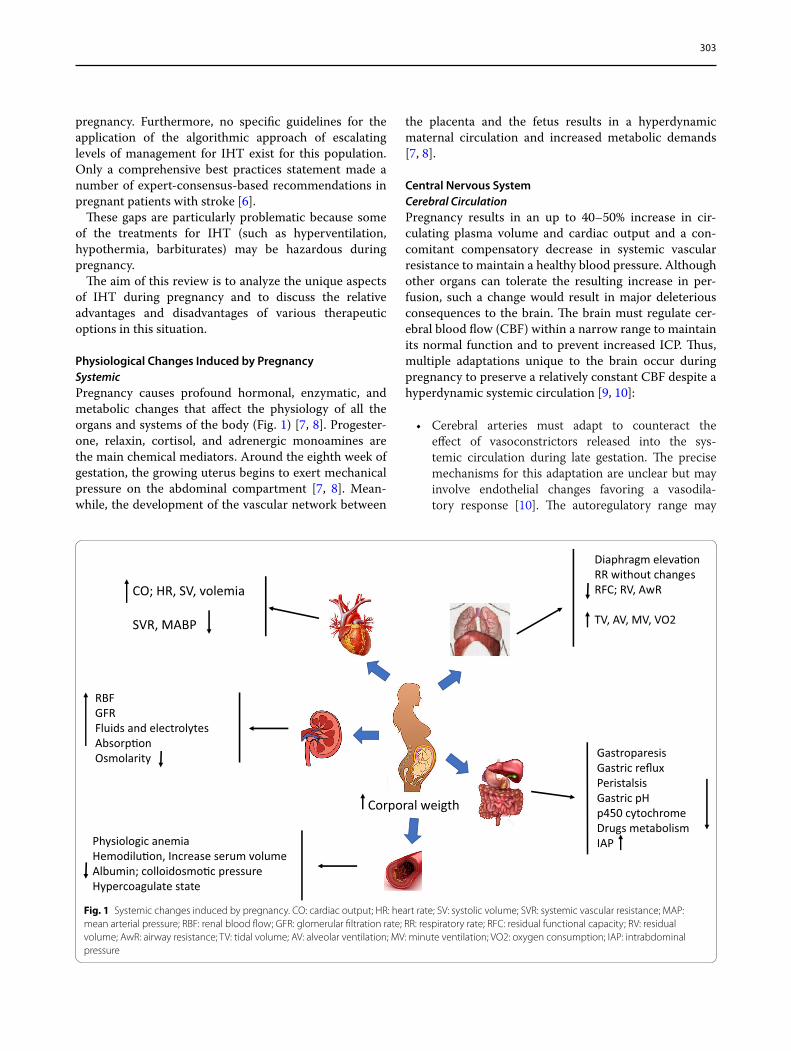
Physiological Changes Induced by PregnancySystemicPregnancy causes profound hormonal, enzymatic, and metabolic changes that affect the physiology of all the organs and systems of the body (Fig. 1) [7, 8]. Progester-one, relaxin, cortisol, and adrenergic monoamines are the main chemical mediators. Around the eighth week of gestation, the growing uterus begins to exert mechanical pressure on the abdominal compartment [7, 8]. Mean-while, the development of the vascular network between
the placenta and the fetus results in a hyperdynamic maternal circulation and increased metabolic demands [7, 8].
Central Nervous SystemCerebral CirculationPregnancy results in an up to 40–50% increase in cir-culating plasma volume and cardiac output and a con-comitant compensatory decrease in systemic vascular resistance to maintain a healthy blood pressure. Although other organs can tolerate the resulting increase in per-fusion, such a change would result in major deleterious consequences to the brain. The brain must regulate cer-ebral blood flow (CBF) within a narrow range to maintain its normal function and to prevent increased ICP. Thus, multiple adaptations unique to the brain occur during pregnancy to preserve a relatively constant CBF despite a hyperdynamic systemic circulation [9, 10]:
• Cerebral arteries must adapt to counteract the effect of vasoconstrictors released into the sys-temic circulation during late gestation. The precise mechanisms for this adaptation are unclear but may involve endothelial changes favoring a vasodila-tory response [10]. The autoregulatory range may
CO; HR, SV, volemia
SVR, MABP
Diaphragm eleva�onRR without changesRFC; RV, AwR
TV, AV, MV, VO2
RBFGFRFluids and electrolytesAbsorp�onOsmolarity Gastroparesis
Gastric refluxPeristalsisGastric pHp450 cytochromeDrugs metabolismIAP Physiologic anemia
Hemodilu�on, Increase serum volumeAlbumin; colloidosmo�c pressureHypercoagulate state
Corporal weigth
Fig. 1 Systemic changes induced by pregnancy. CO: cardiac output; HR: heart rate; SV: systolic volume; SVR: systemic vascular resistance; MAP: mean arterial pressure; RBF: renal blood flow; GFR: glomerular filtration rate; RR: respiratory rate; RFC: residual functional capacity; RV: residual volume; AwR: airway resistance; TV: tidal volume; AV: alveolar ventilation; MV: minute ventilation; VO2: oxygen consumption; IAP: intrabdominal pressure

304
widen during pregnancy, with a decreased lower limit and increased upper limit of autoregulation, thereby enhancing cerebral tolerance to hypoten-sion or hypertension (Fig. 2) [11, 12]. Failure of these autoregulatory adjustments may play a role in the development of preeclampsia [13].
• Structural changes have been reported in cerebral arterioles, but not in pial vessels, in pregnant rats. Those changes consist of hypotrophic remodeling, with luminal enlargement and thinning of the arte-riolar wall, and by decreasing distal vascular resist-ance, they can help accommodate increased flow to sustain the greater metabolic demands of some brain structures [10, 14]. However, it has been conjectured that this remodeling of small vessels can render them more susceptible to injury from acute hypertension [10].
• Impact of preeclampsia/eclampsia on cerebral circu-lation: During pregnancy, cerebral circulation under-goes structural and functional adaptations aimed at maintaining intracranial homeostasis [10–14]. These modifications tend to maintain the permeability of the blood–brain barrier (BBB) and counteract the dominant vasoconstrictor state, which especially occurs in the last trimester of gestation [10–14]. Cer-ebral autoregulation becomes adjusted to the circu-latory changes and dominant vasoconstriction [10–14]. Posterior reversible encephalopathy syndrome related to preeclampsia/eclampsia is characterized by endothelial dysfunction, which compromises cerebral autoregulation [15]. As a consequence, increases in mean arterial pressure (MAP) translate into increased capillary hydrostatic pressure and BBB per-meability, resulting in vasogenic edema, microbleeds, and inflammation [15]. The mechanisms involved are variable and complex; standing out among them are
hyperperfusion, alterations of the factors that oppose the vasoconstrictor state, increases in the electrical conductivity of the BBB, and decreases or blocking of the mediators (sFlt1) that keep the permeability of the BBB intact by counteracting the effects of the vas-cular endothelial growth factor [15].
BBBBBB permeability remains mostly unchanged during nor-mal pregnancy despite increased circulating levels of fac-tors that augment vascular permeability (such as vascular endothelial growth factor and placental growth factor) [16]. Disruption of the BBB resulting in increased per-meability has been observed in animal models of preec-lampsia [15, 17], but whether this mechanism contributes to the pathogenesis of the disease in humans remains a matter of debate [18]. Overexpression of aquaporin 4 is known to occur during the second half of gestation and could affect the formation and resolution of brain edema [19].
Seizure ThresholdPregnancy induces a state of neuronal hyperexcitability, thereby increasing susceptibility to seizures [9, 10]. This phenomenon is more pronounced during the latter part of gestation. Its mechanisms are not well elucidated, but downregulation of γ-aminobutyric acid (GABA) A recep-tors and upregulation of aquaporin 4 may be important contributors [9, 10].
Subarachnoid Space and Cerebrospinal FluidFrom week 16 of gestation, the gravid uterus and the intraabdominal pressure increase and accentuate aor-tocaval compression [7, 8]. The resulting congestion of epidural venous plexuses from the thoracic to the sacral levels leads to compression of the subarachnoid space and reduction in thecal cerebrospinal fluid (CSF) volume [20]. These changes may also affect the volume and dis-tribution of intracranial CSF. Additionally, CSF becomes more alkaline during pregnancy, with lower CO2 pressure and protein content [7, 8].
The changes noted in the volume of the CSF of the spinal space increase the sensitivity of anesthetics deliv-ered regionally (epidural); at the same time, they may decrease the intracranial compliance, thereby potentially increasing the risk of IHT [21–23]. There are no reports suggesting any relationship between changes in CSF vol-ume and development of hydrocephalus. The pH of CSF is alkalinized during the second and third trimesters of pregnancy; together with a decrease in CO2 levels, both adaptive phenomena follow the systemic physiological changes [21–23]. These modifications can sometimes Fig. 2 Changes in cerebrovascular autoregulation during pregnancy

305
trigger neurovegetative symptoms. Additionally, these changes may decrease the seizure threshold because it is associated with a decrease in GABA levels. CSF protein levels also decrease in the aforementioned gestation peri-ods, thereby increasing the concentrations of anesthetic drugs, especially in their nonionized form [21–23].
Pharmacokinetic ChangesPregnancy results in increased volumes of distribution (because of larger body mass and expansion of plasma volume) and augmented renal clearance (because of increased renal blood flow and glomerular filtration rate) [24]. Concomitant increases in volume of distribu-tion and clearance can affect the half-lives of medications in opposing directions. As a general rule, a larger vol-ume of distribution will diminish the effect to first drug boluses, whereas augmented clearance will lower serum concentrations of the drug during a steady state. Plasma protein concentrations decrease after the second trimes-ter, and drug-binding capacity is consequently reduced by 20–30% by the end of gestation [24]. Meanwhile, the effect of pregnancy across hepatic enzymes is variable [24]. Pharmacokinetics studies during pregnancy have revealed consistent results for various classes of drugs, and they can be used to guide drug administration [25].
These pharmacokinetic changes may result in clinically significant consequences. Serum concentration of highly protein-bound medications (such as midazolam, pheny-toin, valproic acid) should be expected to be higher late
in pregnancy. Instead, serum concentrations of renally cleared drugs (such as heparin, opiates, levetiracetam, aminoglycosides) are likely to be lower than expected [8, 24, 25]. Drugs predominantly metabolized by the liver (such as barbiturates) can be variably affected and there-fore require careful monitoring of trough concentrations [8, 24, 25].
Pharmacokinetic considerations must also include the fetus [8, 24, 25]. Drugs that can cross the placental bar-rier can reach higher concentrations in the fetal circula-tion because of the lower metabolic capacity and volume of distribution of the fetus. This can be particularly dan-gerous when administering sedatives and other respira-tory depressants. Thus, drug administration to critically ill pregnant patients should optimally be coordinated by a multidisciplinary team that includes pharmacists and obstetricians.
Causes of IHT During PregnancyThe prevalence and causes of IHT during pregnancy have not been evaluated in detail. It can be caused by decom-pensation of a preexisting pathology (e.g., tumor) or by acute disease (e.g., trauma or vascular events) [7, 8, 26]. A summary of the different etiologies of IHT and their mechanisms are presented in Table 1.
The exact incidence of IHT during pregnancy is unknown. The median age of pregnant women with neu-rological/neurosurgical complications ranges between 20 and 30 years [4, 5]. Conditions causing IHT during
Table 1 Causes and mechanisms of production of intracranial hypertension during pregnancy
Etiology of intracranial hypertension Mechanism of production
Tumors Mass lesionVasogenic edema
Traumatic Brain Injury with or without polytrauma Mass lesionsVasogenic, cytotoxic edemaExtracranial causes (intratho-
racic or intrabdominal hypertension)
Secondary insults (hyper-capnia)
Idiopathic Intracranial Hypertension Unclear
PreeclampsiaPosterior Reversible Encephalopathy Syndrome
Vasogenic edema
Cerebrovascular DiseasesSpontaneous Intracerebral HemorrhageSecondary Intracerebral HemorrhageArteriovenous Malformations RuptureSubarachnoid HemorrhageCerebral venous Thrombosis
Mass effect, Vasogenic edemaCytotoxic edema, Hydro-
cephalus, CSF circulation alteration
CNS infectionsEncephalitis/MeningitisBrain Abscess
Vasogenic, cytotoxic edemaMass lesion, Vasogenic edema
Acute fatty liver Vasogenic, cytotoxic edema
Shunt Malfunction Hydrocephalus
306
pregnancy are more prevalent in the second and third tri-mesters [4, 5]. The most common causes of IHT in preg-nancy are preeclampsia (2–8%), head trauma (0.5–6%), ischemic or hemorrhagic stroke (10–34 cases per 100,000 pregnancies), and tumor complications (3.2–3.6 cases per million live births) and are favored by increased vascula-ture associated with high levels of estrogen and proges-terone [4, 5]. Stroke is more frequent in the intrapartum (40%) and postpartum (50%) periods [4, 5]. Other risk factors for the development of IHT are arterial hyperten-sion, coagulopathy, and cardiomyopathy [4, 5].
Definition of IHT During PregnancyTraditionally, IHT is defined by sustained ICP greater than 20 mm Hg [1, 2]. The latest version of the traumatic brain injury guidelines modified the threshold to 22 mm Hg [27].
Ideally, IHT should be evaluated under consideration of the context in which it develops [28, 29]. However, there is insufficient information on the physiological range of ICP during pregnancy or on what should be the optimal definition of IHT in this setting.
IHT Evaluation During PregnancyThe clinical presentation of IHT, such as headache and vomiting, is often nonspecific and unreliable, whereas the classic Cushing’s triad (arterial hypertension, irregular breathing, bradycardia) is a late expression of IHT when brain herniation is already occurring [1]. No differences in terms of clinical presentation have been observed between the pregnant and not pregnant population.
Papilledema is most prominent when IHT develops slowly, and its absence does not exclude the presence of IHT. Pupillary asymmetry is not a reliable marker of IHT; in fact, only 33% of individuals with pupillary asymme-try greater than 3 mm have ipsilateral focal lesions [30]. The presence of a clinical picture compatible with cere-bral herniation (abnormal motor posturing, depression of consciousness, nonreactive mydriasis) demands immedi-ate and aggressive therapy even before neuroimaging and ICP monitoring.
Computed tomography (CT) with adequate mater-nal protection is the neuroimaging modality of choice in pregnant patients with suspected acute brain injury. Although the fetus is more susceptible to the risks from radiation exposure, the radiation dose of a head CT scan is less than 0.5 rads [31]. Fetal abnormalities, growth retardation, and loss of pregnancy have not been dem-onstrated with exposures less than 5 rads [31]. The exact dose of fetal radiation exposure from noncontrast head CT is between 0.001 and 0.01 mGy (1 mGy = 0.1 rad), which is well below the dose considered safe (5 mGy) [31, 32]. In addition, the use of intravenous contrast is not
contraindicated in the case of life-threatening maternal illness [31, 32]. Noncontrast MRI is a safe option dur-ing pregnancy because of the absence of maternal–fetal exposure to ionizing radiation [31, 32]. MRI (1.5 or 3.0 T) does not increase the risk of adverse fetal outcomes at any trimester of the pregnancy [31, 32]. Gadolinium is not recommended because it may increase risk of fetal adverse effects, especially during the first trimester [31, 32].
Scans should be reviewed systematically for the pres-ence of space-occupying lesions associated with paren-chyma distortion or displacement, hydrocephalus, midline shift, effacement of basal cisterns, and sulci over the cerebral convexities [33].
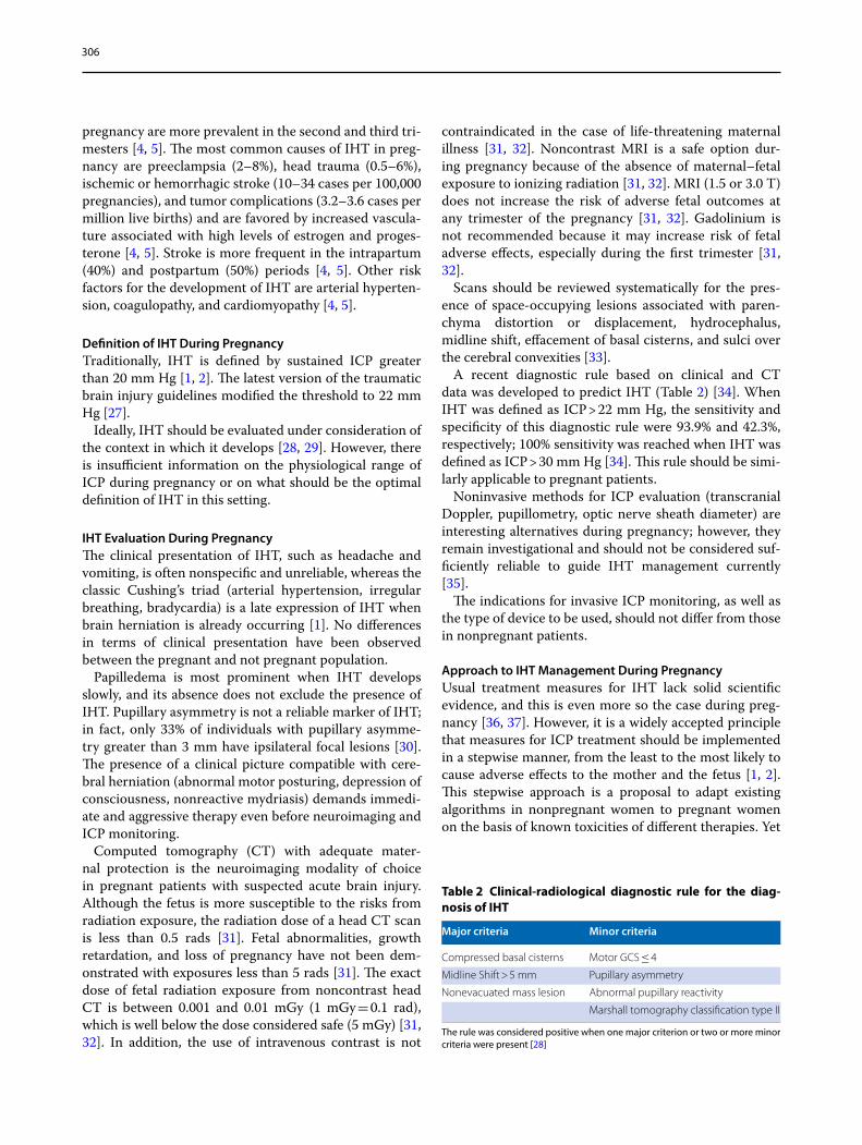
A recent diagnostic rule based on clinical and CT data was developed to predict IHT (Table 2) [34]. When IHT was defined as ICP > 22 mm Hg, the sensitivity and specificity of this diagnostic rule were 93.9% and 42.3%, respectively; 100% sensitivity was reached when IHT was defined as ICP > 30 mm Hg [34]. This rule should be simi-larly applicable to pregnant patients.
Noninvasive methods for ICP evaluation (transcranial Doppler, pupillometry, optic nerve sheath diameter) are interesting alternatives during pregnancy; however, they remain investigational and should not be considered suf-ficiently reliable to guide IHT management currently [35].
The indications for invasive ICP monitoring, as well as the type of device to be used, should not differ from those in nonpregnant patients.
Approach to IHT Management During PregnancyUsual treatment measures for IHT lack solid scientific evidence, and this is even more so the case during preg-nancy [36, 37]. However, it is a widely accepted principle that measures for ICP treatment should be implemented in a stepwise manner, from the least to the most likely to cause adverse effects to the mother and the fetus [1, 2]. This stepwise approach is a proposal to adapt existing algorithms in nonpregnant women to pregnant women on the basis of known toxicities of different therapies. Yet
Table 2 Clinical-radiological diagnostic rule for the diag-nosis of IHT
The rule was considered positive when one major criterion or two or more minor criteria were present [28]
Major criteria Minor criteria
Compressed basal cisterns Motor GCS ≤ 4
Midline Shift > 5 mm Pupillary asymmetry
Nonevacuated mass lesion Abnormal pupillary reactivity
Marshall tomography classification type II

307
it is important to keep in mind that this approach has not been specifically studied for efficacy or fetal safety.
The measures must be “additive,” meaning that when we decide to implement one, we should not abandon the previous one. It is also important to take some time (often approximately 30 min) to evaluate the effective-ness of the therapy before proceeding to the next step [1, 2, 38].
At each step, clinicians should consider the possibil-ity of repeating neuroimaging, especially when there is an abrupt change in the clinical picture or in ICP levels without a clear explanation to justify it [2, 38] (Fig. 3).
First‑line TherapiesGeneral MeasurementsThe first steps should be directed to determine the cause of the IHT, preventing early complications and maintain-ing cerebral functions [1, 2]. First-level therapies (Fig. 3) include general measures such as placing the head in a neutral position (neither flexed nor extended) aligned with the rest of the body and raised 30 degrees above the horizontal [1, 2, 38]. This position helps prevent
microaspirations of gastric contents and may reduce the risk of pneumonia, which is of particular importance in pregnant patients who have increased intrabdominal pressure and higher risk of aspiration [2, 7, 39].
Head elevation should be maintained even if left lat-eral decubitus is necessary to alleviate the hemodynamic effects from compression of the uterus on the aorta and inferior vena cava. Clinicians should not forget to check orotracheal tube restraints and cervical collars to ensure that they are not compressing the jugular veins and restricting venous outflow [2, 39].
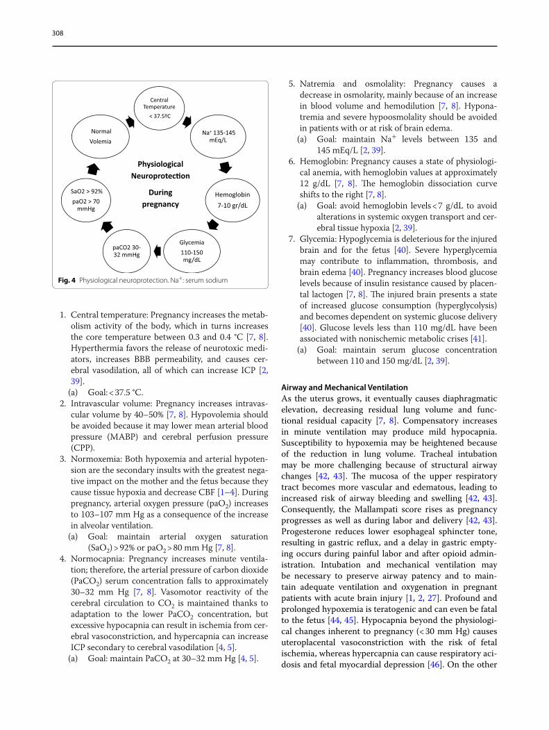
Physiological neuroprotection is a set of measures aimed at maintaining physiological homeostasis, which if altered, can negatively affect ICP [2, 27, 39]. These measures should also be applied when IHT occurs during pregnancy (Fig. 4).
Physiological neuroprotection aims at maintaining homeostasis of crucial physiological variables to avoid secondary cerebral insults. The following goals are extrapolated from nonpregnant women, adapted to this situation:
CT Scan
30-40 min
ControlHV
Osmotherapy
CSFDr
MVSeda�on
Analg
Gral MeasPNp
EMLPrimaryDC ??
CSF Dr +
GoalsCPP 50-60
mmHgICP < 23 mmHg
FetalMonitoring
BBT
DC
Hypoth
MABP targets change during pregnancy
trimester
Fetal BradychardiaNot reported for IHT control
Hemodynamic unstabilityCardiac depressionFetal malforma�ons and hemorrhagesNeonatal abs�nence sybdrome
Indica�ons, �mming and complica�ons not differentTo non pregnancy
Maternal and fetal brain ischemiaFetal hypoxia
Maternal anf fetal osmolarity increaseFetal deshidrata�on and hypoxiaFetal bradychardiaAcid-Basic status modifica�ons
Indica�ons and complica�ons not differentTo non pregnancy
Airway management difficultHypoxemia teratogenicHypocapnia associated to Fetal and brain hypoxiaFor concerns about seda�on, analgesia and NMB duringPregnancy, see table 3
Targets to achive according to physiological modifica�on induced by pregnancy , see figure 4Indica�ons and �mming of Surgery without differences to non pregnancy
Fetal and maternal concerns aboutICP control measurements
Fig. 3 Management steps for control of intracranial hypertension (IHT). Interventions should be implemented in a stepwise, additive manner allow-ing for sufficient intervals and, when pertinent radiological reevaluation, between steps to ensure that proceeding to the next step is necessary. Fetal monitoring is indicated throughout this process and indispensable as the more advanced (and less safe) steps are implemented. CPP: cerebral perfusion pressure; ICP: intracranial pressure; MABP: mean arterial blood pressure; EML: evacuate mass lesion; DC: decompressive craniectomy; PNp: physiological neuroprotection; MV: mechanical ventilation; min: minutes; CT: computed tomography; CSF Dr: cerebrospinal fluid drainage; Analg: analgesia; HV: hyperventilation; BBT: barbiturates; Hypoth: hypothermia
308
1. Central temperature: Pregnancy increases the metab-olism activity of the body, which in turns increases the core temperature between 0.3 and 0.4 °C [7, 8]. Hyperthermia favors the release of neurotoxic medi-ators, increases BBB permeability, and causes cer-ebral vasodilation, all of which can increase ICP [2, 39].
(a) Goal: < 37.5 °C.2. Intravascular volume: Pregnancy increases intravas-
cular volume by 40–50% [7, 8]. Hypovolemia should be avoided because it may lower mean arterial blood pressure (MABP) and cerebral perfusion pressure (CPP).
3. Normoxemia: Both hypoxemia and arterial hypoten-sion are the secondary insults with the greatest nega-tive impact on the mother and the fetus because they cause tissue hypoxia and decrease CBF [1–4]. During pregnancy, arterial oxygen pressure (paO2) increases to 103–107 mm Hg as a consequence of the increase in alveolar ventilation.
(a) Goal: maintain arterial oxygen saturation (SaO2) > 92% or paO2 > 80 mm Hg [7, 8].
4. Normocapnia: Pregnancy increases minute ventila-tion; therefore, the arterial pressure of carbon dioxide (PaCO2) serum concentration falls to approximately 30–32 mm Hg [7, 8]. Vasomotor reactivity of the cerebral circulation to CO2 is maintained thanks to adaptation to the lower PaCO2 concentration, but excessive hypocapnia can result in ischemia from cer-ebral vasoconstriction, and hypercapnia can increase ICP secondary to cerebral vasodilation [4, 5].
(a) Goal: maintain PaCO2 at 30–32 mm Hg [4, 5].
5. Natremia and osmolality: Pregnancy causes a decrease in osmolarity, mainly because of an increase in blood volume and hemodilution [7, 8]. Hypona-tremia and severe hypoosmolality should be avoided in patients with or at risk of brain edema.
(a) Goal: maintain Na+ levels between 135 and 145 mEq/L [2, 39].
6. Hemoglobin: Pregnancy causes a state of physiologi-cal anemia, with hemoglobin values at approximately 12 g/dL [7, 8]. The hemoglobin dissociation curve shifts to the right [7, 8].
(a) Goal: avoid hemoglobin levels < 7 g/dL to avoid alterations in systemic oxygen transport and cer-ebral tissue hypoxia [2, 39].
7. Glycemia: Hypoglycemia is deleterious for the injured brain and for the fetus [40]. Severe hyperglycemia may contribute to inflammation, thrombosis, and brain edema [40]. Pregnancy increases blood glucose levels because of insulin resistance caused by placen-tal lactogen [7, 8]. The injured brain presents a state of increased glucose consumption (hyperglycolysis) and becomes dependent on systemic glucose delivery [40]. Glucose levels less than 110 mg/dL have been associated with nonischemic metabolic crises [41].
(a) Goal: maintain serum glucose concentration between 110 and 150 mg/dL [2, 39].
Airway and Mechanical VentilationAs the uterus grows, it eventually causes diaphragmatic elevation, decreasing residual lung volume and func-tional residual capacity [7, 8]. Compensatory increases in minute ventilation may produce mild hypocapnia. Susceptibility to hypoxemia may be heightened because of the reduction in lung volume. Tracheal intubation may be more challenging because of structural airway changes [42, 43]. The mucosa of the upper respiratory tract becomes more vascular and edematous, leading to increased risk of airway bleeding and swelling [42, 43]. Consequently, the Mallampati score rises as pregnancy progresses as well as during labor and delivery [42, 43]. Progesterone reduces lower esophageal sphincter tone, resulting in gastric reflux, and a delay in gastric empty-ing occurs during painful labor and after opioid admin-istration. Intubation and mechanical ventilation may be necessary to preserve airway patency and to main-tain adequate ventilation and oxygenation in pregnant patients with acute brain injury [1, 2, 27]. Profound and prolonged hypoxemia is teratogenic and can even be fatal to the fetus [44, 45]. Hypocapnia beyond the physiologi-cal changes inherent to pregnancy (< 30 mm Hg) causes uteroplacental vasoconstriction with the risk of fetal ischemia, whereas hypercapnia can cause respiratory aci-dosis and fetal myocardial depression [46]. On the other
Central Temperature
< 37.5ºC
Na+ 135-145 mEq/L
Hemoglobin7-10 gr/dL
Glycemia110-150 mg/dL
paCO2 30-32 mmHg
SaO2 > 92%paO2 > 70
mmHg
NormalVolemia
PhysiologicalNeuroprotec�on
Duringpregnancy
Fig. 4 Physiological neuroprotection. Na+: serum sodium

309
hand, chronic hypercapnia is associated with fetal car-diac malformations [47]. The optimal way to ventilate a pregnant woman is controversial, especially in terms of whether to use the protective lung strategy [48, 49]. In general, and according to recent evidence, it seems pru-dent to start mechanical ventilation with a controlled mode, tidal volumes between 6 and 8 mL/kg, minimum respiratory rates to ensure adequate minute ventilation (with a target PaCO2 of 30–32 mm Hg), and inspirated fraction of oxygen (FiO2) and positive end expiratory pressure (PEEP) necessary to achieve paO2 > 70 mm Hg and SaO2 > 92%. To prevent volutrauma, plateau pres-sure should be kept at < 24 cm H2O and driving pressure at < 13 cm H2O[48–50].
Hemodynamic ManagementMaintaining adequate CPP is vital [1, 2, 38]. Regardless of the type of injury, arterial hypotension should be avoided and quickly corrected [1, 2, 38, 50]. Pregnant patients typically have low blood pressure during the first two tri-mesters and may become hypertensive during the third trimester. Therefore, the CPP target may need to vary depending on the stage of gestation. CPP depends on MABP and ICP. During normal pregnancy, MABP drops approximately 4–5 mm Hg during the first 7 weeks and then drops another 2 mm Hg until weeks 20–21. From then onward, it gradually increases until the end of preg-nancy [51].
In hypertensive pregnant women, the initial decrease is smoother (2 mm Hg) until week 21, and the subsequent rise is sharper (between 6 and 17 mm Hg) [51].
Despite the variability of MABP during normal or path-ological pregnancy (arterial hypertension, preeclamp-sia), the target CPP between 50 and 60 mm Hg should remain unchanged [4–11]. Alterations of autoregulation in preeclampsia/eclampsia can influence CPP targets [12, 15]. To individually optimize the target CPP, multimodal monitoring (ICP, brain tissue oxygen pressure (PbtO2), transcranial Doppler, autoregulation test, monitoring of pressure reactivity index), together with continuous fetal monitoring, could provide valuable information.
We must remember than pregnant women are physi-ologically hemodiluted, so ensuring normal intravascu-lar volume with either isotonic or hypertonic fluids is essential. If the desired MAP is not achieved, vasopres-sors and/or inotropes should be used according to the individual hemodynamic profile [1, 2, 4]. The target MAP may vary depending on the duration of pregnancy and the ICP; however, it should be adjusted to maintain a CPP between 50 and 60 mm Hg [1, 2, 4, 5, 38, 50]. When administering vasopressors, clinicians should carefully consider the potential for inducing placental vasocon-striction. Maternal and fetal blood flow are comparable
[52]. Ten to twelve percent of the total cardiac output corresponds to uterine blood flow (500–700 mL/min) [52]. Eighty percent of it is distributed in the placenta and the remaining 20% in the myometrium [52]. Umbilical flow averages 350–400 mL/min [52]. Uterine blood ves-sels possess smooth muscle, autonomic innervation, and alpha and beta adrenergic receptors [52]. The placental vessels that make up the trophoblast lack smooth mus-cle and adrenergic receptors, forming a high-flow, low-resistance system that does not respond to endogenous or exogenous adrenergic stimuli [52]. Furthermore, this system does not have autoregulation capacity, and there-fore its flow passively follows blood pressure [52].
Rhythmic uterine contractions increase placental flow, whereas hypertonic contractions (labor, oxytocin) decrease it [52]. The vasopressor recommended during pregnancy is phenylephrine (alpha 1 agonist) because it does not modify uterine tone or alter the fetal acid–base state [53]. Data from experimental animals have shown that norepinephrine can cause increased uterine tone, placental hypoperfusion, and fetal bradycardia [54]. According to current definitions and guidelines, antihy-pertensive therapy is recommended when arterial blood pressure (ABP) levels are > 140/90 mm Hg [55–58]. Mon-otherapy is preferred, and first-line oral agents include labetalol, alpha methyldopa, long-acting nifedipine, or another permitted beta blocker (metoprolol, proprano-lol) [55–58]. Angiotensin-converting enzyme inhibitors and angiotensin receptor blockers should be avoided because of increased risk of fetal renal damage in the sec-ond half of pregnancy [55–58]. In cases of severe arterial hypertension, defined by ABP values > 160/110 mm Hg, the first-line agents recommended according to the dif-ferent scientific organizations are summarized in Table 3 [55–58].
Sedation: AnalgesiaThe approach to analgesia/sedation during pregnancy is controversial, and current guidelines do no refer to preg-nant patients [59]. There is no evidence that one strategy has shown superiority over others [43, 48]. In general, it is preferable to use short-acting agents to minimize confounding of the neurological examination [2, 48, 59, 60]. During pregnancy, the pain threshold is increased because of high levels of progesterone, which has sedative and analgesic properties through the induction of endor-phins and enkephalins and through serotonin release [7, 8, 61, 62]. This may allow for lower use of opiates. Yet pain thresholds are variable, so undertreatment of severe pain should be avoided because this could worsen IHT.
Targets of sedation and analgesia should be pain and agitation control, ventilator synchrony, and ICP con-trol without hemodynamic side effects. Protocolization

310
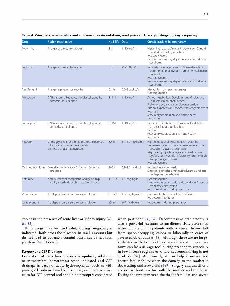
may limit excessive sedation [48, 59, 60]. In deeply sedated patients and in those treated with neuromus-cular blocking (NMB) agents, electroencephalography could be a helpful tool. Simplified electroencephalogra-phy providing a quantitative bispectral index has shown good correlation with sedation–agitation scales [48, 59, 63]. The Nociception Coma Scale has recently emerged as a valid tool to assess pain in patients with disorders of consciousness [63].The determination of the ade-quacy of analgesia for these patients still relies on the observation of indirect signs of pain, for example, tach-ycardia, systemic hypertension, and ICP elevation dur-ing painful interventions [59, 63]. Available drugs, with their properties, potential uses, and adverse effects in pregnancy, during pregnancy are outlined in Table 4. In general terms, regarding sedation and analgesia for pregnant patients, physicians should always con-sider the risk of fetal toxicity and respiratory depres-sion. Because there are no specific guidelines regarding sedation and analgesia during pregnancy or during IHT, extrapolation of the evidence obtained from non-pregnant women is recommended [48, 59, 60]. Table 4 details the maternal–fetal risks of commonly used sed-atives, analgesics, and neuromuscular blockers (NMB). However, lorazepam is a good choice because it lacks active metabolites and has less residual sedative effect, although its half-life is longer [48, 59, 60]. Propofol has
the advantage of its short half-life but must be balanced against the risk of propofol infusion syndrome [48, 59, 60]. Remifentanil and fentanyl can be used safely, even when hemodynamic instability is present [48, 59, 60]. Dexmedetomidine is another good choice because of its pharmacokinetic and pharmacodynamic properties [48, 59, 60]. Fetal risks are similar for all sedative/anal-gesic drugs, including postpartum respiratory depres-sion and floppy baby syndrome [48, 59, 60].
NMB is not routinely indicated for ICP control except for specific situations, such as intubation, shivering (hypothermia, controlled normothermia), unconven-tional ventilatory modes (high airway resistance), venti-lator asynchrony, compartment intrabdominal syndrome, or severe acute respiratory distress syndrome. Addition-ally, NMB can be briefly employed during a dangerous increase of ICP (e.g., secondary to severe and refrac-tory agitation or cough crisis) [48, 64, 65]. NMB pre-cludes neurological examination, masks seizures, and predisposes to infections, deep venous thrombosis, and decubitus ulcers; NMB use is also linked to prolonged mechanical ventilation and may heighten the risk of criti-cal illness myoneuropathy [22, 48, 64, 65].
During pregnancy, short-acting agents that do not cause histamine release (to avoid arterial hypotension or broncoconstriction), such as vecuronium or cisatracu-rium, are preferred [48, 64, 65]. Cisatracurium is the first
Table 3 First-line drugs for management of severe arterial hypertension during pregnancy
Drug FDA cat‑egory
Mechanism of action Dose Route Adverse effects
Labetalol C Alfa and beta blocker 10–20 mg. Then 20–80 mg every 20–30 min (max 300 mg)
Infusion: 1–2 mg/min
IV Materno-fetal bradycardiaHypotension
Hydralazine C Peripheral vasodilator(direct smooth muscle relaxant to inhibition of
inositol trisphosphate-induced Ca2+ release)
5–10 mg every 20–40 min. Infusion: 0.5–10 mg/h
IV-IM TachycardiaHypotensionHeadacheFlushingNauseaICP increase
Nifedipine C Ca2+ channels blocker 10–30 mg every 6–8 h Oral HeadachesFacial flushingTachycardia
Diazoxide C Peripheral vasodilator(Potassium channel activator)
30–50 mg every 5–15 min IV FlushingHypotensionICP increase
Nicardipine C Ca2+ channels blocker 5–15 mg/h IV HypotensionHeadacheTachyarrhythmia
Clevidipine C Ca2+ channels blocker 1–21 mg/h IV HypotensionTachyarrhythmia
Sodium Nitroprusside C Peripheral vasodilator (oxide nitric production) 0.25–5 ug/kg/min IV HypotensionCyanide toxicityICP increase
311
choice in the presence of acute liver or kidney injury [48, 64, 65].
Both drugs may be used safely during pregnancy if indicated. Both cross the placenta in small amounts but do not lead to adverse neonatal outcomes or neonatal paralysis [48] (Table 3).
Surgery and CSF DrainageEvacuation of mass lesions (such as epidural, subdural, or intracerebral hematomas) when indicated and CSF drainage in cases of acute hydrocephalus (such as with poor-grade subarachnoid hemorrhage) are effective strat-egies for ICP control and should be promptly considered
when pertinent [66, 67]. Decompressive craniectomy is also a powerful measure to ameliorate IHT, performed either unilaterally in patients with advanced tissue shift from space-occupying lesions or bilaterally in cases of severe cerebral edema [68]. Although there are no large-scale studies that support this recommendation, craniec-tomy can be a salvage tool during pregnancy, especially in low-income regions or where neuromonitoring is not available [68]. Additionally, it can help maintain and ensure fetal viability when the damage to the mother is devastating and irreversible [69]. Surgery and anesthesia are not without risk for both the mother and the fetus. During the first trimester, the risk of fetal loss and severe
Table 4 Principal characteristics and concerns of main sedatives, analgesics and paralytic drugs during pregnancy
Drug Action mechanism Half‑life Dose Considerations in pregnancy
Morphine Analgesia. μ receptor agonist 3 h 1–10 mg/h Histamine release. Arterial hypotension. Contrain-dicated in renal dysfunction
Not teratogenicNeonatal respiratory depression and withdrawal
syndrome
Fentanyl Analgesia. μ receptor agonist 3 h 25–100 μg/h Nonhistamine release and active metabolism. Consider in renal dysfunction or hemodynamic instability
Not teratogenicNeonatal respiratory depression and withdrawal
syndrome
Remifentanil Analgesia μ receptor agonist 3 min 0.5–2 μg/kg/min Metabolism by serum esterasesNot teratogenic
Midazolam GABA agonist. Sedative, anxiolysis, hypnotic, amnesic, antiepileptic
3–11 h 1–10 mg/h Active metabolites. Development of tolerance. Less safe if renal dysfunction
Prolonged sedation after discontinuationArterial hypotension. Unclear if teratogenic effectNeonatalrespiratory depression and floppy babysyndrome
Lorazepam GABA agonist. Sedative, anxiolysis, hypnotic, amnesic, antiepileptic
8–15 h 1–10 mg/h No active metabolites. Less residual sedation. Unclear if teratogenic effect
Neonatalrespiratory depression and floppy babysyndrome
Propofol GABA, glycine, muscarinic, and nicotinic recep-tors agonist. Sedative/anxiolytic,
amnestic, and anticonvulsant
30 min 5 to 50 mg/kg/min High hepatic and extrahepatic metabolismDecreases systemic vascular resistance and can
provoke myocardial depressionMay be employed during acute renal or liver
dysfunction. Propofol infusion syndrome (high and prolonged doses)
Not teratogenic
Dexmedetomidine Selective presynaptic α2 agonist. Sedative, analgesic
2–3 h 0.2–1.5 mg/kg/h No respiratory depressionDecreases catecholamines. Bradycardia and arte-
rial hypotension (bolus)
Ketamine NMDA receptor antagonist. Analgesic, hyp-notic, anesthetic and sympathomimetic
1.5–3 h 1–5 mg/kg/h Not teratogenicUterine contractions (dose-dependent). Neonatal
respiratory depressionNot a first choice during pregnancy
Vecuronium No depolarizing neuromuscular blocker 0.5–2 h 1–2 mg/kg/min Contraindicated in renal or liver failureNo problems for fetus
Cisatracurium No depolarizing neuromuscular blocker 25 min 2–4 mg/kg/min No problems during pregnancy
312
congenital malformations should be discussed, consid-ering in certain cases the possibility of interrupting the pregnancy, whereas during the second and third trimes-ters, surgical procedures can be performed with a good margin of safety [26]. Beyond week 34, it is generally advisable to proceed with cesarean delivery at the time of the neurosurgery [26].
CSF drainage is an effective and transitory measure to lower ICP, especially in the presence of acute hydro-cephalus [2, 27, 67]. The indications for ventriculostomy are not affected by pregnancy. Instead, controlled lum-bar drainage is a less desirable option during pregnancy because of the compression of the thecal space [20].
OsmotherapyMannitol and hypertonic saline solution (HSS) share similar distribution in the extracellular space, but man-nitol crosses the placental barrier and is excreted in fetal urine [27, 70]. Animal studies have shown that mannitol increases maternal osmolality and may cause fetal dehy-dration, hypovolemia, hypoxia, bradycardia, and changes in acid–base status [70]. It is unclear if this phenome-non is dose dependent [4, 70]. The safety of intravenous hypertonic saline in pregnancy is not well established. In the past, intrauterine instillation of hypertonic saline was used to induce abortions in the midtrimester [71]; how-ever, this experience is most likely irrelevant for the eval-uation of the risks related to intravenous administration. There are no human studies assessing mannitol or HSS during pregnancy. Low doses of mannitol (0.25–0.5 g/kg) are generally considered safe [72]. Yet HSS may be more physiological, and we therefore favor the use of HSS when central venous access is available and the patient does not have signs of heart failure.
There are no safety data on the use of hypertonic lac-tate in pregnancy.
HyperventilationPhysiologically, pregnant women maintain PaCO2 levels between 30 and 32 mm Hg. Therefore, the PaCO2 goal when hyperventilating a pregnant patient should prob-ably be between 26 and 28 mm Hg [4, 7, 8]. There are no studies on hyperventilation during pregnancy; however, despite the physiological and adaptive decrease in CO2 levels and the alkalinity of CSF during pregnancy, vascu-lar reactivity to CO2 remains unchanged or can even be increased [73, 74].
Hyperventilation should be considered after safer measures have failed to bring the ICP under control, and it should only be induced under strict fetal monitoring.
To our knowledge, there are no studies evaluating fetal complications from maternal hyperventilation; how-ever, hypocapnia produces systemic vasoconstriction
(myocardial, renal, splanchnic), and the uteroplacen-tal circulation does not escape this effect [4, 75, 76]. Hence, acceptable indications for hyperventilation should be restricted to brain herniation, potentially lethal increases in ICP (Lundberg A waves), or IHT secondary to increased CBF (hyperemia). Hyperventilation should ideally be used as briefly as possible, but if used for more than a few minutes, it should not be stopped suddenly to avoid rebound increases in ICP [75, 76].
Refractory IHT: Second‑line TherapiesWhen there is no response to the aforementioned therapies, IHT is defined as refractory [2, 27]. In this situation, available therapies are scarce, and they are associated with severe complications. None of the sec-ond-tier options have been formally evaluated during pregnancy.
The use of barbiturates at high doses is not a safe option because of its strongly negative impact on hemo-dynamics and its profound maternal immunosuppressive effects [2]. Barbiturates cross the placenta and distrib-ute widely in the fetus [77]. During the first trimester of pregnancy, they can cause malformations (spinal cord) [77]. Additionally, their administration is associated with fetal hemorrhages, likely related to vitamin K deficiency [77]. Neonatal abstinence syndrome may also occur after delivery when barbiturates are prescribed during the third trimester of pregnancy [77].
Indomethacin is a nonsteroidal antiinflammatory agent that causes cerebral vasoconstriction and has been used as a rescue measure (not included in current pro-tocols) for the control of refractory IHT [2]. It has toco-lytic capacity by reducing the number and frequency of uterine contractions [78]. It should not be used beyond 32 weeks’ gestation and produces a decrease in fetal diu-resis and oligohydramnios [78]. Additionally, it can cause jaundice, premature closure of the arteriovenous ductus, necrotizing enterocolitis, intraventricular hemorrhage, and leukomalacia in the fetus [78].
Therapeutic hypothermia has anecdotally been reported during pregnancy in the context of cardiac arrest but not for control of IHT [79]. Hypothermia can induce fetal bradycardia; therefore, strict monitoring of the fetal heart rate (FHR) is mandatory. There are case reports of induced hypothermia (33–35 °C) between 13 and 20 weeks’ gestation resulting in favorable maternal functional and good fetal outcomes [79].
As previously mentioned, decompressive craniec-tomy is a valid option to treat refractory IHT [68]. Data from randomized controlled trials are very supportive of the benefit of decompressive hemicraniectomy for young patients with hemispheric stroke [80] but do not show clear benefits in functional outcome after severe

313
traumatic brain injury [81, 82]. There have been no trials of surgical decompression for other causes of refractory IHT. Thus, decompressive craniectomy should be used very selectively and after careful evaluation of individual circumstances (prognosis of the cause of the IHT, fetal viability, etc.).
In fact, given the safety concerns with other rescue treatments, craniectomy should be particularly consid-ered as a preferable alternative in pregnant patients with refractory IHT.
Maternal–fetal MonitoringApart from classic systemic maternal monitoring (elec-trocardiography, SaO2, capnography, arterial pressure, etc.), serial fetal monitoring by using conventional or Doppler ultrasonography is essential [26, 83, 84]. Fetal monitoring should be considered in all pregnant patients with IHT and may include FHR and obstetrical ultra-sound parameters, such as fetal physical characteristics, weight and position, placental localization and blood flow, umbilical flow, and amniotic fluid volume [26, 83, 84].
The analysis of the variability of the FHR is a crucial parameter because its alteration denotes severe fetal dis-tress [26, 83, 84]. The normal FHR ranges from 110 to 160 beats per minute [85]. Periodic acceleration phenomena associated with fetal movements (more than 15 beats of 15 s duration) and FHR variability (spontaneous changes between 6 and 25 beats per minute according to fetal car-diac demands) are physiological [85]. Decelerations and absence of variability denote severe fetal distress [85].
Fetal and Maternal Considerations About DeliveryMaternal homeostasis is vital to fetal well-being. The pri-oritization of fetal health over maternal health can lead to unnecessary delays in maternal care and suboptimal treatment of life-threatening conditions, such as IHT, resulting in poor outcomes for both mother and fetus.
The survival of the fetus depends on uterine perfusion and oxygen delivery [7, 8, 86]. The uteroplacental cir-culation has no autoregulatory properties; thus, blood flow passively depends on maternal MAP, and peripheral vasoconstriction can compromise uterine perfusion [7, 8, 52, 86]. The fetus is usually considered viable when it has a 50% chance of extrauterine survival [86, 87]. If neo-natal facilities are available, this usually means at least 25–26 weeks’ gestation or an estimated weight of 750 g [86, 87]. By using cutoffs of 24 weeks’ gestation and an estimated weight of 500–600 g, the chances of survival are reduced to 20–30% [86, 87]. Decisions on fetal viabil-ity are based on the best estimation of gestational age. When estimating the fetal age in the emergency room or intensive care unit, if the fundus of the uterus extends
beyond the umbilicus, the fetus is potentially viable [86, 87]. When emergency neurosurgery is indicated and the fetus is viable, cesarean delivery should be performed under general anesthesia either immediately before or along with the neurosurgical intervention [4, 26, 72, 88, 89].
The decision whether to interrupt a pregnancy depends on the severity of the mother’s clinical situation and the age and clinical condition of the fetus, and it should always be reached by consensus from a multidisciplinary team [72, 86–89]. A shared decision-making approach is recommended and should include the patient (if possi-ble) and family.
In cases of preterm labor or early uterine contractions during pregnancy, tocolytics (oxytocin receptor antago-nist, beta adrenergic agonists, or calcium channel block-ers) can be used, but the safety of them is unknown. Oxytocin and its synthetic analogues can be used with a good safety margin; however, the lowest possible dose should be employed at a slow infusion rate because they can potentially cause vasodilation, arterial hypotension, and increased ICP [90, 91].
Maternal Brain DeathIntractable IHT may result in maternal brain death. In some of these instances, fetal viability is preserved and the life of the developing fetus still depends on somatic functions of the deceased mother [92]. In some cases, interventions on the mother’s body are justified to pre-serve the life of the fetus [92]. This point is extremely controversial and complex because of the bioethical dilemmas specifically related to pregnancy [93]. Deci-sion-makers must consider the maternal–fetal unit, and all aspects of care must be discussed in a multidiscipli-nary manner, with inclusion of medical, ethical, and legal experts [93]. Regarding the care of a pregnant woman who has suffered a catastrophic and irreversible brain injury, decision-makers should consider the following factors: (1) prognosis of the injury, gestational age and its impact, and of the therapeutic interventions on the fetus; (2) the mother’s wishes; (3) commitment to decision-making by the mother’s close relatives or surrogates; and (4) current local laws [93].
A systematic review that included 30 cases of brain-dead pregnant mothers found that the mean gestational age at the time of brain death was 22 weeks and at the time of delivery was 29.5 weeks; 12 children survived and developed normally over the subsequent 2 years [94]. Fetus survival depended on several factors, including the mother’s gestational age and complications such as diabetes insipidus, hemodynamic instability, hypopitui-tarism, hypothermia, metabolic instability, and neonatal acute distress respiratory syndrome [92, 94].
314
OutcomeThere are no studies specifically addressing the mater-nal and fetal outcomes related to IHT during pregnancy. Yet 60% of deaths in the peripartum period are due to hypertensive disorders (preeclampsia/eclampsia) and venous thromboembolism [4]. Preeclampsia, eclampsia, and hemolysis, elevated liver enzymes, and low platelet count (HELLP) syndrome are associated with 1.5% mor-tality in developed countries, but in developing coun-tries, the mortality rate may range between 10 and 33% [95]. Traumatic brain injury is the cause responsible for maternal death in 10% of cases [96]. Poor functional outcome (modified Rankin score of 3 or more) has been reported after 16% of ischemic strokes and nearly 40% of hemorrhagic strokes. The mortality rate during the peripartum period is 2.7% and 11.7% for ischemic and hemorrhagic stroke, respectively [97–99].
ConclusionsIHT is a rare complication during pregnancy, and its man-agement remains empirical and extrapolated from data on nonpregnant patients. However, this special situa-tion demands attention to special considerations, includ-ing physiological changes related to the pregnancy (e.g., expansion of circulating volume, hypocapnia, raised intraabdominal pressure), pharmacokinetic changes affect-ing multiple classes of medications, and the risks to the developing fetus. ICP management protocols should there-fore be adjusted accordingly to optimize maternal and fetal outcomes. The numerous gaps in knowledge identified in this review highlight the need for more research on this infrequent but important clinical situation.
Author details1 Neurointensive Care Unit, Sanatorio Pasteur, Catamarca, Argentina. 2 Intensive Care, Hospital Carlos Malbran, Catamarca, Argentina. 3 Anesthesia and Intensive Care, San Martino Policlinico Hospital, Investigational Research for Critical Care for Oncology and Neurosciences, Genoa, Italy. 4 Division of Neurological Surgery, University of Sao Paulo Medical School, Sao Paulo, Brazil. 5 Neuroscience Intensive Care Unit, Mayo Clinic, Rochester, MN, USA.
Author contributionsDAG: Search and analysis of the literature; design and writing of the original manuscript and later versions; reply to reviewers. CR and WSP: Revision and first edition of the manuscript. AAR: Analysis, global content review; grammar adaptation and correction; final editing of the manuscript and of the response to the reviewers. The final manuscript was approved by all authors.
Source of supportNone.
Conflicts of interestThe authors have nothing to disclose.
Ethical approval/informed consentAll procedures performed in studies involving human participants were in accordance with the ethical standards of the institutional and/or national
research committee and with the 1964 Helsinki declaration and its later amendments or comparable ethical standards.
Publisher’s NoteSpringer Nature remains neutral with regard to jurisdictional claims in pub-lished maps and institutional affiliations.
Received: 30 March 2021 Accepted: 12 August 2021Published online: 7 September 2021
References 1. Stochetti N, Maas AIR. Traumatic intracranial hypertension. N Eng J Med.
2014;370:2121–30. 2. Godoy DA, Videtta W, Di Napoli M. Practical approach to posttraumatic
intracranial hypertension according to pathophysiologic reasoning. Neurol Clin. 2017;35:613–40.
3. Wilson MH. Monro-Kellie 2.0: the dynamic vascular and venous patho-physiological components of intracranial pressure. J Cereb Blood Flow Metab. 2016;36:1338–50.
4. Frontera JA, Ahmed W. Neurocritical care complications of pregnancy and puerperum. J Crit Care. 2014;29(6):1069–81.
5. Burn MS, Sheth SS, Sheth KN. Neurocritical care of the pregnant patient. Handb Clin Neurol. 2020;171:205–13.
6. Ladhani NNN, Swartz RH, Foley N, Nerenberg K, Smith EE, Gubitz G, et al. Canadian stroke best practice consensus statement: acute stroke man-agement during pregnancy. Int J Stroke. 2018;13:743–58.
7. Cunningham F, Leveno K, Bloom S, Hauth J, Gilstrap L, Wenstrom K. Maternal physiology. In: Obstetrics W, editor. 22nd edition. New York: McGraw Hill; 2005. p. 121–50.
8. Datta S, Kodali B, Segal S. Maternal physiological changes during pregnancy. Labor Postpartum Period. 2010. https:// doi. org/ 10. 1007/ 978-0- 387- 88602-2_1.
9. Johnson AC, Cipolla MJ. The cerebral circulation during pregnancy: adapting to preserve normalcy. Physiology (Bethesda). 2015;30(2):139–47.
10. Cipolla MJ. The adaptation of the cerebral circulation to preg-nancy: mechanisms and consequences. J Cereb Blood Flow Metab. 2013;33(4):465–78.
11. Cipolla MJ, Bishop N, Chan SL. Effect of pregnancy on autoregulation of cerebral blood flow in anterior versus posterior cerebrum. Hypertension. 2012;60:705–11.
12. Janzarik WG, Ehlers E, Ehmann R, Gerds TA, Schork J, Mayer S, Gabriel B, Weiller C, Prömpeler H, Reinhard M. Dynamic cerebral autoregulation in pregnancy and the risk of preeclampsia. Hypertension. 2014;63:161–6.
13. Janzarik WG, Jacob J, Katagis E, Markfeld-Erol F, Sommerlade L, Wuttke M, Reinhard M. Preeclampsia postpartum: Impairment of cerebral autoregu-lation and reversible cerebral hyperperfusion. Pregnancy Hypertens. 2019;17:121–6.
14. Cipolla MJ, Sweet JG, Chan SL. Cerebral vascular adaptation to pregnancy and its role in the neurological complications of eclampsia. J Appl Physiol. 2011;110(2):329–39.
15. Jones-Muhammad M, Warrington JP. Cerebral blood flow regulation in pregnancy, hypertension, and hypertensive disorders of pregnancy. Brain Sci. 2019;9:224.
16. Carmeliet P, Moons L, Luttun A, Vincenti V, Compernolle V, De Mol M, Wu Y, Bono F, Devy L, Beck H, Scholz D, Acker T, DiPalma T, Dewerchin M, Noel A, Stalmans I, Barra A, Blacher S, VandenDriessche T, Ponten A, Eriksson U, Plate KH, Foidart JM, Schaper W, Charnock-Jones DS, Hicklin DJ, Herbert JM, Collen D, Persico MG. Synergism between vascular endothelial growth factor and placental growth factor contributes to angiogenesis and plasma extravasation in pathological conditions. Nat Med. 2001;7:575–83.
17. Wallace K, Bean C, Bowles T, Spencer SK, Randle W, Kyle PB, Shaffery J. Hypertension, anxiety, and blood-brain barrier permeability are increased in postpartum severe preeclampsia/hemolysis, elevated liver enzymes, and low platelet count syndrome rats. Hypertension. 2018;72:946–54.
18. Burwick RM, Togioka BM, Speranza RJ, Gaffney JE, Roberts VHJ, Frias AE, Rincón M. Assessment of blood–brain barrier integrity and neuroinflam-mation in preeclampsia. Am J Obstet Gynecol. 2019;221(269):e1-269.e8.

315
19. Quick AM, Cipolla MJ. Pregnancy-induced up-regulation of aquaporin-4 protein in brain and its role in eclampsia. FASEB J. 2005;19:170–5.
20. Takiguchi T, Yamaguchi S, Tezuka M, Furukawa N, Kitajima T. Compression of the subarachnoid space by the engorged epidural venous plexus in pregnant women. Anesthesiology. 2006;105:848–51.
21. Hirabayashi Y, Shimizu R, Saitoh K, Fukuda H, Igarashi T. Acid-base state of cerebrospinal fluid during pregnancy and its effect on spread of spinal anaesthesia. Br J Anaesthesia. 1996;77:352–5.
22. Altemus M, Fong J, Yang R, Damast S, Luine V, Ferguson D. Changes in cerebrospinal fluid neurochemistry during pregnancy. Biol Psychiatry. 2004;56:386–92.
23. Lee LK. Physiological adaptations of pregnancy affecting the nervous system. Semin Neurol. 2007;27:405–10.
24. Ansari J, Carvalho B, Shafer SL, Flood P. Pharmacokinetics and pharmaco-dynamics of drugs commonly used in pregnancy and parturition. Anesth Analg. 2016;122:786–804.
25. Pariente G, Leibson T, Carls A, Adams-Webber T, Ito S, Koren G. Pregnancy-associated changes in pharmacokinetics: a systematic review. PLoS Med. 2016;13:e1002160.
26. Chowdhury T, Chowdhury M, Schaller B, Cappellani RB, Daya J. Perioperative considerations for neurosurgical procedures in the gravid patient: continuing professional development. Can J Anaesth. 2013;60(11):1139–55.
27. Carney N, Totten AM, O’Reilly C, Ullman JS, Hawryluk GW, Bell MJ, et al. Guidelines for the management of severe traumatic brain injury. Neuro-surgery. 2017;80:6–15.
28. Vik A, Nag T, Fredriksli OA, Skandsen T, Moen KG, Schirmen Mikalsen K, et al. Relationship of “dose” of intracranial hypertension to outcome in severe traumatic brain injury. J Neurosurg. 2008;109:678–84.
29. Jha RM, Elmer J, Zusman BE, Desai S, Puccio AM, Okonkwo DO, et al. Intracranial pressure trajectories: a novel approach to informing severe traumatic brain injury phenotypes. Crit Care Med. 2018;46:1792–802.
30. Chesnut RM, Gautille T, Blunt BA, et al. The localizing value of asymmetry in pupil size in severe head injury: relation to lesion type and location. Neurosurgery. 1994;34:840–6.
31. Chen MM, Coakley FV, Kaimal A, Laros RK Jr. Guidelines for computed tomography and magnetic resonance imaging use during pregnancy and lactation. Obstet Gynecol. 2008;112:333–40.
32. Committee Opinion No. 723: Guidelines for diagnostic imaging during pregnancy and lactation. Obstet Gynecol. 2017; 130: e210-e216. Erratum in: Obstet Gynecol. 2018; 132: 786.
33. Marshall LF, Marshall SB, Klauber MR, van Berkum CM. A new classifica-tion of head injury based on computerized tomography. J Neurosurg. 1991;75(Suppl):S14–20.
34. Alali AS, Temkin N, Barber J, Pridgeon J, Chaddock K, Dikmen S, Hendrick-son P, Videtta W, Lujan S, Petroni G, Guadagnoli N, Urbina Z, Chesnut RM. A clinical decision rule to predict intracranial hypertension in severe traumatic brain injury. J Neurosurg. 2018;8:1–8.
35. Fernando SM, Tran A, Cheng W, Rochwerg B, Taljaard M, Kyeremanteng K, English SW, Sekhon MS, Griesdale DEG, Dowlatshahi D, McCredie VA, Wijdicks EFM, Almenawer SA, Inaba K, Rajajee V, Perry JJ. Diagnosis of elevated intracranial pressure in critically ill adults: systematic review and meta-analysis. BMJ. 2019;366:l4225.
36. Meyer MJ, et al. Acute management of acquired brain injury part I: an evidence-based review of non-pharmacological interventions. Brain Inj. 2010;24:694–705.
37. Meyer MJ, et al. Acute management of acquired brain injury part II: an evidence-based review of pharmacological interventions. Brain Inj. 2010;24:706–21.
38. Hawryluk GWJ, Aguilera S, Buki A, Bulger E, Citerio G, Cooper DJ, Arrastia RD, Diringer M, Figaji A, Gao G, Geocadin R, Ghajar J, Harris O, Hoffer A, Hutchinson P, Joseph M, Kitagawa R, Manley G, Mayer S, Menon DK, Meyfroidt G, Michael DB, Oddo M, Okonkwo D, Patel M, Robertson C, Rosenfeld JV, Rubiano AM, Sahuquillo J, Servadei F, Shutter L, Stein D, Stocchetti N, Taccone FS, Timmons S, Tsai E, Ullman JS, Vespa P, Videtta W, Wright DW, Zammit C, Chesnut RM. A management algorithm for patients with intracranial pressure monitoring: the Seattle international severe traumatic brain injury consensus conference (SIBICC). Intensive Care Med. 2019;45:1783–94.
39. Godoy DA, Videtta W, Santa Cruz R, Silva X, Aguilera-Rodríguez S, Carreño-Rodríguez JN, Ciccioli F, Piñero G, Ciro JD, da Re-Gutiérrez S, Domeniconi
G, Fischer D, Hernández O, Lacerda-Gallardo A, Mejía J, Panhke P, Romero C, Lora FS, Soler-Morejón C, Sufan JL, Montes JM, Fuenzalida LC, Parahnos JL, Jibaja M. En representación del Consorcio Latinoamericano de Injuria Cerebral (LABIC). General care in the management of severe traumatic brain injury: Latin American consensus. Med Intensiva. 2020;44(8):500–8.
40. Godoy DA, Di Napoli M, Rabinstein AA. Treating hyperglycemia in neuro-critical patients: benefits and perils. Neurocrit Care. 2010;13:425–38.
41. Vespa P, Boonyaputthikul R, McArthur D, Miller C, et al. Intensive insulin therapy reduces microdialysis glucose values without altering glucose utilization or improving the lactate/pyruvate ratio after traumatic brain injury. Crit Care Med. 2006;34:850–6.
42. Kinsella SM, Winton AL, Mushambi MC, Ramaswamy K, Swales H, Quinn AC, Popat M. Failed tracheal intubation during obstetric general anaes-thesia: a literature review. Int J Obstet Anesth. 2015;24:356–74.
43. Mushambi MC, Kinsella SM, Popat M, Swales H, Ramaswamy KK, Winton AL, Quinn AC; Obstetric Anaesthetists’ Association; Difficult Airway Society. Obstetric Anaesthetists’ Association and Difficult Airway Society guidelines for the management of difficult and failed tracheal intubation in obstetrics. Anaesthesia. 2015; 70:1286–1306.
44. Itskovitz J, LaGamma EF, Rudolph AM. The effect of reducing umbilical blood flow on fetal oxygenation. Am J Obstet Gynecol. 1983;145:813–8.
45. Dilts PV Jr, Brinkman CR 3rd, Kirschbaum TH, Assali NS. Uterine and systemic hemodynamic interrelationships and their response to hypoxia. Am J Obstet Gynecol. 1969;103:138–57.
46. Walker AM, Oakes GK, Ehrenkranz R, McLaughlin M, Chez RA. Effects of hypercapnia on uterine and umbilical circulations in conscious pregnant sheep. J Appl Physiol. 1976;41:727–33.
47. Haring OM. Cardiac malformations in rats induced by exposure of the mother to carbon dioxide during pregnancy. Circ Res. 1960;8:1218–27.
48. Pacheco LD, Saade GR, Hankins GD. Mechanical ventilation during pregnancy: sedation, analgesia, and paralysis. Clin Obstet Gynecol. 2014;57(4):844–50.
49. Schwaiberger D, Karcz M, Menk M, Papadakos PJ, Dantoni SE. Respiratory failure and mechanical ventilation in the pregnant patient. Crit Care Clin. 2016;32(1):85–95.
50. Munnur U, Bandi V, Guntupalli KK. Management principles of the critically ill obstetric patient. Clin Chest Med. 2011;32:53–60.
51. Ayala DE, Hermida RC, Mojón A, Fernández JR, Silva I, Ucieda R, Iglesias M. Blood pressure variability during gestation in healthy and complicated pregnancies. Hypertension. 1997;30:611–8.
52. Nag DS, Samaddar DP, Chatterjee A, Kumar H, Dembla A. Vasopres-sors in obstetric anesthesia: a current perspective. World J Clin Cases. 2015;3:58–64.
53. Lee A, Ngan Kee WD, Gin T. A quantitative, systematic review of rand-omized controlled trials of ephedrine versus phenylephrine for the man-agement of hypotension during spinal anesthesia for cesarean delivery. Anesth Analg. 2002;94:920–6.
54. Stevens AD, Lumbers ER. Effects of intravenous infusions of noradrenaline into the pregnant ewe on uterine blood flow, fetal renal function, and lung liquid flow. Can J Physiol Pharmacol. 1995;73(2):202–8.
55. Butalia S, Audibert F, Côté AM, Firoz T, Logan AG, Magee LA, et al. Hyper-tension Canada’s 2018 guidelines for the management of hypertension in pregnancy. Can J Cardiol. 2018;34:526–31.
56. Podymow T, August P. Update on the use of antihypertensive drugs in pregnancy. Hypertension. 2008;51:960–9.
57. Nij Bijvank SW, Duvekot JJ. Nicardipine for the treatment of severe hyper-tension in pregnancy: a review of the literature. Obstet Gynecol Surv. 2010;65:341–7.
58. Finger JR, Kurczewski LM, Brophy GM. Clevidipine versus nicardipine for acute blood pressure reduction in a neuroscience intensive care popula-tion. Neurocrit Care. 2017;26:167–73.
59. Devlin JW, Skrobik Y, Gélinas C, et al. Clinical practice guidelines for the prevention and management of pain, agitation/sedation, delirium, immobility, and sleep disruption in adult patients in the ICU. Crit Care Med. 2018;46(9):e825–73.
60. Neuman G, Koren G. Safety of procedural sedation in pregnancy. J Obstet Gynaecol Can. 2013;35(2):168–73.
61. Gintzler AR. Endorphin-mediated increases in pain threshold during pregnancy. Science. 1980;210:193–5.
62. Jayaram A, Carp H. Progesterone-mediated potentiation of spinal sufen-tanil in rats. Anesth Analg. 1993;76:745–50.
316
63. Le Roux P, Menon DK, Citerio G, et al. Consensus summary statement of the international multidisciplinary consensus conference on multimodal-ity monitoring in neurocritical care: a statement for healthcare profes-sionals from the neurocritical care society and the European Society of Intensive. Intensive Care Med. 2014;40:1189–209.
64. Sanfilippo F, Santonocito C, Veenith T, Astuto M, Maybauer MO. The role of neuromuscular blockade in patients with traumatic brain injury: a systematic review. Neurocrit Care. 2015;22:325–34.
65. Renew JR, Ratzlaff R, Hernandez-Torres V, et al. Neuromuscular blockade management in the critically Ill patient. J Intensive Care. 2020;8:37.
66. Bullock RM, Chesnut R, Ghajar J, Gordon D, Hartl R, Newell DW, Servadei F, Walters BC, Wilberger JE. Guidelines for the surgical management of traumatic brain injury author group. Neurosurgery. 2006;58(3):S2-vi.
67. Rabinstein AA, Lanzino G, Wijdicks EF. Multidisciplinary management and emerging therapeutic strategies in aneurysmal subarachnoid haemor-rhage. Lancet Neurol. 2010;9(5):504–19.
68. Hawryluk GW, Rubiano AM, Totten AM, O’Reilly C, Ullman JS, Bratton SL, Ghajar J. Guidelines for the management of severe traumatic brain injury: 2020 update of the decompressive craniectomy recommendations. Neurosurgery. 2020;87(3):427–34.
69. Whitney N, Raslan AM, Ragel BT. Decompressive craniectomy in a neurologically devastated pregnant woman to maintain fetal viability. J Neurosurg. 2012;116(3):487–90.
70. Basso A, Fernandez A, Althabe O, Sabini G, Piriz H, Belitzky R. Passage of mannitol from mother to amniotic fluid and fetus. Obstet Gynecol. 1977;49:628–31.
71. Hern WM. Midtrimester abortion Obstet Gynecol Annu. 1981;10:375–422. 72. Reitman E, Flood P. Anaesthetic considerations for nonobstetric surgery
during pregnancy. Br J Anaesth. 2011;107(suppl 1):i72-8. 73. Riskin-Mashiah S, Belfort MA, Saade GR, Herd JA. Cerebrovascular
reactivity in normal pregnancy and preeclampsia. Obstet Gynecol. 2001;98:827–32.
74. Steinback CD, King EC, Davenport MH. Longitudinal cerebrovascular reactivity during pregnancy: a case study. Appl Physiol Nutr Metab. 2015;40(6):636–9.
75. Godoy DA, Seifi A, Garza D, Lubillo-Montenegro S, Murillo-Cabezas F. Hyperventilation therapy for control of posttraumatic intracranial hyper-tension. Front Neurol. 2017;8:250.
76. Godoy DA, Rovegno M, Lazaridis C, Badenes R. The effects of arte-rial CO2 on the injured brain: two faces of the same coin. J Crit Care. 2020;61:207–15.
77. Nobay F, Acquisto NM. Barbiturates. In: Wexler P, editor. Encyclopedia of toxicology. 3rd ed. Cambridge: Academic Press; 2014. p. 363–7.
78. Abou-Ghannam G, Usta IM, Nassar AH. Indomethacin in pregnancy: applications and safety. Am J Perinatol. 2012;29(3):175–86.
79. Oguayo KN, Oyetayo OO, Stewart D, Costa SM, Jones RO. Successful use of therapeutic hypothermia in a pregnant patient. Tex Heart Inst J. 2015;42(4):367–71.
80. Hofmeijer J, Kappelle LJ, Algra A, Amelink GJ, van Gijn J, van der Worp HB; HAMLET investigators. Surgical decompression for space-occupying cerebral infarction (the hemicraniectomy after middle cerebral artery infarction with life-threatening edema trial [HAMLET]): a multicentre, open, randomised trial. Lancet Neurol. 2009; 8: 326–33.
81. Cooper DJ, Rosenfeld JV, Murray L, Arabi YM, Davies AR, D’Urso P, Koss-mann T, Ponsford J, Seppelt I, Reilly P, Wolfe R; DECRA Trial Investigators; Australian and New Zealand Intensive Care Society Clinical Trials Group. Decompressive craniectomy in diffuse traumatic brain injury. N Engl J Med. 2011; 364: 1493–502. Erratum in: N Engl J Med. 2011; 365: 2040.
82. Hutchinson PJ, Kolias AG, Timofeev IS, Corteen EA, Czosnyka M, Timothy J, Anderson I, Bulters DO, Belli A, Eynon CA, Wadley J, Mendelow AD, Mitchell PM, Wilson MH, Critchley G, Sahuquillo J, Unterberg A, Servadei F, Teasdale GM, Pickard JD, Menon DK, Murray GD, Kirkpatrick PJ; RESCUEicp Trial Collaborators. Trial of decompressive craniectomy for traumatic intracranial hypertension. N Engl J Med. 2016; 375: 1119–30.
83. Tuncali B, Aksun M, Katircioglu K, Akkol I, Savaci S. Intraoperative fetal heart rate monitoring during emergency neurosurgery in a parturient. J Anesth. 2006;20:40–3.
84. Kilpatrick CC, Puig C, Chohan L, Monga M, Orejuela FJ. Intraoperative fetal heart rate monitoring during non-obstetric surgery in pregnancy: a practice survey. South Med J. 2010;103:212–5.
85. Ayres-de-Campos D, Arulkumaran S. FIGO intrapartum fetal monitoring expert consensus panel. FIGO consensus guidelines on intrapartum fetal monitoring. Int J Gynaecol Obstet. 2015;131(1):3–29.
86. Desjardins G. Management of the injured pregnant patient. Trauma Org. Trauma in Pregnancy. 2008.
87. Jain V, Chari R, Maslovitz S, Farine D; Maternal Fetal Medicine Committee, Bujold E, Gagnon R, Basso M, Bos H, Brown R, Cooper S, Gouin K, McLeod NL, Menticoglou S, Mundle W, Pylypjuk C, Roggensack A, Sanderson F. Guidelines for the management of a pregnant trauma patient. J Obstet Gynaecol Can. 2015;37(6):553–74.
88. Ng J, Kitchen N. Neurosurgery and pregnancy. J Neurol Neurosurg Psy-chiatry. 2008;79:745–52.
89. Wang LP, Paech MJ. Neuroanesthesia for the pregnant woman. Anesth Analg. 2008;107:193–200.
90. Unterrainer AF, Steiner H, Kundt MJ. Caesarean section and brain tumour resection. Br J Anaesth. 2011;107:111–2.
91. Freo U, Pitton M, Carron M, Ori C. Anesthesia for urgent sequential ventriculoperitoneal shunt revision and cesarean delivery. Int J Obstet Anesth. 2009;18:284–7.
92. Barr JJ. When death is not the end: continuing somatic care during post-mortem pregnancy. Linacre Q. 2019;86:275–82.
93. Smok D, Prager KM. The ethics of neurologically complicated pregnan-cies. Handb Clin Neurol. 2020;171:227–42.
94. Esmaeilzadeh M, Dictus C, Kayvanpour E, Sedaghat-Hamedani F, Eichbaum M, Hofer S, Engelmann G, Fonouni H, Golriz M, Schmidt J, Unterberg A, Mehrabi A, Ahmadi R. One life ends, another begins: Man-agement of a brain-dead pregnant mother—a systematic review. BMC Med. 2010;8:74.
95. Curiel-Balsera E, Prieto-Palomino MA, Muñoz-Bono J, Ruiz de Elvira MJ, Galeas JL, Quesada García G. Análisis de la morbimortalidad materna de las pacientes con preeclampsia grave, eclampsia y síndrome HELLP que ingresan en una Unidad de Cuidados Intensivos gineco-obstétrica Analysis of maternal morbidity and mortality among patients admitted to Obstetric Intensive Care with severe preeclampsia, eclampsia or HELLP syndrome. Med Intensiva 2011; 35: 478–483.
96. Kho GS, Abdullah JM. Management of severe traumatic brain injury in pregnancy: a body with two lives. Malays J Med Sci. 2018;25:151–7.
97. Yoshida K, Takahashi JC, Takenobu Y, Suzuki N, Ogawa A, Miyamoto S. Strokes associated with pregnancy and puerperium: a nationwide study by the Japan Stroke Society. Stroke. 2017;48:276–82.
98. Abalos E, Cuesta C, Grosso AL, Chou D, Say L. Global and regional esti-mates of preeclampsia and eclampsia: a systematic review. Eur J Obstet Gtnecol Reprod Biol. 2013;170:1–7.
99. Grear KE, Bushnell CD. Stroke and pregnancy: clinical presentation, evalu-ation, treatment, and epidemiology. Clin Obstet Gynecol. 2013;56:350–9.