idiopathic intracranial hypertension...
TRANSCRIPT
Idiopathic Intracranial
Hypertension Update
Christopher J. Borgman, OD, FAAO
Southern College of Optometry
Memphis, TN

Disclosures…
I have no disclosures to report.

“Doc these headaches are killing me!...
will new glasses help?”
Case:
Case History…
26 YO AAF------- “fits the profile”
CC: “progressive loss of side vision and now central vision”
OS>>OD, (+)HA’s behind eyes, onset 2-3 mo, referred by PCP after no improvement with
oral antibiotics for “sinus infection”
BCVA = 20/25- OD, 20/1000 OS with Snellen
Color Vision = 1/7 OD, UTT OS with HRR Plates
CT = ortho at D & N
IOP = 16 mmHg OD, 18 mmHg OS with Goldmann
BP= 114/73 in-office
PMH = (+)HTN—controlled, (-)DM, (-)pregnant
Meds = Atenolol
Allergies = NKDA
MRI results for patient
MRI = WNL
MRA = WNL
MRV = bilateral stenosis of transverse sinuses
LP opening pressure = 295 mm/H20
LP Cytology = normal
Idiopathic Intracranial Hypertension (IIH)
Aka: Pseudotumor Cerebri (PTC)
Defn: increased ICP without a mass effect and with normal CSF composition
MOA: intracranial venous drainage obstruction ; decreased CSF drainage
*Headaches = 90% of cases
Most common Sx
Blurred vision, loss of VF (up to 96%), visual obscurations, permanent visual
loss (25%)
*Papilledema = most common Sn ; 89-95% of cases
F>>M (90% vs. 10%) ; females of child-bearing age
Risk factors = obesity (70% of IIH), delayed CSF absorption, venous outflow
abnormalities/increased cerebral venous sinus pressure
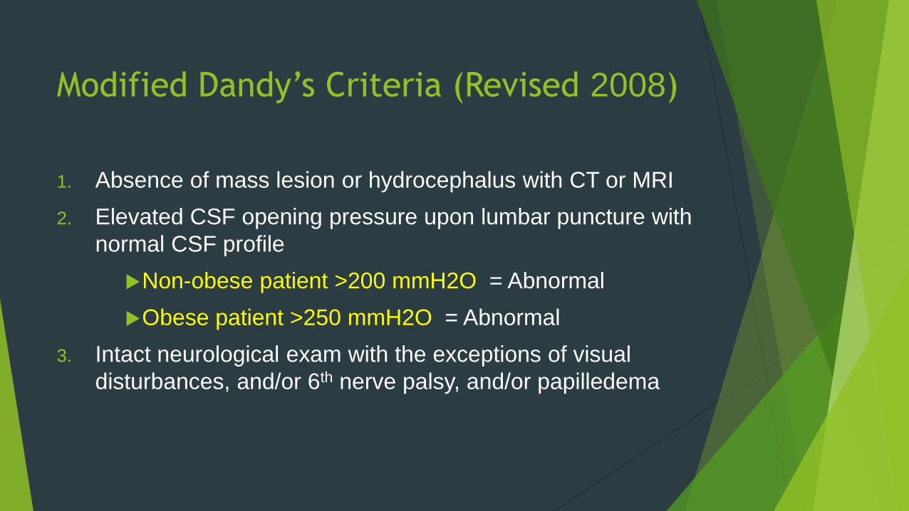
Modified Dandy’s Criteria (Revised 2008)
1. Absence of mass lesion or hydrocephalus with CT or MRI
2. Elevated CSF opening pressure upon lumbar puncture with
normal CSF profile
Non-obese patient >200 mmH2O = Abnormal
Obese patient >250 mmH2O = Abnormal
3. Intact neurological exam with the exceptions of visual
disturbances, and/or 6th nerve palsy, and/or papilledema
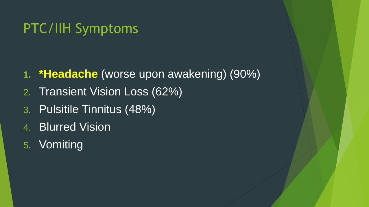
PTC/IIH Symptoms
1. *Headache (worse upon awakening) (90%)
2. Transient Vision Loss (62%)
3. Pulsitile Tinnitus (48%)
4. Blurred Vision
5. Vomiting
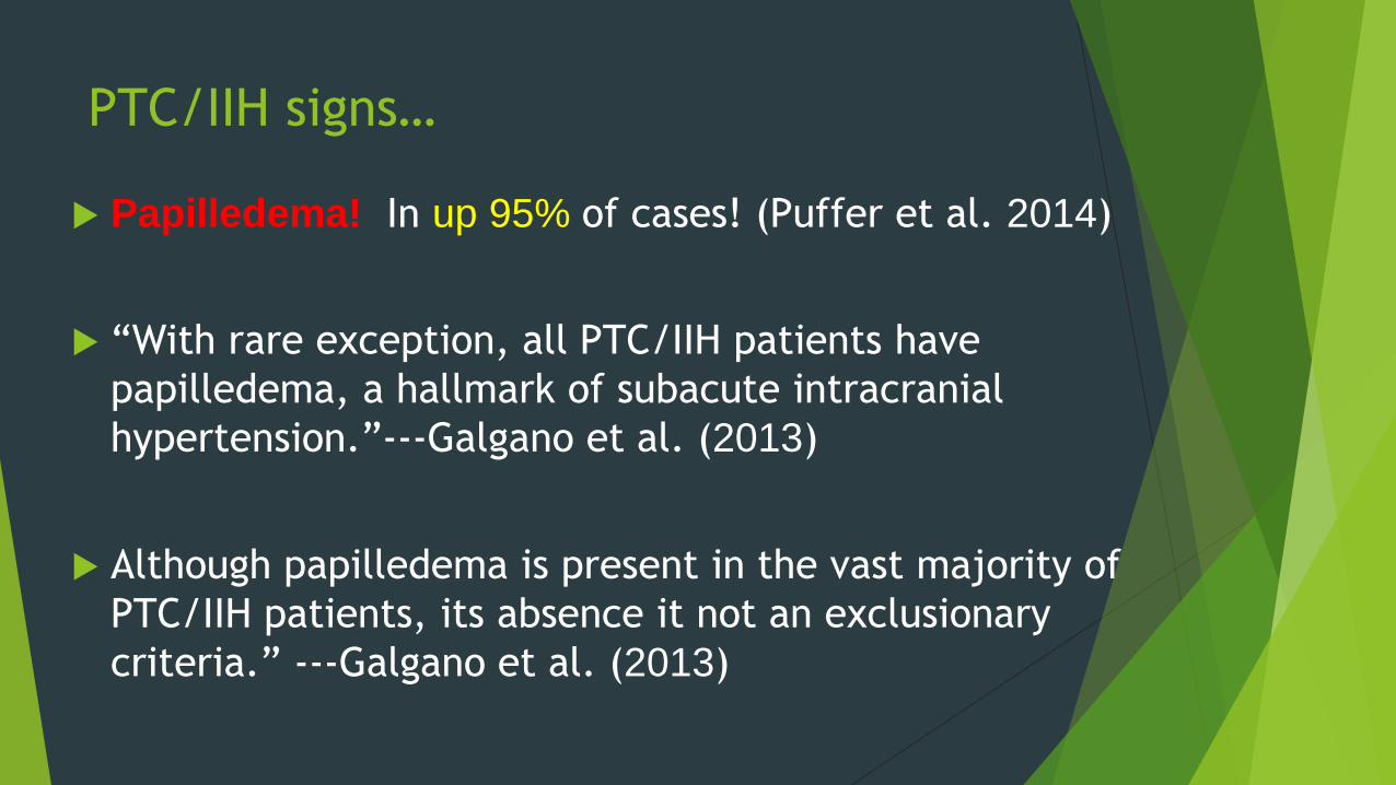
PTC/IIH signs…
Papilledema! In up 95% of cases! (Puffer et al. 2014)
“With rare exception, all PTC/IIH patients have
papilledema, a hallmark of subacute intracranial
hypertension.”---Galgano et al. (2013)
Although papilledema is present in the vast majority of
PTC/IIH patients, its absence it not an exclusionary
criteria.” ---Galgano et al. (2013)
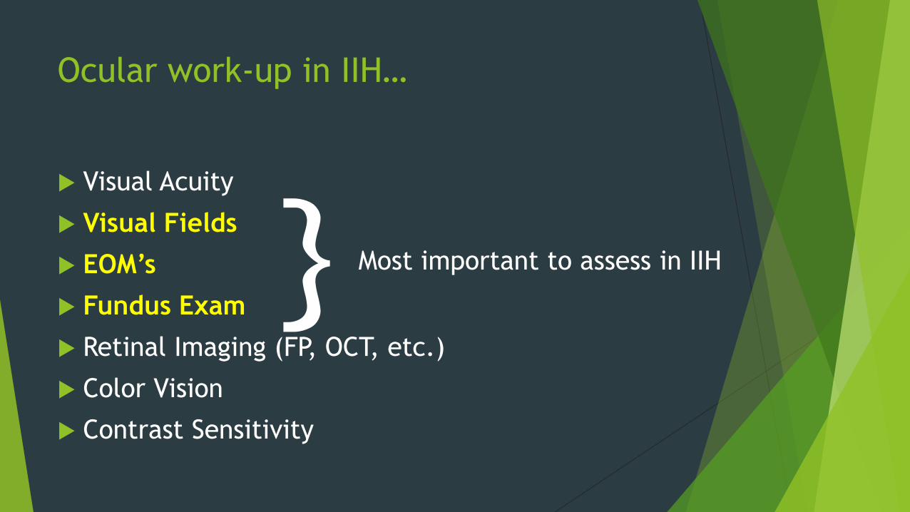
Ocular work-up in IIH…
Visual Acuity
Visual Fields
EOM’s
Fundus Exam
Retinal Imaging (FP, OCT, etc.)
Color Vision
Contrast Sensitivity
} Most important to assess in IIH
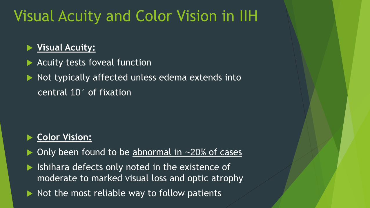
Visual Acuity and Color Vision in IIH
Visual Acuity:
Acuity tests foveal function
Not typically affected unless edema extends into
central 10° of fixation
Color Vision:
Only been found to be abnormal in ~20% of cases
Ishihara defects only noted in the existence of
moderate to marked visual loss and optic atrophy
Not the most reliable way to follow patients
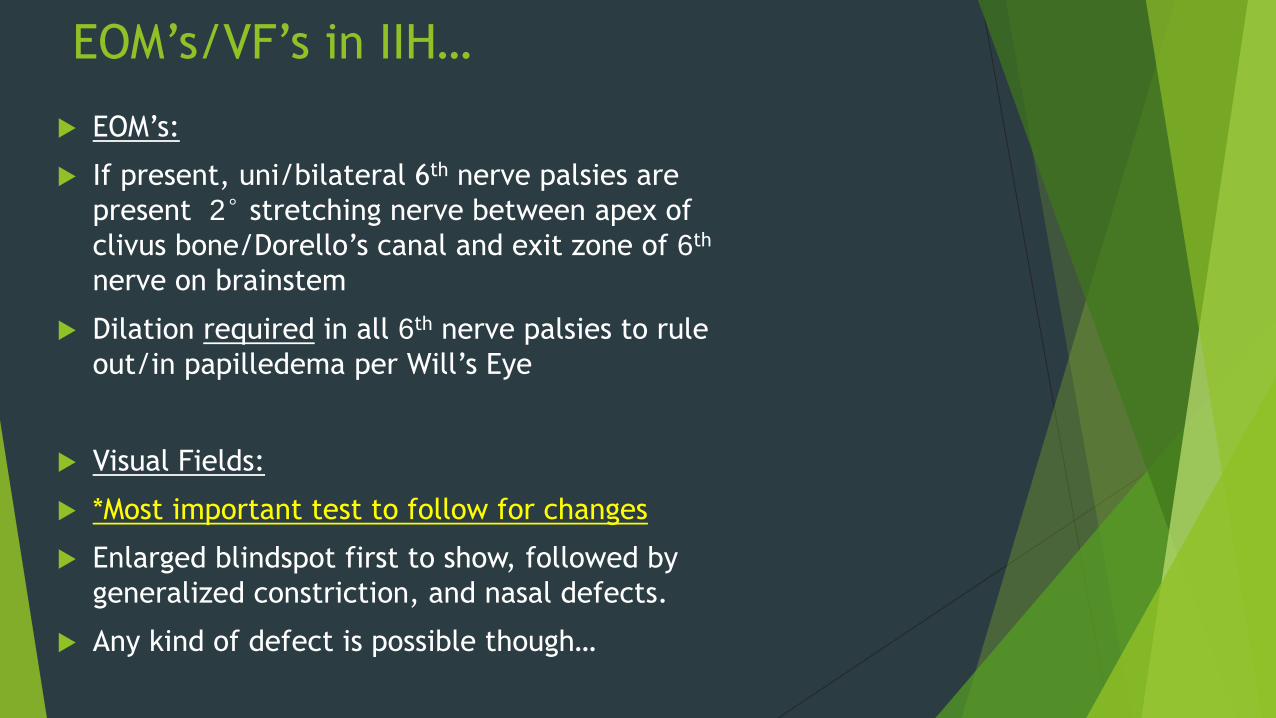
EOM’s/VF’s in IIH…
EOM’s:
If present, uni/bilateral 6th nerve palsies are
present 2° stretching nerve between apex of
clivus bone/Dorello’s canal and exit zone of 6th
nerve on brainstem
Dilation required in all 6th nerve palsies to rule
out/in papilledema per Will’s Eye
Visual Fields:
*Most important test to follow for changes
Enlarged blindspot first to show, followed by
generalized constriction, and nasal defects.
Any kind of defect is possible though…
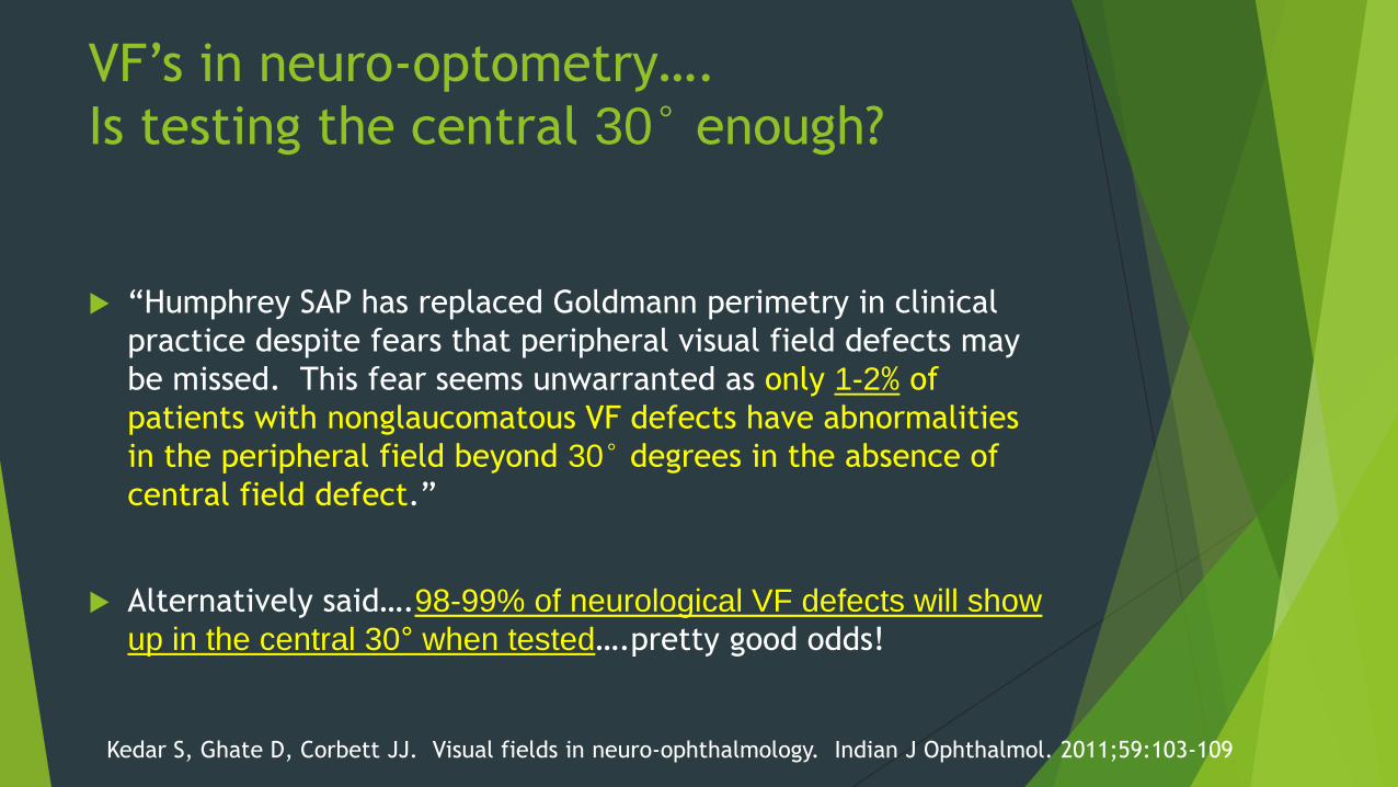
VF’s in neuro-optometry….
Is testing the central 30° enough?
“Humphrey SAP has replaced Goldmann perimetry in clinical
practice despite fears that peripheral visual field defects may
be missed. This fear seems unwarranted as only 1-2% of
patients with nonglaucomatous VF defects have abnormalities
in the peripheral field beyond 30° degrees in the absence of
central field defect.”
Alternatively said….98-99% of neurological VF defects will show
up in the central 30° when tested….pretty good odds!
Kedar S, Ghate D, Corbett JJ. Visual fields in neuro-ophthalmology. Indian J Ophthalmol. 2011;59:103-109
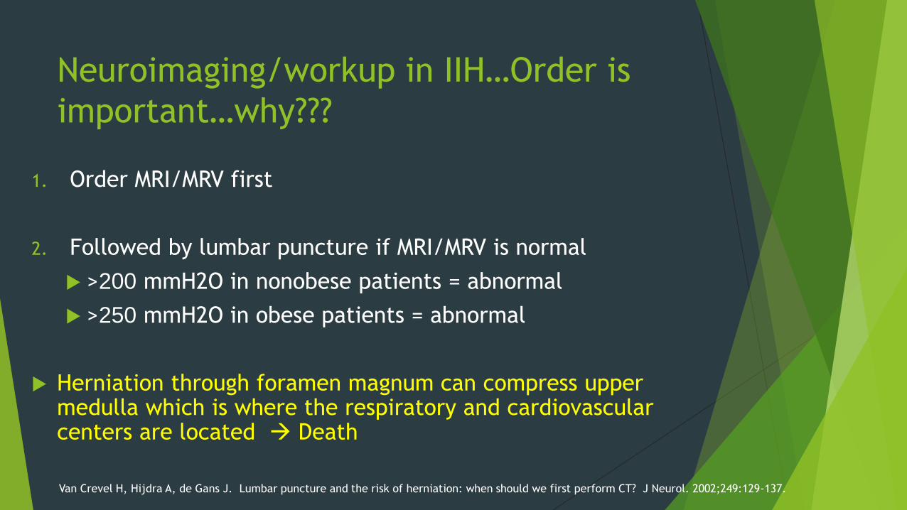
Neuroimaging/workup in IIH…Order is
important…why???
1. Order MRI/MRV first
2. Followed by lumbar puncture if MRI/MRV is normal
>200 mmH2O in nonobese patients = abnormal
>250 mmH2O in obese patients = abnormal
Herniation through foramen magnum can compress upper medulla which is where the respiratory and cardiovascular centers are located Death
Van Crevel H, Hijdra A, de Gans J. Lumbar puncture and the risk of herniation: when should we first perform CT? J Neurol. 2002;249:129-137.
What does herniation look like?
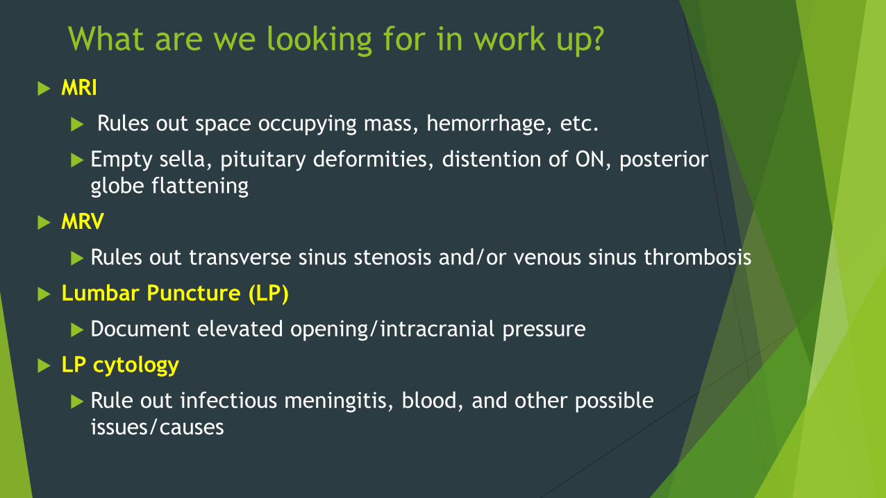
What are we looking for in work up?
MRI
Rules out space occupying mass, hemorrhage, etc.
Empty sella, pituitary deformities, distention of ON, posterior
globe flattening
MRV
Rules out transverse sinus stenosis and/or venous sinus thrombosis
Lumbar Puncture (LP)
Document elevated opening/intracranial pressure
LP cytology
Rule out infectious meningitis, blood, and other possible
issues/causes
CSF Hydrodynamics
Oh crap……you’re kidding me right?
Where is CSF made again???
Answer: Choroid Plexus in Lateral, 3rd, & 4th Ventricles
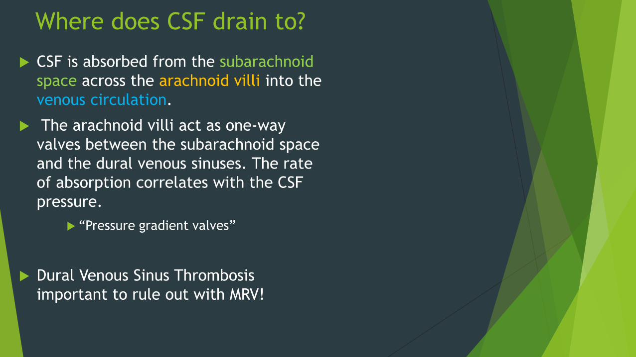
Where does CSF drain to?
CSF is absorbed from the subarachnoid
space across the arachnoid villi into the
venous circulation.
The arachnoid villi act as one-way
valves between the subarachnoid space
and the dural venous sinuses. The rate
of absorption correlates with the CSF
pressure.
“Pressure gradient valves”
Dural Venous Sinus Thrombosis
important to rule out with MRV!
Any blockage along this CSF drainage
pathway or in the venous sinus drainage
can result in increased ICP!
Bottom Line MOA = obstruction of
intracranial venous drainage
Pathophysiology of IIH…
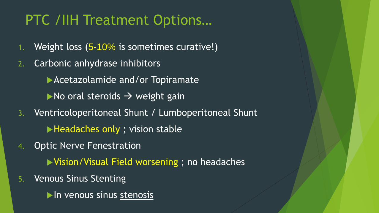
PTC /IIH Treatment Options…
1. Weight loss (5-10% is sometimes curative!)
2. Carbonic anhydrase inhibitors
Acetazolamide and/or Topiramate
No oral steroids weight gain
3. Ventricoloperitoneal Shunt / Lumboperitoneal Shunt
Headaches only ; vision stable
4. Optic Nerve Fenestration
Vision/Visual Field worsening ; no headaches
5. Venous Sinus Stenting
In venous sinus stenosis
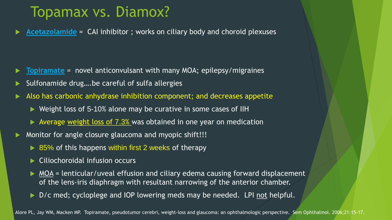
Topamax vs. Diamox?
Acetazolamide = CAI inhibitor ; works on ciliary body and choroid plexuses
Topiramate = novel anticonvulsant with many MOA; epilepsy/migraines
Sulfonamide drug….be careful of sulfa allergies
Also has carbonic anhydrase inhibition component; and decreases appetite
Weight loss of 5-10% alone may be curative in some cases of IIH
Average weight loss of 7.3% was obtained in one year on medication
Monitor for angle closure glaucoma and myopic shift!!!
85% of this happens within first 2 weeks of therapy
Ciliochoroidal infusion occurs
MOA = lenticular/uveal effusion and ciliary edema causing forward displacement
of the lens-iris diaphragm with resultant narrowing of the anterior chamber.
D/c med; cycloplege and IOP lowering meds may be needed. LPI not helpful.
Alore PL, Jay WM, Macken MP. Topiramate, pseudotumor cerebri, weight-loss and glaucoma: an ophthalmologic perspective. Sem Ophthalmol. 2006;21:15-17.
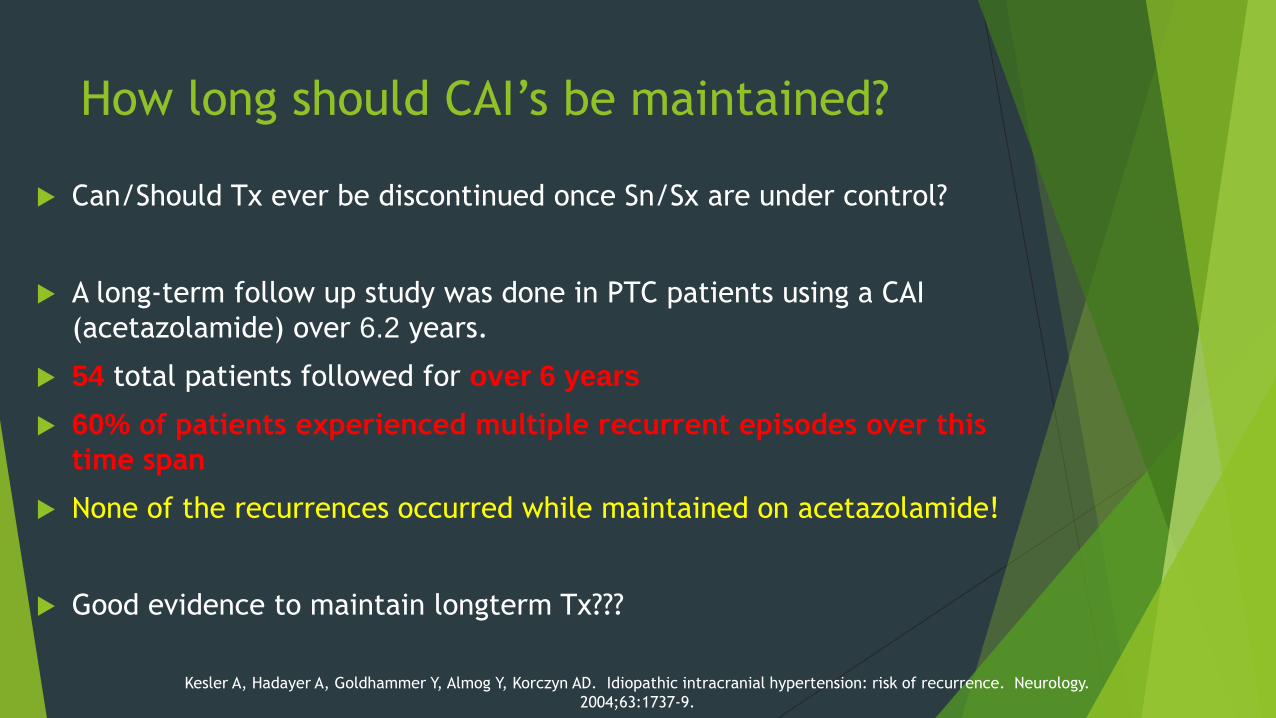
How long should CAI’s be maintained?
Can/Should Tx ever be discontinued once Sn/Sx are under control?
A long-term follow up study was done in PTC patients using a CAI
(acetazolamide) over 6.2 years.
54 total patients followed for over 6 years
60% of patients experienced multiple recurrent episodes over this
time span
None of the recurrences occurred while maintained on acetazolamide!
Good evidence to maintain longterm Tx???
Kesler A, Hadayer A, Goldhammer Y, Almog Y, Korczyn AD. Idiopathic intracranial hypertension: risk of recurrence. Neurology.
2004;63:1737-9.
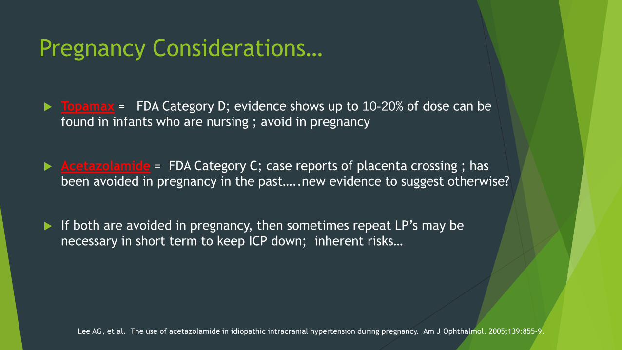
Pregnancy Considerations…
Topamax = FDA Category D; evidence shows up to 10-20% of dose can be
found in infants who are nursing ; avoid in pregnancy
Acetazolamide = FDA Category C; case reports of placenta crossing ; has
been avoided in pregnancy in the past…..new evidence to suggest otherwise?
If both are avoided in pregnancy, then sometimes repeat LP’s may be
necessary in short term to keep ICP down; inherent risks…
Lee AG, et al. The use of acetazolamide in idiopathic intracranial hypertension during pregnancy. Am J Ophthalmol. 2005;139:855-9.
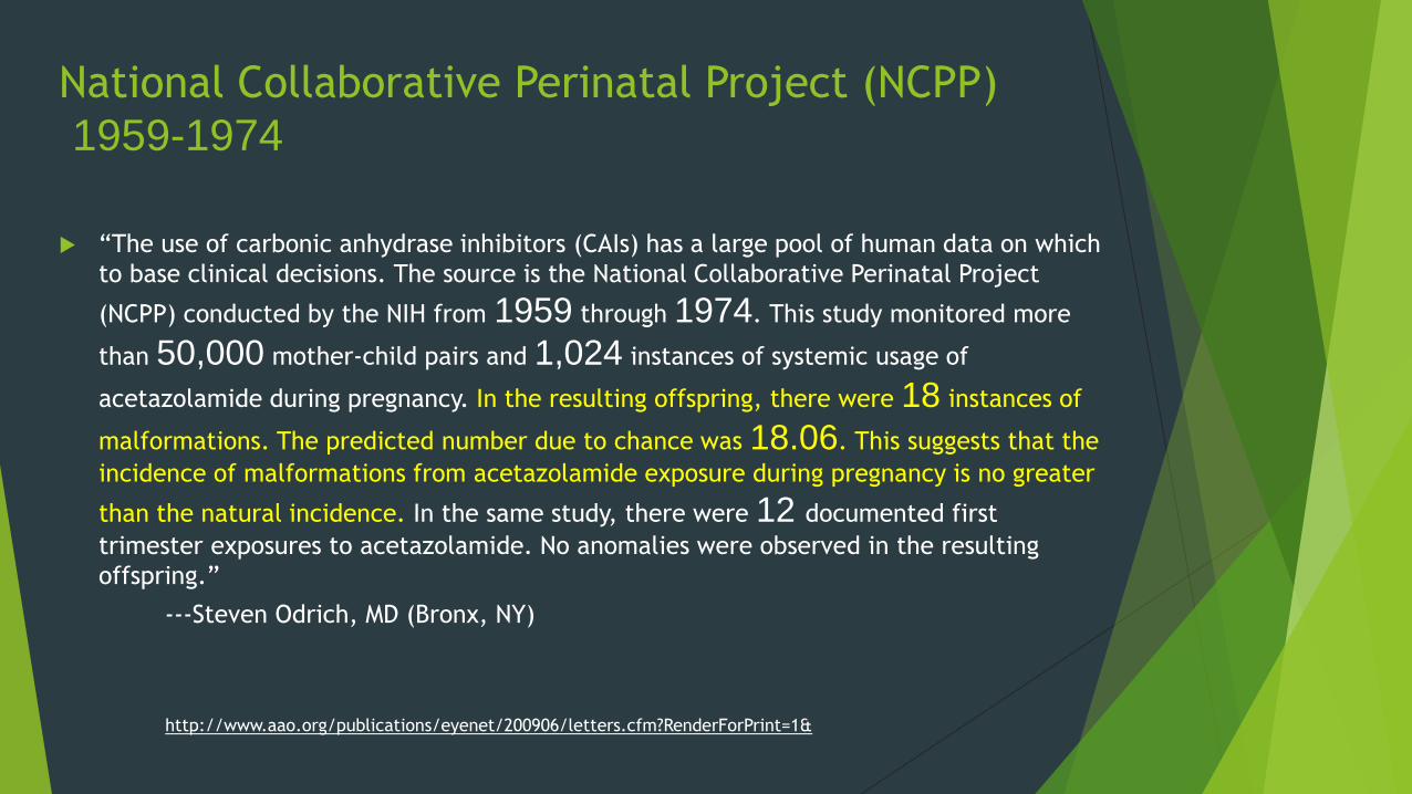
National Collaborative Perinatal Project (NCPP)
1959-1974
“The use of carbonic anhydrase inhibitors (CAIs) has a large pool of human data on which
to base clinical decisions. The source is the National Collaborative Perinatal Project
(NCPP) conducted by the NIH from 1959 through 1974. This study monitored more
than 50,000 mother-child pairs and 1,024 instances of systemic usage of
acetazolamide during pregnancy. In the resulting offspring, there were 18 instances of
malformations. The predicted number due to chance was 18.06. This suggests that the
incidence of malformations from acetazolamide exposure during pregnancy is no greater
than the natural incidence. In the same study, there were 12 documented first
trimester exposures to acetazolamide. No anomalies were observed in the resulting
offspring.”
---Steven Odrich, MD (Bronx, NY)
http://www.aao.org/publications/eyenet/200906/letters.cfm?RenderForPrint=1&
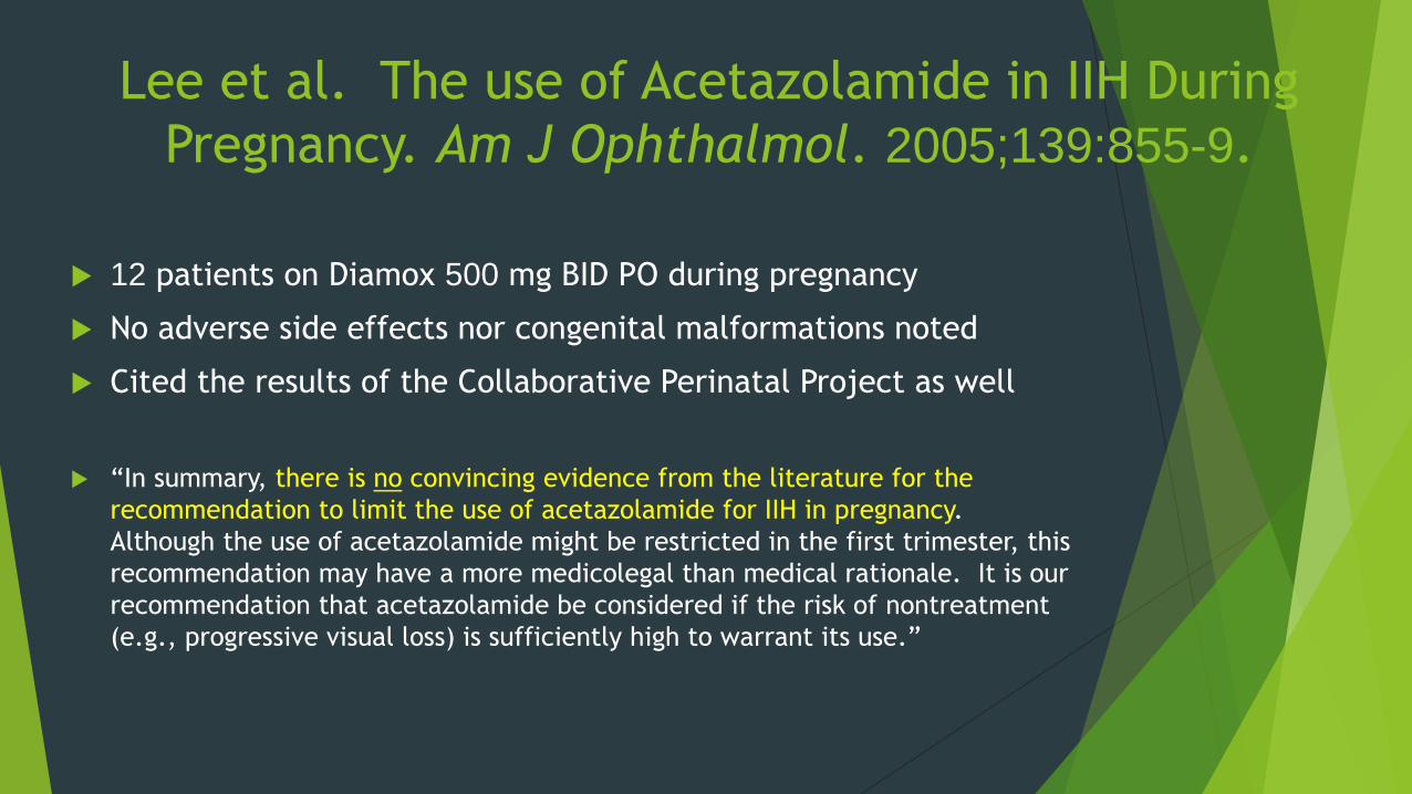
Lee et al. The use of Acetazolamide in IIH During
Pregnancy. Am J Ophthalmol. 2005;139:855-9.
12 patients on Diamox 500 mg BID PO during pregnancy
No adverse side effects nor congenital malformations noted
Cited the results of the Collaborative Perinatal Project as well
“In summary, there is no convincing evidence from the literature for the
recommendation to limit the use of acetazolamide for IIH in pregnancy.
Although the use of acetazolamide might be restricted in the first trimester, this
recommendation may have a more medicolegal than medical rationale. It is our
recommendation that acetazolamide be considered if the risk of nontreatment
(e.g., progressive visual loss) is sufficiently high to warrant its use.”
Question to y’all:
Is it appropriate for O.D.’s to prescribe Diamox
for these patients long term???
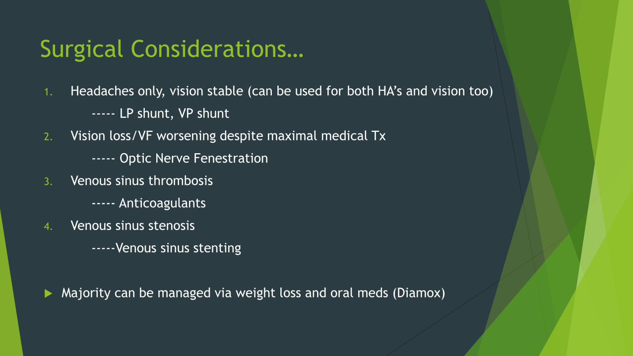
Surgical Considerations…
1. Headaches only, vision stable (can be used for both HA’s and vision too)
----- LP shunt, VP shunt
2. Vision loss/VF worsening despite maximal medical Tx
----- Optic Nerve Fenestration
3. Venous sinus thrombosis
----- Anticoagulants
4. Venous sinus stenosis
-----Venous sinus stenting
Majority can be managed via weight loss and oral meds (Diamox)
1A) Ventriculo-Peritoneal Shunts (VPS)
and
Ventriculo-Atrial Shunts (VAS) in IIH
1B) Lumboperitoneal shunts in IIH
• L3/L4 or L4/L5 spaces most commonly used
• Drain into peritoneal space like VPS
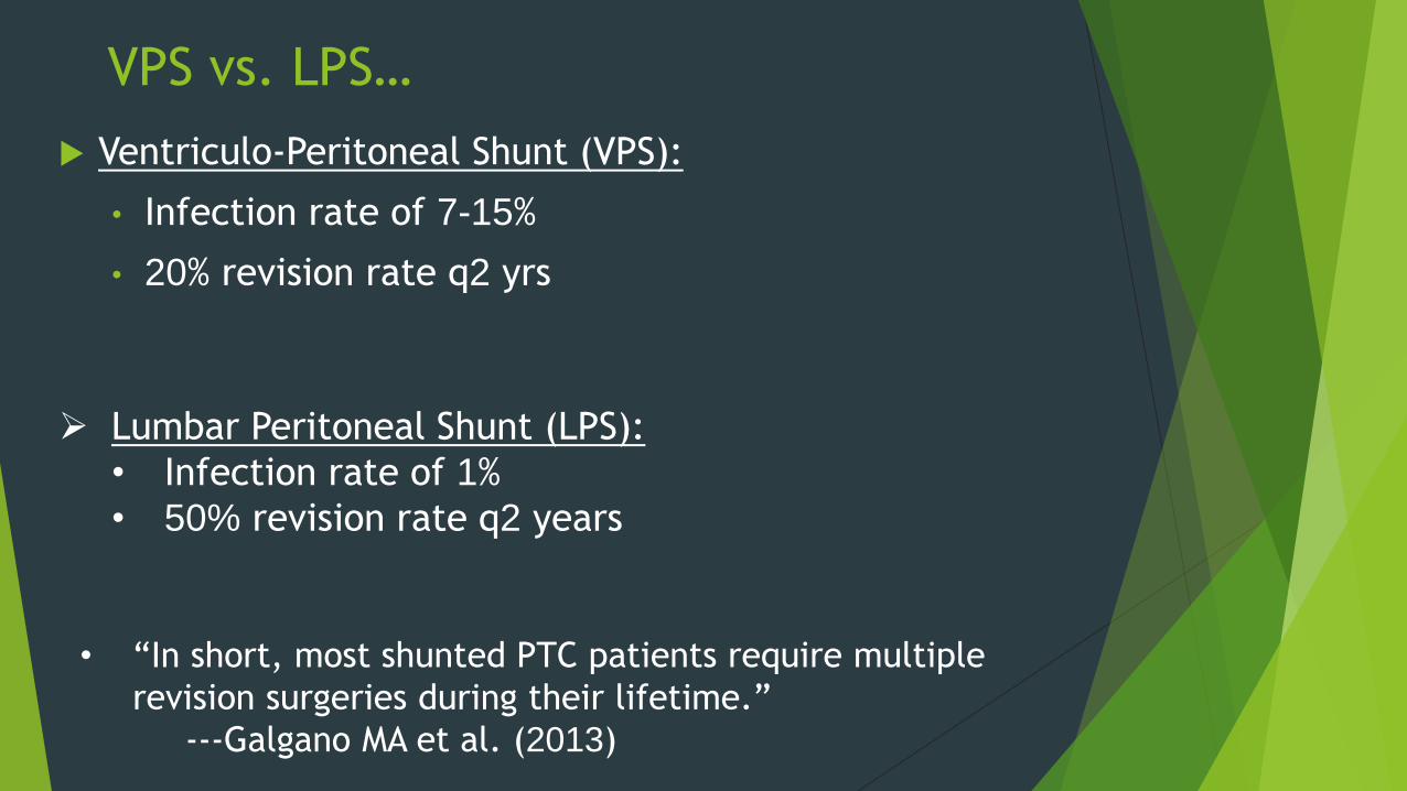
VPS vs. LPS…
Ventriculo-Peritoneal Shunt (VPS):
• Infection rate of 7-15%
• 20% revision rate q2 yrs
Lumbar Peritoneal Shunt (LPS):
• Infection rate of 1%
• 50% revision rate q2 years
• “In short, most shunted PTC patients require multiple
revision surgeries during their lifetime.”
---Galgano MA et al. (2013)
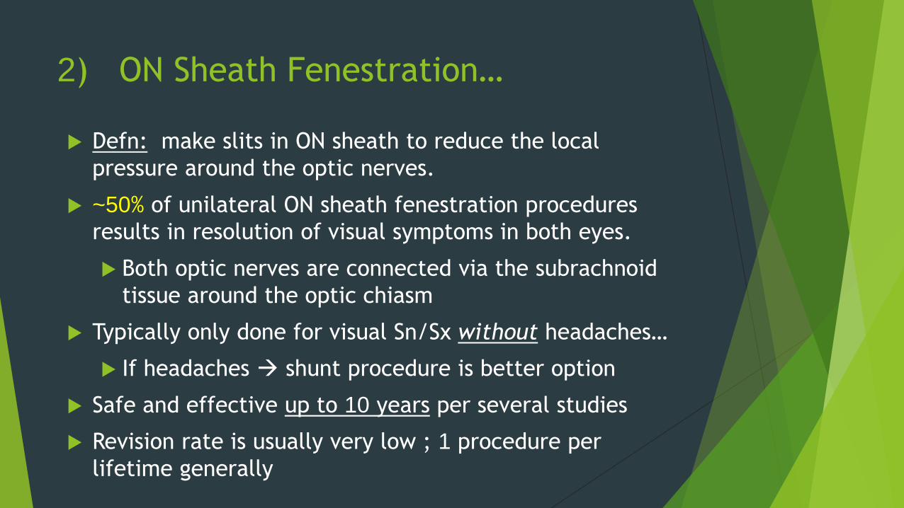
2) ON Sheath Fenestration…
Defn: make slits in ON sheath to reduce the local
pressure around the optic nerves.
~50% of unilateral ON sheath fenestration procedures
results in resolution of visual symptoms in both eyes.
Both optic nerves are connected via the subrachnoid
tissue around the optic chiasm
Typically only done for visual Sn/Sx without headaches…
If headaches shunt procedure is better option
Safe and effective up to 10 years per several studies
Revision rate is usually very low ; 1 procedure per
lifetime generally
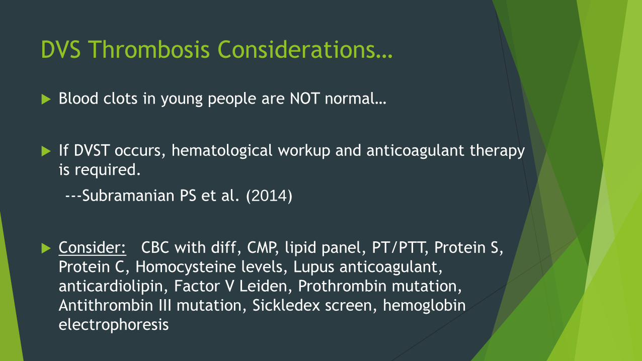
DVS Thrombosis Considerations…
Blood clots in young people are NOT normal…
If DVST occurs, hematological workup and anticoagulant therapy
is required.
---Subramanian PS et al. (2014)
Consider: CBC with diff, CMP, lipid panel, PT/PTT, Protein S,
Protein C, Homocysteine levels, Lupus anticoagulant,
anticardiolipin, Factor V Leiden, Prothrombin mutation,
Antithrombin III mutation, Sickledex screen, hemoglobin
electrophoresis
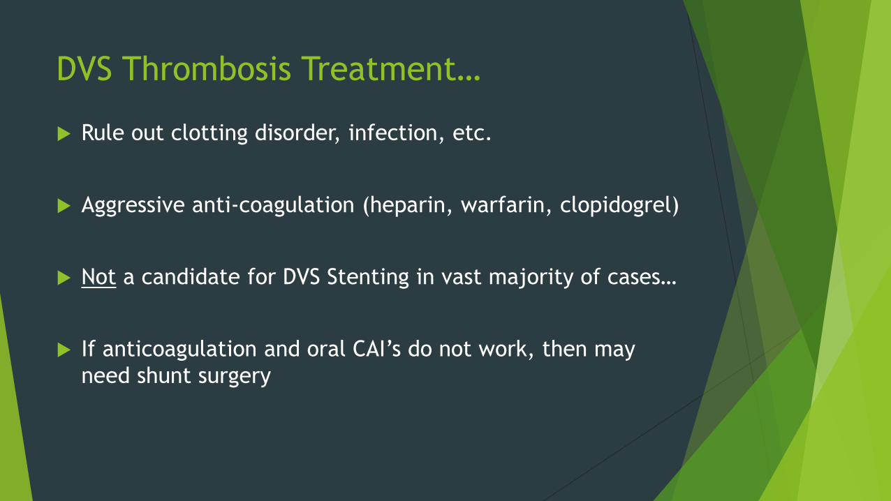
DVS Thrombosis Treatment…
Rule out clotting disorder, infection, etc.
Aggressive anti-coagulation (heparin, warfarin, clopidogrel)
Not a candidate for DVS Stenting in vast majority of cases…
If anticoagulation and oral CAI’s do not work, then may
need shunt surgery

Dural Venous Sinus Stenosis
Dural Venous Sinus Stenosis (DVSS)
Defn: focal, narrowed section of dural venous sinuses
causing back up/turbulent venous blood flow
*Most common at junction of
Sigmoid and Transverse sinuses
Not a true blood clot like DVST is…
Treatment = weight loss, oral CAI,
and/or DVS Stenting procedure
DVSS in IIH vs. Normals…
Focal stenosis has been demonstrated in 90+% of
IIH patients using advanced imaging techniques.
Furthermore, focal stenosis in the same sinus
territory was only demonstrated in 6.8% of
asymptomatic control subjects.
Might be on to something here….
4) Dural Venous Sinus Stenting
Right transverse sinus is dominant in 73% of cases
MOA: Increases drainage of venous blood from
venous sinus system which helps with the
pressure dependent valves, arachnoid villi
granulations, allowing them to clear CSF in to the
venous system more efficiently/quickly
decreasing intracranial pressure
High frequency of resolved or improved HA’s and
papilledema with this method
DVS Stenting…
Not every patient is a candidate for DVS Stenting
Criteria Needed:
1. Presence of venous sinus stenosis (MRI/MRV) ; not thrombosis…
2. Transvenous manometry across the stenosis >10 mmHg differential
Catheter with stent and manometer placed in femoral vein and “fished”
upwards to location of sinus stenosis
Post-Op Medications:
Plavix 75 mg x 6-12 weeks then d/c
ASA 325 mg for life
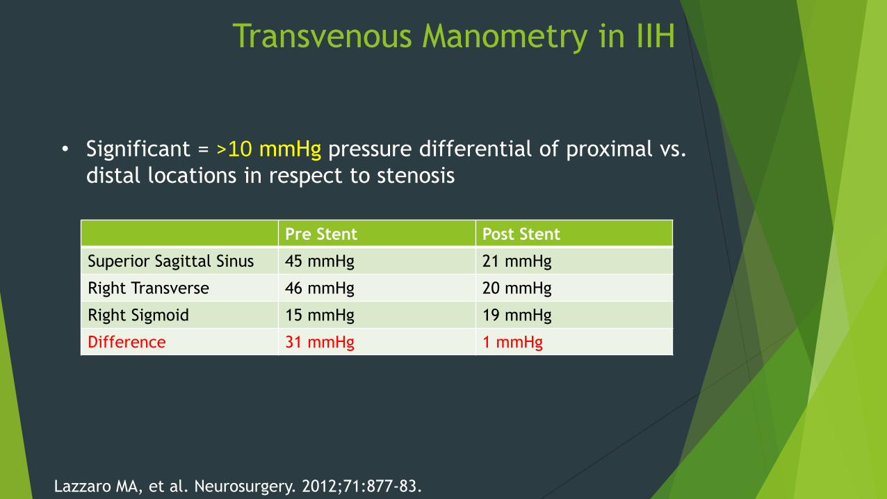
Transvenous Manometry in IIH
• Significant = >10 mmHg pressure differential of proximal vs.
distal locations in respect to stenosis
Lazzaro MA, et al. Neurosurgery. 2012;71:877-83.
Pre Stent Post Stent
Superior Sagittal Sinus 45 mmHg 21 mmHg
Right Transverse 46 mmHg 20 mmHg
Right Sigmoid 15 mmHg 19 mmHg
Difference 31 mmHg 1 mmHg
Dural Venus Sinus Stenting for IIH
Suboccipital/Subtemporal Cranial
Decompression
Very invasive ; but historically pretty successful…
Remove part of skull to allow for more room inside…
Not gold standard anymore
Can be used in severely refractory cases unresponsive to
traditional surgical procedures for ICP and IIH
Optometry IIH/PTC Summary…
Make the diagnosis
Get MRI/MRV
Refer for LP
Neurology should start Diamox/Topamax
Monitor x 1 month post med Tx, then q3-4 months until
resolution/stability (Varies)
Serial OCT, FP, and HVF’s are necessary to gauge Tx/stability
Relay findings to managing neurologist/PCP regularly.
Encourage weight loss
What happened to patient?
“No-showed” to neurologist twice!
Has not returned phone calls or letters…
Phone has been disconnected…
Possible candidate for DVSS? Diamox only? Both?
“You cannot care more for a patient than what they care for themselves.”----Joseph Sowka, OD and Alan Kabat, OD
References Savino PJ, Danesh-Meyer H. Wills Eye Hospital Color Atlas & Synopsis of Clincal Ophthalmology: Neuro-Ophthalmology. McGraw-Hill: Singapore. 2003;74-79.
Lai LT, Danesh-Meyer HV, et al. Visual outcomes and headache following interventions for idiopathic intracranial hypertension. J Clin Neurosci. 2014:
Spitze A, Malik A, Lee AG. Surgical and endovascular interventions in idiopathic intracranial hypertension. Curr Opin Neurol. 2014; 27:69-74.
Subramanian PS, Haq A. Cerebral venous sinus thrombosis and stenosis in pseudotumor cerebri syndrome. Int Ophthalmol Clin. 2014;54:61-71.
Hoffmann J, Goadsby PJ. Update on intracranial hypertension and hypotension. Curr Opin Neurol. 2013;26:240-7.
Galgano MA, Deshaies EM. An update on the management of pseudotumor cerebri. Clin Neurol Neurosurg. 2013;115: 252-9.
Biousse V. Idiopathic intracranial hypertension: diagnosis, monitoring and treatment. Rev Neurol (Paris). 2012;168:673-83.
Puffer RC, Mustafa W, Lanzino G. Venous sinus stenting for idiopathic intracranial hypertension: a review of the literature. J Neurointerv Surg. 2013;5:482-6.
Dykhuizen MJ, Hall J. Cerebral venous sinus system and stenting in pseudotumor cerebri. Curr Opin Ophthalmol. 2011;22:458-62.
Pula JH, Daily J, DeSanto J. Radiology update in neuro-ophthalmology. Curr Opin Ophthalmol. 2011;22:451-7.
Kedar S, Ghate D, Corbett JJ. Visual fields in neuro-ophthalmology. Indian J Ophthalmol. 2011;59:103-9.
Abubaker K, Ali Z, et al. Idiopathic intracranial hypertension: lumboperitoneal shunts versus ventriculoperitoneal shunts--case series and literature review. Br J Neurosurg. 2011;25:94-9.
Rowe FJ. Assessment of visual function in idiopathic intracranial hypertension. Br J Neurosurg. 2011:25:45-54.
Heidary G, Rizzo JF 3rd. Use of optical coherence tomography to evaluate papilledema and pseudopapilledema. Semin Ophthalmol. 2010;25:198-205.
Uretsky S. Surgical interventions for idiopathic intracranial hypertension. Curr Opin Ophthalmol. 2009;20:451-5.
Agid R, Farb RI. Neuroimaging in the diagnosis of idiopathic intracranial hypertension. Minerva Med. 2006;97:365-70.
Alore PL, Jay WM, Macken MP. Topiramate, pseudotumor cerebri, weight-loss and glaucoma: an ophthalmologic perspective. Semin Ophthalmol. 2006;21:15-7.
Lee Ag, Pless M, Falardeau J, Capozzoli T, Wall M, Kardon RH. The use of acetazolamide in idiopathic intracranial hypertension during pregnancy. Am J Ophthalmol. 2005;139:855-9.
National Collaborative Perinatal Project (NCPP), 1959-1974.
Sowka JW, Gurwood AS, Kabat AG. Brain and orbital tumor. The Handbook of Ocular Disease Management: 16th Ed. 2014: 76A-80A.
Lazzaro MA, Darkhabani Z, Remler BF, Hong SH, Wolfe TJ, Zaidat OO. et al. Venous sinus pulsatility and the potential role of dural incompetence in idiopathic intracranial hypertension. Neurosurgery. 2012;71:877-83
Xu K, Yu T, Yuan Y, Yu J. Currrent status of the application of intracranial venous sinus stenting. Int J Med Sci. 2015;12:780-9.
Kanagalingam S, Subramanian PS. Cerebral venous sinus stenting for pseudotumor cerebri: a review. Saudi J Ophthalmol. 2014.