acute inflammatory myelopathies
TRANSCRIPT

UC San FranciscoUC San Francisco Previously Published Works
TitleAcute inflammatory myelopathies
Permalinkhttps://escholarship.org/uc/item/3wk5v9h9
AuthorCree, BAC
Publication Date2014-02-11
DOI10.1016/B978-0-444-52001-2.00027-3 Peer reviewed
eScholarship.org Powered by the California Digital LibraryUniversity of California

Chapter 28
Acute inflammatory myelopathies
BRUCE A.C. CREE*
Department of Neurology, University of California, San Francisco, USA
INTRODUCTION
Spinal cord inflammation canpresentwith symptoms sim-ilar to those of compressive myelopathies: bilateral weak-ness and sensory changes below the spinal cord level ofinjury, often accompanied by bowel and bladder impair-ment and sparing cranial nerve and cerebral function.Because of the widespread availability of magnetic reso-nance imaging (MRI) and computed tomography (CT)imaging, compressive etiologies can be rapidly excluded,leading to the consideration ofnon-compressive etiologiesfor myelopathy. The differential diagnosis of non-compressivemyelopathy is broad and includes infectious,parainfectious, toxic, nutritional, vascular, and systemicas well as idiopathic inflammatory etiologies (Table 28.1).
This review will focus on the idiopathic forms of spi-nal cord inflammation and their relationship to centralnervous system (CNS) demyelinating diseases, systemicinflammatory or autoimmune disease, and asmanifesta-tions of paraneoplastic illness (Table 28.2). Although thepathoetiologies vary widely, the clinical presentations ofthese myelopathies are similar, therefore differentiatingbetween these and other causes of non-compressivemyelopathy can be challenging. In addition to the clinicalpresentation, imaging studies of the spinal cord andbrain, spinal fluid analysis, and serological studies canhelp reveal a diagnosis in many cases.
Although controlled treatment trials have not beenundertaken, the treatment strategy for acute myelitsuses high-dose corticosteroids in nearly all circum-stances in an effort to reduce tissue injury caused byinflammation. In cases refractory to corticosteroid treat-ment, plasmapharesis is sometimes utilized to reduce theserum concentrations of autoantibodies presumed todamage the blood–spinal cord barrier or gray andwhite-matter spinal cord structure. The prognosis forrecovery depends largely on the extent of spinal cord
injury caused by the acute inflammation and the likeli-hood of recurrence differs depending on the etiology.Additional important diagnostic and prognostic featuresinclude whether the myelitis is partial or transverse,febrile illness, the number of vertebral spinal cordsegments involved on MRI at the time of acute attack,the rapidity from symptom onset to maximum deficit,and the severity of involvement.
METHODOLOGIC CONSIDERATIONS
Large observational cohort studies or randomized con-trolled trials concerning myelitis have never been under-taken. Consequently, nearly the entire neurologicknowledge is based on case series and reports. As such,a review of the literature faces the methodologic chal-lenge of not being able to systematically review all casesand case series. Therefore, unintentional biases areinherent in the selection and interpretation of case series.Despite this limitation, certain observations, particularlywhen made by more than one group of investigators,may be clinically useful for formulating a differentialdiagnosis and treatment plan. Potentially useful clinicaland laboratory studies will be reviewed with citation ofrelevant case series. The primary literature consistsexclusively of case reports for certain etiologies of mye-litis. Because keyword indexing is not consistent for casereports, systematic review of all case reports for eachpathoetiology is not practical. Therefore, only select casereports containing observations not found in case seriesare reviewed and cited.
CLINICAL PRESENTATIONANDDEFINITIONS
Recognition of these clinical syndromes localizes thelesion and helps with ordering appropriate imaging stud-ies that can verify the anatomic lesion and provide
*Correspondence to: Bruce A.C. Cree,M.D., Ph.D.,M.C.R., Associate Professor of Clinical Neurology, Clinical Research Director,UCSF Multiple Sclerosis Center, 675 Nelson Rising Lane, Suite 221, San Francisco, CA 94158, USA. Tel: þ1-415-514-2466,
Fax: þ1-415-514-2470, E-mail: [email protected]
Handbook of Clinical Neurology, Vol. 122 (3rd series)Multiple Sclerosis and Related DisordersD.S. Goodin, EditorCopyright © 2014 Bruce Cree. Published by Elsevier B.V. All rights reserved

important clues as to pathoetiology. Although classicexamples of non-compressive myelopathies are givenfor each spinal cord syndrome, in practice inflammationof different etiologies can present with any of these ana-tomic syndromes.
The term transverse myelitis is often used synony-mously with any form of spinal cord inflammation; how-ever, it more specifically refers to inflammation thatinvolves both the anterior and posterior portion of thespinal cord, i.e., the inflammation is transverse fromthe anterior to posterior in the horizontal plane. As such,transversemyelitis typically presents with subacute bilat-eral limb weakness and sensory changes accompanied bybowel and bladder dysfunction without impairment ofcranial nerve and cerebral function. Additional clinicalfeatures that help localize the area of injury include a spi-nal sensory level, diminished or absent reflexes at thelevel of the lesion, hyperreflexia below the level of thelesion, presence of respiratory compromise, and a
Lhermitte’s symptom. When transverse myelitis is bilat-eral and complete, all spinal cord tracts are involved,causing pyramidal, sensory, and autonomic dysfunctionbelow the level of the lesion. Examples of etiologies thatcan cause complete transverse myelitis include neuro-myelitis optica (NMO), paraneoplastic myelopathies,and necrotizing infectious myelitis.
Transverse myelitis may also be unilateral, meaningthe right or left side, as long as there is clinical involve-ment of both anterior and posterior cord function, i.e.,motor weakness and sensory symptoms consistent withdorsal column injury, often with contralateral spinotha-lamic injury. This pattern is also known as the hemicordor Brown-Sequard syndrome (Brown-Sequard, 1849). Inthis setting, pyramidal weakness is accompanied by ipsi-lateral dorsal column dysfunction and contralateral spi-nothalamic loss. Bowel and bladder impairment oftenstill occurs but may be less obvious than with bilateral,complete transverse myelitis. Although the classic
Table 28.1
Differential diagnosis of non-inflammatory myelopathy
Traumatic/compressive Toxic/metabolic
Trauma Vitamin deficiency (B12, B1, E, folate)Disc herniation Nitrous oxide abuseCervical spondylosis with stenosis Abetalipoproteinemia
Epidural abscess or hematoma Medication-induced (amiodarone,Extramedullary and extradural tumors methotrexate, amphotericin, etc.)Cyst (synovial or arachnoid) OrganophosphatesCongenital spinal stenosis Konzo (cassava ingestion)
Posterior longitudinal ligament ossification Lathyrism (legume ingestion)Epidural lipomatosis Heroin/hepatic myelopathyArnold–Chiari malformation Fluorosis
Rheumatoid arthritis or ankylosing Clioquinolspondylitis-associated subluxation Hashimoto’s encephalopathyOsteomyelitis Neoplastic
Paget disease Lymphoma (primary CNS or metastatic)Diffuse idiopathic skeletal hyperostosis LeukemiaExtramedullary hematopoiesis Glioma
Hereditary/neurodegenerative Vascular
Hereditary spastic paraplegia (HSP) Thromboembolic infarctFriedreich’s ataxia Arteriovenous fistulaLeukodystrophies Fibrocartilaginous embolism
Motor neurone disease (ALS, PLS) Hypoperfusion injuryMitochondrial Prothrombotic disorders (infection,Krabbe’s disease neoplasm, vasculitis, DIC, etc.)
Other Arteriovenous malformationSyringomyelia Decompression sickness (Caisson disease)Radiation myelopathy
Superficial siderosisHIV vacuolar myelopathy
ALS, amyotrophic lateral sclerosis; CNS, central nervous system; DIC, disseminated intravascular coagulation; HIV, human immunodeficiency
virus; PLS, primary lateral sclerosis.
614 B.A.C. CREE

Brown-Sequard syndrome is caused by penetrating orcompressive injury to the spinal cord, multiple sclerosis(MS) may also present with this syndrome.
Spinal cord inflammation that spares either the ante-rior or posterior portion of the cord is not consideredtransverse myelitis but is rather classified as partial mye-litis. Partial myelitis may be either unilateral or bilateraland can be associated with sphincter impairment. As in
transversemyelitis, alterations in deep tendon reflexes, asensory level, and Lhermitte symptom can help localizethe level, although respiratory impairment is highlyunusual for partial myelitis. Thus the symptoms of par-tial myelitis may be restricted to only unilateral or evenmonomelic forms with incomplete sensory or motorimpairment. In these circumstances the only clinical fea-ture that may help localize the injury to the spinal cord is
Table 28.2
Differential diagnosis of acute transverse myelitis
Demyelinating Viral – Herpesviruses (DNA)
Multiple sclerosis Herpes simplex virus type-2 (HSV)
Neuromyelitis optica Varicella-zoster virus (VZV)
Idiopathic transverse myelitis Cytomegalovirus (CMV)
Acute disseminated encephalomyelitis (ADEM) Human herpesvirus 6 and 7 (HHV)
Postvaccinial Epstein–Barr virus (EBV)Associated with acute demyelinating polyneuropathy Viral – Paramyxoviruses (RNA)
Systemic autoimmune disease Measles
Systemic lupus erythmatosus (SLE) MumpsPrimary Sj€ogren syndrome Viral – Orthomixovirus (RNA)
Neurosarcoidosis Influenza A virus (including H1N1)
Behcet’s disease Viral – Picornaviruses (RNA)
Mixed connective tissue disease (MCTD) Coxsackieviruses A and BSystemic sclerosis Enterovirus-70 and -71
Vogt–Koyanagi–Harada Echovirus 30Primary angiitis of the central nervous system Hepatitis B, C, EAtopic myelitis Poliovirus 1, 2, and 3Paraneoplastic Viral – Flaviviruses (RNA)
Anti-amphiphysin (breast carcinoma) West Nile virusAnti-CRMP-5 (small cell lung cancer) Japanese encephalitis virusNecrotizing myelopathy Tick-borne encephalitis virus
Bacterial St. Louis encephalitis virusMycoplasma pneumoniae Dengue virusBorrelia burgdorferi (Lyme disease) Orthoretroviruses (RNA)
Treponema pallidum (syphilis) HTLV-1 and 2Mycobacterium tuberculosis (TB) HIVBrucella melitensis (brucellosis) Parasitic
Salmonella non-typhi Neurocysticercosis
Salmonella para-typhi B SchistosomaScrub typhus Gnathostoma angiostrongylosisBartonella henselae (cat-scratch) Larva migrans
Listeria monocytogenes Angiostrongylosis cantonensisLeptospirosis ToxoplasmosisTrophymera whipplei (Whipple’s) Trypanosomiasis
Coxiella burnetiiFungal
ActinomycesCoccidiodesAspergillusBlastomyces dermatidesCladophialophoro bantianaCryptococcus
Those in bold indicate common causes for transverse myelitis.
HIV, human immunodeficiency virus; HTLV, human T-lymphotropic virus; TB, tuberculosis.
ACUTE INFLAMMATORY MYELOPATHIES 615

the absence of cortical or cranial nerve symptoms. By farthe most common cause of partial myelitis is MS.
One form of a partial myelitis is the posterior cordsyndrome in which only the dorsal columns are affectedeither unilaterally or bilaterally, resulting in loss of finetouch, vibration, and proprioception but without corti-cospinal or anterior spinothalamic tract involvement.Clinically, patients will manifest limb incoordinationdue to proprioceptive loss but will have full strengthand intact pain and temperature sensation.
When the anterior cord is selectively partially affected,pyramidal weakness will be the primary clinical manifes-tation. The spinal thalamic tracts may also be affected,resulting in loss of pain and temperature sensation. Auto-nomic impairment is also common with loss of sphincterfunction. As with other types of partial myelitis, theinvolvementmay be unilateral or bilateral. When the ante-rior horn cells are damaged the weakness will be flaccidand deep tendon reflexes will be absent at the level ofinjury. Because the dorsal columns are spared, vibrationand proprioception sensation remain intact. The classicexample of an anterior cord syndrome is infarction ofthe spinal cord caused by anterior spinal artery occlusion,although MS can also cause this syndrome. Selectiveinvolvement of the anterior horns of the spinal cord result-ing in flaccid ascending paralysis is a hallmark of polio-myelitis, an infection that was nearly eradicated but ison the resurgence due to failure of global vaccinationefforts (Heymann and Aylward, 2004).
Inflammatory injury to the central cord results in aclinical syndrome similar to that caused by a syrinx.The crossing spinothalamic fibers are affected, resultingin dissociated sensory loss. Pain and temperature sensa-tion are impaired below the level of the lesion whereasfine touch, vibration, and proprioception remain intact.Central cord injury is often accompanied by corticosp-inal and autonomic impairment below the level of thelesion. Although a central cord syndrome occurs inNMO, it is relatively uncommon in MS.
The conus medullaris may be selectively affected,causing sphincter dysfunction and sacral sensory loss.Although motor impairment can occur, it is typically rel-atively mild compared to the autonomic and sensory lossbecause of selective involvement of the sacral spinalcord segments. Conus medullaris inflammation mayspread caudally, resulting in an ascending transversemyelitis. Parainfectious etiologies such as postviral mye-litis typify the conus syndrome.
Cauda equina inflammation can occur, resulting insensory loss, motor impairment, and autonomic dys-function in the distribution of the affected nerve roots.Flaccid weakness affects the lower limbs and is oftenasymmetric. Although cauda equina inflammation is apolyradiculitis and not myelitis, it sometimes can extend
into the conus medullaris, resulting in a mixed polyradi-culitis with myelitis syndrome. Infectious and parainfec-tious causes of ascending polyradiculopathies includeneuroborreliosis, cytomegalovirus, and acute inflamma-tory demyelinating polyradiculopathy.
The exception to the rule that cranial nerve symptomsare spared with spinal cord injury is the trigeminal nerve.Afferent sensory fibers of the trigeminal nerve descendthrough the upper cervical cord prior to decussating andascending to the thalamic nuclei. Therefore, with high cer-vical cord lesions, partial facial sensory loss may occur.
DIAGNOSTIC EVALUATION
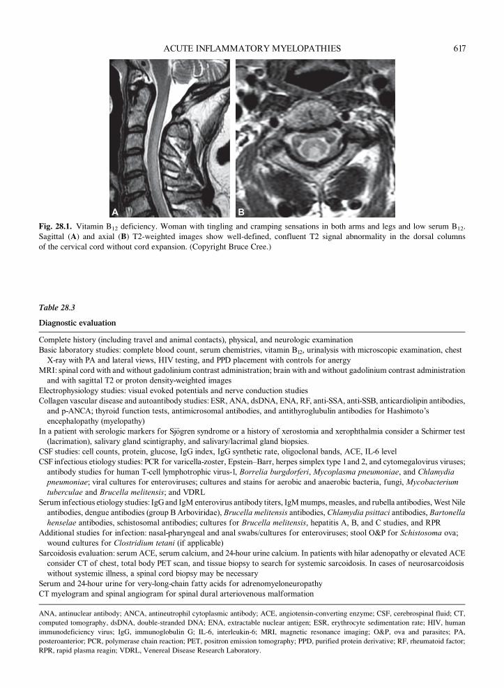
The diagnostic evaluation of patients presenting withacute myelopathy begins with a detailed clinical history,including full review of systems, as well as past medical,family, social, and travel histories. Important clues todiagnostic etiology can be garnered from basic labora-tory studies that include a complete blood count with dif-ferential, serum chemistries, as well as tests for commonmetabolic diseases that may present with acute myelop-athy such as vitamin B12 deficiency (Fig. 28.1). Neuroim-aging and cerebrospinal fluid (CSF) evaluation are alsocrucial diagnostic studies. Additional laboratory studiesmay include visual evoked potentials (VEPs) andsomatosensory evoked potentials (SSEPs) if structuralimaging is not revealing. If an infectious etiology is sus-pected by the presence of fever, cough, rash, or historyof exposure, then specific tests can be ordered to con-firm the precise infectious etiology. A comprehensiveapproach to the diagnostic evaluation of acute myelop-athies, with consideration of the potentially very broaddifferential, is presented in Table 28.3.
Although beyond the scope of this review, numerousinfectious etiologies have been associated with myelitis(Table 28.2). Additional symptoms, signs, and aspectsof the patient’s historymay suggest an infectious etiology.For example, the presence of fever, meningismus, rash(lyme, zoster, enterovirus), cough (Mycoplasma pneumo-niae, Chlamydia pneumoniae), diarrhea (enterovirus,Salmonella), an immunocompromised state (herpeszoster, cytomegalovirus), a history of recent travel (tuber-culosis, parasitic infections), recurrent genital infection(herpesvirus), mosquito bite (West Nile virus), radicularburning pain with or without vesicles suggestive of zosterradiculitis, or adenopathy may suggest specific infectiousetiologies. Infectious etiologies of myelitis can be viral,bacterial, fungal, and, rarely, parasitic. It is always impor-tant to consider treatable infections such as syphilis,herpesviruses, human immunodeficiency virus (HIV),Lyme disease, and tuberculosis. The most commonlyimplicated viruses are West Nile virus, varicella-zoster,herpes simplex type-2, and cytomegalovirus.
616 B.A.C. CREE
A B
Fig. 28.1. Vitamin B12 deficiency. Woman with tingling and cramping sensations in both arms and legs and low serum B12.
Sagittal (A) and axial (B) T2-weighted images show well-defined, confluent T2 signal abnormality in the dorsal columns
of the cervical cord without cord expansion. (Copyright Bruce Cree.)
Table 28.3
Diagnostic evaluation
Complete history (including travel and animal contacts), physical, and neurologic examinationBasic laboratory studies: complete blood count, serum chemistries, vitamin B12, urinalysis with microscopic examination, chest
X-ray with PA and lateral views, HIV testing, and PPD placement with controls for anergyMRI: spinal cord with and without gadolinium contrast administration; brain with and without gadolinium contrast administrationand with sagittal T2 or proton density-weighted images
Electrophysiology studies: visual evoked potentials and nerve conduction studiesCollagen vascular disease and autoantibody studies: ESR, ANA, dsDNA, ENA, RF, anti-SSA, anti-SSB, anticardiolipin antibodies,and p-ANCA; thyroid function tests, antimicrosomal antibodies, and antithyroglubulin antibodies for Hashimoto’s
encephalopathy (myelopathy)In a patient with serologic markers for Sj€ogren syndrome or a history of xerostomia and xerophthalmia consider a Schirmer test(lacrimation), salivary gland scintigraphy, and salivary/lacrimal gland biopsies.
CSF studies: cell counts, protein, glucose, IgG index, IgG synthetic rate, oligoclonal bands, ACE, IL-6 level
CSF infectious etiology studies: PCR for varicella-zoster, Epstein–Barr, herpes simplex type 1 and 2, and cytomegalovirus viruses;antibody studies for human T-cell lymphotrophic virus-1, Borrelia burgdorferi, Mycoplasma pneumoniae, and Chlamydiapneumoniae; viral cultures for enteroviruses; cultures and stains for aerobic and anaerobic bacteria, fungi, Mycobacteriumtuberculae and Brucella melitensis; and VDRL
Serum infectious etiology studies: IgG and IgM enterovirus antibody titers, IgMmumps, measles, and rubella antibodies,West Nileantibodies, dengue antibodies (group B Arboviridae), Brucella melitensis antibodies, Chlamydia psittaci antibodies, Bartonellahenselae antibodies, schistosomal antibodies; cultures for Brucella melitensis, hepatitis A, B, and C studies, and RPR
Additional studies for infection: nasal-pharyngeal and anal swabs/cultures for enteroviruses; stool O&P for Schistosoma ova;wound cultures for Clostridium tetani (if applicable)
Sarcoidosis evaluation: serum ACE, serum calcium, and 24-hour urine calcium. In patients with hilar adenopathy or elevated ACEconsider CT of chest, total body PET scan, and tissue biopsy to search for systemic sarcoidosis. In cases of neurosarcoidosiswithout systemic illness, a spinal cord biopsy may be necessary
Serum and 24-hour urine for very-long-chain fatty acids for adrenomyeloneuropathy
CT myelogram and spinal angiogram for spinal dural arteriovenous malformation
ANA, antinuclear antibody; ANCA, antineutrophil cytoplasmic antibody; ACE, angiotensin-converting enzyme; CSF, cerebrospinal fluid; CT,
computed tomography, dsDNA, double-stranded DNA; ENA, extractable nuclear antigen; ESR, erythrocyte sedimentation rate; HIV, human
immunodeficiency virus; IgG, immunoglobulin G; IL-6, interleukin-6; MRI, magnetic resonance imaging; O&P, ova and parasites; PA,
posteroanterior; PCR, polymerase chain reaction; PET, positron emission tomography; PPD, purified protein derivative; RF, rheumatoid factor;
RPR, rapid plasma reagin; VDRL, Venereal Disease Research Laboratory.
ACUTE INFLAMMATORY MYELOPATHIES 617

Neuroimaging
Once a clinician has recognized that a spinal cord injuryhas occurred, a next step in the diagnostic evaluation is todetermine whether the myelopathy is non-compressiveor caused by compression of the cord using imaging.MRI is the preferred imaging modality because of thesignificantly superior ability to visualize the spinal corditself as well as other soft-tissue structures compared toCT. When the cause of the myelopathy is unknown, inalmost all cases infusion of the MRI contrast agentgadolinium-diethylenetriaminepentacetate (DTPA) isindicated. Gadolinium-DTPA is useful to demonstratedisruption of the blood–spinal cord barrier, as in the set-ting of active spinal cord inflammation or infarct, or toshow increased blood flow such as found with tumorsand arteriovenous malformations.
However, if an MRI cannot be obtained emergently,CTmyelography is a reasonable alternative. Themain dis-advantage of this imagingmodality is the limited ability tovisualize the spinal cord. It is important to keep in mindthat determining the region of the spinal cord to imagebased on clinical features in some cases can miss a com-pressive lesion just superior to the field of imaging thatcould be surgically decompressed. This is especially thecase if the suspected lesion is near the cervicothoracicor thoracolumbar junction because cord lesions can causeclinical deficits that localize to lower spinal cord segment.For this reason, it is always best to image the region wherethe clinical signs and symptoms localize, as well as thesuperior spinal cord, possibly using a sagittal survey.
Once the MRI excludes compressive etiology, addi-tional imaging characteristics may be helpful with thedifferential diagnosis, particularly the appearance andpattern of the lesion(s). Table 28.4 describes the imagingpatterns associated with some of the more commoncauses of acute transverse myelitis (ATM). Followingreview of the spinal imaging, a brain MRI should be per-formed to determine if other demyelinating lesionswithin the CNS are present. Patients with MS andNMO are much more likely to have lesions on the brainMRI. In addition,MS-associated spinal cord lesions tendto be asymmetric, peripherally located within the cordaxis, and tend to extend over fewer than two spinal cordsegments (Fig. 28.2). Patients with NMO are more likelyto have lesions that extend over three ormore spinal cordsegments and tend to be centrally located. Approxi-mately 25% are associated with cord swelling, andmay have patchy gadolinium enhancement (Fig. 28.3).
Cerebrospinal fluid
Once neuroimaging has excluded a compressive etiol-ogy, the next step in the diagnostic work-up is a lumbar
puncture to determine if there are signs of inflammationwithin the CSF. If the CSF is non-inflammatory then vas-cular, toxic/metabolic, neurodegenerative, or neoplasticmyelopathies become much more likely and the subse-quent work-up should focus on these etiologies.
CSF is an essential component of the evaluation ofevery patient with suspected myelitis (Table 28.5). Aftermeasuring the opening pressure, CSF studies shouldinclude cell count with differential, protein, and glucose.Measurements of intrathecal immunoglobulin synthesiswith oligoclonal bands (OCBs) and an immunoglobulinG (IgG) index or synthesis rate must be sent on everypatient. This requires drawing a serum sample at the timeof the lumbar puncture for comparative analysis of gam-maglobulins and should be performed on every patientwith suspected myelitis. In addition, cytology for evalu-ation of neoplasm should be included. If the CSF showssigns of inflammation (pleiocytosis, elevated protein,OCBs, or elevated IgG index), then the subsequent diag-nostic studies should focus on demyelinating, infectious,or other inflammatory causes of acute myelitis (AM).
The evaluation of common infectious causes of mye-litis includes: Venereal Disease Research Laboratory(VDRL), Lyme Western blot, and polymerase chainreaction (PCR) studies for Herpetoviridae (varicella-zoster virus, herpes simplex virus types 1 and 2, cytomeg-alovirus, human herpesvirus 6 and 7, Epstein–Barrvirus), West Nile virus, and tuberculosis (Table 28.6).In addition, bacterial, fungal, and acid-fast bacilluscul-tures should be considered. One milliliter of acellularsupernatant should be sent for interleukin-6 (IL-6)enzyme-linked immunosorbent assay (see section onimmunology, below). Lastly, several milliliters of frozenCSF sample should be reserved for additional PCR stud-ies. As in neuroimaging, certain CSF patterns or findingsmay be helpful in narrowing the differential diagnosis.
A low CSF glucose (less than 60% of serum glucose)generally suggests an infection (fungal, bacterial, ormycobacterial), especially when associated with elevatedCSF protein. However, an isolated low CSF glucose canoccur in neurosarcoidosis, leptomeningeal carcinomato-sis, subarachnoid hemorrhage, and even systemic lupuserythematosus (SLE) with CNS involvement. An elevatedprotein is the most common CSF abnormality in patientswith spinal cord disease and is present in approximately50% of patients with transverse myelitis. However, ele-vated CSF protein is non-specific and is associated withspinal cord tumors, paraneoplastic myelopathies, radia-tion myelopathies, vascular malformations, infection,syringomyelia with spinal block, and spinal cord trauma.
Elevation in the CSF white blood cell count (WBC)defines inflammatory myelitis. The WBC differentialcan be very helpful in understanding whether an infec-tious or autoimmune process is at play. The presence
618 B.A.C. CREE

Table 28.4
Imaging in acute myelitis
Etiology ofmyelitis Location Lesion length Pattern of T2 involvement
Pattern of contrastenhancement
Cord swelling(enlargement) Other characteristics
MS 60–75% cervical �2 cordsegments
Peripheral, ovoid, paracentral �15% of cord plaquesenhance
Atypical (atrophymore common
>50% have multiple lesions, ofwhich �50% are clinically silent
NMO �80% cervical �3 cordsegments
Centrally located, can bedorsal
Patchy �25% �25% will have an average of 3–4brain lesions
Idiopathic TM Usually thoracic �3 cordsegments
Diffuse, patchy, or peripheral Variable (diffuse, patchy,peripheral)
Variable Many infectious etiologies havesame profile (diagnosis of
exclusion)ADEM Usually thoracic Variable Multifocal, flame-shaped,
and can be largeVariable Common Myelitis occurs in 11–28% of cases;
meningeal enhancement is
unusualSarcoidosis Cervical or thoracic Variable Central (62%) > anterior,
lateral, posteriorUsually patchy, but can bediffuse, nodular,
multifocal,leptomeningeal
Up to 35 May involve the intradural nerveroots. Thickening of the roots
may occur. About 25% with >1lesion
VZV Usually thoracic Variable Typically posterior (Hiraiet al., 1996)
Patchy or focal atdermatomal level
Common Enhancement may involve thedorsal root
CMV Cauda equina andconus medullaris
Variable Thickened cauda equina Leptomeningeal, dorsalroot, and diffuse nerveenhancement
Can cause a focalspace-occupyinglesion
Usually a polyradiculitis
HSV Variable Can be >1 lesion Diffuse Can occur HSV-2 >> HSV-1 causes myelitis.Can have associated hemorrhage.
Poliomyelitis Variable Increased signal in anterior
horns
Anterior horns Focal Can also be seen in postvaccinial
poliomyelitis, West Nile virus,enterovirus-71, or Lyme
Lyme Variable Can be normal or nodular,
increased signal
Nodular leptomeningeal,
intraspinal or anteriorhorn cell
Focal Can cause ATM, meningomyelitis,
or polio-like paralytic syndrome
Paraneoplastic Variable Variable, can be holocord,or highly specific symmetric
tract involvement
Patchy Antiampiphysin
ADEM, acute disseminated encephalomyelitis; ATM, acute transverse myelitis; CMV, cytomegalovirus; HSV, herpes simplex virus; MS, multiple sclerosis; NMO, neuromyelitis optica; TM, transverse
myelitis; VZV, varicella-zoster virus.

A B C
Fig. 28.3. Neuromyelitis optica (NMO). Magnetic resonance images shown include sagittal T2 (A), axial gadolinium-
enhanced T1 (B). Longitudinally extensive T2 signal abnormality in the cervical cord (A), accompanied by patchy intrame-
dullary enhancement on gadolinium-enhanced T1-weighted imaging (B). The patient subsequently developed monocular
vision loss and was seropositive for the NMO-immunoglobulin G (IgG) antibody. The brain magnetic resonance imaging
scan was normal (C). (Copyright Bruce Cree.)
A B C
Fig. 28.2. Multiple sclerosis. Sagittal T2-weighted (A) and sagittal T1 plus gadolinium (B) cervical spine images in a patient with
multiple sclerosis presenting with partial myelitis. Note the sharply marginated, short-segment plaque that is peripherally located
within the cord axis and predominantly located within the white matter of the cervical spinal cord. The patient also had multiple
plaques in the periventricular white matter (C). (Copyright Bruce Cree.)
620 B.A.C. CREE

Table 28.5
Cerebrospinal fluid (CSF) studies for evaluation of acute transverse myelitis
Studies Volume CSF Method Considerations
Cell count withdifferential
1 mL 1. Hemocytometer for count2.Wright-stained cytocentrifugepreparation for differential
CSF specimens should be transported at ambient temperatureas soon as possible after collection. Cellular degenerationof CSF can begin within 1 hour of collection
Glucose 0.5 mL Spectrophotometric(glucose oxidase)
Measure the serum glucose as well. CSF levels are usually>55% of serum glucose and >40 mg/dL. As serumglucose rises above 200 mg/dL, the CSF/serum ratio fallsfrom about 0.55 to a minimum of 0.31. Sample can be
stable for up to 10 days if refrigeratedTotal protein 0.5 mL Spectrophotometric
(pyrogallol red)Sample can be stable for up to 10 days if refrigerated
Oligoclonalbands
(OCB)
2 mL (and 2 mLof blood)
Isoelectric focusing withimmunoblotting,
preferably with antihuman IgG
labeled with alkalinephosphatase
It is important to include a serum sample to test in parallelwith the CSF. If serum collected the same day as the CSF isunavailable, a sample collected within 72 hours of the CSF
is acceptable. Isoelectric focusing is superior toimmunofixation with sensitivity for detecting OCBs inexcess of 95%
IgG index 1 mL (and 1 mLof blood)
Rate nephelometry As with OCB, a serum sample must accompany the CSFsample. A bloody contamination of CSF due to a traumaticlumbar puncture can significantly elevate the IgG index
IgG, immunoglobulin G.
Table 28.6
Infections associated with acute myelitis and the utility of biomarkers used in the diagnosis of their most commonly
associated infections and clinical syndromes
CSF studies Sensitivity Specificity Associated CNS infection
VDRL 71% 99% NeurosyphilisLyme (Borrelia) PCR 17–21% Neuroborreliosis (Lyme)
Enterovirus PCR >90% Aseptic meningitisHerpes simplex virus (HSV-1) PCR 98% 94% EncephalitisHerpes simplex virus (HSV-2) PCR 100%* 99%* EncephalitisVaricella-zoster virus (VZV) PCR 80%* 98%* Varied CNS infections (including myelitis)
Cytomegalovirus (CMV) PCR 82–100%* 89–100%* Encephalitis or polyradiculitisEpstein–Barr virus (EBV) PCR 88–100%* 89–100%* Primary CNS lymphomaSerologic assays
Rapid plasma reagin (RPR) 75% 99% NeurosyphilisWest Nile virus (WNV)IgM
IgG
50% 95% Encephalitis86% 69%
Early LymeIgM ELISA
40–78% 89–94% Borreliosis (Lyme){
IgM Western blot 32% 100% Borreliosis (Lyme){
Late LymeIgG ELISA
89–100% 72–89% Borreliosis (Lyme){
IgG Western blot 83% 95% Borreliosis (Lyme){
*Among HIVþpatients.{Systemic borreliosis (Lyme), not CNS.
CNS, central nervous system; CSF, cerebrospinal fluid; ELISA, enzyme-linked immunosorbent assay; HIV, human immunodeficiency virus; PCR,
polymerase chain reaction; VDRL, Venereal Disease Research Laboratory.
ACUTE INFLAMMATORY MYELOPATHIES 621

of eosinophils can suggest NMO, parasitic or fungalinfection, or the presence of a foreign material suchas surgical hardware following a spinal operation. Thepresence of neutrophils in the CSF is highly suggestiveof bacterial or mycobacterial infection, but can also beseen in sarcoidosis, NMO, or other autoimmune causesof transverse myelitis as well as acute viral infections.The presence of eosinophils in the CSF,>5% neutrophilsin the CSF, or a pleiocytosis of >50 cells/cm3 is atypicalfor MS, and increases the suspicion for other diagnoses.If infection is likely, the use of CSF cultures and PCRanalysis is invaluable for identifying the cause.Table 28.6 reviews the sensitivity and specificity of somecommon CSF tests.
The presence of two ormore OCBs in the CSF that arenot found in the corresponding serum sample is consid-ered indicative of intrathecal synthesis of gammaglobu-lins. OCBs are present in >95% of patients withclinically definite MS (CDMS), and can be a confirma-tory test for this diagnosis once systemic inflammatoryand infectious etiologies have been excluded.
The presence of intrathecal synthesis of OCBs can befound in other conditions that cause inflammation in theCNS, including NMO, paraneoplastic disorders, SLE,neurosarcoidosis, Behcet’s disease, various forms of
cerebral angiitis, and many CNS infections, includingaseptic meningitis, neuroborreliosis, and neurosyphilis.Within NMO, OCBs are positive in as many as one-thirdof cases. Thus, although OCBs are a sensitive test forMS, they are not specific.
The IgG index is calculated by the following equation:IgG index¼ (CSF IgG/albumin)/(serum IgG/albumin).This ratio generally falls between 0.3 and 0.6 for normalpatients depending on the laboratory. Like OCBs, thistest assesses an abnormal intrathecal humoral response.Similar caveats to the interpretation of OCB also holdfor interpreting the IgG index. Accurate calculation ofthe IgG index requires that the CSF sample not be con-taminated by a significant amount of blood caused bya traumatic lumbar puncture.
Serologic studies
Serologic tests for autoimmune or inflammatory diseasecan be very helpful in determining the underlyingetiology of ATM. Screening tests for these diseasesshould be assessed in every patient presenting withATM and are listed in Table 28.7, along with each test’ssensitivity and specificity. The NMO antibody (alsoknown as antiaquaporin-4 or NMO IgG) is a specific
Table 28.7
Autoimmune and inflammatory diseases associated with acute transverse myelitis and the utility of common biomarkers
used in their diagnosis
CSF studies Sensitivity Specificity Associated diseases
Oligoclonal bands (performed by isoelectric focusingwith immunoblotting)
>95% MS61% CIS24% NMO
Elevated IgG index 70–80% MSAngiotensin-converting enzyme (ACE) 24–55% 94–95% NeurosarcoidosisSerologic assays
Anti-aquaporin-4 antibody (NMO-IgG) 54–73% 91% NMOAngiotensin-converting enzyme (ACE) �60% 80–95% SarcoidosisAntinuclear antibodies (ANA) 93% 57% SLE
85% 54% Systemic sclerosis48% 52% Sj€ogren syndrome44% NMOSD
Anti-double-stranded DNA (dsDNA) 66% 99.5% SLE
Anti-SSA (anti-Ro52) 63% Sj€ogren syndrome35% Myositis19% Systemic sclerosis
16% NMOSD5% SLE
Anti-ribonucleoprotein (when ANA is also þ) 34% 88% SLE
Anti-smith (when ANA is also þ) 39% 84% SLEAnti-scl70 (ELISA) 43% 90% Systemic sclerosis
CIS, clinically isolated syndrome; CSF, cerebrospinal fluid; ELISA, enzyme-linked immunosorbent assay; IgG, immunoglobulin G; MS, multiple
sclerosis; NMO, neuromyelitis optica; NMOSD, neuromyelitis optica spectrum disorder; SLE, systemic lupus erythematosus.
622 B.A.C. CREE

serum autoantibody that binds to the dominant CNSwater channel protein aquaporin-4 (AQP4).A seropositive result effectively establishes a diagnosisof NMO, or NMO spectrum disorder if the patient hasnot had a prior optic neuritis (see section onNMO, above).
Several systemic inflammatory diseases are associatedwith ATM and include SLE, Sj€ogren syndrome, antipho-spholipid antibody syndrome (APLS), sarcoidosis, ormixed connective tissue disease (MCTD). Many collagenvascular diseases are associated with myelitis and arereviewed in further detail in the section on systemicinflammatory diseases, below. A screening panel of sero-logic studies to assess for systemic inflammatory diseaseincludes: erythrocyte sedimentation rate, C-reactive pro-tein, antinuclear antibodies (ANA), double-strandedDNA antibodies (ds-DNA), extractable nuclear antigenpanel, Sj€ogren’s antibodies, antiphospholipid panel, rheu-matoid factor, thyroid function tests, antimicrosomalantibodies, and antithyroglobulin antibodies.
Numerous infectious etiologies are associated withmyelitis (Table 28.2). There is no straightforwardapproach to determining which of the myriad possiblediagnostic tests should be ordered in AM patients. Pro-spective studies that could determine which tests providethe greatest yield have not been done and therefore acomprehensive evaluation for infectious etiologiesrequires many laboratory assessments. A summary ofdiagnostic studies to consider is included in Table 28.3.The presence of additional signs or symptoms of infec-tious disease, e.g., fever, cough, diarrhea, can be usefulin determining which additional studies to pursue toestablish an infectious cause. Even in the absence of sys-temic signs of infection screening, serologic tests forAM should include: HIV, West Nile virus antibodies,Mycoplasma antibodies, Chlamydia pneumoniae anti-bodies, rapid plasma regain, and Lyme serology.
HISTORICASPECTSOFMYELITISANDTHEPROGRESSIVENECROTIC
MYELOPATHYDEBATE
Early accounts attributed spinal cord necrosis to aninflammatory process (Gowers, 1899). Pathologic “soft-ening of the spinal cord”was assumed to be secondary toinflammation from all causes, including trauma, com-pressive injuries, malignancies, infections, acute rheu-matism, and other chronic systemic illnesses.However, not everyone accepted the premise that all spi-nal cord softening was inflammatory and a dissentingopinion suggested that vascular thrombosis was the pri-mary cause of spinal cord softening and that inflamma-tion was usually a secondary event (Bastian, 1882). Theseperspectives framed a debate over the cause of
progressive necrotic myelopathy, with opposing authorsmaintaining that either thrombosis or inflammation wasthe underlying etiology. This history of this debate isworth considering because even today, with the advancesin MRI and CSF analysis, there remain many acute mye-lopathy cases wherein a vascular or inflammatory etiol-ogy is not clearly established. Even the most recentlyproposed ATM diagnostic criteria (Transverse MyelitisConsortium Working Group, 2002) do not definitivelydistinguish between vascular and inflammatory etiolo-gies, generating etiological uncertainty in many cases.
Naturally, the causes of myelitis have evolved over thelast century. In 1900 syphiliswaswidespread, the causativeagent, Treponema pallidum, was not yet discovered(Schaudinn, 1905), and the first antimicrobial treatment,Salvarsan, was not yet developed. Several reported casesof myelitis were due to syphilitic complications, includingarteritic thrombosis (Singer, 1902). With the evolvingunderstanding that encephalomalacia could be caused byvascular thrombosis it was suggested that “acutemyelitis”was not due to inflammation but secondary to thrombosisand myelomalacia was likened to thrombotic encephalo-malacia (Bastian, 1910). Infectious myelitis was proposedtobe aconsequenceofendarteritis, resulting in thrombosisand secondary spinal cord injury. In 1926, Foix andAlajouanine coined the term “subacute necrotic myelitis”in describing two cases of spinal necrosis, where an “endo-mesovascularitis” was described associated with vascularhyperplasia (Foix andAlajouanine, 1926). Subsequent sim-ilar cases of subacute necrotic myelitis enjoyed the “FoixandAlajouanine” eponym. One of the pathologic featuresof these caseswas intramedullary and extramedullary vas-cular hyperplasia and this feature, among other observa-tions, led to the hypothesis that these cases were due to“angioma racemosum venosum,” now known as spinaldural arteriovenous malformations (Wyburn-Mason,1943; Ferrell et al., 2009). Other opinions considered thecases of suacute necrotic myelitis to be caused by spinalthromboplebitis (Mair and Folkerts, 1953; Blackwood,1963). Since it became clear that more than one etiologycontributed to subacutenecroticmyelitis, theFoix andAla-jouanine eponym was eventually abandoned.
The clinical characteristics of progressive necroticmyelopathy were not strongly indicative of a unifyingetiology. The average age of onset was in mid 30s to late40s, with a range of 3–74 years (Spiegel, 1936; Mancalland Rosales, 1964). Men were affected as often aswomen and no clear preceding illness could be identi-fied, although many cases were characterized by aninfectious prodrome. Other associated concurrent ill-nesses included carcinoma, trauma, exposure to heavymetals, recent childbirth, hypertension, and radiotherapy(Folliss and Netsky, 1970). That an inflammatory etiol-ogy could contribute to the clinical syndrome of
ACUTE INFLAMMATORY MYELOPATHIES 623

progressive necrotic myelopathy is indicated by theobservation of a pleocytosis in several cases (VanGehuchten, 1927; Low, 1929; Moersch and Kernohan,1934; Jaffe and Freeman, 1943; Hoffman, 1955; Beharet al., 1957).
In these early case series the prognosis for acute mye-lopathy was dismal. In one series, acute spinal necrosis ofobscure origin was uniformly fatal in from 10 days to3 months (Jaffe and Freeman, 1943). Other series foundthat if death did not occur in the acute stage, invariablypatients succumbed from the effects of bedsores and uri-nary infection (Hoffman, 1955; Veron et al., 1974). How-ever, the possibility of long-term survival was alsodescribed in certain cases (Moersch and Kernohan, 1934;Adams and Kubik, 1952). As with Foix and Alajouannine,these case series grouped together patients with myelopa-thies of vacular and inflammatory etiologies.
The term “acute transversemyelitis” was first used bySuchett-Kaye in 1948 in describing a case of postinfec-tious myelitis as a complication of pneumonia.A possible causal link between recent infection and mye-litis was suggested in a review of 25 pediatric cases oftransverse myelitis in which 15 cases were associatedwith recent infection. Presentations included weakness(9/25), limb pain or paresthesias (7/25), back pain(4/25), abdominal pain (2/25), and sphincter disturbance(3/25). Ten cases showed sensory dissociation suggestiveof anterior spinal artery involvement and two cases werepresumed to be arteriovenous malformations. CSF wascharacterized by a leukocytosis (21/25) and elevated pro-tein (20/25). Patients presenting with a high and mid tho-racic sensory level were less likely to have experienced aprior infection, suggesting possibly a vascular etiology inthese cases due to the more limited blood supply in theupper thoracic cord (Paine and Byers, 1953).
Further evidence for an infectious etiology fortransverse myelopathy came from a case series fromColumbia University of 44 adults and 23 children affectedby transverse myelopathy. A potential link with priorinfection or vaccination was found in 20/67 patients(11 upper respiratory tract infections, 2 bacterial skininfections, 1 varicella primary infection, 1 dengue fever,1 shingles, 3 infections of unknown etiology and 1 vaccina-tion).Neither gendernor age ofonset provided insight intopathogenesis. Presenting symptoms included weakness(25%), sensory disturbance (25%), back pain (25%), radic-ular pain (21%), and sphincter disturbance (3%). Duringthe course of illness, virtually all patients were afflictedbyweakness, sensory disturbance, and sphincter dysfunc-tion. CSF results were not tabulated but counts as high as8800 were reported and >50% of patients had elevatedprotein. In the majority of cases the causative etiologyremained obscure, although in 8 patients a plausible etiol-ogy was identified:MS (4), carcinoma (2), syphilis (1), and
an arteriovenous malformation (1). Recovery wasdescribed as “good” in a third, “fair” in a third, and “poor”in a third of patients (Altrocchi, 1963a, b).
Predictors of outcomes
Clinical predictors of recovery were first reported in acase series of acute transverse myelopathy from JohnsHopkins Hospital (Lipton and Teasdall, 1973). Twelveof 34 patients experienced a viral prodrome. Acute trans-verse myelopathy was defined as paralysis of both legs,associated with bilateral sensory loss and urinary andfecal retention in patients with no antecedent neurologicor systemic illness. The presenting symptoms and defi-cits during the course of the illness were similar to thosedescribed in other series. The time to maximum deficitvaried between 1 hour and 14 days. CSF pleocytosiswas reported in 50% and elevated protein in 33%.Patients who retained deep tendon reflexes and postcol-umn function tended to have a better prognosis, whereasthosewho developed spinal shockwith lost reflexes had apoor prognosis. The overall outcomes were “good” in9 patients, “fair” in 9 patients, and “poor” in 11 patients.Five died from complications of ATM and 8 died later.At follow-up, only 1 patient developed MS. Infarctionswere identified in 2 patients, non-specific necrosis in2 patients, meningomyelitis in 1 patient, and an intrame-dullary capillary telangiectasia in 1 patient at autopsy.
Additional clinical predictors of recovery werereported in a case series of 52 patients with acute trans-verse myelopathy from Massachusetts General Hospital(RopperandPoskanzer, 1978).Theclinicalmanifestations,including presenting symptoms, were similar to thosedescribed in other series and as in other series patientdemographics provided no etiologic clues. Unfortunately,CSF results were notwell summarized and the presence ofOCBswas not reported. Eleven of 52 patients had a hyper-acute, catastrophic course. In 10/11 of these patients backpain was the presenting symptom. Seven of 11 of thesepatientshadapooroutcome;only1/11hadagoodoutcome.Thirty-six of 52 patients had a subacute, progressive onsetwith ascending paresthesias or legweakness evolving overdays to weeks. In this group, 15/32 had a good outcome,and 17/32hada fairoutcome.Seventeenof52hadanteced-ent illness; 1 patient had recent chickenpox and 1 hadrecently received oral polio vaccination. Seven patientshad coexistingmedical conditions (including cancer, Feltysyndrome, postoperative state, pregnancy, type 1 diabetesmellitus). Seven patients ultimately developed MS,although 3/7 had the NMO phenotype.
In a case series of 31 patients with ATM in whomMSand NMO were excluded, severity of the clinical deficitduring the nadir of the attack, radiographic involvementon MRI affecting two or more vertebral levels, and
624 B.A.C. CREE

abnormal SSEPs were predictors of a poor outcome(al Deeb et al., 1997). Interestingly, treatment with corti-costeroids did not appear to influence the outcome. Theauthors concluded that ATM was a restricted form ofpostinfectious encephalomyelitis.
Additional predictors of outcome were reported in aseries of 53 patients who presented with AM and werefollowed for a median time of 6.2 years (Gajofattoet al., 2010). Forty-two patients were eventually diag-nosed with MS, 6 with monophasic AM, and 5 withrecurrent AM. A history of connective tissue diseasewas associated with recurrent AM and not for MS(OR¼0.2, p<0.001). As expected, patients with brainMRI abnormalities were at increased risk for MS.Patients presenting with motor dysfunction at onset,and especially those with symmetric motor dysfunction,were at higher risk for having a residual expanded dis-ability status scale score (EDSS) score >2 comparedto patients who presented with asymmetric or withoutmotor dysfunction ( p¼0.01). In contrast, the presenceof OCBs was protective and was associated with an oddsratio of 0.1 for EDSS score >2 compared to patientswithout OCBs. Motor dysfunction at presentation alsowas associated with a shorter time to relapse (9.0 versus17.9 months, p¼0.01). In a subset of 11 patients whounderwent CSF analysis, the cystatin C densitometricvalue was correlated with increased EDSS scores at lastfollow-up (r¼0.69, p¼0.03). CSF 14-3-3 protein and tauwere also examined; however, correlation between theseCSF biomarkers and disease recurrence or severity wasnot found.
Estimates of prevalence
The first estimate of the relative prevalence of ATMcompared to MS came from a study of Jewish patientsfrom Israel between 1955 and 1975 (Berman et al.,1981). During this time, 62 patients developed ATMwhereas 747 patients in Israel were diagnosed withMS. ATM was defined as: (1) acute paraparesis withmotor, sensory, and sphincter impairment; (2) spinalsensory level (patients with patchy deficits or Brown-Sequard syndrome were excluded); (3) non-progressiveclinical course; (4) no spinal cord compression; and (5)absence of other known neurologic disease to accountfor the symptoms, such as syphilis, trauma, malignancy,encephalitis, or spinal cord irradiation. The estimatedannual incidence of acute transverse myelitis was 1.34cases/million/year. As with other prior case series therewas no gender preference and the age range was broad.Thirty-seven percent of patients had either a viral or bac-terial infection prior to symptom onset by 5–21 days.Infection wasmore common in the<40 years age group.Thirty-one of 50 had abnormal CSF. Recovery followed
a similar distribution desecribed in other case series.Only 1 patient with ATM developed MS.
Using the same definition of acute transverse mye-lopathy, similar observations were made in a Danishseries of 31 patients with respect to antecedent illness(41%) and prognosis (one-third with good, one-third withfair, and one-third with poor outcomes) (Christensenet al., 1990). As was previously observed, spinal shockand back pain were associated with a poor prognosis.Only one of the patients in this series developed MS10 years after the onset ofmyelitis. OCBswere identifiedin 1/13 patients (whether the patient with MS had OCBswas not specified). Nevertheless, this observation sug-gests that OCBs are uncommon in postinfectious acutetransverse myelopathy and might be useful for distin-guishing MS cases.
Another population-based approach using a similardefinition of ATM estimated the incidence to be 4.6cases/million/year (Jeffery et al., 1993). Thirty-threecases were identified in five New Mexico hospitalsbetween 1980 and 1990. Cases were divided into: parain-fectious (15), MS (7), spinal cord ischemia clinicallydefined by lack of dissemination and preservation ofposterior column sensation (4), and idiopathic (7).Seventy-three percent of postinfectious cases had ante-cedent respiratory illnesses. Postinfectious cases tendedto be weaker, showed ascending spinal cord dysfunction,and had edema on imaging. OCBs were identified in 3/5MS, 0/4 postinfectious patients, and in 1/2 idiopathiccases, but were not reported in the ischemic cases.Relapses occurred in 2/14 postinfectious, 3/5 MS, 1/6 idi-opathic, and 0/4 ischemic cases. Unfortunately, the pre-dictive value ofOCBswith respect tomyelitis recurrencewas not reported. Ischemia cases tended to be older,although one patient was age 13. A good prognosisoccurred in the majority of MS cases (5/6), about a thirdof post-infectious cases (5/16), and did not occur in theischemic cases.
The observation that OCBs may not be identified inpostinfectious cases of myelitis (as suggested by theNew Mexico experience) was replicated in a case seriesof 31 patients with ATM in whom MS and NMO wereexcluded by follow-up (al Deeb et al., 1997). Myelitis fol-lowed a febrile illness in 81% of patients and OCBs werenot present in any patient.
A retrospective study of 45 patients who presentedwith AM found that 22% of patients eventually werediagnosed withMS (Harzheim et al., 2004). Patients withknown MS at the time of ATM were excluded, as wereother patients presenting with myelopathy of compres-sive, traumatic, vascular etiologies or patients who hada history of spinal cord radiation. Single vertebral seg-ment spinal cord lesions were most common in patientswho developed MS (8/10 patients). Importantly, the
ACUTE INFLAMMATORY MYELOPATHIES 625

definition of “transverse” myelitis used in this studycould have allowed inclusion of cases of partial myelitis.Unfortunately, the duration of follow-up was notspecified. Additional diagnostic etiologies included para-infectious myelitis (38%), rheumatoid arthritis (1 case),and hypersensitivity vasculitis (1 case). An infectiouscause was found in only 3 patients (Borrelia burgdorferi,Treponema pallidum, Staphylococcus aureus). Anetiology for myelitis could not be identified in 38% ofcases. An interesting feature of this case series is that12 patients (27%) had electrophysiologic evidence ofperipheral nerve impairment (6 patients with parainfec-tious ATM, 4 with idiopathic ATM, 1 with hypersensitiv-ity vasculitis, and 1 with rheumatoid arthritis). None ofthe patients who developed MS had evidence of periph-eral nerve involvement. MRI documented spinal cordpathology in 96% of cases. In patients who ultimatelydevelopedMSor thosewith possibleMS theMRI showeda single vertebral segmental lesion, whereas in other casesspinal cord lesions spanned more than one segment.
A retrospective chart review of patients presentingwith ATM between January 1997 and December 2000identified 45 patients. Thirty-eight percent of patientshad an antecedent febrile illness. Thirty-six percentofpatients were diagnosed with idiopathic ATM. Elevenpercent of patients were ultimately diagnosed with MSduring follow-up and another 11% were thought to havepossible MS. Additional etiologies were attributed torheumatoid arthritis and hypersensitivity vasculitis eachin a single patient.
However, in a retrospective case series of 21 ATMpatients, methylprednisolone was more often associatedwith a good outcome (non-significant trend) (Kalita andMisra, 2001b). Nine patients received methylpredniso-lone treatment and 12 did not; 67% ofmethylprenisolonepatients had a good outcome (Barthel index �12) com-pared to 33% of paients who were not treated with meth-ylprednisolone. Patients with complete paraplegia whohad evidence of denervation on electromyogram andunrecordable central motor conduction time to tibialSSEP had a poor outcome regardless of treatment. Thisstudy is limited by a small sample size and confoundedby imbalance between the methylprednisolone-treatedand untreated groups with respect to severity of myelitisand age at presentation.
IDIOPATHICACUTE TRANSVERSEMYELITIS
In 2002, the Transverse Myelitis Consortium WorkingGroup (TMCWG) proposed diagnostic criteria for idio-pathic ATM based on expert opinion. The diagnostic cri-teria require clinical evidence of bilateral sensory,motor, or autonomic dysfunction referable to the spinal
cord, with a clearly defined sensory level that progressesto the nadir over 4–21 days from onset. Neuroimagingmust eliminate structural etiologies. Evidence support-ing an inflammatory etiology is also required either byMRI evidence of gadolinium enhancement within thecord or by CSF findings of pleiocytosis or elevation ofthe IgG index. In addition, there must be no history ofradiation near the spine for 10 years, no serologic evi-dence of connective tissue disease or infection, no brainMRI abnormalities consistent with MS, no history ofoptic neuritis, and no clinical evidence of an anterior spi-nal artery infarct. If all diagnostic criteria are met, this isconsidered to be definite idiopathicATM.Adiagnosis ofpossible idiopathic ATM can be made if the inflamma-tory criteria (MRI or CSF) are not met. The intent ofthese criteria was to identify a relatively homogeneouspatient cohort for the purpose of forwarding research(Cree and Wingerchuk, 2005).
When the TMCWG-proposed diagnostic criteria areapplied to a cohort of patient with ATM, a relativelysmall proportion of patients meet criteria for idiopathictransverse myelitis. In a cohort of 288 patients with clin-ically diagnosed ATM from nine French hospitals,45 patients (15.6%) met criteria for idiopathic transversemyelitis (de Seze et al., 2005). Of these patients T2 signalabnormaity extended beyond two vertebral segments in95% of patients. When performed, brain MRI and VEPswere normal. OCBs were identified in 8 patients (18%).Twenty-nine patients were described as having a goodoutcome and 16 as having a poor outcome. As in othercase series, spinal shock at presentation was associatedwith a poor outcome. The authors concluded that theTMCWG criteria identified a relatively homogeneousgroup of patients.
However, not all case series found that patients meet-ing TMCWG criteria had a homogeneous idiopathic dis-ease. In a retrospective study of 24 patients who metproposed TMCWG diagnostic criteria for ATM, and21 patients who met diagnostic criteria for possibleATM, 5 patients (11%) developed MS during a meanfollow-up of 3.5 years (Bruna et al., 2006). Womenand younger patients were at increased risk for develop-ing MS. The highest Rankin score reached was associ-ated with a poor outcome. Sixty-seven percent ofpatients received methylprednisolone; a discernible ben-efit favoring treatment was not detected. Similarly, a ret-rospective review of medical records of cases of ATMwho met TMCWG diagnostic criteria from a single uni-versity hospital in Pakistan identified 20 patients(Kahloon et al., 2007). In this series, 60% of patients ulti-mately were classified as having idiopathic ATM, 30%with parainfectious ATM, and 10% with MS. The med-ical record review was conducted prior to the develop-ment of NMO-IgG.
626 B.A.C. CREE

In contrast, in a population-based study of ATMusing the TMCWG criteria at a single center in NorthCanterbury, New Zealand, none of 15 idiopathic ATMcases developed MS (Young et al., 2009). Patients wereclassified depending on whether lesions consistent withdemyelination were identified on brainMRI. In addition,another category of myelitis was defined, “partialATM,” that allowed inclusion of patients with myelitiswho had unilateral signs or symptoms or did not havea clear sensory level. Sixty-one patients were includedin the analysis and at a mean of 30 months of follow-up, 36% of patients met diagnostic criteria for MS.The subgroup of patients who most frequently devel-oped CDMS was the “partial” ATM with brain lesionsgroup (71%), followed by ATM patients with brainlesions (50%), and then followed by partial ATMwithoutbrain lesions (41%). None of the 15 patients classified asdefinite or possible idiopathic ATM developed MS. Theoverall incidence of ATM was 24.6 (18.2–31.1, 95% con-fidence interval (CI)) per million, considerably higherthan prior estimates (Berman et al., 1981; Jeffery et al.,1993). However, when cases of partial ATM and ATMwith brain lesions were excluded, the incidence was 6.2(2.9–9.6, 95% CI). Anti-AQP4 antibody serologic assess-ment was not available at the time this cohort wasidentified.
Immunology
CSF IL-6 levels may be useful both prognostically anddiagnostically. CSF IL-6 levels are markedly elevatedin patients with ATM (Kaplin et al., 2005). Furthermore,there is a strong correlation between CSF IL-6 obtainedat the time of acute clinical evaluation and long-term dis-ability. A similar correlation was observed in NMOpatients (Icoz et al., 2010). Taken together, these studiesdemonstrate that IL-6 levels can be useful for distin-guishing inflammatory from non-inflammatorymyelop-athies and may be useful prognostically. For thesereasons CSF IL-6 should be measured in all patients pre-senting with ATM. Accurate measurement requires thatthe assessment be made prior to treatment with cortico-steroids and that the assay be performed on acellularCSF supernatant.
The pathologic effects of IL-6 were investigated intissue culture and animal experiments. (Kaplin et al.,2005). IL-6 induced cellular injury in organotypic spinalcord tissue cultures through activation of the JAK/STATpathway that resulted in increased activity of iNOS andpoly(ADP-ribose) polymerase. In contrast, brain organo-typic cell cultures were not injured by incubation withIL-6 due to increased expression of soluble IL-6 recep-tor. Rats were intrathecally infused with IL-6 and devel-oped a progressive inflammatory myelopathy with
demyelination and axonopathy. In contrast, intraventric-ular injection of IL-6 did not induce cerebral injury. Theauthors suggest that increased expression of soluble IL-6receptors in rat brain antagonizes IL-6 signaling andthereby reduces IL-6 activation of the JAK/STAT path-way. The relative facility of IL-6 signaling may underliethe spinal cord’s particular susceptibility to inflamma-tory injury.
Because CSF IL-6 levels are elevated in transversemyelitis and because IL17 is known to regulate IL-6expression, IL-17 levels were measured in stimulatedperipheral blood mononuclear cell supernatants frompatients with transverse myelitis, MS, and other neuro-logic diseases, as well as healthy controls (Graberet al., 2008). Both IL-17 and IL-6 levels were increasedin transverse myelitis patients relative to MS patients,patients with other neurologic diseases, and healthy con-trols. Additional experiments showed that stimulatedperipheral blood mononuclear cell secretion of IL-6induces astrocyte IL-6 production. These findings sug-gest that IL-17 and IL-6 production from peripheralblood mononuclear cells may induce astrocyte IL-6production.
Treatment
The initial treatment of ATM is determined by the pre-senting clinical symptoms, the appearance on MRI, andthe findings on CSF. Once an inflammatory etiology isidentified by CSF analysis, the clinician must decidewhether or not infection is a likely etiology. Any sys-temic symptoms suggestive of infection must prompta thorough infectious work-up (fever, chills, rash,etc.). If the clinical symptoms, CSF profile, and appear-ance onMRI are indicative of an autoimmune or inflam-matory myelitis, serologies looking for systemicautoimmune or inflammatory diseases should beobtained and intravenous (IV) corticosteroids initiated.Although corticosteroid treatment appears to helpresolve acute inflammation in the setting of MS andNMO-associated myelitis, not all studies have found abenefit of IV corticosteroids for all causes of myelitis(Kalita andMisra, 2001a, b; Pidcock et al., 2007). Severalsmall studies in pediatric patients found improved out-comes with IV methylprednisolone (Sebire et al.,1997a; Lahat et al., 1998; Defresne et al., 2001a).Amore recent study looking at ATM treatment in adultsalso found IV methylprednisolone beneficial, but not inpatients affected by complete loss of motor and sensoryfunction (Greenberg et al., 2007). In this study, 122patients who presented with ATM were analyzed retro-spectively for response to treatment. Patients weregrouped in four treatment categories: IV methylprednis-olone (IVMP, n¼66), plasmapheresis (PLEX, n¼32),
ACUTE INFLAMMATORY MYELOPATHIES 627

IV cyclophosphamide (CTX, n¼ 13), and PLEX plusCTX (n¼ 11). The decision as to which of these treat-ments was utilized was made on a per-patient, per-physician basis. Patients with recurrent ATM and withhigher American Spinal Injury Association (ASIA)scores were more likely to receive CTX with or withoutPLEX. Patients with systemic autoimmune disease weremore likely to receive CTX. Patients with longer spinalcord lesions were more likely to receive PLEX or CTXwith or without PLEX. Patients treated with CTX, PLEX,or CTX plus PLEX appeared to experience greaterdegrees of neurologic recovery compared to patientstreated with IVMP. However, these patients also hadhigher nadir EDSS scores compared to patients treatedwith IVMP alone. Patients with ASIAA scores (completeimpairment of sensory and motor function) appearednot to benefit from treatment with IVMP or PLEX butappeared to benefit from treatment with CTX with orwithout PLEX. This study is limited by its retrospectivedesign, lack of standardization with respect to assign-ment to treatment group, and the overrepresentationof patients with systemic autoimmune disease in thegroups treated with CTX. Nevertheless, the observationssuggest that, for patients with greater degrees of neuro-logic impairment, treatment with CTX with or withoutPLEX may be of greater benefit than IVMP orPLEX alone.
Prospective studies and randomized controlled trialsare needed to determine whether the potential benefitsof CTXand PLEXoutweigh their known risks. Until con-vincing data regarding efficacy of these potentiallyharmful treatments are established, the decision as towhich patient should receive these therapies will needto be made on a case-by-case basis. In contrast, evenin the absence of convincing efficacy data, the potentialbenefits of high-dose glucocorticoids probably outweightheir risks, thereby justifying use in the majority ofpatients.
Plasma exchange was found to be helpful followingfailure of IV corticosteroid treatment, especially inpatients with NMO or NMO spectrum disorders (inde-pendent of NMO-IgG positivity (Keegan et al., 2002;Paus et al., 2003; Bonnan et al., 2009) and in one patientin whom corticosteroids treatment was contraindicated(Yucesan et al., 2007).
IV immunoglobulin (IVIg) is also proposed to behelpful in corticosteroid-refractory myelitis associatedwith acute disseminated encephalomyelitis (ADEM)(Ravaglia et al., 2007). Nineteen patients with ADEMwere treated with 2 g/kg IVIg after failure to respondto 6–8 grams of IVMP. The Scripps Neurological RatingScale was used to assess outcome. Ten of 19 patientsappeared to respond to IVIg with improvement of motorfunction. Milder disability and lower CSF albumin were
associated with a beneficial response. This study did notdirectly assess the effect of IVIg in the setting ofmyelitis;however, 23 of the 24 patients hadmyelitis as a componentof encephalomyelitis (n¼2), combined encephalomyelitisand radiculoneuritis (n¼9), or myeloradiculoneuritis(n¼ 12). It is possible that corticosteroid-refractory mye-litis patients might benefit from treatment with IVIg.
In addition to PLEX and IVIg, uncontrolled caseseries suggest that use of immune suppressants includ-ing CTX (idiopathic transverse myelitis, SLE) (D’Cruzet al., 2004; Greenberg et al., 2007), rituximab (NMO)(Cree et al., 2005; Jacob et al., 2008a), and azathioprine(NMO), (Bichuetti et al., 2010) might be helpful.
An interesting case reported functional recovery in apatient with chronic myelopathy from ATM followingintradural injections of acidic fibroblast growth factor(Lin et al., 2006). A total of 20 mg was administeredover a 15-month period by three intradural injections.During 18 months of follow-up after the first injection,the patient gradually improved clinically. This is thefirst case report that suggests use of a neurotrophicfactor could be of benefit in patients with chronic mye-lopathy from ATM. Further investigation into intrathe-cal administration of neurotrophic factors seemswarranted.
PEDIATRIC ACUTE TRANSVERSEMYELITIS
Although more commonly described in adults, ATMalso occurs in children. One estimate suggested that280 children are affected by ATM annually in the UnitedStates (Banwell, 2007). Approximately 20% of ATMcases are diagnosed before the age of 18 (Kerr et al.,2005).Most studies of ATM in children are single-centerretrospective case series, although a few population-based studies have been performed. ATM in childrendoes not have a consensus definition and theTMCWG-proposed diagnostic criteria have not beenapplied consistently to studies of acquired myelopathyin children. As such, variation in prevalence and outcomeacross different studies is likely to be due, at least in part,to different inclusion and exclusion criteria.
Three population-based studies attempted to estimatethe incidence of pediatric ATM. TheCanadian PaediatricSurveillance Program estimated the incidence of ATMto be 0.2 per 100 000 children (Banwell et al., 2009). Thissurvey found that ATM affected girls about as often asboys (0.81:1) and found no seasonal variation or peak ageat symptom onset. A prospective study of pediatricATM in the United Kingdom found a similar incidenceof ATM in children under the age of 16 years – 0.172 per100 000 per year (De Goede et al., 2010). This study wasstarted prior to the publication of the TMCWG criteria
628 B.A.C. CREE

and patients were acquired based on a diagnosis of“acquired myelopathy.” Of the 41 cases identified, therewere 25 boys and 16 girls. The substantial male predom-inance found in this study underscores the clinical differ-ences between ATM and MS. Abnormalities on spinalcord imaging were identified in 27/39 children (69%).Twenty-six children underwent brain MRI and abnor-malities were identified in 17 suggestive of either MSor ADEM. Outcomes with respect to further episodesof CNS demyelination consistent with MS were notdescribed and follow-up was limited to only 6 months.In 27/41 patients a parainfectious etiology was thoughtpossible. Hopefully, this study will continue to recruit,apply the TWCWG criteria, and provide the basis forlonger-term follow-up of this important pediatric cohort.
Finally, a population-based study ofADEM in Fukuo-koa prefecture, Japan identified only 4 pediatric ATMpatients. The estimated incidence was 0.11 per 100 000person-years (Torisu et al., 2010). The mean age of onsetwas 6.3 years and 3/4 patients were girls. Two of the 4patients had an antecedent febrile illness and none hada recent history of recent vaccination exposure.
Observations from case series
As with adults, ATM overlaps with other CNS demyelin-ating diseases, including MS, NMO, and ADEM. Thefirst case series of ATM in children described the clinicalcharacteristics of 25 patients (Paine and Byers, 1953).The mean age of onset was 8 years (range 6 months to15 years) and girls were affected twice as often as boys.Antecedent febrile illness was reported in 60% of caseswith a mean time from infection to symptom onset of10 days. Pain was present in 90% of children and sphinc-ter dysfunction occurred in 95%. Thoracic sensory levelswere present in the majority of patients (60%), followedby lumbar (26%) and cervical (11%). MS was eventuallydiagnosed in 4% of children.
The secondpublishedcase series ofpediatricATMwasa single-center retrospective case series and described theclinical characteristics of 21 childrenwith acute transversemyelopathy (Dunne et al., 1986). Compressive, traumatic,and radiation-induced etiologies were excluded. Five chil-dren presented acutely and 16 presented with progressivesymptoms. A midsummer seasonal distribution wasnoted. A bimodal age distribution was also present, withpeak incidences in children under 4 and adolescents. Anantecedent illness was identified in 8/21 children (67%).Twelve of 21 patients (57%) presented with pain. In addi-tion to motor and sensory impairments consistent withmyelopathy, bladder dysfunction occurred in 18/21(86%) patients and constipation in 9/21 (43%) patients.None of the patients developed MS.
Another single-center (Hopital de Bicetre, Paris,France) retrospective case series described the clinicalcharacteristics in 22 children with ATM (Defresneet al., 2003). Themean age of symptom onset was 7 yearsand the ratio of boys to girls was 0.85. Pain (88%) andfever (56%) were the most common presenting symp-toms and a history of an antecedent illness was foundin 58%.Optic neuritis was present in 4 patients consistentwith NMO,MS, or ADEM, although at 1 year of follow-up MS and NMO were not diagnosed in any patient.
Transverse myelitis often can be the presenting man-ifestation ofMS or ADEM in children. In a study of 296children presenting with an initial CNS demyelinatingevent, 42 children presented with transverse myelitis,although details of the inclusion/exclusion criteria werenot provided (Mikaeloff et al., 2004). Of these, 13 (30%)were eventually diagnosed with MS, 2 (5%) were diag-nosed with monophasic ADEM, and 27 (64%) remainedmonosymptomatic during 2.9�3 years of follow-up(range 0.5–14.9 years). This study did not examine thepredictive value of abnormal brain MRI scans basedon clinical presentation.
Another single-center (Johns HopkinsMedical Institu-tions) retrospective case series described the clinical char-acteristics and outcomes of 47 children with ATM(Pidcock et al., 2007). The majority of children (42/47 or89%)hadamonophasic illness.Twopatientshad recurrentATM, and one child developed SLE, NMO, and MS.The age of onset had a bimodal distribution, with 15/47(38%) children presenting before the age of 3 and 23/47(49%) children presenting in adolescence, similar to priorobservations fromanother case series (Dunne et al., 1986).At nadir, 80% of children were paraplegic and requiredcatheterization. An antecedent illness occurred in 47%of children and vaccination was administered within30 days of symptom onset in 28%. However, 38% ofchildren were in an age associated with vaccination.Anti-NMO IgG antibodies were not measured in thisseries. OCBs were present in only 2 patients (5%).
A review of the Japanese literature identified 50 chil-dren with ATM found similar demographics and out-comes to those of western pediatric ATM series. Thefollowing criteria were used: loss of motor and sphincterfunction, bilateral segmental sensory loss, radiographicor electrophysiologic confirmation, absence of compres-sive disease, and maximum severity reached by 4 weeks(Miyazawa et al., 2003). It is important to recognize thatthese criteria do not exclude myelopathies with cata-strophic onset that could possibly be associated with spi-nal cord infarcts. Seventeen boys, 26 girls, and 7 childrenof unspecified sex were identified. The mean age was8.0 years (SD�3.8 years). A preceding infection wasidentified in 22/33 patients (67%). Seventeen of20 patients (85%) had an abnormal spinal cord MRI.
ACUTE INFLAMMATORY MYELOPATHIES 629

Radiographic features
The imaging characteristics of pediatric ATM weredescribed in a retrospective case series of 35 childrenwho presented with either definite or possible ATMusing the TMCWG criteria (Alper et al., 2011). Twopatients developed optic neuritis and were seropositivefor NMO-IgG and were excluded from the study. Sixpatients were excluded becauseMRI data were not avail-able or were not obtained in a timelymanner. None of theremaining 27 patients developed NMO or MS at a meanfollow-up of 5.2 years. Twenty-one of 27 patients (28%)had abnormal spinal cord MRI scans and 7 patients hadmultifocal lesions. In all patients with MRI abnormali-ties, the central gray matter was hyperintense and in 7patients there was white-matter involvement as well.Fourteen of 21 patients (67%) had spinal cord lesions�3 vertebral segments and the mean length was 6.4 seg-ments. None of 22 patients tested hadOCBs in their CSF.Two of 7 patients tested positive for NMO IgG and boththese patients experienced relapses consistent with thisdiagnosis. The brain MRI studies were normal in allbut one child. The disease course was monophasic inall 27 children. This study highlighted the distinct imag-ing characteristics of pediatric ATM affecting the cen-tral gray matter with or without also causing signalchanges in white-matter tracts and having a preponder-ance to be longitudinally extensive.
Comparison to ADEM
The clinical features of pediatric patients with ATM andADEMwith spinal cord involvement were compared at asingle hospital (Royal Children’s Hospital in Melbourne,Australia). A radiology database was used to identify22 children with ATM that met TMCWG criteria and12 with ADEM that met the consensus definition (Yiuet al., 2009). The mean age of onset for ATM was7.5 years (range 4 months–15 years) and for ADEMwas 7.2 years (range 2–14 years). Children with ATMwere more likely to by hyporeflexic or areflexic whereaschildren with ADEM were more likely to be hyperre-flexic. A sensory level was more often present inATM than ADEM. Severe urinary retention requiringcatheterization was similar between the two groups.Fever was more common in ADEM than ATM. All otherclinical characteristics were similar. A good to normaloutcome occurred in 82% of ATM and 100% of ADEMcases. Poor prognostic factors for ATM included flaccidparaparesis, respiratory failure, and age <6 months.That so many clinical features are common to ATMand ADEM with spinal cord involvement suggests thatATM might be a localized form of ADEM. However,histopathology of ATM and ADEM cases was not
available for comparison. Thus it is possible that thetwo conditions simply share common clinical manifesta-tions but are unrelated pathophysiologically. Overlap ofATM with acute axonal polyneuropathy has also beenreported in a single case (Howell et al., 2007) and periph-eral nervous system involvement also occurs in ADEM(Bernard et al., 2008).
Possible infectious etiologies
That pediatric ATM might be triggered by infection issuggested by the high frequency of antecedent illnessesreported in case series and population-based studies. Inone small case series, putative infectious triggers wereidentified in 5/6 cases. This study utilized a retrospectivechart review to identify 6 patients with ATM from apediatric neurology database of 3159 patients in CapeTown, South Africa (Govender et al., 2010). The medianage was 6.5 years (range 5–10) and there were two boysand four girls. Antecedent infections occurred in fivechildren and included herpes simplex virus 1, cytomega-lovirus, Epstein–Barr virus, and Mycoplasma pneumo-niae. Diffuse involvement of the spinal cord on MRIincluding gray- and white-matter structures was presentin all children. All children received treatment with cor-ticosteroids and recovery was complete in 3/6.
A specific infectious etiology was identified in twoother studies. In a UK population-based study an infec-tion was found in 3/41 patients – one case each of influ-enzaA, influenza B, and human herpesvirus 6 (DeGoedeet al., 2010). Lastly, in a Chinese case series an infectiousetiology was identified in four cases:Mycoplasma pneu-moniae (1), varicella-zoster (1), and Epstein–Barr virus(2) (Shian and Chi, 1994). That no single infectious causewas found in these studies suggests either that multipleinfectious agents can trigger ATM or that infection withthese common organisms is coincidental. Infectionsassociated with ATM are listed in Table 28.2.
Based on the experience with acute inflammatorydemyelinating polyneuropathy, investigators in Indiasought to determine whether Campylobacter jejuniand subsequent development of anti-GM1 antibodiesmight be associated with pediatric ATM (Kalra et al.,2009). This prospective study examined the possibleassociation of anti-GM1 antiganglioside antibodies in15 children with ATM and 15 age- and sex-matched con-trols. ATM was defined as: (1) no prior neurologic ill-ness; (2) acute-onset bilateral spinal cord dysfunction;and (3) absence of other plausible etiology by MRI.Themean age was 7.9 years and the male-to-female ratiowas 1.5:1. Sixty percent of children had a preceding ill-ness (either gastrointestinal or upper respiratory tractinfection). Stool cultures for C. jejuni were negative.One patient had positive IgG and IgM antibodies.
630 B.A.C. CREE

Anti-GM1 ganglioside IgG antibodies were present in7/15 patients (46%) compared to 1/15 controls (6.6%,p¼0.035). Spinal cord MRI was abnormal in all chil-dren, with 13/15 having longitudinally extensive lesions.All children were treated with corticosteroids. By 1 year,eight children completely recovered, four partiallyrecovered, and three remained non-ambulatory. Sevenchildren had persistent urologic dysfunction. None ofthe children developed MS or NMO at 1 year offollow-up, although NMO-IgG was not evaluated. Theauthors speculated that antiganglioside antibodies couldhave a pathogenic role in some patients with ATM.
Outcomes and treatment
Children affected by acute transverse myelopathy mayhave a somewhat better prognosis compared to adults.In the first reported pediatric cases series of ATM,recovery was good to complete in 15/25 cases, whereas4/25 failed to recover function (Paine and Byers, 1953).However, 38% were afflicted by chronic bladder dys-function. The second pediatric ATM case series foundan overall good prognosis in the majority of cases(12/21) (Dunne et al., 1986). A poor outcome in 5 patientswas associatedwith severe deficits evolving over 3 hours,suggestive of spinal cord ischemia, although without theexpected sparing of dorsal column sensory function.Recovery occurred within 3 months and furtherimprovement did not occur after 6 months, except in 1patient. Twelve had a normal or good recovery and 9had a fair or poor recovery. The patients with acute cat-astrophic presentations had poor recoveries, with 2patients permanently wheelchair-bound. Fourteenpatients were treated with corticosteroids and no overtimprovement was observed following treatment.
A Chinese case series of seven children with trans-verse myelitis found that recovery was complete in 6/7(86%) within 1–6 months of the acute illness (Shianand Chi, 1994). The mean age was 8.6 years and five boysand two girls were included in this series. An infectiousetiology was identified in four cases:Mycoplasma pneu-moniae (1), varicella-zoster (1) and Epstein–Barr virus(2), perhaps suggesting that infectious etiologies ofATM were predictive of good recoveries.
The population-based study from Fukuokoa prefec-ture in Japan also reported full recovery in all 4 patients;3 patients received corticosteroids and 1 received IVIg(Torisu et al., 2010). In the review of the Japanese caseliterature, several clinical features were identified thatfavored a better prognosis and included older age atsymptom onset, hyperreflexia, and the presence ofBabinski signs signifying corticospinal tract involvementwithout anterior horn cell injury. Use of corticosteroids
was not associated with a more favorable outcome(Miyazawa et al., 2003).
The population-based UK study also reported gener-ally favorable prognosis. Outcomes were normal in 19,good in 8, fair in 3, and poor in 6 (De Goede et al.,2010). Factors associated with a poor outcome includedflaccid paraplegia, sphincter impairment, and reaching anadir within 24 hours of onset. Factors associated with agood prognosis included preceding infection (similar tothe Chinese case series), recovery in <7 days, age<10 years, and a lumbar versus thoracic or cervical level.Thirty patients received treatment with high-dose cortico-steroids and, of these, 7 (23%)were left with a fair or pooroutcome. Because the majority of patients were treatedwith corticosteroids, and confounding by the indicationto treat, any conclusions with regard to corticosteroidtreatment efficacy cannot be reached from this study.
That treatment with high-dose corticosteroids mightimprove neurologic recovery was suggested by severalstudies from the Hopital de Bicetre. Five children withacute transverse myelopathy were treated with IV meth-ylprednisolone and compared to a historic group of10 untreated patients (Sebire et al., 1997b). Treatmentwith methylprednisolone was associated with a shortermedian time to independent ambulation (23 versus97 days). Furthermore, the proportion of patients witha full recovery within 12 months was significantly higher(80% versus 10%) in the methylprednisolone-treatedgroup. The authors suggested that high-dose methyl-prednisolone might be an effective treatment for pediat-ric acute transverse myelopathy. A follow-up study of12 children with ATM, that also used historic controlsfound similar results (Defresne et al., 2001b).
The Hopital de Bicetre series of 22 patients similarlyreported a generally favorable prognosis and also foundclinical features that were predictive of poor recovery.Thirty-one percent of patients recovered completely,minimal sequelae were present in 25%, and mild tosevere sequelae were present in 44% (Defresne et al.,2003). A poor prognosis was associated with paraplegiaand a nadir was reached within 24 hours (suggestive ofvascular compromise), similar to the observations fromother case series (Dunne et al., 1986; De Goede et al.,2010). This series found that use of high-dose corticoste-roids was associated with a favorable outcome. The lon-gitudinal extent of spinal cord involvement appears notto be correlated with recovery because a case series of3 patients with longitudinally extensive ATM describedcomplete recovery following treatment with high-dosecorticosteroids (Andronikou et al., 2003). Based on thesestudies, high-dose corticosteroids became the standardof care for treatment of pediatric ATM.
However, favorable outcomes were not reported inall case series. In the Johns Hopkins Medical Institutions
ACUTE INFLAMMATORY MYELOPATHIES 631

study, two children died from respiratory failure(Pidcock et al., 2007). At a mean time of follow-up of3.2 years, 40% of children were left wheelchair-bound,80% had severe bladder symptoms, and 27% requiredassistance for activities of daily living. Poor outcomeswere associated with a longer time from symptom onsetto diagnosis, requirement for respiratory support, higherWBC counts in CSF, higher sensory levels, longitudinalextent of the lesion, and younger age of onset.
Urologic complications
A common theme to most studies of pediatric myelitis isthat many children experience long-term urologic dys-function despite treatment. A retrospective case seriesof 21 children (mean age 5.4 years) with ATM found thatmajor residual sphincter impairment was present in 23%of patients, minor sequelae were present in 39% ofpatients, and normal function was restored in 38% ofpatients (Leroy-Malherbe et al., 1998). Factors associatedwith a favorable recovery included early return of motorfunction and early management of bladder dysfunction.Another case series found that residual bladder dysfunc-tion was present in the majority (4/5) of children withATM 2–10 years after onset (Cheng et al., 1999).A single-center retrospective case series of 10 pediatricATM patients found that residual bladder dysfunctionoccurred in all patients and that antispasmodic treatmentwas only partially effective in the majority of patients.Antispasmodic therapy completely resolved symptomsin only 4 patients (Ganesan and Borzyskowski, 2001).
The high frequency of sphincter control impairmentwas found in another retrospective medical recordreview of 22 children with ATM in which 19/22 children(86%) were affected by bladder dysfunction. Further-more, 17/22 (77%) were affected by bowel dysfunction,with a mean follow-up time of 7.2 years (Tanaka et al.,2006).Motor recovery and absence of leg spasticity werenot paralleled by normal sphincter function. When initi-ated within 2 years of onset, clean intermittent catheter-ization appeared to preserve bladder compliance anddecreased upper urinary tract disease. In contrast,another study of 14 children found that motor and blad-der outcomes in ATM were correlated (DaJusta et al.,2008). Children who had residual motor impairmenttended also to have residual bladder dysfunction.
Acute myelopathy
A consistent feature associated with a poor prognosis ofpediatric myelopathy is acute onset (Dunne et al., 1986;Defresne et al., 2003; De Goede et al., 2010). A caseseries of 5 adolescent patients who presented with rapidmyelopathy with maximum deficit between 10 minutesand 6 hours also experienced poor recovery
(Wilmshurst et al., 1999). MRI showed involvement ofthe anterior cord in all patients and electrophysiologicstudies showed drop-out of anterior horn cells in 5/5patients studied. None of the patients underwent spinalangiography. The authors speculate that fibrocartilagi-nous emboli might be causative in some cases (Toroet al., 1994).
IDIOPATHIC DEMYELINATINGDISEASE
Multiple sclerosis
The most common cause of myelitis is MS. Whereas MSinfrequently presents with acute transversemyelitis, par-tial myelitis is one of the most common presentations ofbout onset MS. Furthermore, the transition from relaps-ing MS to progressive MS is characterized by a clinicalmyelopathy in the majority of patients. Many patientswho present with myelopathies of unclear etiology willultimately be diagnosed with MS.
For example, in a case series of 264 patients thatincluded all forms of myelopathy, 72 patients had mye-lopathies of unknown etiology (Marti-Fabregas et al.,1989). Clinical follow-up at a mean of 28 months wasavailable for 57 patients. Twenty-three had either defi-nite or probable MS, 7 patients had monophasic myelitiswithout developing MS, 4 amyotrophic lateral sclerosis,1 Brucella, and 1 cervical spondylosis. Twenty-one casesremained classified as myelopathy of unknown etiology.
Although the hallmark of MS is multifocal disease,meaning signs and symptoms referable to multiple areasof the CNS, some patients experience clinical involve-ment restricted to the spinal cord. Case series in thepre-MRI era describe spinal cord-restricted MS charac-terized by recurrent myelitis. In a survey of 269 MSpatients in Israel identified by chart review between1955 and 1959, relapsing spinal cord-restricted diseaseoccurred in 18 of 269 patients (7%) (Leibowitz et al.,1967). In another case series, 37 of 1271 MS patients(3%) experienced recurrent symptoms solely referableto the spinal cord (Poser et al., 1978).
As with brainMRI, spinal cord imaging inMS greatlyexpanded the understanding of the anatomic character-istics of the disease in vivo. The MRI features of spinalcord MS plaques were summarized in a case series of68 MS patients who underwent spinal cord imaging(Tartaglino et al., 1995). Of 124 spinal cord plaques iden-tified, 112 were no more than two vertebral segments inlength. Plaques �3 vertebral segments were associatedwith cord swelling or atrophy. A total of 108 plaqueswere identified on cross-sectional imaging. Eighty-onepercent occupied less than half the cord. Eighty percentof plaques were distributed in the dorsal and lateral por-tions of the cord and 20%were distributed in the anteriorcord. These observations are consistent with the
632 B.A.C. CREE

impression that most spinal cord MS plaques are <3vertebral segments in length, tend to affect restrictedportions of the spinal cord rather than the entire cross-sectional area of the cord, and in the majority of casesinvolve the dorsal column. Although this study hascertain limitations, e.g., not all patients received bothcervical and thoracic spine studies and contrast wasnot administered for all scans, its description of theradiographic features of MS spinal cord plaques isconsistent with clinical experience. Despite myelitisbeing a major manifestation of MS, spinal cord imaginghas not received the comparable technical attention thatbrain MRI has enjoyed in MS clinical research. Conse-quently, fewer advances in spinal cord imaging havebeenmade compared to brainMRI inMS and the qualityof spinal cord imaging is often inferior in comparison.
CLINICALLY ISOLATED SPINAL CORD SYNDROME
AND RISK OF MS
That partial myelitis is often the heralding manifestationofMSwas shown in several case series. In recent clinicaltrials of patients presenting with a clinically isolated syn-drome (CIS) who also had abnormalities on brain MRIconsistent with MS, spinal cord presentations consti-tuted 19–22% of cases (Beck et al., 2002; Polmanet al., 2008). These and other studies showed thatdisease-modifying therapies used for the treatment ofMS were effective in delaying the time to a second clin-ical attack in patients who presented with CIS and whohad brain MRI scans suggestive of MS (Filippi et al.,2004; Comi et al., 2009).
The prognostic utility of abnormal brain MRI scanswas first demonstrated in a prospective observationalcohort study of CIS patients who presented with eithera brainstem or spinal cord CIS (Miller et al., 1989).Thirty-three patients with a clinically isolated spinal cordsyndrome were identified. All patients underwent base-line brain MRI. Eighteen of 33 (56%) had multifocalwhite-matter lesions at baseline. Of the 18 patients withmultifocal white-matter brain MRI lesions, 13 patients(72%) were eventually diagnosed with MS using com-bined clinical/MRI criteria at an average of 14.1 monthsof follow-up. In contrast, of the 15/33 patients (44%) whohad a normal brain MRI at baseline at an average of18.2 months of follow-up, only 1 patient (7%) had arelapse and the same patient developed new lesions onbrain MRI. The relative risk for development of MSfor patients with spinal cord CIS and abnormal baselinebrain MRI was 36 times that for patients with normalbaseline brain MRI ( p<0.001). The caveats of thisimportant study are the potential for referral bias anda relatively short period of follow-up (under 2 years).
A follow-up study of the same cohort found similarresults (Morrissey et al., 1993b). Fifty-nine percent ofpatients with abnormal brainMRI and 9%with a normalbrain MRI developed CDMS. In this study the odds ofdeveloping CDMS (as opposed to the combined clini-cal/radiographic endpoint used in the earlier study) withspinal cord CIS and abnormal brain MRI at presentationwere 15.7 times the odds of developing CDMS with spi-nal cord CIS and normal brain MRI at presentation.
A small case series of 15 spinal cord CIS patients thatexcluded patients with ATM also found that white-matter lesions on brain MRI were prognostically useful(Ford et al., 1992). Of the 15 cases of partial myelitis,12 (80%) had abnormal brainMRI scans. During an aver-age follow-up period of 38months, 11 of these 12 patients(92%) developed CDMS. One patient with an abnormalbrain MRI did not develop CDMS during 53 months offollow-up. Of the 3 patients with a normal brain MRI, 1patient developed MS and the other 2 did not. Thus theestimated risk for developing MS for a partial myelitispatient with an abnormal brain MRI is 92%, whereas,the estimated risk for a partial myelitis patient with anormal brain MRI is 33%. Despite the small numbersand restrospective nature of this case series, this studyillustrated the utility of brain MRI in patients who pre-sent with a partial myelitis. This study is also consistentwith subsequent work that showed that partial myelitis isoften associated with an abnormal brain MRI and is theinitial presentation of MS in the majority of cases.
Several small case series found different estimatesfor the risk of developing MS following myelitis. In aseries of 24 patients who presented with partial myelitis,22 (92%) developed CDMS, 1 (4%) developed probableMS, and 1 (4%) developed NMO during up to 13 yearsof follow-up (Simnad et al., 1997). Fourteen of these24 patients presented with spinal cord-restricted symp-toms and, of these, 11 (79%) had an abnormal brainMRI and 3 patients (21%) had a normal brain MRI.All 3 patients with a normal brain eventually developedCDMS. In contrast, another case series found that only1/15 patientswith spinal cordCIS and anormal brainMRIdeveloped MS during 14 months of follow-up (Milleret al., 1989). In a third case series, 4 of 12 patients whopresented with spinal cord CIS who had normal brainMRI scans developed CDMS after a mean of 4.1 years(range: 1.5� 10.4 years) (Bashir and Whitaker, 2000).
An early prospective study designed to determine thelong-term risk of MS studied 81 patients with CIS whounderwent a brain MRI study at presentation and werefollowed for 10 years (O’Riordan et al., 1998). Twenty-three patients presented with spinal cord CIS. Two of8 patients (25%) with a normal baseline brainMRI devel-oped MS compared to 10/15 (67%) patients with anabnormal baseline brain MRI. The odds ratio for
ACUTE INFLAMMATORY MYELOPATHIES 633

developing MS is approximately 2.6-fold higher forpatients with abnormal brainMRI scans relative to thosewith a normal brain MRI, although this result was notstatistically significant, as would be expected from sucha small study. Important considerations that limit theinterpretation of these studies are their small sizes, ret-rospective study designs, varying duration of follow-up,and different definitions of AM used by the study inclu-sion criteria.
VEPs and brainstem auditory evoked potentials(BAEPs) can be used to look for evidence of dissemi-nated disease in patients who present with myelopathythat could herald the onset of MS. As part of a largerstudy of patients with possible MS, 32 patients with clin-ical symptoms restricted to the spinal cord underwentVEPs, BAEPs, and SSEPs (Hume and Waxman, 1988).Of these 32 patients, 14 (44%) had either abnormal VEPsor BAEPs, indicating disseminated disease. At 2.5 yearsof follow-up, only 4 patients developed CDMS and all 4had abnormal VEPs or BAEPs. Although MRI was notperformed in all patients, abnormal evoked potentialswere found in 5 of 8 patients who had normal brainMRIs, suggesting that in some patients electrophysio-logic studies could provide complementary evidenceof disseminated disease.
That at least some patients who present with acutemyelopathy do not develop MS was found in a caseseries of 42 patients at the University of Milan, Italy,who presented with myelopathy of unclear etiology(Filippi et al., 1990).Most of the patients described in thisseries presented with progressive myelopathy; however,13 patients were included who presented with an acutespinal cord syndrome with either motor and/or sensoryinvolvement but did not experience a second clinicalattack, with a mean time of follow-up of 13 years (range3–30 years). Four of these patients had normal brainMRI studies, VEPs, BAEPs, and CSF. Thus, at leastsome patients who present with spinal cord CIS appearto have a true CIS and do not develop evidence of multi-focal disease or progression consistent with MS. Unfor-tunately, patients who presented with spinal cord CISand then developed MS (second attack) were notincluded. Therefore the risk ofMS based on acute spinalcord presentations could not be inferred by this study.
The negative prognostic value of a normal brainMRIin spinal cord CIS patients may be enhanced by CSF anal-ysis that does not show intrathecal synthesis of gamm-globulins. Twenty-seven patients presenting withclinical myelopathy of unclear etiology underwent com-prehensive evaluation at presentation to determine thediagnostic and prognostic value of neuroimaging, elec-trophysiologic assessments, and CSF analysis(Martinelli et al., 1995). The inclusion criteria were sim-ilar to those of another study of ATM (Jeffery et al.,
1993). Spinal cord MRI was abnormal in 15 patients(56%). Patients were followed for an average for24 months (range 6–60 months). Six patients (22%) werediagnosed with CDMS after experiencing supraspinalinvolvement. The remaining 21 patients did not experi-ence further clinical relapses. Five of these patientshad abnormalities on brain MRI consistent with CNSdemyelination and two of these patients had OCBs intheir CSF. Sixteen patients had normal brain MRI scansand, of these patients, 9 had normal spinal cord MRIscans. Only 1 patient had OCBs present in the CSF.The negative predictive value of having a normal brainMRI scan and CSF without OCBs present was 100% inthis case series. Clinical characteristics overrepresentedin the spinal cord CIS group relative to the MS groupincluded pain and sudden onset. Absent cortical SSEPsor motor evoked potentials were significantly morecommon in patients who experienced either a partialor no recovery, demonstrating the prognostic utility ofSSEPs in AM.
Another prospective study of patients with acute par-tial myelitis examined the predicitve value of clinicalsigns, spinal cord and brain MRI, CSF, and VEPs fordevelopment of MS in 55 patients (Cordonnier et al.,2003). After a mean follow-up of 35 months (range12–86), 30 were diagnosed with MS. Sensory symptoms,posterior lateral spinal cord lesions, abnormal brainMRI, and OCBs in CSF were predictive of the MS diag-nosis. The number of spinal cord lesions on MRI waspredictive of a poor outcome for patients who developedMS and was correlated with a higher number of relapses.
To further address the question of the risk for devel-oping MS in patients who have normal brain MRI scansat the time of myelitis presentation, 30 spinal cord CISpatients were followed prospectively for a mean of61 (range 24–126) months (Scott et al., 2005). Only 3patients (10%) experienced second clinical relapses thatled to a diagnosis of CDMS within 5 years of diseaseonset. All 3 patients also developed cerebral MRIlesions; however, OCBs were present in only 1 patientat the time of the heralding myelitis. Interestingly, in thiscohort 14 patients (46.6%) experienced further spinalcord relapses, raising alternate diagnostic possibilitiesof NMO spectrum disorder, recurrent transverse myeli-tis, as well as reconsideration ofMS. In 13 patients (43%)myelitis remained a monophasic disease.
A study that is particularly useful for informingpatients who present with spinal cord CIS and have noevidence of dissemination in space by MRI as to theirrisk for developing MS followed 58 spinal cord CIS,who had a normal brain MRI and VEPs, prospectivelyfor a mean of 61.8 (�2.6) months (Perumal et al.,2008). Patients with longitudinally extensive myelitiswere excluded from this study and, although the study
634 B.A.C. CREE

described the cases of myelitis as “ATM,” the entry cri-teria allowed for inclusion of partial myelitis. During thistime 17 patients (22%) developed MS during the first2 years of follow-up and no patients developedMS after2 years of follow-up. Abnormal intrathecal synthesis ofgammaglobulins was found in 100% of patients whodeveloped MS and in 37% of patients who did notdevelop MS (r¼0.6, p<0.001). Recurrent myelitiswas not described in any patient and spinal cord CISremained a monophasic illness in 78% of patients. Thisstudy also underscores the utility of CSF analysis in help-ing further stratify individual patient risk.
A retrospective case series of 73 patients with acutepartial myelitis were followed for a mean of 46 months(range 12–90 months) (Sellner et al., 2008). Thirty-twopatients were diagnosed with MS (44%), whereas spinalcord CIS remained a monophasic event in 35 patients(48%) and recurred as relapsing myelitis in 6 patients(8.2%). Patients who developed MS were more likelyto have a family history of MS, a higher EDSS at onset,and to have lesions on brain MRI compared to patientswho maintained a diagnosis of spinal cord CIS.
The largest study of partial myelitis to date examinedthe predictive value of brainMRI andCSFwith respect tothe risk for MS in 114 patients with partial myelitis. Par-tial myelitis was confirmed by spinal cord imaging (Ruetet al., 2011). Compressive, vascular, and acute transverseetiologies (including NMO) were excluded. During themean time of follow-up of 4.0� 1.9 years, 78 patients(68%) developed MS. Importantly, no partial myelitispatient received disease-modifying therapy until afterthe MS diagnosis was confirmed. Thirty-six patients(32%) did not experience a second relapse duringfollow-up, with 78% of patients followed for at least2 years. Patients who developed MS were younger andlacked bladder involvement at the time of myelitis com-pared to patients who maintained a diagnosis of spinalcord CIS. In addition, patients who developed MS weremore likely to have �2 cord lesions on MRI, �9 brainlesions, �3 periventricular lesions, and intrathecal IgGsynthesis compared to patients whomaintained a diagno-sis of spinal cord CIS. Multivariate logistic modelingidentified three predictors of MS: (1) age �40 years atonset; (2) inflammatory CSF; and (3) �3 periventricularlesions on brainMRI. In this dataset, these variablesweremore accurate than the proposed dissemination in spaceInternational Panel criteria for MS (Polman et al., 2005).As with most studies of partial myelitis, the conclusionsof this study are limited by its retrospective nature.
That spinal cord CIS patients with abnormal brainstudies are at high risk for experiencing a second clinicalattack was confirmed in a subgroup analysis of theCHAMPS clinical trial, a randomized, blinded,placebo-controlled study of the impact of once-weekly
interferon b-1a on the risk of conversion from an initialdemyelinating event to CDMS (Beck et al., 2002).Eighty-three enrolled patients presented with spinal cordCIS and had two or more brain lesions typical of MS.Forty-one received interferon b-1a and 42 received pla-cebo. Because of the favorable results of an interim anal-ysis, the study was stopped 22 months after the lastpatient was enrolled. At the end of the study 19/42placebo-treated patients developed CDMS (Kaplan–Meier cumulative probability 44%) and, using a com-bined MRI/CDMS outcome measure, 33/42 developedMS (Kaplan–Meier cumulative probability 82%). In con-trast, 9/41 patients receiving interferon b-1a developedCDMS (Kaplan–Meier cumulative probability 19%),and 21/41 patients developed MS using the combinedMRI/CDMS outcome measure (Kaplan–Meier cumula-tive probability 55%). Thus this study showed that, forspinal cord CIS patients with an abnormal brain MRIscan, the 2-year risk of a second relapse was 82%. Fur-thermore, treatment with interferon significantlyreduces this risk.
In contrast to partial myelitis, patients who presentwithATMare at lower risk for being eventually diagnosedwithMS. That patients who are ultimately diagnosed withtypical MS rarely experience ATM was supported by acase series of Japanese patients with CNS demyelinatingdisease (Fukazawa et al., 1990). Sixty-two Japanese MSpatients were separated into two groups: those with a his-tory of ATM (16) and those without (46). Patients with ahistory ofATMwere significantly less likely to have brain-stem, cerebellar, or cerebral clinical or MRI involvementthan patients without a history of ATM. This study sup-ports the proposition that there is a discrete form of opti-cospinal MS in Japanese (and other Asian) patients thathaspredominant involvementof theoptic nerve and spinalcord. A limitation of this and other studies of Japaneseopticospinal MS is that many of these patients, in retro-spect, probably had NMO, as evidenced by more recentcase series that showed that many Japanese opticospinalMS patients will test seropositive for the anti-AQP4 anti-body (Lennon et al., 2004).
In a retrospective study of 24 patients who met pro-posed TMCWG diagnostic criteria for ATM and21 patients who met diagnostic criteria for possibleATM, 5 patients (11%) developed MS during a meanfollow-up of 3.5 years (Bruna et al., 2006). Womenand younger patients were at increased risk for develop-ing MS. The highest Rankin score reached was associ-ated with a poor outcome.
Taking these studies together, themajority of patientswith partial myelitis have abnormalities on brain MRIthat are consistent with MS and will experience addi-tional relapses. A minority of patients presenting withAM have normal brain MRI at the time of presentation.
ACUTE INFLAMMATORY MYELOPATHIES 635

Although some of these patients will experience otherclinical relapses consistent with MS, their overall riskappears to be substantially less than that of patientswho have MS-like lesions on brain MRI. Even withextended follow-up, some patients will not experienceevidence of an ongoing disease process. Such patientshave a true “clinically isolated syndrome,” meaning aone-time neurologic CNS inflammatory event. Patientspresenting with ATM carry a substantially lower riskfor developing MS. In addition to heralding the onsetof NMO, some patients with ATM experience a mono-phasic disease.
PROGRESSIVE MYELOPATHY AND RISK OF MS
Although partial myelitis is strongly suggestive of MS,patients presenting with progressive myelopathy canpose a significant diagnostic challenge, especially inthe pre-MRI era. Several case series describing chronicprogressive myelopathy had to rely on either long-termclinical follow-up or autopsy in order to establish anMS diagnosis. In a series of adult-onset chronic spasticparaplegia, 10 of 52 cases from physicians’ records and11 of 35 autopsied cases were diagnosed with MS(Marshall, 1955). In a later case series, MS was the lead-ing cause of patients presenting with spastic paraplegiaand affected 166/255 patients (65%), followed by amyo-trophic lateral sclerosis and malignancies (Hubbe andMouritzen Dam, 1973). Only 22 patients in this seriesremained without a diagnosis.
As with relapsing forms of MS, VEPs and CSF oph-thalmologic examination are helpful in establishing adiagnosis of MS (Bynke et al., 1977). In a case of chronicmyelopathy, 13 of 24 patients (54%) had laboratory-supported evidence of MS with disseminated demyelin-ation. With the development of CT imaging, additionalevidence of disseminated disease could be found inpatients with chronic progressive myelopathy (Patyet al., 1979). In a series of 72 patients, CT imagingshowed cortical atrophy in 38 patients, slightly outper-forming VEPs, blink responses, and CSF analysis forlaboratory-supported evidence of MS. Although themajority of patients had at least one abnormal test,and 44% of patients had OCBs and abnormal VEPs con-sistent withMS, 17% of patients with chronic progressivemyelopathy had normal values for all tests.
Several case series in the pre-MRI era describe spinalcord-predominant forms of MS. In a survey of 269 MSpatients in Israel identified by chart review between 1955and 1959, spinal cord-restricted disease was described in75/269 patients (29%) (Leibowitz et al., 1967). Of these75 patients, 57 (76%) had a progressive course. Of the57 with a progressive course, 20 cases of a pure pyrami-dal syndrome were included (clinically reminiscent of
primary lateral sclerosis). Thus, of all cases of MS,22% had a spinal progressive course. The mean age ofonset of the spinal progressive course was older than thatfor all other cases (37.4 versus 30 years), consistent withthe observation that primary progressive MS (PPMS)patients have an older age of onset compared to boutonset patients.
In another case series of 1271 MS patients, 109 (8.6%)experienced symptoms solely referrable to the spinalcord (Poser et al., 1978). As in the case series from Israel,themean age of onset for the spinal cord-restricted caseswas older (35.4 versus 31.1 years for the entire series). Ofthe spinal cord-restricted cases, 36% had a progressivecourse from onset. In contrast, the non-spinal cord-restricted cases, i.e., multifocal cases, were less likelyto have a progressive-from-onset disease course (17%).This observation is consistent with the clinical impres-sion that patients with PPMS tend to experience progres-sive myelopathy from disease onset.
As with relasping forms of MS, MRI substantiallyimproved the ability to detect disseminated disease. Inan early case series of myelopathy of unclear etiology,7 patients presented with a progressive paraparesis(Edwards et al., 1986). Brain MRI and CSF analysisallowed diagnosis of PPMS in only a single case. Intwo other cases, the brain MRI was consistent withMS; however, the CSF was normal and PPMS was notdiagnosed. In four cases, brain MRI studies were nor-mal. CSF was abnormal in 2 of these 4 patients, suggest-ing an inflammatory etiology. Thus, of these 7 patientswith a progressive myelopathy, 5 had evidence of eitherdisseminated disease or inflammatory CSF. The diagnos-tic evaluation, including MRI, CSF, and evoked poten-tials, was unrevealing in 2 patients. A comprehensiveevaluation for hereditary spastic paraplegias or adult-onset leukodystrophies was not described.
The utility of cranial MRI for diagnosis of MS wassubsequently demonstrated in a case series of 20 patientswith amyelopathy of undetermined etiology. Thirteen ofthe 20 patients had brain lesions consistent with MS. Tenof these 13 patients also underwent CSF analysis andOCBs were detected in 9. Similarly, VEPs were per-formed in 9 of the 10 patients who underwent CSF anal-ysis and were abnormal in 5. This study suggested thatcranial MRI was a more sensitive method for detectingevidence of disseminated demyelination than either CSFanalysis or VEPs. Nevertheless, 3 patients in this serieshad normal brain MRI scans but had OCBs, suggestingthat MS might be confined to the spinal cord in somepatients. This study also showed that a diagnosis ofPPMS can be established in the majority of patients withmyelopathy of unclear etiology (Miska et al., 1987).
In the University of Milan case series, 29 of42 patients presented with an insidious myelopathy with
636 B.A.C. CREE

either a motor or motor-sensory onset (Filippi et al.,1990). Thirteen patients (45%) met diagnostic criteriafor PPMS. Six additional patients had brain lesions butwere OCB-negative. These patients are likely to haveMS despite the absence of OCBs; however, adult-onsetleukodystrophies were not excluded. Thus, 66% of pro-gressive myelopathies have either MS or (much lesslikely) a hereditary leukodystrophy. This study also dem-onstrated the utility of paraclinical studies, brain MRIscans, evoked potentials, and CSF analysis for OCBsin establishing, or ruling out, a diagnosis of MS.
Eleven of the 29 patients presented with a progressivepure motor myelopathy. Nine patients (82%) had abnor-mal tibial SSEPs, indicating involvement of sensorypathways. The 2 patients with normal SSEPs had diseaseduration of 19 and 30 years and did not develop MS, asevidenced by brainMRI studies without white demyelin-ation, normal CSF, and normal BAEPs. Five of the 9patients with abnormal SSEPs had either an abnormalbrain MRI or abnormal VEP, demonstrating dissemina-tion in space, and all of these patients had OCBs, con-firming the diagnosis of PPMS. Thus, 45% of patientspresenting with insidious motor myelopathy meet diag-nostic criteria for PPMS. Interestingly, an abnormalSSEP in patients presenting with insidious-onset motorimpairment did not add further diagnostic value to brainMRI and CSF studies. Nevertheless, a normal SSEP washelpful in effectively excluding MS in 2 patients.
Eighteen of the 29 patients presented with progres-sive mixed motor and sensory myelopathy and, of these,13 (72%) hadmultiple brain lesions onMRI strongly sug-gestive ofMS. OCBs were present in 8/18 (44%) patients;all of these had abnormal brain MRI studies. Fivepatients had multiple brain lesions but did not haveOCBs; 3 of these patients had abnormal VEPs consistentwithMS. VEPs also were abnormal in 1 patient who had asingle lesion on brain MRI and did not have OCBs.BAEPs were abnormal in 2 patients who had normalbrain MRIs and did not have CSF OCBs. Thus, of18 patients who presented with a sensory-motor myelop-athy, 8 (44%) filled diagnostic criteria for PPMS, withOCBs present in CSF. Eight other patients (44%) whodid not have OCBs present in CSF had evidence of dis-semination in space either by MRI or evoked potentials.Only 2 patients had disease restricted to the spinal cordand OCBs were absent in these 2 patients. These obser-vations support the use of brain MRI studies as well asVEPs and BAEPs to document dissemination in spacefor patients presenting with motor and sensory myelop-athy. Another smaller study of 19 patients made similarobservations regarding a relatively greater representa-tion of MS in patients presenting with chronic sensoryand motor myelopathy versus pure motor myelopathy(Jeffery, 1996).
Neuromyelitis optica
NMO is an inflammatory, demyelinating, chronic CNSdisease characterized by recurrent severe attacks ofmyelitis and optic neuritis. The disease was firstdescribed in the late 18th century and was the sourceofmuch debate as to whether it constituted a distinct dis-ease or was a form of MS. Recently, a biomarker forNMO, NMO-IgG, was identified. This autoantibodybinds to AQP4, a water channel ubiquitously expressedthroughout the CNS and in the stomach and kidneys.Because this autoantibody is reasonably sensitive andhighly specific for NMO, it is useful diagnostically indistinguishing NMO from typicalMS. A pathogenic rolefor the autoantibody is now established; however, addi-tional as yet to be elucidated mechanisms are requiredfor disease pathogenesis. Apparently successful treat-ments with plasmapheresisfor acute flares and B-celldepletion for maintenance of remission support the con-cept that NMO may be, at least in part, a humorallymediated disease.
The NMO-IgG was discovered at the Mayo Clinicthrough recognition of an unusual immunohistochemi-cal staining pattern using a rat brain slice preparationto identify novel paraneoplastic autoantibodies(Lennon et al., 2004). A distinct pattern characterizedby staining of the subpial and Virchow–Robin spacesof unclear etiology was recognized. When serum froman NMO patient was found to have the same stainingpattern, the researchers obtained serum samples fromadditional NMO patients from North America and acohort of Japanese patients with opticospinal MS.Thirty-five of 45 patients who had NMO based on clin-ical criteria were found to have the same unique stainingpattern (73% sensitivity). An additional cohort of14 patients whose serum had undergone paraneoplasticantibody assessment and were known to have this stain-ing pattern was retrospectively identified and their med-ical records were obtained. Remarkably, every casereviewed was found to have a history consistent withNMO or recurrent transverse myelitis. Subsequentinvestigation in a cohort of 22 patients who presentedwith optic neuritis andmyelitis, but whowere consideredto have MS and not NMO, identified only 2 patients astesting positive for the antibody (91% specificity). Thus,without knowing the target for the antibody, a biomarkerfor NMO was found.
Clues to the antigen recognized by the antibody camefrom the immunohistochemical staining pattern. Theantibody stained the abluminal face of cerebral micro-vessels and the pericapillary regions of astrocytes. Fur-thermore, it colocalized with laminin. The antibodyalso recognized distal urine-collecting tubules in therenal medulla and parietal cells in the gastric mucosa.
ACUTE INFLAMMATORY MYELOPATHIES 637

This distribution pattern led to the hypothesis that theantigen recognized by the NMO-IgG was AQP4, a waterchannel (Agre and Nielsen, 1996). AQP4 is the predom-inant water channel in the CNS (Tait et al., 2008). It isexpressed at high concentrations in astrocyte foot pro-cesses facing microvessels, interneuronal synaptic junc-tions, and ventricular ependyma. It is coexpressed withthe potassium channel Kir4.1, is associated with the dys-trophin protein complex, and regulates water fluxbetween brain and blood and brain and spinal fluid(Amiry-Moghaddam et al., 2004). AQP4 appears to becrucial for elimination of interstitial water, and deletionof AQP4 appears to exacerbate vasogenic edema.
That the NMO-IgG bound to AQP4 was demon-strated by the following series of experiments(Lennon et al., 2005). First, NMO-IgG serum was foundnot to bind to CNS tissue from transgenic mice carryingdeletions of the AQP4 genes. Second, NMO-IgG serumrecognized human embryonic kidney (HEK) cells trans-fected with the AQP4 gene. Third, NMO-IgG serumimmunoprecipitates green fluorescent protein-labeledAQP4 but not other members of the dystroglycan com-plex proteins. These elegant experiments conclusivelyproved that the antigen recognized by the NMO-IgGantibody was AQP4. Both the immunohistochemicaland the AQP4-transfected HEK cell assays for detectinganti-AQP4 antibody from patient sera were subse-quently validated by several groups (Takahashi et al.,2006; Paul et al., 2007; Marignier et al., 2008; Kalluriet al., 2010). These studies definitively prove that theNMO-IgG is an anti-AQP4 antibody. Hence, NMO-IgG can also be referred to as the anti-AQP4 antibody.
Diagnostic criteria and utility of theanti-AQP4 antibody
Several sets of diagnostic criteria for NMO have beenused (Cree et al., 2002). Current diagnostic criteria incor-porate use of the anti-AQP4 antibody because it can reli-ably differentiate NMO from typical MS (Wingerchuket al., 2006). The currently accepted diagnostic criteriaare as follows: (1) optic neuritis; (2) AM; and (3) at leasttwo of three supportive criteria: (a) contiguous spinalcord lesion extending three or more spinal cord seg-ments; (b) brain MRI not meeting criteria for MS; and(c) anti-AQP4 antibody seropositivity. These criteriaallowed inclusion of NMO patients who tested seropos-itive for the anti-AQP4 antibody but who had brain MRIlesions, extraoptic nerve and spinal cord clinical mani-festations, or milder attacks. These criteria were 99%sensitive and 90% specific for differentiating NMOfromMS with optic nerve and spinal cord presentations.The utility of these criteria was validated in an indepen-dent, prospectively gathered dataset that found the
revised criteria to have greater specificity (83.3% versus25%) but lower sensitivity (87.5% versus 93.7%) than ear-lier proposed criteria that did not include the anti-AQP4antibody (Wingerchuk et al., 1999; Saiz et al., 2007). Fur-thermore, both positive (87.5% versus 62.5%) and nega-tive (83.3% versus 75%) predictive values were improvedwith the newer criteria.
Anti-AQP4 antibody is clinically useful not only fordifferentiating between NMO and MS with optic nerveand spinal cord presentations, but also for its predictivevalue following acute attacks of myelitis. In a retrospec-tive study, 55% of patients presenting with myelitis whowere seropositive for the anti-AQP4 antibody experi-enced a second demyelinating event of either recurrentmyelitis or optic neuritis during the next year comparedwith 0% of seronegative patients (Weinshenker et al.,2006). Interestingly, in a study of recurrent optic neuri-tis, seropositivity for the anti-AQP4 antibody was asso-ciated with only a 50% risk of myelitis over an 8.9-yearmedian follow-up interval (Matiello et al., 2008). Threeseparate cases of seropositive recurrent optic neuritisduring 9–12 years of follow-up never developed trans-verse myelitis (Dinkin et al., 2008). These cases ofanti-AQP4 antibody-seropositive recurrent optic neuritissuggest that either another factor in addition to anti-AQP4 antibody is necessary for patients to develop mye-litis or that an inhibitor of myelitis may be present insome patients, restricting involvement to the opticnerves.
Anti-AQP4 antibodies and pathogenesis
A correlation between anti-AQP4 antibody titer andoptic neuritis and myelitis severity was found in onestudy (Takahashi et al., 2007). Spinal cord MRI at thenadir of myelitis also correlated with higher anti-AQP4antibody titers. Anti-AQP4 antibody titers declined fol-lowing treatment with high-dose methylprednisoloneand remained low during periods of remission inducedby immune suppression. These observations providesupportive evidence that the anti-AQP4 antibody is alsoa biomarker for disease activity in NMO.
Because the anti-AQP4 antibody bound to a CNSwater channel, it raised the possibility that the antibodymight be pathogenic and that NMO could be an autoim-mune channelopathy. Several lines of evidence sug-gested that AQP4 is a pathogenic target. A case reportof an acute NMO spinal cord lesion found diminishedAQP4 staining by immunohistochemistry (Misu et al.,2006). Loss of AQP4 was found in central gray matter,particularly in a periventricular pattern where stainingfor glial fibrillary astrocytic protein (GFAP) was alsosubstantially reduced. Furthermore, the periventricularareas characterized by loss of AQP4 and GFAP staining
638 B.A.C. CREE
correspond to the areas where antibody and complementare known to deposit in NMO lesions (Lucchinetti et al.,2002). In the areas surrounding the lesions, reactive glio-sis with intense GFAP stainingwas present. Unlike AQP4and GFAP, myelin basic protein staining was relativelypreserved in the lesions. In contrast to the NMO case,spinal cord lesions from MS cases did not show lossof AQP4 and GFAP. The fact that tissue staining forGFAP was reduced and was lost for AQP4 in NMO spi-nal cord lesions suggests astrocytic podocytes could bedegraded by anti-AQP4 autoantibodies and complementdeposition. This study did not specify the stage of demy-elination associated with the lesion, degree of astrocyteloss, or extent of tissue necrosis – factors that could con-found the interpretation that AQP4 is targeted in NMOpathogenesis. It is also possible that AQP4 was endocy-tosed by astrocytes in response to the acute attack or thatAQP4 staining was blocked by the presence of anti-AQP4 antibodies.
In a pathologic series of nine cases, AQP4 immuno-histochemical reactivity in MS and NMO lesions wasfurther examined (Roemer et al., 2007). In contrast toa stage-dependent loss of AQP4 in MS lesions, AQP4was always lost in NMO lesions. Furthermore, the pat-tern of NMO loss corresponded to the pattern of IgGand complement deposition observed in NMO lesions.Interestingly, two types of NMO lesions were noted.One type, seen primarily in optic nerve and spinal cord,was associated with inflammatory infiltrates, IgG andcomplement deposition, AQP4 loss, and demyelination.The second type, seen in the spinal cord and medulla,particularly the area postrema, showed inflammation,IgG and complement deposition, and AQP4 loss withoutdemyelination. These observations suggest that AQP4loss could occur independently of demyelination.
The observation of loss of AQP4 reactivity in the areapostrema is particularly interesting because reversibleT2 signal abnormalities were found on brain MRI stud-ies of NMO patients who experienced intractable hic-cups and vomiting (Misu et al., 2005). It is possiblethat the reversible aspect of these symptoms and imag-ing findings associated with NMO plaques at this loca-tion are the consequence of autoantibody-mediatedfocal disruption of AQP4 function.
A follow-up series found consistent loss of AQP4 inacute inflammatory NMO lesions with or without activedemyelination (Misu et al., 2007). However, in morechronic lesions, AQP4 staining could be detected regard-less of whether demyelination accompanied the lesions.This observation correlates loss of AQP4 reactivity withthe acute pathogenic process in NMO and suggests thatloss of AQP4 reactivity may be reversible because of thereturn of AQP4 reactivity in chronic lesions. Loss ofAQP4 reactivity in NMO spinal cord and optic nerve
lesions was found in another study (Sinclair et al.,2007). That NMO lesions lost AQP4 reactivity, whereaschronicMS lesions showed increased gene expression ofAQP4 and osteopontin, highlights the differences inpathogenesis between these two disease states.
Taken together, these pathologic studies have impor-tant implications for NMO. First, AQP4 appears to betargeted in acutely forming lesions by the anti-AQP4antibody. Second, it appears that demyelination occursafter loss of AQP4 because acute spinal cord plaquesshow loss of AQP4 but preserved myelin basic proteinreactivity (Misu et al., 2006, 2007). In contrast, morechronic plaques show loss of both myelin basic proteinand AQP4 (Roemer et al., 2007). It is not known whetherloss of AQP4 function results in demyelination orwhether demyelination occurs through other mecha-nisms. Third, depending on the lesion location, such asthe area postrema, loss of AQP4 is associated withintense inflammation but apparently is uncoupled fromdemyelination.
The NMO autoantibody is an IgG subclass I antibodyand is capable of fixing complement. The pathogenicpotential of the AQP4 autoantibody was studied usingAQP4-transfected HEK cell line that does not normallyexpress AQP4 (Hinson et al., 2007). First, anti-AQP4antibodies from NMO patient sera were shown to bindto the extracellular domain of AQP4. Second, bindingof anti-AQP4 antibodies resulted in the rapid endocyto-sis of AQP4, which subsequently formed large cytoplas-mic aggregates. Interestingly, removal of anti-AQP4serumwas followed by redistribution of AQP4 to the cellsurface, implying reversibility of AQP4 loss under someconditions. Third, the anti-AQP4 serum could causeC9neo complement deposition on cell membranes medi-ating cell lysis. This process was found to be specific foranti-AQP4-IgG antibodies and not for anti-AQP4-IgMantibodies. Taken together, these studies show thatanti-AQP4 antibody can cause endocytosis of AQP4from plasma membranes and can fix complement, caus-ing cell lysis. An important caveat to these in vitro obser-vations is that the studies were performed on transfectedHEK cells. Nevertheless, the implication is that anti-AQP4 antibodies could have direct pathogenic effectson AQP4, thereby explaining the loss of AQP4 reactivityfrom NMO lesions.
Proof that anti-AQP4 antibodies have a direct patho-genic effect on astrocytes came from a series of studiesinwhich human anti-AQP4 antibodies were shown to havepathogenic effects in animals.Anti-AQP4 antibodieswerecloned from plasma cells isolated from the CSF of NMOpatients (Bennett et al., 2009). These human antibodieswere expressed and purified and then parenterallyadministered to mice. In the setting of experimentalautoimmune encephalomyelitis the recombinant human
ACUTE INFLAMMATORY MYELOPATHIES 639

anti-AQP4 antibodies caused astrogliopathy with loss ofAQP4, demonstrating for the first time in an experimentalsystem a role for an antigen-specific human autoantibodyin the pathogenesis of a CNS demyelinating disease. In asecond study, human serum containing anti-AQP4antibodies induced astrogliaopathy in mice with T-cell-mediated experimental autoimmune encephalomyelitis(Bradl et al., 2009). Finally, in a third study human seracontaining anti-AQP4 antibodies and human complementwere injected directly into mouse brain and caused astro-glial injury (Saadoun et al., 2010). Taken together thesestudies prove the pathogenic potential of anti-AQP4 anti-bodies. Importantly, all three studies used variousmethods to allow the antibodies access to their CNS tar-gets. This indicates that the anti-AQP4 antibodies areunable to induce astroglial injury on their own. Anotherprocess bywhich the blood–brain barrier is disruptedmustoccur to allow the antibodies access to their targets onastroglial podocytes.
Why spinal cord and optic nerve lesions in NMOare demyelinated is not immediately obvious from stud-ies on AQP4. AQP4 localizes to the astrocytic podocytessurrounding nodes of Ranvier and paranode processes(Hinson et al., 2007). It is plausible that if the comple-ment cascade is activated at these paranodal processesby anti-AQP4 antibodies, then an inflammatory responsecould cause secondary injury to oligodendroglial cellsthat are in contact with the astrocytes. Alternatively,axonal injury at the nodes of Ranvier could result insecondary demyelination. Glutamate toxicity could alsocontribute to injury in NMO because expression of theastrocytic glutamate transporter GLT1 is in part depen-dent on the presence of AQP4 (Zeng et al., 2007). Thus,it is possible that oligodendroglial cells might besusceptible to focal increases in glutamate concentrationas a consequence of downregulation of astrocytic AQP4by the anti-AQP4 antibody (Hinson et al., 2007).
Because the AQP4 water channel is expressed ubiqui-tously in astrocytes, it is perhaps difficult to understandrestriction of NMO for the optic nerves and spinal cord.With the revised NMO diagnostic criteria, it becameclear that brain MRI lesions were present in up to 50%of individuals with anti-AQP4 antibody (Pittock et al.,2006b). Most of these lesions were non-specific; how-ever, 10% were similar to lesions seen in MS, whereas5% had cerebral, brainstem, or diencephalic involvementthat was unusual for MS. Interestingly, hypothalamicand periventricular lesions in NMO correspond to areasof high AQP4 expression (Pittock et al., 2006a).
Although many brain lesions in NMO are asymptom-atic, hypothalamic involvement in NMO was associatedwith endocrinopathies (Kira et al., 1991; Vernant et al.,1997; Yamasaki et al., 2000; Poppe et al., 2005). It isnot known why brain lesions in NMO are often
asymptomatic, in contrast to spinal cord and optic nervelesions that rarely, if ever, are asymptomatic. Further-more, NMO brain lesions that are hyperintense onT2-weighted imaging are typically not hypointense onT1-weighted imaging and often resolve over time(Cabrera-Gomez et al., 2008). It is possible that suchlesions could correspond to a more transient pathologicprocess, such as edema, rather than demyelination. Thus,asymptomatic lesions may be associated with temporaryloss of AQP4 without demyelination, whereas symptom-atic lesions could be associated with both loss of AQP4and demyelination.
It seems likely that the pathologic process in NMO ismore complex than complement-mediated tissue injurycaused by anti-AQP4 antibodies. That at least one offour NMO cases are not seropositive for anti-AQP4 anti-bodies demonstrates that the clinical manifestations ofNMO can occur completely independently of the anti-AQP4 antibody. It is possible that in such cases otherautoantibodies may be involved. Indeed, a pilot studyfound three novel autoantibodies in a case of NMO(Lalive et al., 2006). One of these antibodies directedagainst cleavage and polyadenylation specificity factor73 declined following treatment with rituximab.Although potentially interesting, a relationship betweenthis or other autoantibodies and NMO is speculative.
NMO treatment
Based on the experience in MS, high-dose glucocorti-coids are the primary therapy for acute attacks of trans-verse myelitis and optic neuritis in NMO. Unfortunately,NMO attacks often only partially respond, or do notrespond at all, to treatment with glucocorticoids. In thissetting, plasmapheresis is often used. A randomized,sham-controlled trial of plasma exchange inglucocorticoid-refractory CNS demyelinating diseasesincluded two cases of NMO (Weinshenker et al., 1999).An NMO patient who received active plasma exchangesexperienced a positive response to treatment whereas theother patient who received sham exchange did not.
A retrospective case series of plasmapheresis used totreat glucocorticoid-refractory severe attacks of CNSdemyelination in MS, NMO, and transverse myelitisfound a marked or moderate improvement in 60% ofNMO patients (Keegan et al., 2002). A study of 6 anti-AQP4 seropositive patients who suffered fromglucocorticoid-refractory attacks found moderate clini-cal improvement in three cases following plasmaphere-sis treatment. The clinical response was brisk in thesecases, with onset of improvement following the firstor second exchange (Watanabe et al., 2007b). Two casereports also describe benefit for lymphocytapheresis(Aguilera et al., 1985; Nozaki et al., 2006).
640 B.A.C. CREE

Maintenance of remission in NMO is challenging.Disability in NMO is largely caused by severe attacksof demyelination. Unlike MS, only some NMO patientsdevelop secondary progressive changes (Wingerchuket al., 2007). Thus, preventing attacks in NMO may pre-vent cumulative disability. Anecdotal experience foundthat NMO does not respond to immunomodulatory ther-apy (Wingerchuk et al., 1999). A multicenter, retrospec-tive case series of 26 patients found that NMO patients(n¼ 19) treatedwith immune suppressionwere less likelyto relapse than NMO patients treated with interferon(n¼7) (Papeix et al., 2007). All 7 (100%) of theinterferon-treated patients relapsed by 12 months,whereas only 25% of immune suppressive-treatedpatients relapsed by 36 months. Indeed, one study sug-gested that interferon b-1b treatment increased relapsesin Japanese opticospinal MS patients (Warabi et al.,2007). The opticospinal MS patients in this study werenot assessed for anti-AQP4 seropositivity; however, theirclinical features were more consistent with NMO (longi-tudinally extensive spinal cord lesions, severe optic nerveinjury, and CSF pleocytosis).
That interferon might cause flares in NMO was sug-gested by a study of 56 Japanese patients with RRMStreated with interferon b-1b (Shimizu et al., 2010). Four-teen patients subsequently tested seropositive for anti-AQP4 antibody. Seven of 14 anti-AQP4-seropositivepatients (NMO and NMO spectrum disorder) had severetransverse myelitis relapses (EDSS �7) within 3 monthsof starting treatment with interferon b-1b. Althoughcause and effect could not clearly be established, thisreport suggests that interferon treatment, at least insome NMO patients, may trigger exacerbations. If thisobservation is correct, then interferon is not safe fortreatment in NMO. The other important implicationfor these studies is that, in all open-label studies ofNMO in which pre- and posttreatment relapse ratesare compared, if some NMO patients were treated withinterferon then the pretreatment relapse rate might beexaggerated by interferon-caused relapses. This willinadvertently bias these open-label studies to favor atreatment effect when the reduction in relapsing activitycould be due, at least in part, to cessation ofinterferon use.
The first of these studies used combined treatmentof azathioprine and prednisone in 7 newly diagnosedNMO patients (two attacks) (Mandler et al., 1998).Azathioprine is a broad-spectrum immune suppressantand is approved by the US Food and Drug Administra-tion (FDA) for renal transplant rejection and severe rheu-matoid arthritis. Serious complications of azathioprinetreatment include myelosuppression, lymphoma, malig-nancies, hepatotoxicity, and opportunistic infections.During 18 months of follow-up, no patients experienced
further relapses and the median EDSS graduallydeclined. Based on this series, azathioprine plus predni-sone became the standard of care in NMO patients.Unfortunately, some patients continue to suffer fromrelapses despite treatment with azathioprine andprednisone.
A retrospective case series of 9 NMO patients com-pared the annualized relapse rates for each patient dur-ing periods when they received or did not receive dailyglucocorticoids (Watanabe et al., 2007a). Relapse rateswere significantly lower during periods when patientswere treated with at least 10 mg/day of prednisone.The investigators suggested that daily glucocorticoidscould be beneficial in preventing NMO relapses.Because daily glucocorticoid treatment was typically ini-tiated in response to ongoing disease activity, it is notclear whether the purported treatment response reflectsa change in the disease activity because of disease dura-tion or a treatment effect. Some NMO patients becomeglucocorticoid-dependent; nevertheless, daily glucocor-ticoids are inexpensive and have the potential to be com-bined with other immune-suppressing therapies. Twocase reports also suggested that IVIg may be beneficialin preventing NMO relapses (Bakker and Metz, 2004;Okada et al., 2007).
Mitoxantrone is a broad-spectrum immunosuppres-sant and is FDA-approved for the treatment of MS,acute myeloid leukemia, and symptomatic hormone-refractory prostate cancer. Known serious adversereactions include leukemia, cardiotoxicity, hepatotoxic-ity, myelosuppression, ovarian failure, and infections. Inan open-label case series of 5 NMO patients treated withmitoxantrone, 3 of them did not experience relapses dur-ing the average follow-up time of 13 months (Weinstock-Guttman et al., 2006). Although a statistical analysis wasnot performed, the investigators noted clinical andradiographic improvement in mitoxantrone-treatedNMO patients. A 20-patient, open-label, retrospectiveSouth Korean study of mitoxantrone in NMO spectrumdisorder patients, all of whom tested seropositive for theanti-AQP4 antibody, found similarly favorableresponses to treatment (Kim et al., 2010). The annualizedrelapse rate pretreatment was 2.8 (1–5.7) and posttreat-ment was 0.7 (0–2.3), a statistically significant decrease.Ten of 20 (50%) patients became relapse-free. EDSSscores also significantly improved posttreatment. Themean pretreatment EDSS score was 5.6 (1.5–9) and post-treatment EDSS score was 4.4 (1–7). Mitoxantrone waswell tolerated. No cases of promyelocytic leukemia werereported in this series. One patient discontinued treat-ment due to asymptomatic change in left ventricularejection fraction. To date, mitoxantrone is the only med-ication that may be beneficial in NMO that is FDA-approved for MS.
ACUTE INFLAMMATORY MYELOPATHIES 641

Mycophenolate mofetil is a reversible inhibitor ofinosine monophosphate dehydrogenase, an enzyme nec-essary for de novo purine biosynthesis. Mycophenolatemofetil inhibits lymphocyte proliferation and activationand is US FDA-approved for prevention of solid-organtransplant rejection. A retrospective case series fromthe Mayo Clinic in 24 patients with NMO spectrum dis-order who all were seropositive for anti-AQP4 foundthat the annualized relapse rate declined after treatmentwith mycophenolate mofetil was initiated (Jacob et al.,2009). Seventeen of 24 patients were previously treatedwith other immune therapies. The annualized relapserate pretreatment was 1.28 (0.23–11.78) and posttreat-ment was 0.09 (0–1.56). Nineteen of 24 (79%) patientshad an improvement in relapse rate. Although statisti-cally significant benefits on relapses were found, disabil-ity scores were not significantly improved. The medianpretreatment EDSS score was 6 (0–8) and posttreatmentwas 5.5 (0–10). Overall, mycophenolate mofetil was welltolerated. Importantly, 9/25 patients received concomi-tant treatment with corticosteroids. Thus independentbenefits of mycophenolate mofetil could not be deter-mined in this study. Nonetheless, because of its excellenttolerability, ease of administration, and relatively lowcost, mycophenolate mofetil should be considered as afirst-line treatment in NMO.
Rituximab was also used in treatment-refractoryNMO cases (Cree et al., 2005). Rituximab is a monoclo-nal antibody directed against CD20, a cell surfacemarker expressed on pre-B and B cells. Rituximabcauses depletion of B cells and is FDA-approved forthe treatment of non-Hodgkin’s lymphoma and rheuma-toid arthritis. Because several lines of evidence suggestthat NMO is at least in part a humorally mediated dis-ease, B-cell depletion in NMO might be expected to bebeneficial. Serious complications of rituximab includedsevere and even fatal infusion reactions, hepatitis, newor reactivated viral infections, and progressive multifo-cal leukoencephalopathy. In this open-label case series, 7of 8NMOpatients experienced a substantial reduction inrelapsing activity following rituximab treatment, with acompensatory improvement in neurologic function.A retrospective follow-up study of 26 patients and pre-liminary results from an open-label clinical trial of20 NMO patients found similar results (Jacob et al.,2008b). Some patients with very aggressive disease donot appear to respond immediately to treatment withrituximab, indicating that if rituximab has benefit inNMO, its effects may not be immediate (Capobiancoet al., 2007; Jacob et al., 2008b).
Eculizumab is amonoclonal antibody directed againstthe complement protein C5, thereby preventing cleavagebyC5 convertase and halting complement-dependent celllysis. Eculizumab is US FDA-approved for treatment of
paroxysmal nocturnal hemoglobinuria and is adminis-tered by IV infusion every 2 weeks (Hillmen et al.,2006). Because the anti-AQP4 antibody-mediated astro-glial injury is complement-dependent, eculizumab mightblock humorally mediated injury in NMO. A single-center, open-label study of eculizumab in NMO is cur-rently underway. Preliminary observations suggest thatNMO relapses may be inhibited by eculizumab.
The identification of anti-AQP4 antibodies as a bio-marker for NMO is of proven diagnostic value. In thesetting of diagnostic uncertainty, a seropositive testfor anti-AQP4 can be very helpful for prognosis andpotentially for selecting treatment options. Becausethe biomarker is a pathogenic autoantibody targetingAQP4, a water channel abundant on astrocytic podo-cytes, NMO is, at least in part, a humorally mediated dis-ease. Why NMO has a predilection for the optic nervesand spinal cord is hard to explain given the ubiquitousexpression of AQP4. Furthermore, anti-AQP4 antibodiesdo not readily explain the link between AQP4 loss anddemyelination, nor the striking inflammation seen inNMO. Indeed, it seemsmore likely that vigorous inflam-mation, perhaps initiated by Th17 T cells, is neededinitially in order to expose astrocytic targets to anti-AQP4 antibodies. Lastly, the fundamental cause of thepresumed autoimmunity inNMO remains elusive. In thisregard, NMO is very similar to MS.
Recurrent myelitis
Although many myelitis cases are monophasic, recur-rence can occur. MS and NMO account for the majorityof recurrent cases. However, when MS and NMO areexcluded, some cases of recurrent idiopathic myelitisremain. Recurrence in this setting can be at the samelevel as the initial injury or at different spinal cord levels.In aMalaysian study of 52 patients with a demyelinatingspinal cord syndrome, 24 patients had no recurrence at amean time of follow-up of 5.6 years (Tan, 1989). Twenty-eight patients relapsed, 18 developed MS, with12 described as opticospinal MS (possible NMO). Tenpatients had recurrent myelitis involving the same seg-ment of the cord. In this series women were more likelyto relapse than men. An important caveat of this study isthatMRIwas not performed and thus the anatomic char-acteristics of the spinal cord lesions are unknown.
That recurrent ATM may be a syndrome distinctfrom MS was proposed in two case series. In the firstseries, three cases of recurrent ATM with normal brainMRI and no clinical involvement above the cord werereported (Tippett et al., 1991). All patients had inflamma-tory CSF but OCBs were not present. In the secondseries, three cases of recurrent ATM with normal VEPswere described (Pandit and Rao, 1996). CSF showed a
642 B.A.C. CREE

lymphocytic predominance and OCBs were present in 1patient. Two patients underwent brain MRI and bothstudies were normal. Two of 2 had cord edema.
Nine cases of progressive idiopathic myelopathy weredefined by persistent lower motor neuron signs and spi-nal cord atrophy identified by MRI changes or gliosis ornecrosis seen on biopsy or at autopsy (Katz and Ropper,2000). The average age of onset was 59. One patientdeveloped optic neuritis, consistent with NMO, and alsohad a history of myasthenia gravis. Two patients hadabnormal VEPs and 1 patient had changes on brainMRI consistent with MS. The remaining 5 patients hada relapsing course and none had OCBs. The coursewas saltatory, with pain and lower motor neuron signscommon at presentation. Treatments included high-dosecorticosteroids, cyclophoshamide, plasmapheresis, andlomustine (for presumed primary malignancy), but werenot obviously effective. The authors suggest that thesecases are indistinguishable from a spinal cord-restrictedform of NMO.
Thirty-seven cases of recurrent transverse myelitiswere described, 15 of which were idiopathic, and 22 ofwhich were associated with MS (Kim, 2003). For the idi-opathic group the mean age of onset was 43 years andmen were affected more often than women (4:1). Recur-rences occurred as often as four times, with an averageof 2.5. Fifty-three percent had paresthesias and numb-ness, 33% hadweakness, and 14%had pain. Forty percenthad symmetric sensory and motor disturbance as well asbladder dysfunction, thereby meeting proposed criteriaforATM. Thirty-three percent of patients presented withcervical and 67% presented with thoracic myelitis. Onlyone of the five cervical myelitis cases had cervical recur-rence, whereas all thoracic presenting cases had thoracicmyelitis. On average three vertebral segments wereinvolved. Cord lesions were most likely to involve theposterior columns, followed by the spiothalamic tracts,followed by the spinocerebellar trats on MRI. OCBswere absent in all 15 cases and only three cases had ele-vation in the IgG index.MS patients with recurrent trans-verse myelitis had a similar age of onset (41 years) butwere more often women (16:6: 73%). Recurrences oftransverse myelitis were less frequent (2.09) Presenta-tion of transverse myelitis was similar, as were the imag-ing characteristics. CSF showed intrathecal synthesis ofgammaglobulins in 50% of cases: OCBs were present in7/20 cases and the IgG index was elevated in 7/20 cases inthe CSF, with elevations in both in two cases.
Thirteen cases of recurrent ATM, as defined by theTMCWG criteria, were identified by medical recordreview (Hummers et al., 2004). Of these 13 cases, 10 testedseropositive for anti-Ro antibodies. Anti-Ro antibodieswere seropositive in 4/12 control ATM patients, 11 ofwhom had monophasic ATM. The authors conclude that
anti-Ro antibodies are associated with recurrent myelitis.However, 3 of the patientswith recurrentmyelitis also hadoptic neuritis consistent with NMO. Because anti-Ro anti-bodies are common in NMO patients, it is not clearwhether the anti-Ro antibodies are associated with recur-rent myelitis or are biomarkers for NMO. NMO-IgG wasnot available at the time of this study.
A case series of recurrent transverse myelitis not sec-ondary to MS, collagen vascular disease, or detectableinfection describes 3 patients with spinal cord involve-ment >3 segments (Seifert et al., 2005). The mean ageof onset was 57�6 years, with 3–7 years of follow-up.VEPs and brain MRI were normal. When assessed, aCSF pleocytosis was present during each attack of mye-litis. OCBs were present in 2/3 patients. All patientsbecame severely disabled despite a variety of treatments.The authors note the rarity of the condition (only threecases identified over a 10-year period) and speculateas to the potential relationship between recurrent myeli-tis and NMO. In each of these case series the AQP4 sero-logic assay was not available. Thus, it is uncertain as towhether cases of recurrent myelitis have NMO.
Recurrent transverse myelitiswas also found in aminority of ATM patients identified by a retrospectivechart review from a university-based hospital in HongKong (Chan et al., 2006). Forty-five patients who pre-sented with ATM were identified. During a meanfollow-up of 64 months, 20 had non-recurrent idiopathicATM (63%), 5 patients developed NMO or NMO spec-trum disorder, 3 patients developed recurrent ATM, 2patients developed SLE, 1 patient developed anti-Roantibodies, and 1 patient developed classic MS.
SYSTEMIC INFLAMMATORY DISEASES
In ATM patients, the presence of other findings on gen-eral physical examination may suggest a systemic auto-immune disease. These could include: xerostomia,xerophthalmia (Sj€ogren syndrome), a history of venousthrombosis or multiple miscarriages (antiphospholipidsyndrome), malar rash, arthritis, pericarditis, anemia,nephropathy, SLE, uveitis, pulmonary symptoms (sar-coidosis), or arthralgias, malaise, Raynaud phenome-non, Sj€ogren syndrome, sclerodactyly and myopathy(mixed connective tissue disease), or oral/genital ulcera-tions (Behcet’s).
Table 28.7 lists the common available labs and theirassociated conditions. In addition to these studies, a uri-nalysis with microscopic analysis for hematuria may bewarranted and, depending on the clinical level of suspi-cion, lip/salivary gland biopsy, chest CT scan with IVcontrast agent, and Schirmer test should be considered.That myelitis occurs in the setting of systemic collagenvascular diseases underscores the need for a detailed
ACUTE INFLAMMATORY MYELOPATHIES 643

history and examination (both general and neurologic),including a full review of systems.
Systemic lupus erythematosus
The most common autoantibodies associated with lupusmyelitis are ANA and are reported in nearly all cases oflupus myelitis. Antiphospholipid antibodies are reportedin 43–73% of lupus myelitis. When longitudinally exten-sive, SLE-associatedmyelitis can be associated with anti-AQP4 antibodies overlapping with NMO. The overlapbetween NMO and SLE myelitis is underscored by theco-occurrence of myelitis and optic neuritis in somecases. The extent of myelitis can be variable, involvingonly a single spinal cord segment or multiple segments,typically affecting the thoracic cord. Myelitis can be thepresenting manifestation of SLE or occur as a latercomplication.
Myelopathy as a complication of SLE was firstreported in a 19-year-oldwomanwho presentedwith para-plegia and succumbed to a febrile illness. At autopsy, herlumbar spinal cordwas found to be necrosed secondary toarterial thrombosis (Piper, 1953). In following years casesof paraplegia andmyelitis were reported as complicationsof SLE (Clark and Bailey, 1954; Siekert and Clark, 1955).Over 100 additional case reports of myelitis associatedwith SLE have subsequently accrued in the rheumatologicliterature, although a few case series from centers servinglarge numbers of SLE patients have emerged in the lastdecade. In a review of large case series myelitis, parapar-esis and quadraparesis were described as rare manifesta-tions ofSLE, affectingonly 7 patients of 2316 cases (0.3%)(West, 1994).
A survey of approximately 600 patients with SLEreceiving care at two academic institutions found14 cases of myelopathy (estimated prevalence is 2.3%)(Kovacs et al., 2000). These cases were added to theexisting English and German-language literature thatcomprised 91 cases from 35 publications. The age ofonset was available for 98/105 total cases and was32 for SLE and 33 for myelitis (range 9–77 years). Mye-litis was the initial manifestation of SLE in 39% ofpatients. Ninety-one percent of cases were women.The most common level of involvement was mid tho-racic, extending from T5 to T8. Optic neuritis wasdescribed in 27/55 cases (48%) and was not reportedon in the remaining 50 cases. The co-occurrence of opticneuritis and myelitis raises the question as to whetherthese patients had NMO. Antiphospholipid antibodieswere assessed in 64/105 patients and were identified in41 (64%), raising the possibility that spinal cord injurymight be mediated by arterial or venous thrombosisrather than inflammatory injury. MRI of the spinal cordwas reported in 55 patients and was consistent with
myelitis in 39 patients (70%). Outcome data were avail-able for 86/105 cases. Complete recovery occurred in50%, partial recovery in 29%, and no improvement ordeterioration occurred in 21%. Treatment consisted pri-marily of IV corticosteroids and seemed to be associatedwith improved outcomes, although controlled data werenot available. The addition of cyclophosphamide to cor-ticosteroids appeared to be associated with improvedoutcomes relative to corticosteroids alone. Any addi-tional benefit of plasmapheresis to corticosteroids andcyclophosphamide could not be discerned.
In a case series of 15 patients who presented with clin-ical myelopathy as the initial manifestation of SLE, 9/15had radiographic evidence of spinal cord inflammationand 1 other patient, who could not tolerate MRI, had evi-dence of a leukocytosis on CSF analysis (D’Cruz et al.,2004). Seven of these 10 patients met diagnostic criteriafor SLE and the other 3 had a lupus-like illness that didnot fulfill American College of Rheumatology classifi-cation criteria for neuropsychiatric SLE. All 10 patientswere seropositive for ANA and 70% (7/11) were antipho-spholipid antibody-positive. Treatment consisted of cor-ticosteroids alone in 3 patients, cyclophosphamide alonein 2 patients, and combined corticosteroids with cyclo-phosphamide in 5 patients. All patients experiencedsome recovery in function. One patient completelyrecovered. Five patients had partial recovery and wereable to walk without assistance and 4 patients requiredeither a cane or crutches to ambulate.
In a retrospective case series of 22 SLE myelitispatients from a single center, two distinct patterns of spi-nal cord injury were described (Birnbaum et al., 2009).Eleven patients presented with signs of gray-matterinjury (flaccid paralysis with hyporeflexia) and11 patients presented with signs of corticospinal injury(spasticity and hyperreflexia). Patients with gray-matterinvolvement were more likely reach the nadir of weak-ness in less than 6 hours (8/11), to have irreversible para-plegia (10/11), to have amonophasic course (10/11), and topresent with proteomes of fever (11/11) and urinary reten-tion (10/11). In contrast, patients with corticospinal tractinjury were more likely to be seropositive for lupus anti-coagulant (6/11 patients), to meet diagnostic criteria forNMO (5/11), and to test positive for anti-AQP4 anti-bodies (4/11). Spinal fluid abnormalities were commonto both categories with leukocytosis, neutrophilia, andelevated protein; however, CSF glucose was low (mean33 mg/dL) and inflammatory features were much moreprominent in patients with gray-matter presentations.The authors proposed that SLE myelitis is comprisedof two distinct clinical syndromes rather than being a sin-gle entity. However, the clinical distinction does not nec-essarily represent separate pathologic processes. As anend-organ that may be targeted by systemic
644 B.A.C. CREE

inflammatory disease, the severity of acute inflamma-tion could determine which of these clinical subtypesbecomes manifest. Rapid swelling of the spinal cordrestricted to the spinal canal space likely results in com-pression of the dorsal venous plexus and subsequentdiminished perfusion of the central gray matter fromcentripetally oriented radicular arteries. Central cordgray matter is critically dependent on perfusion fromthese arteries and ischemia could result in necrosis ofthese gray-matter structures. Reduced perfusion wouldbe associated with less contrast enhancement despiteprominence of inflammatory CSF. The argumentsagainst this inflammatory severity hypothesis are theobservations regarding lupus anticoagulant and anti-AQP4 antibodies that are present in the corticospinaltract form of SLE myelitis that argue for separate path-ologic targets of autoantibodies.
Longitudinally extensive myelitis affecting four ormore vertebral segments on MRI can be a presentingmanifestation of SLE (Espinosa et al., 2010). The clinicalmanifestations of 22 cases (20 from the literature plustwo additional cases) were reviewed. Seventy-seven per-cent were women with a mean age at onset of myelitisbeing 29.3�9.4 years. Of these cases only 1 patienthad SLE-associated antiphospholipid antibody syn-drome. Myelitis was the first symptom of SLE in 5/11(23%) patients. Fifty percent of patients had hemato-logic, articular, or dermatologic involvement concurrentwith the symptoms of myelitis. One patient also hadoptic neuritis, raising the possible diagnosis of NMO.The anti-AQP4 antibody was not assessed in this patient.The two cases reported by these authors were seronega-tive for the anti-AQP4 antibody. Eleven of 17 (65%)patients were seropositive for anti-double-strandedDNA antibodies, 14/19 (74%) had low complement levels,and 9/15 (60%) had both findings. Increased signal on T2imaging was most frequently found in the cervical andupper thoracic spinal cord. Eight patients presented withfever, areflexia, and urinary sphincter dysfunction sim-ilar to the gray-matter involvement cases described byBirnbaum et al. (2009). Patients presenting with feverhad a significantly greater CSF leukocytosis than thepatients who did not present with fever (758 versus57 white blood cells, p¼0.007), similar to a prior report(Birnbaum et al., 2009). However, there was no differ-ence in whether patients experienced plegia or paresisand recovery was similar between the patients presentingwith fever and those who did not.
One limitation to interpreting the rheumatologicalmyelitis literature is that anti-AQP4 antibody testingdid not become commercially available until 2005.Because most of the literature describes earlier cases,assessment for this biomarker could not have been done.Since the NMO IgG assay became commercially
available, several cases of SLE myelitis have beenreported to be seropositive for anti-AQP4 antibodies(Birnbaum and Kerr, 2007, 2008; Mehta et al., 2008;Nasir et al., 2009; Squatrito et al., 2010). In the seriesof 22 SLE myelitis patients from Johns Hopkins Univer-sity, the anti-AQP4 antibody was assessed in all patients:4 patients tested seropositive, 5 patients met diagnosticcriteria for NMO, and 4 additional patients satisfied cri-teria for NMO spectrum disorder. In 1976 the first caseof co-occurrence of SLE and NMO was reported (Apriland Vansonnenberg, 1976). Subsequently over 40 casesof NMO and SLE co-occurrence have been reported.Given that the prevalence of SLE is estimated to be53 cases per 100 000 and the prevalence of NMO is esti-mated to be 1 case per 100 000, the co-occurrence ofthese two diseases in so many reported patients cannotbe due to chance.
A case series of 88 NMO patients and 83 patients withlongitudinally extensive myelitis also found that SLEcould co-occur with NMO (Pittock et al., 2008). Five of171 (2.9%) patients met the diagnostic criteria for SLE.All of these patients tested seropositive for NMO IgG.Forty-six patients with either SLE or Sj€ogren syndromewithout myelitis were assessed for anti-AQP4 antibodiesand these patients tested seronegative. That only patientswith NMO or longitudinally extensive myelitis testedseropositive for the anti-AQP4 antibody argues that thisantibody is specifically associated with myelitis.
Although no treatment trials specific for lupusmyelitishave been performed, the impression from case series isthat cyclophosphamide in conjunction with corticoste-roids is associated with better outcomes than corticoste-roids alone. A small controlled trial investigated IVcyclophosphamide versus IV methylprednisolone in32 patients with acute neuropsychiatric manifestationsof SLE (Barile-Fabris et al., 2005). All patients were trea-ted with 3 days of IV methylprednisolone 1 gram/day fol-lowed by one of two regimens: methylprednisolone 1gram daily for 3 days, monthly for 4 months, thenbimonthly for 6months, and subsequently every 3monthsfor 1 year or cyclophosphamide 0.75 g/m2 body surfacemonthly for 1 year then every 3 months for 1 year. Oralprednisone (1 mg/kg/day) was started on the fourth dayof treatment, for no more than 3 months, and taperedaccording to disease activity/remission. Overall, 18/19patients treated with cyclophosphamide responded totreatment compared to 7/13 patients treated with methyl-prednisolone (p<0.03). Of patients with myelitis in thisstudy, 2 were assigned to the methylprednisolone arm.One of these patients discontinued treatment due to preg-nancy and 1 patient experienced a relapse ofmyelitiswhenshe was switched to receive methylprednisolone every3 months. Three patients with myelitis were treated withcyclophosphamide. Two of the patients died from
ACUTE INFLAMMATORY MYELOPATHIES 645

Evans syndrome (hemolytic anemia in conjunction withthrombocytopenic purpura); the third patient initiallyimproved with treatment but discontinued cyclophospha-mide after the fifth infusion and subsequently succumbedto abdominal vasculitis.A case report decribes response tomycophenolate mofetil and dexamethasone in a womanwith longitudinally extensive transverse myelitis thatwas refractory to treatment with cyclophosphamide(Tomietto et al., 2007). The patient presented with intrac-table hiccuping, a presenting manifestation of NMO;however, the anti-AQP4 antibody was not assessed.
The European League Against Rheumatism (EULAR)current recommendations for SLE myelitis include: (1)diagnostic studies (gadolinium-enhanced MRI and CSFanalysis); (2) timely (as soon as possible) induction treat-mentwith high-dose glucocorticoids followedby IV cyclo-phosphamide; and (3) maintenance therapy with lessintensive immunosuppression to prevent recurrencemay be considered (Bertsias et al., 2010). EULAR recom-mendations also consider plasmapheresis for cases refrac-tory to induction therapy (Neuwelt, 2003; Bartolucci et al.,2007). In addition, plasmapheresis may be beneficial inpatients in whom corticosteroids are contraindicated(Yucesan et al., 2007). Taken together, these studies sug-gest that myelitis is associated with SLE in a minority ofpatients. Some, but not all, SLE patients with myelitis willtest seropositive for the anti-AQP4 antibodies, placingthese patients at risk for further attacks of myelitis oroptic neuritis. Because SLEandNMOco-occur at a higherrate than what would be expected by chance for these tworare diseases, this suggests that NMO can be a manifesta-tion of SLE, and vice versa. That SLE patients can be sero-positive for the anti-AQP4 antibody suggests that thepredisposition for autoantibody production in SLE canalso result in this organ-specific antibody as a manifesta-tion of underlying breakdown of immune tolerance toself-antigens. When myelitis occurs in SLE patients,assessment for the NMO IgG antibody should be routine.However, not all SLE patients should be routinelyassessed for the anti-AQP4antibody because this antibodyappears to be restricted to SLE patients affected bymyelitis.
Antiphospholipid antibody syndrome
The term “lupoid sclerosis” was first applied to 6patients with an MS like-illness, 5 of whom presentedwith progressive myelopathy (Fulford et al., 1972). Lab-oratory findings were suggestive of SLE with positiveANA, false-positive reactions for syphilis, antimito-chondrial antibodies, raised serum IgM levels, and anti-thyroid antigastric antibodies. One patient developedarthralgia, fever, and a pleural effusion 3 years afterthe onset of neurologic signs and would be considered
to have SLE. In the other patients 2 had arthralgias, 3had skin manifestations, and 1 had iridocyclitis, butnone would have met diagnostic criteria for SLE. Inthe 5 patients with progressive myelopathy the CSFshowed elevated protein, normal cell counts, and a“paretic curve” for the Lange reaction, a now obsoletetest that assessed the presence of gammaglobulins inthe CSF. The paretic curve was characteristic ofpatients with general paresis (tertiary neurosyphilis)and MS.
A similar case of “lupoid sclerosis” was described inwhich a patient presented with progressive myelopathyand had a positive ANA, false-positive VDRL, andwas also found to have antiphospholipid antibodies,leading the authors to speculate that the myelopathywas related to the presence of the antiphospholipid anti-bodies (Harris et al., 1985). Although the term lupoidsclerosis is no longer used in describing patients withclinical overlap of MS and SLE, these early observationswere important in highlighting the need for systematicassessment of autoantibodies in patients presenting withmyelitis.
The association between antiphospholipid antibodiesand SLE myelitis was further illustrated in a case seriesin which 11/12 SLE patients with a history of transversemyelitis from two institutions tested positive for anticar-diolipin antibodies. Eight of 11 had both IgG and IgMantibodies. The patient who was seronegative for antic-ardiolipin antibodies had a false-positive VDRL and pro-longed partial thromboplastin time at the time ofmyelitis. Thus, all 12 patients with SLE and transversemyelitis had evidence of antiphospholipid antibodies.The authors concluded that there was a strong correla-tion between transverse myelitis in SLE and antipho-spholipid antibodies (Lavalle et al., 1990). In contrast,antiphospholipid antibodies were detected in only 2/18myelitis patients who did not have SLE, suggesting thatantiphospholipid antibodies may have a role in SLEmye-litis (Medina-Rodriguez et al., 1990).
Antiphospholipid antibodies have also been proposedto have a pathogenic role in pediatric SLE, although theextreme rarity of the conditionmakes strong conclusionsimpossible. In a case series of 57 children with SLE, onechild had transverse myelitis and was persistently posi-tive for both IgM and IgG anticardiolipin antibodiesbut had no other manifestations of antiphospholipid syn-drome (Campos et al., 2003). In a study of 137 childrenwith neuropsychiatric SLE, two children had transversemyelitis. Both tested positive for anticardiolipin anti-bodies and anti-b2 glycosylphosphatidylinositol anti-bodies (1 was positive for lupus anticoagulant) (Avcinet al., 2008).
However, not all case series of SLE myelitis havefound a correlation with antiphospholipid antibodies.
646 B.A.C. CREE

In a case series of 10 patients, anticardiolipin antibodieswere moderately positive in 2 patients and weakly positivein 4 patients. Lupus anticoagulant was present in only 1patient (Mok et al., 1998). Similar findings were observedinacase seriesof667SLEpatients, 52ofwhommetcriteriafor antiphospholipid antibody syndrome. Livedo reticu-laris and deep venous thrombosis were the two most com-mon clinical manifestations of antiphospholipid antibodysyndrome. Although the authors regarded transversemyelitis as a clinical manifestation of the antiphospholipidantibody syndrome, reanalysis of their data argues thatthe association is by chance. Of the 52 patients with def-inite antiphospholipid antibody syndrome, only 1 patienthad transverse myelitis, whereas there were 4 transversemyelitis patients of the 615 SLE patients who did not meetcriteria for definite antiphospholipid antibody syndrome.This observation demonstrates that SLE myelitis is notoverrepresented in patients with antiphospholipid anti-body syndrome (p¼0.33, chi-square Fisher exact)(Alarcon-Segovia et al., 1992).
Antiphospholipid antibodies have also been found inSLE patients who may also have NMO. A case reportdescribed a 32-year-old woman with antiphospholipidantibodies who presented with intractable hiccupsfollowed by transverse myelitis (Ruiz-Arguelles et al.,1998). D-dimers at the time of the myelitis werenegative, suggesting that the etiology of myelitis wasinflammatory rather than thrombotic. Her symptomsresolved following treatment with corticosteroids, hepa-rin, and plasmapheresis. That the patient presented withintractable hiccups followed by myelitis raises the ques-tion whether this patient could have had anti-NMO anti-bodies as well. Antiphospholipid antibodies have alsobeen reported in NMO patients; however, whether thepresence of antiphospholipid antibodies is related totransverse myelitis is not at all clear (Fukazawa et al.,1993; Karussis et al., 1998; Ferreira et al., 2005; Mehtaet al., 2008). Given that the anti-AQP4 antibody is provento have a pathogenic role, and that when tested manySLEmyelitis patients are seropositive for NMO, it seemsmore likely that the presence of antiphospholipid anti-bodies in SLE myelitis is incidental rather than causal.
The possible relationship between antiphospholipidantibodies and transverse myelitis in SLE was recentlyexplored by grouping data from case reports and caseseries (Katsiari et al., 2011). Seventy SLE patients wereidentified who met proposed criteria for ATM in whomantiphospholipid antibodies were reported. Thirty-eightpatients (54%) were seropositive for antiphospholipidantibodies, a rate similar to that for the overall popula-tion of SLE patients. Antiphospholipid antibody-positiveand -negative patients did not differ with respect todemographics or clinical and laboratory SLE manifesta-tions. The presence of antiphospholipid antibodies did
not correlate with relapsing ATM, additional CNS lupusmanifestations, or outcome. Although anticoagulationwas more frequently used in antiphospholipidantibody-positive patients, no additional therapeuticbenefit was found, suggesting either that SLE myelitiswas non-thrombotic or that anticoagulation was an inef-fective treatment for spinal cord thromboses. Antipho-spholipid antibody-negative patients more oftenpresented with paralysis (50% versus 21%, p¼0.01)and were less likely to recover neurologic function. Itis possible that seronegative status served as a proxyfor gray-matter involvement, as has been proposed(Birnbaum et al., 2009). However, unlike lupus anticoag-ulant, the presence of antiphospholipid antibodies wasnot previously correlated with gray versus white-matterdisease. Given the small numbers it is also possible thatthis association is due to chance.
Sjogren syndrome
Sj€ogren syndrome is a connective tissue disease charac-terized primarily by mononuclear infiltration of the lac-rimal and salivary glands, causing xerophthalmia andxerostomia (Sj€ogren, 1933). Similar mononuclear infil-trates or vasculitic lesions are associated with extra-glandular manifestations, including involvement of theperipheral nervous system and CNS (Sj€ogren, 1935;Attwood and Poser, 1961). Case series of Sj€ogren syn-drome with neurologic involvement have described anMS-like illness (Alexander et al., 1986; de Seze et al.,2001). Whether Sj€ogren syndrome can cause anMS-like illness, or whether patients withMS and Sj€ogrensyndrome have the misfortune of two separate illnesses,is debated (Noseworthy et al., 1989; Miro et al., 1990;Sandberg-Wollheim et al., 1992).
That myelopathy is associated with Sj€ogren syndromeperhaps is more clear. The first report of theco-occurrence of myelopathy with Sj€ogren syndromedescribed three different forms of spinal cord involve-ment: (1) necrotizing vasculitis of the anterior spinalartery; (2) transverse myelitis; and (3) a chronic progres-sive myelopathy (Alexander et al., 1981). Additional casereports of the co-occurrence of Sj€ogren syndrome withmyelopathy followed (Manabe et al., 2000). In a seriesof 82 patients with neurologic involvement in primarySj€ogren syndrome, 29/82 (35%) had spinal cord involve-ment (Delalande et al., 2004). Of these 29 patients,12 had acute myelopathy, 16 had a chronic myelopathy(mimicking PPMS in 13 patients), and 1 had a progressivemyeloradiculitis. Of the 12 who had an ATM, 2 had aconcomitant optic neuritis, suggesting NMO. Extendedlesions involving much of the spinal cord weredescribed for any of the patients with acute myelopathy.As with SLE myelitis, there are no controlled trials
ACUTE INFLAMMATORY MYELOPATHIES 647

for Sj€ogren myelitis; however, case reports describepositive responses to plasmapheresis and corticosteroids(Konttinen et al., 1987).
It seems that these cases could be reclassified as havingneuromyelitis spectrum disorder. In the 1999Mayo ClinicNMO series, 4/71 (5.6%) patients were reported to haveSj€ogren syndrome, although details were not provided.Additional cases describing the co-occurrence of Sj€ogrensyndrome with NMO were reported prior to the generalavailability of the anti-AQP4 antibody assay (Mochizukiet al., 2000; Arabshahi et al., 2006; Gokcay et al., 2007).Subsequent to the commercial availability of the anti-AQP4 antibody test, multiple cases of Sj€ogrensyndrome-associatedmyelopathy have tested seropositivefor the anti-AQP4 antibody (Hamnik et al., 2008; Javedet al., 2008; Sofat and Venables, 2008; Kim et al., 2009;Min et al., 2009; Wandinger et al., 2010; Kahlenberg,2011). As with SLE, there appears to be overlap betweenNMO and Sj€ogren syndrome. In a series of 153 NMOspectrum disorder patients, 2 had concomitant Sj€ogrensyndrome, and both were anti-AQP4 antibody seroposi-tive. In the same study, 4/10 patients from a FrenchNMO cohort had Sj€ogren syndrome. Unfortunately,how many of these patients were seropositive for anti-AQP4 antibodies was not specified, although 5/14 (36%)of patients with NMO disorder and either SLE or Sj€ogrensyndrome were seropositive in this series. NMO IgG wasnot detected in SLE or Sj€ogren syndrome patients withouta history of myelitis or optic neuritis.
It appears that when NMO IgG antibody testing wasassessed, the majority of cases of Sj€ogren myelitis testedseropositive (21/25 cases). That a minority of cases ofSj€ogren syndrome-associated NMO tested seronegativefor the NMO antibody is to be expected because the anti-body, although highly specific, is not 100% sensitive forNMO (Javed et al., 2008; Chahin et al., 2009; Kim et al.,2009; Rabadi et al., 2010). As with SLE, theco-occurrence of these rare autoimmune diseases isunlikely to be due to chance. Rather, the autoantibodyproduction that typifies both Sj€ogren syndrome andNMO is likely related to a common breach in immunetolerance, giving rise to production of ANA, anti-SSA,anti-SSB, and anti-AQP4 antibodies.
Mixed connective tissue disease
MCTD combines features of SLE, rheumatoid arthritis,scleroderma, and myositis and is considered an overlapsyndrome (Sharp et al., 1972). MCTD causes arthralgias,malaise, Raynaud phenomenon, Sj€ogren syndrome,sclerodactyly, and myopathy. The ANA is positive witha speckled pattern and anti-U1 antibodies are present.Neurologic symptoms can occur in up to 10% of MCTDpatients and typically involve trigeminal neuralgia,
peripheral neuropathy, aseptic meningitis, cerebellardysfunction, seizures, and psychiatric symptoms(Sharp, 1975). Only eight cases of myelitis are describedwith MCTD (Weiss et al., 1978; Pedersen et al., 1987;Obara and Tanaka, 1991; Yamaguchi et al., 1991;Miyata et al., 1993; Flechtner and Baum, 1994; Mokand Lau, 1995; Weatherby et al., 2000; Bhinder et al.,2007). Unlike with SLE and Sj€ogren syndrome, whetherthese cases of myelitis represent the randomco-occurrence of two autoimmune conditions orwhether there is causal overlap is not clear. The anti-AQP4 antibody status of these cases was not described;however, the longitudinally extensive lesions onMRI forone case (Bhinder et al., 2007), the presence of optic neu-ropathy in a second case (Flechtner and Baum, 1994), andpathologic features of an autopsied case (Weiss et al.,1978) are consistent with the diagnosis of NMO. Treat-ment typically consisted of corticosteroids with azathio-prine with recovery of neurologic function insome cases.
Behcet’s disease
Behcet’s disease is a chronic, relapsing multiorganinflammatory disorder characterized by aphthous andgenital ulcerations and uveitis (Behcet, 1937). The CNSmanifestations of the disease are protean and includemeningoencephalitis, cranial neuropathies, cerebrove-nous sinus thrombosis, seizures, movement disorders,ataxia, cognitive dysfunction, and psychosis (Siva andSaip, 2009). Myelitis is a rare complication of Behcet’sdisease. In a series of 162 patients with parenchymalCNS involvement in Behcet’s disease, spinal cordinvolvement as manifested by clinical signs of myelopa-thy occurred in 23 patients. In 10 of these 23 patients, thespinal cord was the only portion of the CNS involved(Akman-Demir et al., 1999). Spinal cord involvementwas described in 7 of 50 patients in another case seriesof neuro-Behcet’s disease (Kidd et al., 1999). Of these7 patients the myelitis was partial in 3, transverse in 2,and presented as a Brown-Seqard syndrome in 2. In addi-tion to this series a number of case reports describe mye-lopathic involvement in Behcet’s disease (Morrisseyet al., 1993a; Yoshioka et al., 1996; Mascalchi et al.,1998; Kocer et al., 1999; Green and Mitchell, 2000;Harmouche et al., 2000; Lee et al., 2001; Lannuzelet al., 2002; Moskau et al., 2003; Calguneri et al.,2005; Deshpande et al., 2005; Mullins et al., 2009;Fukae et al., 2010; Metreau-Vastel et al., 2010). Behcet’sassociated myelitis is typically longitudinally extensiveand extension of upper cervical cord lesions into brain-stem structures can occur. Single vertebral segmentinvolvement is reported (Kidd et al., 1999; Calguneriet al., 2005). Unlike in SLE and Sj€ogren syndrome,
648 B.A.C. CREE
NMO has not yet been associated with Behcet’s disease.Only one case of longitudinally extensive myelitisreported assessment of the anti-AQP4 antibody andthe patient tested seronegative (Fukae et al., 2010).Although pathology in Behcet’s associated myelitis isnot available, autopsy studies in neuro-Behcet’s foundintraparenchymal plasma cell and monocellular infil-trates as well as thrombosed medium-size veins consis-tent with a vasculitic process (Kocer et al., 1999).Corticosteroids and broad-spectrum immune suppres-sants are the primary therapy used in neuro-Behcet’smyelitis, with improvement in function reported in themajority of cases. Interferon-a has been usedwith appar-ent success in corticosteroid-refractory cases (Calguneriet al., 2005; Monastirli et al., 2010).
Vogt–Koyanagi–Harada syndrome
Vogt–Koyanagi–Harada syndrome, also known asuveomeningoencephalitis, is a syndrome of presumedautoimmune etiology characterized by multiorganinvolvement of melanin-making cells. It is characterizedby: (1) bilateral chronic iridocyclitis; (2) posterior uveitis;(3) neurologic dysfunction including tinnitus, meningis-mus, cranial neuropathy, or CSF pleocytosis or othercentral nervous dysfunction; and (4) cutaneous findingsof vitiligo, alopecia, or poliosis (Snyder and Tessler,1980). Two cases of Vogt–Koyanagi–Harada-associatedmyelitis were reported (Lubin et al., 1981; Dahbour,2009). Although NMO serologies were not assessed ineither case, the radiographic features in the second caseare not consistent with longitudinally extensive myelitis.Cases of co-occurrence of Vogt–Koyanagi–Harada andMS have been reported and the neurologic follow-up ofthese two cases of myelitis with respect to the develop-ment of other neurologic dysfunction consistent withMS is unknown.
Granulomatous angiitis of the CNS(primary CNS vasculitis)
Myelitis is an uncommon complication of granuloma-tous angiitis of the CNS. A review of granulomatousangiitis of the CNS documented spinal cord involvementin 7/61 cases (Sigal, 1987). Perhaps the first documentedcase of primary CNS angiitis described a 26-year-oldwoman who presented with myelopathy and experiencedrelentless progression, leading to death 2 years later.At autopsy arteritis of unclear etiology was found(Harbitz, 1922). Several other autopsied cases of granu-lomatous CNS angiitis with spinal cord involvement havebeen reported (Newman and Wolf, 1952; Kolodny et al.,1968; Harrison, 1976; Rawlinson and Braun, 1981;Ropper et al., 2003). In each of these cases of CNS angii-tis with myelitis, autopsy typically revealed widespread
vasculitis affecting the brain, spinal cord, and leptome-ninges. Primary CNS angiitis restricted to the spinal cordhas been described in a single case of fulminant,treatment-refractory, cervical myelopathy that was fatalwithin 6 months of onset. Autopsy showed acute andchronic inflammatory changes affecting intramedullaryvessels with moderate necrosis of the vessel walls asso-ciated with patchy necrosis and demyelination of theentire cervical spinal cord (Feasby et al., 1975).
When reported, the CSF associated with myelitic vas-culitis is inflammatory with leukocytosis and elevatedprotein. Patient demographics do not provide clues topathogenesis. Men and women appear to be equally atrisk and the age at onset varies from young adulthoodto late in life. Overall the prognosis appears to be dismal:until recently, all reported cases of granulomatous angii-tis of the CNS with myelitis succumbed to complicationsof the disease within months to 3 years. A recent case ofbiopsy-proven spinal cord vasculitis involving a65-year-old man who presented with a progressive mye-lopathy is noteworthy because he recovered with cortico-steroid and cyclophosphamide treatment (Rourkeet al., 2009).
Other systemic vasculitides
There are two cases of antineutrophil cytoplasmic anti-body (ANCA)-associated myelitis. One involved a34-year-old man with a history of chronic hepatitisC infection who also had markers of systemic autoim-munity, including ANA, ds-DNA, p-ANCA, andc-ANCA, and who developed myelitis. He was treatedwith corticosteroids with only partial improvement(Zandman-Goddard et al., 2003). The second reportdescribes a 65-year-old man with p-ANCA-associatedrenal and pulmonary vasculitis who developed a longitu-dinally extensive transverse myelitis (Hamilton et al.,2010). This patient was seronegative for the anti-AQP4antibody and had a poor response to treatment withrituximab; however, he recovered with high-dose corti-costeroids and plasmapheresis. Biopsies were not per-formed in these cases and the spinal cordinflammation is presumed to be vasculitic. A single caseof recurrent longitudinally extensive myelitis in a65-year-old man with a history of urticarial vasculitiswas described. An elevated erythrocyte sedimentationrate was the only marker of systemic inflammation.Although CNS biopsies were not performed, vasculitisaffecting the CNS is presumed because the patient alsodeveloped seizures and had atrophic areas on brain MRIconsistent with prior ischemic events (Bolla et al., 1998).A case of inflammatory myelopathy in a 64-year-oldman associated with immune complex cutaneous vascu-litis caused by antibiotic treatment for bacterial
ACUTE INFLAMMATORY MYELOPATHIES 649

endocarditis has also been described (Nikol et al., 1996).MRI was not performed because of the presence of apacemaker and a spinal cord biopsy was not done.
Rheumatoid arthritis
Rheumatoid arthritis is usually not associated with CNScomplications, although rare cases of rheumatoid cere-bral vasculitis are recognized (Sigal, 1987). Thereappears to be a single case of rheumatoid arthritis-associated myelitis affecting an 81-year-old woman.Noteworthy features of this case include a normalMRI of the brain and spinal cord, floridly inflammatoryCSF (1400 WBC, 80% polymorphonuclear), and positivetests for the lupus anticoagulant and anticardiolipin anti-bodies (Staub et al., 1992).
Psoriatic arthritis
Although historically thought to be a form of rheuma-toid factor-seronegative rheumatoid arthritis, psoriaticarthritis is now considered to be a distinct disease entity(Rath et al., 2010). A case of longitudinally extensivetransverse myelitis was also reported in a patient withlong-standing psoriatic arthritis. Anti-AQP4 antibodieswere assessed and were negative. A causal relationshipwas proposed because themyelitis followed an exacerba-tion of oligoarthritis.
Reactive arthritis
Reactive arthritis is a postinfectious arthropathy that fol-lows episodes of urinary tract infections or diarrhea.Extra-articular involvement can affect the skin, eyes,heart, and urogenital and musculoskeletal systems.CNS involvement is uncommon and only two cases ofmyelitis are documented in the literature (Montanaroand Bennett, 1984; Agarwal et al., 2009). In neither casewas anti-AQP4 antibody status assessed.
Scleroderma
Systemic sclerosis is an autoimmune disease that causesmultiorgan fibrosis and is associated with antitopoisome-rase, anti-U3, and anti-RNA polymerase antibodies. CNSmanifestations of scleroderma are rare and theco-occurrence ofmyelopathy in patientswith sclerodermais reported in only a few cases (Brown andMurphy, 1985).In a series of 50 scleroderma patients, 20 patients werefound to have nervous system involvement and 4 of thesepatients had myelopathy (Averbuch-Heller et al., 1992).However, the CSF was non-inflammatory and imagingwas not reported, therefore it is unclear whether the clin-ical signs in these cases can be attributed to myelitis. Sim-ilarly, transverse myelopathy of unclear etiology wasreported in a case of linear scleroderma; however, the
spinal cord appeared normal on MRI and the CSF wasnon-inflammatory. Thus it seems doubtful that this casewas caused bymyelitis and perhaps could have beenmoreaccurately classified as a myelopathy (Littman, 1989).A single case describes an association of progressive scle-rosis with longitudinally extensive transverse myelitis(anti-AQP4 antibody-seropositive) (Franciotta et al.,2011). Yet, in another case,myelitis associatedwith sclero-derma was associated with CSF leukocytosis and was notlongitudinally extensive, and therefore is unlikely to berelated to NMO (Torabi et al., 2004). The extreme rarityof scleroderma associated myelopathy, and absence of aconsistent clinical pattern, suggests that the neurologicalsyndromes observed in these cases could be due to chance.
Ankylosing spondylitis
Ankylosing spondylitis is an autoimmune, chronic inflam-matory arthropathy affecting the joints of the spine andpelvis (Graham andOgryzlo, 1947). There is a strong asso-ciation with human leukocyte antigen (HLA)-B27, with90% of patients carrying this genotype. Extra-articularmanifestations are also common: approximately 40% ofpatients are affected by uveitis. CNS complications caninclude compressive myelopathy as a consequence ofarthropathy and cauda equina syndrome (Ramos-Remus et al., 1995; Ahn et al., 2003). However, intrapar-enchymal CNS complications of ankylosing spondylitisare very unusual. Only a few cases of myelopathy havebeen associated with the disease (Dolan and Gibson,1994). CSF may show a pleocytosis; however, in somecases spinal cord imaging is normal (Oh et al., 2001). Inone case myelitis and arachnoiditis co-occurred in apatient with long-standing ankylosing spondylitis (Lanet al., 2007). Themechanism bywhich ankylosing spondy-litis causes arachnoiditis and myelitis is not known.
Sarcoidosis
Sarcoidosis is a systemic granulomatous disease ofundetermined etiology whose histopathologic hallmarkis the presence of non-caseating granulomas in affectedtissues. The lungs, mediastinal and peripheral lymphnodes, liver, skin, spleen, eyes, phalangeal bones, andparotid glands are commonly affected; however, otherorgans may be involved. Myelopathy attributed tosystemic sarcoidosis was first noted in 1941; however,details of the case and pathologic confirmation werenot provided (Longcope, 1941). Multiple cases of intra-parenchymal and meningeal spinal cord sarcoidosiswere described in the pre-MRI literature, although manyof these cases did not present with a clinical myelopathy(Erickson et al., 1942; Jefferson, 1957; Walker, 1961;Silverstein et al., 1965; Matthews, 1965; Wiederholtand Siekert, 1965; James et al., 1967; Herring and
650 B.A.C. CREE

Urich, 1969). The first detailed description of sarcoidosiswith symptomatic involvement of the spinal cord wasreported in a 52-year-old woman who had presented witha slowly progressive thoracic myelopathy (Aszkanazy,1952). At autopsy a diffusely thinned spinal cord withinfiltrative granulomatous nodules was found.
In the pre-MRI literature several similar cases of sar-coidosis presenting as a spinal cord tumor have beendescribed (Banerjee and Hunt, 1972; Semins et al.,1972; Snyder et al., 1976; Day and Sypert, 1977). Symp-toms of systemic sarcoidosis preceded the onset of mye-lopathy in most cases. When reported, CSF typicallyshowed elevation in protein, consistent with a spinalblock, and a lymphocytic leukocytosis was sometimespresent. Sarcoidosis causing a compressive myelopathyfrom leptomeningeal granulomas has also beendescribed (Wood and Bream, 1959; Baruah et al.,1978). These cases are noteworthy because the patientsrecovered following debulking of the extramedullarymass and treatment with corticosteroids.
A review of case series published from 1941 to 1972identified 5092 cases of sarcoidosis. Neurologic involve-ment occurred in 244 patients and 17 patients (0.3%) hadspinal cord involvement (Delaney, 1977). In a morerecent Mayo Clinic cohort, 83 of 2894 sarcoidosispatients had neurologic involvement. Myelopathy waspresent in 15 (0.5%) (Aksamit, 2008). Of these patients,63% had an abnormalMRI. Linear signal abnormality onT2-weighted imaging associated with patchy gadoliniumenhancement was the most common abnormal finding.Other radiographic manifestations include: enlargementof the spinal cord with T2 hyperintensity withoutenhancement, subpial enhancement, and thickening withenhancement of the cauda equina.
Diagnostic criteria for neurosarcoidosis have beenproposed based on a series of 68 patients with neurologicmanifestations of sarcoidosis (Zajicek et al., 1999). Nine-teen patients had clinical signs of spinal cord disease. In10 patients clinical signs of sarcoidosis were restricted tothe spinal cord, whereas other portions of the neuroaxiswere involved in the other 9. Twelve patients met diag-nostic criteria for definite neurosarcoidosis (histologicdemonstration of non-caseating granulomas in theaffected tissue). Spinal cord biopsy led to a diagnosisof definite neurosarcoidosis in 2 patients. Meningealenhancement was present in 38% of all cases and in57% of biopsy-proven cases. Because meningealenhancement is an infrequent pattern for other inflamma-tory myelopathies, when observed, this finding providesan important clue for suspecting neurosarcoidosis (orother infiltrative processes).
A recent case series of neurosarcoidosis from Van-derbilt University found that 10/54 (19%) patients had aclinical myelopathy and 13/54 (24%) had radiographic
evidence of spinal cord involvement on MRI (Pawateet al., 2009). Only 1 patient had radiographic involve-ment restricted to the spinal cord. Tissue confirmationby biopsy of spinal dural nodule was reported in 1patient. This series illustrates the rarity of spinalcord-restricted neurosarcoidosis and the utility ofMRI in identifying the pathologic extent of involve-ment, given that the brain MRI is in most cases alsoabnormal. The imaging characteristics of spinal cordlesions are similar to those of other inflammatory mye-lopathies, with increased signal on T2-weighted imag-ing, expansion of the cord. and the presence ofcontrast enhancement. Longitudinally extensive lesions,reminiscent of NMO, also can occur. Given the risksassociated with spinal cord biopsy, when possible,biopsy of affected meninges or brain may establish adiagnosis of definite neurosarcoidosis in patients pre-senting with a myelopathy.
In another recent case series of 30 patients with pre-sumed neurosarcoidosis, clinical involvement of the spi-nal cord was reported in 5 patients. None of thesepatients had a benign outcome. One patient had involve-ment of the conus medullaris on contrast¼enhancedMRI and subsequently underwent a meningeal biopsyto confirm the diagnosis (Joseph and Scolding, 2009).Sarcoidosis of the cauda equina is rare, with only 19 casesreported in the literature (Goodman et al., 2007).
These recent case series underscore the utility of plainchest X-rays in the evaluation of possible neurosarcoido-sis because this study is abnormal in 50–60% of patientswith neurosarcoidosis. In the only published case-controlstudy of spinal cord sarcoidosis, 31 patients with sarcoid-osis (22 biopsy-proven) who presented with myelopathywere compared to 29 patients with myelopathies of otheretiologies, e.g., MS, NMO, Sj€ogren’s, tumor, degenera-tive, and infectious (Cohen-Aubart et al., 2010). Patientswith sarcoidosis were more likely to have elevatedC-reactive protein, lactate dehydrogenase, lymphopenia,and hypergammaglobulinemia than the controls. Incontrast, and somewhat counterintuitively, angiotensin-converting enzyme levels were not significantly differ-ent between cases and controls. Chest X-rays, chestCT scans, and total body gallium scans were abnormalin 45–55% of patients (see Table 28.3 for a diagnosticapproach to sarcoidosis-associated myelitis). On spinalcord MRI, sarcoidosis patients were more likely to havelongitudinally extensive lesions, to have medullary andmeningeal enhancement, and to have central cord signalchanges compared to controls (Fig. 28.4). Treatmentconsisted primarily of corticosteroids in combinationwith a variety of immune suppressants. The overall prog-nosis was poor: only 2 patients recovered completely and19/31 (61%) patients had moderate to severe handicaps atapproximately 5 years of follow-up.
ACUTE INFLAMMATORY MYELOPATHIES 651

Neurosarcoidosis remains a challenging diagnosis andtissue is required to establish a definite diagnosis. Severalrecent studies found that 18 F-fluorodeoxyglucose (FDG)was useful in detecting spinal cord sarcoidosis (Dubeyet al., 2002; Bolat et al., 2009; Ota et al., 2009; Kimet al., 2011). One case series found that the standarduptake value of FDG-positron emission tomographywas higher in spinal sarcoidosis than in myelopathiescaused by canal stenosis, underscoring its potential diag-nostic utility (Sakushima et al., 2011).
Given the markedly unfavorable prognosis of spinalcord sarcoidosis, more aggressive treatment than corti-costeroids may be warranted. Several case reports sug-gest that infliximab, a tumor necrosis-a inhibitor, isuseful in cases of spinal cord sarcoidosis refractory to
more conservative management (Pritchard andNadarajah, 2004; Sollberger et al., 2004; Saleh et al.,2006; Santos et al., 2010). Given the extreme rarity of spi-nal cord sarcoidosis, its overall dismal prognosis withtraditional management, and the impressive resultsreported with infliximab, clinicians should be aware ofits potential application to this often disabling diseaseand consider early treatment.
Common variableimmunodeficiency (CVID)
CVID is a clinically heterogeneous group of primaryimmune deficiency disorders characterized by hypogam-maglobulinemia, resulting in an increased propensity for
A B
C
Fig. 28.4. Sarcoidosis. Magnetic resonance images shown include sagittal T2 (A), gadolinium-enhanced sagittal T1, and (C) axialgadolinium-enhanced T1 in a woman with gradual onset of weakness in legs and inflammatory cerebrospinal fluid. Note extensive
nodular enhancement that appears to originate from the meninges and spread inward into the spinal cord consistent with sarcoid
granulomatous disease in this patient with pulmonary sarcoidosis. (Copyright Bruce Cree.)
652 B.A.C. CREE

infection, and is caused by a variety of predisposinggenetic alleles (Weiler and Bankers-Fulbright, 2005).Treatment consists primarily of supplementation withIVIg. Myelitis has been associated with several cases ofCVID. In a case series of neurologic complications ofCVID, 5 patients presented with a thoracic myelopathy.The clinical manifestations in 1 patient were similar tothose of PPMS. A myelopathy that was followed byencephalopathy characterized the other 4 patients. CSFwas inflammatory in these 4 patients (Rudge et al.,1996). CVID has been associated with increased levels oftumor necrosis factor-a (Aukrust et al., 1996). This obser-vation suggested that tumor necrosis factor-a inhibitorscould have a role in the treatment of cases ofCVID refrac-tory to IVIg treatment. Infliximabwasusedsuccessfully in1 patient with myelitis and CVID (Kumar et al., 2008).
Atopic myelitis (Kira’s disease)
The occurrence of partial myelitis in patients with atopicdermatitis and other atopic disorders was first describedin 4 Japanese patients with hyperIgEemia who presentedwith a cervical myelitis (Kira et al., 1997). Two of thepatients had a history of atopic dermatitis and 2 wereacutely affected at the time of the myelitis. The myelitiswas characterized by dorsal column involvement span-ning a single vertebral segment. Intrathecal synthesisof gammaglobulins was not present and none of thepatients developed MS over the follow-up interval of1–4 years. HyperIgEemia, atopic dermatitis, and miteantigen-specific IgEs were subsequently found to bemore common in cases of partial myelitis of unkown eti-ology compared to MS patients and healthy controls(Kira et al., 1998). Subsequent single-center and nation-wide surveys found an association between atopic der-matitis and myelitis in Japan (Kira et al., 2002;Osoegawa et al., 2003).
Because most patients recover with corticosteroidtreatment there are few pathologic studies of atopicmyelitis. Twopatients with atopicmyelitiswho underwentspinal cord biopsy because of concern for gliomas werefound to have perivascular cuffing with CD8þ T-celland eosinophilic infiltrates, findings consistent with anatopic pathoetiology (Kikuchi et al., 2001). In subsequentstudies atopic myelitis was found to span potentiallyseveral spinal cord segments, raising the question as towhether atopic myelitis was related to NMO. However,atopicmyelitis is distinguished fromNMOby the absenceof the anti-AQP4 antibody. In addition, CSF levels of IL-9and CCL11/eotaxin are elevated in atopic myelitis cases,whereas in NMO patients CSF levels of Il-17 andinterferon-g levels are elevated (Tanaka et al., 2008).
Although initially described in Japan, cases of atopicmyelitis have been described in European patients (Zoliet al., 2005; Gregoire et al., 2006). Interestingly, several
patients with atopic myelitis have developed focal amyo-trophy, suggesting a potential link between atopic mye-litis and Hopkin’s syndrome, a rare poliomyelitis-likeillness associated with acute asthma in children(Hopkins, 1974; Kira et al., 2008).
PARANEOPLASTICMYELITIS
In 1897 Lubarsch first proposed that malignancies couldcause myelitis indirectly, possibly through elaborationof a toxin, based on a case of necrotic myelopathy thatoccurred in a patient who had gastric carcinoma. Subse-quently, cases of otherwise unexplained necrotizing mye-lopathy were associated with a wide variety ofmalignancies, including prostate cancer (Nonne, 1903;Whiteley et al., 1979; Gray et al., 1980), lung cancer(Nonne, 1919; D’Antona, 1926; L’hermitte and Bussierede Robert, 1941; Mancall and Rosales, 1964; Ojeda,1984; Glantz et al., 1994; Lins et al., 2003), leg sarcoma(Feindel, 1921), squamous cell skin cancer (Moersch andKernohan, 1934), stomach cancer (Juba, 1938), thyroidcancer (Jaffe and Freeman, 1943; Kuroda et al., 1993),non-Hodgkin’s lymphoma (Williams et al., 1962; Richterand Moore, 1968; Nishida and Ziegler, 1973; Whiteleyet al., 1979; Gray et al., 1980; Grignani et al., 1992;Drach et al., 1996; Anderson and Borsaru, 2008), breastcancer (Mancall and Rosales, 1964; Sieben et al., 1981;Ojeda, 1984; Mueller et al., 2008), ovarian cancer (Caserecords of theMassachusettsGeneralHospital, 1976), leu-kemia (Reznik, 1979; Grisold et al., 1980; Gieron et al.,1987), Hodgkin’s lymphoma (Lester et al., 1979; Danseyet al., 1988; Hughes et al., 1992), renal cell carcinoma(Handforth et al., 1983;Wilson et al., 1983), hepatocellularcarcinoma (Misumi et al., 1988), multiple myeloma(Storey and McKelvie, 1991), and squamous cell esopha-geal cancer (Urai et al., 2009). No single tumor type isclearly associated with paraneoplastic myelopathy,although lung and breast cancer account for approxi-mately one-third of reported cases, as might be expectedbased on the prevalence of these malignancies.
Clinical presentation
Presenting symptoms are typical of thoracic myelopathyand include back pain, numbness, and bilateral leg weak-ness. The myelopathy is rapidly progressive and trans-verse, with paraparesis, sphincter disturbance, and asensory level. The paraparesis frequently progresses toa flaccid, areflexic plegia and can ascend to involvethe cervical cord, causing arm weakness and respiratorycompromise. Necrosis of anterior horn cells is thought toaccount for the flaccid, areflexia plegia and the ascend-ing quality can be mistaken for acute inflammatorydemyelinating polyneuropathy, although the loss ofcutaneous sensation and sphincter impairment point toa myelopathic process.
ACUTE INFLAMMATORY MYELOPATHIES 653

Pathology
The pathologic extent of myelopathy spanned a mini-mum of three spinal cord segments and in most caseswas far more extensive, sometimes involving the entirespinal cord. Macrophage infiltration was present innearly all cases, with varying degrees of other inflamma-tory cells. Most pathologists considered the inflamma-tory changes to be insufficient to cause the observedextent of tissue necrosis. Hyalinization and hyperplasiaof the vascular endothelium were present in many cases.Patchy tissue necrosis of both gray and white matter wasalways present in all autopsied or biopsied cases. Meta-static involvement of the spinal cord, roots, meninges, orepidural spaces was, by definition, not present. Macro-phage infiltration and the vascular proliferative changesand hyalinization were thought to be secondary, reactiveprocesses. The unusual vasculature, originally describedby Foix and Alajouanine (Foix and Alajouanine, 1926),and now thought to represent spinal dural arteriovenousmalformations, was not present in these cases.
The pathologic process is usually restricted to the spi-nal cord, with a predilection to involve the thoracic cord.In some cases there is involvement of the cerebellum(Renkawek and Kida, 1983; Ojeda, 1984), thalamus(Nishida and Ziegler, 1973), brainstem, mammillary bod-ies, and temporal lobe (Gieron et al., 1987), internal cap-sule (Richter and Moore, 1968), centrum semiovale(Ojeda, 1984), and parietal lobe (Whiteley et al., 1979;Gray et al., 1980). That necrosis is not restricted to thespinal cord implies that a common pathologic processafflicts multiple areas of the nervous system simulta-neously. This argues against vascular injury, or otherfocal pathologies, and suggests causation by a more dif-fuse process such as toxic, metabolic, or immune-mediated injury.
Evidence for autoimmunity
That the underlying pathology is immune-mediatedcomes from the observation that treatment with immunesuppression may stabilize and even improve the neuro-logic deficit (Dansey et al., 1988; Grignani et al., 1992;Hughes et al., 1992; Glantz et al., 1994; Drach et al.,1996; Anderson and Borsaru, 2008; Mueller et al.,2008). Furthermore, in some cases intrathecal synthesisof gammaglobulins, a common laboratory abnormalityfound in MS and other paraneoplastic syndromes, waspresent (Dansey et al., 1988; Hughes et al., 1992;Kuroda et al., 1993; Anderson and Borsaru, 2008). Acti-vated T-helper cells were found in one case (Kurodaet al., 1993) and antioligodendroglial antibodies wereidentified in another (Lins et al., 2003), although the anti-genic target was not identified.
Imaging
Most cases were reported prior to the availability of spi-nal cord imaging by MRI. The first case that includedMRI of the spinal cord was reported in a 14-year-oldboy with myelomonocytic leukemia and progressivemyelopathy (Gieron et al., 1987). The cervical spineMRI showed extensive T2 signal abnormality in theupper cervical cord. Additional cases in which spinalcord MRI was performed typically showed spinal cordswelling with multisegmental T2-hyperintensity and cor-responding patchy contrast enhancement (Glantz et al.,1994; Drach et al., 1996; Anderson and Borsaru, 2008;Mueller et al., 2008; Urai et al., 2009). These MRIchanges are similar to the longitudinally extensivelesions characteristically observed during AM in NMO(Wingerchuk et al., 1999). When CSF results werereported, a mild to marked pleocytosis was sometimes,but not always, present, as was elevation in protein.
Prognosis and treatment
Survival is measured in weeks to months and the prox-imal cause of death is often bronchopneumonia orrespiratory failure. In untreated patients the outcomewas uniformly fatal; spontaneous remission of neuro-logic symptoms was not reported. In the last 20 yearsthere have been several cases reports suggesting stabi-lization or improvement in neurologic function follow-ing immune suppression and treatment of theunderlying malignancy (Dansey et al., 1988; Grignaniet al., 1992; Hughes et al., 1992; Glantz et al., 1994;Drach et al., 1996; Anderson and Borsaru, 2008;Mueller et al., 2008; Urai et al., 2009). Furthermore,spinal cord MRI signal changes may improve orresolve with immune suppression (Glantz et al., 1994;Drach et al., 1996; Anderson and Borsaru, 2008;Mueller et al., 2008). Nevertheless, relentless neuro-logic decline occurs in some patients despite treatment(Storey and McKelvie, 1991; Kuroda et al., 1993; Glantzet al., 1994). Many patients ultimately succumbed tocomplications from the underlying malignancy(Hughes et al., 1992; Glantz et al., 1994; Urai et al.,2009). Only three cases surviving both the malignancyand the myelopathy were reported (Dansey et al., 1988;Grignani et al., 1992; Anderson and Borsaru, 2008). Inthe surviving cases, histologic examination of the cordis not possible for confirmation of the pathologicchanges described in earlier cases of malignancy-associated necrotizing myelopathy (with the exceptionof 1 patient in whom a spinal cord biopsy was per-formed) (Glantz et al., 1994). Therefore the relationshipbetween the surviving cases to those autopsied isspeculative.
654 B.A.C. CREE

Nevertheless, assuming that the surviving cases arepathologically related to the autopsied series has impor-tant ramifications. First, cases of transverse myelitis notrelated to collagen vascular disease, idiopathic inflam-matory demyelinating diseases, or infections should becomprehensively assessed for occult malignancies. Sec-ond, immunologic treatments of the myelopathy may beclinically beneficial. Based on the available case reports,corticosteroids including intrathecal dexamethasoneshould be considered and broad-spectrum immune sup-pression with cyclophosphamide, or other cytotoxicagents, can be employed if treatment with corticoste-roids does not yield the hoped-for clinical response.Third, repeat imaging with MRI and CSF samplingmay be helpful in following the paraneoplastic syn-drome’s response to treatment.
Association with neuromyelitis optica
Sometimes, the optic nerves and chiasm are alsoinvolved in necrotizing myelopathy (Richter andMoore, 1968; Kuroda et al., 1993). These cases are strik-ingly similar to NMO, whose pathology also involvesboth gray- and white-matter necrosis as well as vascularhyalinization (Lucchinetti et al., 2002). The potentialrelationship between paraneoplastic myelopathy andNMO has previously been suggested (Katz andRopper, 2000; Okai et al., 2006). Vascular hyalinization,frequently observed in cases of necrotizing myelopathy,is a pathologic hallmark of NMO. Demyelination isobserved in necrotizing myelopathy but is consideredto be secondary to the necrotic lesions and may alsobe a secondary event following astroglial injury inNMO (Hinson et al., 2007). Necrotic changes in thebrainstem and cerebrum sometimes occur in necrotizingmyelopathy and brainstem and cerebral involvement inNMO is frequent (Pittock et al., 2006a). At least 20 casesof NMO occurred in the setting of malignancy (Richterand Moore, 1968; Kuroda et al., 1993; Antoine et al.,2004; Mueller et al., 2008; Pittock and Lennon, 2008;De Santis et al., 2009). Anti-AQP4 autoantibodies, pre-sent in approximately 70% of typical NMO cases(Lennon et al., 2004, 2005), have been reported in 17cases of NMO or paraneoplastic myelopathy (Muelleret al., 2008; Pittock and Lennon, 2008; De Santis et al.,2009). Taken together, the overlap between these condi-tions is considerable and it seems possible anti-AQP4autoantibodies may participate in the pathogenesis ofat least some cases of paraneoplastic myelopathy. Ifso, then one might expect to find astroglial pathologythat seems to be the hallmark of NMO (Misu et al.,2007), also in cases of necrotizing myelopathy. How-ever, not all cases of paraneoplastic myelopathy areseropositive for the anti-AQP4 antibody (Urai et al.,
2009). Paraneoplastic syndromes develop as aconsequence of immune-mediated attack of tumorcells and subsequent production of autoantibodies.Paraneoplastic autoantibodies may be involved in con-trolling tumor spread and it is possible that anti-AQP4antibodies could be protective against metastasisbecause AQP4 expression enhances the migration ofmetastatic potential of tumor cells (Hu andVerkman, 2006).
Anti-Ri autoantibodies
Anti-Ri antibodies, also known as ANNA-2 antibodies,are syndromically associated with paraneoplastic opso-clonus (Luque et al., 1991). However, the spectrum ofneurologic disorders associated with ANNA-2 is broadand includes myelopathy (Pittock et al., 2003). Of31 ANNA-2-seropositive patients for whom clinicalinformation was available, 5 (18%) manifested signsof myelopathy as some point during their neurologicillness. However, none of these patients presented withsyndromic myelopathy: all already had other manifes-tations of paraneoplastic disease, including ataxia,oscillopsia, Lambert–Eaton syndrome, and radiculopa-thy. Thus, although some patients seropositive forANNA-2 antibodies will develop myelopathy,ANNA-2 seropositivity has not yet been described inpatients presenting with isolated myelopathy. Of thepatients with available follow-up information, 1 patientimproved neurologically following radiation and che-motherapy for breast cancer and 1 patient who had sur-gery for small cell lung cancer did not improve. Thereare two other reports of anti-Ri-associated myelopathyand in both cases themyelopathy was not syndromic butrather a late manifestation of the paraneoplastic syn-drome. In one case, remission of neurologic symptomswas attributed to treatment with corticosteroids(Rajabally et al., 2008) and to treatment with cyclophos-phamide plus corticosteroids in the other (Stich andRauer, 2006).
A report of myelitis associated with anti-Ri anti-bodies in the absence of a detectable tumor in a65-year-old woman is included in this section becauseof possible benefit from immune-suppressive treatment(Leypoldt et al., 2006). This patient had a contrast-enhancing myelopathy with radiographic involvementby MRI extending from C6 to T3 and from T8 to T12.CSF showed amild pleocytosis (up to 25WBCs) and evo-lution of OCBs. The clinical course was characterized bymultiple steroid-responsive recurrences, followed byadditional treatments with cyclophosphamide, azathio-prine, mycophenolate mofetil, and finally IVIg. Thepatient eventually stabilized, presumably because ofuse of aggressive immune suppression.
ACUTE INFLAMMATORY MYELOPATHIES 655

ANNA-3 autoantibodies
Anti-ANNA-3 autoantibodies were found in an83-year-old woman with adenocarcinoma who presentedwith insidious thoracic myelopathy. An MRI scan of thethoracic cord was unrevealing. The patient’s myelopathyimproved with local chest irradiation; however, she suc-cumbed to recurrence of the malignancy 3 years later(Chan et al., 2001).
Antiamphiphysin autoantibodies
Antiamphiphysin antibodies, syndromically associatedwith stiff-person syndrome (DeCamilli et al., 1993), havealso been associated with myelopathy (Pittock et al.,2005). Of a series of 63 patients with antiamphiphysinantibodies, 18 manifested evidence at some point duringthe course of their illness and in 6 themyelopathywas theonly neurologic manifestation (one man with malignantmelanoma and five women: three with breast cancer, onewith small cell lung cancer, and one without detectedtumor). The man had a rapidly progressive spastic para-paresis resulting in wheelchair confinement within6 weeks associated with a T2 lesion spanning more thanthree vertebral segments on MRI that enhanced follow-ing contrast administration. The woman with small celllung cancer was seropositive for anti-CRMP-5 anti-bodies and had a rapidly progressive quadraparesis witha non-contrast-enhancing longitudinally extensive spinalcord lesion. Anti-CRMP 5 antibodies were previouslyassociated with myelopathy and optic neuropathy mim-icking NMO (Cross et al., 2003) and may have contrib-uted to the myelopathy in this patient. The womanwithout detectable tumor had an insidiously progressiveparaparesis associated with a contrast, long spinal cordlesion. Clinical and radiographic information on thethree patients with breast cancer was not available.
CONCLUSIONS
Paraneoplastic myelopathy is associated with a widevariety of tumors. The initial recognition of this syn-drome described a rapidly progressive, usually fatalthoracic myelopathy that destroyed both gray andwhite-matter structures. Compressive and thromboticlesions were not found and the etiology was elusive.The clinical course and pathologic features are alsofound in patients who do not have a malignancy(idiopathic necrotizing myelopathy). The identificationof several autoantibodies, including anti-AQP4, anti-Ri, anti-ANNA3 and antiamphiphysin, in more recentlycharacterized cases suggests that paraneoplastic mye-lopathy may be immunologically mediated, althoughthe mechanism of tissue injury does not appear to becellularly mediated.
One of the features of paraneoplastic necrotizingmyelopathy that is somewhat different from other para-neoplastic conditions is the relative male predominance.Of the pathologically definite cases reviewed for whomthe sex was specified, 19 of 34 (56%) were men, whereasthere is a strong female predominance in paraneoplasticcases in general (76% in one case series) (Candler et al.,2004). Given the relative rarity of the diagnosis, and thepotential ascertainment bias in a literature review of casereports, it remains possible that the male predominanceobserved in this review of necrotizing myelopathy is dueto artifact.
The recognition that myelopathy can be associatedwith neoplasia should prompt a comprehensive evalua-tion for occult malignancy in every patient presentingwith transverse myelopathy of unclear etiology. Treat-ment trials of paraneoplastic myelopathy are unlikelyto be undertaken because of the scarcity of the syn-drome. Nevertheless, treatment with immune suppres-sion should be considered based on the available casereports because of the high risk of mortality in thissyndrome.
SUMMARY
ATM is a clinical syndrome associated with diverse eti-ologies. Idiopathic transverse myelitis remains thedefault diagnosis for unexplained non-compressivemyelopathy with radiographic or CSF evidence ofinflammation. In a recent large case series of 170patients presenting with acute non-compressive myelop-athy, 40.6% (69/170) of patients had an identifiable causeon initial evaluation; however, on follow-up, an etiologywas secured in 71.2% (121/170) of cases (mean follow-upof 73.2 months). The most commonly identified causeswere demyelinating disease (MS 27%,NMO6%), infarc-tion (15%), parainfectious myelitis (12%), and systemicinflammatory disease (8%, e.g., SLE and Sj€ogren syn-drome) (Debette et al., 2009).
Determining the etiology of transverse myelitis is amajor diagnostic challenge due to the broad differentialdiagnosis that includes a myriad of infectious, systemicinflammatory and idiopathic CNS diseases. Effectiveinterpretation of clinical symptoms and signs, high-quality neuroimaging, and biomarkers such as CSFIL-6 levels and the NMO-IgG can help identify the causeof the myelitis, and potentially guide treatment. Despitethe absence of randomized trials needed to demonstrateproof of efficacy, empiric treatment with IV corticoste-roids for idiopathic cases is warranted. In some casesrefractory to treatment with glucocorticoids, plasmapharesis, IVIg and possibly immune suppression mightbe justified. Validated diagnostic criteria, biomarkers,and improved imaging will enhance study of ATM, its
656 B.A.C. CREE

idiopathic form, and its associated causes. Hopefully,with improved understanding of ATM, treatmentstailored to the underlying disease can be developed.
REFERENCES
Adams RD, Kubik CS (1952). The morbid anatomy of the
demyelinative disease. Am J Med 12: 510–546.Agarwal MP, Giri S, Sharma V et al. (2009). Concurrent reac-
tive arthritis andmyelitis – a case report. Int ArchMed 2: 17.Agre P, Nielsen S (1996). The aquaporin family of water chan-
nels in kidney. Nephrologie 17: 409–415.
Aguilera AJ, Carlow TJ, Smith KJ et al. (1985).
Lymphocytaplasmapheresis in Devic’s syndrome.
Transfusion 25: 54–56.
Ahn BO, Kang KK, Ahn GJ et al. (2003). Efficacy of
DA-8159, a new PDE5 inhibitor, for inducing penile erec-
tion in rabbits with acute spinal cord injury. Int J Impot Res
15: 405–411.Akman-Demir G, Serdaroglu P, Tasci B (1999). Clinical pat-
terns of neurological involvement in Behcet’s disease:
evaluation of 200 patients. The Neuro-Behcet Study
Group. J Neurol Brain 122: 2171–2182.Aksamit AJ (2008). Neurosarcoidosis. Continuum 14: 181–196.al Deeb SM, Yaqub BA, Bruyn GW et al. (1997). Acute trans-
verse myelitis. A localized form of postinfectious enceph-
alomyelitis. J Neurol Brain 120: 1115–1122.Alarcon-Segovia D, Perez-Vazquez ME, Villa AR et al.
(1992). Preliminary classification criteria for the antipho-
spholipid syndrome within systemic lupus erythematosus.
Arth Rheum Semin Arthritis Rheum 21: 275–286.
Alexander GE, Provost TT, Stevens MB et al. (1981). Sj€ogrensyndrome: central nervous system manifestations.
Neurology 31: 1391–1396.Alexander EL, Malinow K, Lejewski JE et al. (1986). Primary
Sj€ogren’s syndrome with central nervous system disease
mimicking multiple sclerosis. Ann Intern Med 104:323–330.
Alper G, Petropoulou KA, Fitz CR et al. (2011). Idiopathic
acute transverse myelitis in children: an analysis and dis-
cussion of MRI findings. Mult Scler 17: 74–80.
Altrocchi PH (1963a). Acute transverse myelopathy. Arch
Neurol 9: 111–119.Altrocchi PH (1963b). Acute transverse myelopathy. Trans
Am Neurol Assoc 88: 186–188.
Amiry-Moghaddam M, Frydenlund DS, Ottersen OP (2004).
Anchoring of aquaporin-4 in brain: molecular mechanisms
and implications for the physiology and pathophysiology
of water transport. Neuroscience 129: 999–1010.Anderson DW, Borsaru A (2008). Case report: Lymphoma-
related resolving paraneoplastic myelopathywithMRI cor-
relation. Br J Radiol 81: e103–e105.Andronikou S, Albuquerque-Jonathan G, Wilmshurst J et al.
(2003). MRI findings in acute idiopathic transverse mye-
lopathy in children. Pediatr Radiol 33: 624–629.Antoine JC, Camdessanche JP, Absi L et al. (2004). Devic dis-
ease and thymoma with anti-central nervous system and
antithymus antibodies. Neurology 62: 978–980.
April RS, Vansonnenberg E (1976). A case of neuromyelitis
optica (Devic’s syndrome) in systemic lupus erythemato-
sus. Clinicopathologic report and review of the literature.
Neurology 26: 1066–1070.
Arabshahi B, Pollock AN, Sherry DD et al. (2006). Devic dis-
ease in a child with primary Sj€ogren syndrome. J Child
Neurol 21: 285–286.
Aszkanazy CL (1952). Sarcoidosis of the central nervous sys-
tem. J Neuropahtol Exp Neurol 11: 392–400.Attwood W, Poser CM (1961). Neurologic complications of
Sj€ogren’s syndrome. Neurology 11: 1034–1041.AukrustP,LienE,KristoffersenAKet al. (1996).Persistent acti-
vation of the tumor necrosis factor system in a subgroup of
patientswithcommonvariable immunodeficiency–possible
immunologic and clinical consequences. Blood 87: 674–681.Avcin T, Benseler SM, Tyrrell PN et al. (2008). A followup
study of antiphospholipid antibodies and associated neuro-
psychiatric manifestations in 137 children with systemic
lupus erythematosus. Arth Rheum 59: 206–213.Averbuch-Heller L, Steiner I, Abramsky O (1992). Neurologic
manifestations of progressive systemic sclerosis. Arch
Neurol 49: 1292–1295.Bakker J, Metz L (2004). Devic’s neuromyelitis optica treated
with intravenous gamma globulin (IVIG). Can J Neurol Sci
31: 265–267.Banerjee T, Hunt WE (1972). Spinal cord sarcoidosis. Case
report. J Neurosurg 36: 490–493.
Banwell BL (2007). The long (-itudinally extensive) and the
short of it: transverse myelitis in children. Neurology 68:1447–1449.
Banwell B, Kennedy J, Sadovnick D et al. (2009). Incidence of
acquired demyelination of the CNS in Canadian children.
Neurology 72: 232–239.
Barile-Fabris L, Ariza-Andraca R, Olguin-Ortega L et al.
(2005). Controlled clinical trial of IV cyclophosphamide
versus IV methylprednisolone in severe neurological man-
ifestations in systemic lupus erythematosus. Ann Rheum
Dis 64: 620–625.Bartolucci P, Brechignac S, Cohen P et al. (2007). Adjunctive
plasma exchanges to treat neuropsychiatric lupus: a retro-
spective study on 10 patients. Lupus 16: 817–822.Baruah JK, Glasauer FE, Sil R et al. (1978). Sarcoidosis of the
cervical spinal canal: case report. Neurosurgery 3: 216–218.
Bashir K, Whitaker JN (2000). Importance of paraclinical and
CSF studies in the diagnosis of MS in patients presenting
with partial cervical transverse myelopathy and negative
cranial MRI. Mult Scler 6: 312–316.Bastian HC (1882). Special disease of the spinal cord. In:
R Quain (Ed.), A Dictionary of Medicine, 5th edn.
D. Appleton, New York, pp. 1474–1477.
Bastian HC (1910). Observations on thrombotic softening of
the spinal cord: a missuse of so called “acute myelitis”.
Lancet 176: 1531–1534.
BeckRW,Chandler DL, Cole SR et al. (2002). Interferon beta-
1a for early multiple sclerosis: CHAMPS trial subgroup
analyses. Ann Neurol 51: 481–490.
Behar AJ, Feldman S, Halpern L (1957). Subacute necrotic
myelopathy with bulbar involvement. Confin Neurol 17:329–338.
ACUTE INFLAMMATORY MYELOPATHIES 657

Behcet H (1937). Uber residivierende, apht€ose, durch ein virusverursachte Geschw€ure am Mund, am Auge und an den
Genitalien. Derm Woschenscr 105: 1152–1157.Bennett JL, Lam C, Kalluri SR et al. (2009). Intrathecal path-
ogenic anti-aquaporin-4 antibodies in early neuromyelitis
optica. Ann Neurol 66: 617–629.BermanM, Feldman S, Alter M et al. (1981). Acute transverse
myelitis: incidence and etiologic considerations.
Neurology 31: 966–971.Bernard G, Riou E, Rosenblatt B et al. (2008). Simultaneous
Guillain-Barre syndrome and acute disseminated encepha-
lomyelitis in the pediatric population. J Child Neurol 23:752–757.
Bertsias GK, Ioannidis JP, Aringer M et al. (2010). EULAR
recommendations for the management of systemic lupus
erythematosus with neuropsychiatric manifestations:
report of a task force of the EULAR standing committee
for clinical affairs. Ann Rheum Dis 69: 2074–2082.Bhinder S, Harbour K,Majithia V (2007). Transverse myelitis,
a rare neurological manifestation of mixed connective tis-
sue disease – a case report and a review of literature. Clin
Rheumatol 26: 445–447.Bichuetti DB, Lobato de Oliveira EM, Oliveira DM et al.
(2010). Neuromyelitis optica treatment: analysis of
36 patients. Arch Neurol 67: 1131–1136.Birnbaum J, Kerr D (2007). Devic’s syndrome in a woman
with systemic lupus erythematosus: diagnostic and thera-
peutic implications of testing for the neuromyelitis optica
IgG autoantibody. Arth Rheum 57: 347–351.Birnbaum J, Kerr D (2008). Optic neuritis and recurrent mye-
litis in a woman with systemic lupus erythematosus. Nat
Clin Pract Rheumatol 4: 381–386.Birnbaum J, Petri M, Thompson R et al. (2009). Distinct sub-
types of myelitis in systemic lupus erythematosus. Arth
Rheum 60: 3378–3387.Blackwood W (1963). Spinal thrombophelebitis. In:
WH Blackwood, A Meyer, RM Norman et al. (Eds.),
Greenfield’s Neuropathology, Wilkins & Wilkins,
Baltimore.
Bolat S, Berding G, Dengler R et al. (2009).
Fluorodeoxyglucose positron emission tomography
(FDG-PET) is useful in the diagnosis of neurosarcoidosis.
J Neurol Sci 287: 257–259.
Bolla G, Disdier P, Verrot D et al. (1998). Acute transverse
myelitis and primary urticarial vasculitis. Clin
Rheumatol 17: 250–252.
Bonnan M, Valentino R, Olindo S et al. (2009).
Plasma exchange in severe spinal attacks associated with
neuromyelitis optica spectrum disorder. Mult Scler 15:487–492.
Bradl M, Misu T, Takahashi T et al. (2009). Neuromyelitis
optica: pathogenicity of patient immunoglobulin in vivo.
Ann Neurol 66: 630–643.
Brown JJ, Murphy MJ (1985). Transverse myelopathy in pro-
gressive systemic sclerosis. Ann Neurol 17: 615–617.Brown-Sequard CM (1849). De la transmission des impres-
sions sensitives par la moelle epiniere. Compt Rend Soc
Biol 1: 192–194.
Bruna J, Martinez-Yelamos S, Martinez-Yelamos A et al.
(2006). Idiopathic acute transverse myelitis: a clinical
study and prognostic markers in 45 cases. Mult Scler 12:169–173.
Bynke H, Olsson JE, Rosen I (1977). Diagnostic value of
visual evoked response, clinical eye examination and
CSF analysis in chronic myelopathy. Acta Neurol Scand
56: 55–69.Cabrera-Gomez J, Saiz-Hinarejos A, Graus F et al. (2008).
Brain magnetic resonance imaging findings in acute
relapses of neuromyelitis optica spectrum disorders. Mult
Scler 14: 248–251.Calguneri M, Onat AM, Ozturk MA et al. (2005). Transverse
myelitis in a patient with Behcet’s disease: favorable out-
come with a combination of interferon-alpha. Clin
Rheumatol 24: 64–66.Campos LM, Kiss MH, D’Amico EA et al. (2003).
Antiphospholipid antibodies and antiphospholipid syn-
drome in 57 children and adolescents with systemic lupus
erythematosus. Lupus 12: 820–826.
Candler PM, Hart PE, Barnett M et al. (2004). A follow up
study of patients with paraneoplastic neurological disease
in the United Kingdom. J Neurol Neurosurg Psychiatry
75: 1411–1415.Capobianco M, Malucchi S, di Sapio A et al. (2007). Variable
responses to rituximab treatment in neuromyelitis optica
(Devic’s disease). Neurol Sci 28: 209–211.
Case records of the Massachusetts General Hospital (1976).
Weekly clinicopathological exercises. Case 26-1976.
N Engl J Med 294: 1447–1454.
Chahin S, Garwood MD, St Louis EK et al. (2009).
IntramedullarySj€ogren syndrome.Neurology73: 2041–2042.Chan KH, Vernino S, Lennon VA (2001). ANNA-3 anti-
neuronal nuclear antibody: marker of lung cancer-related
autoimmunity. Ann Neurol 50: 301–311.ChanKH, TsangKL, FongGC et al. (2006). Idiopathic inflam-
matory demyelinating disorders after acute transversemye-
litis. Eur J Neurol 13: 862–868.Cheng W, Chiu R, Tam P (1999). Residual bladder dysfunc-
tion 2 to 10 years after acute transverse myelitis.
J Paediatr Child Health 35: 476–478.Christensen PB, Wermuth L, Hinge HH et al. (1990). Clinical
course and long-term prognosis of acute transverse mye-
lopathy. Acta Neurol Scand 81: 431–435.Clark EC, Bailey AA (1954). Neurologic and psychiatric find-
ings in lupus erythematosus. Trans Am Neurol Assoc 13:
15–18.Cohen-Aubart F,GalanaudD,GrabliD et al. (2010). Spinal cord
sarcoidosis: clinical and laboratory profile and outcome of
31 patients in a case-control study. Medicine 89: 133–140.
Comi G, Martinelli V, Rodegher M et al. (2009). Effect of gla-
tiramer acetate on conversion to clinically definite multiple
sclerosis in patients with clinically isolated syndrome
(PreCISe study): a randomised, double-blind, placebo-
controlled trial. Lancet 374: 1503–1511.Cordonnier C, de Seze J, Breteau G et al. (2003). Prospective
study of patients presenting with acute partial transverse
myelopathy. J Neurol 250: 1447–1452.
658 B.A.C. CREE

Cree BA, Wingerchuk DM (2005). Acute transverse myelitis:
is the “idiopathic” form vanishing? Neurology 65:1857–1858.
Cree BA, Goodin DS, Hauser SL (2002). Neuromyelitis
optica. Semin Neurol 22: 105–122.Cree BA, Lamb S,Morgan K et al. (2005). An open label study
of the effects of rituximab in neuromyelitis optica.
Neurology 64: 1270–1272.Cross SA, Salomao DR, Parisi JE et al. (2003). Paraneoplastic
autoimmune optic neuritis with retinitis defined by CRMP-
5-IgG. Ann Neurol 54: 38–50.D’Antona S (1926). Sulla necrosi spinale acuta nel corso dei
tumori maligni, con un contributo alla conoscenza delle
“ernie spinali malaciche”. Neurologica 3: 65.
Dahbour SS (2009). MRI documented acute myelitis in a
patient with Vogt–Koyanagi–Harada syndrome: first
report. Clin Neurol Neurosurg 111: 200–202.
DaJusta DG, Wosnitzer MS, Barone JG (2008). Persistent
motor deficits predict long-term bladder dysfunction in
children following acute transverse myelitis. J Urol 180
(Suppl): 1774–1777.Dansey RD, Hammond-Tooke GD, Lai K et al. (1988).
Subacute myelopathy: an unusual paraneoplastic complica-
tion of Hodgkin’s disease. Med Pediatr Oncol 16: 284–286.Day AL, Sypert GW (1977). Spinal cord sarcoidosis. Ann
Neurol 1: 79–85.D’Cruz DP, Mellor-Pita S, Joven B et al. (2004). Transverse
myelitis as the first manifestation of systemic lupus erythe-
matosus or lupus-like disease: good functional outcome
and relevance of antiphospholipid antibodies.
J Rheumatol 31: 280–285.De Camilli P, Thomas A, Cofiell R et al. (1993). The synaptic
vesicle-associated protein amphiphysin is the 128-kD auto-
antigen of stiff-man syndrome with breast cancer. J Exp
Med 178: 2219–2223.De Goede CG, Holmes EM, Pike MG (2010). Acquired
transverse myelopathy in children in the United
Kingdom – a 2 year prospective study. Eur J Paediatr
Neurol 14: 479–487.De Santis G, Caniatti L, De Vito A et al. (2009). A possible
paraneoplastic neuromyelitis optica associated with lung
cancer. Neurol Sci 30: 397–400.de Seze J, Devos D, CastelnovoG et al. (2001). The prevalence
of Sj€ogren syndrome in patients with primary progressive
multiple sclerosis. Neurology 57: 1359–1363.de Seze J, Lanctin C, Lebrun C et al. (2005). Idiopathic acute
transversemyelitis: application of the recent diagnostic cri-
teria. Neurology 65: 1950–1953.Debette S, de Seze J, Pruvo JP et al. (2009). Long-term out-
come of acute and subacute myelopathies. J Neurol 256:
980–988.Defresne P, Meyer L, Tardieu M et al. (2001a). Efficacy of
high dose steroid therapy in children with severe acute
transverse myelitis. J Neurol Neurosurg Psychiatry 71:272–274.
Defresne P,Meyer L, TardieuM et al. (2001b). Efficacy of high
dose steroid therapy in children with severe acute transverse
myelitis. J Neurol Neurosurg Psychiatry 71: 272–274.
Defresne P, Hollenberg H, Husson B et al. (2003). Acute trans-
verse myelitis in children: clinical course and prognostic
factors. J Child Neurol 18: 401–406.Delalande S, de Seze J, Fauchais AL et al. (2004). Neurologic
manifestations in primary Sj€ogren syndrome: a study of
82 patients. Medicine 83: 280–291.Delaney P (1977). Neurologic manifestations in sarcoidosis:
review of the literature, with a report of 23 cases. Ann
Intern Med 87: 336–345.Deshpande DM, Krishnan C, Kerr DA (2005). Transverse
myelitis after lumbar steroid injection in a patient with
Behcet’s disease. Spinal Cord 43: 735–737.DinkinMJ,Cestari DM, SteinMC et al. (2008).NMOantibody-
positive recurrent optic neuritis without clear evidence of
transverse myelitis. Arch Ophthalmol 126: 566–570.Dolan AL, Gibson T (1994). Intrinsic spinal cord lesions in 2
patients with ankylosing spondylitis. J Rheumatol 21:
1160–1161.Drach LM, Enzensberger W, Fabian T et al. (1996).
Paraneoplastic necrotising myelopathy in a case of AIDS
with lymphoma. J Neurol Neurosurg Psychiatry 60: 237.Dubey N, Miletich RS, Wasay M et al. (2002). Role of fluor-
odeoxyglucose positron emission tomography in the diag-
nosis of neurosarcoidosis. J Neurol Sci 205: 77–81.Dunne K, Hopkins IJ, Shield LK (1986). Acute transverse mye-
lopathy in childhood. Dev Med Child Neurol 28: 198–204.Edwards MK, Farlow MR, Stevens JC (1986). Cranial MR in
spinal cord MS: diagnosing patients with isolated spinal
cord symptoms. AJNR Am J Neuroradiol 7: 1003–1005.Erickson TC, Odom G, Stern K (1942). Boeck’s disease (sar-
coid) of the central nervous system: report of a case, with
complete clinical and pathologic study. Arch Neurol
Psychiatry 48: 613–621.
Espinosa G, Mendizabal A, Minguez S et al. (2010).
Transverse myelitis affecting more than 4 spinal segments
associated with systemic lupus erythematosus: clinical,
immunological, and radiological characteristics of
22 patients. Semin Arthritis Rheum 39: 246–256.Feasby TE, Ferguson GG, Kaufmann JC (1975). Isolated spi-
nal cord arteritis. Can J Neurol Sci 2: 143–146.
Feindel R (1921). Uber akute aufsteigende
R€uckenmarksnekrose. Z ges Neurol Psychiat 68: 147.Ferreira S, Marques P, Carneiro E et al. (2005). Devic’s syn-
drome in systemic lupus erythematosus and probable anti-
phospholipid syndrome. Rheumatology 44: 693–695.Ferrell AS, Tubbs RS, Acakpo-Satchivi L et al. (2009). Legacy
and current understanding of the often-misunderstood
Foix–Alajouanine syndrome. Historical vignette.
J Neurosurg 111: 902–906.Filippi M, Martinelli V, Locatelli T et al. (1990). Acute- and
insidious-onset myelopathy of undetermined aetiology:
contribution of paraclinical tests to the diagnosis of multi-
ple sclerosis. J Neurol 237: 171–176.
Filippi M, Rovaris M, Inglese M et al. (2004). Interferon beta-
1a for brain tissue loss in patients at presentation with syn-
dromes suggestive of multiple sclerosis: a randomised,
double-blind, placebo-controlled trial. Lancet 364:1489–1496.
ACUTE INFLAMMATORY MYELOPATHIES 659

Flechtner KM, Baum K (1994). Mixed connective tissue dis-
ease: recurrent episodes of optic neuropathy and transverse
myelopathy. Successful treatment with plasmapheresis.
J Neurol Sci 126: 146–148.
Foix C,Alajouanine T (1926). Lamyelite necrotique subaigue.
Myelite centrale angio-hypertrophique a evolution pro-
gressive, paraplegie amyotrophique lentement ascendante,
d’abord spasmodique, puis flasque, s’accompagnant de dis-
sociation albuminocytologique. Rev Neurol 33: 1–42.Folliss AGH, Netsky MG (1970). Progressive necrotic mye-
lopathy. In: PJ Vinken, GW Bruyn (Eds.), Handbook of
Clinical Neurology, Vol. 9. North-Holland Publishing,Amsterdam, pp. 452–468.
Ford B, Tampieri D, Francis G (1992). Long-term follow-up of
acute partial transverse myelopathy. Neurology 42:250–252.
Franciotta D, Zardini E, Caporali R et al. (2011). Systemic
sclerosis in aquaporin-4 antibody-positive longitudi-
nally extensive transverse myelitis. J Neurol Sci 303:139–141.
Fukae J, Noda K, Fujishima K et al. (2010). Subacute longitu-
dinal myelitis associated with Behcet’s disease. InternMed
49: 343–347.
Fukazawa T, Hamada T, Tashiro K et al. (1990). Acute trans-
verse myelopathy in multiple sclerosis. J Neurol Sci 100:217–222.
Fukazawa T, Moriwaka F, Mukai M et al. (1993).
Anticardiolipin antibodies in Japanese patients with multi-
ple sclerosis. Acta Neurol Scand 88: 184–189.Fulford KW, Catterall RD, Delhanty JJ et al. (1972).
A collagen disorder of the nervous system presenting as
multiple sclerosis. Brain 95: 373–386.Gajofatto A, Monaco S, Fiorini M et al. (2010). Assessment of
outcome predictors in first-episode acute myelitis: a retro-
spective study of 53 cases. Arch Neurol 67: 724–730.Ganesan V, Borzyskowski M (2001). Characteristics and
course of urinary tract dysfunction after acute transverse
myelitis in. Dev Med Child Neurol 43: 473–475.Gieron MA, Margraf LR, Korthals JK et al. (1987).
Progressive necrotizing myelopathy associated with leuke-
mia: clinical, pathologic, and MRI correlation. J Child
Neurol 2: 44–49.Glantz MJ, Biran H,Myers ME et al. (1994). The radiographic
diagnosis and treatment of paraneoplastic central nervous
system disease. Cancer 73: 168–175.G€okcay F, Celebisoy N, G€okcay A et al. (2007). Primary
Sj€ogrens syndrome presenting as neuromyelitis optica.
Pediatr Neurol 36: 58–60.Goodman BP, Driver-Dunckley ED, Leslie KO et al. (2007).
A case of gait unsteadiness–an atypical manifestation of
an unusual disease. Lancet Neurol 6: 1029–1032.Govender R, Wieselthaler NA, Ndondo A et al. (2010).
Acquired demyelinating disorders of childhood in the
Western Cape, South Africa. J Child Neurol 25: 48–56.Gowers WR (1899). In: WRS Gowers, S Taylor (Eds.),
A Manual of Diseases of the Nervous System, 3rd edn.
Disease of the nerves and spinal cord, Vol. 1.
P. Blackstone’s, Philadelphia, pp. 344–376.
Graber JJ, Allie SR,Mullen KMet al. (2008). Interleukin-17 in
transversemyelitis andmultiple sclerosis. J Neuroimmunol
196: 124–132.Graham W, Ogryzlo MA (1947). Ankylosing (Marie-
Strumpell) spondylitis; an analysis of 100 cases. Can
Med Assoc J 57: 16–21.Gray F, Hauw JJ, Escourolle R et al. (1980). Necrotic myelop-
athies and neoplastic pathologie. Three clinico-
pathological cases (author’s transl). Rev Neurol 136:235–246.
Green AL, Mitchell PJ (2000). Spinal cord Neurobehcet’s dis-
ease detected on magnetic resonance imaging. Australas
Radiol 44: 201–203.Greenberg BM, Thomas KP, Krishnan C et al. (2007).
Idiopathic transverse myelitis: corticosteroids,
plasma exchange, or cyclophosphamide. Neurology 68:1614–1617.
Gregoire SM,Mormont E, Laloux P et al. (2006). Atopic mye-
litis: a clinical, biological, radiological and histopatholog-
ical diagnosis. J Neurol Sci 247: 231–235.
Grignani G, Gobbi PG, Piccolo G et al. (1992). Progressive
necrotic myelopathy as a paraneoplastic syndrome: report
of a case and some pathogenetic considerations. J Intern
Med 231: 81–85.Grisold W, Lutz D, Wolf D (1980). Necrotizing myelopathy
associated with acute lymphoblastic leukemia. Case report
and review of literature. Acta Neuropathol 49: 231–235.
Hamilton AJ, Whitehead DJ, Bull MD et al. (2010). Systemic
panca-associated vasculitis with central nervous involve-
ment causing recurrent myelitis: case report. BMC
Neurol 10: 118.Hamnik SE, Hacein-Bey L, Biller J et al. (2008).
Neuromyelitis optica (NMO) antibody positivity in
patients with transverse myelitis and no visual manifesta-
tions. Semin Ophthalmol 23: 191–200.Handforth A, Nag S, Sharp D et al. (1983). Paraneoplastic
subacute necrotic myelopathy. Can J Neurol Sci 10:204–207.
Harbitz F (1922). Unknown forms of arteritis with special ref-
erence to their relation to syphilitic arteritis and periarteritis
nodosa. Am J Med Sci 163: 250–272.Harmouche H,Mouti O, el-Alaoui Faris M et al. (2000). Acute
myelitis and Behcet’s disease: three case reports. Rev Med
Interne 21: 1047–1051.Harris EN, Gharavi AE, Mackworth-Young CG et al. (1985).
Lupoid sclerosis: a possible pathogenetic role for antipho-
spholipid antibodies. Ann Rheum Dis 44: 281–283.Harrison Jr PE (1976). Granulomatous angitis of the central
nervous system. Case report and review. J Neurol Sci 29:335–341.
Harzheim M, Schlegel U, Urbach H et al. (2004).
Discriminatory features of acute transverse myelitis: a ret-
rospective analysis of 45 patients. J Neurol Sci 217:
217–223.Herring AB, Urich H (1969). Sarcoidosis of the central ner-
vous system. J Neurol Sci 9: 405–422.
Heymann DL, Aylward RB (2004). Eradicating polio. N Engl
J Med 351: 1275–1277.
660 B.A.C. CREE

Hillmen P, Young NS, Schubert J et al. (2006). The comple-
ment inhibitor eculizumab in paroxysmal nocturnal hemo-
globinuria. N Engl J Med 355: 1233–1243.Hinson SR, Pittock SJ, Lucchinetti CF et al. (2007).
Pathogenic potential of IgG binding to water channel extra-
cellular domain in neuromyelitis optica. Neurology 69:2221–2231.
Hirai T, Korogi Y, Hamatake S et al. (1996). Case report:
varicella-zoster virus myelitis – serial MR findings. Br
J Radiol 69: 1187–1190.
Hoffman HL (1955). Acute necrotic myelopathy. Brain 78:377–394.
Hopkins IJ (1974). A new syndrome: poliomyelitis-like illness
associated with acute asthma in childhood. Aust Paediatr J
10: 273–276.Howell KB, Wanigasinghe J, Leventer RJ et al. (2007).
Concomitant transverse myelitis and acute motor axo-
nal neuropathy in an adolescent. Pediatr Neurol 37:378–381.
Hu J, Verkman AS (2006). Increased migration and metastatic
potential of tumor cells expressing aquaporin water chan-
nels. FASEB J 20: 1892–1894.Hubbe P, Mouritzen Dam A (1973). Spastic paraplegia of
unknown origin. A follow-up of 32 patients. Acta Neurol
Scand 49: 536–542.Hughes M, Ahern V, Kefford R et al. (1992). Paraneoplastic
myelopathy at diagnosis in a patient with pathologic stage
1A Hodgkin disease. Cancer 70: 1598–1600.Hume AL, Waxman SG (1988). Evoked potentials in sus-
pected multiple sclerosis: diagnostic value and prediction
of clinical course. J Neurol Sci 83: 191–210.Hummers LK, Krishnan C, Casciola-Rosen L et al. (2004).
Recurrent transverse myelitis associates with anti-Ro
(SSA) autoantibodies. Neurology 62: 147–149.Icoz S, Tuzun E, KurtuncuM et al. (2010). Enhanced IL-6 pro-
duction in aquaporin-4 antibody positive neuromyelitis
optica patients. Int J Neurosci 120: 71–75.Jacob A, Weinshenker BG, Violich I et al. (2008a). Treatment
of neuromyelitis optica with rituximab: retrospective anal-
ysis of 25 patients. Arch Neurol 65: 1443–1448.
Jacob A, Weinshenker BG, Violich I et al. (2008b). Treatment
of neuromyelitis optica with rituximab: retrospective anal-
ysis of 25 patients. Arch Neurol 65: 1443–1448.
JacobA,MatielloM,Weinshenker BGet al. (2009). Treatment
of neuromyelitis optica with mycophenolate mofetil: retro-
spective analysis of 24 patients. Arch Neurol 66: 1128–1133.
Jaffe D, Freeman W (1943). Spinal necrosis and softening of
obscure origin. Necrotic myelitis versus myelomalacia;
review of literature and clinicopathological case studies.
Arch Neurol Psychiatr (Chic) 49: 683–707.
James DG, Sharma OP, Bradstreet P (1967). The Kveim–
Siltzbach test. Report of a new British antigen. Lancet 2:1274–1275.
Javed A, Balabanov R, Arnason BG et al. (2008). Minor sali-
vary gland inflammation in Devic’s disease and longitudi-
nally extensive myelitis. Mult Scler 14: 809–814.
Jefferson M (1957). Sarcoidosis of the nervous system. Brain
80: 540–556.
Jeffery DR (1996). Chronic progressive myelopathy: diagnos-
tic analysis of cases with and without sensory involvement.
J Neurol Sci 142: 153–156.Jeffery DR, Mandler RN, Davis LE (1993). Transverse mye-
litis. Retrospective analysis of 33 cases, with differentia-
tion of cases associated with multiple sclerosis and
parainfectious events. Arch Neurol 50: 532–535.
Joseph FG, Scolding NJ (2009). Neurosarcoidosis: a study of
30 new cases. J Neurol Neurosurg Psychiatry 80: 297–304.Juba A (1938). Myelitis necroticans subacuta (Foix–
Alajouanine). Deutsch Z Nervenheilk 148: 17.Kahlenberg JM (2011). Neuromyelitis optica spectrum disor-
der as an initial presentation of primary Sj€ogren’s syn-
drome. Semin Arthritis Rheum 40: 343–348.
Kahloon AA, Arif H, Baig SM et al. (2007). Characteristics of
acute transverse myelitis at Aga Khan University Hospital,
Karachi. J Pak Med Assoc 57: 215–218.
Kalita J, Misra UK (2001a). Is methyl prednisolone useful in
acute transverse myelitis? Spinal Cord 39: 471–476.Kalita J, Misra UK (2001b). Is methyl prednisolone useful in
acute transverse myelitis? Spinal Cord 39: 471–476.Kalluri SR, Illes Z, Srivastava R et al. (2010). Quantification
and functional characterization of antibodies to native
aquaporin 4 in neuromyelitis optica. Arch Neurol 67:1201–1208.
Kalra V, Sharma S, Sahu J et al. (2009). Childhood acute trans-
verse myelitis: clinical profile, outcome, and association
with antiganglioside antibodies. J Child Neurol 24:466–471.
Kaplin AI, Deshpande DM, Scott E et al. (2005). IL-6 induces
regionally selective spinal cord injury in patients with the
neuroinflammatory disorder transverse myelitis. J Clin
Invest 115: 2731–2741.
Karussis D, Leker RR, Ashkenazi A et al. (1998). A subgroup
of multiple sclerosis patients with anticardiolipin
antibodies and unusual clinical manifestations: do they
represent a new nosological entity? Ann Neurol 44:629–634.
Katsiari CG, Giavri I, Mitsikostas DD et al. (2011). Acute
transverse myelitis and antiphospholipid antibodies in
lupus. No evidence for anticoagulation. Eur J Neurol 18:556–563.
Katz JD, Ropper AH (2000). Progressive necroticmyelopathy:
clinical course in 9 patients. Arch Neurol 57: 355–361.Keegan M, Pineda AA, McClelland RL et al. (2002). Plasma
exchange for severe attacks of CNS demyelination: predic-
tors of response. Neurology 58: 143–146.Kerr D, Chrishnan C, Pidcock F (2005). Acute transverse mye-
litis. In: H Singer, EH Kossoff, AL Hartman, TO Crawford
(Eds.), Treatment of Pediatric Neurologic Disorders.
Taylor and Francis, Boca Raton, FL, pp. 445–451.
Kidd D, Steuer A, Denman AM et al. (1999). Neurological
complications in Behcet’s syndrome. Brain 122:
2183–2194.Kikuchi H, Osoegawa M, Ochi H et al. (2001). Spinal cord
lesions of myelitis with hyperIgEemia and mite antigen
specific IgE (atopic myelitis) manifest eosinophilic inflam-
mation. J Neurol Sci 183: 73–78.
ACUTE INFLAMMATORY MYELOPATHIES 661

KimKK (2003). Idiopathic recurrent transverse myelitis. Arch
Neurol 60: 1290–1294.Kim SM, Waters P, Vincent A et al. (2009). Sj€ogren’s syn-
drome myelopathy: spinal cord involvement in Sj€ogren’ssyndrome might be a manifestation of neuromyelitis
optica. Mult Scler 15: 1062–1068.KimSK, ImHJ, KimWet al. (2010). F-18 fluorodeoxyglucose
and F-18 fluorothymidine positron emission tomography/
computed tomography imaging in a case of neurosarcoido-
sis. Clin Nucl Med 35: 67–70.
Kim SH, KimW, Park MS et al. (2011). Efficacy and safety of
mitoxantrone in patients with highly relapsing neuromye-
litis optica. Arch Neurol 68: 473–479.Kira J, Harada M, Yamaguchi Y et al. (1991).
Hyperprolactinemia in multiple sclerosis. J Neurol Sci
102: 61–66.Kira J, Yamasaki K, Kawano Y et al. (1997). Acute myelitis
associated with hyperIgEemia and atopic dermatitis.
J Neurol Sci 148: 199–203.Kira J, Kawano Y, Yamasaki K et al. (1998). Acute myelitis
with hyperIgEaemia and mite antigen specific IgE: atopic
myelitis. J Neurol Neurosurg Psychiatry 64: 676–679.Kira J, Osoegawa M, Horiuchi I et al. (2002). History of aller-
gic disorders in common neurologic diseases in Japanese
patients. Acta Neurol Scand 105: 215–220.Kira J, Isobe N, Kawano Y et al. (2008). Atopic myelitis with
focal amyotrophy: a possible link to Hopkins syndrome.
J Neurol Sci 269: 143–151.Kocer N, Islak C, Siva A et al. (1999). CNS involvement in
neuro-Behcet syndrome: an MR study. AJNR Am
J Neuroradiol 20: 1015–1024.Kolodny EH, Rebeiz JJ, Caviness Jr VS et al. (1968).
Granulomatous angiitis of the central nervous system.
Arch Neurol 19: 510–524.Konttinen YT, Kinnunen E, von Bonsdorff M et al. (1987).
Acute transverse myelopathy successfully treated with
plasmapheresis and prednisone in a patient with primary
Sj€ogren’s syndrome. Arth Rheum 30: 339–344.Kovacs B, Lafferty TL, Brent LH et al. (2000). Transverse
myelopathy in systemic lupus erythematosus: an analysis
of 14 cases and review of the literature. Ann Rheum Dis
59: 120–124.Kumar N, Hagan JB, Abraham RS et al. (2008). Common var-
iable immunodeficiency-associated myelitis: report of
treatment with infliximab. J Neurol 255: 1821–1824.KurodaY,MiyaharaM, Sakemi T et al. (1993). Autopsy report
of acute necrotizing opticomyelopathy associated with thy-
roid cancer. J Neurol Sci 120: 29–32.Lahat E, Pillar G, Ravid S et al. (1998). Rapid recovery from
transversemyelopathy in children treated withmethylpred-
nisolone. Pediatr Neurol 19: 279–282.Lalive PH, Menge T, Barman I et al. (2006). Identification of
new serum autoantibodies in neuromyelitis optica using
protein microarrays. Neurology 67: 176–177.Lan HH, Chen DY, Chen CC et al. (2007). Combination of
transverse myelitis and arachnoiditis in cauda equina syn-
drome of long-standing ankylosing spondylitis: MRI fea-
tures and its role in clinical management. Clin
Rheumatol 26: 1963–1967.
Lannuzel A, Lamaury I, Charpentier D et al. (2002).
Neurological manifestations of Behcet’s disease in a
Caribbean population: clinical and imaging findings.
J Neurol 249: 410–418.
Lavalle C, Pizarro S, Drenkard C et al. (1990). Transverse
myelitis: a manifestation of systemic lupus erythematosus
strongly associated with antiphospholipid antibodies.
J Rheumatol 17: 34–37.Lee SH,Yoon PH, Park SJ et al. (2001).MRI findings in neuro-
Behcet’s disease. Clin Radiol 56: 485–494.
Leibowitz U, Halpern L, Alter M (1967). Clinical studies of
multiple sclerosis in Israel. 5. Progressive spinal syndromes
and multiple sclerosis. Neurology 17: 988–992.Lennon VA, Wingerchuk DM, Kryzer TJ et al. (2004).
A serum autoantibody marker of neuromyelitis optica: dis-
tinction from multiple sclerosis. Lancet 364: 2106–2112.Lennon VA, Kryzer TJ, Pittock SJ et al. (2005). IgG marker of
optic-spinal multiple sclerosis binds to the aquaporin-4
water channel. J Exp Med 202: 473–477.Leroy-Malherbe V, Sebire G, Hollenberg H et al. (1998).
Neurogenic bladder in children with acute transverse mye-
lopathy. Arch Pediatr 5: 497–502.Lester EP, Feld E, Kinzie JJ et al. (1979). Necrotizing myelop-
athy complicating Hodgkin’s disease. Arch Neurol 36:583–585.
Leypoldt F, Eichhorn P, Saager C et al. (2006). Successful
immunosuppressive treatment and long-term follow-up
of anti-Ri-associated paraneoplastic myelitis. J Neurol
Neurosurg Psychiatry 77: 1199–1200.L’hermitte J, Bussiere de Robert MME (1941). Myelomalacie
et cancers visceraux. Rev Neurol 73: 611–614.Lin PH, Chuang TY, Liao KK et al. (2006). Functional recov-
ery of chronic complete idiopathic transverse myelitis after
administration of neurotrophic factors. Spinal Cord 44:254–257.
Lins H, Kanakis D, Dietzmann K et al. (2003). Paraneoplastic
necrotizing myelopathy with hypertrophy of the cauda
equina. J Neurol 250: 1388–1389.Lipton HL, Teasdall RD (1973). Acute transverse myelopathy
in adults. A follow-up study. Arch Neurol 28: 252–257.
Littman BH (1989). Linear scleroderma: a response to neuro-
logic injury? Report and literature review. J Rheumatol 16:1135–1140.
Longcope WT (1941). Sarcoidosis, or Benier–Boeck–
Schumann disease. JAMA 117: 1321–1327.Low AA (1929). Acute ascending myelomalacia with clinical
picture of Landry’s paralysis. CPC report of a case. Arch
Neurol Psychiatr (Chic) 21: 594–606.Lubarsch K (1897). Ueber Ruckenmarksveranderungera bei
carcinomatosen. Z Klin Med 31: 389.
Lubin JR, Loewenstein JI, Frederick Jr AR (1981). Vogt–
Koyanagi–Harada syndrome with focal neurologic signs.
Am J Ophthalmol 91: 332–341.
Lucchinetti CF,Mandler RN,McGavernD et al. (2002). A role
for humoral mechanisms in the pathogenesis of Devic’s
neuromyelitis optica. Brain 125: 1450–1461.
Luque FA, Furneaux HM, Ferziger R et al. (1991). Anti-Ri: an
antibody associated with paraneoplastic opsoclonus and
breast cancer. Ann Neurol 29: 241–251.
662 B.A.C. CREE

Mair WG, Folkerts JF (1953). Necrosis of the spinal cord due
to thromboplebitis (subacute necrotic myelitis). Brain 76:563–757.
Manabe Y, Sasaki C, Warita H et al. (2000). Sj€ogren’s syn-drome with acute transverse myelopathy as the initial man-
ifestation. J Neurol Sci 176: 158–161.Mancall EL, Rosales RK (1964). Necrotizing myelopathy
associated with visceral carcinoma. Brain 87: 639–656.Mandler RN, Ahmed W, Dencoff JE (1998). Devic’s neuro-
myelitis optica: a prospective study of seven patients trea-
ted with prednisone and azathioprine. Neurology 51:1219–1220.
Marignier R, De Seze J, Vukusic S et al. (2008). NMO-IgG and
Devic’s neuromyelitis optica: a French experience. Mult
Scler 14: 440–445.Marshall J (1955). Spastic paraplegia of middle age; a clinico-
pathological study. Lancet 268: 643–646.
Marti-Fabregas J,Martinez JM, Illa I et al. (1989).Myelopathy
of unknown etiology. A clinical follow-up and MRI study
of 57 cases. Acta Neurol Scand 80: 455–460.
Martinelli V, Comi G, Rovaris M et al. (1995). Acute myelop-
athy of unknown aetiology: a clinical, neurophysiological
and MRI study of short- and long-term prognostic factors.
J Neurol 242: 497–503.Mascalchi M, Cosottini M, Cellerini M et al. (1998). MRI of
spinal cord involvement in Behcet’s disease: case report.
Neuroradiology 40: 255–257.
Matiello M, Lennon VA, Jacob A et al. (2008). NMO-IgG pre-
dicts the outcome of recurrent optic neuritis. Neurology 70:2197–2200.
Matthews WB (1965). Sarcoidosis of the nervous system.
J Neurol Neurosurg Psychiatry 28: 23–29.Medina-Rodriguez F, Garcia E, Fraga A (1990). Transverse
myelitis and antiphospholipid antibodies. J Rheumatol 17:1250.
Mehta LR, Samuelsson MK, Kleiner AK et al. (2008).
Neuromyelitis optica spectrum disorder in a patient with
systemic lupus erythematosus and anti-phospholipid anti-
body syndrome. Mult Scler 14: 425–427.Metreau-Vastel J, Mikaeloff Y, Tardieu M et al. (2010).
Neurological involvement in paediatric Behcet’s disease.
Neuropediatrics 41: 228–234.Mikaeloff Y, Suissa S, Vallee L et al. (2004). First episode of
acute CNS inflammatory demyelination in childhood:
prognostic factors for multiple sclerosis and disability.
J Pediatr 144: 246–252.
Miller DH,Ormerod IE, Rudge P et al. (1989). The early risk of
multiple sclerosis following isolated acute syndromes of
the brainstem and spinal cord. Ann Neurol 26: 635–639.Min JH, Kim HJ, Kim BJ et al. (2009). Brain abnormalities in
Sj€ogren syndrome with recurrent CNS manifestations:
association with neuromyelitis optica. Mult Scler 15:1069–1076.
Miro J, Pena-Sagredo JL, Berciano J et al. (1990). Prevalence
of primary Sj€ogren’s syndrome in patients with multiple
sclerosis. Ann Neurol 27: 582–584.
Miska RM, Pojunas KW,McQuillen MP (1987). Cranial mag-
netic resonance imaging in the evaluation of myelopathy of
undetermined etiology. Neurology 37: 840–843.
Misu T, Fujihara K, Nakashima I et al. (2005). Intractable hic-
cup and nausea with periaqueductal lesions in neuromyeli-
tis optica. Neurology 65: 1479–1482.Misu T, Fujihara K, Nakamura M et al. (2006). Loss of
aquaporin-4 in active perivascular lesions in neuromyelitis
optica: a case report. Tohoku J Exp Med 209: 269–275.Misu T, Fujihara K, Kakita A et al. (2007). Loss of aquaporin 4
in lesions of neuromyelitis optica: distinction frommultiple
sclerosis. Brain 130: 1224–1234.Misumi H, Ishibashi H, KanayamaK et al. (1988). Necrotizing
myelopathy associated with hepatocellular carcinoma. Jpn
J Med 27: 333–336.Miyata M, Suzuki K, Sakuma F et al. (1993). Anticardiolipin
antibodies are associated with pulmonary hypertension in
patients with mixed connective tissue disease or systemic
lupus erythematosus. Int Arch Allergy Immunol 100:351–354.
Miyazawa R, Ikeuchi Y, Tomomasa T et al. (2003).
Determinants of prognosis of acute transverse myelitis in
children. Pediatr Int 45: 512–516.
Mochizuki A, Hayashi A, Hisahara S et al. (2000). Steroid-
responsive Devic’s variant in Sj€ogren’s syndrome.
Neurology 54: 1391–1392.
Moersch FP, Kernohan JW (1934). Progressive necrosis of the
spinal cord. Arch Neurol Psychiatr (Chic) 31: 504–526.Mok CC, Lau CS (1995). Transverse myelopathy complicat-
ing mixed connective tissue disease. Clin Neurol
Neurosurg 97: 259–260.Mok CC, Lau CS, Chan EY et al. (1998). Acute transverse
myelopathy in systemic lupus erythematosus: clinical pre-
sentation, treatment, and outcome. J Rheumatol 25 (3):467–473.
Monastirli A, Chroni E, Georgiou S et al. (2010).
Interferon-alpha treatment for acute myelitis and intesti-
nal involvement in severe Behcet’s disease. Q J Med 103:787–790.
Montanaro A, Bennett RM (1984).Myelopathy in Reiter’s dis-
ease. J Rheumatol 11: 540–541.Morrissey SP, Miller DH, Hermaszewski R et al. (1993a).
Magnetic resonance imaging of the central nervous system
in Behcet’s disease. Eur Neurol 33: 287–293.Morrissey SP, Miller DH, Kendall BE et al. (1993b). The sig-
nificance of brain magnetic resonance imaging abnormal-
ities at presentation with clinically isolated syndromes
suggestive of multiple sclerosis. A 5-year follow-up study.
Brain 116: 135–146.
Moskau S, Urbach H, Hartmann A et al. (2003). Multifocal
myelitis in Behcet’s disease. Neurology 60: 517.Mueller S, Dubal DB, Josephson SA (2008). A case of para-
neoplastic myelopathy associated with the neuromyelitis
optica antibody. Nat Clin Pract Neurol 4: 284–288.Mullins GM, Elamin M, Saidha S et al. (2009). Acute aseptic
meningitis and diffuse myelitis as the presenting features
of neurological Behcet disease. J Clin Neurosci 16:1702–1704.
Nasir S, Kerr DA, Birnbaum J (2009). Nineteen episodes of
recurrent myelitis in a woman with neuromyelitis optica
and systemic lupus erythematosus. Arch Neurol 66:1160–1163.
ACUTE INFLAMMATORY MYELOPATHIES 663

NeuweltCM(2003). The role of plasmapheresis in the treatment
of severe central nervous system neuropsychiatric systemic
lupus erythematosus. Ther Apheresis Dialys 7: 173–182.Newman W, Wolf A (1952). Non-infectious granulomatous
angiitis involving the central nervous system. Trans Am
Neurol Assoc 56: 114–117.Nikol S, Huehns TY, Pilz G et al. (1996). Immune-complex
allergic vasculitis in association with the development of
transverse myelitis. A case report. Angiology 47: 1107–1110.Nishida T, Ziegler DK (1973). Acute necrotic myelopathy in
associationwith lymphoma cells of the reticulo-endothelial
system, acute torticollis, and recurrent cardiac arrest.
Confin Neurol 35: 346–355.Nonne M (1903). Uber acute Querlahmung bei maligner
Neubildung der Wirbelsaule. Berl Klin Wschr 40: 728.Nonne M (1919). Myelodegeneratio transversa carcino-
toxaemica. Med Klinik 15: 943.
Noseworthy JH, Bass BH, Vandervoort MK et al. (1989). The
prevalence of primary Sj€ogren’s syndrome in a multiple
sclerosis population. Ann Neurol 25: 95–98.
Nozaki I, Hamaguchi T, Komai K et al. (2006). Fulminant
Devic disease successfully treated by lymphocytapheresis.
J Neurol Neurosurg Psychiatry 77: 1094–1095.
Obara K, Tanaka K (1991). A case of mixed connective tissue
disease(MCTD)associatedwith transversemyelitis respond-
ing to pulse therapy. Rinsho shinkeigaku 31: 1197–1201.Oh DH, Jun JB, Kim HT et al. (2001). Transverse myelitis in a
patient with long-standing ankylosing spondylitis. Clin
Exp Rheumatol 19: 195–196.Ojeda VJ (1984). Necrotizing myelopathy associated with
malignancy. A clinicopathologic study of two cases and lit-
erature review. Cancer 53: 1115–1123.Okada K, Tsuji S, Tanaka K (2007). Intermittent intravenous
immunoglobulin successfully prevents relapses of neuro-
myelitis optica. Intern Med 46: 1671–1672.Okai AF, Muppidi S, Bagla R et al. (2006). Progressive necro-
tizing myelopathy: part of the spectrum of neuromyelitis
optica? Neurol Res 28: 354–359.O’Riordan JI, Thompson AJ, Kingsley DP et al. (1998). The
prognostic value of brain MRI in clinically isolated syn-
dromes of the CNS. A 10-year follow-up. Brain 121:495–503.
OsoegawaM, Ochi H,Minohara M et al. (2003). Myelitis with
atopic diathesis: a nationwide survey of 79 cases in Japan.
J Neurol Sci 209: 5–11.Ota K, Tsunemi T, Saito K et al. (2009). 18 F-FDG PET suc-
cessfully detects spinal cord sarcoidosis. J Neurol 256:1943–1946.
Paine RS, Byers RK (1953). Transverse myelopathy in child-
hood. Am J Dis Child 85: 151–163.
Pandit L, Rao S (1996). Recurrent myelitis. J Neurol
Neurosurg Psychiatry 60: 336–338.Papeix C, Vidal JS, de Seze J et al. (2007). Immunosuppressive
therapy is more effective than interferon in neuromyelitis
optica. Mult Scler 13: 256–259.Paty DW, Blume WT, Brown WF et al. (1979). Chronic pro-
gressive myelopathy: investigation with CSF electrophore-
sis, evoked potentials, and CT scan. AnnNeurol 6: 419–424.
Paul F, Jarius S, Aktas O et al. (2007). Antibody to aquaporin 4
in the diagnosis of neuromyelitis optica. PLoSMed 4: e133.PausS,PromseA,SchmidtSet al. (2003).Treatment of steroid-
unresponsive tumefactive demyelinating disease with
plasma exchange. Neurology 61: 1022, author reply 1022.Pawate S, Moses H, Sriram S (2009). Presentations and out-
comes of neurosarcoidosis: a study of 54 cases. Q J Med
102: 449–460.Pedersen C, Bonen H, Boesen F (1987). Transverse myelitis in
mixed connective tissue disease. Clin Rheumatol 6:
290–292.Perumal J, Zabad R, Caon C et al. (2008). Acute transverse
myelitis with normal brain MRI : long-term risk of MS.
J Neurol 255: 89–93.
Pidcock FS, Krishnan C, Crawford TO et al. (2007). Acute
transverse myelitis in childhood: center-based analysis of
47 cases. Neurology 68: 1474–1480.
Piper PG (1953). Disseminated lupus erythematosus with
involvement of the spinal cord. JAMA 153: 215–217.Pittock SJ, Lennon VA (2008). Aquaporin-4 autoantibodies in
a paraneoplastic context. Arch Neurol 65: 629–632.Pittock SJ, Lucchinetti CF, Lennon VA (2003). Anti-neuronal
nuclear autoantibody type 2: paraneoplastic accompani-
ments. Ann Neurol 53: 580–587.Pittock SJ, Lucchinetti CF, Parisi JE et al. (2005).
Amphiphysin autoimmunity: paraneoplastic accompani-
ments. Ann Neurol 58: 96–107.
Pittock SJ, Weinshenker BG, Lucchinetti CF et al. (2006a).
Neuromyelitis optica brain lesions localized at sites of high
aquaporin 4 expression. Arch Neurol 63: 964–968.
Pittock SJ, Lennon VA, Krecke K et al. (2006b). Brain abnor-
malities in neuromyelitis optica. Arch Neurol 63: 390–396.Pittock SJ, Lennon VA, de Seze J et al. (2008). Neuromyelitis
optica and non organ-specific autoimmunity. Arch Neurol
65: 78–83.PolmanCH, Reingold SC, EdanG et al. (2005). Diagnostic cri-
teria for multiple sclerosis: 2005 revisions to the
“McDonald criteria”. Ann Neurol 58: 840–846.Polman C, Kappos L, Freedman MS et al. (2008). Subgroups
of the BENEFIT study: risk of developing MS and treat-
ment effect of interferon beta-1b. J Neurol 255: 480–487.Poppe AY, Lapierre Y, Melancon D et al. (2005).
Neuromyelitis optica with hypothalamic involvement.
Mult Scler 11: 617–621.Poser S, Herrmann-Gremmels I, Wikstrom J et al. (1978).
Clinical features of the spinal form of multiple sclerosis.
Acta Neurol Scand 57: 151–158.PritchardC,NadarajahK (2004). Tumour necrosis factor alpha
inhibitor treatment for sarcoidosis refractory to conven-
tional treatments: a report of five patients. Ann Rheum
Dis 63: 318–320.Rabadi MH, Kundi S, Brett D et al. (2010). Neurological pic-
tures. Primary Sj€ogren syndrome presenting as neuromye-
litis optica. J Neurol Neurosurg Psychiatry 81: 213–214.Rajabally YA, Qaddoura B, Abbott RJ (2008). Steroid-
responsive paraneoplastic demyelinating neuropathy and
myelopathy associated with breast carcinoma. J Clin
Neuromusc Dis 10: 65–69.
664 B.A.C. CREE

Ramos-Remus C, Gomez-Vargas A, Guzman-Guzman JL
et al. (1995). Frequency of atlantoaxial subluxation and
neurologic involvement in patients with ankylosing spon-
dylitis. J Rheumatol 22: 2120–2125.
Rath JJ, Ronday HK,Wirtz PW (2010). Acute transverse mye-
litis in psoriatic arthritis. J Neurol 257: 457–458.Ravaglia S, Piccolo G, Ceroni M et al. (2007). Severe steroid-
resistant post-infectious encephalomyelitis: general fea-
tures and effects of IVIg. J Neurol 254: 1518–1523.Rawlinson DG, Braun CW (1981). Granulomatous angiitis of
the nervous system first seen as relapsing myelopathy.
Arch Neurol 38: 129–131.Renkawek K, Kida E (1983). Combined acute necrotic
myelopathy (ANM) and cerebellar degeneration
associated with malignant disease. Clin Neuropathol 2:90–94.
Reznik M (1979). Acute ascending poliomyelomalacia after
treatment of acute lymphocytic leukemia. Acta
Neuropathol 45: 153–157.Richter RB, Moore RY (1968). Non-invasive central nervous
system disease associated with lymphoid tumors. Johns
Hopkins Med J 122: 271–283.Roemer SF, Parisi JE, Lennon VA et al. (2007). Pattern-
specific loss of aquaporin-4 immunoreactivity distin-
guishes neuromyelitis optica from multiple sclerosis.
Brain 130: 1194–1205.Ropper AH, Poskanzer DC (1978). The prognosis of acute and
subacute transverse myelopathy based on early signs and
symptoms. Ann Neurol 4: 51–59.Ropper AH, Ayata C, Adelman L (2003). Vasculitis of the spi-
nal cord. Arch Neurol 60: 1791–1794.Rourke T, Hamidian JA, Pretorius P et al. (2009).
Intraoperative, imaging and pathological features in spinal
cord vasculitis. J Clin Neurosci 16: 1093–1095.Rudge P, Webster AD, Revesz T et al. (1996).
Encephalomyelitis in primary hypogammaglobulinaemia.
Brain 119: 1–15.RuetA, DeloireMS, Ouallet JC et al. (2011). Predictive factors
formultiple sclerosis in patients with clinically isolated spi-
nal cord syndrome. Mult Scler 17: 312–318.
Ruiz-Arguelles GJ, Guzman-Ramos J, Flores-Flores J et al.
(1998). Refractory hiccough heralding transverse
myelitis in the primary antiphospholipid syndrome.
Lupus 7: 49–50.Saadoun S, Waters P, Bell BA et al. (2010). Intra-cerebral
injection of neuromyelitis optica immunoglobulin G and
human complement produces neuromyelitis optica lesions
in mice. 133: 349–361.
Saiz A, Zuliani L, Blanco Y et al. (2007). Revised diagnos-
tic criteria for neuromyelitis optica (NMO). Application
in a series of suspected patients. J Neurol 254:1233–1237.
Sakushima K, Yabe I, Shiga T et al. (2011). FDG-PET SUV
can distinguish between spinal sarcoidosis and myelopathy
with canal stenosis. J Neurol 258: 227–230.Saleh S, Ghodsian S, Yakimova V et al. (2006). Effectiveness
of infliximab in treating selected patients with sarcoidosis.
Respir Med 100: 2053–2059.
Sandberg-Wollheim M, Axell T, Hansen BU et al. (1992).
Primary Sj€ogren’s syndrome in patients with multiple scle-
rosis. Neurology 42: 845–847.Santos E, Shaunak S, Renowden S et al. (2010). Treatment of
refractory neurosarcoidosis with infliximab. J Neurol
Neurosurg Psychiatry 81: 241–246.Schaudinn H (1905). Vorlaufiger Bericht €uber das
Vorkommen von Spirochaeten in syphilitischen
Krankheitsprodukten und bei Papillomen. Arbeit
Kaiserlich Gesundheit XXII: 527–534.
Scott TF, Kassab SL, Singh S (2005). Acute partial transverse
myelitis with normal cerebralmagnetic resonance imaging:
transition rate to clinically definite multiple sclerosis. Mult
Scler 11: 373–377.
Sebire G, Hollenberg H, Meyer L et al. (1997a). High dose
methylprednisolone in severe acute transversemyelopathy.
Arch Dis Child 76: 167–168.
Sebire G, Hollenberg H, Meyer L et al. (1997b). High dose
methylprednisolone in severe acute transversemyelopathy.
Arch Dis Child 76: 167–168.
Seifert T, Enzinger C, Ropele S et al. (2005). Relapsing acute
transverse myelitis: a specific entity. Eur J Neurol 12:681–684.
Sellner J, Luthi N, Buhler R et al. (2008). Acute partial trans-
verse myelitis: risk factors for conversion to multiple scle-
rosis. Eur J Neurol 15: 398–405.SeminsH,NugentGR,Chou SM (1972). Intramedullary spinal
cord sarcoidosis. Case report. J Neurosurg 37: 233–236.Sharp GC (1975). Mixed connective tissue disease. Bull
Rheum Dis 25: 828–831.
Sharp GC, Irvin WS, Tan EM et al. (1972). Mixed connective
tissue disease – an apparently distinct rheumatic disease
syndrome associated with a specific antibody to an extract-
able nuclear antigen (ENA). Am J Med 52: 148–159.Shian WJ, Chi CS (1994). Acute transverse myelitis in chil-
dren: clinical analysis of seven cases. ChinMed J 54: 57–61.
Shimizu J, Hatanaka Y, HasegawaM et al. (2010). IFNbeta-1b
may severely exacerbate Japanese optic-spinal MS in neu-
romyelitis optica spectrum. Neurology 75: 1423–1427.Sieben GM, De Reuck JL, De Bruyne JC et al. (1981).
Subacute necrotic myelopathy. Its appearance eight
years after cure of a breast carcinoma. Arch Neurol 38:775–777.
Siekert RG, Clark EC (1955). Neurologic signs and symptoms
as early manifestations of systemic lupus erythematosus.
Neurology 5: 84–88.
Sigal LH (1987). The neurologic presentation of vasculitic and
rheumatologic syndromes. A review. Medicine 66: 157–180.Silverstein A, Feuer MM, Siltzbach LE (1965). Neurologic
sarcoidosis. Study of 18 cases. Arch Neurol 12: 1–11.
SimnadVI, Pisani DE, Rose JW (1997).Multiple sclerosis pre-
senting as transverse myelopathy: clinical and MRI fea-
tures. Neurology 48: 65–73.
Sinclair C, Kirk J, Herron B et al. (2007). Absence of
aquaporin-4 expression in lesions of neuromyelitis optica
but increased expression in multiple sclerosis lesions and
normal-appearing white matter. Acta Neuropathol 113:187–194.
ACUTE INFLAMMATORY MYELOPATHIES 665

Singer DH (1902). Pathology of so-called myelitis. Brain 25:332–340.
Siva A, Saip S (2009). The spectrum of nervous system
involvement inBehcet’s syndrome and its differential diag-
nosis. J Neurol 256: 513–529.Sj€ogren H (1933). Zur Kenntnis der Keratoconjunctivitis
sicca: Keratitis filiformis bei Hyopfunktion der
Tranendrusen. Acta Opthalmol (Suppl. 2): 1–151.Sj€ogren H (1935). Zur Kenntnis der Keratoconjunctivitis
sicca. II. Allgemeine Symptomatologie und Aetiologie.
Acta Opthalmol 13: 1–39.Snyder DA, Tessler HH (1980). Vogt–Koyanagi–Harada syn-
drome. Am J Ophthalmol 90: 69–75.Snyder R, Towfighi J, Gonatas NK (1976). Sarcoidosis of the
spinal cord. Case report. J Neurosurg 44: 740–743.Sofat N, Venables PJ (2008). Is Sj€ogrenmyelopathyDevic dis-
ease? Ann Rheum Dis 67: 730–731.
Sollberger M, Fluri F, Baumann T et al. (2004). Successful
treatment of steroid-refractory neurosarcoidosis with
infliximab. J Neurol 251: 760–761.
Spiegel HA (1936). Myelomalacia in a three year old child.
Report of a case. Arch Pediatr 53: 500–504.Squatrito D, Colagrande S, Emmi L (2010). Devic’s syndrome
and primary APS: a new immunological overlap. Lupus 19:1337–1339.
Staub HL, Capobianco K, Neto PR et al. (1992). Rheumatoid
arthritis, transverse myelitis and antiphospholipid anti-
bodies. Clin Exp Rheumatol 10: 626–627.Stich O, Rauer S (2006). Qualitative evidence of anti-Ri spe-
cific intrathecal antibody synthesis and quantification of
anti-Ri antibodies in serial samples from a patient with
anti-Ri syndrome. J Neurol Neurosurg Psychiatry 77:282–283.
Storey E, McKelvie PA (1991). Necrotizing myelopathy asso-
ciated with multiple myeloma. Acta Neurol Scand 84:98–101.
Suchett-Kaye AI (1948). Acute transverse myelitis complicat-
ing pneumonia; report of a case. Lancet 2: 417.Tait MJ, Saadoun S, Bell BA et al. (2008). Water movements
in the brain: role of aquaporins. Trends Neurosci 31: 37–43.
Takahashi T, Fujihara K, Nakashima I et al. (2006).
Establishment of a new sensitive assay for anti-human
aquaporin-4 antibody in neuromyelitis optica. Tohoku
J Exp Med 210: 307–313.Takahashi T, Fujihara K, Nakashima I et al. (2007). Anti-
aquaporin-4 antibody is involved in the pathogenesis of
NMO: a study on antibody titre. Brain 130: 1235–1243.Tan CT (1989). Prognosis of patients who present with an epi-
sode of myelopathy of unknown origin in Malaysia: a ret-
rospective study of 52 patients. Aust NZ JMed 19: 297–302.
Tanaka ST, Stone AR, Kurzrock EA (2006). Transverse mye-
litis in children: long-term urological outcomes. J Urol 175:1865–1868, discussion 1868.
TanakaM,Matsushita T, Tateishi T et al. (2008). Distinct CSF
cytokine/chemokine profiles in atopic myelitis and other
causes of myelitis. Neurology 71: 974–981.
Tartaglino LM, Friedman DP, Flanders AE et al. (1995).
Multiple sclerosis in the spinal cord: MR appearance and
correlation with clinical parameters. Radiology 195:725–732.
Tippett DS, Fishman PS, Panitch HS (1991). Relapsing trans-
verse myelitis. Neurology 41: 703–706.
Tomietto P, D’Agostini S, Annese V et al. (2007).
Mycophenolate mofetil and intravenous dexamethasone
in the treatment of persistent lupus myelitis. J Rheumatol
34: 588–591.Torabi AM, Patel RK,Wolfe GI et al. (2004). Transverse mye-
litis in systemic sclerosis. Arch Neurol 61: 126–128.
Torisu H, Kira R, Ishizaki Y et al. (2010). Clinical study of
childhood acute disseminated encephalomyelitis, multiple
sclerosis, and acute transverse myelitis in Fukuoka
Prefecture, Japan. Brain Dev 32: 454–462.
Toro G, Roman GC, Navarro-Roman L et al. (1994). Natural
history of spinal cord infarction caused by nucleus pulposus
embolism. Spine 19: 360–366.
Transverse Myelitis Consortium Working Group (2002).
Proposed diagnostic criteria and nosology of acute trans-
verse myelitis. Neurology 59: 499–505.
Urai Y, Matsumoto K, Shimamura M et al. (2009).
Paraneoplastic necrotizing myelopathy in a patient with
advanced esophageal cancer: An autopsied case report.
J Neurol Sci 280: 113–117.Van Gehuchten P (1927). Un cas de myelite necrotique aigue.
Rev Neurol 1: 505–519.Vernant JC, Cabre P, Smadja D et al. (1997). Recurrent optic
neuromyelitis with endocrinopathies: a new syndrome.
Neurology 48: 58–64.Veron JP, Escourolle R, Buge A et al. (1974). Acute necrotic
myelopathy. Eur Neurol 11: 83–96.Walker AG (1961). Sarcoidosis of the brain and spinal cord.
Postgrad Med J 37: 431–436.
Wandinger KP, Stangel M, Witte T et al. (2010).
Autoantibodies against aquaporin-4 in patients with neuro-
psychiatric systemic lupus erythematosus and primary
Sj€ogren’s syndrome. Arth Rheum 62: 1198–1200.Warabi Y, Matsumoto Y, Hayashi H (2007). Interferon beta-
1b exacerbates multiple sclerosis with severe optic nerve
and spinal cord demyelination. J Neurol Sci 252: 57–61.
Watanabe S,Misu T,Miyazawa I et al. (2007a). Low-dose cor-
ticosteroids reduce relapses in neuromyelitis optica: a ret-
rospective analysis. Mult Scler 13: 968–974.
Watanabe S, Nakashima I, Misu T et al. (2007b). Therapeutic
efficacy of plasma exchange inNMO-IgG-positive patients
with neuromyelitis optica. Mult Scler 13: 128–132.
Weatherby SJ, Davies MB, Hawkins CP et al. (2000).
Transverse myelopathy, a rare complication of mixed con-
nective tissue disease: comparison with SLE related trans-
verse myelopathy. J Neurol Neurosurg Psychiatry 68:
532–533.Weiler CR, Bankers-Fulbright JL (2005). Common variable
immunodeficiency: test indications and interpretations.
Mayo Clin Proc 80: 1187–1200.Weinshenker BG, O’Brien PC, Petterson TM et al. (1999).
A randomized trial of plasma exchange in acute central ner-
vous system inflammatory demyelinating disease. Ann
Neurol 46: 878–886.
666 B.A.C. CREE

Weinshenker BG, Wingerchuk DM, Vukusic S et al. (2006).
Neuromyelitis optica IgG predicts relapse after longitudi-
nally extensive transversemyelitis. AnnNeurol 59: 566–569.Weinstock-Guttman B, Ramanathan M, Lincoff N et al.
(2006). Study of mitoxantrone for the treatment of recur-
rent neuromyelitis optica (Devic disease). Arch Neurol
63: 957–963.
Weiss TD, Nelson JS, Woolsey RM et al. (1978). Transverse
myelitis in mixed connective tissue disease. Arth Rheum
21: 982–986.
West SG (1994). Neuropsychiatric lupus. Rheum Dis Clin
North Am 20: 129–158.Whiteley AM, Hauw JJ, Escourolle R (1979). A pathological
survey of 41 cases of acute intrinsic spinal cord disease.
J Neurol Sci 42: 229–242.Wiederholt WC, Siekert RG (1965). Neurological manifesta-
tions of sarcoidosis. Neurology 15: 1147–1154.
Williams RA, Billings JJ, De Gruchy GC (1962). Acute mye-
litis complicating lymphosarcoma. Med J Aust 49: 128–133.Wilmshurst JM, Walker MC, Pohl KR (1999). Rapid onset
transverse myelitis in adolescence: implications for patho-
genesis and prognosis. Arch Dis Child 80: 137–142.Wilson JW,MoralesA,SharpD (1983).Necrotizingmyelopathy
associated with renal cell carcinoma. Urology 21: 390–392.Wingerchuk DM, Hogancamp WF, O’Brien PC et al. (1999).
The clinical course of neuromyelitis optica (Devic’s syn-
drome). Neurology 53: 1107–1114.
WingerchukDM,LennonVA, Pittock SJ et al. (2006). Revised
diagnostic criteria for neuromyelitis optica. Neurology 66:1485–1489.
Wingerchuk DM, Pittock SJ, Lucchinetti CF et al. (2007).
A secondary progressive clinical course is uncommon in
neuromyelitis optica. Neurology 68: 603–605.
Wood EH, Bream CA (1959). Spinal sarcoidosis. Radiology
73: 226–233.
Wyburn-Mason R (1943). Venous abnormalities. In: The vas-
cular abnormalities and tumours of the spinal cord and its
membranes, Henry Kimpton, London, pp. 1–196.
Yamaguchi Y, Yoshimura T, Hosokawa S et al. (1991). A case
of mixed connective tissue disease with subacute
transverse myelopathy. Rinsho shinkeigaku 31 (10):1099–1102.
Yamasaki K, Horiuchi I, Minohara M et al. (2000).
Hyperprolactinemia in optico-spinal multiple sclerosis.
Intern Med 39: 296–299.
Yiu EM, Kornberg AJ, Ryan MM et al. (2009). Acute trans-
verse myelitis and acute disseminated encephalomyelitis
in childhood: spectrum or separate entities? J Child
Neurol 24: 287–296.
Yoshioka H, Matsubara T, Miyanomae Y et al. (1996). Spinal
cord MRI in neuro-Behcet’s disease. Neuroradiology 38:661–662.
Young J, Quinn S, Hurrell M et al. (2009). Clinically isolated
acute transverse myelitis: prognostic features and inci-
dence. Mult Scler 15: 1295–1302.
Yucesan C, ArslanO,AratM et al. (2007). Therapeutic plasma
exchange in the treatment of neuroimmunologic disorders:
review of 50 cases. Transfus Apher Sci 36: 103–107.
Zajicek JP, Scolding NJ, Foster O et al. (1999). Central ner-
vous system sarcoidosis – diagnosis and management.
Q J Med 92: 103–117.Zandman-Goddard G, Levy Y, Weiss P et al. (2003).
Transverse myelitis associated with chronic hepatitis C.
Clin Exp Rheumatol 21: 111–113.Zeng XN, Sun XL, Gao L et al. (2007). Aquaporin-4
deficiency down-regulates glutamate uptake and GLT-1
expression in astrocytes. Mol Cell Neurosci 34: 34–39.ZoliA,MarianoM, Fusari A et al. (2005). Atopicmyelitis: first
case report outside Japan? Allergy 60: 410–411.
ACUTE INFLAMMATORY MYELOPATHIES 667