2.1. inflammation and pro-inflammatory mediators -...
TRANSCRIPT
Review of Literature
7
2.1. Inflammation and pro-inflammatory mediators:
Inflammation is derived from a Latin word „„inflammatio‟‟ means to set on fire, is
an important process in the body‟s defense system, which acts to remove and repair
damaged tissue or to neutralize harmful agents (Ferrero et al., 2006; Maslinska and
Gajewski 1998). The cascade includes elevated permeability in microvessels, attachment
of circulating cells to the vessels in the vicinity of injury site, migration of several cell
types, growth of new tissue and blood vessels (Geert, 2006). Inflammation may release or
generate a diverse population of pro-inflammatory mediators like bradykinins, serotonin,
histamines, prostaglandins and nitric oxide. These substances contribute to the classic
clinical picture of heat (calor), redness (rubor), pain (dolor), swelling (tumor) and
diminished function associated with inflammation and may produce hyperalgesia or
allodynia (Howard, 2006).
Inflammation can be classified into two categories: acute inflammation and
chronic inflammation. Acute inflammation is the initial response of the immune system
against pathogens and tissue injury. It is a rapid self-limiting process, mediated by
eicosanoids and vasoactive amines which increase the movement of plasma and
leukocytes into infected site (Charles et al., 2008). The classical hallmarks of acute
inflammation are reddening, heat, pain, oedema and loss of function (Delas and
Hortelano, 2009).
Acute inflammation helps the body ward off infections; it lasts for short period
and generally is regarded as therapeutic inflammation (Aggarwal et al., 2009; Lin and
Karin, 2007). Early in the inflammatory response, pro-inflammatory mediators such as
prostaglandins and leukotrienes play an important role (Samuelsson et al., 1987). The
progression from acute inflammation to chronic inflammation as in many widely
occurring human diseases is widely viewed due to excess of pro-inflammatory mediators
(Serhan et al., 2009).
In chronic inflammation, various cytokines and growth factors are released,
resulting recruitment of higher order immune cells such as leukocytes, lymphocytes and
Review of Literature
8
fibroblasts. The inflammation can lead to persistent tissue damage by these cells
(Aggarwal et al., 2009; Lin and Karin, 2007). In addition, chronic inflammation can also
lead to a number of diseases such as hay fever, periodontitis, rheumatoid arthritis,
arteriosclerosis, cardiovascular diseases, diabetes, obesity, pulmonary diseases,
neurologic diseases and cancer (Nandini et al., 2009; Aggarwal et al., 2006).
Inflammation plays a critical role in the promotional stage of carcinogenesis.
Chronic inflammation has been linked to various steps involved in tumorigenesis,
including cellular transformation, promotion, survival, proliferation, invasion,
angiogenesis, and metastasis (Mantovani, 2005; Coussens and Werb, 2002).
Inflammatory response and tissue damage are induced by inflammatory mediators
generated through up-regulation of inducible pro-inflammatory genes COX-2 and iNOS.
During the inflammatory process, large amounts of the pro-inflammatory mediator‟s like
nitric oxide and prostaglandins are generated by the inducible iNOS and COX-2
respectively (Vane et al., 1994; Jong et al., 2006; Akira and Hajime, 2007). They have
been associated with pathophysiology of certain types of human cancers as well as
inflammatory disorders. Continuous production of these molecules in chronic
inflammation has been linked to the development of cancer (Israf et al., 2007).
2.2. Role of Arachidonic acid in Inflammation:
The potent mediators of inflammation are derivatives of arachidonic acid (AA) a
20-carbon poly unsaturated fatty acid produced from membrane phospholipids.
Arachidonic acid, the major poly unsaturated fatty acid present in mammalian systems is
the precursor for PGs synthesis by cyclooxygenase pathway. Under normal conditions the
concentration of free AA within the cells is low. Most of it is stored as part of
phospholipids in the membranes of the cells (Brash, 2001).
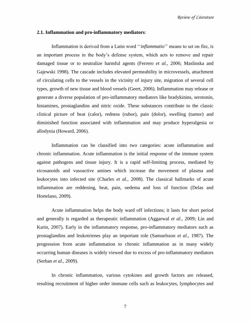
The availability of free AA is essential for the biosynthesis of eicosanoids.
Therefore, this mediator is released from the phospholipids membranes by the action of
various phospholipase enzymes, which are activated in response to multiple stimuli such
Review of Literature
9
as mechanical trauma, cytokines and growth factors (Figure-2) (Stratton and Alberts,
2002).
In most cells, arachidonic acid may be released at the endoplasmic reticulum and
nuclear membrane, predominantly via the translocation of type IV cytosolic
phospholipase A2. Arachidonic acid released from the membrane is rapidly metabolized
in several enzymatic and non-enzymatic pathways to yield an important family of
oxygenated products, collectively termed eicosanoids (Stables and Gilroy, 2011;
Simmons et al., 2004).
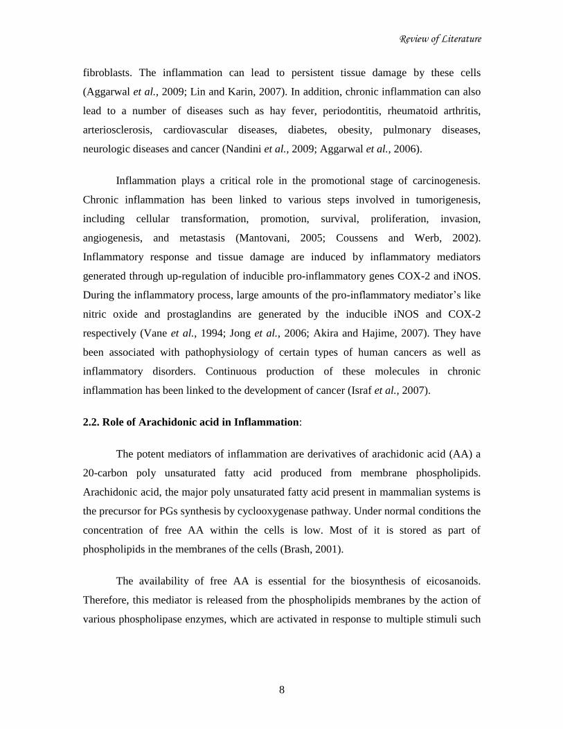
The arachidonic acid metabolism generally occurs via one of four major avenues
(i) the cyclooxygenase (COX) pathway, involved in the formation of prostaglandins
(PGs), thromboxanes (Txs), and prostacyclin, (ii) the lipoxygenase (LOX) pathway,
which produces leukotrienes (LTs) and lipoxins, (iii) the cytochrome P450
monooxygenase pathway, which produces epoxyeicosatrienoic and
hydroxyeicosatetraenoic acids and (iv) non-enzymatic lipid peroxidation which produces
isoprostanes (Figure-3) (Howard, 2006).
Review of Literature
10
Figure. 2. Schematic representation of Arachidonic acid release process. Membrane-
bound phospholipids are converted to arachidonic acid by the action of phospholipase
enzymes, which are activated in response to external stimuli (Stratton and Alberts, 2002).
Figure. 3. Four major pathways of Arachidonic acid metabolism. EETs-
epoxyeicosatrienoic acids; FLAP-5-lipoxygenase-activating protein; HETEs-
hydroxyeicosatetraenoic acids; HPETE - hydroxyperoxyeicosatetraenoic acid; 12-KETE-
12-ketoeicosatetraenoic acid; LOX - lipoxygenase; LT- leukotriene; PG- prostaglandin;
TXA2-thromboxane A2 (Howard, 2006).
Review of Literature
11
2.3. Cyclooxygenase pathway:
Cyclooxygenase converts arachidonic acid to endoperoxide containing
intermediates to produce prostaglandins and thromboxanes. Two distict active catalytic
sites exist on COX: the cyclooxygenase active site (CAS), which converts arachidonic
acid to prostaglandin G2 (PGG2) and the peroxidase active site (PAS) which transform
PGG2 to PGH2. The PGH2 is the precursor of several bioactive prostanoids, which are
formed by the action of specialized tissue isomerases. The five prostanoids synthesized
by this pathway include PGE2, PGD2, PGF2α, PGI2, and TxA2 (Figure-4). The production
of individual prostanoids is catalyzed by specific synthases and they have distinct
biological functions (Davies et al., 2002; Rocca, 2006).
2.3.1. Isoforms of cyclooxygenase:
Cyclooxygenase also known as Prostaglandin endoperoxide H synthase (PGHS,
EC.1.14.99.1) and exists in two isoforms; PGHS-1 (COX-1) and PGHS-2 (COX-2),
which catalyses the oxidation of AA to prostanoids. COX-1 and COX-2 enzymes are
heme proteins, homodimers that are widely distributed (Morham et al., 1995). These
enzymes are located in the lumenal portion of the endoplasmic reticulum membrane and
the nuclear envelope (Chandrasekharan and Simmons, 2004).
COX-2 is twice more abundant at the nuclear envelope than within the
endoplasmic reticulum, whereas the concentration of COX-1 is equal at both locations
(Bonventre et al., 1997). COX-1 functions predominantly in the endoplasmic reticulum
and COX-2 mostly in the nucleus (Van et al., 2002). Therefore, it appears that COX-1
and COX-2 are two distinct prostanoid biosynthetic systems with separate biological
functions for their products. COX-1 is expressed constitutively in most mammalian
tissues and plays a role in the production of PGs that control normal physiological
processes such as regulation of gastric response. Therefore, it is kept responsible for the
„housekeeping‟ prostaglandins synthesis. In contrast, COX-2 is an inducible enzyme
responsible for the production of pro-inflammatory PGs causing inflammation and pain
(Masferrer et al., 1994).

Review of Literature
12
Figure. 4. Schematic representation of cyclooxygenase (COX) pathway. Formation of
the main prostanoids from arachidonic acid (AAc) is depicted. AAc is converted to PGH2
through a two-step process that involves COX activity to convert arachidonic acid to
PGG2 followed by a peroxidase reaction, mediated also by COX enzymes to produce
PGH2. This leads to the formation of PEG2, a bioactive prostanoid, via concerted
activation of PGE synthase (PGES). The formation of the several PGs is carried out by
tissue-specific isomerases. PG - Prostaglandin; Tx - Thromboxane; PGDS - PGD
synthase; PGES - PGE synthase; PGFS - PGF synthase; PGIS - Prostacyclin synthase;
TxAS - Thromboxane A synthase (Luis et al., 2010).
Review of Literature
13
COX-1 and COX-2 isozymes catalyze the same reactions, but are encoded by two
different and specific genes, located on human chromosomes 9 and 1 respectively. The
gene for COX-1 is approximately 22kb in length, contains 11 exons and 10 introns,
whereas COX-2 gene is about 8kb, contains 10 exons and 9 introns (Bakhle, 1999). The
gene for COX-1 is transcribed as a 2.8kb mRNA, where as the gene for COX-2 is
transcribed as 4.6, 4.0 and 2.8kb mRNA variants. The molecular weight of COX-1
protein is approximately 67kD containing 600-602 amino acids. COX-2 protein
molecular weight is from 68-72kD and contains 603-604 amino acids (Tanabe and
Tohnai, 2002; Reddy et al., 2007).
The total structure of these enzymes is very similar. The structure of COX
consists of three domains; an N-terminal epidermal growth factor domain, a membrane-
binding motif, and a C-terminal catalytic domain containing the peroxidase and
cyclooxygenase active sites (Kurumbail et al., 1996). These enzymes are approximately
60% identical in terms of amino acid composition, and their catalytic regions are widely
conserved. The studies on their crystal structures have revealed that COX-2 has a larger
active site. Furthermore, the active sites of these two isoforms differ by only two
aminoacids, at positions 523 (Ile for COX-1 and Val for COX-2) and 513 (His for COX-1
and Arg for COX-2) (Zhang et al., 1996).
2.3.2. Role of COX-2 in Inflammation and Cancer:
COX-2 is the more important source of prostanoid formation in inflammatory
processes (Colin and Carlo, 2003). The prostanoids are metabolites that exert their
biological effects in the proximity of the sites of their synthesis, in autocrine or paracrine
manner. These mediators play an important role in the inflammatory process. In inflamed
tissues, their biosynthesis is significantly increased, and they contribute to the
development of the main signs of acute inflammation. Moreover, during an inflammatory
response, the level and profile of PG production change significantly (Emanuela and
Garret, 2011).
Review of Literature
14
COX-2 is also expressed constitutively, in few tissues such as in brain, kidney and
seminal vesicles, but is induced by various inflammatory and mitogenic stimuli (Jean et
al., 2006). It is highly induced by various growth factors, cytokines, endotoxins, pro-
inflammatory molecules and tumor promoters in various cell types and has emerged as
the isoform primarily responsible for PGs production in acute and chronic inflammatory
conditions (Luisa, 2004). Over-expression of COX-2 is associated with high levels of
PGE2 and has been demonstrated in several malignancies of breast, lung, prostrate, skin,
cervix and head and neck (Dannenberg et al., 2001). Higher prostaglandin levels have
been shown to stimulate proliferation of cells and mediate immune suppression (Ji and
Marnett, 1992).
Recently, COX-2 has been shown to be involved in the suppression of apoptosis,
which is critical in tumor cell death (Bobbili et al., 2003). COX-2 is over-expressed in
many cancers including breast cancer; the expression of the COX-2 gene is associated
with high tumor grade, which suggests it may serve as a prognostic biomarker for the
presence of breast cancer. COX-2 is expressed at an intermediate or high level in
epithelial cells of invasive breast cancers. Researchers also found high expression of
COX-2 in highly invasive estrogen independent MDA-MB-231 breast cancer cell lines
(Half et al., 2002; Teri et al., 2006). Liu et al., (2001) reported that the tumorigenesis is
induced by COX-2 over-expression. In their study, the murine COX-2 gene was inserted
downstream of a murine mammary tumor virus promoter. As a consequence, hyperplasia
and carcinoma of the mammary gland were observed and associated with strong COX-2
expression in mammary gland epithelial cells with increased PGE2 levels. While COX-2
expression is virtually absent from normal mammary parenchyma and its over-expression
was observed in roughly one third of human breast cancers (Figure-5).
In parallel with the COX enzyme family, there also exists constitutive isoforms of
Nitric oxide synthase (NOS), produce NO to maintain physiological functions, including
regulation of vasodilation and neurotransmission. Like COX-2, iNOS also plays an
important role in the mediation of inflammation (Weinberg, 2000).
Review of Literature
15
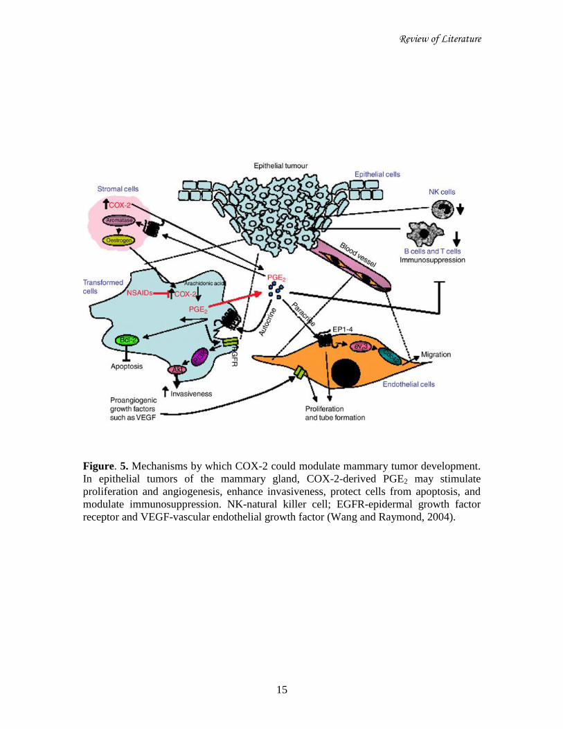
Figure. 5. Mechanisms by which COX-2 could modulate mammary tumor development.
In epithelial tumors of the mammary gland, COX-2-derived PGE2 may stimulate
proliferation and angiogenesis, enhance invasiveness, protect cells from apoptosis, and
modulate immunosuppression. NK-natural killer cell; EGFR-epidermal growth factor
receptor and VEGF-vascular endothelial growth factor (Wang and Raymond, 2004).
Review of Literature
16
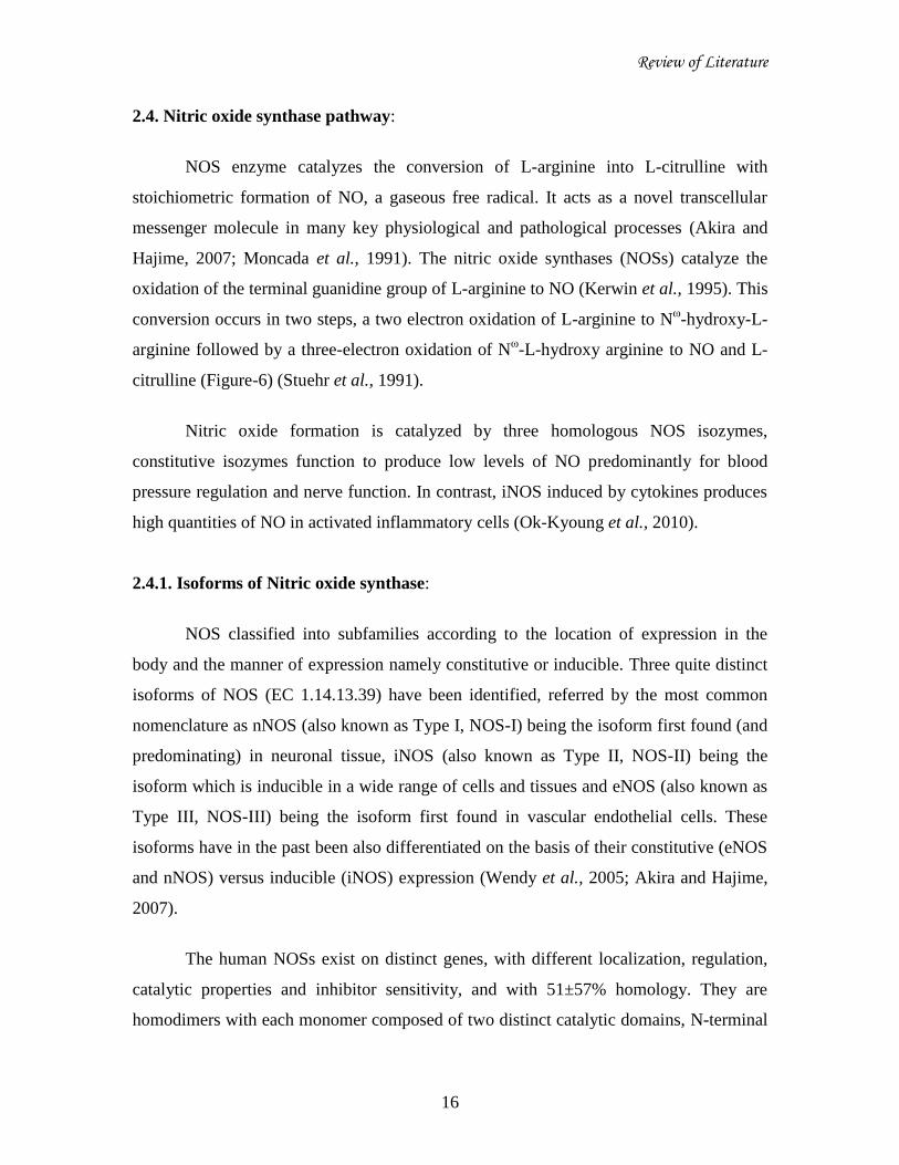
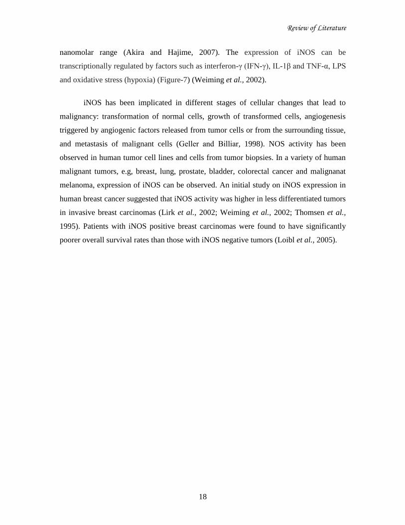
2.4. Nitric oxide synthase pathway:
NOS enzyme catalyzes the conversion of L-arginine into L-citrulline with
stoichiometric formation of NO, a gaseous free radical. It acts as a novel transcellular
messenger molecule in many key physiological and pathological processes (Akira and
Hajime, 2007; Moncada et al., 1991). The nitric oxide synthases (NOSs) catalyze the
oxidation of the terminal guanidine group of L-arginine to NO (Kerwin et al., 1995). This
conversion occurs in two steps, a two electron oxidation of L-arginine to Nω-hydroxy-L-
arginine followed by a three-electron oxidation of Nω-L-hydroxy arginine to NO and L-
citrulline (Figure-6) (Stuehr et al., 1991).
Nitric oxide formation is catalyzed by three homologous NOS isozymes,
constitutive isozymes function to produce low levels of NO predominantly for blood
pressure regulation and nerve function. In contrast, iNOS induced by cytokines produces
high quantities of NO in activated inflammatory cells (Ok-Kyoung et al., 2010).
2.4.1. Isoforms of Nitric oxide synthase:
NOS classified into subfamilies according to the location of expression in the
body and the manner of expression namely constitutive or inducible. Three quite distinct
isoforms of NOS (EC 1.14.13.39) have been identified, referred by the most common
nomenclature as nNOS (also known as Type I, NOS-I) being the isoform first found (and
predominating) in neuronal tissue, iNOS (also known as Type II, NOS-II) being the
isoform which is inducible in a wide range of cells and tissues and eNOS (also known as
Type III, NOS-III) being the isoform first found in vascular endothelial cells. These
isoforms have in the past been also differentiated on the basis of their constitutive (eNOS
and nNOS) versus inducible (iNOS) expression (Wendy et al., 2005; Akira and Hajime,
2007).
The human NOSs exist on distinct genes, with different localization, regulation,
catalytic properties and inhibitor sensitivity, and with 51±57% homology. They are
homodimers with each monomer composed of two distinct catalytic domains, N-terminal
Review of Literature
17
oxygenase domain containing binding sites for heme, BH4 and L-arginine and are linked
by a CaM-recognition site to a C-terminal reductase domain that contains binding sites
for FAD, FMN and NADPH (Knowles and Moncada, 1994; Wendy et al., 2005). The
nNOS, iNOS and eNOS genes were located on human chromosomes 12, 17 and 7
respectively. The gene for constitutive nNOS is >200kb in length, contains 29 exons, 28
introns and for eNOS is 21-22kb in length, contains 26 exons, 25 introns, whereas iNOS
gene is about 37kb, contains 26 exons, 25 introns. The molecular weight of nNOS, iNOS
and eNOS proteins is approximately 161kD, 131kD and 133kD containing 1434, 1153
and 1203 amino acids respectively (Nakane et al.,1993; Hall et al.,1994; Geller et
al.,1993; Sherman et al.,1993; Charles et al., 1993; Janssens et al.,1992; Marsden et
al.,1992).
NOSs have essential roles in the maintenance of homeostasis, e.g., regulating
blood vessel tone (eNOS), and providing neurotransmitter and neuromodulator (nNOS)
functions. On the other hand, numerous reports have implicated that sustained and/or
excess NO generation, most of which is attributable to iNOS expression, often occurs in
pathogenic conditions. In particular, iNOS has drawn considerable attention for its
critical functions in inflammation-related diseases (Cedergren et al., 2002; Donnelly and
Barnes, 2002; Hao et al., 2001; Cross and Wilson 2003).
2.4.2. Role of iNOS in Inflammation and Cancer:
NO synthesized from L-arginine by iNOS is a multifunctional mediator involved
in the vasodilatation observed during inflammatory responses and an important biological
signaling molecule and cellular cytotoxin. The excessive production of this free radical is
pathogenic to the host tissue, since NO can bind with other superoxide radicals which
directly damages the function of normal cells (Griffith and Stuehr, 1995; Moncada et al.,
1991). iNOS is expressed in a variety of cell types under both normal and pathological
conditions, including macrophages, microglial cells, keratinocytes, hepatocytes,
astrocytes and vascular endothelial and epithelial cells. With infectious and pro-
inflammatory stimuli, iNOS protein is highly induced to produce NO in a micromolar
range, whereas NO generation from nNOS and eNOS enzymes is constant and within the
Review of Literature
18
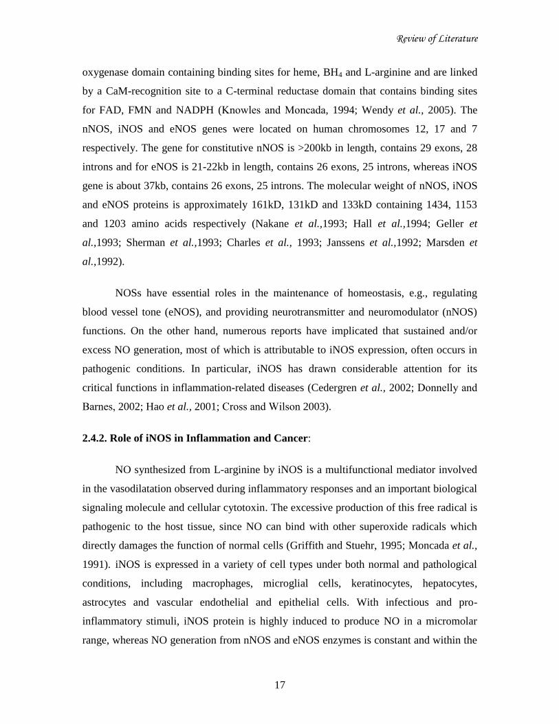
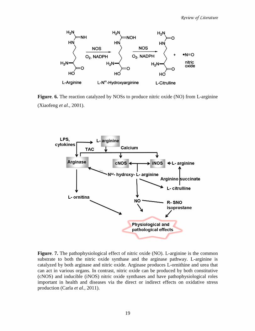
nanomolar range (Akira and Hajime, 2007). The expression of iNOS can be
transcriptionally regulated by factors such as interferon-γ (IFN-γ), IL-1β and TNF-α, LPS
and oxidative stress (hypoxia) (Figure-7) (Weiming et al., 2002).
iNOS has been implicated in different stages of cellular changes that lead to
malignancy: transformation of normal cells, growth of transformed cells, angiogenesis
triggered by angiogenic factors released from tumor cells or from the surrounding tissue,
and metastasis of malignant cells (Geller and Billiar, 1998). NOS activity has been
observed in human tumor cell lines and cells from tumor biopsies. In a variety of human
malignant tumors, e.g, breast, lung, prostate, bladder, colorectal cancer and malignanat
melanoma, expression of iNOS can be observed. An initial study on iNOS expression in
human breast cancer suggested that iNOS activity was higher in less differentiated tumors
in invasive breast carcinomas (Lirk et al., 2002; Weiming et al., 2002; Thomsen et al.,
1995). Patients with iNOS positive breast carcinomas were found to have significantly
poorer overall survival rates than those with iNOS negative tumors (Loibl et al., 2005).
Review of Literature
19
Figure. 6. The reaction catalyzed by NOSs to produce nitric oxide (NO) from L-arginine
(Xiaofeng et al., 2001).
Figure. 7. The pathophysiological effect of nitric oxide (NO). L-arginine is the common
substrate to both the nitric oxide synthase and the arginase pathway. L-arginine is
catalyzed by both arginase and nitric oxide. Arginase produces L-ornithine and urea that
can act in various organs. In contrast, nitric oxide can be produced by both constitutive
(cNOS) and inducible (iNOS) nitric oxide synthases and have pathophysiological roles
important in health and diseases via the direct or indirect effects on oxidative stress
production (Carla et al., 2011).
Review of Literature
20
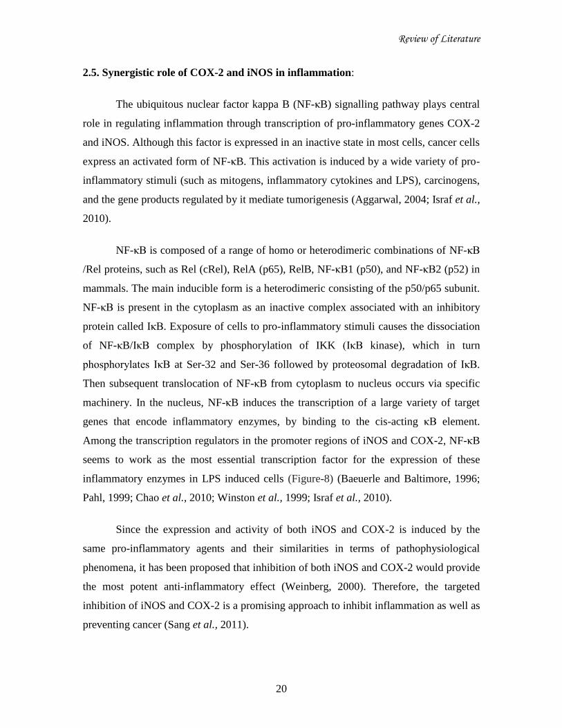
2.5. Synergistic role of COX-2 and iNOS in inflammation:
The ubiquitous nuclear factor kappa B (NF-κB) signalling pathway plays central
role in regulating inflammation through transcription of pro-inflammatory genes COX-2
and iNOS. Although this factor is expressed in an inactive state in most cells, cancer cells
express an activated form of NF-κB. This activation is induced by a wide variety of pro-
inflammatory stimuli (such as mitogens, inflammatory cytokines and LPS), carcinogens,
and the gene products regulated by it mediate tumorigenesis (Aggarwal, 2004; Israf et al.,
2010).
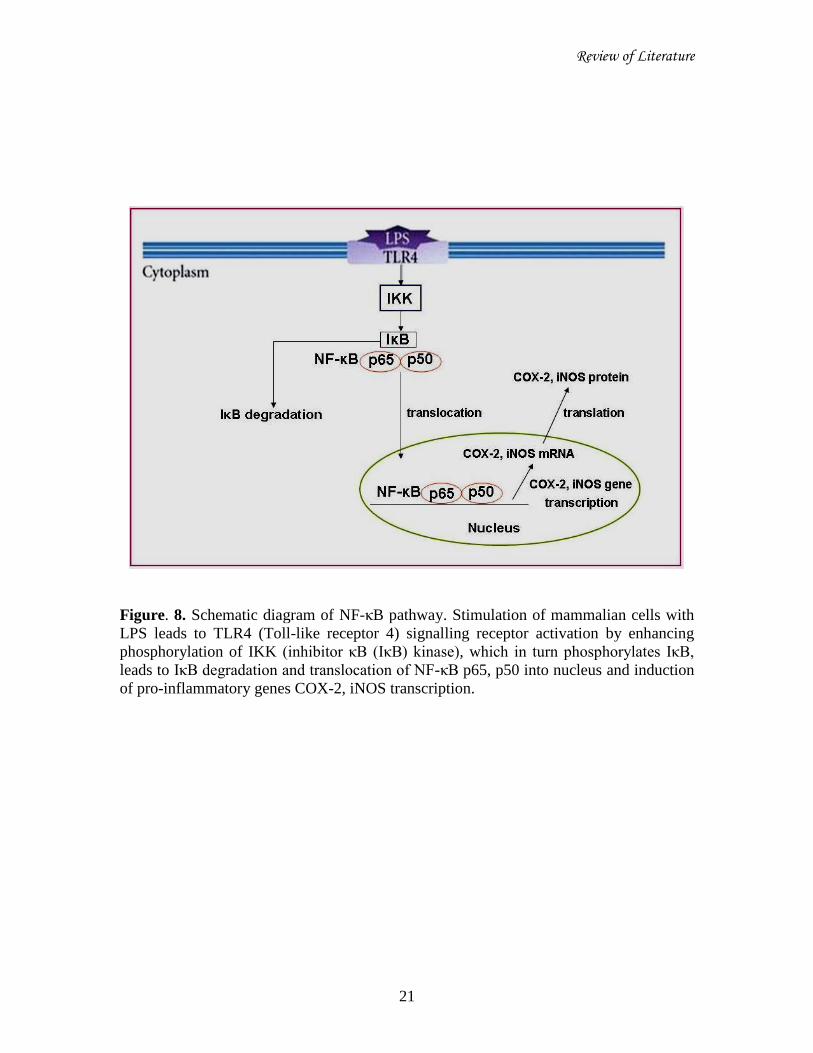
NF-κB is composed of a range of homo or heterodimeric combinations of NF-κB
/Rel proteins, such as Rel (cRel), RelA (p65), RelB, NF-κB1 (p50), and NF-κB2 (p52) in
mammals. The main inducible form is a heterodimeric consisting of the p50/p65 subunit.
NF-κB is present in the cytoplasm as an inactive complex associated with an inhibitory
protein called IκB. Exposure of cells to pro-inflammatory stimuli causes the dissociation
of NF-κB/IκB complex by phosphorylation of IKK (IκB kinase), which in turn
phosphorylates IκB at Ser-32 and Ser-36 followed by proteosomal degradation of IκB.
Then subsequent translocation of NF-κB from cytoplasm to nucleus occurs via specific
machinery. In the nucleus, NF-κB induces the transcription of a large variety of target
genes that encode inflammatory enzymes, by binding to the cis-acting κB element.
Among the transcription regulators in the promoter regions of iNOS and COX-2, NF-κB
seems to work as the most essential transcription factor for the expression of these
inflammatory enzymes in LPS induced cells (Figure-8) (Baeuerle and Baltimore, 1996;
Pahl, 1999; Chao et al., 2010; Winston et al., 1999; Israf et al., 2010).
Since the expression and activity of both iNOS and COX-2 is induced by the
same pro-inflammatory agents and their similarities in terms of pathophysiological
phenomena, it has been proposed that inhibition of both iNOS and COX-2 would provide
the most potent anti-inflammatory effect (Weinberg, 2000). Therefore, the targeted
inhibition of iNOS and COX-2 is a promising approach to inhibit inflammation as well as
preventing cancer (Sang et al., 2011).
Review of Literature
21
Figure. 8. Schematic diagram of NF-κB pathway. Stimulation of mammalian cells with
LPS leads to TLR4 (Toll-like receptor 4) signalling receptor activation by enhancing
phosphorylation of IKK (inhibitor κB (IκB) kinase), which in turn phosphorylates IκB,
leads to IκB degradation and translocation of NF-κB p65, p50 into nucleus and induction
of pro-inflammatory genes COX-2, iNOS transcription.
Review of Literature
22
2.6. Anti-inflammatory drugs:
A number of drugs have been developed to cure the diseases of chronic
inflammation origin. These drugs can be divided into two groups; steroidal anti-
inflammatory drugs and non-steroidal anti-inflammatory drugs. Steroids are the chemical
compounds released by the adrenal gland and have anti-inflammatory action by different
mechanisms. As an example, glucocorticoids are the steroidal hormones which enhance
the expression of nearly 130 genes which include the anti-inflammation, phagocytosis,
antioxidative stress and suppress the pro-inflammatory genes (Franchimont et al., 2003;
Yona and Gordon 2007; Barnes, 1998).
In addition, glucocorticoids may express non genomic pathways by restricting
ATP consuming activities and these effects are much more rapid than genomic effects
(Goulding, 2004). Corticosteroids, another type of steroid hormones, inhibit the activity
of phospholipase A2 and diminish the production of AA upon activation of cells by pro-
inflammatory molecules (Vane and Botting, 1998). PGs and LTs are thus inhibited by
corticosteroids through the action of phospholipase A2 (Nguyen and Lee, 1992).
However, a number of side effects are revealed as a result of glucocorticoid use in
inflammatory diseases. Glucocorticoids enhance glucose levels by degrading proteins and
modulating fatty acid metabolism partly. This catabolic interference by corticosteroids
leads to tissue remodeling, osteoporosis, insulin resistance and diabetes (Kleiman and
Tuckermann, 2007). Long term use of glucocorticoids increases the apoptosis of
hypertrophic chondrocytes in growth plate which reduces the longitudinal growth of
bones (De Luca, 2006).
The second category of anti-inflammatory drugs is NSAIDs. Approximately, 60
million Americans use the non steroidal anti-inflammatory drugs annually to treat
inflammation related diseases and especially rheumatological disorders and arthritis
(Cryer, 2005). NSAIDs show their effect by inhibiting the action of COX instead of
phospholipase A2 and do not prevent the activity of LOX (Vane and Botting, 1998).
NSAIDs block the production of PGs by inhibiting both COX-1 and COX-2. It is known
that about 1% of chronic users of NSAIDs, such as patients with chronic inflammatory

Review of Literature
23
diseases develop gastrointestinal (GI) complications such as mucosal damage and
bleeding (Singh et al., 2009).
Moreover, some researchers have found acute renal failure as a result of
NSAIDs use (Griffin et al., 2000). NSAIDs inhibit the production of renal prostaglandins
and negatively affect glomerular filtration rate and salt excretion (Clive and Stoff, 1984).
These drugs appear to produce at least some of their beneficial effects by inhibiting
COX-2 and their deleterious side effects by inhibiting COX-1. Hence, synthetic anti-
inflammatory drugs are more associated with negative effects rather than positive effects.
Thus, selective inhibition of the induced enzyme, without affecting the homeostatic one,
might avoid the side effects of currently available NSAIDs. NSAIDs have also been
shown to inhibit iNOS but the pharmacological inhibitors of iNOS are not yet in clinical
use while selective inhibitors of COX-2 have recently been launched on the market
(Simon et al., 1999; Singh et al., 2009; Esko, 2002).
Selective COX-2 inhibitors (COXibs) have same anti-inflammatory benefits as
traditional NSAIDs with little effect on COX-1, but as inhibitors of the enzyme
responsible for the production of most inflammatory PGs, their drug efficacy is upheld.
COXibs have proven to be effective in suppressing experimental tumorigenesis.
Furthermore, several recently reported randomized clinical trials have shown that
COXibs significantly reduce the incidence of colorectal adenomas in humans.
Dismayingly, these trials also identified an increased risk for cardiovascular events
associated with COXib use, suggesting that COXibs may not be sufficiently safe for
general use as cancer chemopreventive agents (Simon et al., 1999; Louise, 2007). In
view of the gastric side effects of conventional NSAIDs and the recent market
withdrawal of rofecoxib and valdecoxib due to their adverse cardiovascular side effects
there is need to develop alternative anti-inflammatory agents with reduced gastric and
cardiovascular problems (Reddy et al., 2007).
Review of Literature
24
2.7. Importance of natural drugs:
Natural compounds are now gaining more pharmacological attention as many
unexplored plant products are showing a wide range of activities like anti-inflammatory
and anti-cancer (Chang, 2000; Raghav et al., 2007). It is estimated that about 80% of the
world‟s population primarily those of developing countries rely on plant-derived
medicines for their healthcare needs (Gurib, 2006). In many developed countries popular
use of traditional/complementary and alternative medicine is also expanded due to great
concern about the adverse effects of modern drugs (World Health Organization, 2002).
It is estimated that approximately one quarter of the best selling drugs worldwide
were natural products or derived from natural products (Balunas and Kinghorn, 2005).
Nearly 25% of all prescribed drugs are derived from plants with or without further
modification (Newman et al., 2000; Raskin and Ripoll, 2004) and still several
pharmacologically active plant-derived compounds remain unexplored (Mendelson and
Balick, 1995; Raskin et al., 2002). The anti-inflammatory activities of plants are due to
the secondary metabolites. These bioactive compounds consist of polyphenols,
flavonoids, alkaloids, terpenoids, steroids, carotenoids, coumarins and curcumins (Saeed
et al., 2010).
The majority of naturally occurring phenolics retain antioxidative and anti-
inflammatory properties which appear to contribute to their chemopreventive or
chemoprotective activity. Since inflammation is closely linked to tumor promotion,
substances with potent anti-inflammatory activities are anticipated to exert
chemopreventive effects on carcinogenesis, particularly in the promotion stage. Examples
are curcumin, a yellow pigment of turmeric (Curcuma longa L), the green tea polyphenol
epigallocatechin gallate (EGCG) and resveratrol from grapes (Vitis vinifera) that strongly
suppress tumor promotion (Surh et al., 2001). Thus, searching for inflammatory
inhibitors with chemotherapeutic potential from natural sources is an alternative approach
in the development of anti-inflammatory and anti-cancer drugs.
Review of Literature
25
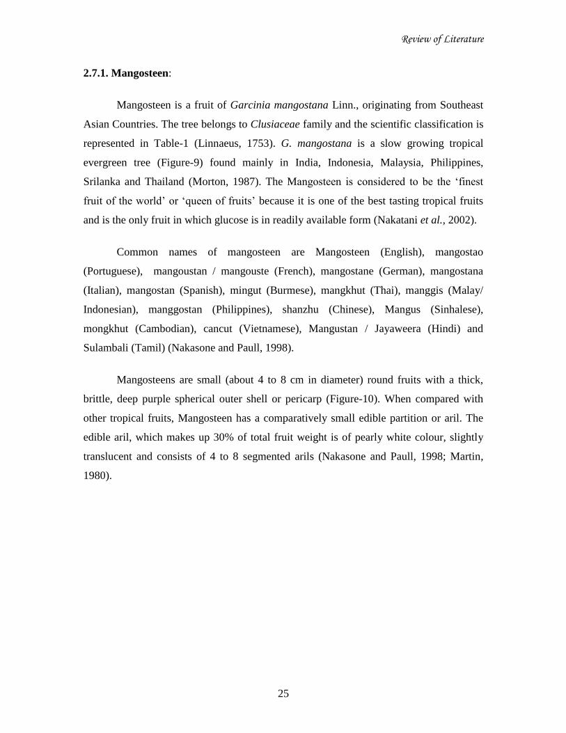
2.7.1. Mangosteen:
Mangosteen is a fruit of Garcinia mangostana Linn., originating from Southeast
Asian Countries. The tree belongs to Clusiaceae family and the scientific classification is
represented in Table-1 (Linnaeus, 1753). G. mangostana is a slow growing tropical
evergreen tree (Figure-9) found mainly in India, Indonesia, Malaysia, Philippines,
Srilanka and Thailand (Morton, 1987). The Mangosteen is considered to be the „finest
fruit of the world‟ or „queen of fruits‟ because it is one of the best tasting tropical fruits
and is the only fruit in which glucose is in readily available form (Nakatani et al., 2002).
Common names of mangosteen are Mangosteen (English), mangostao
(Portuguese), mangoustan / mangouste (French), mangostane (German), mangostana
(Italian), mangostan (Spanish), mingut (Burmese), mangkhut (Thai), manggis (Malay/
Indonesian), manggostan (Philippines), shanzhu (Chinese), Mangus (Sinhalese),
mongkhut (Cambodian), cancut (Vietnamese), Mangustan / Jayaweera (Hindi) and
Sulambali (Tamil) (Nakasone and Paull, 1998).
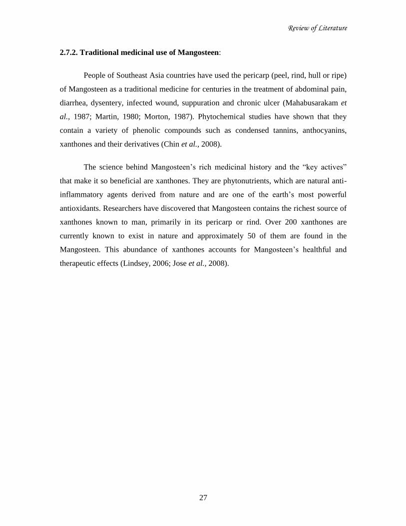
Mangosteens are small (about 4 to 8 cm in diameter) round fruits with a thick,
brittle, deep purple spherical outer shell or pericarp (Figure-10). When compared with
other tropical fruits, Mangosteen has a comparatively small edible partition or aril. The
edible aril, which makes up 30% of total fruit weight is of pearly white colour, slightly
translucent and consists of 4 to 8 segmented arils (Nakasone and Paull, 1998; Martin,
1980).
Review of Literature
26
Figure. 9. Garcinia mangostana L., (Akao et al., 2008).
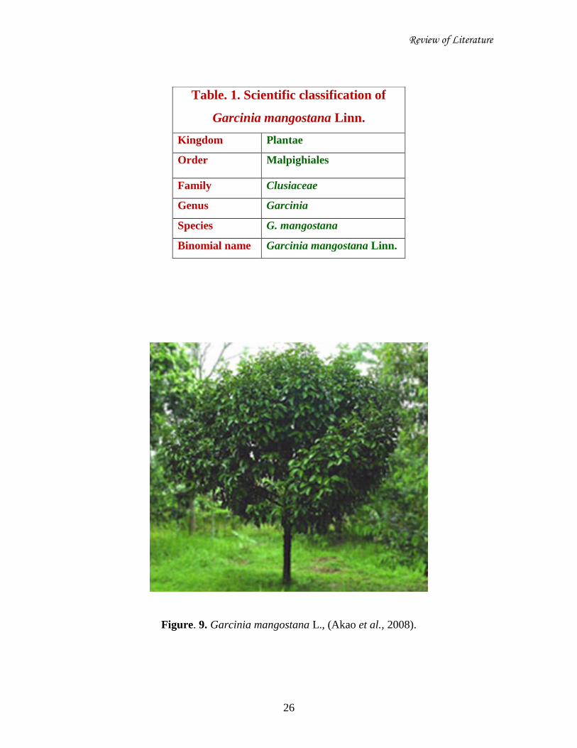
Table. 1. Scientific classification of
Garcinia mangostana Linn.
Kingdom Plantae
Order Malpighiales
Family Clusiaceae
Genus Garcinia
Species G. mangostana
Binomial name Garcinia mangostana Linn.
Review of Literature
27
2.7.2. Traditional medicinal use of Mangosteen:
People of Southeast Asia countries have used the pericarp (peel, rind, hull or ripe)
of Mangosteen as a traditional medicine for centuries in the treatment of abdominal pain,
diarrhea, dysentery, infected wound, suppuration and chronic ulcer (Mahabusarakam et
al., 1987; Martin, 1980; Morton, 1987). Phytochemical studies have shown that they
contain a variety of phenolic compounds such as condensed tannins, anthocyanins,
xanthones and their derivatives (Chin et al., 2008).
The science behind Mangosteen‟s rich medicinal history and the “key actives”
that make it so beneficial are xanthones. They are phytonutrients, which are natural anti-
inflammatory agents derived from nature and are one of the earth‟s most powerful
antioxidants. Researchers have discovered that Mangosteen contains the richest source of
xanthones known to man, primarily in its pericarp or rind. Over 200 xanthones are
currently known to exist in nature and approximately 50 of them are found in the
Mangosteen. This abundance of xanthones accounts for Mangosteen‟s healthful and
therapeutic effects (Lindsey, 2006; Jose et al., 2008).
Review of Literature
28
Figure. 10. Mangosteen (Akao et al., 2008).
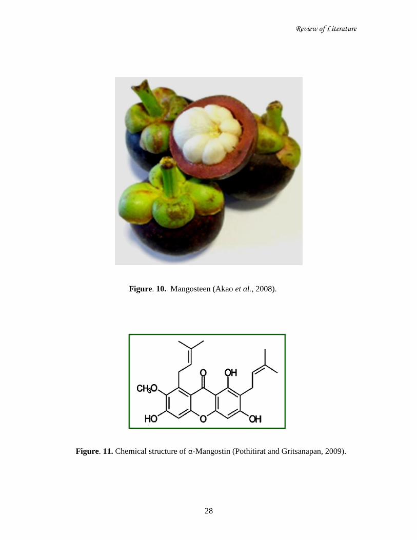
Figure. 11. Chemical structure of α-Mangostin (Pothitirat and Gritsanapan, 2009).
Review of Literature
29
2.7.3. α-Mangostin:
The first xanthone out of 50 isolated from Mangosteen‟s pericarp was named as
α-Mangostin (Schmid, 1855). Later, Dragendorff (1930) and Murakami (1932) elucidated
the Mangostin structure. Yates and Stout (1958) established the molecular formula
(C24H26O6), and type and position of substituents of α-Mangostin (Figure.11).
Several studies have stated that xanthones particularly α-Mangostin, which is a
major prenylated xanthone exhibits anti-allergic, anti-bacterial, anti-fungal, antioxidant,
anti-inflammatory and anti-tumoral activities (Akao et al., 2008; Chairungsrilerd et al.,
1996; Chen et al., 2008; Gopalakrishnan et al., 1997; Jung et al., 2006; Sakagami et al.,
2005).
With this background the present study was designed to evaluate the anti-
inflammatory potential of α-Mangostin and its mode of action by screening the effect of
α-Mangostin on the expression of pro-inflammatory genes COX-2 and iNOS using LPS
stimulated MDA-MB-231 human breast cancer cells as model.