acute colonic pseudo-obstruction with feeding intolerance...
TRANSCRIPT
Clinical StudyAcute Colonic Pseudo-Obstruction with Feeding Intolerance inCritically Ill Patients: A Study according to Gut Wall Analysis
Chenyan Zhao,1,2 Tingbin Xie,1,3 Jun Li,4 Minhua Cheng,1,2 Jialiang Shi,1 Tao Gao,1
Fengchan Xi,1 Juanhong Shen,1 Chun Cao,1,2 and Wenkui Yu1,2
1Research Institute of General Surgery, Jinling Hospital, No. 305 Zhongshan East Road, Nanjing 210002, China2Medical School, Nanjing University, No. 22 Hankou Road, Nanjing 210002, China3Southern Medical University, No. 1023 Shatai South Road, Guangzhou 510515, China4Jining No.1 People’s Hospital, No. 99 Jianshe West Road, Jining 272000, China
Correspondence should be addressed to Wenkui Yu; [email protected]
Received 26 September 2016; Revised 16 December 2016; Accepted 26 January 2017; Published 12 March 2017
Academic Editor: Greger Lindberg
Copyright © 2017 Chenyan Zhao et al. This is an open access article distributed under the Creative Commons Attribution License,which permits unrestricted use, distribution, and reproduction in any medium, provided the original work is properly cited.
Objective. To compare the differences between acute colonic pseudo-obstruction (ACPO) with and without acute gut wallthickening. Methods. ACPO patients with feeding tolerance were divided into ACPO with no obvious gut wall thickening(ACPO-NT) group and ACPO with obvious acute gut wall thickening (ACPO-T) group according to computed tomographyand abdominal radiographs. Patients’ condition, responses to supportive measures, pharmacologic therapy, endoscopicdecompression, and surgeries and outcomes were compared. Results. Patients in ACPO-T group had a significantly higherAPACHE II (11.82 versus 8.25, p = 0 008) and SOFA scores (6.47 versus 3.54, p < 0 001) and a significantly higher 28-daymortality (17.78% versus 4.16%, p = 0 032) and longer intensive care unit stage (4 versus 16 d, p < 0 001). Patients in ACPO-NTgroup were more likely to be responsive to supportive treatment (62.50% versus 24.44%, p < 0 001), neostigmine (77.78% versus17.64%, p < 0 001), and colonoscopic decompression (75% versus 42.86%, p = 0 318) than those in ACPO-T group. Of thepatients who underwent ileostomy, 81.25% gained benefits. Conclusions. ACPO patients with gut wall thickening are moresevere and are less likely to be responsive to nonsurgical treatment. Ileostomy may be a good option for ACPO patients with gutwall thickening who are irresponsive to nonsurgical treatment.
1. Introduction
Feeding intolerance (FI) is a common and clinically importantproblem in critically ill patients. FI manifested as gastroin-testinal symptoms such as abdominal distension, diarrhea,vomiting, and gastric retention, and inadequate enteralcalorie intake is the biggest challenge in maintaining enteralnutrition (EN) in critically ill patients, which could cause orexacerbate malnutrition and has been associated with longerintensive care unit (ICU) stay and higher morbidity andmortality [1, 2]. Gastrointestinal dysfunctions are the majorcause of FI. Most doctors and studies have focused on theupper gastrointestinal dysfunctions especially gastroparesissince they are more likely to present nausea, vomiting, andgastric retention; particularly, gastric residual volume (GRV)
is the most frequently used parameter to monitor FI, whereasthe lower gastrointestinal factors were often neglected [3, 4].However, the stomach and small intestine may functionproperly, but FI still exists which could be the result ofcolonic dysfunction.
Acute colonic pseudo-obstruction (ACPO), also knownas Ogilvie’s syndrome, is characterized by acute dilation ofthe large bowel with obstructive symptoms in the absenceof mechanical obstruction [5]. The remarkable changes inACPO patients are the massive dilation and dysmotility ofcolon which could cause FI, and if left untreated, ACPOcan lead to colonic necrosis and perforation [6]. In criticalpatients, we have observed an interesting phenomenon thatpatients who are feeding intolerant and have a great dilatedlarge bowel are sometimes accompanied with a markedly
HindawiGastroenterology Research and PracticeVolume 2017, Article ID 9574592, 11 pageshttps://doi.org/10.1155/2017/9574592

thickening gut wall which could be detected by imaging testand those who are with acute gut wall thickening often havea poor outcome. In general, ACPO was mainly caused by animbalance between parasympathetic and sympathetic inner-vation [7]. However, in critical patients, severe trauma, majorsurgery, sepsis, shock, or mesenteric vascular occlusion willcause great stress, intestinal ischemia/reperfusion, and acuteinflammation in the gut wall, inducing acute edema in thecolon which could be another reason for the dysfunction ofthe colon [8, 9].
Supportive treatment, cholinergic drugs, decompression,and surgery were usually sequentially used to treat patientswith ACPO [5, 7]. However, to the best of our knowledge,there is no study distinguishing the two different ACPOs interms of treatment. Studies have evaluated the responses ofACPO patients to these treatments, but got conflictingresults. A retrospective study by Mehta et al. [10] enrolled27 patients with ACPO who have received supportive treat-ment, and only eight (30%) of them achieved spontaneousresolution, which is contrary to that of Loftus et al. [11],who showed that a majority of patients with ACPO wereresponsive to supportive measures. Both of them have evalu-ated the predictive factors for response to neostigmine,finding that postoperative patients, females, or older-agedpatients were more likely to be responder of neostigminewhile the presence of electrolyte imbalance and antimotilitymedication use were the risk factors for poor response toneostigmine. However, none of them has taken the gut walledema into consideration [10, 11]. Considering the differentpathophysiologies and clinical courses of general ACPOand ACPO with acute gut wall thickening (edema), therecommended treatment protocol for ACPO may not besuitable for both of the two ACPOs.
Therefore, this study was designed to compare theoutcomes of the two different ACPOs and their responsesto different treatments, in order to alert doctors to distinguishthe two ACPOs.
2. Methods
2.1. Patients and Design. This study was approved by theethics committee of Jinling Hospital and the Medical Schoolof Nanjing University, and the protocols were registered atClinicalTrials.gov (NCT02939508). This is a prospective,single-center, observational study conducted at the 39-bedsurgical intensive care unit (SICU) of the General SurgicalDepartment of Jinling Hospital, affiliated to the MedicalSchool of Nanjing University from July 1, 2014, to July 1,2016. Patients in our ICU aged 18–75 years who had FI anddiagnosed with ACPO were included. EN was started within24–48 h after ICU admission if possible unless electively notfed by the attending doctors. FI was thought to be present ifat least 50% of the calculated needs via enteral feeding couldnot be reached after 72 h EN attempt due to signs of nausea,vomiting, gastric retention (a single GRV > 250ml), abdom-inal pain, abdominal distension, ileus or severe diarrhea, orno feeding because of any clinical conditions (active gastroin-testinal hemorrhage, obstruction, intestinal necrosis, gastro-intestinal fistula or perforation, and so forth). FI was not
registered if the patient was electively not fed or if the enteralfeeding was disrupted or withheld due to procedures [12, 13].All patients with FI and fed via the jejunum pathway werescreened, and those who were diagnosed with ACPO wereincluded. ACPO was identified if acute dilation of the colonor cecum (colonic diameter > 6 cm or cecal diameter > 9 cm)was observed on computed tomography (CT) or abdominalradiography [5]. Patients with any of the following conditionswere excluded: (1) mechanical gastrointestinal obstruction(including tumor and stercoral obstruction); (2) gastrointesti-nal hemorrhagewithin 72 hours before inclusion; (3) presenceof intra-abdominal abscesses at inclusion; (4) presence ofintestinal perforation, necrosis, or fistula at inclusion; (5)history of inflammatory bowel disease (ulcerative colitis orCrohn’s disease) or radiation enteritis; (6) pregnancy; (7)contraindications of neostigmine administration; and (8)disconcerting with endoscopy or surgical treatment or treat-ment abandonment. The enrolled patients were divided intotwo groups according to the gut wall thickness (edema) onCT (ileocecus): ACPO with no obvious thickening (edema)of the colonic gut wall (ACPO-NT) group and ACPO withobvious acute thickening (edema) of the colonic gut wall(ACPO-T) group.
2.2. Treatment Protocol. Once the patients had confirmeddiagnosis of ACPO, they started the treatment according toour protocol depicted in Figure 1. Specific processes wereas follows:
Supportive measures: Firstly, hydro-electrolyte and thy-roid function were assessed and corrected if abnormal.Blood glucose was maintained within normal levels. Patientsprone to sepsis were administered antibiotics empiricallyand adjusted to more targeted antibiotics according to theblood culture and drug sensitivity test results. Patients weresubject to fasting and gastrointestinal decompression viastomach and anal tubes. GRV was assessed four times aday. Pharmaceuticals affecting bowel movement, such asopiates, anticholinergic drugs, and calcium channel blockers,were suspended as quickly as possible.
Pharmacologic therapy with cholinergic drugs: Intrave-nous administration of neostigmine was considered if thececal diameter was >10 cm after applying the treatmentdescribed above without signs of amelioration within 24hours or cecal diameter of >12 cm [14, 15].
Colonoscopic decompression: This procedure wasapplied when obvious cecum distension was present(diameter > 10 cm) for more than 3 days and when therewere no signs of improvement after 24–48 hours of supportiveor neostigmine treatment or if neostigmine was contraindi-cated [16]. Colonoscopic decompression was performed by2 experienced endoscopy physicians at the bedside or at anendoscopy room.
Surgical intervention: Surgery was indicated whencolonic distension lasted more than 6 days or obvious cecaldistention (diameter > 10 cm) continued after 48–72 hoursof supportive or pharmacologic management and colono-scopic decompression [17]. Ileostomy was performed underepidural or local anesthesia by a team of experienced sur-geons. If the patients had colonic necrosis or perforation,
2 Gastroenterology Research and Practice

ileostomy was superseded by colectomy under generalanesthesia [18].
2.3. Date Collection. The following data were recorded: (1)demographic data including age, sex, and body mass index(BMI) at grouping; (2) primary diagnosis, reasons for inten-sive care, and interventions before or at grouping; and (3)severity of illness, as assessed by the Acute Physiology andChronic Health Evaluation II (APACHE II) and SequentialOrgan Failure Assessment (SOFA) scores within 24 hoursfrom the time of grouping.
2.4. Follow-Up. After ICU admission, gastrointestinal dys-function symptoms such as FI, vomiting, abdominal disten-sion, and defecation were recoded every day. GRV andintra-abdominal pressure (IAP) which was reflected bybladder pressure were measured four times a day after ICUadmission. Abdominal CTs were performed on days 1, 3, 5,
and 7 and every week after inclusion, unless requiredmore fre-quently because of an additional illness. Colonic and cecaldiameters were evaluated independently by two trained fellowswho were blinded to this study. Responses to treatment (sup-portive treatment, neostigmine, colonoscopy, and surgery)and colonic recovery time were also assessed and recordedthrough vital signs, laboratory tests, and CT examination.
Colonic recovery was identifiedwhenmeeting the followingcriteria: (1) EN reached or surpassed 50% of the targeted needsduring 3 days or longer and (2) evident improvement of colonicdistension(colonicdiameter < 6 cmorcecumdiameter < 9 cm)or edema of the colonic wall shown on the abdominal CT [16].Recurrence was defined as patients diagnosed with recurrentACPO within 72 hours of recovery after a certain treatment.
2.5. End Points. The primary outcome was the 28-daymortality of patients in the two groups. Secondary outcomes
Patients diagnosed with acute colonicpseudo-obstruction
Assessing ischemia and perforation
Supportive treatment for 24 hEvaluate and treat reversible causes
Resolution No response or cecal diameter > 12 cm
Neostigmine i.v. 2 mg each time
Re-admission whilerecurrence or partial
response
No response
Colonoscopicdecompression
Ileostomy
No response
Resolution
Figure 1: Treatment protocol.
3Gastroenterology Research and Practice

included ICU mortality, hospitalization mortality, and theduration of the stay in the ICU and in the hospital of thetwo groups. Complications of the two groups such as intra-abdominal hypertension (IAH; IAP is found to be 12mmHgorhigher,confirmedbyat least twomeasurements);abdominalcompartment syndrome (ACS; IAP above 20mmHgwith newonset organ failure confirmed by minimally two standardizedmeasurements); sepsis; gastrointestinal hemorrhage; colonicperforation; colonic necrosis; andneworgandysfunctionwerealsorecorded.
2.6. Statistical Analysis. Statistical analysis was performedwith SPSS 20.0 (SPSS, Inc., an IBM Company, Chicago, IL).Categorical variables were compared using the χ2 test orFisher’s exact test. The parametric tests will be applied whennormality (and homogeneity of variance) assumptions aresatisfied; otherwise, the equivalent nonparametric test will beused. Parametric tests were conducted using t-tests, and non-parametric tests were conducted using the Mann-WhitneyU test. Multivariate analysis by binary logistic regressionwas done for the risk of 28-day mortality and failure of non-surgical treatment. A 2-tailed p < 0 05 was considered asstatistically significant.
3. Results
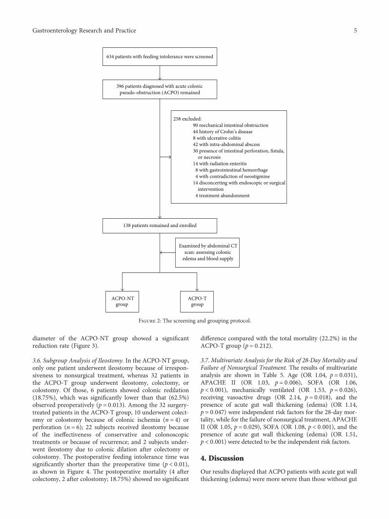
3.1. General Information. From July 1, 2014, to July 1, 2016,634 patients were enrolled for FI via the jejunum pathway.Of these, 396 patients remained due to the diagnosis ofACPO. Of the remaining 396 subjects, 258 patients wereexcluded: 90 with mechanical intestinal obstruction; 44 withhistory of Crohn’s disease; 8 with ulcerative colitis; 42 withintra-abdominal abscess; 30 with intestinal perforation,fistula, or necrosis at inclusion; 14 with radiation enteritis;8 with gastrointestinal hemorrhage within 72 h; 4 withcontradiction of neostigmine; 14 disconcerting with endo-scopic or surgical intervention (patients or their families);and 4 treatment abandonment. Thus, 138 patients (32 menand 106 women) were studied. The subjects were classifiedinto two groups, the ACPO-NT group (n = 48) andACPO-T group (n = 90), according to colon conditions.The process of patient screening and grouping is presentedin Figure 2.
3.2. Comparison of the General Data between the ACPO-NTGroup and the ACPO-T Group. There were no differences insex ratio (p = 0 224) and BMI (p = 0 536) between the twogroups. The mean age was significantly lower in the ACPO-NT group (p = 0 007). However, APACHE II (p = 0 008) andSOFA scores (p < 0 001), mechanical ventilation (p < 0 001),sepsis incidence (p = 0 004), and vasoactive drug usage(p = 0 004) were significantly higher in the ACPO-T group.Details are listed in Table 1.
3.3. Comparison of the Gastrointestinal DysfunctionParameters at the Time of Grouping between the ACPO-NTGroup and the ACPO-T Group. Although the occurrence ofvomiting (p = 0 356), abdominal distension (p = 0 192), IAP(p = 0 671), and days without defecation (p = 0 607) showedno statistical difference between the groups, the incidence
of gastric retention (p = 0 002) and GRV (p = 0 002) weresignificantly higher in the ACPO-T group during the 28-day period. The colonic diameters (p = 0 110) and cecaldiameters (p = 0 853) were not statistically different at group-ing between the groups as Table 2 shows.
3.4. Comparison of the Treatment Responses. The overallefficacy of the nonsurgical treatment in the ACPO-NT groupreached 97.91% versus 64.4% in the ACPO-T group(p < 0 01). Patients in the ACPO-NT group had significantlyhigher efficacy in conservative treatment (62.50% versus24.44%, p < 0 001). Neostigmine efficacy was only 17.64% inthe ACPO-T group, which was significantly lower than thatin the ACPO-NT group (77.78%, p < 0 001). Colonoscopicdecompressionhadbetter results in theACPO-NTgroup thanin the ACPO-T group, though with no significant difference(75% versus 42.86%, p = 0 318). One patient in the ACPO-NT group irresponsive to nonsurgical treatment underwentileostomy and eventually recovery, while 32 inACPO-Tgroupirresponsive to nonsurgical treatment underwent ileostomy,colectomy, or colostomy, which were also effective in 26subjects as Table 3 shows.
3.5. Comparison of Outcomes between the Two Groups.Within 28 days, 16 (17.78%) patients died in the ACPO-Tgroup and 2 (4.17%) deaths occurred in the ACPO-NT group(p = 0 032). During the ICU period, 18 patients died(20.00%) in the ACPO-T group and 1 (2.08%) deathoccurred in the ACPO-NT group, with a significant differ-ence (p = 0 003). During hospitalization, 20 (22.22%)patients died in the ACPO-T group versus 3 (6.25%) in theACPO-NT group (p = 0 017). Median ICU stay was signifi-cantly shorter in the ACPO-NT group than in the ACPO-Tgroup (4 versus 16 d, p < 0 001). Similarly, the ACPO-NTgroup has significantly less median hospitalization days (15versus 36 d, p < 0 001) (Table 4).
The incidence of IAH (p = 0 008) and ACS (p < 0 001)was significantly higher in the ACPO-T group than inthe ACPO-NT group. During treatment process, subjectsdiagnosed with sepsis were 58 (64.44%) in the ACPO-Tgroup and 12 (25.00%) in the ACPO-NT group (p < 0 001).Two (4.17%) patients suffered gastrointestinal hemor-rhage in the ACPO-NT group versus 8 (8.89%) in theACPO-T group (p = 0 494). No colonic perforation or necro-sis occurred in the ACPO-NT group while 6 (6.67%)patients in the ACPO-T group had colonic perforationand 4 (4.44%) had colonic necrosis, though showing nostatistical difference (p = 0 092 and p = 0 298, resp.). TheACPO-T group had a significantly higher rate of neworgan dysfunction than the ACPO-NT group (p < 0 001)(Table 4).
At the time of grouping, there was no significant dif-ference in colonic or cecal diameter between the twogroups (p = 0 110 and p = 0 853, resp.). After treatment,colonic and cecal diameters of both groups showed adecreasing trend. On days 7 and 14, the colonic diameterof the ACPO-NT group was significantly less than thatof the ACPO-T group. Similarly, on days 3, 7, and 14, the cecal
4 Gastroenterology Research and Practice
diameter of the ACPO-NT group showed a significantreduction rate (Figure 3).
3.6. Subgroup Analysis of Ileostomy. In the ACPO-NT group,only one patient underwent ileostomy because of irrespon-siveness to nonsurgical treatment, whereas 32 patients inthe ACPO-T group underwent ileostomy, colectomy, orcolostomy. Of those, 6 patients showed colonic redilation(18.75%), which was significantly lower than that (62.5%)observed preoperatively (p = 0 013). Among the 32 surgery-treated patients in the ACPO-T group, 10 underwent colect-omy or colostomy because of colonic ischemia (n = 4) orperforation (n = 6); 22 subjects received ileostomy becauseof the ineffectiveness of conservative and colonoscopictreatments or because of recurrence; and 2 subjects under-went ileostomy due to colonic dilation after colectomy orcolostomy. The postoperative feeding intolerance time wassignificantly shorter than the preoperative time (p < 0 01),as shown in Figure 4. The postoperative mortality (4 aftercolectomy, 2 after colostomy; 18.75%) showed no significant
difference compared with the total mortality (22.2%) in theACPO-T group (p = 0 212).
3.7. Multivariate Analysis for the Risk of 28-Day Mortality andFailure of Nonsurgical Treatment. The results of multivariateanalysis are shown in Table 5. Age (OR 1.04, p = 0 031),APACHE II (OR 1.03, p = 0 006), SOFA (OR 1.06,p < 0 001), mechanically ventilated (OR 1.53, p = 0 026),receiving vasoactive drugs (OR 2.14, p = 0 018), and thepresence of acute gut wall thickening (edema) (OR 1.14,p = 0 047) were independent risk factors for the 28-day mor-tality, while for the failure of nonsurgical treatment, APACHEII (OR 1.05, p = 0 029), SOFA (OR 1.08, p < 0 001), and thepresence of acute gut wall thickening (edema) (OR 1.51,p < 0 001) were detected to be the independent risk factors.
4. Discussion
Our results displayed that ACPO patients with acute gut wallthickening (edema) were more severe than those without gut
634 patients with feeding intolerance were screened
396 patients diagnosed with acute colonicpseudo-obstruction (ACPO) remained
258 excluded:90 mechanical intestinal obstruction44 history of Crohn’s disease8 with ulcerative colitis42 with intra-abdominal abscess
or necrosis14 with radiation enteritis
30 presence of intestinal perforation, fistula,
8 with gastrointestinal hemorrhage4 with contradiction of neostigmine
14 disconcerting with endoscopic or surgicalintervention
4 treatment abandonment
138 patients remained and enrolled
Examined by abdominal CTscan: assessing colonic
edema and blood supply
ACPO-NTgroup
ACPO-Tgroup
Figure 2: The screening and grouping protocol.
5Gastroenterology Research and Practice

wall thickening (edema), with higher ICU scores, longer FIdays, more frequent shock state, more frequent perforationor necrosis, more frequent presence of ACS and Multiple
Organ Dysfunction Syndrome (MODS), and finally, longerICU stay and higher mortality rates. Regarding the treatmentresponses, nonsurgical treatments (supportive treatment,
Table 1: Characteristics of the study population.
Characteristics ACPO-NT (N = 48) ACPO-T (N = 90) p
Age (year)∗ 48.38± 14.47 57.16± 11.46 0.007
Sex, male, n (%)† 14 (29.2) 18 (20.0) 0.224
BMI∗ 23.73± 2.32 23.33± 2.65 0.536
APACHE II∗ 8.25± 5.04 11.82± 5.22 0.008
SOFA∗ 3.54± 3.04 6.47± 3.04 <0.001Primary diagnosis, n (%)†
Abdominal/multitrauma 22 (45.83) 18 (20.00)
Vascular surgery 10 (20.83) 4 (4.44)
Pancreatic surgery 4 (8.33) 6 (6.67)
Gastrointestinal surgery 2 (4.17) 12 (13.33)
SAP 0 (0.00) 14 (15.56)
Biliary surgery 0 (0.00) 8 (8.89)
Peritonitis 0 (0.00) 8 (8.89)
Hepatic surgery 0 (0.00) 6 (6.67)
Others 10 (20.83) 4 (4.44)
Reason for intensive care, n (%)†
IAH/ACS 18 (37.50) 24 (26.67)
Sepsis 10 (20.83) 20 (22.22)
Gastrointestinal hemorrhage 4 (8.33) 8 (8.89)
Intra-abdominal hemorrhage 4 (8.33) 6 (6.67)
Acute renal failure 2 (4.17) 12 (13.33)
Others 8 (16.67) 2 (2.22)
Mechanically ventilated, n (%)† 10 (20.83) 56 (62.22) <0.001Sepsis, n (%)† 6 (12.50) 32 (35.56) 0.004
Receiving vasoactive drugs, n (%)† 6 (12.50) 32 (35.56) 0.004
Receiving IV opioid, n (%)† 18 (37.50) 46 (51.11) 0.127
Receiving pharmacologic paralysis, n (%)† 4 (8.33) 12 (13.33) 0.578
ACPO-NT: acute colonic pseudo-obstruction without obvious thickening of the colonic gut wall; ACPO-T: acute colonic pseudo-obstruction with obviousacute thickening of the colonic gut wall; BMI: body mass index; APACHE II: Acute Physiology and Chronic Health Evaluation II; SOFA: Sequential OrganFailure Assessment; SAP: severe acute pancreatitis; IAH/ACS: intra-abdominal hypertension/abdominal compartment syndrome.∗Values are expressed as mean ± SD; †values are expressed as n (%).
Table 2: Comparison of the gastrointestinal dysfunction-related parameters at the time of grouping between the groups.
Characteristics ACPO-NT (N = 48) ACPO-T (N = 90) p
Gastric retention, n (%)‡ 6 (12.50) 34 (37.78) 0.002
GRV (ml)† 133 (53–285) 288 (76–507) 0.002
IAP (mmHg)∗ 9.38± 6.84 10.01± 5.31 0.671
Colonic diameter (cm)∗ 9.82± 1.9 9.07± 1.75 0.110
Cecal diameter∗ 13.75± 2.85 12.9± 3.14 0.853
Vomiting, n (%)‡ 8 (16.67) 10 (11.11) 0.356
Abdominal distention, n (%)‡ 20 (41.67) 48 (53.33) 0.192
Without defecation for 3 days, n (%)‡ 38 (79.17) 46 (51.11) 0.001
Time span to last defecation (day)∗ 4.04± 2.18 4.31± 2.00 0.607
ACPO-NT: acute colonic pseudo-obstruction without obvious thickening of the colonic gut wall; ACPO-T: acute colonic pseudo-obstruction with obviousacute thickening of the colonic gut wall; GRV: gastric residual volume; IAP: intra-abdominal pressure.∗Values are expressed as mean ± SD; †values are expressed as median (range); ‡values are expressed as n (%).
6 Gastroenterology Research and Practice

neostigmine administration, or endoscopic decompression)were more likely to be effective for ACPO patients withoutgut wall edema, while ileostomy could be an effective surgicaltreatment for ACPO patients with gut wall thickening.Multivariate analysis also revealed that acute thickening ofthe colonic wall is a risk factor for the 28-day mortality andfailure of nonsurgical treatment in ACPO patients.
Up until now, the incidence of ACPO remains unclear.Vanek and Al-Salti reviewed 400 cases and found that moststudied ACPO patients have underwent retroperitoneal orspine surgery or had cerebral, spine, or pelvic trauma, andother conditions like imbalance of electrolyte and infectioncould also cause ACPO [19]. General ACPO is consideredto be the result of colonic autonomic dysregulation withdecreased activity of parasympathetic and increased activityof sympathetic stimulation. The neurotransmitters whichcan stimulate colonic movement mainly include acetylcho-line, neurokinin A, and substance P, while the inhibitorynerves express vasoactive intestinal polypeptide and nitricoxide [6]. Thus, administration of cholinergic drugs such asneostigmine which acts as a reversible acetylcholinesteraseinhibitor can achieve good results. Those patients usuallyhave good general condition, having transient ischemia and
hypoxemia or mild systemic and local inflammation. How-ever, in our practice, sepsis, shock, or intestinal ischemia/reperfusion induced acute inflammation and edema in thegut wall will also cause gastrointestinal dysfunction andcolonic dilation. This could be the other type of ACPO; here,we call it “critical illness-associated ACPO (CIACPO).” Inthe present study, we divided our patients into the ACPO-NT and ACPO-T groups to represent the conventionalACPO and critical illness-associated ACPO. We found thatcritical illness-associated ACPO patients were more likelyto suffer shock, ACS, and MODS. The explanations must berelated to its pathophysiology.
The gastrointestinal tract comprises a series of importantand complex functions, including digestion, absorption,endocrine, and mechanical and immune barriers [20, 21].As the biggest immune organ of the body, and the biggestpool of bacteria, acute gastrointestinal dysfunction has beenconsidered to be the motor of MODS in critical patients[22, 23]. CIACPO is one of the acute gastrointestinal dys-functions (AGID) mainly induced by sepsis or ischemia/reperfusion. Contrary to general ACPO in which the mainimpairment of the gut is motility caused by dysregulation ofthe colonic nervous system [7], multiple barrier dysfunctions
Table 3: Comparison of the efficacy of nonsurgical treatment and ileostomy between the groups.
Characteristics All ACPO-NT ACPO-T p
Nonsurgical treatment 105/138 (73.91) 47/48 (97.91) 58/90 (64.4) <0.001Conservative treatment 52/138 (37.68) 30/48 (62.50) 22/90 (24.44) <0.001Neostigmine 26/86 (30.23) 14/18 (77.78) 12/68 (17.64) <0.001Colonoscopic decompression 27/59 (45.00) 3/4 (75.00) 24/56 (42.86) 0.318
Surgery 27/33 (81.81) 1/1 (100) 26/32 (81.25) 0.212
ACPO-NT: acute colonic pseudo-obstruction without obvious thickening of the colonic gut wall; ACPO-T: acute colonic pseudo-obstruction with obviousacute thickening of the colonic gut wall.All values are expressed as effective/total (efficacy %).
Table 4: Comparison of outcomes.
ACPO-NT (N = 48) ACPO-T (N = 90) p
ICU mortality, n (%)∗ 1 (2.08) 18 (20.00) 0.003
Hospitalization mortality, n (%)∗ 3 (6.25) 20 (22.22) 0.017
28-day mortality, n (%)∗ 2 (4.16) 16 (17.78) 0.032
ICU stage (day)† 4 (3, 7) 16 (11, 25) <0.001Hospitalization (day)† 15.00 (14, 20) 36.00 (23, 46) <0.001Complications
IAH, n (%)∗ 22 (45.83) 62 (68.89) 0.008
ACS, n (%)∗ 4 (8.33) 34 (37.78) <0.001Sepsis, n (%)∗ 12 (25.00) 58 (64.44) <0.001Gastrointestinal hemorrhage, n (%)∗ 2 (4.17) 8 (8.89) 0.494
Colonic perforation, n (%)∗ 0 (0.0) 6 (6.67) 0.092
Colonic necrosis, n (%)∗ 0 (0.0) 4 (4.44) 0.298
New organ dysfunction, n (%)∗ 4 (8.33) 36 (40.00) <0.001ACPO-NT: acute colonic pseudo-obstruction without obvious thickening of the colonic gut wall; ACPO-T: acute colonic pseudo-obstruction with obviousacute thickening of the colonic gut wall; ICU: intensive care unit; IAH: intra-abdominal hypertension; ACS: abdominal compartment syndrome.∗Values are expressed as n (%); †values are expressed as median (interquartile range).All values were calculated from the time of grouping.
7Gastroenterology Research and Practice

are happening in AGID [24]. Animal studies have shownthat, in sepsis or intestinal ischemia/reperfusion models,hyperpermeability of the intestinal mucosal barrier andvascular as well as microcirculation dysfunction could causea massive leakage of intravascular fluid and albumin intothe interstitial space causing edema of the gut wall or intothe enteric lumen causing diarrhea or abdominal distension,which could be one of the reasons for more frequent ACS orshock [9, 25, 26]. ACS and shock could then greatly impactthe perfusion of organs, thereby leading to MODS [27]. Onthe other hand, the change of intraluminal environmentand dysmotility will cause intestinal flora disturbance andtoxin release [24]. With the hyperpermeability of the intesti-nal mucosal barrier and vascular endothelium, the disturbedbacteria and their toxin will easily translocate into the circula-tion, aggravating Systemic Inflammatory Response Syndrome(SIRS) and sepsis and inducing septic shock and MODS.
The remission rates of nonsurgical treatments includingsupportive treatment, neostigmine administration, andendoscopic decompression have been reported with a widerange of 70–90%, 60–100%, and 60–90%, respectively [5].Elsner et al. showed that the recovery rate of colonicpseudo-obstruction from nonsurgical treatment was 70–85%, and the rest required surgical intervention although ata different percentage [28]. In our study, 47 out of 48(97.91%) patients in the ACPO-NT group recovered fromsequential nonsurgical treatments, while in the ACPO-Tgroup, the nonsurgical recovery rate was only 64.4%. Theefficiency of nonsurgical intervention in ACPO-T was muchlower than that in ACPO-NT (17.64% versus 62.5% and42.86% versus 75% for neostigmine and colonoscopy, resp.).We thought that the wide range of efficiency of nonsurgicaltreatments could be attributed to the indiscrimination ofthe two different ACPOs. As for patients with acute colonicedema, colonic dysfunction was caused by inflammationand colonic edema instead of autonomic regulation dys-function; actually, those patients easily progressed to ACS[29] as shown in our results, which could be the reasonwhy they were less responsive to neostigmine than in theACPO-NT group. Meanwhile, endoscopic decompressioncould aspirate gas and fluid in the colon, but the inflamma-tion and edema would not subside in the short term, thusdelaying the recovery. Like gastroparesis, the gastrointestinaldecompression could relieve the symptom, but the recoverystill required time.
At present, surgery is the last resort for ACPO when non-surgical intervention is irresponsive or colonic ischemia andperforation occurs [17]. Measures such as colectomy, colos-tomies, and cecostomy are commonly applied [30]. A studyby Vanek and Al-Salti [19] which showed that among the129 ACPO patients who have received an ostomy, successfuldecompression for tube cecostomy, cecostomy, and ileost-omy or colostomy was achieved in 100%, 95%, and 73%,respectively. Though cecostomy seemed to be more likely to
3
4
5
6
7
8
9
10
11Co
loni
c dia
met
ers (
cm)
0 2 4 86 10 12 14 16 18 20 22 24 26 28
⁎⁎
⁎⁎
ACPO‒NT groupACPO‒T group
Days0 2 4 86 10 12 14 16 18 20 22 24 26 28
3456789
1011121314151617
ACPO‒NT groupACPO‒T group
Days
Ceca
l dia
met
ers (
cm)
⁎⁎
⁎⁎
⁎⁎
(a) (b)
Figure 3: Colonic and cecal diameters in the CT scan at grouping and after 1, 3, 7, 14, and 28 days of treatment. (a) Changes of colonicdiameter. On days 7 and 14, the colonic diameter of the ACPO-NT group was significantly less than that of the ACPO-T group. (b)Changes of cecal diameter. On days 3, 7, and 14, the cecal diameter of the ACPO-NT group showed a significant reducing rate, ∗∗p < 0 01.
0
5
10
15
Pre‒stomy Post‒stomy
Day
of f
eedi
ng in
tole
ranc
e
⁎⁎
Figure 4: Comparison of the preoperative and postoperative days offeeding intolerance in patients who received ileostomy, colectomy,or colostomy. After surgery, patients had significantly less days offeeding intolerance, ∗∗p < 0 01.
8 Gastroenterology Research and Practice

achieve better outcomes than ileostomy or colostomy forconventional ACPO patients, we chose to apply ileostomyin our study if the nonsurgical treatment failed to takeCIACPO into consideration. Interestingly, ileostomy miti-gated the postoperative FI (62.5% preoperatively versus23.08% postoperatively, p = 0 013) and reduced the dilationof the large bowel and recurrence of nonmechanical obstruc-tion significantly.
Other research has indicated that terminal ileum could bethe main place where bacterial translocation occurs, due toileocecal reflux under intestinal ischemia/reperfusion condi-tions [31, 32]. Therefore, ileostomy may prevent bacterialtranslocation besides having the merits of minimal trauma.On the other hand, since nutrition was mainly absorbed inthe small bowels [33], ileostomy could make the large bowelsrest and EN could be avoided in the large bowel, thus avoid-ing the occurrence of FI caused by colonic dysfunction whichmay also be secondary to colostomy or colectomy; thus, notsurprisingly, ileostomy could avoid the occurrence of FIafterwards. For these reasons, patients in this study all under-went ileostomy when surgical interventions were needed, andcolectomy was only applied in cases of colonic necrosis andperforation.
There are still some limitations in our study. Firstly,abdominal CT was an important measurement for colonlesion in our study. Although abdominal CT had a distinctadvantage over other examinations to assess colonic edemaand dilation, there were no unified criteria or scale systemto evaluate intestinal lesions. Misjudgment was possible, eventhough two experienced radiologists viewed the CT indepen-dently in our study. Because our SICU focuses on gastroin-testinal surgery, our data and management experience ofAGID may not fit the critical patients in other ICUscompletely. Secondly, the sample size is relatively small espe-cially for the ACPO-NT group; thus, the significance betweenthe groups may not be detected in some aspects. Futureinvestigations based on a large survey sample are needed.
Thirdly, we only used ileostomy as the last resort for treat-ment. Other surgical methods should also be applied to makea comparison with each other. Thirdly, in the process ofscreening patients, we also found that some patients withcolonic gut wall edema but without colonic dilation alreadyhad colonic dysfunction and FI. Thus, it is probable to spec-ulate that ACPO with gut wall edema is proceeded by or is avariant of it. As a matter of fact, critical illness-related ileuswas introduced by van der Spoel et al. in 2001 [34], thoughthey have not included patients with colonic dilation ormentioned the gut wall edema. In that case, comparisonbetween colonic edema with dilation and colonic edemawithout dilation may be necessary, too, in future.
In conclusion, acute gut wall thickening (edema) is arisk factor for failure of nonsurgical treatment in ACPOpatients and is associated with worse outcome than ACPOpatients without acute gut wall thickening (edema). ForACPO patients with acute gut wall thickening (edema),ileostomy could be a good surgical method to relieve thesymptoms and to enhance the recovery of ACPO. Thisstudy is the first to discriminate the two different ACPOs,to discuss the different pathophysiologies, and to provideevidence for treatment.
Conflicts of Interest
The authors declare that they have no conflicts of interest.
Authors’ Contributions
Chenyan Zhao and Tingbin Xie contributed equally to thiswork. Wenkui Yu conceptualized the research aims,planned the analyses, and guided the literature review.Chenyan Zhao, Jun Li, Minhua Cheng, and Jialiang Shiparticipated in the collection of data. Tingbin Xie, TaoGao, and Fengchan Xi did the statistical analysis. WenkuiYu conceived and designed this study. Chenyan Zhao and
Table 5: Multivariate analyses of the possible risk factors for the 28-day mortality and failure of nonsurgical treatment.
28-day mortality Failure of nonsurgical treatmentOR 95% CI p value OR 95% CI p value
Age (per year) 1.04 1.02–1.13 0.031 0.93 0.65–1.56 0.095
Sex (male) 1.26 0.79–1.54 0.298 1.02 0.84–1.23 0.302
BMI (kg/m2)
18.5–25 1 (reference) 1 (reference)
<18.5 1.16 0.82–1.91 0.247 1.08 0.73–1.48 0.495
>25 0.87 0.53–1.03 0.064 0.96 0.57–1.28 0.251
APACHE II (per point) 1.04 1.01–1.11 0.006 1.05 1.01–1.23 0.029
SOFA (per point) 1.06 1.02–1.17 <0.001 1.08 1.03–1.14 <0.001Mechanically ventilated 1.53 1.24–2.06 0.026 1.25 0.93–1.92 0.060
Receiving vasoactive drugs 2.14 1.55–2.76 0.018 1.09 0.89–1.58 0.081
Receiving IV opioid 0.96 0.46–1.25 0.213 0.83 0.33–3.48 0.607
Acute gut wall thickening (edema) 1.14 1.03–1.29 0.047 1.51 1.27–1.69 <0.001Receiving pharmacologic paralysis 1.02 0.83–2.31 0.365 0.99 0.72–2.27 0.212
OR: odds ratio; CI: confidence interval; BMI: body mass index; APACHE II: Acute Physiology and Chronic Health Evaluation II; SOFA: Sequential OrganFailure Assessment.
9Gastroenterology Research and Practice

Tingbin Xie wrote the first draft of the paper, and allother authors provided comments and approved the finalmanuscript.
Acknowledgments
This research was supported by the National Natural ScienceFoundation (81270884) and a grant from the 12th Five-YearPlan Major Project (AWS12J001).
References
[1] A. R. Blaser, J. Starkopf, U. Kirsimagi, and A. M. Deane,“Definition, prevalence, and outcome of feeding intolerancein intensive care: a systematic review and meta-analysis,”Acta Anaesthesiologica Scandinavica, vol. 58, no. 8,pp. 914–922, 2014.
[2] E. Mahanna, E. Crimi, P. White, D. S. Mann, and B. G. Fahy,“Nutrition and metabolic support for critically ill patients,”Current Opinion in Anaesthesiology, vol. 28, no. 2, pp. 131–138, 2015.
[3] S. Buyukcoban, M. Akan, U. Koca, M. Y. Eğlen, M. Çiçeklioğlu,and Ö. Mavioğlu, “Comparison of two different enteral nutri-tion protocol in critically ill patients,” Turkish Journal ofAnaesthesiology and Reanimation, vol. 44, no. 5, pp. 265–269, 2016.
[4] A. Reintam Blaser, L. Starkopf, A. M. Deane, M. Poeze, andJ. Starkopf, “Comparison of different definitions of feedingintolerance: a retrospective observational study,” ClinicalNutrition, vol. 34, no. 5, pp. 956–961, 2015.
[5] J. D. Vogel, D. L. Feingold, D. B. Stewart et al., “Clinicalpractice guidelines for colon volvulus and acute colonicpseudo-obstruction,” Diseases of the Colon and Rectum,vol. 59, no. 7, pp. 589–600, 2016.
[6] A. Jain and H. D. Vargas, “Advances and challenges in themanagement of acute colonic pseudo-obstruction (Ogilviesyndrome),” Clinics in Colon and Rectal Surgery, vol. 25,no. 1, pp. 37–45, 2012.
[7] R. De Giorgio and C. H. Knowles, “Acute colonic pseudo-obstruction,” The British Journal of Surgery, vol. 96, no. 3,pp. 229–239, 2009.
[8] P. Feuerstadt and L. J. Brandt, “Update on colon ischemia:recent insights and advances,” Current GastroenterologyReports, vol. 17, no. 12, 45 pages, 2015.
[9] S. D. Moore-Olufemi, H. Xue, B. O. Attuwaybi et al.,“Resuscitation-induced gut edema and intestinal dysfunction,”The Journal of Trauma, vol. 58, no. 2, pp. 264–270, 2005.
[10] R. Mehta, A. John, P. Nair et al., “Factors predicting successfuloutcome following neostigmine therapy in acute colonicpseudo-obstruction: a prospective study,” Journal of Gastroen-terology and Hepatology, vol. 21, no. 2, pp. 459–461, 2006.
[11] C. G. Loftus, G. C. Harewood, and T. H. Baron, “Assessment ofpredictors of response to neostigmine for acute colonic pseudo-obstruction,”TheAmerican Journal ofGastroenterology, vol. 97,no. 12, pp. 3118–3122, 2002.
[12] A. Reintam, P. Parm, R. Kitus, J. Starkopf, and H. Kern, “Gas-trointestinal failure score in critically ill patients: a prospectiveobservational study,” Critical Care (London, England), vol. 12,no. 4, article R90, 2008.
[13] A. Reintam Blaser, M. L. Malbrain, J. Starkopf et al., “Gastro-intestinal function in intensive care patients: terminology,
definitions andmanagement. Recommendations of the ESICMWorking Group on abdominal problems,” Intensive CareMedicine, vol. 38, no. 3, pp. 384–394, 2012.
[14] M. D. Saunders and M. B. Kimmey, “Systematic review: acutecolonic pseudo-obstruction,” Alimentary Pharmacology &Therapeutics, vol. 22, no. 10, pp. 917–925, 2005.
[15] R. McNamara and M. J. Mihalakis, “Acute colonic pseudo-obstruction: rapid correction with neostigmine in theemergency department,” The Journal of Emergency Medicine,vol. 35, no. 2, pp. 167–170, 2008.
[16] M. Revelli, L. Bacigalupo, L. Cevasco et al., “Degree of colonicdistension: intrapatient comparison between CT colonographyand CT with water enema,” Clinical Imaging, vol. 40, no. 3,pp. 425–430, 2016.
[17] G. Legnani, M. Zago, F. Varoli, C. Rebuffat, and A. Battilana,“Acute pseudo-obstruction of the colon (Ogilvie syndrome).Apropos of a case,” Annales de Gastroentérologie et d'Hépato-logie, vol. 28, no. 1, pp. 17–20, 1992.
[18] R. De Giorgio, G. Barbara, V. Stanghellini et al., “Review arti-cle: the pharmacological treatment of acute colonic pseudo-obstruction,” Alimentary Pharmacology & Therapeutics,vol. 15, no. 11, pp. 1717–1727, 2001.
[19] V. W. Vanek and M. Al-Salti, “Acute pseudo-obstruction of thecolon (Ogilvie's syndrome). An analysis of 400 cases,” Diseasesof the Colon and Rectum, vol. 29, no. 3, pp. 203–210, 1986.
[20] M. Zareie, K. Johnson-Henry, J. Jury et al., “Probiotics preventbacterial translocation and improve intestinal barrier functionin rats following chronic psychological stress,” Gut, vol. 55,no. 11, pp. 1553–1560, 2006.
[21] J. D. Soderholm andM. H. Perdue, “Stress and gastrointestinaltract. II. Stress and intestinal barrier function,” AmericanJournal of Physiology. Gastrointestinal and Liver Physiology,vol. 280, no. 1, pp. G7–G13, 2001.
[22] G. A. Nieuwenhuijzen and R. J. Goris, “The gut: the 'motor' ofmultiple organ dysfunction syndrome?” Current Opinion inClinical Nutrition and Metabolic Care, vol. 2, no. 5, pp. 399–404, 1999.
[23] C. L. Leaphart and J. J. Tepas 3rd, “The gut is a motor oforgan system dysfunction,” Surgery, vol. 141, no. 5, pp. 563–569, 2007.
[24] S. Nullens, M. Staessens, C. Peleman et al., “Beneficial effects ofanti-interleukin-6 antibodies on impaired gastrointestinalmotility, inflammation and increased colonic permeability ina murine model of sepsis are most pronounced when adminis-tered in a preventive setup,” PLoS One, vol. 11, no. 4, articlee0152914, 2016.
[25] Y. C. Yeh, C. Y. Wu, Y. J. Cheng et al., “Effects of dexmedeto-midine on intestinal microcirculation and intestinal epithelialbarrier in endotoxemic rats,” Anesthesiology, vol. 125, no. 2,pp. 355–367, 2016.
[26] B. P. Yoseph, N. J. Klingensmith, Z. Liang et al., “Mechanismsof intestinal barrier dysfunction in sepsis,” Shock (Augusta,Ga), vol. 46, no. 1, pp. 52–59, 2016.
[27] M. Smit, K. T. Buddingh, B. Bosma, V. B. Nieuwenhuijs, H. S.Hofker, and J. G. Zijlstra, “Abdominal compartment syn-drome and intra-abdominal ischemia in patients with severeacute pancreatitis,” World Journal of Surgery, vol. 40, no. 6,pp. 1454–1461, 2016.
[28] J. L. Elsner, J. M. Smith, and C. R. Ensor, “Intravenous neostig-mine for postoperative acute colonic pseudo-obstruction,” TheAnnals of Pharmacotherapy, vol. 46, no. 3, pp. 430–435, 2012.
10 Gastroenterology Research and Practice

[29] W. Ertel and O. Trentz, “The abdominal compartment syn-drome,” Der Unfallchirurg, vol. 104, no. 7, pp. 560–568, 2001.
[30] L. Laine, “Management of acute colonic pseudo-obstruction,”The New England Journal of Medicine, vol. 341, no. 3,pp. 192–193, 1999.
[31] A. A. Barra, A. L. Silva, L. Rena Cde et al., “Analysis of thediversity of the intestinal microbiota of rats subjected to resec-tion of the ileocecal valve and creation of artificial sphincter,”Revista do Colégio Brasileiro de Cirurgiões, vol. 39, no. 6,pp. 521–528, 2012.
[32] G. Piton and G. Capellier, “Biomarkers of gut barrier failure inthe ICU,” Current Opinion in Critical Care, vol. 22, no. 2,pp. 152–160, 2016.
[33] M. Boland, “Human digestion—a processing perspective,”Journal of the Science of Food and Agriculture, vol. 96, no. 7,pp. 2275–2283, 2016.
[34] J. I. van der Spoel, H. M. Oudemans-van Straaten,C. P. Stoutenbeek, R. J. Bosman, and D. F. Zandstra, “Neostig-mine resolves critical illness-related colonic ileus in intensivecare patients with multiple organ failure—a prospective,double-blind, placebo-controlled trial,” Intensive Care Medi-cine, vol. 27, no. 5, pp. 822–827, 2001.
11Gastroenterology Research and Practice

Submit your manuscripts athttps://www.hindawi.com
Stem CellsInternational
Hindawi Publishing Corporationhttp://www.hindawi.com Volume 2014
Hindawi Publishing Corporationhttp://www.hindawi.com Volume 2014
MEDIATORSINFLAMMATION
of
Hindawi Publishing Corporationhttp://www.hindawi.com Volume 2014
Behavioural Neurology
EndocrinologyInternational Journal of
Hindawi Publishing Corporationhttp://www.hindawi.com Volume 2014
Hindawi Publishing Corporationhttp://www.hindawi.com Volume 2014
Disease Markers
Hindawi Publishing Corporationhttp://www.hindawi.com Volume 2014
BioMed Research International
OncologyJournal of
Hindawi Publishing Corporationhttp://www.hindawi.com Volume 2014
Hindawi Publishing Corporationhttp://www.hindawi.com Volume 2014
Oxidative Medicine and Cellular Longevity
Hindawi Publishing Corporationhttp://www.hindawi.com Volume 2014
PPAR Research
The Scientific World JournalHindawi Publishing Corporation http://www.hindawi.com Volume 2014
Immunology ResearchHindawi Publishing Corporationhttp://www.hindawi.com Volume 2014
Journal of
ObesityJournal of
Hindawi Publishing Corporationhttp://www.hindawi.com Volume 2014
Hindawi Publishing Corporationhttp://www.hindawi.com Volume 2014
Computational and Mathematical Methods in Medicine
OphthalmologyJournal of
Hindawi Publishing Corporationhttp://www.hindawi.com Volume 2014
Diabetes ResearchJournal of
Hindawi Publishing Corporationhttp://www.hindawi.com Volume 2014
Hindawi Publishing Corporationhttp://www.hindawi.com Volume 2014
Research and TreatmentAIDS
Hindawi Publishing Corporationhttp://www.hindawi.com Volume 2014
Gastroenterology Research and Practice
Hindawi Publishing Corporationhttp://www.hindawi.com Volume 2014
Parkinson’s Disease
Evidence-Based Complementary and Alternative Medicine
Volume 2014Hindawi Publishing Corporationhttp://www.hindawi.com