active motif’s epigenetics 2013 brochure · 2019. 1. 22. · epigenetics 2013. japan +81 (0)3...
TRANSCRIPT

2013epigenetics
ChIPHistonesDNA MethylationSample PreparationEpigenetic ServicesAntibodies
products & services
Enabling Epigenetics Research

NORTH AMERICA 877 222 9543 EUROPE +32 (0)2 653 0001 JAPAN +81 (0)3 5225 3638 www.activemotif.com
Antibodies for Epigenetic Research ...................................................1
Chromatin Immunoprecipitation ChIP Products Overview ..................................................................4 ChIP Services.........................................................................................5 Introduction to ChIP-IT® Kits ........................................................6 ChIP-IT® Express ..................................................................................7 ChIP-IT® Express Enzymatic ............................................................7 ChIP-IT® High Sensitivity .................................................................8 ChIP-IT® ChIP-Seq ..............................................................................9 ChIP-IT® qPCR Analysis Kit ..............................................................9 Re-ChIP-IT® .......................................................................................... 10 ChIP-IT® Express HT ......................................................................... 10 RNA ChIP-IT® ....................................................................................... 11 RNA ChIP-IT® Control Kit ............................................................... 11 ChIP-IT® Control Kits ....................................................................... 12 ChIP Control qPCR Primer Sets .................................................. 12 Ready-to-ChIP Chromatin ............................................................. 12 ChIP-IT® Express Shearing Kit ...................................................... 12 ChIP-IT® Express Enzymatic Shearing Kit ................................ 12 Bridging Antibody for Mouse IgG ............................................. 12 Chromatin IP DNA Purification Kit .............................................13 GenoMatrix™ Whole Genome Amplification Kit ................. 14 EpiShear™ Sonication Products.................................................... 15
Histone Purification Histone Purification Kit .................................................................. 16 Histone Purification Mini Kit ........................................................ 16
Chromatin Assembly (in vitro) Chromatin Assembly Kit ................................................................ 17 HeLa Core Histones ......................................................................... 17 Nucleosome Assembly Control DNA ...................................... 17
Histone Modifying Enzymes ............................................................... 18
Histone Peptide Array MODified™ Histone Peptide Array ............................................. 19 MODified™ Array Labeling Kit ...................................................... 19
Recombinant Histone Proteins Recombinant Histones (unmodified)....................................... 20 Recombinant Methylated Histones ......................................... 20 Recombinant Acetylated Histones .......................................... 20 Recombinant Phosphorylated Histones ................................ 20 Recombinant Biotinylated Histones ........................................ 20
Histone Modification ELISAs Histone H3 monomethyl Lys4 ELISA ........................................ 21 Histone H3 dimethyl Lys4 ELISA ................................................ 21 Histone H3 trimethyl Lys4 ELISA ................................................ 21 Histone H3 acetyl Lys9 ELISA ...................................................... 21 Histone H3 dimethyl Lys9 ELISA ................................................ 21 Histone H3 trimethyl Lys9 ELISA ................................................ 21 Histone H3 phospho Ser10 ELISA ............................................... 21 Histone H3 acetyl Lys14 ELISA ..................................................... 21 Histone H3 monomethyl Lys27 ELISA ...................................... 21 Histone H3 trimethyl Lys27 ELISA ............................................. 21 Histone H3 phospho Ser28 ELISA .............................................. 21 Total Histone H3 ELISA ................................................................... 21
Histone Acetylation & Deacetylation HAT Assay Kit (Fluorescent) ......................................................... 22 HDAC Assay Kits (Fluorescent & Colorimetric) ................... 22 Recombinant p300 protein, catalytic domain ..................... 22 Recombinant GCN5 protein, active ......................................... 22
Histone Demethylation Histone Demethylase Assay (Fluorescent) .............................23 Recombinant LSD1 protein, active .............................................23
DNA Methylation Activity & Enrichment DNA Methylation / Demethylation Overview ................... 24 DNA Methylation Services .......................................................... 25 Hydroxymethyl Collector™ .......................................................... 26 hMeDIP Assay & 5-hmC antibodies ......................................... 27 MethylCollector™ Ultra.................................................................. 28 MeDIP Assay & 5-mC antibodies ............................................... 29 Bridging Antibody for Mouse IgG ............................................ 29 DNMT Activity / Inhibition Assay ............................................ 30 Recombinant DNMT1 protein, active ...................................... 30 MethylDetector™ ...............................................................................31 HypoMethylCollector™ ..................................................................32
Methylated DNA Standards and Controls Methylated DNA Standard Kit ....................................................33 Fully Methylated Jurkat DNA .......................................................33 Jurkat genomic DNA ........................................................................33
5-Hydroxymethylcytosine Enzymes Recombinant Tet1 protein, active ............................................. 34 PvuRts1I restriction enzyme ........................................................ 34 b-Glucosyltransferase enzyme ................................................... 34
5-Formylctyosine (5-fC) & 5-Carboxylctyosine (5-caC) ..........35
NOMe-Seq .................................................................................................. 36
Table of ConTenTs
epigenetics 2013
www.activemotif.com JAPAN +81 (0)3 5225 3638 EUROPE +32 (0)2 653 0001 NORTH AMERICA 877 222 9543 1
anTibodies
Antibodies for Epigenetic Researchhigh-quality antibodies to histones, histone modifications and chromatin-modifying proteins
At Active Motif, we are committed to providing the highest quality antibodies for studying chromatin and the biology of the nucleus. We manufacture our histone and histone modifica-tion antibodies in-house, allowing us to control antibody quality and performance. We are the only company to test our histone modification antibodies for specificity using our ground-breaking MODified™ Histone Peptide Array (see page 19). Active Motif
also offers a wide range of antibodies against chromatin modi-fiers, proteins involved in DNA methylation and transcription factors. Our staff scientists validate these antibodies for use in the applications you need them to work in, such as chromatin immunoprecipitation (ChIP), ChIP-Seq, MeDIP, Western blot and immunofluorescence (IF). To see a complete list of all our antibodies, please visit us at www.activemotif.com/abs.
The aCTive MoTif anTibody differenCe
• Quality first – we’d rather fail an antibody development project than sacrifice quality
• Highly characterized – all antibodies are tested stringently in multiple applications
• Consistent – we go to great lengths to minimize lot-to-lot variability
• Convenient – most antibodies are available in two pack sizes, including economical sample sizes
F I G U R E 1 :Detection of INCENP by immunofluorescence.HeLa cells stained with INCENP monoclonal antibody (Catalog No. 39259) at a dilution of 1:1,000. Red: INCENP. Green: alpha tubulin antibody (Catalog No. 39527). Blue: DAPI staining.
Abbreviated lists showing some of the antibodies we offer in various research areas are shown on the next 2 pages. For complete, up-to-date lists of antibodies available in each category, please visit the links below.
Histone & Histone Modifications www.activemotif.com/hismodabs Chromatin Associated Proteins www.activemotif.com/chromabs Transcription Factors www.activemotif.com/tfabs Nuclear Receptors www.activemotif.com/nrabs Stem Cell Biology www.activemotif.com/stemcellabs
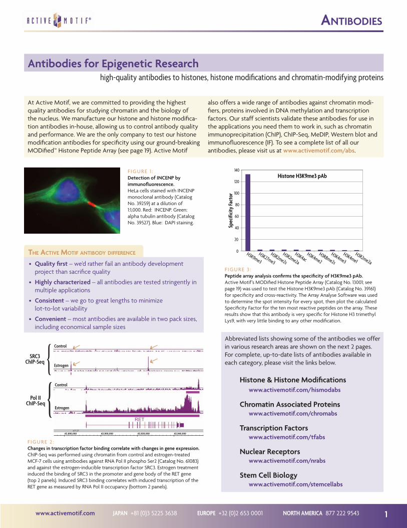
F I G U R E 3 :Peptide array analysis confirms the specificity of H3K9me3 pAb.Active Motif’s MODified Histone Peptide Array (Catalog No. 13001; see page 19) was used to test the Histone H3K9me3 pAb (Catalog No. 39161) for specificity and cross-reactivity. The Array Analyse Software was used to determine the spot intensity for every spot, then plot the calculated Specificity Factor for the ten most reactive peptides on the array. These results show that this antibody is very specific for Histone H3 trimethyl Lys9, with very little binding to any other modification.
F I G U R E 2 :Changes in transcription factor binding correlate with changes in gene expression.ChIP-Seq was performed using chromatin from control and estrogen-treated MCF-7 cells using antibodies against RNA Pol II phospho Ser2 (Catalog No. 61083) and against the estrogen-inducible transcription factor SRC3. Estrogen treatment induced the binding of SRC3 in the promoter and gene body of the RET gene (top 2 panels). Induced SRC3 binding correlates with induced transcription of the RET gene as measured by RNA Pol II occupancy (bottom 2 panels).
{{
RET
42,880,000 42,900,000 42,920,000 42,940,000q11.21

NORTH AMERICA 877 222 9543 EUROPE +32 (0)2 653 0001 JAPAN +81 (0)3 5225 3638 www.activemotif.com2
anTibodies
Description Applications Cat. No.
HISTONES & HISTONE MODIFICATIONSHistone H1 pAb ChIP, IF, IP, WB 39575
Histone H2A pAb ChIP, WB 39235
Histone H2AT120ph pAb DB, WB 39391
Histone H2AS129ph pAb ChIP, DB, IF, IP, WB 39271
Histone macroH2A1.1 pAb IF, IHC, WB 39871
Histone macroH2A2 pAb IF, IHC, WB 39873
Histone H2A.X pAb IF, WB 39689
Histone H2A.XS139ph pAb DB, IF, WB 39117
Histone H2A.Z pAb ChIP, DB, WB 39943
Histone H2B pAb ChIP, WB 39237
Histone H2BK5ac pAb ChIP, ChC, ChS, DB, WB 39123
Histone H2BS14ph mAb IF, IP, WB 61011
Histone H2BK120ac pAb ChIP, ChC, ChS, DB, WB 39119
Histone H2BK120ub1 mAb ChIP, WB 39623
Histone H3 mAb ChIP, IF, TRF, WB 39763
Histone H3, C-terminal pAb ChIP, ELISA, WB 39163
Histone H3ac (pan-acetyl) pAb ChIP, DB, WB 39139
Histone H3R2me2s pAb DB, WB 39703
Histone H3K4me1 pAb ChIP, ChC, ChS, DB, ELISA, IF, IP, WB 39297
Histone H3K4me2 pAb ChIP, ChC, ChS, DB, ELISA, IP, WB 39141
Histone H3K4me3 pAb ChIP, ChC, ChS, DB, ELISA, IF, IP, WB 39159
Histone H3R8me2a pAb DB, WB 39651
Histone H3K9ac pAb ChIP, ChC, ChS, DB, WB 39137
Histone H3K9me1 mAb ChIP, DB, WB 39681
Histone H3K9me2 mAb ChIP, DB, IF, TRF, WB 39683
Histone H3K9me3 pAb ChIP, ChC, ChS, DB, ELISA, IF, IP, WB 39161
Histone H3S10ph pAb ChIP, DB, IF, WB 39253
Histone H3K14ac pAb ChIP, ChC, ChS, DB, ELISA, IP, WB 39599
Histone H3R17me2a pAb DB, WB 39709
Histone H3K18ac pAb ChIP, DB, IF, WB 39587
Histone H3K27ac pAb ChIP, ChC, ChS, DB, IF, IP, WB 39133
Histone H3K27me1 pAb DB, IF, WB 39377
Histone H3K27me2 pAb ChIP, DB, IF, WB 39245
Histone H3K27me3 pAb ChIP, ChC, ChS, DB, IF, IP, WB 39155
Histone H3S28ph mAb ELISA, IF, WB 39098
Histone H3K36ac pAb ChIP, ChC, ChS, DB, IF, WB 39379
Histone H3K36me2 pAb ChIP, ChC, ChS, DB, IF, IP, WB 39255
Histone H3K36me3 pAb ChIP, ChC, ChS, DB, IP, WB 61101
Histone H3T45ph pAb DB, WB 39737
Histone H3K56ac pAb ChIP, ChC, ChS, DB, WB 39281
Histone H3K56me1 pAb DB, WB 39273
Histone H3K79me1 pAb ChIP, DB, WB 39145
Histone H3K79me2 pAb ChIP, ChC, ChS, DB, WB 39143
Histone H4 pAb WB 39269
Histone H4ac (pan-acetyl) pAb ChIP, DB, IF, WB 39243
Histone H4R3me2a pAb DB, IF, WB 39705
Histone H4K5ac pAb ChIP, DB, WB 39699
Histone H4K12ac pAb ChIP, ChC, ChS, DB, IF, WB 39165
Description Applications Cat. No.
Histone H4K16ac pAb ChIP, ChC, ChS, DB, IP, WB 39167
Histone H4K20me1 mAb IF, WB 39727
Histone H4K20me2 pAb ChIP, IF, WB 39173
Histone H4K20me3 pAb ChIP, IF, WB 39180
DNA METHYLATION3-Methylcytosine (3-mC) pAb DB 61111
5-Carboxylcytosine (5-caC) pAb DB, IF 61225
5-Formylcytosine (5-fC) pAb DB, IF 61223
5-Hydroxymethylcytosine (5-hmC) mAb DB, MeDIP 39999
5-Hydroxymethylcytosine (5-hmC) pAb DB, IF, IHC, MeDIP 39769
5-Methylcytosine (5-mC) mAb DB, FACS, IHC, IP, MeDIP 39649
5-Methylcytosine (5-mC) pAb DB, IP, MeDIP 61255
DNMT1 mAb ChIP, IHC, IP, WB 39204
DNMT2 pAb WB 39205
DNMT3A mAb ChIP, IF, IHC, WB 39206
DNMT3B mAb ChIP, IF, IP, WB 39207
DNMT3L pAb WB 39907
MBD1 pAb WB 39857
MBD2 pAb WB 39547
MBD3 mAb WB 39216
MBD4 pAb WB 39217
MeCP2 mAb ChIP, IF, IHC, IP, WB 61285
Uhrf1 mAb IF, IHC, IP, WB 61341
CHROMATIN MODIFIERSBRD4 pAb ChIP, IP, WB 39909
CARM1 pAb WB 39251
CGBP pAb WB 39203
DOT1L pAb WB 39953
EZH2 mAb ChIP, IF, IP 39875
EZH2 pAb Ch 39901
EZH2 phospho Thr345 pAb DB, WB 61241
GCN5 mAb ELISA, IF, WB 39975
HDAC1 mAb ChIP, IF, IHC, IP, WB 39531
HDAC2 mAb ChIP, IF, IHC, IP, WB 39533
HDAC3 pAb ChIP, WB 40968
HDAC5 pAb ChIP, WB 40970
HDAC6 pAb ChIP, WB 40971
JARID1C pAb ChIP, IP, WB 39229
Jhd2 pAb WB 39263
JMJD2A mAb WB 39815
JMJD2D pAb WB 39247
LSD1 pAb ChIP, ChC, ChS, IP, WB 39186
MLL pAb ChIP, IP, WB 61295
MLL1/HRX mAb ChIP, IP, WB 39829
MMSET / WHSC1 mAb ChIP, ChC, ChS, IF, IP, WB 39879
PARP-1 N-terminal pAb ChIP, IHC, IP, WB 39559PARP-2 pAb ChIP, IP, WB 39743

www.activemotif.com JAPAN +81 (0)3 5225 3638 EUROPE +32 (0)2 653 0001 NORTH AMERICA 877 222 9543 3
anTibodies
Description Applications Cat. No.
PRMT5 pAb WB 61001PRMT6 pAb WB 61003
SIRT1 mAb IF, IP, WB 39353
SIRT2 pAb IF, IP, WB 61043
SIRT6 pAb WB 39911SUV39H1 mAb ChIP, IP, WB 39785
CHROMATIN REMODELERSAiolos pAb IF, IP, WB 39393
Boris / CTCFL pAb WB 39851
BRG-1 mAb IF, WB 39807
BRM mAb ChIP, IF, WB 39805
CHD1 pAb ChIP, WB 39729
CHD2 pAb WB 39363
CTCF pAb ChIP, ChC, ChS, IP, WB 61311
HIRA mAb WB 39557
HMG-2 pAb WB 39029
HMGA2 / HMGI-C pAb WB 61041
HP1a mAb ChIP, ELISA, ICC, IF, IHC 39977
HP1b mAb ELISA, IF, IHC, IP, WB 39979
HP1g mAb ELISA, ICC, IF, IHC, IP, WB 39981
Rsf1 pAb IF, IP, WB 39579
SATB1 pAb WB 39839
STEM CELLS5-Carboxylcytosine (5-caC) pAb DB, IF 612255-Formylcytosine (5-fC) pAb DB, IF 612235-Hydroxymethylcytosine (5-hmC) mAb DB, MeDIP 399995-Hydroxymethylcytosine (5-hmC) pAb DB, IF, IHC, MeDIP 397695-Methylcytosine (5-mC) pAb DB, IP, MeDIP 61255Dicer mAb WB 39817EED mAb ChIP, ChC, ChS, IHC, WB 61203EZH2 mAb ChIP, IF, IP 39875GATA-1 pAb ChIP, WB 39025KLF4 pAb WB 39745LIN28A pAb WB 61191c-Myc pAb (65 kDa form) WB 39012NKX2.5 pAb WB 61267Notch1 mAb WB 61147Notch3 mAb WB 61149Oct-4 pAb WB 39811PLZF mAb ChIP, IF, WB 39987Ring1B mAb ChIP, ChC, ChS, IF, IP, WB 39663Sox2 pAb ChIP, IF, IHC, IP, WB 39823Sp1 pAb ChIP, IP, WB 39058Suz12 pAb ChIP, IP, WB 39357YY1 pAb ChIP, IP, WB 39071
Description Applications Cat. No.
CANCER5-Methylcytosine (5-mC) pAb DB, IP, MeDIP 61255BRD4 pAb ChIP, IP, WB 39909CHD1 pAb ChIP, WB 39729DOT1L pAb WB 39953EZH2 mAb ChIP, IF, IP 39875EZH2 pAb Ch 39901HDAC2 mAb ChIP, IF, IHC, IP, WB 39533HDAC6 pAb ChIP, WB 40971Histone H3K4me2 pAb ChIP, ChC, ChS, DB, ELISA, IP, WB 39141Histone H3K4me3 pAb ChIP, ChC, ChS, DB, ELISA, IF, IP, WB 39159Histone H3K27me3 pAb ChIP, ChC, ChS, DB, IF, IP, WB 39155Histone H3K36me2 pAb ChIP, ChC, ChS, DB, IF, IP, WB 39255Histone H3K79me2 pAb ChIP, ChC, ChS, DB, WB 39143JARID1C pAb ChIP, IP, WB 39229LSD1 pAb ChIP, ChC, ChS, IP, WB 39186MLL pAb ChIP, IP, WB 61295MMSET / WHSC1 mAb ChIP, ChC, ChS, IF, IP, WB 39879MOZ pAb WB 39867c-Myc pAb (65 kDa form) WB 39012PARP-1 N-terminal pAb ChIP, IHC, IP, WB 39559PHF8 pAb WB 39711SIRT1 mAb IF, IP, WB 39353SIRT2 pAb IF, IP, WB 61043ZEB1 pAb WB 61119
POLYCOMBBMI-1 mAb ChIP, IP 39993CBX8 pAb WB 61237EED mAb ChIP, ChC, ChS, IHC, WB 61203EZH2 mAb ChIP, IF, IP 39875EZH2 pAb Ch 39901PCL2 mAb WB 61153Phc1 mAb IF, IP, WB 39723Phc2 mAb ChIP, IF, IP 39661Ring1B mAb ChIP, ChC, ChS, IF, IP, WB 39663Suz12 mAb ChIP, IF, IHC, IP 39877Suz12 pAb ChIP, IP, WB 39357
ChIP Chromatin immunoprecipitation
ChC ChIP-chipChS ChIP-SeqDB Dot blotICC ImmunocytochemistryIF Immunofluorescence
IHC ImmunohistochemistryIP ImmunoprecipitationMeDIP Methyl DNA
immunoprecipitationTRF Time-resolved FRETWB Western blot
Applications Key
The antibodies shown above are only a small sample of the over 600 antibodies currently offered. For a complete, up-to-date list of all antibodies available, please visit www.activemotif.com/abs.

NORTH AMERICA 877 222 9543 EUROPE +32 (0)2 653 0001 JAPAN +81 (0)3 5225 3638 www.activemotif.com4
ChIP Products Overview
Active Motif has consistently been the leader in introducing innovations that make ChIP faster, simpler and more reproduc-ible. We were the first to introduce magnetic beads into a ChIP kit, the first to offer enzymatic chromatin shearing and, build-ing on these improvements, the first with kits for performing
sequential, high-throughput and RNA ChIP. We now offer a highly sensitive ChIP kit optimized to work with limited sample amounts and low-binding affinity antibodies, as well as a kit for performing ChIP-Seq. The diagram below is an overview of the ChIP process and highlights Active Motif products for each step.
a complete selection of proven reagents and equipment for the entire ChIP procedure
ChroMaTin iMMunopreCipiTaTion flowCharT
Chromatin Preparation
ChromatinImmunoprecipitationChIP Antibodies
• ChIP & ChIP-Seq validated antibodies
• ChIP Antibody Validation Services
• MODified™ Histone Peptide Array
ChIP Kits• ChIP-IT® Express• ChIP-IT® High Sensitivity• ChIP-IT® ChIP-Seq• Re-ChIP-IT®• RNA ChIP-IT®
Chromatin Shearing• EpiShear™ Sonicators• ChIP-IT® Sonication &
Enzymatic Shearing Kits• Dounce Homogenizers
Pre-Made Chromatin• Ready-to-ChIP Chromatin
PCR Analysis• ChIP-IT® Control Kits
qPCR Analysis• ChIP-IT® qPCR Analysis Kit• ChIP-IT® qPCR Control Kits• ChIP Control qPCR Primer Sets
Epigenetic Services• FactorPath™ ChIP-Seq & ChIP-chip• TranscriptionPath™ ChIP-Seq & ChIP-chip• HistonePath™ ChIP-Seq & ChIP-chip
DNA Clean-up• Chromatin IP
DNA Purification KitDownstream Analysis
Cycle
DRx
n
C C A T G310
C A A G
www.activemotif.com JAPAN +81 (0)3 5225 3638 EUROPE +32 (0)2 653 0001 NORTH AMERICA 877 222 9543 5
epigeneTiC serviCes
ChIP Servicesutilize our experienced research team for your epigenetics studies
Chromatin immunoprecipitation (ChIP) is a powerful tool for studying protein/DNA interactions. ChIP-Seq takes ChIP a step further by combining it with Next-Gen sequencing in order to generate whole-genome data sets. However, the various techni-cal and bioinformatic challenges associated with ChIP-Seq present a barrier to many researchers in need of this data. To
overcome this barrier, researchers can utilize our expertise to facilitate their research into transcription factor binding, transcriptional regulation and histone distribution. Active Motif’s Epigenetic Services team has over 9 years of success providing a wide variety of ChIP services. For complete details, please give us a call or visit us at www.activemotif.com/services.
Contact us about our ChIP antibody validation service.
F I G U R E 1 :HistonePath ChIP-Seq maps H3K4me3 peaks to a Zfp gene cluster.ChIP–Seq was performed using chromatin from mouse livers and an antibody against H3K4me3 (Catalog No. 39159). Sequence tags were mapped to gener-ate a whole-genome data set. The image above focuses on a 3 Mb window containing a Zfp gene cluster on chromosome 13. H3K4me3 peaks are present at the start site of all Zfp genes. Gene annotations run from right to left.
F I G U R E 2 :A representative TranscriptionPath gene profile.TranscriptionPath-Seq, an RNA Pol II ChIP-Seq, provides a genome-wide view of transcription rates. A typical Pol II profile is presented above. Most genes will display an RNAPII peak at the promoter indicative of the paused state. RNAPII occupancy in the gene body tends to be fairly even across the gene body. Occupancy then increases toward the 3´ end of the gene and often extends beyond the 3´ gene boundary. The increased occupan-cy at the 3´ end is associated with pausing, as transcription is terminated.
ChIP-Seq ServicesSeveral types of ChIP-Seq are offered; ChIP is performed on different types of targets to answer different questions:
• FactorPath™ ChIP-Seq – discover, identify and quantitate transcription factor and cofactor binding sites
• HistonePath™ ChIP-Seq – map histone modifications or histone modifying enzymes across the genome
• TranscriptionPath™ ChIP-Seq – measure transcription rates globally as a function of RNA Pol II occupancy
In addition, we offer MethylPath™ MeDIP-Seq and a number of other services for DNA methylation research (see page 25).
Active Motif Epigenetic Services
• Experience – over 1,500 genome-wide data sets generated
• Quality – QC steps ensure high-quality whole-genome data
• Support – all services include bioinformatics analysis

NORTH AMERICA 877 222 9543 EUROPE +32 (0)2 653 0001 JAPAN +81 (0)3 5225 3638 www.activemotif.com6
Introduction to ChIP-IT® Kitsfind which ChIP-IT Kit is best suited for your specific application
Chip-iT® produCTs
The different ways in which ChIP is used have multiplied in recent years, making it critical to correctly match the specific kit with the intended application. Active Motif has a long history of innovation in kits for different ChIP applications, having simpli-fied the protocol with magnetic beads, developed enzymatic shearing procedures, introduced a high-throughput ChIP kit, and
a Re-ChIP kit for sequential ChIP assays on a single sample. Our most recent kits have been optimized for sample amounts and antibody types that require high sensitivity ChIP, and for performing ChIP-Seq. The descriptions below provide brief summaries of our ChIP-IT® Kits so you can determine which ones are best suited for your specific needs.
ChIP-IT Express (page 7)ChIP-IT Express was the first ChIP kit to utilize magnetic beads. Because magnetic beads have much lower background binding than traditional agarose beads, this made it possible to speed up and simplify the ChIP protocol by eliminating some of the steps required in more conventional protocols. Washing and elution steps are also faster because centrifugation steps were replaced by rapid magnetic pull-down. This reduced the amount of time required to a single day, and the number of cells to 100,000.
ChIP-IT Express Kits provide reagents for 10 chromatin prepa-rations by sonication, 2 shearing optimizations, and 25 ChIP reactions. ChIP DNA purification components are not included, but can be purchased separately if users plan to perform certain downstream applications, such as qPCR.
ChIP-IT High Sensitivity (page 8)ChIP-IT High Sensitivity was designed for ChIP with limited amounts of sample or when using low-affinity antibodies or those directed against low-abundance targets. The kit employs specialized protein G agarose beads and an antibody blocker to minimize non-specific binding. Filtration-based capture and washes are used to eliminate sample loss, making the kit more sensitive than magnetic-bead methods, like ChIP-IT Express.
The kit provides reagents to prepare 16 chromatin samples by sonication and perform 16 ChIP reactions. Each ChIP reaction requires as little as 1,000 cell equivalents for highly abundant target proteins, or 50,000 for low abundance proteins. Due to overnight incubation and other steps that increase capture ef-ficiency, the procedure takes 3 days. DNA purification compo-nents are included in the kit.
ChIP-IT ChIP-Seq (page 9)This kit was designed for performing ChIP-Seq on the Illumina® sequencing platforms. It combines the ChIP-IT High Sensitivity Kit (above), the ChIP-IT qPCR Analysis Kit for validation of ChIP DNA (see page 9), and a set of library construction reagents suitable for the preparation of 10 Next-Generation sequencing libraries. As this kit is used to construct ChIP-Seq libraries, chromatin should be prepared from at least 3 million cells.
ChIP-IT Express Enzymatic (page 7)ChIP-IT Express Enzymatic was the first kit with reagents for shearing chromatin by enzymatic digestion instead of by sonication. As not everybody owns a sonicator or is proficient in its use, Active Motif developed this user-friendly method to shear chromatin for ChIP by digesting it, eliminating problems associated with sonication. Enzymatic shearing is a good choice for users who will not do enough ChIP to make it worth buying and mastering the use of a sonicator.
ChIP-IT Express Enzymatic is a magnetic-bead based kit that is identical to ChIP-IT Express except for the different chromatin preparation reagents. The kit provides reagents sufficient to make 10 chromatin preparations by enzymatic digestion, as well as 2 shearing optimizations, and to perform 25 ChIP reactions.
Re-ChIP-IT (page 10)Re-ChIP-IT takes advantage of the same magnetic-bead based ChIP method developed for the ChIP-IT Express Kit. However, Re-ChIP-IT was designed for sequential ChIP, in which two ChIP reactions using different antibodies are performed in series on the same sample. This makes it possible to assay for the simul-taneous binding of two transcription factors or histone modifi-cations at the same genomic region of interest. The kit provides reagents sufficient to perform 25 Re-ChIP reactions. It does not include any shearing components; users can choose to purchase either the ChIP-IT Express Shearing or Enzymatic Shearing Kit.
ChIP-IT Express HT (page 10)ChIP-IT Express HT leverages our magnetic-bead based ChIP method to make possible high-throughput ChIP in a 96-well format. The kit provides reagents sufficient to perform 96 ChIP reactions. No shearing components are included; users can pur-chase the ChIP-IT Express Shearing or Enzymatic Shearing Kit.
RNA ChIP-IT (page 11)Non-coding RNAs play important roles in chromatin structure and transcriptional silencing. The RNA ChIP-IT Kit uses our magnetic-bead based ChIP method to enable the study of RNA-protein interactions in chromatin. All components have been optimized to recover RNA from ChIP for RT-PCR analysis.

www.activemotif.com JAPAN +81 (0)3 5225 3638 EUROPE +32 (0)2 653 0001 NORTH AMERICA 877 222 9543 7
ChroMaTin iMMunopreCipiTaTion
ChIP-IT® Express & ChIP-IT® Express Enzymaticmagnetic beads make ChIP fast, easy & more reproducible
ChIP-IT® Express Kits use protein G-coated magnetic beads in-stead of traditional agarose beads, making it possible to perform ChIP in just 1 day. Kits are available that use your choice of either sonication or enzymatic digestion for preparing sheared chro-
matin. Both feature fast, reproducible protocols that make ChIP more successful while reducing your time and effort. For more information on ChIP-IT Express Kits, please visit our website at www.activemotif.com/chipitexpress.
Product Format Cat. No.
ChIP-IT® Express 25 rxns 53008
ChIP-IT® Express Enzymatic 25 rxns 53009
Chip-iT express advanTages
• No pre-blocking needed – magnetic beads are inert
• Get results quickly – 4-hour immunoprecipitation and streamlined wash steps enable ChIP to be completed in just 1 day
• Optimized components, buffers and protocols – Active Motif has sweated all the details, so you don’t have to
Faster, more streamlined method for ChIPChIP-IT Express Kits improve on traditional ChIP by reducing or eliminating several time-consuming steps. These kits utilize protein G magnetic beads, which have much lower background binding than traditional agarose beads. This reduced background has made it possible to eliminate some of the steps required in more conventional protocols like pre-clearing and block-ing. Washing is much easier because the spin steps have been replaced by rapid magnetic pull-down. Collectively, all of the improvements included in ChIP-IT Express give you the capabil-ity to perform your reactions in PCR tubes with a multi-channel pipettor, which reduces your effort and improves consistency (Figure 1).( A bar magnet is included with all ChIP-IT Express Kits, but you can also use commercially available magnetic stands.)
Efficient ChIP enables you to use fewer cellsConventional ChIP requires at least two million cells as starting material, which can be problematic with some cell lines, and labor-intensive even in the best case. ChIP-IT Express Kits have been optimized to provide superior target gene enrichment. The ChIP-IT Express Kits can routinely produce excellent results with chromatin from as few as 100,000 cells (Figure 2).
Shear chromatin by sonication or enzymatic digestionChromatin shearing is most commonly done by sonication. However, sonication can be difficult to optimize due to emul-sification and overheating of the sample. Because of this, or if you don’t have a sonicator, Active Motif has developed a robust, user-friendly method to shear chromatin by enzymatic digestion (Figure 3). Because Active Motif offers both ChIP-IT Express and ChIP-IT Express Enzymatic Kits, you can choose whichever shearing method you prefer.
F I G U R E 1 :Multiple sample ChIP using ChIP-IT Express.Washing the magnetic beads is fast and easy because the pellet forms against the side of the tube above the level of the supernatant. This speeds the procedure, eliminates sample loss and enables you to ChIP multiple samples in 8-well PCR tubes using a multi-channel pipettor.
F I G U R E 2 :ChIP-IT Express works with 100,000 cells.ChIP-IT Express was performed in dupli-cate on sonicated HeLa cell chromatin (1.0 x 10⁵ cell equivalents per ChIP). Two µg of RNA pol II and Neg IgG antibody was used for IP. GAPDH PCR primers were used for endpoint PCR analysis. The ChIP DNA isolated with RNA pol II antibody generated more product than that from the negative control IgG, demonstrating successful ChIP from 100,000 cells.
RNA
pol I
I
RNA
pol I
I
Neg
IgG
Neg
IgG
F I G U R E 3 :Analysis of enzymatically sheared DNA.HeLa cells were fixed for 10 minutes with formaldehyde and then chromatin was prepared using the ChIP-IT Express Enzymatic Kit protocol & reagents. Chromatin was sheared with the Enzymatic Shearing Cocktail for 5, 10 & 15 minutes before stopping the reaction.
Lane 1: 100 to 1000 bp ladder. Lane 2: Unsheared HeLa DNA. Lane 3: HeLa DNA treated for 5 minutes. Lane 4: HeLa DNA treated for 10 minutes. Lane 5: HeLa DNA treated for 15 minutes.
1000 bp
500 bp
100 bp
1 2 3 4 5

NORTH AMERICA 877 222 9543 EUROPE +32 (0)2 653 0001 JAPAN +81 (0)3 5225 3638 www.activemotif.com8
ChIP for limited sample material and low abundance target proteinsChIP-IT® High Sensitivity
Chip high sensiTiviTy
As the field of epigenetics expands into the research areas of transcriptional regulation and stem cells, the need for accurate and reliable chromatin immunoprecipitation assays suitable for use with limited sample material and difficult-to-ChIP transcrip-tion factor antibodies is more important than ever. Active Mo-
tif’s ChIP-IT® High Sensitivity Kit is designed to meet this need by enabling the enrichment of high-quality ChIP DNA from as little as 1,000 cell equivalents. ChIP-IT High Sensitivity has been validated with hundreds of samples as well as low abundance transcription factors and low-affinity ChIP antibodies.
Benefits of ChIP-IT High SensitivityThe ChIP-IT High Sensitivity Kit utilizes specially formulated buffers for high-quality chromatin preparation from cultured cells or fresh or frozen tissue. Low background protein G agarose beads and an antibody blocker are used to minimize any non-specific binding during the immunoprecipitation reac-tion. ChIP filtration columns are included in the kit for a fast, easy and consistent solution for capture and wash steps. The re-sult is a kit that is capable of delivering both higher signals and reduced background levels as compared with other commer-cially available ChIP kits (Figure 1). These features allow for the detection of low abundance transcription factor binding events, the use of limited sample material in the immunoprecipitation reaction and the detection of low-affinity antibody targets in downstream applications such as ChIP-Seq, ChIP-chip and qPCR.
Product Format Cat. No.
ChIP-IT® High Sensitivity 16 rxns 53040
ChIP-IT® qPCR Analysis Kit 10 rxns 53029
Chip-iT high sensiTiviTy advanTages
• Ideal for low abundance transcription factor enrichment or for use with antibodies with low binding affinities
• Optimized reagents increase signals and reduce the background caused by non-specific binding events
• Enrichment from as little as 1,000 cell equivalents per IP reaction for highly abundant target proteins, and as little as 50,000 cell equivalents for low abundance proteins
• Extensively tested across multiple sample types and anti-bodies with proven performance for ChIP-Seq & qPCR
F I G U R E 2 :ChIP-IT qPCR Analysis Kit enables direct comparison of ChIP efficiency.ChIP reactions were performed with the ChIP-IT High Sensitivity Kit using an RNA pol II antibody on different amounts of chromatin. The top graph shows absolute signals that are independent of the starting amount of chromatin. In the bottom graph, Active Motif’s ChIP-IT qPCR Analysis Kit was used to normalize for the starting amount of chromatin, primer efficiency and ChIP DNA resuspension vol-ume. This data demonstrates the sensitivity of ChIP-IT High Sensitivity between 3,000 and 150,000 cell equivalents per ChIP reaction, and the consistency of the ChIP efficiency as shown by normalization with the ChIP-IT qPCR Analysis Kit.
F I G U R E 1 :The ChIP-IT High Sensitivity Kit detects low abundance proteins.MCF-7 chromatin was prepared and immunoprecipitated according to the rec-ommendations for each manufacturer’s ChIP Kit using an antibody for the low abundance nuclear co-activator 2 (NCOA2) protein and a negative control IgG. Following enrichment, qPCR was performed using the ChIP-IT qPCR Analysis Kit (described on page 9) to normalize the data. While NCOA2 is considered a difficult antibody for ChIP, NCOA2 binding was detected at 20-fold over back-ground at the estrogen-related receptor alpha (ESRRA) promoter when ChIP was performed using the ChIP-IT High Sensitivity Kit. When using competitor kits, NCOA2 was either not enriched at all (Competitors M & I), or there was non-specific binding by the IgG and high background for the negative control primers (Competitor D). Data represents triplicate qPCR values expressed as binding events detected per 1,000 cells.

www.activemotif.com JAPAN +81 (0)3 5225 3638 EUROPE +32 (0)2 653 0001 NORTH AMERICA 877 222 9543 9
ChIP-IT® ChIP-Seqsimplify genome-wide ChIP analysis for Next-Generation sequencing
Chip sequenCing & qpCr analysis
The combination of ChIP with genome-wide analysis using Next-Generation sequencing (ChIP-Seq) is a powerful approach that can provide insights into gene regulation, gene expression mechanisms of chromatin modification and pathway analysis. The ChIP method enriches for protein-DNA complexes using an
antibody against a protein of interest. Oligonucleotide adapters are then ligated to the ChIP-enriched DNA before the size- selected library is amplified and purified for use in Next-Gen sequencing. Analysis of the sequencing data will identify the global binding sites for the protein of interest.
ChIP for Next-Generation sequencingActive Motif’s ChIP-IT® ChIP-Seq Kit provides proven reagents, streamlined protocols and validation controls to give you con-fidence in successful ChIP-Seq using the Illumina® sequencing platforms. The assay utilizes the highly consistent and robust chromatin immunoprecipitation and purification procedure of the ChIP-IT® High Sensitivity Kit (described on page 8), and also includes Active Motif’s ChIP-IT® qPCR Analysis Kit for ChIP DNA validation (described to the right) as well as a set of library construction reagents suitable for the preparation of ten Next-Generation sequencing libraries.
For successful ChIP-sequencing using the ChIP-IT ChIP-Seq Kit, we recommend the preparation of chromatin from at least 3 million cells. This will help to ensure the recovery of 10 ng ChIP-enriched DNA for library preparation. Adapter sequences to generate single end, paired end or barcoded libraries are not included in the kit and should be obtained from Illumina.
ChIP DNA validation with ChIP-IT qPCR AnalysisBefore investing time and money into expensive downstream applications, such as Next-Generation sequencing, it is recom-mended to validate the quality of your ChIP-enriched DNA. Active Motif has drawn upon our years of experience with test-ing and validating hundreds of samples and target antibodies to develop the ChIP-IT qPCR Analysis Kit.
The ChIP-IT qPCR Analysis Kit uses DNA standards to create a single qPCR standard curve that can be used to determine PCR primer pair efficiency. Following qPCR amplification, the output of the qPCR instrument can be copied into the ChIP-IT qPCR Analysis spreadsheet. The spreadsheet performs the calcula-tions to normalize the data with respect to primer pair efficien-cy, the amount of chromatin used in each immunoprecipitation reaction and the resuspension volume of the ChIP DNA. The analyzed data is automatically graphed within the spreadsheet to show the normalized values. By utilizing this normalization strategy, data can easily be analyzed and compared across multiple sample types and experiments, including those performed on different days (see Figure 1 on page 8).
In addition to the DNA standards and control primer sets included within the kit, the manual provides recommendations for data interpretation to evaluate the success of the ChIP reac-tions, helping to determine the quality of the ChIP DNA. These recommendations are based upon the historical experience of Active Motif using this method to analyze hundreds of ChIP reactions. Following these recommendations will provide con-fidence that the ChIP DNA is suitable for sequencing. The data interpretation recommendations are based on the use of the ChIP-IT High Sensitivity or ChIP-IT ChIP-Seq Kit to perform the chromatin immunoprecipitation reactions. If another method is used to perform ChIP that results in higher background levels and lower sensitivity, the recommended threshold levels may not apply. To learn more about the ChIP-IT qPCR analysis Kit, please visit www.activemotif.com/qPCRanalysis.
Product Format Cat. No.
ChIP-IT® ChIP-Seq 10 library constructs 53041
ChIP-IT® qPCR Analysis Kit 10 rxns 53029
F I G U R E 1 :ChIP-IT ChIP-Seq data using Histone H3K4me3 pAb.ChIP was performed using chromatin from mouse livers and a Histone H3K4me3 pAb (Catalog No. 39159) using the ChIP-IT ChIP-Seq Kit. Genome alignment was performed with ELAND and peak calling was performed with MACS. The image above focuses on a Zfp gene cluster on chromosome 13. Gene annotations run from right to left, showing that H3K4me3 binds to the transcription start sites of all Zfp genes shown.

NORTH AMERICA 877 222 9543 EUROPE +32 (0)2 653 0001 JAPAN +81 (0)3 5225 3638 www.activemotif.com10
ChIP-IT® Express for Sequential ChIP & High-throughput ChIPexpect quick, easy, reliable performance
F I G U R E 5 :Chromatin IP performed on HeLa chromatin with ChIP-IT Express HT.PCR carried out using primers specific for the GAPDH gene. Lanes 1-4: ChIP using 2 µg RNA Pol II antibody. Lanes 5-8: ChIP using normal mouse IgG as a negative control. Lane 9: no DNA control. Lane 10: input DNA control.
F I G U R E 4 :True high-throughput ChIP with ChIP-IT Express HT.With the efficient plate-based protocol of ChIP-IT Express HT, you can process up to 96 ChIP reactions at a time.
re-Chip & high-ThroughpuT Chip
High-throughput ChIPPerforming multiple ChIP experiments simultaneously using traditional methods only compounds the difficulty of the assay. That is why Active Motif developed ChIP-IT® Express HT. Now it is possible to perform the time-saving ChIP-IT Express protocol in a 96-well format (Figures 4 & 5), with everything optimized to the level you have come to expect from Active Motif. ChIP-IT Express HT is compatible with our enzymatic or sonication-based shearing kits for chromatin preparation, as well as with our ChIP-IT Control Kits. For complete information, please visit www.activemotif.com/htchip.
F I G U R E 2 :Schematic representation of the Re-ChIP-IT procedure.
F I G U R E 3 :Sequential chromatin immunoprecipitation using Re-ChIP-IT.The lane numbers are the same in each panel to indicate that the DNA is from the same chromatin sample. The left panel shows the results of PCR performed on an aliquot of DNA removed from the experiment after the first ChIP step; the right panel represents PCR results on DNA from chromatin samples after both ChIP steps. For example, chromatin samples subjected to first ChIP using Mouse IgG as a negative control (lanes 1 and 2 in the left panel) were then subjected to a second ChIP with an RNA Pol II antibody (lanes 1 and 2 in the right panel). Chromatin samples in which Mouse IgG was used as either the first antibody (lanes 1 and 2) or second antibody (lanes 5 and 6) show little amplifica-tion of GAPDH DNA in either the left (first ChIP) or right panel (first and second ChIP). Chromatin samples in which the first antibody used was anti-RNA Pol II and the second antibody was anti-TFIIB (lanes 3 and 4) show good amplification of GAPDH DNA after the second ChIP (right panel) indicating co-localization of RNA Pol II and TFIIB at the same region of the GAPDH promoter.
Re-ChIP-IT® for sequential ChIP experimentsDeciphering the Histone Code often requires showing that two marks or associated proteins co-occur at the same site in the genome. Sequential ChIP (or Re-ChIP) involves perform-ing sequential chromatin immunoprecipitations using different antibodies in series on the same sample (Figure 2). This makes it possible to assay for the simultaneous binding of two transcrip-tion factors or histone modifications at the same genomic region of interest.
Sequential chromatin IP was technically challenging and diffi-cult, until now. Active Motif’s Re-ChIP-IT Kit® has been opti-mized for this technique, making it easy to perform sequential ChIP, so you can more easily localize two different proteins or histone modifications to the same genetic locus (Figure 3). For complete information, visit www.activemotif.com/rechipit.
Product Format Cat. No.
Re-ChIP-IT® 25 rxns 53016
ChIP-IT® Express HT 96 rxns 53018
106 842 95 731

www.activemotif.com JAPAN +81 (0)3 5225 3638 EUROPE +32 (0)2 653 0001 NORTH AMERICA 877 222 9543 11
The RNA ChIP-IT methodRNA ChIP-IT uses a modified ChIP protocol that has been optimized for RNA preservation and recovery. RNA-protein interactions are fixed with formaldehyde, and chromatin shear-ing is combined with DNase treatment to yield RNA/protein complexes that can be immunoprecipitated with antibodies to specific proteins. Cross-links are subsequently reversed; RNA is recovered and again treated with DNase to ensure the absence of DNA. The optimized method is quick and has been success-fully used to study several non-coding RNAs in the chromatin context (Figure 1).
In contrast to other kits designed to study RNA-protein interac-tions, called RIP kits, RNA ChIP-IT is designed specifically to extract and immunoprecipitate RNA from chromatin. The kit solves the associated challenges of extracting chromatin while preserving RNA integrity, and removing all DNA, for a clean result that is attributable to RNA alone. The RNA ChIP-IT Kit is the solution optimized for the Epigeneticist studying RNA, rather than the RNA biologist.
Optimized RNA-ChIPTo make the characterization of the role of RNA in genome regulation possible, Active Motif has leveraged its expertise in ChIP to develop the first of its kind kit for RNA-ChIP. The RNA ChIP-IT® Kit was designed to study RNA-protein interactions in a chromatin context, and optimized to recover RNA for RT-PCR analysis. It contains sufficient reagents for 25 assays and employs protocols that utilize magnetic beads, which improve results while reducing time and effort.
Start investigating the role of RNA todayFor complete information, including a downloadable product manual, visit www.activemotif.com/rnachip.
Product Format Cat. No.
RNA ChIP-IT® 25 rxns 53024
RNA ChIP-IT® Control Kit – Human 5 rxns 53025
F I G U R E 1 :RT-PCR analysis and % recovery of RNA-ChIP samples.The RNA ChIP-IT Kit was used on 10 µg samples of DNase I-treated HeLa chromatin with 10 µl of Suz12 antibody (Catalog No. 39357) and 2 µg of Normal Rabbit IgG. Real-time RT-PCR was performed using primers for the lincRNA SFPQ locus. The amplification plot (top) and % input recoveries (bottom) are shown. Dividing the input recovery of the Suz12 antibody by that of the rabbit IgG indicates a 141-fold enrichment of the lincSFPQ region with Suz12 antibody.
rna Chip-iT advanTages
• Specifically tailored to study chromatin-associated RNA
• Designed to remove DNA while maintaining RNA integrity
• Step-by-step protocols for fixation, sonication and immunoprecipitation of chromatin, all optimized for RNA preservation
• Includes all RNase and protease inhibitors at precise concentrations
• Separate control kit available with positive and negative control antibodies and primers for the lincSFPQ locus
RNA ChIP-IT®optimized method to study RNA/protein interactions in a chromatin context
rna ChroMaTin iMMunopreCipiTaTion
Evidence is building that RNA-directed processes play a critical role in orchestrating chromatin architecture and epigenetic memory. Nucleic acids purified from chromatin are 2-5% RNA; these RNAs are non-coding sequences that play important roles
in chromatin structure and transcriptional silencing. However, characterizing these RNAs using conventional ChIP techniques is difficult due to the complexity of chromatin and the large amount of DNA present in chromatin.

NORTH AMERICA 877 222 9543 EUROPE +32 (0)2 653 0001 JAPAN +81 (0)3 5225 3638 www.activemotif.com12
get better chromatin IP results with less effortChIP Accessory Kits and Reagents
Chip aCCessories
F I G U R E 1 :Improved chromatin IP using Bridging Antibody for Mouse IgG.ChIP was performed on Ready-to-ChIP HeLa Chromatin (Cat. No. 53015) with RNA pol II mouse mAb (Cat. No. 39097) alone, the same antibody together with the Bridging Antibody for Mouse IgG (Cat. No. 53017), the Bridging Antibody alone, and Mouse IgG alone. Real-time qPCR was then performed using GAPDH primers, as this is an active gene. The plot above shows the fold enrichment of each ChIP relative to that of Mouse IgG alone. Inclusion of Bridging Antibody clearly increases the capture efficiency of the RNA pol II mouse mAb.
ChIP-IT Express Shearing KitsDesigned to work specifically with our ChIP-IT Express Kits, the sonication and enzymatic shearing kits give you the same shearing components as those found in ChIP-IT Express Kits, but in greater quantities to allow you to optimize your shearing conditions before proceeding with ChIP experiments.
ChIP-IT® Control KitsAs ChIP is a DNA enrichment, not a purification, ChIPs are unavoidably contaminated with non-specific chromatin. This can lead to false positive PCR products that make data inter-pretation difficult. To solve this problem, we offer species-specific (human, mouse and rat) control kits for real-time or endpoint PCR that help confirm the chromatin preparation and immunoprecipitation procedures worked properly and enable you to assess the quality of your antibody and PCR reactions. ChIP-IT Control qPCR Kits include a positive control antibody, a bridging antibody to enhance binding affinity of mouse monoclonal antibodies, a negative control antibody to evaluate non-specific binding, and species-specific positive and negative control qPCR primers. The positive and negative control primers allow you to show that the ChIP worked and to establish back-ground binding levels within the same ChIP reaction. ChIP-IT Control Kits are for endpoint PCR. Each kit provides positive and negative control antibodies, a bridging antibody, a positive control PCR primer set and PCR buffer/DNA loading dye that makes your PCR rxns gel-ready straight out of the thermocycler. For details, see www.activemotif.com/chipcontrols.
ChIP Control qPCR Primer SetsPrimers that amplify positive and negative binding locations are an important component of every ChIP experiment. Unfortu-nately the location of transcription factor binding sites, histone modifications and CpG DNA methylation varies between cell types. Therefore, we offer a large variety of species-specific qPCR primer sets for use as positive and negative controls for many of the more common ChIP targets. Use of our human, mouse, rat, Drosophila, yeast and Zebrafish primer sets will save you the time and effort required to design, synthesize and test your own species/gene-specific control primers, as ours have been tested and validated to work, and are also used regularly by our Epigenetics Services division. To see the many different primer sets available, go to www.activemotif.com/chipprimers.
Ready-to-ChIP ChromatinReady-to-ChIP Chromatin is offered from a number of ENCODE cell lines that have been optimally sheared by sonication and validated in ChIP as a control or test sample. As a result, you can more easily validate your own antibodies and primer sets. The chromatin can be used with all of the ChIP-IT Express Kits and controls, so you can be certain that the only variable in validating a new antibody for ChIP is the antibody itself. For details, please visit www.activemotif.com/chipready.
ChIP-validated antibodies, guaranteed for ChIPWhen performing ChIP, only antibodies of the highest quality that recognize the target protein in its native, chromatin-asso-ciated context will do. For a complete listing of ChIP-validated antibodies, go to www.activemotif.com/chipabs.
Enhanced ChIP and IP when using mouse antibodiesLow antibody binding can greatly reduce the quality of ChIP results. For improved ChIP when using mouse IgG antibodies, Active Motif offers the Bridging Antibody for Mouse IgG. This anti-mouse IgG binds with high affinity to both mouse primary IgG antibody and protein G-conjugated beads. As this greatly increases capture efficiency, ChIP results are markedly improved (Figure 1); Bridging Antibody also improves standard IP and Co-IP.
The permutations in ChIP experiments are numerous. We’ve been listening to your feedback for more than 10 years now, and understand that, for example, you must often optimize sonication conditions before performing ChIP. Or, you may be
unsure of your chromatin quality, or the suitability of your anti-body or primers for use in ChIP. To make your ChIP experiments more successful, we offer a line of accessories designed to make it easier for you to troubleshoot and validate your ChIP results.

www.activemotif.com JAPAN +81 (0)3 5225 3638 EUROPE +32 (0)2 653 0001 NORTH AMERICA 877 222 9543 13
Chromatin IP DNA Purification Kitrapid, simplified purification of your ChIP DNA samples
F I G U R E 2 :DNA recovery as a function of fragment size.The Chromatin IP DNA Purification Kit enables efficient recovery of DNA fragments as small as 50 base pairs. DNA molecular weight markers (10 base pair ladder) were purified with the Chromatin IP DNA Purification Kit, run on a 5% agarose gel and stained with ethidium bromide to visualize the recovered DNA fragments.
Lane 1: DNA not purified.Lane 2: DNA after purification.
DNA that is larger than 50 base pairs is repre-sented equally in the purified and non-purified DNA samples. DNA fragments that are smaller than 50 base pairs (bracket) are not efficiently purified, and thus are not represented in the purified DNA sample (Lane 2, bracket).
F I G U R E 1 :DNA Binding Buffer pH indicator dye.The DNA Purification Binding Buffer has a pH indicator dye so the pH of the solution can be easily determined. The DNA should only be applied to the column if the solution is bright yellow (left), indicating a pH under 7.5. DNA will not bind to the column if the pH is higher than 7.5. (Sodium acetate is included to reduce sample pH, if needed.)
dna purifiCaTion froM Chip’d saMples
Rapid method for use with many sample typesOnce your ChIP experiments are complete, DNA purification can be started immediately. The entire procedure takes only five to ten minutes, depending upon the number of samples to purify. The Chromatin IP DNA Purification Kit is compatible with samples from all of Active Motif’s ChIP-IT® Kits, or from any standard chromatin IP kit or procedure. You can use your choice of mechanical or enzymatic shearing of chromatin, and either agarose or magnetic beads. The kit can also be used to purify methylated DNA samples enriched using other Active Motif kits, including MethylCollector™ Ultra, HypoMethylCollector™, and the hMeDIP and MeDIP Kits. The Chromatin IP DNA Purifi-cation Kit is designed for use with a microcentrifuge for sample processing, but can also be used with a vacuum manifold.
DNA recovery and yieldThe purified DNA is suitable for use in many downstream analy-sis techniques, including PCR (endpoint & quantitative), South-ern blotting, microarray analysis & Next-Gen sequencing. DNA can be successfully recovered from ChIP performed with as few as 10,000 cells. Depending upon the amount of chromatin used in ChIP, DNA recovery will range from 100 ng to 1 µg. DNA frag-ments below 50 base pairs in length are not recovered efficient-ly (Figure 2), but as ChIP experiments usually require chromatin that has been sheared to 200-1500 bp, this is not an issue.
User friendly procedureAfter your ChIP is complete, the ChIP DNA samples are mixed with the kit’s DNA Purification Binding Buffer. Because binding to the included purification columns is pH dependant, the Binding Buffer contains a convenient pH indicator dye (Figure 1). This enables you to see the pH of your samples before apply-ing them to the columns, helping ensure successful purification of your ChIP DNA. After binding, the DNA on the column is washed, then eluted using the included Elution Buffer.
Product Format Cat. No.
Chromatin IP DNA Purification Kit 50 rxns 58002
Chromatin Immunoprecipitation (ChIP) is a powerful, well estab-lished technique for studying interactions between chromatin-associated proteins and specific regions of the genome. The use of ChIP in combination with genome-wide analysis techniques can yield a tremendous amount of information regarding the distribution of transcription factors and histone modifications.
But, many downstream analysis techniques require DNA that has been purified away from the contaminants present in an eluted ChIP sample. Active Motif’s Chromatin IP DNA Purification Kit enables you to quickly clean up your ChIP DNA samples and get them ready for analysis, without the need for labor-intensive, time-consuming phenol/chloroform extractions.
ChroMaTin ip dna purifiCaTion KiT advanTages
• Purify your ChIP DNA samples quickly and easily
• Compatible for use with all of Active Motif’s ChIP-IT® Kits, as well as with ChIP kits from other manufacturers
• Use to purify DNA that was ChIP’d using either agarose or paramagnetic bead methods
• Can also be used to purify DNA from our hMeDIP, MeDIP, MethylCollector™ Ultra and HypoMethylCollector™ Kits
21
100 bp
50 bp

NORTH AMERICA 877 222 9543 EUROPE +32 (0)2 653 0001 JAPAN +81 (0)3 5225 3638 www.activemotif.com14
GenoMatrix™ Whole Genome Amplification
After enriching for methyl-DNA or protein markers of interest by chromatin immunoprecipitation (ChIP), your recovered DNA may be limiting, depending on the number of cells you started with, the efficiency of the capture technique, and the abundance of
the marker. You may not have sufficient DNA for downstream analysis techniques. Active Motif’s GenoMatrix™ Whole Genome Amplification Kit enables you to accurately amplify DNA for use in a variety of downstream analysis techniques.
How much DNA can you expect to recover?The amount of DNA you can expect to recover following ChIP or methyl-DNA pull down experiments will vary based on the method you are using, and if it is ChIP, the target and quality of the antibody. In general, more DNA will be recovered with histone antibodies than antibodies to proteins more peripherally associated with chromatin. But this should not discourage you in pursuing the experiments you need to answer your questions. In Table 1, we give some guidelines on typical yields you can expect from fixed numbers of starting cells. If the yield is going to be insufficient for your downstream analysis, Whole Genome Amplification is worth considering.
amplify your recovered DNA without representational bias
What is GenoMatrix?The GenoMatrix Whole Genome Amplification Kit works with sheared DNA fragments with undefined ends, and converts those fragments into ones that may be readily amplified by PCR, with known sequences at their ends. The kit is also compat-ible with unsheared DNA, as it guides you through the shearing process. An important consideration when amplifying all the fragments of the genome is that they be amplified equally, with-out selectivity for some fragments over others. This is critical for interpretation of data from enrichment experiments such as ChIP. Active Motif’s GenoMatrix Kit utilizes a new approach that virtually eliminates amplification bias, so the amplified material has the same sequence representation as the starting material (Figure 1).
genoMaTrix KiT advanTages
• Bias-free DNA amplification – even from small amounts of starting sample, so enrichment from ChIP and other techniques is preserved in the output DNA
• Quick and simple – can be completed in 2.5 hours
• Versatile – compatible with Active Motif’s ChIP-IT Express, MethylCollector Ultra and HypoMethylCollector Kits
Compatibility with upstream assaysMany upstream assays, such as ChIP or DNA methylation enrichment, may yield insufficient DNA quantities for certain downstream applications. The GenoMatrix Kit was designed to be compatible with Active Motif’s ChIP-IT® Express products, MethylCollector™ Ultra and HypoMethylCollector™ assays. DNA recovered from any of these applications can be easily amplified without sequence bias using GenoMatrix. To learn more about our ChIP-IT Kits, see pages 6-11. For details on MethylCollector Ultra and HypoMethylCollector, see pages 28 and 32.
Number of cells Chromatin input ChIP DNA output
1 x 10⁴ 200 ng 30 pg^
1 x 10⁵ 2 µg 300 pg^
5 x 10⁵ 10 µg 1.3 ng*
1 x 106 20 µg 10 ng
1 x 10⁷ 200 µg 150 ng* * output measured from a RNA pol II ChIP sample^ output estimated based on ChIP samples from larger scale experiments
Table 1: Estimates of DNA resulting from ChIP based on the number of mammalian cells used for each ChIP.
Please visit us at www.activemotif.com/wga for more details.
0
5
10
15
20
25
30
35
BRCA1 BRCA2 CREB1 DHFR
Ct V
alue
FosB MAS1 NEDD4 NFκB
F I G U R E 1 :Quantitative PCR carried out on DNA samples using primers from a sampling of representative genes to verify the lack of sequence bias.Real-time quantitative PCR was carried out on DNA samples using primers indicated below each data set and the Ct value for each sample plotted. The DNA was derived from an original DNA sample amplified using the GenoMatrix Whole Genome Amplification Kit (red bars), amplified with a competitor’s whole genome amplification kit (copper bars) or not subjected to whole genome amplification (purple bars).
Product Format Cat. No.
GenoMatrix™ Whole Genome Amplification Kit 50 rxns 58001
whole genoMe aMplifiCaTion

www.activemotif.com JAPAN +81 (0)3 5225 3638 EUROPE +32 (0)2 653 0001 NORTH AMERICA 877 222 9543 15
EpiShear™ Sonication Productsreproducible preparation of sheared chromatin samples ensures consistent results
ChroMaTin preparaTion by soniCaTion
Complete package for reproducible shearingThe EpiShear Multi-Sample Sonicator (Figure 1) is a high-intensity cup horn sonicator that can shear up to eight vials (20 µl to 1.2 ml sample per vial) simultaneously. The unit includes a fully programmable, digitally controlled generator that is used to control the pulse intensity precisely from 20-100%, and to moni-tor the amount of energy delivered in real time. The 8-sample cup horn sonicator / water bath is mounted inside a compact sound enclosure that takes up far less space than other units, enabling it to be used anywhere in your lab. The optional EpiShear Thermoelectric Chiller provides continuous cooling of the samples. Due to the advanced engineering and high-quality components used in its design and manufacture, the EpiShear Multi-Sample Sonicator is backed by a two-year warranty that is twice as long as those offered by our competitors. For more information, please visit us at www.activemotif.com/cuphorn.
Small sonicator can shear small or large samplesWhile the EpiShear Probe Sonicator is a compact, economical unit (Figure 2), it is still a fully programmable unit with all the features needed to help ensure reproducible results. With the supplied 1/8" microtip probe, you can process samples from 500 µl to 15 ml. Other size probes are available that expand its range from 200 µl to 50 ml. This unit is backed by a two-year warranty. For information, visit www.activemotif.com/probe.
Precise probe positioning enhances reproducibilityThe EpiShear Cooled Sonication Platform (Figure 3) greatly increases reproducibility when using any probe sonicator by enabling you to precisely position the depth of the probe in your sample. It has been machined out of stainless steel and aluminum, and includes a hand crank, a height counter and a vertical alignment tool. After determining your optimal settings, you can recreate them every time. The platform can be used in a sound enclosure or on its base, and includes a Tube Cooler (available for microfuge, 15 ml and 50 ml tubes) that keeps the sample cold, so you don’t need to move the sample to and from an ice bucket during sonication. With programmable sonicators like our EpiShear Probe Sonicator, you can simply set your parameters, press Start, then walk away. For more information, please visit www.activemotif.com/platform.
Active Motif’s EpiShear™ sonication products are ideal for shear-ing chromatin and DNA for use in ChIP, DNA methylation studies and Next-Gen sequencing. The units can also be used for stan-dard cell disruption, RNA shearing and other homogenization applications. They were designed to save you time and effort, and include features like fully programmable generators with
keypads and digital displays that make it easy to program the amplitude and set the total sonication time and duration of the On and Off cycles. Amplitude can be set from 20-100%, enabling you to optimize the exact parameters for the specific require-ments of your cell type and application. This gives you the control needed to get the most reproducible results possible.
F I G U R E 1 :The EpiShear Multi-Sample Sonicator / Chiller.The Multi-Sample Sonicator includes a powerful 750-watt generator with a keypad and digital display (left) that make it easy to program and monitor sonication. The cup horn sonicator is mounted in an acrylic water bath that is housed in a compact sound enclosure (middle) that reduces sonication noise. The unit can process up to 8 samples at once, which rotate continuously in the bath to ensure they are all subjected to equal amounts of sonic energy. The bath and enclosure are plumbed with quick-connect hoses that attach to the optional Thermoelectric Chiller (right), which maintains the samples at 2-4°C during sonication.
F I G U R E S 2 & 3 :The EpiShear Probe Sonicator and Cooled Sonication Platform.The EpiShear Probe Sonicator (left) is a compact 120-watt unit that can process small or large samples. The EpiShear Cooled Sonication Platform (right), sold separately, precisely positions the probe within each sample and keeps it cold, greatly enhancing sample-to-sample reproducibility.
NORTH AMERICA 877 222 9543 EUROPE +32 (0)2 653 0001 JAPAN +81 (0)3 5225 3638 www.activemotif.com16
hisTone purifiCaTion
Histone Purification Kitsisolate pure fractions of core histone proteins while preserving post-translational modifications
Now you can easily purify histones and further separate the fractions of core histones from any cell culture or tissue sample
while maintaining post-translational modifications such as acetylation, methylation and phosphorylation states.
How does it work?Active Motif’s Histone Purification and Histone Purification Mini Kits enable you to isolate core histones from any cell culture or tissue sample while preserving their post-translational modifi-cations (Figure 1). The kits use a unique purification resin and a series of proprietary elution buffers to isolate very pure fractions of histones.
Unlike standard acid extraction techniques, it is possible to isolate core histones as either a single fraction, or to further separate them into H2A/H2B and H3/H4 fractions (Figure 2). Additionally, our unique extraction buffer prevents unwanted enzymatic reactions from occurring, thereby preserving existing histone modifications.
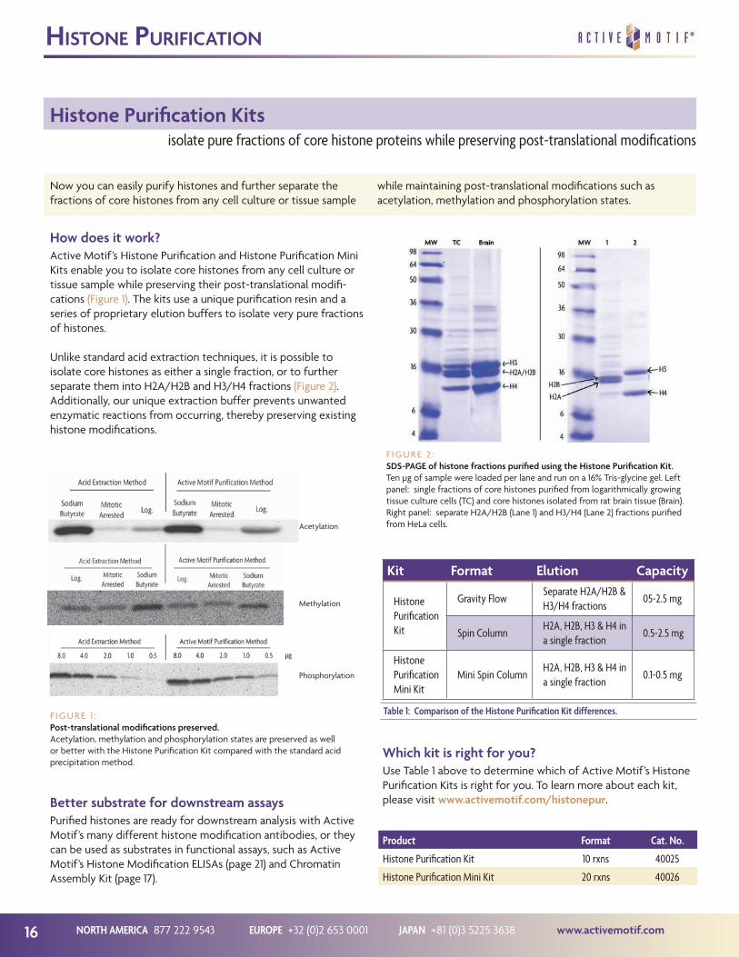
F I G U R E 1 :Post-translational modifications preserved.Acetylation, methylation and phosphorylation states are preserved as well or better with the Histone Purification Kit compared with the standard acid precipitation method.
F I G U R E 2 :SDS-PAGE of histone fractions purified using the Histone Purification Kit.Ten µg of sample were loaded per lane and run on a 16% Tris-glycine gel. Left panel: single fractions of core histones purified from logarithmically growing tissue culture cells (TC) and core histones isolated from rat brain tissue (Brain). Right panel: separate H2A/H2B (Lane 1) and H3/H4 (Lane 2) fractions purified from HeLa cells.
Better substrate for downstream assaysPurified histones are ready for downstream analysis with Active Motif’s many different histone modification antibodies, or they can be used as substrates in functional assays, such as Active Motif’s Histone Modification ELISAs (page 21) and Chromatin Assembly Kit (page 17).
Acetylation
Methylation
Phosphorylation
Product Format Cat. No.
Histone Purification Kit 10 rxns 40025
Histone Purification Mini Kit 20 rxns 40026
Kit Format Elution Capacity
Histone Purification Kit
Gravity FlowSeparate H2A/H2B & H3/H4 fractions
05-2.5 mg
Spin ColumnH2A, H2B, H3 & H4 in a single fraction
0.5-2.5 mg
Histone Purification Mini Kit
Mini Spin ColumnH2A, H2B, H3 & H4 in a single fraction
0.1-0.5 mg
Table 1: Comparison of the Histone Purification Kit differences.
Which kit is right for you?Use Table 1 above to determine which of Active Motif’s Histone Purification Kits is right for you. To learn more about each kit, please visit www.activemotif.com/histonepur.

www.activemotif.com JAPAN +81 (0)3 5225 3638 EUROPE +32 (0)2 653 0001 NORTH AMERICA 877 222 9543 17
ChroMaTin asseMbly
Chromatin Assembly Kitgenerate high-quality chromatin for downstream success
The assembly of genomic DNA and histones into chromatin is a fundamental process that affects a broad range of genome-dependent processes including DNA replication, DNA repair and gene expression. In general, there are ATP-dependent and ATP-independent methods for reconstituting or assembling chromatin in vitro. The ATP-independent method results in a random arrangement of histones on the DNA that does not
accurately reflect the native core nucleosome. To generate an extended array of ordered nucleosomes on a length of DNA greater than 250 bp, the chromatin must be assembled through the ATP-dependent process. Active Motif’s Chromatin Assembly Kit provides an easy and complete solution for ATP-dependent chromatin reconstitution and produces an excellent substrate for downstream assays.
The Chromatin Assembly Kit advantageWith Active Motif’s Chromatin Assembly Kit, you can design your own chromatin. Using an ATP-dependent method, the included purified HeLa core histones are combined with the histone chaperone NAP-1 (h-NAP-1) in a high-salt buffer, which is ideal for proper histone configuration. Purified recombinant human chromatin assembly complex ACF and sample DNA are then added with the complete ATP regeneration system for in vitro assembly of extended, regularly ordered, periodic arrays of nucleosomes.
The resulting chromatin closely resembles natural in vivo chromatin, enabling studies of histone modifications and associated proteins that are crucial to regulation of the target DNA sequence. The assembled chromatin is ready to use in downstream assays such as in vitro transcription assays, chro-matin immunoprecipitation and histone acetyltransferase (HAT) assays. Chromatin assembly is verified by partial digestion with the provided Enzymatic Shearing Cocktail and analyzed by gel electrophoresis. High-quality chromatin should yield six or more distinct bands (Figure 1). For your convenience, supercoiled DNA is provided as a positive control. For details on the Chromatin Assembly Kit, go to www.activemotif.com/chromassembly.
Nucleosome Assembly Control DNAThe Nucleosome Assembly Control DNA will prove to be a key component of your in vitro chromatin assembly reactions. The 187 bp double-stranded DNA was isolated from a ran-domly generated synthetic DNA library based on its ability to bind to histone octamers with high affinity. It can be directly added to your purified histones or histone extracts resulting in mononucleosome formation that can be easily visualized on a gel by mobility shift. The Nucleosome Assembly Control DNA can be used as a control to validate the proper assembly of the components of your in vitro chromatin assembly reactions. Alternatively, the Nucleosome Assembly Control DNA can be used as a template to monitor assembly kinetics in the presence or absence of chromatin-interacting proteins or compounds. For details on the Nucleosome Assembly Control DNA, please visit us at www.activemotif.com/nucleosomeDNA.
F I G U R E 1 :Enzymatic digestion of assembled chromatin.Chromatin assembled from 1 µg samples of circular DNA (Lanes 1 & 2)and linear DNA (Lanes 3 & 4) were digested for 2 and 4 minutes, respec-tively, deproteinated, phenol/chloro-form extracted and run on an agarose gel. Each sample type processed with the Chromatin Assembly Kit resulted in regularly spaced nucleosomes.
Product Format Cat. No.
Chromatin Assembly Kit 10 rxns 53500
HeLa Core Histones 36 µg 53501
Nucleosome Assembly Control DNA 50 µg 53502
why use The ChroMaTin asseMbly KiT?• Generate chromatin from linear or supercoiled DNA
• ATP-dependent method results in an extended array of regularly spaced nucleosomes
• Excellent substrate for gene regulation experiments
• Easy protocol – simply incubate the supplied components with your DNA

NORTH AMERICA 877 222 9543 EUROPE +32 (0)2 653 0001 JAPAN +81 (0)3 5225 3638 www.activemotif.com18
Histone Modifying Enzymes
Histone modifying enzymes are important regulators of genome function; studying their activity offers insight into the mecha-nisms that regulate processes dependent upon the chromatin. To better investigate some of the complex functional questions
about chromatin-associated proteins, nucleosome remodeling, transcriptional regulation, replication and DNA repair, Active Motif offers a variety of recombinant histone modifying enzymes for use in your exploration of chromatin biology.
The importance of histone modificationsThe eukaryotic genome is packaged into the nucleus through the compaction afforded by the incorporation of DNA into chromatin. The primary structural components of chromatin are the highly conserved histone proteins, around which DNA is wrapped and organized. Histones are subject to a variety of reversible post-translational modifications that are tightly regulated such as phosphorylation, acetylation, methylation and ubiquitylation. These modifications are important regulatory events that govern the accessibility and function of regions of the genome. Histone modifications are dynamically regulated and are deposited and removed by enzymes that are generally part of large multi-subunit protein complexes recruited to chromatin by sequence-specific DNA binding proteins.
modifying enzymes enable better understanding of histone modifications
hisTone Modifying enzyMes
F I G U R E 1 :MODified Histone Peptide Arrays treated with G9a methyltransferase.MODified Histone Peptide Arrays (Catalog No. 13001) were treated with A) 25 µM G9a methyltransferase (Catalog No. 31327), B) 25 µM G9a mutant H904K (Catalog No. 31328), or C) no enzyme control, overnight in the presence of 1 mM AdoMet. The arrays were detected using a Histone H3 dimethyl Lys9 antibody. Novel methylation sites were observed on the array treated with wild-type G9a histone methyltransferase, showing the activity of this histone modifying enzyme on the peptide substrates. See page 19 for information on the MODified Histone Peptide Array.
A
B
C
hisTone Modifying enzyMe Classes available
• Acetyltransferases – add acetyl groups to histones or non-histone substrates, which are important for many aspects of genome function, especially transcriptional activation
• Deacetylases – remove acetyl groups from histones and non-histone chromatin proteins, and are often important drug targets, especially in regards to cancer therapeutics
• Methyltransferases – are responsible for adding methyl groups to histones on either lysine or arginine residues
• Demethylases – are organized into two classes: amine oxidase-domain-containing or Jumonji (JmjC)-domain- containing enzymes, both of which are involved in removing methyl groups from histones
• Kinases – regulate cell cycle by phosphorylating specific histone residues
• Bromodomain Proteins – regulate transcription and chromatin remodeling by acting as “Readers” of acetylated lysine residues on histone tails
These modifying enzymes can be used in conjunction with Active Motif’s antibodies, recombinant histones, ELISAs, activity assays (Figure 2) and MODified™ Histone Peptide Array (Figure 1).
To view a full list of our histone modifying enzymes, please visit www.activemotif.com/hismodenz.
F I G U R E 2 :Activity of Recombinant HDAC1.Recombinant HDAC1 protein, active (Catalog No. 31342) was measured for activity using Active Motif’s HDAC Assay (Fluorescent) Kit (Catalog No. 56200). Increasing amounts of HDAC1 protein were incubated with the substrate, then the reaction was developed. HDAC activity is measured as relative fluorescence intensity.

www.activemotif.com JAPAN +81 (0)3 5225 3638 EUROPE +32 (0)2 653 0001 NORTH AMERICA 877 222 9543 19
hisTone pepTide array
MODified™ Histone Peptide Array
Understanding the effects of histone modifications on chroma-tin remodeling and transcriptional regulation requires accurate research tools. With Active Motif’s MODified™ Histone Peptide Array, you can screen histone modification antibodies for cross-
reactivity, ensuring that the antibodies used in your studies are specific. It can also be used to determine if a protein or enzyme binds to specific modifications, and study if this binding is al-tered by the presence of other nearby modifications.
novel peptide array simplifies screening of histone modifications
F I G U R E 1 :Peptide array analysis confirms the specificity of H3K9me3 pAb.The MODified Histone Peptide Array was used with the MODified Array Labeling Kit to screen Active Motif’s Histone H3K9me3 pAb (Catalog No. 39161) for cross-reactivity. Following detection, the image was captured with a CCD camera and analyzed with Active Motif’s Array Analyse Software. The results plotted above show the Specificity Factor for the ten most reactive peptides on the array. The Specificity Factor is the ratio of the average intensity of all spots containing H3K9me3 divided by the average intensity of all spots not containing H3K9me3. The results show this antibody is very specific for Histone H3 trimethyl Lys9, with very little binding to other modifications.
MODified Histone Peptide ArrayThe MODified Histone Peptide Array* is a valuable research tool that can be used to screen antibodies, proteins and enzymes for interactions with histones and their post-translational modifica-tions. Each array contains 384 different histone modification combinations in duplicate. Modifications include acetylation, methylation, phosphorylation and citrullination on the N-termi-nal tails of histones H2A, H2B, H3 and H4.
This unique histone array contains up to four modifications per 19mer peptide to study not only individual modifications, but also to determine if neighboring modifications alter site recogni-tion and binding. The MODified Histone Peptide Array can be used to screen antibodies for cross-reactivity, as in Figure 1, or to study protein and enzyme interactions, as shown on page 18. The simple array protocol works like a Western blot. Either ECL-based or colorimetric detection systems can be used. The image is then captured using film or a CCD camera.
Free software for analysisActive Motif’s Array Analyse Software is a free program designed for use with the MODified Histone Peptide Arrays. This PC compatible software will analyze the spot intensities from the MODified array and generate a graphical analysis of the histone modification interactions (Figure 1). Information about spot intensity, averages and errors can be saved in Excel-compatible files. For added convenience, up to three individual modifica-tions can be displayed in superposition to the experimental data, enabling better visualization of neighboring effects.
Product Format Cat. No.
MODified™ Histone Peptide Array 1 array 5 arrays
13001 13005
MODified™ Array Labeling Kit 5 rxns 13006
Modified array advanTages
• Histone specific – unique array panel tests for specific histone modifications
• Study neighboring effects – each peptide contains up to four modification combinations, enabling analysis of the effects of neighboring modifications
• Detects like a Western blot – fast and easy to use; works with either ECL-based or colorimetric detection
MODified Array Labeling KitFor a complete solution, the MODified™ Array Labeling Kit contains the buffers and reagents needed for easy labeling and chemiluminescent detection of the MODified Histone Peptide Array. The MODified Array Labeling Kit contains blocking buf-fer, wash buffer, rabbit and mouse HRP-conjugated antibodies and ECL reagents for chemiluminescent detection. For added convenience, a positive control c-Myc antibody is included to recognize the array’s control c-Myc tag. Sufficient reagents are provided for the detection of five MODified Histone Arrays.
To learn more about the MODified Histone Peptide Arrays, MODified Array Labeling Kit, or to download the free Array Analyse Software, please visit www.activemotif.com/modified.
*CelluSpots™ arrays are manufactured under license by INTAVIS Bioanalytical Instruments AG and sold through Active Motif as MODified™ Histone Peptide Array.

NORTH AMERICA 877 222 9543 EUROPE +32 (0)2 653 0001 JAPAN +81 (0)3 5225 3638 www.activemotif.com20
reCoMbinanT hisTones
Recombinant Histone Proteins
Active Motif is the first company to offer recombinant histones containing important site- and degree-specific post-translational modifications. The EPL and MLA technologies* enable us to efficiently methylate, acetylate, phosphorylate and biotinylate human histone H3, and to methylate Xenopus histones H3 and
H4. Our recombinant histones are better suited than histone peptides for use as standards and as substrates in functional assays because they more closely resemble native histone proteins. As they are more “natural” than peptides, recombinant histones can make your results more accurate (Figure 2, page 23).
recombinant histones serve as more “natural” substrates and positive controls than peptides
Histones & chromatin structureThe eukaryotic genome is packaged into chromatin, which con-sists of DNA wrapped around highly conserved histone proteins. These histone proteins are subject to a variety of tightly regu-lated, reversible post-translational modifications that control the organization and transcriptional regulation of genomic DNA. To enable a better understanding of these histone modifications, Active Motif has developed modification-specific recombinant histone proteins for use in functional assays. The recombinant histones can also be used as standards for quantification of a specific histone modification, and are therefore included as controls in our Histone ELISAs (see page 21).
How are the histone modifications generated?The Methylated Lysine Analog (MLA) technology can gener-ate methylated histones via a chemical alkylation reaction to introduce a methyl-lysine analog at the desired lysine location, giving us precise control over the site and degree of methylation. The Expressed Protein Ligation (EPL) technology can be used to generate methylated, acetylated and phosphorylated histones in which the histone globular domain is ligated to a peptide containing the N-terminal histone tail with site-specific modifi-cations. This ligation reaction maintains the native histone bonds.
Many, many modified histones, all verifiedActive Motif offers over 50 different recombinant histones to meet your research needs (see table below). All have been verified by high-resolution ESI-TOF mass spectrometry and confirmed by dot blot or immunoblot (Figure 1). For complete details, please visit www.activemotif.com/recombhis.
F I G U R E 1 :Dot blot confirmation of Recombinant Histone H3K79me2.One µg of unmodified, mono-, di- or trimethylated recombinant proteins for H3K27, H3K36, H3K79 and H4K20 were spotted onto a PVDF membrane and probed with Histone H3K79me2 pAb (Catalog No. 39143) at a 1:1,000 dilution. The dot blot confirms the identity of the recombinant H3K79me2 protein.
Biotinylated histones for capture assaysAs a way to expand the versatility of our recombinant histone proteins, Active Motif has also created histones that are linked to biotin via a carbon linker on the N-terminus. The addition of biotin enables streptavidin capture strategies for functional assays, or homogeneous FRET assays with streptavidin-coated donor beads and antibody-conjugated acceptor beads.
Recombinant Methylated Histones
Histone H3K4me1, me2 & me3 Histone H3K27me1, me2 & me3
Histone H3R8me2a (asymmetric) Histone H3K36me1, me2 & me3
Histone H3K9me1, me2 & me3 Histone H3K79me1, me2 & me3
Histone H3K14me1, me2 & me3 Histone H4K5me1, me2 & me3
Histone H3K18me1, me2 & me3 Histone H4K16me1, me2 & me3
Histone H3K23me1, me2 & me3 Histone H4K20me1, me2 & me3
Recombinant Acetylated Histones
Histone H3K4ac Histone H3K18ac
Histone H3K9ac Histone H3K23ac
Histone H3K14ac
Recombinant Biotinylated Histones
Histone H3 biotinylatedHistone H3K9me1 & me3
biotinylated
Histone H3K4me1, me2 & me3 biotinylated
Recombinant Histones (unmodified)
Histone H2A Histone H3 (C110A)
Histone H2B Histone H4
Recombinant Phosphorylated Histones
Histone H3T3ph Histone H3S10ph
*The MLA technology is covered under U.S. Patent No. 8,278,112. EPL is patent pending.

www.activemotif.com JAPAN +81 (0)3 5225 3638 EUROPE +32 (0)2 653 0001 NORTH AMERICA 877 222 9543 21
hisTone ModifiCaTion elisas
Histone Modification ELISAsachieve sensitive and specific detection of histone modifications
To better understand the effects of histone modifications on chromatin remodeling and transcriptional regulation, Active Motif has applied our histone modification antibody expertise to identify optimal antibody pairs for the detection of specific histone modifications in a sandwich ELISA format.
The Histone Modification ELISA Kits provide a sensitive method for detecting changes in the level of specific histone modifications, such as lysine methylation and acetylation, or phosphorylation of serine. These modification sites serve as key targets of histone modifying enzymes.
How do the Histone ELISAs work?The Histone Modification ELISA Kits provide sensitive and spe-cific detection of histone modifications, enabling researchers to evaluate small changes in the level of specific histone modifi-cations from purified core histones (see page 16), or histones isolated by acid extraction. These easy-to use kits are sandwich ELISAs that utilize a capture antibody against histone H3 and a detecting antibody specific to the modification of interest. An HRP-conjugated secondary antibody and developing solutions provide a sensitive colorimetric readout in less than 3 hours.
hisTone ModifiCaTion elisa advanTages
• Sensitive – works with purified core histones or acid extracted samples (Figure 1)
• Fast – assay can be completed in less than 3 hours
• Flexible – stripwell plates enable screening of 1 to 96 samples in a single experiment
• Modification-specific controls – recombinant methylated and acetylated histone proteins are included to enable quantification of samples
• Colorimetric detection – results are easily quantified by spectrophotometry at 450 nmF I G U R E 1 :
Histone H3 trimethyl Lys27 detection with reference standard curve.The Histone H3 trimethyl Lys27 ELISA was used to assay purified HeLa core histones (125-250 ng) and HeLa acid extract (5-10 µg). The Recombinant Histone H3K27me3 protein provided in the kit as a control was assayed from 3.125-100 ng per well as a reference standard curve, showing the sensitivity of the assay.
Validated controls are includedEach kit includes validated modification-specific controls. The included methylated and acetylated recombinant histone proteins can be used to generate a standard curve to quantify the amount of modified histone in each sample. The Total Histone H3 ELISA can be used to normalize the amount of histone modification in your samples when run in parallel with the methylated or acetylated Histone ELISAs. The untreated and paclitaxel-treated acid extracts provided in the phosphorylated Histone ELISAs serve as qualitative controls.
Also availableTo see a list of the more than 50 unique recombinant histones available, see page 20 or visit www.activemotif.com/recombhis.
To see a complete list of acid extracts offered by Active Motif, please visit www.activemotif.com/acidextract.
To learn about the histone modifying enzymes Active Motif sells, see page 18 or visit www.activemotif.com/hismodenz.
For an up-to-date list of available Histone Modification ELISAs, please visit www.activemotif.com/hiselisa.
Product Format Cat. No.
Histone H3 monomethyl Lys4 ELISA 96 rxns 53101
Histone H3 dimethyl Lys4 ELISA 96 rxns 53112
Histone H3 trimethyl Lys4 ELISA 96 rxns 53113
Histone H3 acetyl Lys9 ELISA 96 rxns 53114
Histone H3 dimethyl Lys9 ELISA 96 rxns 53108
Histone H3 trimethyl Lys9 ELISA 96 rxns 53109
Histone H3 phospho Ser10 ELISA 96 rxns 53111
Histone H3 acetyl Lys14 ELISA 96 rxns 53115
Histone H3 monomethyl Lys27 ELISA 96 rxns 53104
Histone H3 trimethyl Lys27 ELISA 96 rxns 53106
Histone H3 phospho Ser28 ELISA 96 rxns 53100
Total Histone H3 ELISA 96 rxns 53110

NORTH AMERICA 877 222 9543 EUROPE +32 (0)2 653 0001 JAPAN +81 (0)3 5225 3638 www.activemotif.com22
hisTone aCeTylaTion & deaCeTylaTion
HAT & HDAC Assay Kits
Active Motif’s HAT & HDAC Assay Kits are easy-to-use, sensi-tive assays that can be used to determine the activity of histone acetyltransferases and histone deacetylases in your cell and nuclear extracts, immunoprecipitates and purified enzymes, as
well as to screen the effects of potential inhibitor compounds. The HAT Assay Kit uses a fluorescent readout, while HDAC Assay Kits are available in both fluorescent and colorimetric formats.
rapid, sensitive assays for HAT & HDAC activity and inhibitor compounds
F I G U R E 1 :HAT inhibitor effects on p300 activity.50 ng p300 were assayed per well with 50 µM acetyl-CoA and 50 µM histone H3 or H4 peptide substrates in the absence or presence of 15 µM anacardic acid, a known HAT inhibitor. The background signal indicates the level of autoacetylation present from the p300 acetyltransferase.
F I G U R E 2 :HDAC activity in HeLa cells.HeLa nuclear extracts were assayed for HDAC activity in duplicate from 0 to 10 µg per well using the fluorescent version of the HDAC Assay Kit. Untreated extract results are shown with a purple line, while extracts inhibited with 1 µM Trichostatin A are shown with a copper line.
The HAT FamilyHistone acetyltransferases (HAT) are enzymes that acetylate conserved lysine amino acids on histones. Generally, histone acetylation is associated with the activation of gene expression, as hyperacetylated chromatin is transcriptionally active. Histone deacetylases (HDAC) remove these acetyl groups from histones. Their action is opposite to that of histone acetyltransferases, as hypoacetylated chromatin is silent. Because HATs and HDACs are involved with other proteins in the regulation of gene expres-sion, their activity is much studied, as are compounds that inhibit HAT and HDAC activity.
How do the HDAC Assay Kits work?The HDAC Assay Kits utilize a peptide substrate that contains an acetylated lysine residue that can be deacetylated by Class I, IIB and IV HDAC enzymes. (Class III HDAC enzymes, or the Sirtuins, require the addition of the NAD+ cofactor in the assay.) Once the substrate is deacetylated, the lysine reacts with the Developing Solution and releases either the chromophore or the fluorophore from the substrate, which produces either a colori-metric or fluorescent product. The colorimetric product absorbs maximally at 405 nm; the fluorescent product can be read with an excitation wavelength of 360 nm and emission wavelength of 460 nm (Figure 2). A deacetylated assay standard is provided in each kit to enable calculation of HDAC activity in pmol/min/mg.
Product Format Cat. No.
HAT Assay Kit (Fluorescent) 1 x 96 rxns 56100
Recombinant p300 protein, catalytic domain 5 µg 31205
Recombinant GCN5 protein, active 5 µg 31204
HDAC Assay Kit (Fluorescent) 1 x 96 rxns 56200
HDAC Assay Kit (Colorimetric) 1 x 96 rxns 56210
How does the HAT Assay Kit work?The HAT Assay Kit is a quick and sensitive method to de-termine the activity of your own source of purified histone acetyltransferases, or to screen for potential inhibitors of HAT activity. HATs will catalyze the transfer of acetyl groups from the provided acetyl-CoA to generate an acetylated peptide and CoA-SH. After stopping the reaction with stop solution, a developer is added that reacts with the free sulfhydryl groups on CoA-SH to give a fluorescent signal (Figure 1).
This fluorescent 96-well plate assay includes N-terminal histone H3 and H4 substrate peptides for screening HAT enzymes and a positive control p300 catalytic domain protein to screen for inhibitors. Anacardic acid is also provided for use as a control, as it is a potent HAT inhibitor. A standard curve can be generated with either b-mercaptoethanol or Coenzyme A in order to relate the fluorescence of your HAT to pmol/min/µg specific activity.

www.activemotif.com JAPAN +81 (0)3 5225 3638 EUROPE +32 (0)2 653 0001 NORTH AMERICA 877 222 9543 23
hisTone deMeThylaTion
Histone Demethylase Assay
Histone methylation is central to many aspects of the biology of the nucleus, including transcriptional regulation and the control of higher order chromatin structure. Histone methyltransferases (HMTs) and histone demethylases (HDMs) work in opposition to
each other to regulate histone methylation. Studying the activity of these enzymes gives insight into the mechanisms by which transcription and the organization of chromatin are regulated.
screen LSD1 for histone demethylase activity
How does it work?The fluorescent Histone Demethylase Assay is a simple way to analyze the efficiency of lysine specific demethylase enzyme (LSD1, also known as KDM1) samples, or to screen inhibitor com-pounds that change histone demethylation activity. The Histone Demethylase Assay is designed to detect the formaldehyde released from the reaction of LSD1 with a methylated substrate. As the LSD1 enzyme demethylates the recombinant histone sub-strate, formaldehyde is released as a by-product, which then re-acts with the Detection Reagent to generate a fluorescent signal equivalent to the overall production of formaldehyde (Figure 1).
Better substrate for more accurate resultsThe provided Recombinant Histone H3K4me2 substrate mimics a native histone substrate, generating results that more closely re-semble in vivo conditions. As shown in Figure 2, the LSD1 enzyme is able to more efficiently demethylate the included recombi-nant histone H3K4me2 protein than a histone H3K4me2 peptide substrate. Because the recombinant histone more closely resem-bles a native histone, the Histone Demethylase Assay enables more accurate analysis of histone demethylation activity.
What’s included?The Histone Demethylase Assay contains a Recombinant Histone H3K4me2 substrate, optimized buffers to enhance enzymatic activity and black 96-well half area microplates to perform the assay. For added convenience, a Demethylation Standard, which can be used to quantify the amount of formaldehyde released, and an aliquot of LSD1 enzyme are included as controls. Please visit www.activemotif.com/lsd1 for more details.
Product Format Cat. No.
Histone Demethylase Assay (Fluorescent) 48 rxns 53200
Recombinant LSD1 protein, active 50 µg 31334
F I G U R E 1 :Fluorescence detection of LSD1 demethylase activity.LSD1 (1 µg) was tested in the absence or presence of the provided recombinant histone H3K4me2 substrate (3.3 µM). The enzymatic reaction was incubated at 37°C for one hour, followed by a one hour incubation with the Detection Reagent before fluorescence intensity was measured.
F I G U R E 2 :Comparison of LSD1 demethylase efficiency using different histone substrates.The positive control LSD1 enzyme from the Histone Demethylase Assay was used to evaluate demethylase activity using either a histone peptide or the kit’s recom-binant protein. One µg of LSD1 was tested with either 70 µM H3K4me2 peptide or with 13 µM recombinant H3K4me2 protein. LSD1 was able to convert 73% of the recombinant histone substrate into a formaldehyde by-product, yet it was only able to convert 14% of the peptide substrate into a formaldehyde by-product.
hisTone deMeThylase assay advanTages
• The recombinant histone H3K4me2 substrate used in the assay mimics a native histone substrate, providing you with results that more closely resemble in vivo conditions
• Complete assay includes a Demethylation Standard for formaldehyde quantification and LSD1 enzyme as a positive control protein
• Simple fluorescent assay detects the formaldehyde by-product using an excitation wavelength of 410 nm and an emission wavelength of 480 nm

NORTH AMERICA 877 222 9543 EUROPE +32 (0)2 653 0001 JAPAN +81 (0)3 5225 3638 www.activemotif.com24
DNA Methylation / Demethylation Overview
The identification of 5-hydroxymethylcytosine (5-hmC), a derivative of the well-characterized 5-methylcytosine (5-mC) epigenetic mark, established a precedent for the role of Tet enzymes in catalyzing DNA demethylation as a mechanism for epigenetic reprogramming. Recent studies reveal that the path-way of demethylation may involve further oxidation of 5-hmC to 5-formylcytosine (5-fC) and 5-carboxylcytosine (5-caC) during
cell differentiation and development. To aid in the study of how DNA methylation and demethylation work in concert to regu-late gene expression, development, and stem cell identity, Active Motif has developed an expansive line of products and technol-ogies to address the most current and cutting edge areas in DNA methylation research. Let our antibodies, assays, enzymes and contract research services assist in your research efforts.
tools to analyze all aspects of DNA methylation
dna MeThylaTion produCTs
• CGBP Antibody (p. 2)• Methylated DNA Standard Kit (p. 33)
• Recombinant Tet1 Protein (p. 34)
• 5-fC Antibody (p. 35)
• Recombinant Tet1 Protein (p. 34)
Tet
DNMT
Dec
arbo
xyla
se
Tet
Tet
• Recombinant Tet1 Protein (p. 34)
• 5-caC Antibody (p. 35)• 5-Carboxylcytosine DNA Standard (p. 35)
• DNMT Activity/Inhibition Assay (p. 30)• DNMT Antibodies (p. 2)• Recombinant DNMT1 Protein (p. 30)• HP1 Antibodies (p. 3)• MBD Antibodies (p. 2)• MeCP2 Antibodies (p. 2)• Uhrf1 Antibody (p. 2)
• MethylCollector Ultra™ (p. 28)• HypoMethylCollector™ (p. 32)• MeDIP Assay (p. 29)• MethylDetector™ (p. 31)• 5-mC Antibodies (p. 29)• Methylated DNA
Standard Kit (p. 33)
• Hydroxymethyl Collector™ (p. 26)• hMeDIP Assay (p. 27)• PvuRts1I enzyme (p. 34)• b-Glucosyltransferase (p. 34)• 5-hmC Antibodies (p. 27)• Methylated DNA
Standard Kit (p. 33)
F I G U R E 1 :Schematic representation of the oxidation of 5-methylcytosine by Tet enzymes and related products.Tet enzymes catalyze the conversion of 5-methylcytosine (5-mC) to 5-hydroxymethylcytosine (5-hmC), 5-formylcytosine (5-fC), and 5-carboxylcytosine (5-caC) via oxidation of the methyl group at the 5-position on the cytosine ring. The schematic depicts the demethylation pathway and the Active Motif products associated with the individual steps of the reaction.

www.activemotif.com JAPAN +81 (0)3 5225 3638 EUROPE +32 (0)2 653 0001 NORTH AMERICA 877 222 9543 25
DNA Methylation Research Services
Determining the impact of differential DNA methylation across multiple samples is important to understanding the underlying mechanisms of development and disease. Our DNA methylation
services give customers access to the state-of-the-art technolo-gies that make a real impact on this ever expanding field. For details, please visit us at www.activemotif.com/services.
utilize our experienced research team to study DNA methylation patterns
dna MeThylaTion researCh serviCes
F I G U R E 3 :Targeted Next-Gen Bisulfite Sequencing.Bisulfite PCR was performed using 9 separate samples and 10 different primer pairs. All 90 amplicons were sequenced in a single Next-Gen sequencing run. This heat map shows methylation data from one of the ten primer pairs across the population of 9 samples.
F I G U R E 4 :Bisulfite sequencing reveals differen-tial methylation in two cell lines.Sanger bisulfite sequencing is performed on customer-selected genomic locations. This data compares the methylation state of a region with 24 CpGs in two different cell lines. Seven clones from each of the cell lines were sequenced.
F I G U R E 1 :Next-Gen sequencing data detects methylation at CpG shores.Next-Gen sequencing was performed on DNA enriched from 1 µg of human PBMC DNA using MethylCollector™ Ultra, and tags were mapped to generate a whole-genome DNA methylation profile. The image shows an example of DNA methylation detected at CpG shores rather than in the CpG island itself. This data agrees with recent findings showing that methylation frequently occurs in the regions adjacent to CpG islands.
F I G U R E 2 :Next-Gen sequencing data correlates well with CpG density.DNA was enriched by MeDIP from 1 µg of adaptor-ligated, human PBMC DNA. Next-Gen sequencing was performed and tags were mapped to generate a whole-genome DNA methylation profile. The image above shows that the enriched regions correlate well with CpG density.
CpG Island
MethylCollector™ Ultra-SeqCpG Density
MeDIP CpG Density
Genome-wide DNA Methylation Services
• MeDIP-Seq – enrichment of methylated DNA with a highly specific 5-methylcytosine antibody
• MethylCollector™ Ultra-Seq – based on the patented MIRA (Methylated CpG Island Recovery Assay) technology
• hMeDIP-Seq – enrichment using the most specific, highly cited 5-hydroxymethylcytosine antibody
The Genome-wide DNA methylation Services include:
– DNA isolation from cells or tissues, followed by enrichment of methylated DNA
– qPCR analysis of positive and negative control sites
– Next-Gen Library generation
– Sequencing of ≥ 30 million tags using the Illumina HiSeq
– Analysis: mapping, peak calling, visualization files and Excel output
Bisulfite Sequencing ServicesBisulfite Sequencing is the only method that enables detec-tion of the methylation status of individual cytosines at base pair resolution. Active Motif Epigenetic Services offers two Bisulfite Sequencing options:
Targeted Next-Gen Bisulfite Sequencing
• Multiplex many samples and multiple amplicons into a single Next-Gen sequencing reaction
• 100X to 10,000X coverage of each amplicon
• No cloning bias
• Multiplexing makes large experiments cost-effective
Sanger Bisulfite SequencingThis traditional bisulfite approach requires cloning of bisulfite converted amplicons and sequencing of 8 to 16 clones.
• Service includes all steps from primer design to analysis
• Recommended for small-scale experiments of < 5 samples

NORTH AMERICA 877 222 9543 EUROPE +32 (0)2 653 0001 JAPAN +81 (0)3 5225 3638 www.activemotif.com26
Hydroxymethyl Collector™
The Hydroxymethyl Collector™ Kit was designed for the highly specific capture of DNA fragments that contain 5-hydroxymeth-ylcytosine (5-hmC) residues. The method takes advantage of an efficient chemical labeling procedure that enables the enriched
samples to be collected as double-stranded DNA fragments. This makes it easy to prepare libraries for various downstream applications, including Next-Generation sequencing (Figure 1). Please visit www.activemotif.com/hydroxy-mc for more details.
biotin-based magnetic bead enrichment of 5-hydroxymethylcytosine
5-hydroxyMeThylCyTosine dna enriChMenT
How does Hydroxymethyl Collector work?The Hydroxymethyl Collector Kit is a fast, simple procedure that can be completed in less than 4 hours. The method utilizes a b-glucosyltransferase enzyme to transfer a modified glucose moiety to 5-hydroxymethylcytosine residues in double-stranded DNA. This modified glucose is then used to chemically attach a biotin conjugate for capture and enrichment using streptavidin-coated magnetic beads and a magnet (Diagram 1).
Why use Hydroxymethyl Collector?Hydroxymethyl Collector is extremely specific in its capture of hydroxymethylated DNA fragments. Utilizing chemical label-ing of 5-hmC residues ensures no cross-reactivity with 5-mC, and the method is not limited by the specific properties or consensus sequences that constrain traditional methods, such as glucosyl-sensitive restriction enzyme digestion. Furthermore, 5-hmC modification occurs independently of sequence context, enabling the detection of both CpG and non-CpG methylated DNA. The use of streptavidin magnetic beads allows for quick and efficient recovery of samples, and the high affinity of the biotin-streptavidin capture enables more stringent binding and wash conditions. The result is reduced background and increased sensitivity, enabling enrichment of DNA fragments containing as few as two 5-hmC residues. Enriched DNA can be used in the analysis of individual genes by PCR and qPCR, or with microarrays and sequencing for genome-wide analysis of 5-hmC.
N
N
NH2O
O
HO
OH
HOHO
N3
Biotin
N
N
NH2O
O
HO
OH
HOHO
N3
N
N
NH2OH
HO
β-Glucosyltransferase + UDP-Azide-Glucose
(1 hr at 37°C)
Biotin Conjugate Solution
(1 hr at 37°C)
Magnetic Streptavidin
Beads (30 min at RT)
Elute the DNA from the Biotin(30 min at RT)
1. Fragmented DNA containing 5-hmC
Enriched DNA containing 5-hmC
is ready for analysis.5. Eluted DNA is purified using the included reagents.
2. Modified Glucosyl-5-hmC
3. Biotinylated Glucosyl-5-hmC
4. DNA is captured for washing and elution.
N
N
NH2O
O
HO
OH
HOHO
N3
N
N
NH2O
O
HO
OH
HOHO
N3
BiotinSA
D I AG R A M 1 :The Hydroxymethyl Collector method.Fragmented dsDNA (100-500 bp) is combined with b-Glucosyltransferase enzyme in the presence of a UDP-Azide-Glucose donor. The enzyme adds this modified glucose onto the 5-hmC residues. A biotin conjugate is then attached and the complex is captured using streptavidin-coated magnetic beads and a magnet. Elution Buffer is added, which releases the 5-hmC enriched DNA fragments from the biotin linker. Finally, the kit’s purification reagents are used to clean up the DNA prior to its use in downstream applications.F I G U R E 1 :
Human tiling array using DNA enriched with Hydroxymethyl Collector.Human brain DNA was enriched using the Hydroxymethyl Collector Kit, then amplified by whole-genome amplification and hybridized to an Affymetrix Human Tiling 2.0R Array A containing chromosomes 1 and 6. This image shows a 1.2 million base pair view of chromosome 6 where there is a clear enrichment of 5-hmC across the entire length of the JARID2 gene.
Product Format Cat. No.
Hydroxymethyl Collector™ 25 rxns 55013
hydroxyMeThyl ColleCTor advanTages
• Chemical labeling ensures specific modification of 5-hmC DNA without cross-reactivity of 5-mC sites
• Biotin/streptavidin binding enables more stringent binding and wash conditions, which reduces the non-specific background without compromising assay sensitivity
• Use of dsDNA throughout the process makes it easier to prepare libraries for Next-Gen sequencing

www.activemotif.com JAPAN +81 (0)3 5225 3638 EUROPE +32 (0)2 653 0001 NORTH AMERICA 877 222 9543 27
hMeDIP
In mammals and other vertebrates, DNA methylation occurs at the C5 position of cytosine (5-mC), mostly within CpG dinucleotides. An alternative form of DNA methylation, 5-hy-droxymethylcytosine (5-hmC), results from the enzymatic con-version of 5-methylcytosine into 5-hydroxymethylcytosine by the TET family of cytosine oxygenases. While its precise function has yet to be determined, it has been postulated that 5-hmC, with the additional DNA methylation variants 5-fC and 5-caC (see page 35), could represent a pathway to demethylate DNA.
Active Motif’s hMeDIP Assay is specific for the isolation and enrichment of DNA containing the 5-hydroxymethylcytosine residue. This enables researchers to separate 5-hmC DNA from DNA containing the traditional 5-methylcytosine methylation to study the specific effects of 5-hmC on gene regulation. The hMeDIP Assay’s ability to distinguish between the two types of DNA methylation is important, as most common approaches to analyze DNA methylation, such as bisulfite conversion, are unable to distinguish between 5-mC and 5-hmC residues.
easily obtain 5-hydroxymethylcytosine-specific DNA fragments
5-hMC dna iMMunopreCipiTaTion
F I G U R E 1 :Real-time PCR results of hMeDIP assay with the included spike controls.Mse I digested human genomic DNA (500 ng) was “spiked” with 25 pg of either an unmethylated, methylated or hydroxymethylated DNA standard. These samples were then processed using the hMeDIP Assay with the 5-hydroxym-ethylcytosine pAb. Eluted DNA was purified and tested using real-time PCR with the included APC PCR primer mix. The fold enrichment represents the amount of IP DNA recovered from each spike normalized against the negative control rabbit IgG reaction. The results show the assay is specific for detecting 5-hydroxymethylcytosine DNA. The APC locus analyzed in this experiment is not methylated in human genomic DNA and therefore should not amplify.
How does the hMeDIP Assay work?The hMeDIP Assay is designed to immunoprecipitate and en-rich for DNA fragments containing 5-hydroxymethylcytosine. The assay contains a highly specific purified 5-hydroxymeth-ylcytosine antibody and the necessary buffers to perform hydroxymethylated DNA immunoprecipitation (hMeDIP). The hMeDIP Assay starts with either double-stranded or single-stranded genomic DNA fragments. Following an overnight incu-bation with the 5-hydroxymethylcytosine antibody, and a 2 hour incubation with protein G magnetic beads, the included magnet is used to capture, wash and elute the immunoprecipitated 5-hmC DNA. The fast, magnetic protocol streamlines the num-ber of wash and incubation steps needed, saving you valuable time. For added convenience, the kit also includes a negative control rabbit IgG antibody and a set of three DNA standards (unmethylated, methylated and hydroxymethylated) that can be used as spike controls to confirm the efficiency of the hMeDIP enrichment with the provided PCR primer mix in real-time PCR (Figure 1).
Antibodies for 5-hydroxymethylcytosine detectionIn addition to the complete hMeDIP Assay, Active Motif also of-fers both monoclonal and polyclonal antibodies for the study of 5-hydroxymethylcytosine. Each antibody has been validated for use in multiple applications, including dot blot and methyl-DNA immunoprecipitation (Figure 2). The polyclonal antibody is avail-able in two formats depending on your preference: whole rabbit serum (Catalog No. 39769) and purified IgG (Catalog No. 39791). For more information about 5-hydroxymethylcytosine anti- bodies and assays, please visit www.activemotif.com/hmc.
F I G U R E 2 :Examination of 5-methylcytosine and 5-hydroxymethylcytosine methylation by dot blot.DNA samples were spotted onto positively charged nylon membrane and blotted with antibodies recognizing 5-Hydroxymethylcytosine (Panel A, Catalog No. 39769, 1:10,000 dilution) or 5-Methylcytosine (Panel B, Clone 33D3, Catalog No. 39649, 1:1,000 dilution). Lane 1: DNA derived from mouse embryonic stem cells (150 ng). Lane 2: DNA derived from mouse spleen (600 ng). Lane 3: 27 base oligonucleotide containing 5-hydroxymethylcytosine (1.2 ng). Lane 4: 33 base oligonucleotide containing 5-methylcytosine (2000 ng).
Product Format Cat. No.
hMeDIP Assay 10 rxns 55010
5-Hydroxymethylcytosine (5-hmC) antibody mAb 100 µg 39999
5-Hydroxymethylcytosine (5-hmC) antibody pAb 100 µl 39769
5-Hydroxymethylcytosine (5-hmC) antibody pAb (IgG) 100 µg 39791
hMeDIP-Seq servicesActive Motif also offers hMeDIP-Seq as a custom service. See page 25 or visit us at www.activemotif.com/services for details.

NORTH AMERICA 877 222 9543 EUROPE +32 (0)2 653 0001 JAPAN +81 (0)3 5225 3638 www.activemotif.com28
5-MeThylCyTosine dna enriChMenT
MethylCollector™ Ultra
Active Motif’s MethylCollector™ Ultra Kit* provides improved enrichment of CpG-methylated DNA compared to alter-native methyl-binding domain protein (MBD) or antibody
immunoprecipitation methods. Enriched DNA is suitable for use in various downstream applications, such as PCR, bisulfite conver-sion, or amplification and labeling for microarray analysis.
fast magnetic assay for specific isolation of CpG-methylated DNA
Why use MethylCollector Ultra?Active Motif’s MethylCollector Ultra Kit improves the enrichment of CpG-methylated DNA by incorporating the Methylated CpG Island Recovery Assay (MIRA), which uses a combination of methyl-binding proteins (MBD2b and MBD3L1) to increase the affinity for methylated DNA frag-ments.1 This unique protein complex provides greater speci-ficity for methylated CpG dinucleotides than alternative MBD or antibody immunoprecipitation (MeDIP) methods, in less than half the time. The kit also includes positive con-trol human, male genomic DNA and methylation-specific PCR primers to ensure your success.
MethylCollector Ultra-Seq (MIRA-Seq) servicesMethylCollector Ultra-Seq is also offered as a custom service by Active Motif’s Epigenetic Services. Customers can submit their cells or DNA and receive fully analyzed genome-wide methylation data in just weeks. See page 25 or visit us at www.activemotif.com/services for details.
Product Format Cat. No.
MethylCollector™ Ultra 30 rxns 55005 *Technology covered under U.S. Patent No. 7,425,415.
F I G U R E 2 :Real-time PCR analysis validates MethylCollector Ultra specificity.100 ng of human, male genomic DNA was digested with Mse I and tested in MethylCollector Ultra. Eluted DNA was analyzed using PCR primers for both methylated, SNRPN (red), and unmethylated, FOXD2 (blue), promoters. The visible separation between the amplification cycles reveals that the enriched sample is specific for methylated DNA.
Break open cells and shearDNA by Sonication orEnzymatic Shearing.
methylatedDNA
Incubate sheared DNA withHis-tagged MBD2b/MBD3L1
protein complexes.
The purified, methylatedDNA is ready
for PCR analysis.
Elute the bound, methylatedDNA, while simultaneously
degrading the protein complexes.
Capture the methylated protein-DNA complexes with nickel-coated magnetic beads.
Wash to remove the unmethylated
DNA fragments.
F I G U R E 1 :Flow chart of the MethylCollector Ultra process.In MethylCollector Ultra, genomic DNA of interest is sheared by either sonication or enzymatic digestion, then incubated with recombinant His-MBD2b/MBD3L1 protein complex. Nickel-coated magnetic beads capture the protein-DNA complexes from the rest of the genomic DNA. Optimized buffers ensure that fragments with little or no methylation are removed. Methylated DNA is then eluted from the beads. Following clean up, the eluted DNA is ready for use in PCR analysis or other applications.
To learn more about Active Motif’s DNA methylation antibodies and assays, please visit www.activemotif.com/dnamt.
REFERENCES1. Rauch, T. and Pfeifer, G. (2005) Lab. Investigation 85: 1172-1180.
MeThylColleCTor ulTra advanTages
• Improved efficiency – provides greater enrichment of CpG-methylated DNA than other MBD capture or antibody immunoprecipitation (MeDIP) methods
• Faster procedure – magnetic protocol can be completed in less than 3 hours
• Uses minimal sample material – requires as little as 1 ng of DNA fragmented by sonication or enzymatic digestion
• Controls ensure success – includes positive control DNA and methylation-specific PCR primers

www.activemotif.com JAPAN +81 (0)3 5225 3638 EUROPE +32 (0)2 653 0001 NORTH AMERICA 877 222 9543 29
why use The Medip assay?• Works efficiently with 100 ng – 1 µg of fragmented,
single-stranded DNA
• Includes both a 5-Methylcytosine antibody and Bridging Antibody for methylated DNA enrichment, as well as a negative control mouse IgG antibody
• Positive control human genomic DNA and PCR primers are provided to verify the efficiency of the enrichment
• Fast magnetic protocol minimizes the number of wash and incubation steps, saving you valuable time
MeDIP
Methylated DNA Immunoprecipitation (MeDIP) is an immuno-capture technique in which an antibody specific for 5-methylcy-tosine is used to immunoprecipitate methylated DNA fragments from the rest of the sample population. One advantage of using the MeDIP antibody capture approach over methyl-binding
protein capture is that the MeDIP antibody is not limited to the study of DNA methylation in the context of CpG dinucleotides. The antibody can also immunoprecipitate DNA fragments that contain CpA or CpT methylation, which has been reported to exist in embryonic stem cells.
How does the MeDIP Assay work?The MeDIP Assay utilizes a highly specific monoclonal antibody that recognizes 5-methylcytosine (5-mC) to immunoprecipitate and enrich for methylated DNA. The assay begins by heat de-naturing fragmented genomic DNA to generate single-stranded fragments. This step is critical because the 5-Methylcytosine antibody does not recognize 5-mC on double-stranded DNA, as shown in Figure 1. The 5-Methylcytosine antibody is then added to the DNA in the presence of a Bridging Antibody and protein G magnetic beads. The reactions undergo an overnight incuba-tion before the included magnet is used to capture, wash and elute the 5-methylcytosine containing DNA from the protein G beads. For added convenience, the kit also includes a negative control mouse IgG antibody, positive control Mse I digested human genomic DNA and real-time PCR primers that can be used to verify the efficiency of the MeDIP enrichment.
specific enrichment of 5-methylcytosine DNA using antibody immunoprecipitation
5-MC dna iMMunopreCipiTaTion
F I G U R E 2 :Real-time PCR results using the MeDIP Assay with human genomic DNA.Mse I digested human genomic DNA (500 ng) was processed in duplicate using either the 5-mC mAb or the negative control mouse IgG in the presence or absence of Bridging Antibody. Eluted DNA was purified and tested using real-time PCR with the included ZC3H13 PCR primer mix. The methylated ZC3H13 locus is specifically enriched in the IP reactions using the 5-mC antibody and the Bridging Antibody, while reactions that lacked Bridging Antibody or contained the negative control mouse IgG showed little to no enrichment.
Antibodies for 5-methylcytosine detectionIn addition to the complete MeDIP Assay, the 5-mC and Bridging antibodies can be purchased separately. The 5-mC antibody is a mouse monoclonal antibody, Clone 33D3, that has been validat-ed to work in applications such as MeDIP, IP, flow cytometry, IHC and dot blot. A polyclonal 5-mC antibody is also available.
Bridging antibody can be used to improve immunoprecipitation reactions. Some isotypes of mouse IgG antibodies do not bind well to protein G-conjugated agarose/magnetic beads. The inclusion of a bridging antibody into the reaction can increase capture efficiency due to the bridging antibody’s strong affinity to both the protein G beads and the mouse IgG (Figure 2).
Product Format Cat. No.
MeDIP Assay 10 rxns 55009
5-Methylcytosine (5-mC) antibody mAb (Clone 33D3) 50 µg 39649
5-Methylcytosine (5-mC) antibody pAb 100 µg 61255
Bridging Antibody for Mouse IgG 500 µg 53017
F I G U R E 1 :Dot blot analysis of DNA methylation antibody binding specificity.Unmethylated (DNA), 5-methylcyto-sine (5-mC) and 5-hydroxymethylcyto-sine (5-hmC) DNA from the Methy-lated DNA Standard Kit (Catalog No. 55008) was probed with 5-Methylcy-tosine antibody (Catalog No. 39649) at a 1:1,000 dilution (Top panel) and 5-Hydroxymethylcytosine antibody (Catalog No. 39769) at a 1:5,000 dilution (Bottom panel). Lane 1: single-stranded DNA. Lane 2: double-stranded DNA.
MeDIP-Seq servicesActive Motif now offers MeDIP-Seq as a custom service. See page 25 or visit us at www.activemotif.com/services for details.

NORTH AMERICA 877 222 9543 EUROPE +32 (0)2 653 0001 JAPAN +81 (0)3 5225 3638 www.activemotif.com30
In mammals and other vertebrates, DNA methylation occurs at the C5 position of cytosine (5-mC), mostly within CpG dinucleotides. Modifications at these sequences by DNA methyltransferase enzymes (DNMTs) can have profound effects
on transcription. Active Motif’s DNMT Activity / Inhibition Assay is a fast, user-friendly assay to simplify the measurement of DNA methyltransferase activity or the efficacy of DNMT inhibitors without the need for radioisotopes or expensive equipment.
non-radioactive assay to screen for DNA methyltransferase activity
Unique method enhances sensitivityThe DNMT Activity / Inhibition Assay provides all the reagents needed to study DNA methyltransferase activity from recombi-nant DNMT enzymes or nuclear extract samples. The sensitive ELISA-based method is unique in that it utilizes a methyl-CpG binding domain (MBD) protein to detect methyltransferase activ-ity. In the DNMT assay method, a universal CpG-enriched DNA substrate has been immobilized on a 96-stripwell plate. Purified DNMTs or DNMT activities from nuclear extracts are added, which catalyze the transfer of methyl groups from the provided AdoMet reagent to the coated DNA substrate. The resulting methylated DNA is recognized and bound by the recombinant MBD2b protein. MBD proteins are capable of binding methylated DNA with a higher affinity than antibody approaches, which increases the sensitivity of the assay. Addition of a polyHistidine antibody conjugated to horseradish peroxidase (HRP) and devel-oping solutions provide a sensitive colorimetric readout that is easily quantified by spectrophotometry. With this method, as little as 0.5 ng of purified enzyme or 0.5 µg of nuclear extract can be detected (Figure 1). The DNMT assay can also be used to screen for DNA methyltransferase inhibitors (Figure 2).
Product Format Cat. No.
DNMT Activity / Inhibition Assay 1 x 96 rxns 55006
Recombinant DNMT1 protein, active 10 µg 31335
F I G U R E 1 :DNMT activity assayed from Jurkat nuclear extracts.The DNMT Activity / Inhibition Assay was used to screen Jurkat nuclear extracts prepared using Active Motif’s Nuclear Extract Kit (Catalog No. 40010). The assay can detect DNMT activity from as little as 0.5 µg of extract with a 1.5 hour incubation and a 5 minute developing time. Data shown are the results from wells assayed in duplicate.
F I G U R E 2 :DNMT inhibition of MCF-7 cells with procaine treatment.The DNMT Activity / Inhibition Assay was used to screen for DNMT inhibition in MCF-7 cells that were either untreated or treated with 5 mM procaine for 96 hours. Nuclear extracts were prepared using Active Motif’s Nuclear Extract Kit (Catalog No. 40010) and assayed at 0.3 – 10 µg per well with a 1.5 hour incubation time and a 10 minute developing time. At 10 µg/well, the 5mM procaine treatment showed a 63% inhibition of DNMT activity as compared to the untreated sample.
Active Motif also offers DNA methylation antibodies and enrichment kits. For more information, please call or visit us at www.activemotif.com/dnamt.
dna MeThylTransferases (dnMT)
DNMT Activity / Inhibition Assay
dnMT assay advanTages
• Non-radioactive – colorimetric assay is easily quantified by spectrophotometry on a microplate reader at 450 nm
• Sensitive – unique MBD protein approach enhances the sensitivity of detection from either purified proteins (DNMT1, DNMT3a & DNMT3b) or nuclear extracts
• Fast – assay can be completed in less than 3 hours
• Flexible – stripwell plate allows screening in low or high throughput

www.activemotif.com JAPAN +81 (0)3 5225 3638 EUROPE +32 (0)2 653 0001 NORTH AMERICA 877 222 9543 31
MethylDetector™simplified bisulfite conversion of DNA with easily verified results
bisulfiTe Conversion
Bisulfite conversion is a useful method to obtain single nucle-otide resolution information about the methylation status of a specific region of DNA. Many DNA methylation analysis methods begin by using bisulfite-treated DNA. During bisulfite conversion, unmethylated cytosines are converted to uracils, while methylat-ed cytosines remain unchanged. The converted cytosines, which are now uracils, will base pair with adenosine instead of guanosine. The DNA is then amplified by PCR and analyzed by sequencing or restriction digest. A methylation profile of the sample can then
be created by comparing the sequence of the converted DNA to untreated DNA.
Active Motif’s MethylDetector™ Kit makes bisulfite conversion fast and efficient by providing optimized reagents, time-saving DNA purification columns and positive control PCR primers specific for bisulfite-converted DNA. As these primers produce a PCR product only if conversion occurred, you can validate that the conversion worked before performing sequencing or other analysis methods.
The MethylDetector advantageIn the MethylDetector method, DNA of interest is rapidly heat denatured in a thermocycler in the presence of the bisulfite conversion reagent. The temperature is then lowered and the conversion reaction is performed. Unlike other methods, MethylDetector does not require a separate denaturation step as the conversion reagent includes a DNA denaturant, saving you time and effort. After DNA conversion, the sample is added to the included DNA purification columns, and a simple, on-column desulfonation reaction is performed. Ready-to-use DNA is then eluted from the columns. For your convenience, the included positive control PCR primers can be used to assess the success of the bisulfite conversion before you spend time and money on DNA sequencing. The included primers only anneal to converted human DNA (Figure 1).
F I G U R E 1 :Agarose gel analysis of PCR products generated with MethylDetector.Three different DNA conversions were performed (Lanes 1-3) and compared to an unconverted DNA control (Lane 5) and to a no DNA control (Lane 4). The presence of PCR product in only the converted samples demon-strates the conversion efficiency and reproducibility of MethylDetector.
Product Format Cat. No.
MethylDetector™ 50 rxns 55001
For additional information about bisulfite conversion, or to see a complete list of Active Motif’s DNA methylation antibodies and assay kits, please call or visit us at www.activemotif.com/dnamt.
Bisulfite sequencing servicesActive Motif Epigenetic Services now offers both Targeted Next-Gen Bisulfite Sequencing and traditional Sanger Bisulfite Sequencing. The bisulfite sequencing services includes primer design and testing, isolation of DNA from cells, bisulfite conver-sion of DNA, PCR amplification and gel analysis of PCR prod-ucts, gel purification of PCR products, cloning of PCR products, amplification of cloned inserts, sequencing and data analysis. See page 25, or go to www.activemotif.com/services.
why use MeThyldeTeCTor?• Works efficiently with high G/C content sequences and
uncut DNA
• Reproducible assay consistently provides 99% conversion efficiency of unmethylated cytosines
• Optimized reagents and protocol with proven human controls allow you to confirm conversion before starting sequencing or other analysis methods
• Simple protocol uses a spin column for desulfonation and elution of bisulfite-treated DNA
• High yield of converted DNA ideal for downstream analysis

NORTH AMERICA 877 222 9543 EUROPE +32 (0)2 653 0001 JAPAN +81 (0)3 5225 3638 www.activemotif.com32
unMeThylaTed dna enriChMenT
HypoMethylCollector™specific isolation of hypomethylated DNA
Active Motif’s HypoMethylCollector™ Kit is a novel assay for the specific enrichment of hypomethylated DNA from cell or tissue samples. The HypoMethylCollector Kit utilizes the CXXC domain from mouse methyl binding protein, Mbd1, to specifically capture DNA fragments lacking CpG methylation. The kit provides
optimized buffers capable of binding DNA fragments with a single non-methylated CpG dinucleotide, while methylated DNA remains unbound. The kit also includes magnetic beads for convenient wash and elution steps, control DNA, and PCR primers suitable for use in endpoint or real-time PCR analysis.
Why use HypoMethylCollector?Methylation at the fifth-carbon of cytosine in a CpG dinucleotide is an extremely important DNA modification in eukaryotes. While CpG islands, short CG-rich regions, are found only in approximate-ly 1% of the genome, more than 60% of human promoters contain CpG islands. Normally, CpG islands are unmethylated; in cases where methylation does occur, the associated gene is silenced. Due to the importance of methylation in the study of develop-ment and disease, Active Motif’s HypoMethylCollector Kit allows for enhanced analysis of CpG island methylation in normal and diseased samples. The HypoMethylCollector Kit makes it possible to identify hypomethylated promoters and to study the effects of compounds that inhibit methylation with a positive readout.
Product Format Cat. No.
HypoMethylCollector™ 30 rxns 55004
How does HypoMethylCollector work?The HypoMethylCollector Kit uses a recombinant His-CXXC protein to specifically bind unmethylated CpG sites. The kit provides two binding buffers: a low-salt buffer for use with samples containing less than 5 CpG dinucleotides and a higher salt buffer for efficient binding of samples with a large number of CpGs. Nickel-coated magnetic beads capture the protein-DNA complexes, which are separated from the rest of the genomic DNA using the included magnet. Following clean up, the eluted DNA is ready for use in PCR analysis, sequencing or other downstream applications.
The specificity of the HypoMethylCollector technique has been validated using both a side-by-side comparison of fractions obtained from HypoMethylCollector and MethylCollector Ultra Kits across multiple loci (Figure 1), and also by bisulfite-sequenc-ing analysis (Figure 2). Each method independently validated the specificity of the HypoMethylCollector Kit for hypomethylated DNA fragments.
F I G U R E 1 :Direct comparison of HypoMethylCollector and MethylCollector Ultra using real-time PCR illustrates each kit’s unique specificity.Both HypoMethylCollector and MethylCollector Ultra were run according to the protocols using either 200 ng or 100 ng respectively of the provided Mse I digested human, male genomic DNA. Real-time PCR analysis was run on unmethylated and methylated promoters for both the unbound and eluted fractions. HypoMethylCollector clearly captures the unmethylated GAPDH locus, while MethylCollector Ultra enriches for the methylated Xist promoter.
F I G U R E 2 :MethylDetector confirms the specificity of the HypoMethylCollector Kit.Active Motif’s HypoMethylCollector Kit was used to enrich for unmethylated DNA fragments. This DNA was bisulfite treated using the MethylDetector Kit (Catalog No. 55001, see page 31 for details). Converted DNA was amplified by PCR. The gel extracted PCR product was cloned and 8 colonies were selected for sequencing using the unmethylated APC promoter region. Only one clone contained a single methylated CpG of the 19 CpG sites within the sequenced region. This data validates that HypoMethylCollector specifically binds DNA fragments lacking CpG methylation.

www.activemotif.com JAPAN +81 (0)3 5225 3638 EUROPE +32 (0)2 653 0001 NORTH AMERICA 877 222 9543 33
Methylated DNA Standards
Active Motif understands the importance of using appropriate controls to validate experimental results. That is why Active Motif offers a Methylated DNA Standard Kit, which can be used to study unmethylated DNA, 5-methylcytosine methylated DNA
and 5-hydroxymethylcytosine methylated DNA. For researchers studying DNA in a genomic context, Active Motif also has fully methylated and unmethylated Jurkat genomic DNA available.
Methylated DNA Standard KitThe methylation of cytosine residues found at CpG dinucleotides is an important event regulating gene expression and genome organization. To better understand the implications of DNA methylation on gene regulation and disease, Active Motif of-fers the Methylated DNA Standard Kit, which contains positive control DNA oligonucleotides for each of the three cytosine methylation states: unmethylated, 5-methylcytosine methylated and 5-hydroxymethylcytosine methylated. The Methylated DNA Standard Kit is ideal for use as a control in experiments utilizing antibodies specific to the different types of DNA methylation.
The Methylated DNA Standard kit includes three recombinant DNA standards derived from the APC gene promoter. Each standard is 338 base pairs and contains multiple cytosine residues in both CpG and non-CpG contexts. Each oligonucleotide is generated to be 100% unmethylated, 5-methylcytosine methy-lated or 5-hydroxymethylcytosine methylated. In addition to the DNA standards, PCR primers specific to the APC promoter are also provided. The identity of the methylated standards are confirmed by dot blot antibody detection (Figure 1).
DNA standards for studying different types of methylation
Product Format Cat. No.
Methylated DNA Standard Kit 3 x 2.5 µg 55008
Fully Methylated Jurkat DNA 10 µg 55003
Jurkat genomic DNA 10 µg 55007
F I G U R E 1 :Dot blot analysis of Methylated DNA Standard Kit samples.Fifty nanograms of the DNA standards included in the Methylated DNA Standard Kit was spotted onto a posi-tively charged nylon membrane. DNA (unmethylated DNA), 5-mC (5-meth-ylcytosine methylated DNA), 5-hmC (5-hydroxymethylcytosine methylated DNA). Top panel: Dot blot probed with 5-Methylcytosine antibody (Cata-log No. 39649) at a 1:1,000 dilution. Bottom panel: Dot blot probed with 5-Hydroxymethylcytosine antibody (Catalog No. 39769) at a 1:5,000 dilu-tion. Lane 1: single-stranded DNA. Lane 2: double-stranded DNA.
Fully Methylated Jurkat DNAActive Motif offers Fully Methylated Jurkat DNA for use as a control in your DNA methylation applications. Whether you’re performing bisulfite sequencing, methylation-specific PCR (MSP), MeDIP or using our MethylCollector™ Ultra or MethylDetector™ Kits, Fully Methylated Jurkat DNA is a convenient positive control for investigating CpG dinucleotide methylation.
Fully Methylated Jurkat DNA is supplied with a BRCA1 primer set. As native Jurkat DNA is not methylated at the BRCA1 locus, this primer set is ideal for use as a control in methylation-specific experiments with Fully Methylated Jurkat DNA. Jurkat genomic DNA is also available.
MeThylaTed dna sTandard KiT advanTages
• Enables inclusion of controls in experiments involving both forms of DNA methylation, 5-methylcytosine and 5-hydroxymethylcytosine
• Includes primers that enable detection of the control DNA using endpoint or real-time PCR
• Validated for use in hMeDIP and dot blot experiments (See Figure 1 on page 27)
• DNA is methylated at both CpG and non-CpG sites
Active Motif offers a broad range of antibodies and assays for the study of DNA methylation. To learn more about the antibodies available for the detection of 5-methylcytosine and 5-hydroxymethylcytosine, please see pages 27 & 29.
For a complete list of available DNA methylation assays, please visit us at www.activemotif.com/dnamt.
MeThylaTed dna sTandards

NORTH AMERICA 877 222 9543 EUROPE +32 (0)2 653 0001 JAPAN +81 (0)3 5225 3638 www.activemotif.com34
5-Hydroxymethylcytosine Enzymes
To assist researchers working to elucidate the functions of 5-hydroxymethylcytosine in gene regulation, Active Motif is pleased to offer enzymes to study not only the mechanism by which 5-methylcytosine (5-mC) is converted into 5-hydroxym-ethylcytosine (5-hmC), but also enzymes that can differentiate
between these two forms of DNA methylation. We offer our Tet1 enzyme to study the mechanism of conversion, our b-glucosyltransferase enzyme for the addition of glucose to hydroxymethylcytosine and our novel PvuRts1I enzyme for the direct discrimination of hydroxymethylated DNA.
additional offerings for the study of hydroxymethylated DNA
5-hydroxyMeThylCyTosine aCCessories
F I G U R E 2 :Direct digestion of 5-hmC DNA as shown by agarose gel.One µg of unmethylated (Un), 5-methylcytosine (mC) or 5-hydroxymethylcytosine (hmC) Methylated DNA Standards (Catalog No. 55008, see page 33 for complete details) was in-cubated in the absence (–) or presence (+) of 1 unit of PvuRts1I enzyme for 30 minutes at 22°C. Each reaction was run on a 2.5% agarose gel alongside a 1 kb DNA ladder. The results display the specificity of the PvuRts1I enzyme, as only the 5-hmC DNA has been digested.
F I G U R E 1 :Detection of Tet1 activity.Double-stranded DNA containing 5-methylcytosine was incubated with 5 µg of recombinant Tet1 enzyme (+Tet1) or without Tet1 (-Tet1). These samples were then spotted onto a membrane along with control, unmethylated DNA that was not treated with enzyme (DNA). The membrane was then incubated with 5-hmC antibody to detect the conversion of 5-methylcytosine into 5-hydroxymethylcytosine.
b-Glucosyltransferase modification of 5-hmCThe recent identification that the TET family of deoxygenases can convert 5-methylcytosine (5-mC) into 5-hydroxymethylcy-tosine (5-hmC) has raised questions about the functional rele-vance of 5-hmC in mammalian genomes. b-glucosyltransferase serves as a valuable tool to analyze 5-hmC because this en-zyme is capable of modifying 5-hydroxymethylcytosine with the addition of a glucose moiety. This glucosylated residue can easily be distinguished from traditional 5-methylcytosine by the use of glucosyl-sensitive restriction enzymes. Alternatively, use of the b-glucosyltransferase enzyme in combination with a radiolabeled UDP-glucose donor allows for direct labeling of hydroxymethylated residues.
Product Format Cat. No.
Recombinant Tet1 protein, active 25 µg 31363
PvuRts1I restriction enzyme 50 Units 55011
b-Glucosyltransferase enzyme 500 Units 55012
Tet1 enzyme converts 5-mC DNA into 5-hmC DNATet1 (Ten-eleven Translocation Gene Protein 1) is a member of the TET family of cytosine oxygenases that convert 5-meth-ylcytosine (5-mC) into 5-hydroxymethylcytosine (5-hmC). For researchers interested in studying the mechanism of how 5-hydroxymethylcytosine is generated, our Recombinant Tet1 protein is an active enzyme that can be used to convert 5-mC containing DNA into 5-hmC containing DNA (Figure 1).
Direct digestion of 5-hydroxymethylcytosineWhile glucosyl-sensitive restriction enzymes offer a method to enzymatically differentiate between 5-mC and 5-hmC, they require the prior addition of a glucose moiety to the 5-hydroxymethylcytosine residue. Active Motif’s novel PvuRts1I restriction enzyme, however, is able to bypass this intermedi-ary step and directly differentiate between 5-mC and 5-hmC residues. PvuRts1I directly cleaves hydroxymethylated DNA in its non-glucosylated form, but will not digest 5-methylcytosine or unmethylated cytosine residues (Figure 2). The enzyme also cleaves glucosylated-5-hmC DNA, but at a lower efficiency. To learn more about this unique enzyme’s digestion properties, please visit www.activemotif.com/hmc.

www.activemotif.com JAPAN +81 (0)3 5225 3638 EUROPE +32 (0)2 653 0001 NORTH AMERICA 877 222 9543 35
F I G U R E 3 :Dot blot analysis of 5-Carboxylcytosine specificity.Varying amounts of DNA from the 5-Carboxylcytosine DNA Standard Kit were spotted onto a nylon membrane. Single-stranded DNA was heat denatured prior to spotting, and double-stranded DNA was added directly to the membrane. The membrane was probed with a 1:2000 dilution of 5-Carboxylcytosine antibody (Catalog No. 61225) followed by a 1:2000 dilution of anti-rabbit HRP-conjugated antibody and ECL reagents.
F I G U R E 2 :Characterization of 5-fC and 5-caC antibodies by whole mount staining.Shown are representative whole mount confocal images of fertilized oocytes co-stained with Active Motif’s 5-Formylcytosine (5-fC) and 5-Carboxylcytosine (5-caC) antibodies (red, Catalog Nos. 61223 and 61225, respectively), and a 5-methylcytosine (5-mC) antibody (green). The 5-fC antibody was used at a 1:4000 dilution and the 5-caC antibody was used at a 1:2000 dilution (Inoue et al.*).
F I G U R E 1 :Replication-dependent loss of 5-Formylcytosine and 5-Carboxylcytosine revealed in a 2-cell metaphase embryo.Shown are representative immunofluorescent images of mitotic chromosome spreads that have been co-stained with Active Motif’s 5-Formylcytosine (left) or 5-Carboxylcytosine (right) antibodies (red, Catalog Nos. 61223 and 61225, respectively), a 5-methylcytosine (5-mC) antibody (green) and DAPI (blue) at the two-cell stage of mouse preimplantation development. The 5-fC and 5-caC antibod-ies were used at a 1:2000 dilution. The images reveal that at the two-cell stage, only one of the two sister chromatids is enriched for 5-fC and 5-caC, consistent with findings that 5-fC and 5-caC levels are diminished by half in blastomeres with each round of DNA replication (Inoue et al.*).
5-Formylctyosine (5-fC) & 5-Carboxylctyosine (5-caC)antibodies to study alternative forms of cytosine methylation
The TET family of cytosine oxygenases, which convert 5-methyl-cytosine (5-mC) into 5-hydroxymethylcytosine (5-hmC), further oxidize 5-hmC into 5-formylcytosine (5-fC) and 5-carboxylcyto-sine (5-caC). These two novel DNA modifications have been found to exist in many vertebrate cell types, including embryonic stem cells. 5-fC and 5-caC appear in the paternal pronucleus after fer-tilization (Figure 2), concomitant with the disappearance of 5-mC.
The levels of 5-fC and 5-caC are gradually diluted out by DNA replication, rather than being enzymatically removed (Figure 1). While this pathway may represent a mechanism by which DNA methylation (5-mC) is removed, these novel modifications may also serve unique functions in pre-implantation development. Active Motif offers 5-fC and 5-caC antibodies and a 5-caC DNA Standard Kit (Figure 3) to help study these methylation variants.
dna MeThylaTion varianTs (5-fC & 5-CaC)
Product Format Cat. No.
5-Formylcytosine (5-fC) antibody (pAb) 100 µl 61223
5-Formylcytosine (5-fC) antibody (pAb) 100 µg 61227
5-Carboxylcytosine (5-caC) antibody (pAb) 100 µl 61225
5-Carboxylcytosine (5-caC) antibody (pAb) 100 µg 61229
5-Carboxylcytosine DNA Standard Kit 0.5 µg 55014
5-fCpAb
5-fCpAb
Merge
5-caCpAb
5-fCpAb
Merge
5-caC Modified DNA Standard
Unmodified DNA Standard
ss ssds ds
5-fC 5-mC DAPI
5-caC 5-mC DAPI
* These images were kindly provided by the laboratory of Yi Zhang, HHMI Investigator at the University of North Carolina at Chapel Hill. The data is described in detail in Inoue et al. (2011) Cell Research 21(12): 1670-1676.

NORTH AMERICA 877 222 9543 EUROPE +32 (0)2 653 0001 JAPAN +81 (0)3 5225 3638 www.activemotif.com36
NOMe-Seq
Active Motif’s NOMe-Seq Kit is the first commercially available assay that can be used to study the relationship between mul-tiple epigenetic modifications on the same DNA molecule. It is a high-resolution, single-molecule Nucleosome Occupancy and Methylome Sequencing kit that provides both a nucleosome footprint and a DNA methylation profile for the same DNA strand. While currently used research methods can determine
nucleosome occupancy or DNA methylation independently, NOMe-Seq is the first technique capable of providing simul-taneous information on both. This provides a means to better understand the different states of chromatin and their effects on gene regulation, and enables researchers to study the role of nucleosome and transcription factor occupancy in the context of DNA methylation for their specific gene of interest.
simultaneous analysis of nucleosome occupancy and DNA methylation profiles
nuCleosoMes and dna MeThylaTion
F I G U R E 1 :The NOMe-Seq method.NOMe-Seq begins with a formaldehyde fixation of cells that preserves nucleosome position as well as protein/DNA binding interactions. After sonication, the fragmented chromatin is enzymatically treated with a GpC methyltransferase to artificially methylate GpC residues that are not protected by bound nucleosomes or proteins. Following reversal of cross-links and DNA purification, bisulfite conversion is performed to identify the methylated cytosines on each DNA strand. For gene-specific analysis, the regions of interest are PCR amplified, cloned and sequenced. The GpC methylation profile will determine the nucleosome or protein binding site positions, while the CpG methylation reveals the endogenous methylation pattern of the gene of interest.
noMe-seq advanTages
• Simultaneous analysis of nucleosome occupancy and CpG methylation on the same DNA strand
• Provides a spatiotemporal relationship between nucleosome occupancy and DNA methylation
• Sensitive enough to detect even subtle changes in nucleosome position
• Lacks nucleosome occupancy bias that is seen when using nuclease digestion techniques
• Chromatin fixation also provides information regarding transcription factor binding sites
How does NOMe-Seq work?NOMe-Seq was developed by the Peter A. Jones lab at USC to study the relationship between nucleosome occupancy, tran-scription factor binding and DNA methylation at specific gene loci.1-4 It begins with formaldehyde fixation of cells to preserve nucleosome position and protein/DNA binding interactions, followed by sonication. The fixed chromatin fragments are then treated with a GpC methyltransferase enzyme to artificially methylate GpC dinucleotides not protected by nucleosomes or other DNA-bound proteins. Because GpC residues are highly abundant, this method provides a high-resolution nucleosome occupancy footprint that can detect even subtle changes in nucleosome position. Following bisulfite conversion, gene-specific loci are sequenced to establish a DNA methylation profile for a single DNA strand. As GpC dinucleotides are not methylated in mammalian genomes, the sequencing results can be mapped to compare the artificially methylated GpC and the endogenously methylated CpG residues. A region of GpC sequence data containing unmethylated cytosines spanning 147 bp or larger represents the position of a nucleosome, while smaller unmethylated regions of 10-80 bp represent sites of transcription factor binding. By overlaying sequencing data for the artificial (GpC) and endogenous (CpG) methylation profiles, a spatiotemporal relationship between nucleosome occupancy and DNA methylation can be achieved for a single DNA strand. To learn more, please visit www.activemotif.com/nome-seq.
Product Format Cat. No.
NOMe-Seq 10 rxns 54000
REFERENCES1. You, J.S. et al. (2011) Proc. Natl. Acad. Sci. 108(35): 14497-14502.2. Wolff, E.M. et al. (2010) PLoS Genetics 6(4): e1000917.3. Kelly, T.K. et al. (2010) Molecular Cell 39(6): 901-911.4. Kelly, T.K. et al. (2012) Genome Research doi:10.1101/gr.143008.112.

More Tools for Epigenetics Research
Get any (or all) of our product information by mail or by downloadIn addition to this product area profile, Epigenetics 2013, Active Motif has created product area profiles that describe our products in other areas of epigenetics and nuclear function. These detailed brochures for our products can be downloaded or requested by mail at www.activemotif.com/info. Product manuals and technical data sheets are also available.
ChIP-IT® Express Products for Chromatin IP
Products for Histone Analysis
Products for DNA Methylation
Products for Transcription Biology
Stem Cell Epigenetics
Antibodies for the Study of Nuclear Function
Antibodies: Applications in Epigenetics
Epigenetic Services
TransAM® Transcription Factor ELISAs
Enabling Epigenetics Research

UNITED STATES
1914 Palomar Oaks Way, Suite 150Carlsbad, CA 92008 USAToll Free: 1 877 222 9543
Direct: 1 760 431 1263Fax: 1 760 431 1351
EUROPE
Office Park NysdamAvenue Reine Astrid, 92B-1310 La Hulpe, Belgium
Germany Free Phone: 0800/181 99 10France Free Phone: 0800/90 99 79
UK Free Phone: 0800/169 31 47 Other Countries, Direct: +32 (0)2 653 0001
Fax: +32 (0)2 653 0050 [email protected]
JAPAN
Azuma Bldg. 7th Floor2-21 Ageba-Cho, Shinjuku-Ku
Tokyo, 162-0824, JapanDirect: +81 (0)3 5225 3638
Fax: +81 (0)3 5261 [email protected]
Enabling Epigenetics Research