activation of the nlrp1b inflammasome by reduction of cytosolic...
TRANSCRIPT

Activation of the Nlrp1b Inflammasome by Reduction of CytosolicATP
Kuo-Chieh Liao, Jeremy Mogridge
Department of Laboratory Medicine and Pathobiology, University of Toronto, Toronto, Ontario, Canada
The efficacy of the innate immune system depends on its ability to mount an appropriate response to diverse infectionsand damaging agents. Key components of this system are pattern recognition receptors that detect pathogen-associatedand damage-associated molecular patterns (PAMPs and DAMPs). Nlrp1b is a pattern recognition receptor that forms acaspase-1 activation platform, known as an inflammasome, upon sensing the proteolytic activity of anthrax lethal toxin.The activation of caspase-1 leads to the release of proinflammatory cytokines that aid in the clearance of the anthrax infec-tion. Here, we demonstrate that Nlrp1b also becomes activated in cells that are subjected to energy stress caused by meta-bolic inhibitors or by nutrient deprivation. Glucose starvation and hypoxia were used to correlate the level of cytosolicATP to the degree of inflammasome activation. Because lowering the ratio of cytosolic ATP to AMP activates the main cel-lular energy sensor, AMP-activated protein kinase (AMPK), we assessed whether AMPK promoted inflammasome activityby using a combination of small interfering RNA (siRNA) and transfection of a dominant negative AMPK subunit. Wefound that AMPK promoted inflammasome activity, but activation of AMPK in the absence of ATP depletion was not suffi-cient for caspase-1-mediated pro-interleukin 1� (pro-IL-1�) processing. Finally, we found that mutation of the ATP-bind-ing motif of Nlrp1b caused constitutive activation, suggesting that ATP might inhibit the Nlrp1b inflammasome instead ofbeing required for its assembly.
Immune cells that respond to infection initiate energy-demand-ing processes, often in inflamed tissues that are low in oxygen
and glucose. These conditions cause energetic stress that results ina metabolic switch from oxidative phosphorylation to glycolysis(1). Although glycolysis is a less efficient means to generate ATP, itis rapid and capable of meeting the needs of activated cells. Theglycolytic pathway is also upregulated by Toll-like receptor signal-ing (2), indicating that immune cells that detect pathogens pre-pare to fight infection by altering their metabolism.
Studies on the NLRP3 inflammasome have revealed additionallinks between metabolism and innate immunity (3). Inflam-masomes are protein complexes that activate the proinflamma-tory caspase-1 in response to pathogen- and damage-associatedmolecular patterns (PAMPs and DAMPs) (4). NLRP3 becomesactivated when cells are exposed to certain microbe-derived orendogenous stimuli that presumably generate a common signal orsignals that trigger inflammasome assembly. Among the stimulithat activate NLRP3 are fatty acids and the islet amyloid polypep-tide (5, 6), which are associated with obesity and type 2 diabetes.These metabolic disorders are exacerbated by the activation of theNLRP3 inflammasome, as this interferes with insulin signalingand thereby decreases insulin sensitivity.
Comparatively, few studies have focused on NLRP1 and itsmurine homolog Nlrp1b. Nlrp1b was discovered to be activatedby anthrax lethal toxin (LeTx) using a genetic approach to deter-mine why macrophages from some strains of mice are rapidlykilled by the toxin while others are not (7). The macrophages thatwere killed were found to express a sensitive allele of Nlrp1b (allele1 or 5) and to undergo a caspase-1-dependent form of cell deathknown as pyroptosis, whereas those that were not killed expresseda resistant allele (allele 2, 3, or 4). Although it was originally be-lieved that the induction of pyroptosis by LeTx was a virulencestrategy used by Bacillus anthracis to subvert the immune re-sponse, subsequent studies have shown that mice that express a
sensitive allele are more resistant to an anthrax infection becauseof the beneficial release of interleukin 1� (IL-1�) (8, 9).
The enzymatic component of LeTx is a metalloprotease thatcleaves mitogen-activated protein kinase kinases (MAPKKs) (10),and this disruption of MAPKK signaling has been suggested to inter-fere with several processes involved in the immune response (11). Itwas recently discovered that LeTx also cleaves Nlrp1b and that thisproteolysis is required for inflammasome activity (12, 13). It remainsto be determined, however, if this cleavage event is sufficient orwhether LeTx also causes a form of cellular dysfunction that is de-tected by Nlrp1b. Monitoring for cellular dysfunction would enableNlrp1b to detect more than just anthrax infections, which is in linewith how pattern recognition receptors are thought to be activated bydiverse pathogens or injury (14).
Given the energy requirements of an immune response and theconnection between innate immune receptors and metabolism,we speculated that energy stress might be a danger signal thatactivates Nlrp1b. Here, we have used a reconstituted system inhuman fibroblasts to demonstrate that reduction of cytosolic ATPlevels induces murine Nlrp1b inflammasome activity. Reducingthe expression of the AMP-activated protein kinase (AMPK), themajor energy sensor in cells, decreased the level of Nlrp1b activa-
Received 18 September 2012 Returned for modification 21 October 2012Accepted 1 December 2012
Published ahead of print 10 December 2012
Editor: S. R. Blanke
Address correspondence to Jeremy Mogridge, [email protected].
Supplemental material for this article may be found at http://dx.doi.org/10.1128/IAI.01003-12.
Copyright © 2013, American Society for Microbiology. All Rights Reserved.
doi:10.1128/IAI.01003-12
570 iai.asm.org Infection and Immunity p. 570–579 February 2013 Volume 81 Number 2
on May 30, 2018 by guest
http://iai.asm.org/
Dow
nloaded from

tion, but signaling by AMPK was not sufficient to induce inflam-masome activity. LeTx did not activate AMPK or cause a globalreduction in ATP levels, suggesting that ATP depletion and LeTxactivity represent distinct activation signals. Finally, we assessedthe requirement for a functional ATPase domain in Nlrp1b. Sur-prisingly, mutation of the Nlrp1b Walker A motif, which is in-volved in ATP-binding, caused constitutive activation. This resultcontrasted with the abolition of activity observed when the same mu-tation was introduced into NLRP3. These results suggest that Nlrp1bdoes not require energy from ATP to activate caspase-1, which wouldallow it to function under conditions of severe energy stress.
MATERIALS AND METHODSCell culture and reagents. HT1080 cells (ATCC) were cultured in Dul-becco’s modified Eagle medium (DMEM) supplemented with 10% fetalbovine serum (FBS) and 1% penicillin-streptomycin. J774A.1 cells thatexpress control short hairpin RNA (shRNA) or shRNA directed againstNlrp1b were described previously (15). Protective antigen (PA) and lethalfactor (LF) were purified as described previously, and each were applied tocells at a final concentration of 10�8 M (16). The proteasome inhibitor,MG-132 (Calbiochem), was used at 10 �M. Sodium azide, 2-deoxyglu-cose (2DG), N-acetyl-cysteine, and staurosporine were purchased fromSigma-Aldrich and used at the indicated concentrations.
Plasmid construction and site-directed mutagenesis. Plasmids en-coding NTAP-Nlrp1b allele 1, NTAP-Nlrp1b allele 3, procaspase-1-T7,and pro-IL-1�– hemagglutinin (HA) have been described previously (17).QuikChange site-directed mutagenesis (Stratagene) was performed ac-cording to the manufacturer’s instructions to generate the pNTAP-Nlrp1b-WA mutant, containing mutations G137A, K138A, and S139A.These three alanine mutations were introduced to pNTAP-Nlrp1b by us-ing the oligonucleotide 5=-GAA GGG GCT GCT GGG ATT GCG GCGGCA ACA CTG GCC AGG CTG GTG AAG-3= and its complement. TheWA mutation was introduced into Nlrp1b allele 3 using the oligonucleo-tide 5=-GAA GGG GCT GCT GGG ATT GCG GCG GCA ACA CTG GCCAGG CTG-3= and its complement.
The AMPK�1 gene was amplified by using the forward primer 5=-CGC GCG GCC GCA ATG GCG ACA GCC GAG AAG CAG-3= and thereverse primer 5=-CGC CGC GAG TTG TGC AAG AAT TTT AATTAG-3= from an Open Biosystems plasmid, clone ID LIFESEQ90091361.The PCR product was digested with NotI and XhoI and then ligated intopcDNA3-His-FLAG. The pcDNA3-His-FLAG vector was generated byamplifying a triple FLAG tag with the forward primer 5=-CGC GGG CCCGAC TAC AAA GAC CAT GAC GGT G-3= and the reverse primer 5=-CGC GCT AGC CTA CTT GTC ATC GTC ATC CTT GTA GTC G-3=from pMZI3F (18) and ligating the PCR product into the ApaI and NheIsites of the pcDNA3-His-HA vector (17). The pcDNA3-His-AMPK�1-T174A-FLAG was made by using the mutagenic oligonucleotide 5=-GGTGAA TTT TTA AGA GCA AGT TGT GGC TCA CC-3= and its comple-ment.
The murine NLRP3 gene was amplified by using the forward primer5=-CGC AAG CTT ATG ACG AGT GTC CGT TGC AAG-3= and thereverse primer 5=-CGC CTC GAG TCA CCA GGA AAT CTC GAA GACTAT AG-3= from an Open Biosystems plasmid, clone ID 40048724. ThePCR product was digested with HindIII and XhoI and then ligated intopNTAP-B (Stratagene 240101-52). The pNTAP-NLRP3-WA mutant,containing G227A, K228A, and T229A, was made by using the oligonu-cleotide 5=-GTG TTC CAG GGA GCA GCA GGC ATC GCG GCA GCCATC CTA GCC AGG AAG ATT ATG-3= and its complement. Addition-ally, an R258W mutation was introduced into pNTAP-NLRP3 by usingthe oligonucleotide 5=-CTA TTT GTT CTT TAT CCA CTG CTG GGAGGT GAG CCT CAG GAC G-3= and its complement.
The murine NLRP6 gene was amplified by using the forward primer5=-CGC GAA TTC GAT GCT GAA GTC TGC AGG CAC C-3= and thereverse primer 5=-CGC CTC GAG TCA TTT TGA ATA TAT GAT GGA
CAG-3= from an Open Biosystems plasmid, clone ID 4972485. The PCRproduct was digested with EcoRI and XhoI and then ligated intopNTAP-A (Stratagene 240101-51).
The ASC (apoptosis-associated speck-like protein containing aCARD) gene was amplified from an Open Biosystems plasmid, clone ID2648391, by using the forward primer 5=-CGC GGA TCC ATG GGG CGGGCA CGA GAT GCC-3= and the reverse primer 5=-CGC GCG GCC GCTCAG CTC TGC TCC AGG TCC ATC AC-3=. The PCR product was di-gested with BamHI and NotI and then ligated into pcDNA3-FLAG.
IL-1� assay and ATP level assay. One million HT1080 cells wereseeded on a 10-cm dish the day before transfection. On the day of trans-fection, 1 �g each of pNTAP-Nlrp1b, pcDNA3–procaspase-1-T7, andpcDNA3–pro-IL-1�–HA were transfected using 9 �l of 1 mg/ml polyeth-yleneimine, pH 7.2. Approximately 24 h after transfection, cells weretreated with LF (10�8 M) and PA (10�8 M) or 50 mM 2DG and 10 mMNaN3 in 5 ml medium for 3 h. The cell supernatant was mixed with 1 �l of�-HA antibody (Sigma-Aldrich H9658) overnight, followed by the addi-tion of 100 �l of protein A Sepharose (GE Healthcare) and 2 h of incuba-tion. Proteins were eluted from the protein A Sepharose beads with SDSloading dye and subjected to immunoblotting using a polyclonal �-HAantibody (Santa Cruz sc805).
Cell pellets were harvested and then lysed with 300 �l of EBC buffer(0.5% NP-40, 20 mM Tris [pH 8], 150 mM NaCl, 1 mM phenylmeth-ylsulfonyl fluoride [PMSF]) for 60 min. Equivalent amounts of celllysate protein (�30 �g) were subjected to SDS-PAGE and immuno-blotted with �-HA (Santa Cruz sc805) and �-�-actin (Sigma-AldrichA5441) antibodies.
Intracellular ATP levels were measured by a CellTiter-Glo lumines-cent cell viability assay (Promega G7571) in accordance with the manu-facturer’s instructions. Approximately 24 h after transfection in a 10-cmdish, cells were trypsinized, and 1 � 105 cells were seeded in each well in a96-well plate. After 2 h, these transfected cells were treated with LeTx,2DG, and NaN3 as indicated for 3 h, and cell lysates were assayed forintracellular ATP.
Knockdown of endogenous AMPK. Half a million HT1080 cells wereseeded on a 10-cm dish the day before transfection. On the day of trans-fection, 15 �l of 10 �M control siRNA (Santa Cruz sc-37007) or 10 �MAMPK siRNA (Santa Cruz sc-45312) were transfected using 15 �l Lipo-fectamine RNAiMAX transfection reagent (Invitrogen 13778-150). Ap-proximately 48 h after transfection, cell lysates were harvested and cyto-solic proteins were extracted by using the NE-PER nuclear andcytoplasmic extraction reagent kit (Fisher Scientific PI78833) in accor-dance with the manufacturer’s instructions. Equivalent amounts of pro-tein (�60 �g) were subjected to SDS-PAGE and immunoblotted with�-phospho-AMPK (Santa Cruz sc-101630), �-total-AMPK (Santa Cruzsc-74461), and �-�-actin (Sigma-Aldrich A5441) antibodies.
Detection of TAP-tagged proteins. One 10-cm dish of HT1080 cellswas transfected with 1 �g plasmid encoding TAP-tagged proteins, 1 �gplasmid encoding procaspase-1-T7, and 1 �g plasmid encoding pro-IL-1�-HA. Approximately 24 h after transfection, cell pellets from each platewere lysed with 300 �l EBC buffer at 4°C for 1 h. Cell lysates from 3 plateswere incubated with 25 �l streptavidin agarose resin (Thermo Scientific20349) for �2 h. Beads were washed 3 times with 1 ml EBC buffer. Pro-teins were eluted with SDS and analyzed by immunoblotting with �-cal-modulin binding peptide antibody (Upstate 07-482).
RESULTSATP depletion activates Nlrp1b. To determine whether reduc-tion of cytosolic ATP can activate the Nlrp1b inflammasome, weused a reconstituted system in which HT1080 human fibroblastswere transfected with plasmids encoding Nlrp1b, procaspase-1,and pro-IL-1�. This system allows for the study of Nlrp1b in iso-lation of other inflammasomes, and we have shown previouslythat LeTx induces Nlrp1b inflammasome assembly in the trans-fected cells as monitored by the cleavage of pro-IL-1� by caspase-1
Activation of Nlrp1b by ATP Depletion
February 2013 Volume 81 Number 2 iai.asm.org 571
on May 30, 2018 by guest
http://iai.asm.org/
Dow
nloaded from
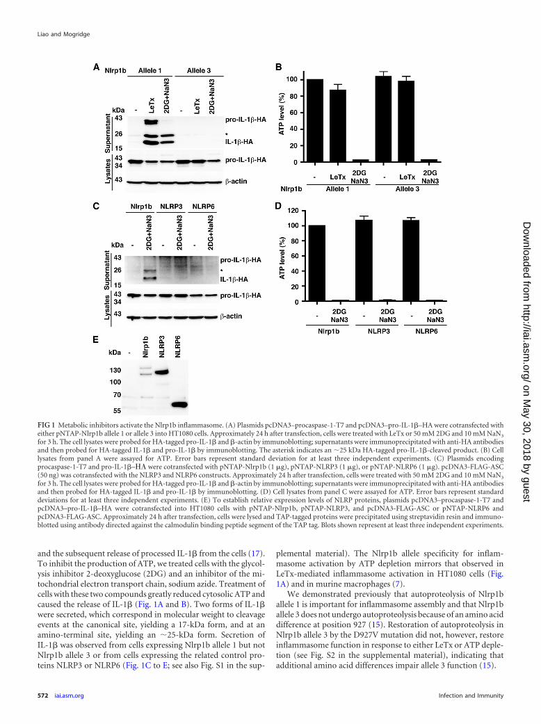
and the subsequent release of processed IL-1� from the cells (17).To inhibit the production of ATP, we treated cells with the glycol-ysis inhibitor 2-deoxyglucose (2DG) and an inhibitor of the mi-tochondrial electron transport chain, sodium azide. Treatment ofcells with these two compounds greatly reduced cytosolic ATP andcaused the release of IL-1� (Fig. 1A and B). Two forms of IL-1�were secreted, which correspond in molecular weight to cleavageevents at the canonical site, yielding a 17-kDa form, and at anamino-terminal site, yielding an �25-kDa form. Secretion ofIL-1� was observed from cells expressing Nlrp1b allele 1 but notNlrp1b allele 3 or from cells expressing the related control pro-teins NLRP3 or NLRP6 (Fig. 1C to E; see also Fig. S1 in the sup-
plemental material). The Nlrp1b allele specificity for inflam-masome activation by ATP depletion mirrors that observed inLeTx-mediated inflammasome activation in HT1080 cells (Fig.1A) and in murine macrophages (7).
We demonstrated previously that autoproteolysis of Nlrp1ballele 1 is important for inflammasome assembly and that Nlrp1ballele 3 does not undergo autoproteolysis because of an amino aciddifference at position 927 (15). Restoration of autoproteolysis inNlrp1b allele 3 by the D927V mutation did not, however, restoreinflammasome function in response to either LeTx or ATP deple-tion (see Fig. S2 in the supplemental material), indicating thatadditional amino acid differences impair allele 3 function (15).
FIG 1 Metabolic inhibitors activate the Nlrp1b inflammasome. (A) Plasmids pcDNA3–procaspase-1-T7 and pcDNA3–pro-IL-1�–HA were cotransfected witheither pNTAP-Nlrp1b allele 1 or allele 3 into HT1080 cells. Approximately 24 h after transfection, cells were treated with LeTx or 50 mM 2DG and 10 mM NaN3
for 3 h. The cell lysates were probed for HA-tagged pro-IL-1� and �-actin by immunoblotting; supernatants were immunoprecipitated with anti-HA antibodiesand then probed for HA-tagged IL-1� and pro-IL-1� by immunoblotting. The asterisk indicates an �25 kDa HA-tagged pro-IL-1�-cleaved product. (B) Celllysates from panel A were assayed for ATP. Error bars represent standard deviation for at least three independent experiments. (C) Plasmids encodingprocaspase-1-T7 and pro-IL-1�–�� were cotransfected with pNTAP-Nlrp1b (1 �g), pNTAP-NLRP3 (1 �g), or pNTAP-NLRP6 (1 �g). pcDNA3-FLAG-ASC(50 ng) was cotransfected with the NLRP3 and NLRP6 constructs. Approximately 24 h after transfection, cells were treated with 50 mM 2DG and 10 mM NaN3
for 3 h. The cell lysates were probed for HA-tagged pro-IL-1� and �-actin by immunoblotting; supernatants were immunoprecipitated with anti-HA antibodiesand then probed for HA-tagged IL-1� and pro-IL-1� by immunoblotting. (D) Cell lysates from panel C were assayed for ATP. Error bars represent standarddeviations for at least three independent experiments. (E) To establish relative expression levels of NLRP proteins, plasmids pcDNA3–procaspase-1-T7 andpcDNA3–pro-IL-1�–HA were cotransfected into HT1080 cells with pNTAP-Nlrp1b, pNTAP-NLRP3, and pcDNA3-FLAG-ASC or pNTAP-NLRP6 andpcDNA3-FLAG-ASC. Approximately 24 h after transfection, cells were lysed and TAP-tagged proteins were precipitated using streptavidin resin and immuno-blotted using antibody directed against the calmodulin binding peptide segment of the TAP tag. Blots shown represent at least three independent experiments.
Liao and Mogridge
572 iai.asm.org Infection and Immunity
on May 30, 2018 by guest
http://iai.asm.org/
Dow
nloaded from

To determine the threshold level of cytosolic ATP that triggersNlrp1b inflammasome activation, we grew cells in minimal me-dium (containing no serum or glucose) with various concentra-tions of either glucose or 2DG (Fig. 2A and B). IL-1� was releasedfrom cells grown in 1 mM glucose but not from cells grown in 10mM glucose. This reduction of glucose in the medium caused adrop in cytosolic ATP levels from �90% to �80% of that ob-served in control cells grown in complete medium. Further de-creasing the concentration of glucose in the medium or adding2DG caused a greater reduction in ATP levels and increased therelease of IL-1�. LeTx, and depletion of ATP to a lesser extent,caused release of pro-IL-1� into the medium, which was likelythrough passive release, because the release of pro-IL-1�, but notprocessed IL-1�, was inhibited by the addition of the osmoticstabilizer PEG-6000 (data not shown). These results suggest thatLeTx is a more effective inducer of pyroptosis than ATP depletionis, possibly because the toxin is a stronger activator of the inflam-masome.
We next assessed whether the Nlrp1b inflammasome functionsunder hypoxia by growing cells in minimal medium with 1% ox-ygen. These conditions caused intracellular ATP levels to drop to�50% of control levels and stimulated the release of IL-1� into
the cell supernatants (Fig. 2C and D; see also Fig. S3 in the supple-mental material). Hypoxia did not reduce the ATP levels in cellsgrown in complete medium to an extent that triggered inflam-masome activation (data not shown). These results suggest thatvarious conditions that lead to the reduction of cytosolic ATPbelow a threshold level activate Nlrp1b.
AMPK promotes inflammasome activation. Depletion of cel-lular ATP is sensed by the master regulator of metabolism, AMPK(19). AMPK is phosphorylated when a high AMP/ATP ratio existsin cells and it in turn phosphorylates numerous substrates to in-crease catabolic and decrease anabolic processes. To determinewhether AMPK signaling is involved in the activation of Nlrp1b byATP depletion or by LeTx, we used siRNA to reduce AMPK levels(Fig. 3A). A combination of 2DG and sodium azide, but not LeTx,caused an increase in phosphorylation of AMPK in control cells.In cells treated with AMPK siRNA, there was a reduced level oftotal and phosphorylated AMPK upon ATP depletion and lessIL-1� was secreted compared to that in control cells, whereas sim-ilar amounts of IL-1� were secreted by control and AMPK knock-down cells in response to LeTx (Fig. 3A; see also Fig. S4A and B inthe supplemental material).
AMPK activity is dependent on phosphorylation of the alpha sub-
FIG 2 Glucose starvation and hypoxia deplete cytosolic ATP and activate the inflammasome. (A) Cells were transfected with plasmids containing pcDNA3–procaspase-1-T7, pcDNA3–pro-IL-1�–HA, and pNTAP-Nlrp1b allele 1. These transfected cells were trypsinized, and 1 � 105 cells were seeded per well in a96-well plate. After 2 h, the cells were left in minimal DMEM containing 20 mM HEPES, pH 7.5, and the indicated concentrations of 2DG or glucose for 3 h. Thecell lysates were then assayed for ATP. (B) Cells were transfected as described for panel A. Approximately 24 h after transfection, cells were incubated in minimalDMEM containing 20 mM HEPES, pH 7.5, and the indicated concentrations of 2DG or glucose. After 3 h of treatment, HA-tagged pro-IL-1� and IL-1� weredetected by immunoblotting. (C) Cells were transfected with plasmids containing pcDNA3–procaspase-1-T7, pcDNA3–pro-IL-1�–HA, and pNTAP-Nlrp1ballele 1 or 3. These transfected cells were trypsinized, and 1 � 105 cells were seeded per well in a 96-well plate. After 2 h, these cells were left in minimal DMEMcontaining 20 mM HEPES, pH 7.5, and incubated under normoxic or hypoxic (1% O2) conditions for 3 h. The cell lysates were then assayed for intracellular ATP.(D) Plasmids encoding pNTAP-Nlrp1b allele 1 or 3, procaspase-1-T7, and pro-IL-1�–HA were transfected into HT1080 cells. Approximately 24 h aftertransfection, cells were incubated in minimal DMEM containing 20 mM HEPES, pH 7.5, in normoxic or hypoxic conditions. After 3 h of treatment, HA-taggedpro-IL-1� and IL-1� were detected as described above. Blots shown represent at least three independent experiments. The asterisk indicates an �25 kDaHA-tagged pro-IL-1�-cleaved product. Error bars represent standard deviations for at least three independent experiments.
Activation of Nlrp1b by ATP Depletion
February 2013 Volume 81 Number 2 iai.asm.org 573
on May 30, 2018 by guest
http://iai.asm.org/
Dow
nloaded from

FIG 3 AMPK facilitates Nlrp1b inflammasome activation. (A) HT1080 cells were first transfected with either control siRNA or AMPK siRNA. After 24 h, thesecells were then transfected with pcDNA3–procaspase-1-T7, pcDNA3–pro-IL-1�–HA, and pNTAP-Nlrp1b. Approximately 24 h after the second transfection,cells were treated with LeTx or 50 mM 2DG and 10 mM NaN3 for 3 h. Cell lysates were probed for phospho-AMPK, total AMPK, HA-tagged IL-1�, and �-actinby immunoblotting. Supernatants were immunoprecipitated with anti-HA antibodies and probed for HA-tagged IL-1� and pro-IL-1� by immunoblotting. (B)Plasmids pNTAP-Nlrp1b, pcDNA3–procaspase-1-T7, and pcDNA3–pro-IL-1�–HA were cotransfected with pcDNA3-His-FLAG vector, pcDNA3-His-AMPK�1-FLAG, or pcDNA3-His-AMPK�1-T174A-FLAG into HT1080 cells. Approximately 24 h after transfection, cells were treated with LeTx or 50 mM 2DGand 10 mM NaN3 for 3 h. Cell lysates were probed for phospho-AMPK, total-AMPK, HA-tagged IL-1�, and �-actin by immunoblotting; supernatants wereimmunoprecipitated with anti-HA antibodies and then probed for HA-tagged IL-1� and pro-IL-1� by immunoblotting. (C) Plasmids encoding procaspase-1-T7, pro-IL-1�–HA, and pNTAP-Nlrp1b were transfected into HT1080 cells. These transfected cells were treated with LeTx, 50 mM 2DG, and 10 mM NaN3 orwith 2 �M staurosporine (STS) as indicated. After 3 h, cell lysates were harvested and probed for phospho-AMPK, total-AMPK, HA-tagged IL-1�, and �-actinby immunoblotting. HA-tagged pro-IL-1� and IL-1� in the supernatants were detected as described above. (D) Cell lysates from panel C were assayed for ATP.(E) Plasmids pcDNA3–procaspase-1-T7, pcDNA3–pro-IL-1�–HA, and pNTAP-Nlrp1b were cotransfected into HT1080 cells. Approximately 24 h after trans-fection, cells were treated with LeTx or 50 mM 2DG and 10 mM NaN3 in the presence or absence of 10 �M cycloheximide (CHX) for 3 h. The cell lysates wereprobed for HA-tagged pro-IL-1� and �-actin by immunoblotting; supernatants were immunoprecipitated with anti-HA antibodies and then probed forHA-tagged IL-1� and pro-IL-1� by immunoblotting. Blots shown represent at least three independent experiments. The asterisk indicates an �25-kDaHA-tagged pro-IL-1�-cleaved product. Error bars represent standard deviations for at least three independent experiments.
574 iai.asm.org Infection and Immunity
on May 30, 2018 by guest
http://iai.asm.org/
Dow
nloaded from

unit at a conserved threonine residue (19). In order to confirm thatAMPK facilitated Nlrp1b activation by ATP depletion, the mutantAMPK�1-T174A subunit was overexpressed, which had a dominantnegative effect, as indicated by the diminished level of AMPK phos-phorylation in ATP-depleted cells (Fig. 3B). Consistent with theknockdown experiment, overexpression of AMPK�1-T174A, butnot wild-type AMPK�1, reduced the amount of IL-1� secreted uponATP depletion (Fig. 3B; see also Fig. S4C in the supplemental mate-rial). AMPK�1-T174A expression had only a minor effect on inflam-masome activation by LeTx (Fig. 3B; see also Fig. S4D in the supple-mental material).
We next assessed whether signaling by AMPK is sufficient toactivate Nlrp1b by treating cells with staurosporine. Staurospor-ine is a kinase inhibitor that causes activation of AMPK in theabsence of ATP depletion (Fig. 3C and D). Staurosporine did notcause the release of IL-1� from cells (Fig. 3C), indicating thatNlrp1b activation is not controlled solely by AMPK.
Because a major function of AMPK is to inhibit translation, wespeculated that inhibition of ribosomal function may explain howAMPK facilitates inflammasome activation. Treatment of cellswith cycloheximide did not, however, stimulate the release ofIL-1� from cells treated with sodium azide and 2DG or with LeTx(Fig. 3E; see Fig. S4E and F in the supplemental material).
Effects of MG-132 and N-acetylcysteine on inflammasomeactivation. Several studies have suggested that reactive oxygenspecies (ROS) generated from the mitochondrial electron trans-port chain are required to trigger NLRP3 assembly downstream ofdiverse stimuli such as extracellular ATP, particulates, and palmi-tate (5, 20). The ROS required to activate NLRP3 may be de-rived from damaged mitochondria or from excessive oxidativephosphorylation. Recently, however, the requirement for ROShas been suggested to be at the priming stage—the expressionof NLRP3—rather than the activation stage (21). To addresswhether activation of the Nlrp1b inflammasome required ROS,we pretreated transfected HT1080 cells with the nonspecificROS scavenger N-acetylcysteine (NAC). We found that NACdid not inhibit IL-1� secretion from cells treated with LeTx(Fig. 4A; see also Fig. S5A in the supplemental material) but diddiminish the amount of IL-1� secreted from cells treated withthe metabolic inhibitors (Fig. 4A; see Fig. S5B and C in thesupplemental material). NAC did not affect cytosolic ATP lev-els (Fig. 4B), however, which suggests that cellular redux po-tential influences inflammasome assembly.
It has been established that proteasome inhibitors block theactivation of Nlrp1b by LeTx (22, 23), so we sought to determinewhether this was also the case for activation by ATP depletion. Theproteasome inhibitor MG-132 blocked the secretion of IL-1� inresponse to both LeTx and 2DG-sodium azide (Fig. 4C; see alsoFig. S5D and E in the supplemental material). MG-132 did not,however, prevent the depletion of cytosolic ATP by 2DG-sodiumazide (Fig. 4D), suggesting that proteasome inhibition diminishesNlrp1b inflammasome activity triggered by both LeTx and ATPdepletion.
Mutation of the Nlrp1b Walker A motif causes constitutiveactivation. The NACHT domains of NLRPs are thought to medi-ate homo-oligomerization by using energy derived from ATP(24). Because this notion seemed at odds with Nlrp1b sensing lowATP levels, we mutated the Walker A motif of the Nlrp1b NACHTdomain to assess its involvement in inflammasome activation. Incontrast to wild-type Nlrp1b, the Walker A mutant was constitu-
FIG 4 Roles of reactive oxygen species and proteasome activity in Nlrp1bactivation. (A) Plasmids pcDNA3–procaspase-1-T7, pcDNA3–pro-IL-1�–HA, and pNTAP-Nlrp1b were transfected into HT1080 cells. Approximately24 h after transfection, cells were left untreated or were treated with LeTx, 50mM 2DG, and 10 mM NaN3 or 50 mM 2DG in the absence or presence of 25mM N-acetyl cysteine (NAC). After 3 h, cell lysates were collected and probedfor HA-tagged pro-IL-1� and �-actin by immunoblotting; supernatants wereimmunoprecipitated with anti-HA antibodies and probed for HA-tagged pro-IL-1� and IL-1� by immunoblotting. (B) Cell lysates from panel A were as-sayed for ATP. (C) Cells were transfected with pcDNA3–procaspase-1-T7,pcDNA3–pro-IL-1�–HA, and pNTAP-Nlrp1b. Approximately 24 h aftertransfection, cells were treated with LeTx or 50 mM 2DG and 10 mM NaN3 inthe presence or absence of 10 �M MG-132. After 3 h, cell lysates were collectedand probed for HA-tagged pro-IL-1�; HA-tagged pro-IL-1� and IL-1� insupernatants were detected as described above. (D) Cell lysates from panel Cwere assayed for ATP. Blots shown represent at least three independent exper-iments. The asterisk indicates an �25-kDa HA-tagged pro-IL-1�-cleavedproduct. Error bars represent standard deviations for at least three indepen-dent experiments.
Activation of Nlrp1b by ATP Depletion
February 2013 Volume 81 Number 2 iai.asm.org 575
on May 30, 2018 by guest
http://iai.asm.org/
Dow
nloaded from

tively active, suggesting that ATP hydrolysis is not required toform a functional inflammasome (Fig. 5A and B). The lower levelof the Walker A mutant may result from its constitutive secretion,as it has been demonstrated previously that inflammasome com-ponents are secreted upon activation (25). The Walker A muta-tion did not, however, activate allele 3 of Nlrp1b (Fig. 5C and D).This was not surprising, as we have shown previously that theinability of Nlrp1b allele 3 to undergo autocatalyic proteolysisprevents the assembly of a functional inflammasome (15).
We next measured NLRP3 activity by using a constitutivelyactive mutant identified in patients with the auto-inflammatorydisorder Muckles-Wells syndrome (26). NLRP3-R258W caused
secretion of IL-1� in the presence of the ASC adaptor but not in itsabsence (Fig. 6A and B). In contrast to what was observed forNlrp1b, mutation of the Walker A motif in NLRP3 did not causeconstitutive activation (Fig. 6C and D). Furthermore, the intro-duction of the Walker A mutation into NLRP3-R258W impairedits activity, indicating that a functional ATPase domain is required
FIG 5 Walker A motif mutant of Nlrp1b is constitutively active. (A) HT1080cells were transfected with pcDNA3–procaspase-1-T7, pcDNA3–pro-IL-1�–HA, and pNTAP-Nlrp1b wild-type (WT) or Walker A (WA) mutant. Approx-imately 24 h after transfection, cells were lysed and TAP-tagged proteins wereprecipitated using streptavidin resin and immunoblotted using antibody di-rected against the calmodulin binding peptide segment of the TAP tag. (B)Plasmids encoding procaspase-1-T7 and pro-IL-1�–HA were cotransfectedinto cells with either pNTAP-Nlrp1b-WT or pNTAP-Nlrp1b-WA. Approxi-mately 24 h after transfection, cells were treated with LeTx, and then cell lysateswere collected and probed for HA-tagged IL-1� and �-actin by immunoblot-ting; supernatants were immunoprecipitated with anti-HA antibodies andprobed for HA-tagged pro-IL-1� and IL-1� by immunoblotting. (C) As de-scribed for panel A, except that both Nlrp1b allele 1 and allele 3 were used. (D)As described for panel B, except that both Nlrp1b allele 1 and allele 3 were used.Blots shown represent at least three independent experiments. The asteriskindicates an �25-kDa HA-tagged pro-IL-1�-cleaved product.
FIG 6 Mutation of the NLRP3 Walker A motif impairs activity. (A) Cells weretransfected with pcDNA3–procaspase-1-T7, pcDNA3–pro-IL-1�–HA, andindicated pNTAP-NLRP3 plasmids along with 50 ng pcDNA3-FLAG-ASC or50 ng empty vector. Approximately 24 h after transfection, TAP-tagged pro-teins were precipitated using streptavidin resin and immunoblotted usingantibody directed against the calmodulin binding peptide segment of the TAPtag. (B) Plasmids encoding the indicated proteins were cotransfected intoHT1080 cells with pcDNA3–procaspase-1-T7 and pcDNA3–pro-IL-1�–HA.Approximately 24 h after transfection, cell lysates were harvested and probedfor HA-tagged IL-1� and �-actin; supernatants were probed for HA-taggedpro-IL-1� and IL-1�. (C) Plasmids pcDNA3-FLAG-ASC, pcDNA3–pro-caspase-1-T7, pcDNA3–pro-IL-1�–HA, and various pNTAP-NLRP3 con-structs were transfected into HT1080 cells. TAP-tagged proteins were detectedas described above. (D) Cells were transfected with pcDNA3-FLAG-ASC,pcDNA3–procaspase-1-T7, pcDNA3–pro-IL-1�–HA, and pNTAP-NLRP3.HA-tagged IL-1� and pro-IL-1� were detected as described above. The aster-isk indicates an �25-kDa HA-tagged pro-IL-1�-cleaved product. Blots shownrepresent at least three independent experiments.
Liao and Mogridge
576 iai.asm.org Infection and Immunity
on May 30, 2018 by guest
http://iai.asm.org/
Dow
nloaded from

for NLRP3 function (Fig. 6D). These data suggest that theNACHT domains of Nlrp1b and NLRP3 have different require-ments for ATP.
Depletion of ATP activates the Nlrp1b inflammasome inJ774A macrophages. To assess whether reduction of cytosolicATP activates the Nlrp1b inflammasome in a macrophage cellline, we generated J774A cells that stably express either a controlshRNA or an shRNA directed against Nlrp1b. Treatment of thecontrol cells with either LeTx or 2DG led to the appearance ofcaspase-1 fragments of �43 kDa and �33 kDa; in addition tothese fragments, several smaller fragments were observed in ly-sates of the LeTx-treated cells (Fig. 7A). Cells that express Nlrp1bshRNA exhibited reduced levels of caspase-1 fragments comparedto the control cells upon treatment with either LeTx or 2DG (Fig.7A; see also Fig. S7 in the supplemental material). As expected, the2DG treatment reduced ATP levels in both control and Nrlp1bknockdown cells (Fig. 7B), indicating that ATP depletion activatesthe Nlrp1b inflammasome. In contrast, LeTx caused a large reduc-tion of ATP in control cells but not in the Nlrp1b knockdown cells,which is likely a consequence of LeTx-induced pyroptosis causingloss of ATP.
DISCUSSION
The innate immune system is able to detect a diversity of infec-tions. It does so by using pattern recognition receptors to senseconserved microbial structures and to discern between normalevents and those that are associated with cellular damage (14). The
damage signals that are detected directly by pattern recognitionreceptors are poorly characterized, although the events that elicitthese signals are being uncovered. We have demonstrated herethat Nlrp1b is activated by energy deprivation in a reconstitutedsystem and in a macrophage cell line. That Nlrp1b can detect adanger signal in addition to LeTx is an attractive idea because thenotion that a pattern recognition receptor has evolved to sense avirulence factor from a single pathogen is counterintuitive.Nlrp1b may therefore be able to detect viral replication, which candeplete cellular ATP, and a number of bacterial pathogens thatsecrete pore-forming toxins as sublytic concentrations of thesetoxins reduce ATP levels (27–30). Activation of Nlrp1b by energystress and the subsequent release of IL-1� could benefit the host inat least two ways. First, the chemokine activity of IL-1� couldrecruit neutrophils to inflamed tissue to combat infection. Sec-ond, IL-1� might facilitate tissue repair and immune cell functionby stimulating glucose uptake (31).
The molecular events that link the reduction of cytosolic ATPlevels to the activation of the Nlrp1b inflammasome are not clear.The observation that a Walker A mutant is constitutively activeraises the idea that Nlrp1b is a direct sensor of ATP levels— de-creased concentrations of ATP might promote the formation ofactive ADP-bound or nucleotide-free Nlrp1b from inactive ATP-bound Nlrp1b. This model does not, however, explain the effectsof AMPK, NAC, or MG-132 on inflammasome activity. It is con-ceivable that in addition to an activating signal, either particularcellular conditions must exist or a distinct inhibitory mechanismmust be overcome for the inflammasome to assemble.
The amino-terminal region of Nlrp1b may be involved in itsauto-inhibition, because cleavage of this region is required forLeTx-mediated inflammasome activation (12, 13). We did not,however, observe cleavage of Nlrp1b in response to ATP depletion(see Fig. S6 in the supplemental material). Nonetheless, there maybe commonalities between the two activating signals. One possi-bility is that LeTx may inactivate an ATP-binding protein, which islikewise inactivated by ATP depletion, to initiate inflammasomeactivation. Alternatively, LeTx may cause a highly localized deple-tion of ATP that was not detected in our assays.
Lee and colleagues implicated AMP deaminase 3 (AMPD3),which coverts AMP to IMP, in LeTx-mediated pyroptosis of a murinemacrophage cell line (32). These results are intriguing, but we do notknow if they relate to the findings reported here. Lee and colleaguesalso demonstrated that a small-molecule activator of AMPK, AICAR,did not influence pyroptosis, which is in agreement with our workshowing that activation of AMPK is not sufficient to trigger theNlrp1b inflammasome. AMPK does, however, enhance inflam-masome assembly caused by ATP depletion. Although the role ofAMPK in this process is unclear, it does not appear to involve thedownregulation of translation, as cycloheximide did not stimulateinflammasome activation.
A recent study has shown that a strain of B. anthracis that producesLeTx activates caspase-1 in macrophages expressing a resistant alleleof Nlrp1b (33). The authors demonstrated that both the bacteriumand the toxin were required to activate caspase-1 and that the dangersignal was mediated by extracellular ATP that had leaked from themacrophages. The authors did not directly test which inflammasomewas responsible for caspase-1 activation by knocking down NLRPexpression, but the data are consistent with NLRP3 activation. Extra-cellular ATP, a well-characterized trigger of NLRP3 (34), binds puri-nergic receptors to cause the opening of pannexin-1 channels. Mem-
FIG 7 Depletion of ATP activates the Nlrp1b inflammasome in J774 macro-phages. (A) J774A cells were grown in minimal medium and treated eitherwith LeTx (5 � 10�10 M LF plus 10�8 M PA) or with 50 mM 2DG for 4 h.Supernatants were collected and probed for caspase-1 by immunoblotting.The blot represents three independent experiments. (B) Cell lysates from panelA were assayed for ATP. Error bars indicate standard deviations from threeindependent experiments.
Activation of Nlrp1b by ATP Depletion
February 2013 Volume 81 Number 2 iai.asm.org 577
on May 30, 2018 by guest
http://iai.asm.org/
Dow
nloaded from

brane openings cause an efflux of potassium that is required for theassembly of the NLRP3 inflammasome.
A Walker A motif mutation in Nlrp1b caused constitutive ac-tivation, whereas the same mutation prevented activity of NLRP3(Fig. 5 and 6). We speculate that the NACHT domain facilitatesauto-inhibition of both proteins and that mutation of the WalkerA motif abolishes auto-inhibition. The NLRP3 Walker A mutantis not active because its NACHT domain is also required for itsoligomerization; oligomerization of Nlrp1b, however, is not de-pendent on a functional NACHT domain, so it is constitutivelyactive. We showed previously that a truncation mutant of Nlrp1bconsisting of the FIIND and CARD domains alone oligomerizesand activates procaspase-1 (17).
Our finding that Nlrp1b detects energy stress adds to a growingbody of work that links metabolism and inflammation. This con-nection is likely a consequence of the requirement of immune cellsto function in inflamed tissues that are deprived of oxygen andglucose. Hypoxia-inducible factor 1� (HIF-1�) is a transcriptionfactor that controls the cellular response to low-oxygen conditionsby upregulating genes involved in glycolysis, angiogenesis, andglucose uptake; in myeloid cells, HIF-1� also increases the expres-sion of tumor necrosis factor alpha (TNF-�) and antimicrobialfactors (35). The importance of HIF-1� for macrophage functionwas demonstrated by the observation that HIF-1�-null macro-phages exhibit impaired inflammatory function and a metabolicdefect that results in an �80% reduction in ATP levels (36). Thus,sensors of oxygen and ATP appear to coordinate to mount aneffective immune response.
ACKNOWLEDGMENTS
This research was supported by NIH grant RO1 AI067683.J. M. holds the Canada Research Chair in Bacterial Pathogenesis.
REFERENCES1. Imtiyaz HZ, Simon MC. 2010. Hypoxia-inducible factors as essential regu-
lators of inflammation. Curr. Top. Microbiol. Immunol. 345:105–120.2. Krawczyk CM, Holowka T, Sun J, Blagih J, Amiel E, DeBerardinis RJ,
Cross JR, Jung E, Thompson CB, Jones RG, Pearce EJ. 2010. Toll-likereceptor-induced changes in glycolytic metabolism regulate dendritic cellactivation. Blood 115:4742– 4749.
3. Tannahill GM, O’Neill LA. 2011. The emerging role of metabolic regu-lation in the functioning of Toll-like receptors and the NOD-like receptorNlrp3. FEBS Lett. 585:1568 –1572.
4. Schroder K, Tschopp J. 2010. The inflammasomes. Cell 140:821– 832.5. Wen H, Gris D, Lei Y, Jha S, Zhang L, Huang MT, Brickey WJ, Ting JP.
2011. Fatty acid-induced NLRP3-ASC inflammasome activation inter-feres with insulin signaling. Nat. Immunol. 12:408 – 415.
6. Masters SL, Dunne A, Subramanian SL, Hull RL, Tannahill GM, SharpFA, Becker C, Franchi L, Yoshihara E, Chen Z, Mullooly N, Mielke LA,Harris J, Coll RC, Mills KH, Mok KH, Newsholme P, Nunez G, YodoiJ, Kahn SE, Lavelle EC, O’Neill LA. 2010. Activation of the NLRP3inflammasome by islet amyloid polypeptide provides a mechanism forenhanced IL-1beta in type 2 diabetes. Nat. Immunol. 11:897–904.
7. Boyden ED, Dietrich WF. 2006. Nalp1b controls mouse macrophagesusceptibility to anthrax lethal toxin. Nat. Genet. 38:240 –244.
8. Terra JK, Cote CK, France B, Jenkins AL, Bozue JA, Welkos SL, LeVineSM, Bradley KA. 2010. Cutting edge: resistance to Bacillus anthracisinfection mediated by a lethal toxin sensitive allele of Nalp1b/Nlrp1b. J.Immunol. 184:17–20.
9. Moayeri M, Crown D, Newman ZL, Okugawa S, Eckhaus M, Cataisson C,Liu S, Sastalla I, Leppla SH. 2010. Inflammasome sensor Nlrp1b-dependentresistance to anthrax is mediated by caspase-1, IL-1 signaling and neutrophilrecruitment. PLoS Pathog. 6:e1001222. doi:10.1371/journal.ppat.1001222.
10. Duesbery NS, Webb CP, Leppla SH, Gordon VM, Klimpel KR, Cope-land TD, Ahn NG, Oskarsson MK, Fukasawa K, Paull KD, Vande
Woude GF. 1998. Proteolytic inactivation of MAP-kinase-kinase by an-thrax lethal factor. Science 280:734 –737.
11. Moayeri M, Leppla SH. 2009. Cellular and systemic effects of anthraxlethal toxin and edema toxin. Mol. Aspects Med. 30:439 – 455.
12. Levinsohn JL, Newman ZL, Hellmich KA, Fattah R, Getz MA, Liu S,Sastalla I, Leppla SH, Moayeri M. 2012. Anthrax lethal factor cleavage ofNlrp1 is required for activation of the inflammasome. PLoS Pathog.8:e1002638. doi:10.1371/journal.ppat.1002638.
13. Hellmich KA, Levinsohn JL, Fattah R, Newman ZL, Maier N, Sastalla I,Liu S, Leppla SH, Moayeri M. 2012. Anthrax lethal factor cleaves mouseNlrp1b in both toxin-sensitive and toxin-resistant macrophages. PLoSOne 7:e49741. doi:10.1371/journal.pone.0049741.
14. Newton K, Dixit VM. 2012. Signaling in innate immunity and inflam-mation. Cold Spring Harb. Perspect. Biol. 4:a006114.
15. Frew BC, Joag VR, Mogridge J. 2012. Proteolytic processing of Nlrp1bis required for inflammasome activity. PLoS Pathog. 8:e1002659. doi:10.1371/journal.ppat.1002659.
16. Kassam A, Der SD, Mogridge J. 2005. Differentiation of human mono-cytic cell lines confers susceptibility to Bacillus anthracis lethal toxin. CellMicrobiol. 7:281–292.
17. Liao KC, Mogridge J. 2009. Expression of Nlrp1b inflammasome com-ponents in human fibroblasts confers susceptibility to anthrax lethaltoxin. Infect. Immun. 77:4455– 4462.
18. Zeghouf M, Li J, Butland G, Borkowska A, Canadien V, Richards D,Beattie B, Emili A, Greenblatt JF. 2004. Sequential peptide affinity (SPA)system for the identification of mammalian and bacterial protein com-plexes. J. Proteome Res. 3:463– 468.
19. Carling D, Mayer FV, Sanders MJ, Gamblin SJ. 2011. AMP-activatedprotein kinase: nature’s energy sensor. Nat. Chem. Biol. 7:512–518.
20. Zhou R, Yazdi AS, Menu P, Tschopp J. 2011. A role for mitochondria inNLRP3 inflammasome activation. Nature 469:221–225.
21. Bauernfeind F, Bartok E, Rieger A, Franchi L, Nunez G, Hornung V.2011. Cutting edge: reactive oxygen species inhibitors block priming,but not activation, of the NLRP3 inflammasome. J. Immunol. 187:613–617.
22. Tang G, Leppla SH. 1999. Proteasome activity is required for anthraxlethal toxin to kill macrophages. Infect. Immun. 67:3055–3060.
23. Squires RC, Muehlbauer SM, Brojatsch J. 2007. Proteasomes controlcaspase-1 activation in anthrax lethal toxin-mediated cell killing. J. Biol.Chem. 282:34260 –34267.
24. Duncan JA, Bergstralh DT, Wang Y, Willingham SB, Ye Z, Zimmer-mann AG, Ting JP. 2007. Cryopyrin/NALP3 binds ATP/dATP, is anATPase, and requires ATP binding to mediate inflammatory signaling.Proc. Natl. Acad. Sci. U. S. A. 104:8041– 8046.
25. Feldmeyer L, Keller M, Niklaus G, Hohl D, Werner S, Beer HD. 2007.The inflammasome mediates UVB-induced activation and secretion ofinterleukin-1beta by keratinocytes. Curr. Biol. 17:1140 –1145.
26. Aganna E, Martinon F, Hawkins PN, Ross JB, Swan DC, Booth DR,Lachmann HJ, Bybee A, Gaudet R, Woo P, Feighery C, Cotter FE,Thome M, Hitman GA, Tschopp J, McDermott MF. 2002. Associationof mutations in the NALP3/CIAS1/PYPAF1 gene with a broad phenotypeincluding recurrent fever, cold sensitivity, sensorineural deafness, and AAamyloidosis. Arthritis Rheum. 46:2445–2452.
27. Ando T, Imamura H, Suzuki R, Aizaki H, Watanabe T, Wakita T,Suzuki T. 2012. Visualization and measurement of ATP levels in livingcells replicating hepatitis C virus genome RNA. PLoS Pathog. 8:e1002561.doi:10.1371/journal.ppat.1002561.
28. Dickman KG, Hempson SJ, Anderson J, Lippe S, Zhao L, Burakoff R,Shaw RD. 2000. Rotavirus alters paracellular permeability and energymetabolism in Caco-2 cells. Am. J. Physiol. Gastrointest. Liver Physiol.279:G757–G766.
29. Dragneva Y, Anuradha CD, Valeva A, Hoffmann A, Bhakdi S, Hus-mann M. 2001. Subcytocidal attack by staphylococcal alpha-toxin acti-vates NF-kappaB and induces interleukin-8 production. Infect. Immun.69:2630 –2635.
30. Lizak M, Yarovinsky TO. 2012. Phospholipid scramblase 1 mediates typeI interferon-induced protection against staphylococcal alpha-toxin. CellHost Microbe 11:70 – 80.
31. Del Rey A, Roggero E, Randolf A, Mahuad C, McCann S, Rettori V,Besedovsky HO. 2006. IL-1 resets glucose homeostasis at central levels.Proc. Natl. Acad. Sci. U. S. A. 103:16039 –16044.
32. Lee S, Wang Y, Kim SO, Han J. 2011. AMPD3 is involved in anthraxLeTx-induced macrophage cell death. Protein Cell 2:564 –572.
Liao and Mogridge
578 iai.asm.org Infection and Immunity
on May 30, 2018 by guest
http://iai.asm.org/
Dow
nloaded from

33. Ali SR, Timmer AM, Bilgrami S, Park EJ, Eckmann L, Nizet V, KarinM. 2011. Anthrax toxin induces macrophage death by p38 MAPKinhibition but leads to inflammasome activation via ATP leakage. Im-munity 35:34 – 44.
34. Mariathasan S, Weiss DS, Newton K, McBride J, O’Rourke K, Roose-Girma M, Lee WP, Weinrauch Y, Monack DM, Dixit VM. 2006.Cryopyrin activates the inflammasome in response to toxins and ATP.Nature 440:228 –232.
35. Peyssonnaux C, Datta V, Cramer T, Doedens A, Theodorakis EA, GalloRL, Hurtado-Ziola N, Nizet V, Johnson RS. 2005. HIF-1alpha expres-sion regulates the bactericidal capacity of phagocytes. J. Clin. Invest. 115:1806 –1815.
36. Cramer T, Yamanishi Y, Clausen BE, Forster I, Pawlinski R, MackmanN, Haase VH, Jaenisch R, Corr M, Nizet V, Firestein GS, Gerber HP,Ferrara N, Johnson RS. 2003. HIF-1alpha is essential for myeloid cell-mediated inflammation. Cell 112:645– 657.
Activation of Nlrp1b by ATP Depletion
February 2013 Volume 81 Number 2 iai.asm.org 579
on May 30, 2018 by guest
http://iai.asm.org/
Dow
nloaded from