activation of the maternal immune system during pregnancy alters behavioral development of rhesus...
TRANSCRIPT
ARCHIVAL REPORT
Activation of the Maternal Immune System DuringPregnancy Alters Behavioral Development of RhesusMonkey Offspring
Melissa D. Bauman, Ana-Maria Iosif, Stephen E.P. Smith, Catherine Bregere, David G. Amaral,and Paul H. PattersonBackground: Maternal infection during pregnancy is associated with an increased risk of schizophrenia and autism in the offspring.Supporting this correlation, experimentally activating the maternal immune system during pregnancy in rodents produces offspring withabnormal brain and behavioral development. We have developed a nonhuman primate model to bridge the gap between clinicalpopulations and rodent models of maternal immune activation (MIA).
Methods: A modified form of the viral mimic, synthetic double-stranded RNA (polyinosinic:polycytidylic acid stabilized with poly-L-lysine)was delivered to two separate groups of pregnant rhesus monkeys to induce MIA: 1) late first trimester MIA (n ¼ 6), and 2) late secondtrimester MIA (n ¼ 7). Control animals (n ¼ 11) received saline injections at the same first or second trimester time points or were untreated.Sickness behavior, temperature, and cytokine profiles of the pregnant monkeys confirmed a strong inflammatory response to MIA.
Results: Behavioral development of the offspring was studied for 24 months. Following weaning at 6 months of age, MIA offspringexhibited abnormal responses to separation from their mothers. As the animals matured, MIA offspring displayed increased repetitivebehaviors and decreased affiliative vocalizations. When evaluated with unfamiliar conspecifics, first trimester MIA offspring deviated fromspecies-typical macaque social behavior by inappropriately approaching and remaining in immediate proximity of an unfamiliar animal.
Conclusions: In this rhesus monkey model, MIA yields offspring with abnormal repetitive behaviors, communication, and socialinteractions. These results extended the findings in rodent MIA models to more human-like behaviors resembling those in both autismand schizophrenia.
Key Words: Animal model, autism spectrum disorder, immuneactivation, macaque, nonhuman primate, poly IC, schizophrenia
Autism spectrum disorder (ASD) and schizophrenia (SZ) arechronic and disabling brain disorders that each affectapproximately 1% of the population (1,2) and are thought
to be caused by complex interactions between genetic andenvironmental factors (3–5). Recent evidence suggests that theprenatal environment, and particularly the maternal immuneenvironment, plays a critical role in some cases of ASD and SZ(6–8). Epidemiologic studies reveal that women exposed to viral,bacterial, or parasitic infections during pregnancy have anincreased risk of having a child that later develops SZ (9–14).Likewise, maternal viral and bacterial infections are associated withan increased risk of ASD in the offspring (15–19). The diversity ofmaternal infections associated with ASD and SZ outcomes suggeststhat the maternal immune response is the critical link betweensickness in the mother and altered neurodevelopment in her child.
From the Department of Psychiatry and Behavioral Sciences (MDB, DGA),and California National Primate Research Center (MDB, DGA), Uni-versity of California, Davis, Davis; The M.I.N.D. Institute (MDG, DGA),University of California, Davis, Sacramento; Department of PublicHealth Sciences (A-MI), Division of Biostatistics, University of California,Davis, Davis; Division of Biology (SEPS, CB, PHP), California Institute ofTechnology, Pasadena; and Center for Neuroscience (DGA), Universityof California, Davis, Davis, California.
Address correspondence to Melissa D. Bauman, Ph.D., University ofCalifornia, The M.I.N.D. Institute, 2825 50th Street #1416, Sacramento,CA 95817; E-mail: [email protected].
Received Apr 10, 2013; revised Jun 22, 2013; accepted Jun 29, 2013.
0006-3223/$36.00http://dx.doi.org/10.1016/j.biopsych.2013.06.025
Understanding the mechanism by which maternal immuneactivation (MIA) during pregnancy increases the risk for SZ andASD is essential to developing novel preventative or therapeuticstrategies. Rodent models have identified molecular, cellular, andbehavioral abnormalities associated with prenatal immune chal-lenge (20). Maternal influenza infection (21–24) or injection of thebacterial endotoxin lipopolysaccaride (25–27) yields offspringwith behavioral abnormalities, neuropathology, and altered geneexpression that are relevant to both SZ and ASD. Similar out-comes are obtained by treating pregnant rodents with the viralmimic, synthetic double stranded RNA (polyinosinic:polycytidylicacid [poly IC]), which stimulates an inflammatory response in theabsence of a specific pathogen (28). Offspring born to pregnantdams treated with poly IC at mid-gestation demonstrate repet-itive behaviors and deficits in social and communication behav-iors that resemble features of ASD, as well as elevated anxiety,deficits in prepulse inhibition, latent inhibition, and workingmemory that resemble clinical features of both ASD and SZ(21,29–32). Neuropathology observed with ASD (localized lossof Purkinje cells) and SZ (enlarged ventricles) have been reportedin poly IC rodent models (33–35), and there are numerous otheralterations in brain structure, neurochemistry, gene expres-sion, and immune function (36–39). The deleterious effects onbrain and behavior in the mouse MIA model appear to bemediated by the maternal cytokine response, in particularinterleukin-6 (40).
While rodent models have laid the foundation for understandingthe effects of MIA on fetal brain development, these models havelimitations. Extrapolating the timing of fetal brain developmentbetween rodents and humans is complicated by the fact that theneural events of the human third trimester occur during the earlypostnatal period in rodents (41). Moreover, there are challenges inrelating the rodent brain to the human brain and rodent behavior
BIOL PSYCHIATRY 2014;75:332–341& 2014 Society of Biological Psychiatry

M.D. Bauman et al. BIOL PSYCHIATRY 2014;75:332–341 333
to human behavior. This is particularly problematic for disorderssuch as ASD and SZ that are characterized by deficits in a range ofcomplex cognitive, social, and affective functions. Indeed, portionsof the human brain, such as prefrontal cortex, which mediate thesefunctions and are heavily impacted in ASD and SZ, are poorlydeveloped in the rodent brain (42). Understanding human disordersinvolving higher cognitive functions will benefit from studies inanimal species more closely related to humans. Nonhumanprimates, such as rhesus macaques (Macaca mulatta), demonstratemany features of human physiology, anatomy, and behavior,making them an appropriate species to study a variety of humanbrain disorders (43). The rhesus monkey lives in a complex,hierarchical social system and uses many forms of human-likecommunication such as facial expressions and social gestures (44).The rich social and cognitive repertoire of rhesus monkeys providesa framework to relate behavioral changes observed in the animalmodel more directly to human mental illness.
We have developed a novel, nonhuman primate model using amodified form of the viral mimic poly IC, which is adapted for use inprimates (polyinosinic:polycytidylic acid stabilized with poly-L-lysine[poly ICLC]). This synthetic RNA is recognized as foreign by theprimate immune system and induces a transient innate inflammatoryresponse (45,46). Pregnant rhesus monkeys were injected with polyICLC over a 72-hour period at the end of the first or second trimester.These gestational ages were selected based on human epidemio-logic data identifying the first and second trimesters as vulnerabletime points where exposure to MIA increases the risk of autism andschizophrenia (14,17). We evaluated sickness behavior, body temper-ature, and cytokine responses in the dams to confirm a strongimmune activation and then analyzed the behavioral developmentof the offspring for 4 years. Here, we present our initial behavioral
Table 1. Behavioral Phenotyping Assays
Behavioral Assay Brief Description
6–12 Months of AgeMother preferencea Following weaning, each infant was tested for 4 days
evaluate one aspect of mother-infant attachment, thpreference for its own mother versus another familifemale (12 2-minute trials/subject).
Postweaning soloobservationsb
At approximately 10 months of age, the animals were oalone in a large, unfamiliar cage for two 5-minute fosamples on 2 separate days to screen for abnormal bsuch as motor stereotypies or self-directed behavior
12–18 Months of AgeJuvenile Y‐maze At approximately 18 months of age, animals were give
access to a novel conspecific in one arm of a Y-mazapparatus. Each animal was tested for six 2-minute trseparate days, meeting an opposite-sex conspecificfirst day and a same-sex conspecific on the second
Juvenile soloobservationsb
At approximately 22 months of age, the animals were oalone in a large, unfamiliar cage for two 5-minute fosamples on 2 separate days to screen for abnormal bsuch as motor stereotypies or self-directed behavior
Juvenile socialapproachc
At approximately 24 months of age, social interactionsnovel conspecific were evaluated using a modified vthe mouse three-chambered social approach assay (minutes/subject).
ASD, autism spectrum disorders; SZ, schizophrenia.aAssays used to control for changes in physical development, reflexes, fear
not directly related to the core features of ASD and SZ.bBehavioral assays targeting repetitive behaviors and restricted interests.cBehavioral assays targeting social and communication domains.
findings through 24 months of age, documenting the emergence ofabnormal behavior in rhesus offspring exposed to MIA.
Methods and Materials
All experimental procedures were developed in consultationwith the veterinary staff at the California National PrimateResearch Center. Protocols were approved by the University ofCalifornia, Davis Institutional Animal Care and Use Committee.Detailed methods are provided in Supplement 1.
Maternal Administration of Poly ICLCTwenty-four multiparous rhesus monkeys were assigned to one
of three experimental groups: 1) first trimester MIA (MIA1), 2)second trimester MIA (MIA2), or 3) saline control animals (CONSaline)(Table S1 in Supplement 1). Pregnant animals in the MIA groupswere injected with .25 mg/kg synthetic double-stranded RNA (polyICLC) (Oncovir, Inc., Washington, DC) via intravenous injection whilerestrained by trained technicians on gestational days 43, 44, and 46(MIA1) or 100, 101, and 103 (MIA2).
Rearing ConditionsInfants were raised in individual cages with their mothers,
where they had visual access to other animals at all times. For 3hours each day, one adult male and four familiar mother-infantpairs were allowed to freely interact in a large cage to provideenrichment and facilitate species-typical social development.Each group consisted of a mixture of male and female offspringof both MIA and control experimental groups. The infants wereweaned from their mothers at 6 months of age but continued thesame socialization routine.
Relevance to Autism Spectrum Disorders and Schizophrenia
toe infant’sar adult
Measures of attachment serve as control parameters for species-typical development and response to separation (48).
bservedcalehaviorss.
Solo observations are conducted to screen for a wide array ofstereotyped behaviors produced by rhesus monkeys(49,53,58).
n visuale testials on 2on theday.
Initial social assays with novel conspecifics were carried outusing the Y-maze testing apparatus and later followed withthe three-chambered social approach assay described below.
bservedcalehaviorss.
Solo observations are conducted to screen for a wide array ofstereotyped behaviors produced by rhesus monkeys(49,53,58).
with aersion of20
The high-throughput social approach assay used in mousemodels (54) paired with the fine-grained focal observationsutilized in our nonhuman primate studies (47,48) provide ascreen for sociability as indexed by the amount of time spentin a chamber with a constrained, novel conspecific.
response development, maternal attachment, and activity levels that are
www.sobp.org/journal

Table 2. Mother Preference
Estimate (SE) p Value
Estimated Trajectory for the Control GroupBaseline (day 1) �.1 (.1) .60Linear change with time (per day) .2 (.0) �.001
Estimated Difference between MIA1 and Control AnimalsBaseline (day 1) .2 (.2) .31Linear change with time (per day) �.2 (.1) �.001
Estimated Difference between MIA2 and Control AnimalsBaseline (day 1) .3 (.2) .09Linear change with time (per day) .1 (.1) .003
Summary (parameter estimates and standard errors) of the mixed-effects models assessing the relationship of group and time withfrequency of reactive behaviors.a Differences from control animals areestimated from mixed-effects regression models fitted to the frequency ofbehaviors and adjusted for gender, day, and the interaction betweengroup and day.
MIA1, first trimester maternal immune activation; MIA2, secondtrimester maternal immune activation.
aThe outcome was first transformed using the fourth root to improveits normality.
334 BIOL PSYCHIATRY 2014;75:332–341 M.D. Bauman et al.
Behavioral ObservationsBehavioral data were collected throughout the first 2 years of
life using our standardized rhesus developmental battery (TableS2 in Supplement 1) (47–50). For the sake of brevity, onlybehavioral assays associated with significant results are presented(Table 1). Unless noted in the material description in Supplement 1,behavioral data were collected using focal animal samples (51) ina predetermined, pseudo-random order, employing a catalog ofbehaviors commonly used for this species (Table S3 inSupplement 1). Behaviors initiated or received by the focalanimal, as well as the behavior of other animals (i.e., mother,other adults, peers) toward the focal animal were recorded,resulting in the quantification of mother-infant and peer socialinteractions throughout development.
Statistical AnalysisPreliminary analyses revealed that the behavioral profiles of
the saline-treated monkeys and the untreated control monkeyswere very similar. They were therefore pooled to form a singlecontrol group. Mixed-effects linear models (52) were used toanalyze the frequency and duration of the behaviors, since all theexperiments involved repeated observations. Suitable transforma-tions were performed for the variables that violated the assump-tion of normality. All core models included fixed effects for group(MIA1, MIA2, and control) and gender (to adjust for genderimbalance across groups and account for its potential effect onfrequency and duration of the behaviors) and a random effect foranimal (to account for the correlated nature of the data). Forexperiments involving stimulus monkeys or where time effectswere detected, additional fixed terms (for stimulus monkeygender, time, interaction of time with group, etc.) were alsoadded to the core model and tested. These terms were retainedin the models only if they were significant. All tests were two-sided, with α ¼ .05.
0
1
2
3
4
5
6
7
8
Freq
uenc
y pe
r 2m
in
Day 4Day 1 Day 3Day 2
MIA2
MIA1
CON
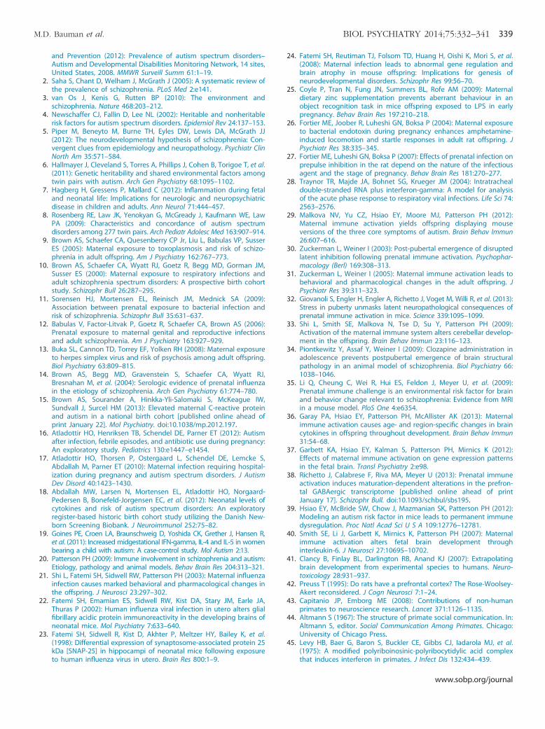
Figure 1. Maternal immune activation (MIA) offspring exhibit abnormalresponses to weaning. Although all animals demonstrate a species-typicalattachment to their own mother, MIA offspring exhibit an unusualresponse in the attachment test. Second trimester MIA (MIA2) offspringproduce significantly more distress or self-soothing behaviors (i.e.,tantrums, convulsive jerk, self-clasp, infant crook tail) than control (CON)offspring. This group difference emerges over the 4 days of testing, withboth MIA groups showing a different pattern over time than controlanimals (p � .001 and p � .003 for the differences in slopes, respectively).Thus, on the final day, MIA2 offspring are highly reactive, control animalsare moderately reactive, and first trimester MIA (MIA1) offspring displaylittle evidence of reactivity (p � .01 for difference from control animals forboth MIA1 and MIA2 groups on day 4).
www.sobp.org/journal
Results
Sickness behavior, temperature and cytokine profiles of thepregnant monkeys confirmed a strong inflammatory response topoly ICLC (Figures S1 and S2 and Tables S4–S7 in Supplement 1).For the sake of brevity, only significant behavioral results from theoffspring are presented in detail. There were no consistentdifferences across offspring in physical growth, motor or reflexdevelopment, adrenal activity, interactions with mothers, ordevelopment of threat detection in the first 6 months of postnatallife (Table S8 in Supplement 1).
Mother PreferenceFollowing weaning at 6 months of age, MIA offspring differed
from control animals during a test designed to evaluate infantattachment to the mother. While all animals, irrespective oftreatment condition, demonstrated a species-typical attachmentto their own mother, we detected differences in the patterns ofthe animals’ responses to the test. Offspring in the MIA2 treat-ment group produced significantly more distress/self-soothingbehaviors that are commonly observed during the attachmentassay (i.e., tantrums, convulsive jerk, self-clasp, infant crook tail)than MIA1 or control offspring. Group differences were notapparent on the first day but emerged over the 4 consecutivedays of testing (Figure 1; Table 2; Figure S9 in Supplement 1). Onthe final day, MIA2 treatment offspring were highly reactive andcontrol offspring were moderately reactive, while MIA1 treatmentoffspring displayed almost no evidence of reactivity.
Solo ObservationsAt 10 months of age, we conducted postweaning solo
observations of the animals alone in a large cage to screen forabnormal motor stereotypic and/or self-directed behaviors that arecommon to captive rhesus monkeys (see Table S1 in Supplement 1for definitions) (53). Compared with control animals, the MIA2
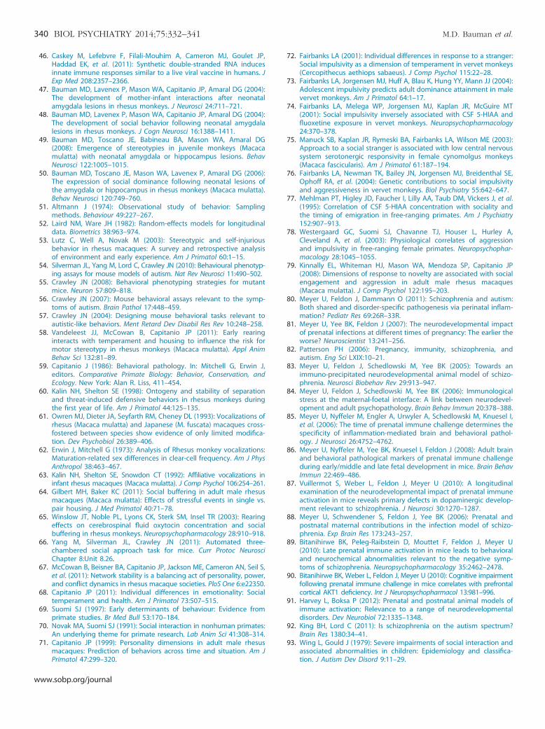
animals produced motor stereotypic and/or self-directed behaviorsmore frequently than control animals (p ¼ .002) (Figure 2A;Table 3). First trimester MIA animals displayed a trend levelincrease in these behaviors compared with control animals (p ¼.06). We also detected trend level differences in the frequency ofaffiliative contact “coo” calls produced by the MIA1 offspring when

MIA1
MIA2
CON
Juvenile Y‐Maze0
1
2
3
4
5
6
nim2 rep sroivaheb evititepe
R
Postweaning Solo0
2
4
6
8
10
12
14
16
nim5 rep sroivaheb evititepe
R
Juvenile Solo0
2
4
6
8
10
12
14
16
nim5 rep sroivaheb evititepe
R
0
5
10
15
20
25
30
35
40
45
Juvenile Solo Juvenile Social Approach
nim5 rep sllac oo
C
0
5
10
15
20
25
30
35
nim01 rep sllac oo
C
0
5
10
15
20
nim01 rep detaitini sroivahe
B
Approach Frequency Proximity Frequency
nim01 rep )s( se
miT
0
50
100
150
200
250
300
Duration of Proximity/Contact with Novel Animal
Interactions with unfamiliar peers during juvenile social approach
Figure 2. (A) Maternal immune activation (MIA) off-spring exhibit increased frequency of motor stereotypiesand self-directed behaviors. Left panel: When observedalone in a large cage at 10 months of age, secondtrimester MIA (MIA2) animals produce significantly morerepetitive behaviors than control animals (CON) (**p #.01). The first trimester MIA (MIA1) offspring also producemore repetitive behaviors than control animals, but thisdifference does not reach statistical significance at 10months (p ¼ .06). Middle panel: When observed alone at22 months of age, MIA1 offspring produce significantlymore repetitive behaviors (*p # .05). Second trimesterMIA animals also produce significantly more repetitivebehaviors than control animals at 22 months (**p # .01).Right panel: When tested at 17 months of age in the Y-maze social preference assay, MIA2 treatment animalsproduce significantly more repetitive behaviors thancontrol animals (**p # .01). (B) Maternal immuneactivation offspring display decreased affiliative vocaliza-tions. Left panel: At 22 months, MIA2 offspring producesignificantly fewer coo calls than control animals (**p �.01). Right panel: When observed with a novel conspe-cific at 24 months of age, MIA1 offspring producesignificantly fewer coo calls than control animals (*p #.05). (C) Maternal immune activation offspring exhibitinappropriate interactions with unfamiliar conspecifics.Left panel: First trimester MIA offspring demonstrateinappropriate social interactions with an unfamiliaranimal, as indexed by high frequency of approaching(*p �0.05) and more frequently moving within arm’sreach of the unfamiliar animal (**p � .01). Right panel:First trimester MIA offspring remained near the unfami-liar animal, as indexed by the duration of time spent inphysical contact or within arm’s reach of the unfamiliaranimal (*p � .05).
M.D. Bauman et al. BIOL PSYCHIATRY 2014;75:332–341 335
observed alone in the large cage (p ¼ .08). Juvenile soloobservations were repeated at 22 months of age. Both MIA groupsproduced significantly more motor stereotypic and/or self-directedbehaviors than control animals (p ¼ .03, .01, respectively)(Figure 2A; Table 3). As observed in the postweaning period,MIA1 offspring produced fewer affiliative contact coo calls thancontrol animals, although the difference remained at trend level. Atthis later time point, however, MIA2 offspring produced signifi-cantly fewer coo calls than control animals (Figure 2B, Table 3).
Interaction with Novel Conspecifics (Y-Maze)At 17 months of age, we conducted an exploratory assay
designed to evaluate social interactions with an unfamiliar con-specific, using a Y-shaped testing chamber in which the exper-imental animal had access to two chutes. A novel stimulus animalwas housed in a holding cage at the end of one chute; the otherarm led to an empty cage. While there were no differences in theamount of time spent in the social versus nonsocial arms of the cage(Table 4) and there were few interactions with the novel animal, wedid detect differences in coo vocalization and repetitive behaviors(Table 4). While there were no differences in the total number of coo
vocalizations, the MIA1 offspring exhibited a trend level difference inthe frequency of affiliative contact coo calls produced when alone inthe nonsocial arm of the Y-maze (p ¼ .06). Paralleling the resultsfrom postweaning and juvenile experiments, the MIA2 offspringproduced significantly more motor stereotypic and/or self-directedbehaviors than control animals (p ¼ .002; Figure 2B).
Interaction with Novel Conspecifics (Two-Chamber SocialApproach)
This test was modeled after the sensitive assay of sociabilityused for mouse models of ASD (54–57). All subjects, irrespectiveof experimental condition, spent significantly more time in thesocial chamber than in the nonsocial chamber (Table 5). The MIA1
offspring, however, differed from control animals in severalbehavioral measures. They produced fewer total affiliative contactcoo calls (Figure 2B, Table 5), and they approached the stimuluscage more frequently than control animals and initiated proximitywith the unfamiliar animal more than twice as frequently as thecontrol animals (Figure 2C, Table 5). Differences were alsodetected in the amount of time spent in contact or proximity(i.e., within arm’s reach) of the stimulus cages within the
www.sobp.org/journal

Table 3. Behaviors During Postweaning and Juvenile Solo Observations
Average Group Frequency Difference from Control Group
MIA1 MIA2 Control Group MIA1 vs. Control Group MIA2 vs. Control Group
Behavior Mean (SD) Mean (SD) Mean (SD) Estimate (SE) p Value Estimate (SE) p Value
PostweaningCoo 27.5 (8.0) 34.6 (9.5) 38.5 (10.9) �10.2 (5.5) .08 �3.5 (5.0) .48Stereotypya 5.8 (8.3) 9.1 (7.6) .5 (.7) 1.4 (.7) .06 2.2 (.6) .002
JuvenileCoo 24.7 (12.1) 21.5 (6.9) 36.1 (9.6) �8.5 (4.8) .09 �14.8 (4.4) .003Stereotypya 10.5 (11.4) 9.5 (8.9) 1.8 (2.3) 1.7 (.7) .03 1.8 (.7) .01
Descriptive statistics and summary (parameter estimates and standard errors) of the mixed-effects models assessing the relationship between groupand frequency of behavior variables. Average group behaviors are based on observed frequency of behaviors. Differences from control groups areestimated from mixed-effects regression models fitted to the frequency of behaviors and adjusted for gender.
MIA1, first trimester maternal immune activation; MIA2, second trimester maternal immune activation.aVariable square-root transformed to improve its normality.
336 BIOL PSYCHIATRY 2014;75:332–341 M.D. Bauman et al.
chambers. Compared with control subjects, both MIA groupsspent more time near the small empty cage in the nonsocialchamber. However, only MIA1 offspring spent more time near thesmall cage containing an unfamiliar conspecific in the socialchamber (Figure 2C). There were no differences in the frequencyof entering or exiting the social and nonsocial chambers or in thefrequency of approaching the empty stimulus cage, suggestingthat the differences in approach frequency were specific to thesocial stimulus and not reflective of global changes in activity.
Discussion
Rhesus monkey offspring exposed to MIA in utero differ fromcontrol offspring in measures of repetitive behaviors, vocalcommunication, and social interactions. These alterations inbehavior overlap with the core diagnostic domains of ASD, andthe latter behaviors may also be relevant for SZ. The developmentof some abnormal behaviors (increased reactivity in MIA2 off-spring and abnormal social behavior in MIA1 offspring) dependson the specific period of MIA exposure during pregnancy, whileother abnormal behaviors (decreased affiliative vocalizations andincreased repetitive behaviors) are present in both MIA groups(Figure S3 in Supplement 1).
Table 4. Duration and Frequency of Behaviors in Juvenile Y‐Maze Paradigm
Average Group Duration
MIA1 MIA2 Control GrouBehavior Mean (SD) Mean (SD) Mean (SD)
Startboxa 19.7 (11.7) 21.7 (12.2) 17.1 (9.7)Social Arma 51.1 (15.4) 50.7 (10.4) 48.5 (21.0)Nonsocial Arm 49.2 (19.7) 47.6 (8.7) 54.4 (24.5)
Average Group FrequencyCoo Aloneb,c 2.3 (3.0) 3.7 (2.9) 4.9 (2.0)Coo to Novelc Animal2 2.1 (1.7) 2.3 (1.4) 3.0 (2.2)Total Coob 4.4 (4.5) 6.0 (4.2) 7.9 (3.3)Stereotypiesc 2.5 (4.2) 4.2 (2.9) .7 (.6)
Average group behaviors are based on observed duration or frequencyestimated from mixed-effects regression models fitted to the duration or fresummary (parameter estimates and standard errors) of the mixed-effects modebehavioral variables.
MIA1, first trimester maternal immune activation; MIA2, second trimester maAnalyses for these duration variables further adjusted for the day of thebAnalyses for these frequency variables further adjusted for the gender ofcFrequency variables square-root transformed to improve their normality.
www.sobp.org/journal
While the majority of rodent MIA models have reportedbehavioral abnormalities in adult offspring, here we describe theemergence of behavior over the first 2 years of life in a nonhumanprimate model. This period for rhesus monkeys is roughlyequivalent to early childhood in humans. Although group differ-ences were not consistently detected at the early time points, by 2years of age the MIA monkey offspring began to demonstrateconsistent patterns of behavioral changes. The first indication ofdifferences between the experimental groups occurred immedi-ately after weaning, at 6 months of age, during an assessment ofemotional attachment to the mother. While all animals, irrespec-tive of treatment condition, demonstrated a species-typical attach-ment to their own mother, the MIA animals’ responses to the testwere different from control animals. The MIA2 offspring displayeda dramatic increase in distress/self-soothing behaviors over the 4-day testing period that was not observed in the control animals. Incontrast, the MIA1 offspring produced almost none of thesebehaviors. Differences in the animals’ responses to the test weremost pronounced on the fourth consecutive day of testing,suggesting that this particular repeated assay can reveal changesin distress/self-soothing behaviors that are not detected in otherparadigms. While we do not know why the MIA2 offspringresponded with increased distress/self-soothing behaviors, mouse
Difference from Control Group
p MIA1 vs. Control Group MIA2 vs. Control Group
Estimate (SE) p Value Estimate (SE) p Value
5.4 (5.9) .37 5.8 (5.3) .292.7 (9.7) .78 2.3 (8.7) .80
�8.1 (11.1) .48 �8.1 (10.0) .43Difference from Control Group
�.8 (.4) .06 .6 (.4) .14�.2 (.4) .53 �.3 (.3) .45
�2.3 (1.9) .24 �2.0 (1.8) .28.6 (.4) .14 1.3 (.4) .002
of behaviors over 2-minute trials. Differences from control groups arequency of behaviors and adjusted for gender. Descriptive statistics andls assessing the relationship between group and duration and frequency of
aternal immune activation.trial and gender of the stimulus monkey.the stimulus monkey.

Table 5. Duration and Frequency of Behaviors in Juvenile Social Approach Paradigm
Average Group Duration Difference from Control Group
MIA1 MIA2 Control Group MIA1 vs. Control Group MIA2 vs. Control Group
Behavior Mean (SD) Mean (SD) Mean (SD) Estimate (SE) p Value Estimate (SE) p Value
Proximity/Contact to Empty Cagea 67.7 (29.1) 71.1 (42.8) 39.1 (24.3) 2.9 (1.2) .02 2.6 (1.0) .02Proximity/Contact to Subject Cage 207.7 (23.9) 101.0 (47.1) 109.9 (85.9) 96.3 (36.8) .02 �9.6 (33.0) .77Social Chamber 427.2 (48.9) 379.8 (75.3) 425.2 (75.0) .9 (39.2) .98 �45.9 (35.2) .21Nonsocial Chamber 172.8 (48.9) 220.2 (75.3) 174.8 (75.0) �.9 (39.2) .98 45.9 (35.2) .21
Average Group Frequency Difference from Control GroupCoo 9.3 (11.4) 22.4 (12.9) 27.4 (12.0) �15.9 (6.7) .03 �4.0 (6.0) .51Approach 15.5 (3.4) 10.1 (5.3) 9.3 (3.6) 4.9 (2.2) .04 .2 (2.0) .91Contact 14.7 (3.4) 9.5 (6.1) 8.8 (4.5) 4.6 (2.6) .09 .1 (2.3) .95Proximity 7.4 (2.1) 2.3 (2.0) 2.0 (1.3) 5.1 (.9) �.001 .2 (.8) .84
Average group behaviors are based on observed duration or frequency of behaviors over 10-minute trials. Differences from control groups areestimated from mixed-effects regression models fitted to the duration of behaviors and adjusted for gender. Descriptive statistics and summary(parameter estimates and standard errors) of the mixed-effects models assessing the relationship between group and duration or frequency of behaviorvariables.
MIA1, first trimester maternal immune activation; MIA2, second trimester maternal immune activation.aVariable square-root transformed to improve its normality.
M.D. Bauman et al. BIOL PSYCHIATRY 2014;75:332–341 337
MIA models also exhibit behaviors indicative of heightenedanxiety (i.e., less time in the center of the open field paradigmand reluctance to explore novel objects) that may provide insightinto this atypical response in the monkey (21).
Additional behavioral changes in monkey MIA offspring beganto emerge during the postweaning (6–12 months) and juvenile(12–24 months) periods. It is important to note that these earlychanges in behavior were subtle, as there were no group differ-ences detected in daily home cage observations or in weeklyobservations of the animals interacting with familiar peers. How-ever, when the MIA animals were removed from these familiarenvironments and observed alone, they consistently producedmore motor stereotypic and/or self-directed behaviors than controlanimals. These behavioral pathologies were most pronounced inthe MIA2 group, as indexed by a high frequency in three differenttesting paradigms. Animals in the MIA1 group also appeared toproduce more repetitive behaviors than control animals, althoughthese differences did not attain statistical significance until theanimals reached 2 years of age. It is well established that restrictedrearing environments, small cage size, and stress-inducing eventscan trigger stereotypies in laboratory animals (53,58,59) and wedesigned our protocols to minimize these factors. The fact that thecontrol animals exhibited a low frequency of motor stereotypicand/or self-directed behaviors indicates that we can reasonablyattribute these behaviors to MIA, rather than general socioenvir-onmental restrictions. The results from this nonhuman primatemodel parallel findings of increased repetitive and compulsivebehaviors of mouse MIA offspring that exhibit high levels ofrepetitive behaviors in marble burying and self-grooming tests (29).
When the animals were removed from their home cageswhere they had constant visual access to familiar animals, we alsocollected data on any social signals, including vocalizations, thatwere produced. During these temporary separations, youngmonkeys often produced affiliative coo calls that are thought toserve the function of reestablishing contact with conspecifics (60–63). Compared with control offspring, both groups of MIAoffspring produced fewer coo calls, although only the MIA2 groupdiffered significantly from control animals under these conditions.Interestingly, the MIA1 offspring continued to exhibit reduced coocalling when removed from their home cage and introduced toan unfamiliar peer, suggesting that the presence of an unfamiliar
animal may differentially impact social buffering for the MIAgroups (64,65). The reduced affiliative vocalizations observed inmacaque MIA offspring are consistent with data from male MIAmice, which display a reduced number of vocalizations as pupswhen they are isolated from their littermates and mother and asadults in the presence of a female (29).
Given that impaired social functioning is a hallmark feature ofboth ASD and SZ, we would expect a valid animal model to alsoproduce impairments in social processing. While MIA offspringdid not differ from control animals during daily interactions withfamiliar peers, group differences were detected during interac-tions with an unfamiliar social partner, which is considered to bea more challenging social encounter. It is important to point outthat the nature of behavioral perturbations in an animal modelmay be complex and species-specific, especially in challengingsocial interactions. In mice, for example, the default response toan unfamiliar conspecific is to approach and investigate. Thus,decreased time spent investigating a novel animal is taken asevidence of diminished sociability (66) and is a common behav-ioral outcome of MIA mouse models (21,29,40). For rhesusmonkeys, the decision to approach and interact with anotheranimal depends on a number of internal (i.e., individual tempera-ment differences) and external (i.e., characteristics of the unfami-liar animal, presence or absence of kin) factors (67–71). For manyspecies of nonhuman primates, immediately approaching anunfamiliar conspecific or behaving impulsively with familiaranimals is met with negative outcomes and physical aggression(72–79). The default for rhesus monkeys is to approach anunfamiliar conspecific with caution and after considerable evalua-tion at a distance. However, when evaluated with an unfamiliarconspecific at 2 years of age, MIA1 offspring exhibited a cleardeviation from the species-typical social protocol for rhesusmonkeys by frequently approaching, contacting, and staying withinarm’s reach of the unfamiliar animal. Thus, both mouse and monkeyMIA models result in deviation from species-typical social norms.
Behavioral changes in mouse MIA models have been inter-preted as bearing resemblance to features of both ASD and SZ(20,80–82), although the timing of the prenatal challenge likelydetermines the ultimate consequences of MIA exposure (83–90).The 165-day macaque monkey pregnancy provides an opportu-nity to further delineate vulnerable periods of gestation during
www.sobp.org/journal
338 BIOL PSYCHIATRY 2014;75:332–341 M.D. Bauman et al.
which MIA alters specific neural networks and ultimately leads todistinct behavioral trajectories over a relatively protracted periodof postnatal development. Our results indicate that experimen-tally inducing MIA at either late first trimester or late secondtrimester produces offspring with overlapping alterations inrepetitive behaviors and affiliative vocalizations, as well as distinctchanges in reactivity and social interactions. While it is prematureto determine if MIA in the primate model is related specifically toASD or SZ or to more general neurodevelopmental issues (91), wecan begin to evaluate the nature and timing of the behavioraloutcomes of the monkey MIA model.
Stereotypic behaviors, for example, are one of the diagnosticfeatures of ASD and were consistently observed throughoutpostnatal development in the MIA2 offspring and to a lesser extentin the MIA1 offspring. While these behaviors support the facevalidity of the model, it is important to recognize that stereotypiesare observed in a variety of developmental, psychiatric, andneurological disorders and are not specific to ASD. However, bothASD and SZ are characterized by changes in social cognition andemotion (92), which were also altered in the macaque MIAoffspring compared with control animals. While both MIA groupsexhibited decreased frequency of the affiliative contact coo callswhen observed alone, only the MIA1 offspring produced fewercoos in a social context. Likewise, only the MIA1 offspring exhibitedinappropriate social interactions with a novel conspecific. Wesuggest that the inappropriate social approach behaviors observedin the animal model may be reminiscent of the active but oddsubtype of social interaction style described in ASD (93) and thecomplex social functioning impairments in SZ (94). We haveinitiated an eye-tracking study to evaluate social processing inthe monkey model and will utilize these data to further clarify thenature of the social impairments and the relevance to ASD and SZ.
The timing of behavioral alterations is another importantconsideration. Autism spectrum disorder, for example, is diag-nosed in early childhood (95), while the onset of psychoticsymptoms of SZ typically occurs during the transition fromadolescence to adulthood (96). In the present study, we firstdetected group differences in response to weaning at 6 monthsof age, which is roughly equivalent to a 2-year-old child. Whilethis time frame is more consistent with the early symptom onsetof ASD, prospective studies of patients who develop SZ also havesocial and neurocognitive impairments that emerge long beforepsychiatric SZ symptoms (97–100). Observations of macaqueoffspring will continue as they mature, which is needed tointerpret the emergence of symptoms over time, as well as thelong-term effects of MIA in primates and the relevance to humanneurodevelopmental disorders.
While the rhesus monkey provides an animal model that closelyparallels human brain organization and cognitive and socialfunctioning, there are ethical and pragmatic limitations in thedevelopment of a nonhuman primate model. The primary limi-tation of the current study is the sample size. A second limitation isthat we must wait until the conclusion of the behavioral studies(approximately 4 years) before initiating brain pathology studiesthat are often simultaneously carried out in rodent models. Thus,the data presented here describe behavioral outcomes but do notprovide a mechanistic neural basis for the specific abnormalities.Mouse MIA models, however, have identified several plausiblemechanisms by which poly IC-induced immune responses candisrupt fetal brain development (101–104). The maternal cytokineresponse to poly IC, in particular interleukin-6 (40), plays a criticalrole in triggering immune activation and endocrine changes in theplacenta (105) and altered cytokine expression in the fetal brain, as
www.sobp.org/journal
well as long-lasting changes in cytokine expression in the brains ofMIA mouse offspring as they mature (36). In the present study, weutilized a modified form of poly IC (poly ICLC), which stimulatescomparable inflammatory responses in humans and nonhumanprimates (45,46,106). While other nonhuman primate models ofMIA have explored maternal immune challenges in the thirdtrimester (107,108), we focused our efforts on the first and secondtrimesters, as human studies have identified these as the gesta-tional windows of vulnerability for ASD and SZ associated withmaternal immune challenge (109). This time frame of early fetalbrain development captures the peak period of macaque neuro-genesis (110–117). Short et al. (107) report that rhesus offspringborn to mothers exposed to influenza in the early third trimesterdemonstrate reduced gray matter volume throughout the cortexand increased white matter in the parietal cortex at 1 year of age.We predict that MIA exposure in the late first and secondtrimesters also produce changes in brain development of theoffspring. We are currently exploring brain pathology in theseanimals to determine if MIA offspring demonstrate structural orfunctional brain pathologies characteristic of ASD or SZ and willinitiate a comprehensive histological evaluation of the brain at theconclusion of the behavioral studies.
While experimentally inducing MIA in the primate model altersbehavioral development, it is important to emphasize that sick-ness during human pregnancy is not uncommon (118,119), andclearly not all women who experience infection during pregnancyhave children later diagnosed with a neurodevelopmental dis-order (120). A number of factors, including genetic susceptibility,the intensity of the infection, and the maternal and/or fetalresponse, as well as the precise timing of the immune challenge,likely influence the degree to which MIA alters fetal braindevelopment and may ultimately determine which diseasephenotype (ASD or SZ) is expressed. With mounting evidenceof the increased risk of psychiatric disorders in offspring exposedto MIA, increased efforts to understand MIA-induced alterations inbrain development are clearly needed.
This work was supported by a grant from the Simons Foundation(SFARI [9900060] to PHP). Additional support was provided by the baseGrant (RR00169) of the California National Primate Research Center.
Biobehavioral assessments at 3 months of age were carried out byDr. John Capitanio and funded in part by the National Center forResearch Resources (R24RR019970 to JPC and P51RR000169 toCalifornia National Primate Research Center) and are currentlysupported by the Office of Research Infrastructure Programs/Officeof the Director (R24OD010962 and P51OD011157, respectively). SEPS iscurrently affiliated with the Department of Immunology, Mayo Clinic,Rochester, Minnesota. CB is currently affiliated with the Department ofNeurosurgery, University Hospital Basel, Basel, Switzerland.
We acknowledge the Research Services and Primate Medicinestaff at the California National Primate Research Center for care ofthe animals and to J. Buser, G. Moadab, and A. Lopez for behavioraldata collection and assistance with manuscript preparation.
Polyinosinic:polycytidylic acid stabilized with poly-L-lysine waskindly provided by Dr. Andres Salazar, M.D., Oncovir, Washington. DC.
The authors report no biomedical financial interests or potentialconflicts of interest.
Supplementary material cited in this article is available online athttp://dx.doi.org/10.1016/j.biopsych.2013.06.025.
1. Autism and Developmental Disabilities Monitoring Network Surveil-lance Year 2008 Principal Investigators; Centers for Disease Control
M.D. Bauman et al. BIOL PSYCHIATRY 2014;75:332–341 339
and Prevention (2012): Prevalence of autism spectrum disorders–Autism and Developmental Disabilities Monitoring Network, 14 sites,United States, 2008. MMWR Surveill Summ 61:1–19.
2. Saha S, Chant D, Welham J, McGrath J (2005): A systematic review ofthe prevalence of schizophrenia. PLoS Med 2:e141.
3. van Os J, Kenis G, Rutten BP (2010): The environment andschizophrenia. Nature 468:203–212.
4. Newschaffer CJ, Fallin D, Lee NL (2002): Heritable and nonheritablerisk factors for autism spectrum disorders. Epidemiol Rev 24:137–153.
5. Piper M, Beneyto M, Burne TH, Eyles DW, Lewis DA, McGrath JJ(2012): The neurodevelopmental hypothesis of schizophrenia: Con-vergent clues from epidemiology and neuropathology. Psychiatr ClinNorth Am 35:571–584.
6. Hallmayer J, Cleveland S, Torres A, Phillips J, Cohen B, Torigoe T, et al.(2011): Genetic heritability and shared environmental factors amongtwin pairs with autism. Arch Gen Psychiatry 68:1095–1102.
7. Hagberg H, Gressens P, Mallard C (2012): Inflammation during fetaland neonatal life: Implications for neurologic and neuropsychiatricdisease in children and adults. Ann Neurol 71:444–457.
8. Rosenberg RE, Law JK, Yenokyan G, McGready J, Kaufmann WE, LawPA (2009): Characteristics and concordance of autism spectrumdisorders among 277 twin pairs. Arch Pediatr Adolesc Med 163:907–914.
9. Brown AS, Schaefer CA, Quesenberry CP Jr, Liu L, Babulas VP, SusserES (2005): Maternal exposure to toxoplasmosis and risk of schizo-phrenia in adult offspring. Am J Psychiatry 162:767–773.
10. Brown AS, Schaefer CA, Wyatt RJ, Goetz R, Begg MD, Gorman JM,Susser ES (2000): Maternal exposure to respiratory infections andadult schizophrenia spectrum disorders: A prospective birth cohortstudy. Schizophr Bull 26:287–295.
11. Sorensen HJ, Mortensen EL, Reinisch JM, Mednick SA (2009):Association between prenatal exposure to bacterial infection andrisk of schizophrenia. Schizophr Bull 35:631–637.
12. Babulas V, Factor-Litvak P, Goetz R, Schaefer CA, Brown AS (2006):Prenatal exposure to maternal genital and reproductive infectionsand adult schizophrenia. Am J Psychiatry 163:927–929.
13. Buka SL, Cannon TD, Torrey EF, Yolken RH (2008): Maternal exposureto herpes simplex virus and risk of psychosis among adult offspring.Biol Psychiatry 63:809–815.
14. Brown AS, Begg MD, Gravenstein S, Schaefer CA, Wyatt RJ,Bresnahan M, et al. (2004): Serologic evidence of prenatal influenzain the etiology of schizophrenia. Arch Gen Psychiatry 61:774–780.
15. Brown AS, Sourander A, Hinkka-Yli-Salomaki S, McKeague IW,Sundvall J, Surcel HM (2013): Elevated maternal C-reactive proteinand autism in a national birth cohort [published online ahead ofprint January 22]. Mol Psychiatry. doi:10.1038/mp.2012.197.
16. Atladottir HO, Henriksen TB, Schendel DE, Parner ET (2012): Autismafter infection, febrile episodes, and antibiotic use during pregnancy:An exploratory study. Pediatrics 130:e1447–e1454.
17. Atladottir HO, Thorsen P, Ostergaard L, Schendel DE, Lemcke S,Abdallah M, Parner ET (2010): Maternal infection requiring hospital-ization during pregnancy and autism spectrum disorders. J AutismDev Disord 40:1423–1430.
18. Abdallah MW, Larsen N, Mortensen EL, Atladottir HO, Norgaard-Pedersen B, Bonefeld-Jorgensen EC, et al. (2012): Neonatal levels ofcytokines and risk of autism spectrum disorders: An exploratoryregister-based historic birth cohort study utilizing the Danish New-born Screening Biobank. J Neuroimmunol 252:75–82.
19. Goines PE, Croen LA, Braunschweig D, Yoshida CK, Grether J, Hansen R,et al. (2011): Increased midgestational IFN-gamma, IL-4 and IL-5 in womenbearing a child with autism: A case-control study. Mol Autism 2:13.
20. Patterson PH (2009): Immune involvement in schizophrenia and autism:Etiology, pathology and animal models. Behav Brain Res 204:313–321.
21. Shi L, Fatemi SH, Sidwell RW, Patterson PH (2003): Maternal influenzainfection causes marked behavioral and pharmacological changes inthe offspring. J Neurosci 23:297–302.
22. Fatemi SH, Emamian ES, Sidwell RW, Kist DA, Stary JM, Earle JA,Thuras P (2002): Human influenza viral infection in utero alters glialfibrillary acidic protein immunoreactivity in the developing brains ofneonatal mice. Mol Psychiatry 7:633–640.
23. Fatemi SH, Sidwell R, Kist D, Akhter P, Meltzer HY, Bailey K, et al.(1998): Differential expression of synaptosome-associated protein 25kDa [SNAP-25] in hippocampi of neonatal mice following exposureto human influenza virus in utero. Brain Res 800:1–9.
24. Fatemi SH, Reutiman TJ, Folsom TD, Huang H, Oishi K, Mori S, et al.(2008): Maternal infection leads to abnormal gene regulation andbrain atrophy in mouse offspring: Implications for genesis ofneurodevelopmental disorders. Schizophr Res 99:56–70.
25. Coyle P, Tran N, Fung JN, Summers BL, Rofe AM (2009): Maternaldietary zinc supplementation prevents aberrant behaviour in anobject recognition task in mice offspring exposed to LPS in earlypregnancy. Behav Brain Res 197:210–218.
26. Fortier ME, Joober R, Luheshi GN, Boksa P (2004): Maternal exposureto bacterial endotoxin during pregnancy enhances amphetamine-induced locomotion and startle responses in adult rat offspring. JPsychiatr Res 38:335–345.
27. Fortier ME, Luheshi GN, Boksa P (2007): Effects of prenatal infection onprepulse inhibition in the rat depend on the nature of the infectiousagent and the stage of pregnancy. Behav Brain Res 181:270–277.
28. Traynor TR, Majde JA, Bohnet SG, Krueger JM (2004): Intratrachealdouble-stranded RNA plus interferon-gamma: A model for analysisof the acute phase response to respiratory viral infections. Life Sci 74:2563–2576.
29. Malkova NV, Yu CZ, Hsiao EY, Moore MJ, Patterson PH (2012):Maternal immune activation yields offspring displaying mouseversions of the three core symptoms of autism. Brain Behav Immun26:607–616.
30. Zuckerman L, Weiner I (2003): Post-pubertal emergence of disruptedlatent inhibition following prenatal immune activation. Psychophar-macology (Berl) 169:308–313.
31. Zuckerman L, Weiner I (2005): Maternal immune activation leads tobehavioral and pharmacological changes in the adult offspring. JPsychiatr Res 39:311–323.
32. Giovanoli S, Engler H, Engler A, Richetto J, Voget M, Willi R, et al. (2013):Stress in puberty unmasks latent neuropathological consequences ofprenatal immune activation in mice. Science 339:1095–1099.
33. Shi L, Smith SE, Malkova N, Tse D, Su Y, Patterson PH (2009):Activation of the maternal immune system alters cerebellar develop-ment in the offspring. Brain Behav Immun 23:116–123.
34. Piontkewitz Y, Assaf Y, Weiner I (2009): Clozapine administration inadolescence prevents postpubertal emergence of brain structuralpathology in an animal model of schizophrenia. Biol Psychiatry 66:1038–1046.
35. Li Q, Cheung C, Wei R, Hui ES, Feldon J, Meyer U, et al. (2009):Prenatal immune challenge is an environmental risk factor for brainand behavior change relevant to schizophrenia: Evidence from MRIin a mouse model. PloS One 4:e6354.
36. Garay PA, Hsiao EY, Patterson PH, McAllister AK (2013): Maternalimmune activation causes age- and region-specific changes in braincytokines in offspring throughout development. Brain Behav Immun31:54–68.
37. Garbett KA, Hsiao EY, Kalman S, Patterson PH, Mirnics K (2012):Effects of maternal immune activation on gene expression patternsin the fetal brain. Transl Psychiatry 2:e98.
38. Richetto J, Calabrese F, Riva MA, Meyer U (2013): Prenatal immuneactivation induces maturation-dependent alterations in the prefron-tal GABAergic transcriptome [published online ahead of printJanuary 17]. Schizophr Bull. doi:10.1093/schbul/sbs195.
39. Hsiao EY, McBride SW, Chow J, Mazmanian SK, Patterson PH (2012):Modeling an autism risk factor in mice leads to permanent immunedysregulation. Proc Natl Acad Sci U S A 109:12776–12781.
40. Smith SE, Li J, Garbett K, Mirnics K, Patterson PH (2007): Maternalimmune activation alters fetal brain development throughinterleukin-6. J Neurosci 27:10695–10702.
41. Clancy B, Finlay BL, Darlington RB, Anand KJ (2007): Extrapolatingbrain development from experimental species to humans. Neuro-toxicology 28:931–937.
42. Preuss T (1995): Do rats have a prefrontal cortex? The Rose-Woolsey-Akert reconsidered. J Cogn Neurosci 7:1–24.
43. Capitanio JP, Emborg ME (2008): Contributions of non-humanprimates to neuroscience research. Lancet 371:1126–1135.
44. Altmann S (1967): The structure of primate social communication. In:Altmann S, editor. Social Communication Among Primates. Chicago:University of Chicago Press.
45. Levy HB, Baer G, Baron S, Buckler CE, Gibbs CJ, Iadarola MJ, et al.(1975): A modified polyriboinosinic-polyribocytidylic acid complexthat induces interferon in primates. J Infect Dis 132:434–439.
www.sobp.org/journal
340 BIOL PSYCHIATRY 2014;75:332–341 M.D. Bauman et al.
46. Caskey M, Lefebvre F, Filali-Mouhim A, Cameron MJ, Goulet JP,Haddad EK, et al. (2011): Synthetic double-stranded RNA inducesinnate immune responses similar to a live viral vaccine in humans. JExp Med 208:2357–2366.
47. Bauman MD, Lavenex P, Mason WA, Capitanio JP, Amaral DG (2004):The development of mother-infant interactions after neonatalamygdala lesions in rhesus monkeys. J Neurosci 24:711–721.
48. Bauman MD, Lavenex P, Mason WA, Capitanio JP, Amaral DG (2004):The development of social behavior following neonatal amygdalalesions in rhesus monkeys. J Cogn Neurosci 16:1388–1411.
49. Bauman MD, Toscano JE, Babineau BA, Mason WA, Amaral DG(2008): Emergence of stereotypies in juvenile monkeys (Macacamulatta) with neonatal amygdala or hippocampus lesions. BehavNeurosci 122:1005–1015.
50. Bauman MD, Toscano JE, Mason WA, Lavenex P, Amaral DG (2006):The expression of social dominance following neonatal lesions ofthe amygdala or hippocampus in rhesus monkeys (Macaca mulatta).Behav Neurosci 120:749–760.
51. Altmann J (1974): Observational study of behavior: Samplingmethods. Behaviour 49:227–267.
52. Laird NM, Ware JH (1982): Random-effects models for longitudinaldata. Biometrics 38:963–974.
53. Lutz C, Well A, Novak M (2003): Stereotypic and self-injuriousbehavior in rhesus macaques: A survey and retrospective analysisof environment and early experience. Am J Primatol 60:1–15.
54. Silverman JL, Yang M, Lord C, Crawley JN (2010): Behavioural phenotyp-ing assays for mouse models of autism. Nat Rev Neurosci 11:490–502.
55. Crawley JN (2008): Behavioral phenotyping strategies for mutantmice. Neuron 57:809–818.
56. Crawley JN (2007): Mouse behavioral assays relevant to the symp-toms of autism. Brain Pathol 17:448–459.
57. Crawley JN (2004): Designing mouse behavioral tasks relevant toautistic-like behaviors. Ment Retard Dev Disabil Res Rev 10:248–258.
58. Vandeleest JJ, McCowan B, Capitanio JP (2011): Early rearinginteracts with temperament and housing to influence the risk formotor stereotypy in rhesus monkeys (Macaca mulatta). Appl AnimBehav Sci 132:81–89.
59. Capitanio J (1986): Behavioral pathology. In: Mitchell G, Erwin J,editors. Comparative Primate Biology: Behavior, Conservation, andEcology. New York: Alan R. Liss, 411–454.
60. Kalin NH, Shelton SE (1998): Ontogeny and stability of separationand threat-induced defensive behaviors in rhesus monkeys duringthe first year of life. Am J Primatol 44:125–135.
61. Owren MJ, Dieter JA, Seyfarth RM, Cheney DL (1993): Vocalizations ofrhesus (Macaca mulatta) and Japanese (M. fuscata) macaques cross-fostered between species show evidence of only limited modifica-tion. Dev Psychobiol 26:389–406.
62. Erwin J, Mitchell G (1973): Analysis of Rhesus monkey vocalizations:Maturation-related sex differences in clear-cell frequency. Am J PhysAnthropol 38:463–467.
63. Kalin NH, Shelton SE, Snowdon CT (1992): Affiliative vocalizations ininfant rhesus macaques (Macaca mulatta). J Comp Psychol 106:254–261.
64. Gilbert MH, Baker KC (2011): Social buffering in adult male rhesusmacaques (Macaca mulatta): Effects of stressful events in single vs.pair housing. J Med Primatol 40:71–78.
65. Winslow JT, Noble PL, Lyons CK, Sterk SM, Insel TR (2003): Rearingeffects on cerebrospinal fluid oxytocin concentration and socialbuffering in rhesus monkeys. Neuropsychopharmacology 28:910–918.
66. Yang M, Silverman JL, Crawley JN (2011): Automated three-chambered social approach task for mice. Curr Protoc NeurosciChapter 8:Unit 8.26.
67. McCowan B, Beisner BA, Capitanio JP, Jackson ME, Cameron AN, Seil S,et al. (2011): Network stability is a balancing act of personality, power,and conflict dynamics in rhesus macaque societies. PloS One 6:e22350.
68. Capitanio JP (2011): Individual differences in emotionality: Socialtemperament and health. Am J Primatol 73:507–515.
69. Suomi SJ (1997): Early determinants of behaviour: Evidence fromprimate studies. Br Med Bull 53:170–184.
70. Novak MA, Suomi SJ (1991): Social interaction in nonhuman primates:An underlying theme for primate research. Lab Anim Sci 41:308–314.
71. Capitanio JP (1999): Personality dimensions in adult male rhesusmacaques: Prediction of behaviors across time and situation. Am JPrimatol 47:299–320.
www.sobp.org/journal
72. Fairbanks LA (2001): Individual differences in response to a stranger:Social impulsivity as a dimension of temperament in vervet monkeys(Cercopithecus aethiops sabaeus). J Comp Psychol 115:22–28.
73. Fairbanks LA, Jorgensen MJ, Huff A, Blau K, Hung YY, Mann JJ (2004):Adolescent impulsivity predicts adult dominance attainment in malevervet monkeys. Am J Primatol 64:1–17.
74. Fairbanks LA, Melega WP, Jorgensen MJ, Kaplan JR, McGuire MT(2001): Social impulsivity inversely associated with CSF 5-HIAA andfluoxetine exposure in vervet monkeys. Neuropsychopharmacology24:370–378.
75. Manuck SB, Kaplan JR, Rymeski BA, Fairbanks LA, Wilson ME (2003):Approach to a social stranger is associated with low central nervoussystem serotonergic responsivity in female cynomolgus monkeys(Macaca fascicularis). Am J Primatol 61:187–194.
76. Fairbanks LA, Newman TK, Bailey JN, Jorgensen MJ, Breidenthal SE,Ophoff RA, et al. (2004): Genetic contributions to social impulsivityand aggressiveness in vervet monkeys. Biol Psychiatry 55:642–647.
77. Mehlman PT, Higley JD, Faucher I, Lilly AA, Taub DM, Vickers J, et al.(1995): Correlation of CSF 5-HIAA concentration with sociality andthe timing of emigration in free-ranging primates. Am J Psychiatry152:907–913.
78. Westergaard GC, Suomi SJ, Chavanne TJ, Houser L, Hurley A,Cleveland A, et al. (2003): Physiological correlates of aggressionand impulsivity in free-ranging female primates. Neuropsychophar-macology 28:1045–1055.
79. Kinnally EL, Whiteman HJ, Mason WA, Mendoza SP, Capitanio JP(2008): Dimensions of response to novelty are associated with socialengagement and aggression in adult male rhesus macaques(Macaca mulatta). J Comp Psychol 122:195–203.
80. Meyer U, Feldon J, Dammann O (2011): Schizophrenia and autism:Both shared and disorder-specific pathogenesis via perinatal inflam-mation? Pediatr Res 69:26R–33R.
81. Meyer U, Yee BK, Feldon J (2007): The neurodevelopmental impactof prenatal infections at different times of pregnancy: The earlier theworse? Neuroscientist 13:241–256.
82. Patterson PH (2006): Pregnancy, immunity, schizophrenia, andautism. Eng Sci LXIX:10–21.
83. Meyer U, Feldon J, Schedlowski M, Yee BK (2005): Towards animmuno-precipitated neurodevelopmental animal model of schizo-phrenia. Neurosci Biobehav Rev 29:913–947.
84. Meyer U, Feldon J, Schedlowski M, Yee BK (2006): Immunologicalstress at the maternal-foetal interface: A link between neurodevel-opment and adult psychopathology. Brain Behav Immun 20:378–388.
85. Meyer U, Nyffeler M, Engler A, Urwyler A, Schedlowski M, Knuesel I,et al. (2006): The time of prenatal immune challenge determines thespecificity of inflammation-mediated brain and behavioral pathol-ogy. J Neurosci 26:4752–4762.
86. Meyer U, Nyffeler M, Yee BK, Knuesel I, Feldon J (2008): Adult brainand behavioral pathological markers of prenatal immune challengeduring early/middle and late fetal development in mice. Brain BehavImmun 22:469–486.
87. Vuillermot S, Weber L, Feldon J, Meyer U (2010): A longitudinalexamination of the neurodevelopmental impact of prenatal immuneactivation in mice reveals primary defects in dopaminergic develop-ment relevant to schizophrenia. J Neurosci 30:1270–1287.
88. Meyer U, Schwendener S, Feldon J, Yee BK (2006): Prenatal andpostnatal maternal contributions in the infection model of schizo-phrenia. Exp Brain Res 173:243–257.
89. Bitanihirwe BK, Peleg-Raibstein D, Mouttet F, Feldon J, Meyer U(2010): Late prenatal immune activation in mice leads to behavioraland neurochemical abnormalities relevant to the negative symp-toms of schizophrenia. Neuropsychopharmacology 35:2462–2478.
90. Bitanihirwe BK, Weber L, Feldon J, Meyer U (2010): Cognitive impairmentfollowing prenatal immune challenge in mice correlates with prefrontalcortical AKT1 deficiency. Int J Neuropsychopharmacol 13:981–996.
91. Harvey L, Boksa P (2012): Prenatal and postnatal animal models ofimmune activation: Relevance to a range of neurodevelopmentaldisorders. Dev Neurobiol 72:1335–1348.
92. King BH, Lord C (2011): Is schizophrenia on the autism spectrum?Brain Res 1380:34–41.
93. Wing L, Gould J (1979): Severe impairments of social interaction andassociated abnormalities in children: Epidemiology and classifica-tion. J Autism Dev Disord 9:11–29.

M.D. Bauman et al. BIOL PSYCHIATRY 2014;75:332–341 341
94. Horan WP, Green MF, DeGroot M, Fiske A, Hellemann G, Kee K, et al.(2012): Social cognition in schizophrenia, Part 2: 12-month stabilityand prediction of functional outcome in first-episode patients.Schizophr Bull 38:865–872.
95. Mandell DS, Novak MM, Zubritsky CD (2005): Factors associated withage of diagnosis among children with autism spectrum disorders.Pediatrics 116:1480–1486.
96. Ziermans TB, Schothorst PF, Sprong M, van Engeland H (2011):Transition and remission in adolescents at ultra-high risk forpsychosis. Schizophr Res 126:58–64.
97. Bachman P, Niendam TA, Jalbrzikowski M, Park CY, Daley M, CannonTD, Bearden CE (2012): Processing speed and neurodevelopment inadolescent-onset psychosis: Cognitive slowing predicts social func-tion. J Abnorm Child Psychol 40:645–654.
98. Niendam TA, Jalbrzikowski M, Bearden CE (2009): Exploring predic-tors of outcome in the psychosis prodrome: Implications for earlyidentification and intervention. Neuropsychol Rev 19:280–293.
99. Cornblatt BA, Auther AM, Niendam T, Smith CW, Zinberg J, BeardenCE, Cannon TD (2007): Preliminary findings for two new measures ofsocial and role functioning in the prodromal phase of schizophrenia.Schizophr Bull 33:688–702.
100. Niendam TA, Bearden CE, Rosso IM, Sanchez LE, Hadley T, Nuech-terlein KH, Cannon TD (2003): A prospective study of childhoodneurocognitive functioning in schizophrenic patients and theirsiblings. Am J Psychiatry 160:2060–2062.
101. Meyer U, Feldon J (2009): Prenatal exposure to infection: A primarymechanism for abnormal dopaminergic development in schizophre-nia. Psychopharmacology (Berl) 206:587–602.
102. Meyer U, Feldon J (2010): Epidemiology-driven neurodevelop-mental animal models of schizophrenia. Prog Neurobiol 90:285–326.
103. Patterson PH (2011): Maternal infection and immune involvement inautism. Trends Mol Med 17:389–394.
104. Patterson PH (2012): Maternal infection and autism. Brain BehavImmun 26:393.
105. Hsiao EY, Patterson PH (2011): Activation of the maternal immunesystem induces endocrine changes in the placenta via IL-6. BrainBehav Immun 25:604–615.
106. Packard AE, Hedges JC, Bahjat FR, Stevens SL, Conlin MJ, Salazar AM,Stenzel-Poore MP (2012): Poly-IC preconditioning protects against
cerebral and renal ischemia-reperfusion injury. J Cereb Blood FlowMetab 32:242–247.
107. Short SJ, Lubach GR, Karasin AI, Olsen CW, Styner M, Knickmeyer RC,et al. (2010): Maternal influenza infection during pregnancy impactspostnatal brain development in the rhesus monkey. Biol Psychiatry67:965–973.
108. Willette AA, Lubach GR, Knickmeyer RC, Short SJ, Styner M, GilmoreJH, Coe CL (2011): Brain enlargement and increased behavioral andcytokine reactivity in infant monkeys following acute prenatalendotoxemia. Behav Brain Res 219:108–115.
109. Brown AS (2012): Epidemiologic studies of exposure to prenatalinfection and risk of schizophrenia and autism. Dev Neurobiol 72:1272–1276.
110. Rakic P (1974): Neurons in rhesus monkey visual cortex: Systematicrelation between time of origin and eventual disposition. Science183:425–427.
111. Rakic P (1975): Timing of major ontogenetic events in the visualcortex of the rhesus monkey. UCLA Forum Med Sci 18:3–40.
112. Rakic P, Nowakowski RS (1981): The time of origin of neurons in thehippocampal region of the rhesus monkey. J Comp Neurol 196:99–128.
113. van Eerdenburg FJ, Rakic P (1994): Early neurogenesis in the anteriorhypothalamus of the rhesus monkey. Brain Res Dev Brain Res 79:290–296.
114. Zecevic N, Rakic P (2001): Development of layer I neurons in theprimate cerebral cortex. J Neurosci 21:5607–5619.
115. Kordower JH, Piecinski P, Rakic P (1992): Neurogenesis of theamygdaloid nuclear complex in the rhesus monkey. Brain Res DevBrain Res 68:9–15.
116. Zecevic N, Rakic P (1991): Synaptogenesis in monkey somatosensorycortex. Cereb Cortex 1:510–523.
117. Smart IH, Dehay C, Giroud P, Berland M, Kennedy H (2002): Uniquemorphological features of the proliferative zones and postmitoticcompartments of the neural epithelium giving rise to striate andextrastriate cortex in the monkey. Cereb Cortex 12:37–53.
118. Laibl VR, Sheffield JS (2005): Influenza and pneumonia in pregnancy.Clin Perinatol 32:727–738.
119. Longman RE, Johnson TR (2007): Viral respiratory disease in preg-nancy. Curr Opin Obstet Gynecol 19:120–125.
120. Selten JP, Frissen A, Lensvelt-Mulders G, Morgan VA (2010): Schizo-phrenia and 1957 pandemic of influenza: Meta-analysis. SchizophrBull 36:219–228.
www.sobp.org/journal