abstracts of the international congress of future medical
TRANSCRIPT

International Congress of Future Medical Pioneers - ICOFMEP - 2021, May 8th-9th, 2021, Virtual Event
Abstracts of the International Congress of Future Medical Pioneers 2021
ICOFMEP 2021
8-9 May 2021, Virtual Event www.futuremedicalpioneers.org
Scientific Programme Committee Chair(s):
Professor Bahar Uslu, PhD Quinnipiac University Frank H. Netter School of
Medicine
Hamden, CT, US
Assoc. Prof. Ahmet Şen Health Sciences University, Samsun Training and
Research Hospital
Samsun, Turkey
Organizational Board
Professor Bahar Uslu, PhD Quinnipiac University Frank H. Netter School of
Medicine
Hamden, CT, US
Assoc. Prof. Ahmet Şen Health Sciences University, Samsun Training and
Research Hospital
Samsun, Turkey
Assoc. Prof. Enes Emre
Başar
Anadolu University, Faculty of Business
Administration
Eskişehir, Turkey
Kenan Demir, MD Health Sciences University, Samsun Training and
Research Hospital
Samsun, Turkey
Özlem Sezer, MD Health Sciences University, Samsun Training and
Research Hospital
Samsun, Turkey
Abstract Review Panel
Each abstract was scored double blind review.
Fevzi Toraman Fatih Kılıçbay Gökşen Derya Reis Köse
Handan Çelik Murat Serkant Ünal Gülsemin Çiçek
İmer Okar Sude Hatun Aktimur Kenan Demir
Mehmet Bilge Çetinkaya Aykut Öztürk Mehmet Cenk Turgut
Ünal Uslu Bahadır Yazıcıoğlu Onur Taşcı
Başak Büyük Betül Sarı Özlem Sezer
Başar Erdivanlı Büşra Akpınar Saim Gubari
Deniz Aka Satar Canan Soyer Çalışkan Samettin Çelik
Mahcube Çubukçu Eda Türe Selim Görgün
Özgür Günal Elif Hilal Ünverdi Şadiye Açıkgöz
Pınar Tosun Taşar Emine Aksoy Yasemin Nasır
Aysın Türkmen Emine Tural Murat Kubat
Emine Özçınar Göksenin Ünlügüzel Üstün

I C O F M E P 2 0 2 1
International Congress of Future Medical Pioneers - ICOFMEP - 2021, May 8th-9th, 2021, Virtual Event II
Table of Contents
Lockdown effect on emergency surgical consults during the covid-19 outbreak: Experience of a
pandemic hospital
Ahmet Burak Ciftçi and Sönmez Ocak ............................................................................................... 1
Fusion Planet: Past, Today and Future of Computer Scientific Solutions in Digital Pathology
Taha Yiğit Alkan, Hüseyin Gökhan Akçay and Havva Serap Toru .................................................... 2
14q32.31q32.33 Microdeletion Detected by Chromosomal Microarray in a Child with Dysmorphism
and Hypotonia
Aslıhan Sanrı and Özlem Sezer ........................................................................................................... 4
Factors Associated with Pressure Ulcers in Adult Critical Care Patients
Başar Erdivanlı ..................................................................................................................................... 5
COVID-19 Through the Eyes of Physiatrists: Back Pain Can be a Symptom and May Predict
Pneumonia in COVID-19
Cuma Uz .............................................................................................................................................. 6
The Effect of Ulipristal Acetate and Vitamin D3 on Folliculogenesis
Damla Gül Fındık, Gülnur Take Kaplanoğlu, Gökçe Nur Arık and Nagva B. Abubaker Alemari ..... 7
Evaluation of Antibody Levels in Pcr Negative Covid-19 Suspected Case Series
Demet Gür Vural, Büşra Usta, Yeliz Tanrıverdi Çaycı, Kemal Bilgin and Asuman Birinci ............. 8
Evaluation of Antinuclear Antibodies in Patients who were Infected with Covid 19
Demet Gür Vural .................................................................................................................................. 9
Depression, Hopelessness, and Social Support in Hemodialysis Atients
Demet Yavuz...................................................................................................................................... 11
Effects of thymoquinone on anxiety-related behavior and auditory potentials in rats exposed to
glucocorticoid
Deniz Kantar ...................................................................................................................................... 12
Spontaneous Tumor Lysis Syndrome in Lung Cancer: Very Rare Case for Solid Tumors
Buğra Özel, Düriye Sıla Karagöz Özen and Ahmet Baran ................................................................ 13
Is Selenium deficiency related to an increased risk for diabetic retinopathy?
Hacer Pınar Öztürk Kurt, Düriye Sıla Karagöz Özen, İpek Genç, Mukadder Erdem and Mehmet
Derya Demirağ ................................................................................................................................... 14
The Development of Family Medicine Identity Scale
Duygu Üstünol, İsmail Kasım and Adem Özkara.............................................................................. 15
Investigation of the relationship between magnesium level and vitamin D, bone mineral density, knee
osteoarthritis and chronic diseases
Ebru Yılmaz and Sena Ünver............................................................................................................. 16
Outcomes of Breast Cancer Brain Metastasis Patients Undergoing Stereotactic Radiotherapy
Ela Delikgöz Soykut and Nilgün Şahin ............................................................................................ 17
Epithelial-mesenchymal transition and Cancer
Elif Önder ........................................................................................................................................... 19

I C O F M E P 2 0 2 1
International Congress of Future Medical Pioneers - ICOFMEP - 2021, May 8th-9th, 2021, Virtual Event III
Ethical Aspects of Embryo Research
E. Elif Vatanoğlu-Lutz ....................................................................................................................... 20
From Tuhfetü'l Tıb* to the Medicine of the Future
Eser Epözdemir, E. Elif Vatanoğlu-Lutz and Bahar Uslu ................................................................ 21
Medically Assisted Reproduction in Turkish Law
Emel Badur ........................................................................................................................................ 22
The Cytotoxicity Effects of Metformin and Lithium Substances in the Human Intestinal Caco-2 Cells
Emine Tural ....................................................................................................................................... 23
A Comparison of Propofol with Ketofol For Sedation Quality and Side Effects in Patients Undergoing
Colonoscopy
Ender Çam, Deniz Karakaya and Sibel Barış ................................................................................... 24
International scientific collaborative activities: Barriers and opportunities ...................................... 25
Şule Başar and Enes Emre Başar ...................................................................................................... 25
Clinical outcomes of intraarticular PRP and corticosteroid combination in advanced osteoarthritis
Erhan Okay ........................................................................................................................................ 27
A COVID-19 case complicated by ecchymosis and cyanosis
Esmeray Mutlu Yılmaz ...................................................................................................................... 28
The frequency of micronodular type neuroendocrine cell hyperplasia detected in gastric endoscopic
biopsies and its relationship with clinicopathological parameters
Oğuzhan Okcu, Ezgi Hacıhasanoğlu and Bayram Şen ...................................................................... 29
Renal Tissue Morphology and Morphometric Alterations with Different Fixatives (Formalin,
Bouin’s, Alfac and B5 Solutions)
Fatma Mert, Bilge Serdaroğlu, İbrahim Alptekin , Ferda Topal Çelikkan and Oya Evirgen ............ 30
New challenge in adolescents: ‘at risk polycystic ovary syndrome’ estimation
Fatma Nurgül Taşgöz ......................................................................................................................... 31
Determination of Cell Viability in LNCaP Prostate Cancer Cells Treated with Boric Acid
Gizem Kabasakal and Mücahit Seçme............................................................................................... 33
Comparison of Insulin Treatment with Mesenchymal Stem Cell Treatment in Experimental Type 1
Diabetes-Induced Rats
Gökçen Gökçe, Murat Tosun, Hasan Hüseyin Demirel and Esra Aslan .......................................... 34
Oxidative Effects and Biochemical Markers Related to Disease Severity Parameters in Cystic Fibrosis
Cases
Göksenin Ünlügüzel Üstün ................................................................................................................ 35
Determination of Sleep Quality in Patients with Rotator Cuff Tears
Gonca Sağlam ................................................................................................................................... 37
Determination of the functions of miRNAs associated with PI3K-Akt signaling pathway and p53
signaling pathway in prostate cancer
Gözde Öztan ...................................................................................................................................... 38
I C O F M E P 2 0 2 1
International Congress of Future Medical Pioneers - ICOFMEP - 2021, May 8th-9th, 2021, Virtual Event IV
Efficacy of Lacosamide Therapy in Focal Onset Refractory Epilepsy of Childhood: A Single Center
Experience
Halil Ural Aksoy, Celil Yılmaz, Dr. Senem Ayca and Dr. Muzaffer Polat ...................................... 39
New treatment options in rare diseases
Işıl Özer .............................................................................................................................................. 40
Evaluation of the Effects of CoenzymeQ10 Treatment on Corneal Epithelium by VEGF and VEGFR
in STZ-induced Diabetes Rats
Çiğdem Karaca, Müberra Akdoğan and Ruhi Türkmen .................................................................... 41
Investigation of Colistin Resistance in Gram Negative Bacteria by Colistin Susceptibility Tube Test
Kübra Hacıeminoğlu Ülker, Yeliz Tanriverdi Çaycı, Eda Köprü and Asuman Birinci .................... 42
Early Detection of Antibiotic Susceptibility of Enterobacteriaceae Family Isolates Using an Inverted
Microscope; Preliminary Study
Kemal Bilgin, Yeliz Tanrıverdi Çaycı, Demet Gür Vural and Asuman Birinci ................................ 43
Optimization Study of Detecting Antibiotic Sensitivity of Pseudomonas aeruginosa Using an Inverted
Microscope
Kemal Bilgin ...................................................................................................................................... 45
The distribution of Enterobacteriales isolated from the urine samples of children, and the evaluation
of antimicrobial susceptibility
Yeliz Tanrıverdi Çaycı, Gülşah Karacan, Moein Yoosefi, Kemal Bilgin, Demet Gür Vural and
Asuman Birinci ................................................................................................................................. 47
Future Expectations in ART
İlay Gözükara and M. Turan Çetin ................................................................................................... 49
Can We Consider Embryos Solely as Biological Material?
Maide Barış ........................................................................................................................................ 50
Common Chest CT Findings in 100 COVID-19 Patients Followed for Pneumonia
Mehmet Akçiçek ................................................................................................................................ 51
Does COVID-19 infection trigger the formation of Anti-nuclear Antibodies?
Melek Bilgin and Eşe başbulut ......................................................................................................... 52
Guillain Barre Syndrome Induced by Covid 19: Discussion with a Case
Süsen Banazılı and Mesut Öterkuş ................................................................................................... 53
Clinical experience in patients with Adult Still’s disease
Muhammed Okuyucu ......................................................................................................................... 54
Distal metatarsal Chevron osteotomy + modified McBride method in hallux valgus, short term 1-3
year results ......................................................................................................................................... 55
Muhammet Salih Ayas ....................................................................................................................... 55
Effects of Avanafil, A Phofodiesterase Type 5 (Pde-5) Inhibitor, on A549 Lung Cancer Cell
Proliferation
Muhammet Çelik................................................................................................................................ 56

I C O F M E P 2 0 2 1
International Congress of Future Medical Pioneers - ICOFMEP - 2021, May 8th-9th, 2021, Virtual Event V
Comparison of posterolateral approach and direct reduction posterior-anterior screw fixation method
versus indirect reduction anterior-posterior screw fixation method for posterior malleolus fixation in
ankle trimalleolar fractures
Muhammet Kalkışım and Kerim Öner............................................................................................... 57
Covid-19 and Comorbidities
Mukadder Erdem, Recai Aci, Mahcube Çubukçu, Adem Keskin, Eda Türe and Ebru Ulaş ............. 58
A Rare Form of Liver Failure and Immune Deficiency: IFNGR1 gene Mutation
Murat Cag .......................................................................................................................................... 59
Commitment to the medical profession and individual competitiveness of Medical Faculty students
in Turkey
Mustafa Bayraktar .............................................................................................................................. 60
Evaluation of staging 18F FDG PET/CT images with conventional and volumetric data in patients
diagnosed with rectal cancer: Do the characteristics of the primary tumor provide information about
the metastatic potential of the tumor?
Nazlı Pınar Karahan Şen and Gamze Çapa Kaya .............................................................................. 61
Is the Ganglion Cell Layer Thickness more sensitive marker than retinal nerve fibre layer thickness
in Multiple Sclerosis Patients?
Nuray Can Usta and Betül Önal Günay ............................................................................................. 62
How can the ocular surface cells be evaluated histologically with the impression cytology method?
İrem İnanç, Burcu Kazancı, Fatma Çorak Eroğlu and Bizden Sabuncuoğlu ..................................... 63
Does the tissue morphology change during the development of the age-related metabolic syndrome?
Mihriban Alemdar, İrem İnanç, Deniz Billur and Nuray Yazıhan .................................................... 64
Histological evaluation of the effectiveness of different microorganism types forming infections in
the abscess formation
Mehmet Batu Ertan, İrem İnanç, Mehmet Yağız Ayduğan, Ebru Evren, Esra Erdemli and Bülent
Erdemli .............................................................................................................................................. 65
Ultrastructural examination of anterior capsule samples taken from patients with congenital or
juvenile cataract cases
İrem İnanç, Pınar Bingöl Kızıltunç, Ferhad Özer, Huban Atilla, and Belgin Can ............................ 66
The Psychosocial Effects of The Covid-19 Pandemic on Adolescents at A Private High School in
Istanbul
Gokce Hazar Otcu, Aydin Arman Canbaz, Sarp Esen, and Meryem Merve Oren ............................ 67
Evaluation of the effect of mesenchymal stem cell administration on prognosis in critical COVID-19
patients in the intensive care unit
Seyfi Kartal ........................................................................................................................................ 68
Polymicrobial Sepsis Causes Edema and Neuronal Damage in Rat Cerebral Cortex; an Experimental
Study
Songül Doğanay and Özcan Budak .................................................................................................. 69
Pediatric Endocrinology and Syrian Refugee Children: A Photo We Want to Look Together
Tuğba Kontbay and Müge Atar ........................................................................................................ 70

I C O F M E P 2 0 2 1
International Congress of Future Medical Pioneers - ICOFMEP - 2021, May 8th-9th, 2021, Virtual Event VI
Methanol Intoxication with Cerebral Hemorrhage: A Case Study
Tuğçehan Sezer Akman, Hale Kefeli Çelik and Ahmet Şen ............................................................. 71
Is There a Relationship between Hip Fracture Surgical Treatment and Devoloping Dementia? One
Year- Prospective Study
Tuğrul Ergün ...................................................................................................................................... 72
Effects of Virtual Reality on Stress Management of Disasters
Volkan Ülker, Bedia Gülen and Yusuf Yürümez .............................................................................. 73
Two siblings with Hyper IgE Syndrome diagnosed by Exome Sequence; DOCK8 mutation
Yeşim Özdemir .................................................................................................................................. 74
Evaluation of protective effects of Ghrelin on Gastric tissue of ovariectomized rats
Özlem Tuğçe Çilingir-Kaya ............................................................................................................... 75
Effect of Chronic Alcohol Abuse on the Spectroscopically Determined Blood Secondary Structure of
Proteins and Lipid Balance
Zozan Güleken ................................................................................................................................... 76
Effects of Rosmarinic Acid Against Nephrotoxicity Induced by Cyclophosphamide in Rats
Dilan Çetinavcı, Engin Yenilmez, Ayşe Firuze Bıyık, Ahmet Alver and Neslihan Sağlam ............. 77
Brucellosis Mimicking Covid-19: A Point of View on Differential Diagnosis in Patients with Fever,
Dry Cough, Arthralgia, and Hepatosplenomegaly
Selim Görgün and Gültekin Ozan Küçük .......................................................................................... 78
Is There a Relationship Between Hip Fracture Surgical Treatment and Devoloping Dementia? One
Year- Prospective Study
Tuğrul Ergün ...................................................................................................................................... 80
Comparison of the VITEK-2 Automated System and the Double Disk Synergy Test for the Detection
of Expanded Spectrum Beta-Lactamase (ESBL) in Escherichia Coli and Klebsiella Pneumoniae
Strains
Hacer İşler .......................................................................................................................................... 81
What the future holds? A Sneak Peek from Obstetrics, Gynecology and Reproductive Medicine
Perspective
Sabri Berkem Okten ........................................................................................................................... 82

I C O F M E P 2 0 2 1
International Congress of Future Medical Pioneers - ICOFMEP - 2021, May 8th-9th, 2021, Virtual Event 1
Lockdown effect on emergency surgical consults during the covid-19
outbreak: Experience of a pandemic hospital
Dr. Ahmet Burak Ciftçi1 and Dr. Sönmez Ocak2
University of Health Sciences, Samsun Training and Research Hospital, Department of General Surgery, Samsun, Turkey 1
E-mail 1: [email protected], E-mail 2: [email protected]
Abstract
Background and aim: The covid-19 pandemic has significantly impacted many lives and health care
systems around the world. Many countries have imposed lockdowns and extraordinary restriction
measures including curfews to control the disease. This study aimed to evaluate the lockdown effect
on surgical emergencies during this outbreak.
Methods: Patients admitted to the emergency department (ED) and referred to general surgery (GS)
was retrospectively recorded during the first lockdown (April 1, 2020-May 31, 2020) and before the
covid-19 pandemic, corresponding time last year (April 1, 2019-May 31,2019). Patient admission
rates to ED and general surgery outpatient clinics, patient characteristics, reasons for consultation,
hospitalization rates, surgical intervention requirements, and reasons for urgent surgery were
compared.
Results: In this study, a significant reduction was observed in the number of patients admitted to ED,
and general surgery consultation is required in the covid-19 lockdown period (72% and 32% decrease
respectively). There was no difference in hospitalization and surgical intervention rates between the
groups (p=0.158 and p=0.871 respectively). The number of patients referred to general surgery with
a diagnosis of perianal disorders was significantly higher in the lockdown group.
Conclusions: Covid-19 lockdown and restrictions have dramatically decreased general surgery
patients' admissions to emergencies. Patients should be advised for future pandemics and lockdowns
not to delay admissions to hospitals in emergent situations. We hope, this study findings will be
guiding health authorities in making rearrangements in hospitals during subsequent waves and
lockdowns in this pandemic.
Keywords: Covid-19 pandemic, lockdown, restrictions, emergency surgery

I C O F M E P 2 0 2 1
International Congress of Future Medical Pioneers 2021 - ICOFMEP - 2021, May 8th-9th, 2021, Virtual Event 2
Fusion Planet: Past, Today and Future of Computer Scientific
Solutions in Digital Pathology
Taha Yiğit Alkan, M.Sc.1, Asst. Prof. Hüseyin Gökhan Akçay2 and
Assoc. Prof. Havva Serap Toru3
Akdeniz University, Computer Engineering Department, Antalya, Turkey 1,2
Akdeniz University, Department of Pathology, Antalya, Turkey 3
E-mail 1: [email protected], E-mail 2: [email protected], E-mail 3: [email protected]
Abstract
The increasing number and variety of the diagnostic and therapeutic techniques have led to a large
data repository in pathology. Meanwhile, pathological data is usually processed with manual
techniques. Although digitalization has started in the preanalytical period, it is not widespread and is
open to further developments. Analytical and postanalytical periods need much more investigation
which not only prevent time consumption but also provide laboratory and optical microscope free
evaluation.
The commercial launch of Whole Slide Imaging (WSI) scanners has been a milestone in digital
pathology (Pantanowitz et al., 2018). With the help of WSI scanners, it is possible to control the entire
biopsy. Thus, the experience on the microscope can be transferred to digital platform which enables
preservation of stains for re-evaluation and archiving.
Pathology is considered as gold standard for diagnoses in various medical applications such as
oncology, surgery and transplantation where intra- and inter-observer disagreements in image-based
decisions are frequently encountered (Allende et al., 2014; Nicholson, 2004; Pournik et al., 2014).
However, mathematical modeling of pathological images can lead to the standardization of these
diagnoses (Choi et al., 2020). Through these models, machines are able to learn from past data to
create classification and scoring tools that can assist pathologists in making revisable and reviewable
decisions. A machine learning decision system usually requires domain expertise to transform the
raw image pixels into a suitable feature vector for representing decisions of interest. Unlike traditional
approaches, deep learning does not need hand-crafted image features that are possibly not well-suited
to the problem. Rather, deep neural networks take example images and expected decisions as input
and automatically learns hidden image features most relevant to the application. Hence, these
automatic methods become feasible also for non-experts.
Although diagnostic AI studies on cancer/metastasis detection, scoring, grading, mitosis, and
proliferation index counting have started in the early 1990s (Moxley-Wyles et al., 2020), the first
certificated AI algorithm for pathology was developed in 2018 (Pantanowitz et al., 2020). While all
healthcare practices are undergoing digital transformation, pathology is still mostly analogous.
Augmentation of AI technology in the fields of immunohistochemistry, special histochemistry,
immunofluorescent staining, and molecular pathology can decrease the workload, provide label-free
evaluation and preserve tissue for molecular techniques. Although there are many challenges and
doubts, there is still hope for reaching molecular data from morphology. Providing technical
infrastructure for molecular examinations is not very easy from technical and economical
perspectives on routine applications. However, in cases where digital pathology infrastructure is
established, accessing molecular information from morphology with AI algorithms can be a cost-
effective solution to be able to perform applications in every medical center. Current practices and
research show that pathologists will play an important role in multidisciplinary and interdisciplinary

I C O F M E P 2 0 2 1
International Congress of Future Medical Pioneers 2021 - ICOFMEP - 2021, May 8th-9th, 2021, Virtual Event 3
studies (Rashidi et al., 2019). Even though the data is sufficient and of high quality, computer science
will be indispensable in the production of automated solutions for pathology. Also, standardization
of these practices will cause legal, ethical, and reliability problems that will reveal new challenges.
Keywords: Digital Pathology, Artificial Intelligence, Image Processing

I C O F M E P 2 0 2 1
International Congress of Future Medical Pioneers 2021 - ICOFMEP - 2021, May 8th-9th, 2021, Virtual Event 4
14q32.31q32.33 Microdeletion Detected by Chromosomal Microarray
in a Child with Dysmorphism and Hypotonia
Aslıhan Sanrı, MD1 and Özlem Sezer, MD2
Department of Pediatric Genetics 1, Department of Medical Genetics 2, University of Health Sciences, Samsun Training and
Research Hospital, Samsun, Turkey
E-mail: [email protected]
Abstract
Terminal and interstitial deletion of chromosome 14q32 region are rare. Although this is quite rare,
the phenotype of 14q32 deletion syndrome has been described before. Common clinical features are
prenatal and/or postnatal growth deficiency, developmental delay, intellectual disability,
microcephaly, hypotonia, high forehead, broad and flat nasal bridge, blepharophimosis, ptosis,
epicanthus, short bulbous nose, long and broad philtrum, thin upper lip, high arched palate, abnormal
dentition, malformed helices, low set ears, short neck, single palmar crease, and congenital heart
defects. We report a patient with interstitial 14q32.31q32.33 deletion detected by chromosomal
microarray. A two-month-old girl was referred to genetic department because of facial dysmorphism
and hypotonia. Parents were nonconsanguineous and the family history was unremarkable for
intellectual disability, developmental delay, and congenital malformations. She was born at term
with a birth weight of 2960 g (10-50th percentile). On physical examination, her weight was 3330 g
(<3rd percentile), height was 54 cm (3-10th percentile) and head circumference was 36 cm (<3rd
percentile). Facial dysmorphic features included hypertelorism, epicanthus, short nose, high
forehead, deep and long philtrum, thin upper lip, retro micrognathia, high arched palate and short
neck. She had mild hypotonia and she could not hold her head. Kidney and trans fontanelle
ultrasonography scans were normal. Echocardiogram revealed a secundum type atrial septal defect.
Hearing spelling and ophthalmological examinations were normal. Because of postnatal growth
deficiency, dysmorphism, hypotonia and congenital cardiac anomaly chromosome analysis and
microarray were requested. The patient’s chromosome analysis was normal, 46, XX. Microarray
showed a 3.7 Mb deletion at chromosome 14q32.31q32.33. A total of 72 genes were deleted and 16
of them are associated with known phenotypes. Parental chromosome analysis and microarray were
normal and showed de novo origin of the deletion. Among the patients with 14q32 microdeletions,
the majority had terminal deletions, in comparison to interstitial deletions. Only a few cases of
interstitial 14q32 microdeletion reported in current literature to date. Our case had interstitial deletion
and there is any reported case with the similar breakpoints with our patient. We believe that our case
contributes to additional phenotype seen in patients with interstitial 14q32 deletion. With increasing
frequency of patients with dysmorphic features, developmental delay and intellectual disability are
found to have a chromosomal imbalance which is submicroscopic often and can be detected with the
specific cytogenetic techniques such as chromosomal microarray. The advances in the use of
microarrays have permitted the increasing number of reports of several microdeletion and
microduplication syndromes especially in patients with developmental delay and/or intellectual
disability. Chromosomal microarray also allows for a more detailed description of location, size and
genes involved in a specific chromosome region, and are helpful to characterize the genotype-
phenotype correlations more precisely.
Keywords: The globalization of science; scientific collaboration; globalizing knowledge economy

I C O F M E P 2 0 2 1
International Congress of Future Medical Pioneers 2021 - ICOFMEP - 2021, May 8th-9th, 2021, Virtual Event 5
Factors Associated with Pressure Ulcers in Adult Critical Care
Patients
Dr. Başar Erdivanlı
Recep Tayyip Erdogan University, Medical Faculty, Rize, Turkey
E-mail: [email protected]
Abstract
Background: Pressure ulcers represent a significant problem in critical care patients. Presence of
ulcers increase duration of hospital stay, morbidity and complexity of the patient care. It is known
that hospital-acquired pressure ulcers are multifactorial. Due to the low number of randomized
controlled studies, systematic reviews reported several factors, of which age, diabetes mellitus,
cardiovascular disease, hypotension, prolonged hospital stay and mechanical ventilation, and
vasopressor administration appeared in all reviews.
Purpose: This study investigated the potential of a relational database to predict occurrence of
pressure ulcers.
Methodology: An adult intensive care database was queried to find factors related to occurrence,
worsening, or healing of pressure ulcers. No laboratory data was used.
Results: The data from a total of 611 patients were evaluated. Patients with pressure ulcer at the day
of admission to critical care unit (n = 38) were excluded from the final analysis. It was understood
that all patients received the same skin care, repositioning and support surfaces. The level of education
of the staff was similar throughout the period of data collection.
Pressure ulcers were observed in 92 patients (16%). Major determinants of development of pressure
ulcers were admission ApacheII and SOFA scores (p < 0.002 and p = 0.025, respectively). Specific
characteristics such as age, body proportions, unintentional weight loss and presence of diabetes
mellitus or hypertension, and diagnosis at the admission were not predictive for development of
pressure ulcers. Patients who had pressure ulcers were slightly older (69±15 vs 64±19 years, p =
0.028), longer critical care unit stay (15-32 vs 1-8 days, p < 0.001), and were more commonly
discharged to palliative care (30% vs 5%, p < 0.001). Propensity score matching according to
development of pressure ulcers yielded similar results.
Conclusion: This retrospective study showed that a relational database may predict development of
pressure ulcers by commonly used metrics like ApacheII and SOFA scores. The inclusion of several
factors in these scoring systems support the notion that occurrence of pressure ulcers cannot be
explained by a single factor. This may explain the neutral effect of factors such as sedation or
vasopressors. We are in opinion that skin care, repositioning and use of support surfaces played a role
in the low occurrence of pressure ulcers.
Keywords: Pressure ulcer, Critical Care, Risk scores, Adult, Human.

I C O F M E P 2 0 2 1
International Congress of Future Medical Pioneers 2021 - ICOFMEP - 2021, May 8th-9th, 2021, Virtual Event 6
COVID-19 Through the Eyes of Physiatrists: Back Pain Can be a
Symptom and May Predict Pneumonia in COVID-19
Dr. Cuma Uz
Kırıkkale High Specialized Hospital, Physical Medicine and Rehabilitation Clinic, Kırıkkale, Turkey E-mail: [email protected]
Abstract
Objective: The aim of the study was to determine whether back pain is a clinical manifestation in
patients with COVID-19, and to determine whether any demographic and disease characteristics
could act as an effective indicator of back pain.
Design: The patients with COVID-19 (N: 99) were recruited from the infectious diseases department
of the secondary care hospital and divided into two groups according to the presence or absence of
back pain. The main outcomes included were demographic and disease characteristics, Nord-
Trøndelag Health Study Physical Activity Level for Work (HUNT), 6-minute walking test (6MWT).
Results: The most common symptoms were fatigue (n = 63, 63.6%), followed by back pain (n = 50,
50.5%). Sedentary lifestyle, oxygen requirement, presence of pneumonia and typical pneumonia
pattern were significantly higher (p = 0.009, p = 0.026, p = 0.001, p = 0.001, respectively), and aerobic
capacity was lower (p = 0001) in the patients with back pain. The presence of back pain continued to
be associated with the presence of pneumonia in multivariate analysis.
Conclusions: Back pain may be associated with the presence of COVID-19 pneumonia and should
be evaluated as an early warning symptom.
Keywords: Coronavirus disease 2019 (COVID-19), SARS-CoV-2, back pain, pneumonia

I C O F M E P 2 0 2 1
International Congress of Future Medical Pioneers 2021 - ICOFMEP - 2021, May 8th-9th, 2021, Virtual Event 7
The Effect of Ulipristal Acetate and Vitamin D3 on Folliculogenesis
Dr. Damla Gül Fındık1, Dr. Gülnur Take Kaplanoğlu2, Gökçe Nur Arık, M.Sc.3
and Dr. Nagva B. Abubaker Alemari4
Gazi University, Faculty of Medicine, Department of Histology and Embryology, Ankara, Turkey 1,2,3,4
E-mail1: [email protected], E-mail 2: [email protected], E-mail 3: [email protected],
E-mail 4: [email protected]
Abstract
Folliculogenesis is a complex process that occurs in the ovaries, with various growth factors and
signal molecules take part. One of the hormones involved in the control of ovulation is progesterone.
Recent studies show that progesterone stimulates ovulation and primary follicle development. High
progesterone concentration suppresses ovulation and secondary follicle development. In the light of
this information, it can be deduced that Ulipristal acetate (UPA) as a progesterone receptor modulator
has a role in folliculogenesis. There are limited studies about the effect of UPA on folliculogenesis.
Vitamin D3 is another factor that has been shown to play an important role in ovarian functions
including follicular development. VitD3 supplementation increases preantral follicle survival, antral
follicle growth, and survival. It has been shown that VitD3 also regulates steroidogenesis by
stimulating progesterone and estradiol synthesis in the ovary. Research on follicular development
mechanisms will provide more comprehensive data on female reproductive life and the development
of new therapeutic approaches against reproductive aging. In this respect, it is important to understand
the effect mechanism in folliculogenesis of UPA and VitD3, which are increasingly used together in
recent years for antifibroid effects in uterine fibroid treatment. In our study, we investigated the
effects of UPA and VitD3 on folliculogenesis by used histochemical methods in a rat model. In study
48 female Wistar-albino rats randomly divided into seven groups: control group, 3 weeks oral VitD3
(1000 IU/kg/day) administration group, 5 weeks oral UPA (3 mg/kg/day) administration group, every
2 days for 5 weeks oral DES (1,35 mg/kg/day) and IM progesterone (1mg/kg) administration group,
after DES and progesterone administration 3 weeks oral VitD3 (1000 IU/kg/day) treatment group, 5
weeks oral UPA (3 mg/kg/day) treatment group, VitD3+UPA treatment group. Follicles in normal
stages have become atretic and hemorrhagic cystic structures were observed in the UPA group. In the
high estrogen–progesterone administration group, the granulosa cell layers of antral follicles were
determined to be relatively thin like the polycystic ovary syndrome. Giant follicles with thinning
walls were also observed in the UPA and VitD3+UPA treatment groups. In the high estrogen–
progesterone applied group, many corpus luteums were detected due to an increase at LH peak and
ovulation caused by estrogen. Corpus luteum degeneration was also observed before maturation
because of negative inhibition of progesterone. The structure of the corpus luteum was normal in the
VitD3 treatment group. We demonstrated that; UPA prevented progesterone negative feedback, and
corpus luteums were observed to be enlarged in the UPA treatment group. UPA and VitD3 cause
excessive vascular dilatation and congestion. As a result, it was observed that VitD3 and VitD3+UPA
treatment groups gave the best results in terms of folliculogenesis in the condition of high estrogen –
progesterone.
Keywords: Folliculogenesis; Progesterone; Ulipristal acetate; Vitamin D
I C O F M E P 2 0 2 1
International Congress of Future Medical Pioneers 2021 - ICOFMEP - 2021, May 8th-9th, 2021, Virtual Event 8
Evaluation of Antibody Levels in Pcr Negative Covid-19 Suspected
Case Series
Asst. Prof. Demet Gür Vural1, Res. Asst. Büşra Usta2 Assoc. Prof. Yeliz
Tanrıverdi Çaycı3, Asst. Prof. Kemal Bilgin 4 and Prof. Asuman Birinci 5
Institution: Ondokuz Mayıs University, Department of Medical Microbiology, Faculty of Medicine, Samsun, Turkey 1
E-mail 1: [email protected] , E-mail 2: [email protected], E-mail 3: [email protected], E-mail 4:
[email protected], E-mail 5: [email protected]
Abstract
Introduction: Real Time-Polimerase Chain Reaction (RT-PCR) test is used as the gold standard test
in the diagnosis of COVID-19. Mostly, viral RNA becomes detectable in-patient samples up to 3 days
before the onset of disease symptoms with the PCR method. The sensitivity of SARS-COV2 RNA
tests is reported to be 55-75 percent. A negative PCR result is not sufficient to exclude the disease in
the presence of compatible symptoms and imaging findings. These patients are evaluated as suspected
COVID-19 and treated.
Examination of antibodies against the SARS-COV2 virus in patients whose PCR test is negative from
patients with suspected COVID-19 disease may be helpful in diagnosis. It has been reported that
SARS-COV2 specific antibodies are formed within 5-14 days from the onset of disease symptoms.
In our study, we investigated SARS-COV2 Ig G levels in patients with negative RT PCR and
clinically suspected Covid-19.
Material Methods: The data of 9 patients whose symptoms were compatible with COVID-19 and
whose RT-PCR test was negative were examined from the data of patients between August-October
2020 at Ondokuz Mayıs University Hospital. Two months after the negative test results, patients were
called, and venous blood samples were collected. SARS-COV2 Ig G was investigated by Enzyme
Linked Immunoassay (ELISA) method according with the manufacturer's recommendations
(Euroimmun, Germany).
Result: The age range of the patients was between 29-72 and 6 patients (66.6%) had comorbid
disease. Pulmonary computed tomography was performed in 8 of 9 patients and the results of 6
patients (75.0%) were reported in accordance with COVID-19. Eight of 9 were hospitalized and 1
patient was taken to the intensive care unit. SARS-COV2 Ig G was found to be positive in 8 patients
(88.8%) in blood samples taken 2 months later.
Conclusion: There may be many reasons behind this negativity in the PCR results, such as the
symptom day on which the swab samples were taken, the way they were taken, the transport or RNA
extraction stage, and the sensitivity of the kits used in the study.
The use of antibody tests against SARS-COV2 together with RT-PCR in the diagnosis of suspected
patients may be useful in the diagnosis of patients.
Keywords: RT-PCR, SARS-COV2 Ig G, Covid-19

I C O F M E P 2 0 2 1
International Congress of Future Medical Pioneers 2021 - ICOFMEP - 2021, May 8th-9th, 2021, Virtual Event 9
Evaluation of Antinuclear Antibodies in Patients who were Infected
with Covid 19
Asst. Prof. Demet Gür Vural
Ondokuz Mayıs University, Department of Medical Microbiology, Faculty of Medicine, Samsun, Turkey
E-mail: [email protected]
Abstract
Introduction: Coronavirus Disease 2019 (COVID-19), caused by Severe Acute Respiratory
Syndrome Coronavirus-2 (SARS-CoV-2) infection, is associated with many different clinical features
that are commonly found in autoimmune diseases, including arthralgias, myalgias, fatigue, sicca, and
rashes. Less common manifestations of autoimmunity have also been observed in COVID-19
patients, including thrombosis, myositis, myocarditis, arthritis, encephalitis, and vasculitis. These
clinical observations, and the increasing proportion of “recovered” patients with persistent post-
COVID-19 symptoms suggest that inflammation in response to SARS-CoV-2 infection promotes
tissue damage in the acute phase and potentially some of the long- term sequelae. In our study, we
aimed to evaluate the frequency of Antinuclear Antibodies in patients after Covid-19 infection.
Material Metods: 45 patients who had infected Covid -19 and ANA test has been studied were
included in our study. ANA IIFA results were retrospectively evaluated. The presence of ANA and
staining pattern was evaluated with Indirect Immunofluorescence Antibody (IIFA). The commercial
IIFA kit (Euroimmun AG, Lübeck, Germany), which contains HEP-2 and monkey liver cells together
as a tissue for ANA IIFA testing, was used. Prepared preparations were evaluated at a fluorescence
microscope (Euroimmun AG, Lübeck, Germany) at a magnification of 400x. The results were
reported qualitatively (+, ++, +++, ++++) according to the fluorescence intensity of the slides and
their patterns.
Results: Of the patients included in the study, 29 (64,5%) were female and 16 (35,5%) were male.
When we look at the distribution of ANA patterns in positive samples; speckled 10 (22,2%), speckled
and cytoplasmic granular 6 (13,3%), homogen and speckled 8(17,8%), granuler and speckled 6
(13,3%) were the most common (Table 1).
Table 1. Distribution of ANA patterns
ANA Patterns %
Speckled 10(22,2%)
Homogen+speckled 8(17,8%)
Speckled+nucleoler 6(13,3%)
Speckled+granular in thecytoplasm 6(13,3%)
Speckled+ Granularchromosomes 2(4,5%)
Nucleoler+homogene 1(2,2%)
Nucleoler+ granular in thecytoplasm 2(4,5%)
Speckled in the cytoplasm 4(9%)
Nucleoler 3(6,6%)
Mitotic cell 3(6,6%)
Conclusion: Though the exact etiology of autoimmune diseases still remains unknown, there are
various factors which are believed to contribute to the emergence of an autoimmune disease in a host

I C O F M E P 2 0 2 1
International Congress of Future Medical Pioneers 2021 - ICOFMEP - 2021, May 8th-9th, 2021, Virtual Event 10
including the genetic predisposition, the environmental triggers such as bacterial infections, including
the gut microbiota, viral fungal and parasitic infections, as well as physical and environmental agents,
hormonal factors and the hosts immune system dysregulation. In order to determine the relationship
between Covid 19 and autoimmune disease, there is a need for studies with extensive patient groups
and the follow-up of autoimmune markers before and after the disease.
Keywords: Covid 19, Anti-nuclear antibody, autoimmunity

I C O F M E P 2 0 2 1
International Congress of Future Medical Pioneers 2021 - ICOFMEP - 2021, May 8th-9th, 2021, Virtual Event 11
Depression, Hopelessness, and Social Support in Hemodialysis Atients
Dr. Demet Yavuz
Department of Internal Medicine, Division of Nephrology, Samsun Training and Research Hospital, Samsun, Turkey
E-mail: [email protected]
Abstract
Introduction: The hemodialysis regimen is an inevitable and mandatory treatment for patients with
end-stage renal disease (ESRD). During the dialysis journey, patients may experience depressive
symptoms, and hopelessness. These problems are common in hemodialysis patients. Social support
has been consistently linked to better health outcomes in this group patients. The objective of the
present study was to investigate the link between depression, hopelessness, and social support in
hemodialysis patients.
Materials and Methods: A total of 131 patients were included in study (58 woman; mean age
48.9±14.2 years; hemodialysis duration 40.6±25.9 months). Demographic data and laboratory values
were evaluated. We used Beck Depression Inventory (BDI) and Multidimensional scale of perceived
social support (MSPSS) and Beck Hopelessness Scale (BHS) in all patients.
Results: BDI, MSPSS and BHS were 14.1±6.3, 47.6±19.8, 5.6±3.3 respectively. There was negative
correlation between BDI and MSPSS (r.-0.485, p=0.001), and BHS inversely correlated with MSPSS
(r.-0.560, p=0.001). In the multivariate linear regression analysis, BDI was independently associated
with MSPSS (β= -0.154; 95% confidence interval, -0.204 to -0.104, p<0.001), BDI was independently
associated with female gender (β= 5.503, 95% confidence interval 3.715 to 7.291 p<0.001) and single
person (β= 2.815, 95% confidence interval 0.868-4.761 p=0.005). In the multivariate linear regression
analysis, BHS was independently associated with MSPSS (β= -0.031; 95% confidence interval, -
0.055 to -0.007, p<0.05), BHS was independently associated with female gender (β= 1.542, 95%
confidence interval 0.793 to 2.290 p<0.001) and single person (β= 0.759, 95% confidence interval
0.029-1.489 p<0.05).
Conclusion: The result of this study indicates that DBI and BHS are negatively correlated with
MSPSS. Hemodialysis patients needed more social and psychological support. The lower social
support that associated with the presence of depression and hopelessness.
Keywords: Depression, Hopelessness, Social Support, Hemodialysis

I C O F M E P 2 0 2 1
International Congress of Future Medical Pioneers 2021 - ICOFMEP - 2021, May 8th-9th, 2021, Virtual Event 12
Effects of thymoquinone on anxiety-related behavior and auditory
potentials in rats exposed to glucocorticoid
Dr. Deniz Kantar
Department of Biophysics, Faculty of Medicine, Akdeniz University, Antalya, Turkey
E-mail: [email protected]
Abstract
The present study was carried out to determine the role of thymoquinone (TMQ) in modulating the
auditory evoked responses, anxiety-related behavior, and serotonin level in rats that stress induced by
corticosterone (cort) administration. Rats were divided into 4 groups (n=6); Sham(S), TMQ (TMQ),
corticosterone (CORT) and Cort+TMQ (CTMQ) groups. Corticosterone (3 mg/kg) in normal saline
was administered intraperitoneally (i.p.) and TMQ (20 mg/kg) in corn oil was administered by
gavage. After half an hour, elevated plus maze test were performed to assess anxiety-related behavior.
Besides, loadness-dependent auditory evoked potentials were recorded and loadness dependence and
oscilatory responses were analyzed. Then, rats were sacrificed and their brains were removed for
serotonin estimation. Cort injection resulted in a reduction in the open arm duration ratio in the EPM
test. TMQ treatment prevented the observed alterations in the CTMQ group. Cort injection led to a
significant increase in the levels of loadness dependence of auditory potentials and decreased auditory
delta/theta responses. TMQ treatment reversed the cort related alterations in auditory evoked potential
parameters. Moreover, cort injection resulted in a reduction in the serotonin content and TMQ
treatment prevented the observed alterations in the CTMQ group. In conclusion, this study
demonstrates that TMQ prevented cort-induced serotonergic changes, which may partly contribute to
the improvement of auditory network dynamics and anxiety-related behavior in rat.
Keywords: Thymoquinone, Anxiety, Auditory Evoked Potential, Serotonin

I C O F M E P 2 0 2 1
International Congress of Future Medical Pioneers 2021 - ICOFMEP - 2021, May 8th-9th, 2021, Virtual Event 13
Spontaneous Tumor Lysis Syndrome in Lung Cancer: Very Rare
Case for Solid Tumors
Dr. Buğra Özel1, Dr. Düriye Sıla Karagöz Özen2 and Dr. Ahmet Baran3
Health Sciences University, Samsun Research and Training Hospital,
Department of Internal Medicine 1,2, Department of Medical Oncology 3
E-mail 1: [email protected], E-mail 2: [email protected], E-mail 3: [email protected]
Abstract
Introduction/purpose: Here we report a 67-years-old male patient with a diagnosis of lung cancer
who presented with spontaneous tumor lysis syndrome.
Case: A 67-year-old male patient was admitted to the emergency department with physical
instability, weakness, and fatigue. He has diagnosed with neuroendocrine lung cancer 1 year ago. He
was treated with topotecan chemotherapy 21 days before admission. Physical examination revealed
conjunctival pallor, scleral icterus. Cardiovascular system and respiratory system examinations were
normal. Tender hepatomegaly was palpated. The patient was oliguric. Laboratory results were
summarized in table 1. Twenty-one days after the last chemotherapy, the patient was diagnosed with
spontaneous tumor lysis syndrome as a result of clinical and laboratory examinations and
hospitalized. Hydration was started rapidly with intravenous NaCl 0.09% with the diagnosis of acute
renal failure and tumor lysis syndrome. Oral and intravenous hydration treatments were done
according to their close follow-up. Venous blood gas, urea, creatine, and electrolyte follow-ups were
performed daily. There was no need for hemodialysis during the patient's hospitalization.
Hyperuricemia, hyperkalemia, and hyperphosphatemia resolved. He was discharged on the seventh
day of hospitalization. The last laboratory findings are summarized in Table 1.
Conclusion: This is a tumor lysis syndrome case in a patient with a solid tumor diagnosis. Although
it can be observed during hematological malignancies, spontaneous tumor lysis syndrome is a very
rare entity for solid tumors. This report is important for clinicians to keep in mind.
Table 1. Laboratory results
First Visit Last Visit Referans Range Units
pH 7.297 7.36 (7.35-7.45)
PaCO2 39.4 32 (35.0-45.0) mMol/L
HCO3 19.2 18.3 (22.0-24.0) mMol/L
K+ 5.61 3.69 (3.5-5.1) mEq/L
Ca+2 9.65 8.79 (8.6-10.2) mg/dl
P 5.05 3.59 (2.8-4.5) mg/dl
Uric asid 19.76 6.67 (3.4-7.0) g/dl
Creatine 2.94 1.11 (0.6-1.2) mg/dl
Keywords: Tumor Lysis Syndrome, Lung Cancer, Solid Tumors

I C O F M E P 2 0 2 1
International Congress of Future Medical Pioneers 2021 - ICOFMEP - 2021, May 8th-9th, 2021, Virtual Event 14
Is Selenium deficiency related to an increased risk for diabetic
retinopathy?
Dr. Hacer Pınar Öztürk Kurt1, Dr. Düriye Sıla Karagöz Özen2, Dr. İpek Genç3,
Dr. Mukadder Erdem4 and Prof. Mehmet Derya Demirağ5
Clinic of Internal Medicine, Health Sciences University, Samsun Education and Research Hospital, Samsun, Turkey 1,2,5
Clinic of Ophthalmology, Health Sciences University, Samsun Education and Research Hospital, Samsun, Turkey 3
Clinic of Biochemistry, Health Sciences University, Samsun Education and Research Hospital, Samsun, Turkey 4
E-mail 1: [email protected], E-mail 2: [email protected], E-mail 3: [email protected],
E-mail 4: [email protected], E-mail 5: [email protected]
Abstract
Aim: Diabetic retinopathy is one of the leading causes of visual loss among adults. Oxidative stress
plays an important role in the pathogenesis of diabetic retinopathy. This study aims to determine
whether selenium deficiency is related to the elevated risk of diabetic retinopathy in patients with
type 2 diabetes mellitus.
Materials and Methods: The patients were selected among patients who applied to the Health
Sciences University, Samsun Research and Training Hospital Internal Medicine outpatient clinics.
115 Patients with type 2 Diabetes Mellitus (DM) were included in the study. The retinopathy group
included 47 patients, and the non-retinopathy group included 68 patients. Plasma samples were
collected from the patients to determine selenium levels.
Results: Mean age of the retinopathy group was 56.5±10 years and the mean age of the non-
retinopathy group was 53.2±9 (p = 0.070). Gender distribution between the two groups was similar
(p = 0.801). The mean selenium level of the retinopathy group was 70.11±17.28 μg/l, and the mean
selenium level of the non-retinopathy group was 80.20±19.10 μg/l. The mean selenium level of the
retinopathy group was significantly lower than that of the non-retinopathy group (p = 0.005). The
median duration of DM was significantly higher in the retinopathy group than that of the non-
retinopathy group [10 (1-25) and 6 (1-21) respectively and p = 0.002]. Logistic regression analyses
showed that higher levels of blood selenium values were an independent preventive factor against
retinopathy occurrence [OR and 95% CI=0.965(0.939-0. 991)] while the duration of DM was an
independent risk factor for retinopathy occurrence [OR and 95% CI= 1.131 (1.050-1.219)].
Conclusion: In our study, selenium levels differed significantly between the retinopathy group, and
the non-retiopathy group. Our findings may improve preventive choices against diabetic retinopathy.
Besides controlling hyperglycemia and high blood pressure, we can measure blood selenium levels
of patients with diabetic retinopathy and replace it if deficient.
Keywords: Diabetes mellitus, diabetic retinopathy, oxidative stress, selenium deficiency

I C O F M E P 2 0 2 1
International Congress of Future Medical Pioneers 2021 - ICOFMEP - 2021, May 8th-9th, 2021, Virtual Event 15
The Development of Family Medicine Identity Scale
Dr. Duygu Üstünol1, Dr. İsmail Kasım2 and Prof. Dr. Adem Özkara3
Sulakyurt State Hospital, Kırıkkale, Turkey 1
Health Sciences University, Ankara City Hospital, Ankara, Turkey 2,3
E-mail 1: [email protected], E-mail 2: [email protected], E-mail 3: [email protected]
Abstract
Objective: It is to put a scale into use of the academic community which measures how well the
doctors can interiorize the features concerning family practice the training of family practice
specialization in our country, by developing a family practice identity scale that is peculiar to Turkey.
Materials and Methods: It is the study of developing a scale. 5-point Likert scale is generated by
creating the questions about core proficiencies of family medicine were defined by WONCA and
professional identity. The scale is evaluated for extent and content by a 16-person specialist group.
The aforementioned scale is studied to verify the validation and reliability in Turkey. Barttlet’s test
result is analyzed to be determined the situation of factor analysis availability of data. The subscales
of the scale are described by exploratory factor analysis. Cronbach’s Alfa value is estimated to be
specified item compliance values of obtained factors. In addition to this, Mann Whitney U test,
ANOVA test, Kruskal Wallis test are performed where necessary. The scale has been performed on
351 people who work as academicians, specialists, and residents in family medicine in Turkey
between 23.05.2019-07.07.2019. In order to expedite the interpretability of acquired scores, the
subscale scores and total scores are transformed according to the 100-point system. To convert, the
below-mentioned formula is utilized.
𝑆𝑐𝑜𝑟𝑒100 (𝑂𝑏𝑡𝑎𝑖𝑛𝑒𝑑 𝑡𝑜𝑡𝑎𝑙 𝑠𝑐𝑜𝑟𝑒
𝑂𝑏𝑡𝑎𝑖𝑛𝑒𝑑 𝑚𝑎𝑥𝑖𝑚𝑢𝑚 𝑠𝑐𝑜𝑟𝑒 𝑓𝑟𝑜𝑚 𝑡ℎ𝑒 𝑠𝑢𝑏𝑠𝑐𝑎𝑙𝑒) ∗ 100
Findings: The answers given by 350 doctors are reported according to the statistical analyses
performed. Of the 350 family physicians who participated in the study, %64,6 (n=226) are women
and %35,4 (n=124) are men. Of the participant, %57,1 (n=200) are family medicine residents, %29,7
(n=104) are family medicine specialists, %13,1 (n=46) are family medicine academicians.
Participants' median age is appointed as 31.00 and their mean age as 34.72. The average age of the
female participants is 32, and the median age is 29. The average age of the male participants is as 39,
and their median age is 35. During the analyses, while the first four factors whose eigenvalues are the
highest are kept fixed, the questions from the other factors are distributed according to their content
similarities. As a result, the scale of four-factor structure with forty-six questions is obtained. The
sub-scales are named by the contents of the questions: Patient-doctor communication, professional
satisfaction, the scope of the working area and comprehensive approach, biopsychosocial approach.
It has been concluded that the scale is a valid and reliable questionnaire in Turkey after these advanced
statistical analyses.
Discussions and Result: "The Scale of Family Practice Identification" is developed successfully.
With the aforementioned scale, by observing professional progress of residents, the doctors that have
occupational identity and sense of belonging can be trained for the community of family practice.
Keywords: Family medicine, family medicine identity, professional identity, scale development

I C O F M E P 2 0 2 1
International Congress of Future Medical Pioneers 2021 - ICOFMEP - 2021, May 8th-9th, 2021, Virtual Event 16
Investigation of the relationship between magnesium level and vitamin
D, bone mineral density, knee osteoarthritis and chronic diseases
Dr. Ebru Yılmaz¹ and Dr. Sena Ünver ²
Kocaeli Government Hospital, Department of Physical Medicine and Rehabilitation, Kocaeli, Turkey 1,2
E-mail 1: [email protected], E-mail 2: [email protected]
Abstract
Magnesium (Mg) is obligatory for maintaining numerous physiological cellular functions. Mg
deficiency is linked with a number of health conditions including osteoporosis, hypertension, diabetes
mellitus, atherosclerosis and coronary heart disease, and malignancies (colon and breast). Although
calcium (Ca) and vitamin D have been the master focus of nutritional prevention of osteoporosis,
several minerals such as copper, zinc, selenium, and Mg are also known to be important. Mg is
dominantly located within the cartilage and bone of a human body. In Mg deficiency, there are
decreased synthesis, release, and action of parathyroid hormone (PTH) and 1,25(OH)2D. In several
studies, a significant association has been found between bone density and the intake of Mg and
dietary Mg restriction promotes osteoporosis. Moreover, there is some evidence about the link
between Mg level and prevalence of knee osteoarthritis (OA). The aim of the study was to evaluate
the relationship between Mg level and vitamin D3, bone mineral densitometry (BMD), knee OA and
chronic diseases. A total of 98 patients (92 female, 6 male) between the ages of 40 and 75 who
presented to the outpatient clinic with complaints of knee pain were included. Data on age, sex, body
mass index (BMI), smoking, menopausal status, duration of menopause, family history of
osteoporosis, the presence of chronic diseases (hypertension=HT, diabetes mellitus=DM,
hyperlipidemia=HPL, coronary artery disease=CAD, hypothyroidism=HPT). The serum levels of
Mg, Ca, 25(OH)-vitamin D3, alkaline phosphatase (ALP) and PTH measurements were performed to
whole patients. Moreover, all patients underwent weight-bearing bilateral anteroposterior
radiography of the knee by using X-Ray, and BMD of femoral neck and lumbar vertebrae (L1-L4)
by using dual-energy X-Ray absorptiometry (DEXA). The presence of osteoporosis was accepted as
T scores ≤ -2.5. The mean age of the study population was 59.15±10.58 years. Forty-seven (48%)
patients had osteoporosis: two of them was male (4.3%) and the others were female (95.7%). Of all
patients, 8.2% (n=8), 22.4% (n=22), 45.9% (n=45), 20.4% (n=20), and 3.1% (n=3) had KL Grade 0,
1, 2, 3, and 4 knee OA, respectively. The percentages of chronic diseases were 35.7% for HT (n=35),
20.4% for DM (n=20), 4.1% for HPL (n=4), 10.2% for CAD (n=10), and 21.4% for HPT (n=21). A
statistically significant relationship was found between the level of Mg and age, smoking, duration
of menopause, presence of chronic disease, PTH level, vitamin D level and femoral neck T score (p<
0.05). The optimizing Mg status through diet and supplementation seems to be a safe and beneficial
treatment for the regulation of vitamin D and PTH metabolism, osteoporosis and various chronic
diseases. Future studies are needed to investigate the relationship between Mg and knee OA.
Keywords: Magnesium; vitamin D; osteoporosis; chronic diseases; knee osteoarthritis
I C O F M E P 2 0 2 1
International Congress of Future Medical Pioneers 2021 - ICOFMEP - 2021, May 8th-9th, 2021, Virtual Event 17
Outcomes of Breast Cancer Brain Metastasis Patients Undergoing
Stereotactic Radiotherapy
Dr. Ela Delikgöz Soykut1 and Dr. Nilgün Şahin2
Health Sciences University, Samsun Training and Research Hospital, Radiation Oncology Clinic, Samsun, Turkey 1
Health Sciences University, Samsun Training and Research Hospital, Radiation Oncology Clinic, Samsun, Turkey 2
E-mail 1: [email protected], E-mail 2: [email protected]
Abstract
Background: In recent years, stereotactic radiotherapy (SRT) has been preferred to whole brain
radiotherapy (WBRT) in the treatment of patients diagnosed with brain metastasis, especially
considering the number and size of the lesions.
Purpose: It was aimed to examine the treatment results of patients who underwent SRT with a
diagnosis of breast cancer brain metastasis.
Methodology: Between January 2015 and September 2018, breast cancer brain metastasis patients
who received SRT alone or were administered SRT and WBRT together, or those who underwent
SRT due to progression were retrospectively analyzed. The clinical and pathological characteristics
of the patients were accessed from the automation system and the patient file archive. Survival curves
were determined by Kaplan-Meier analysis, and prognostic significance of variables showing
significance in univariate analysis were determined by multivariate analysis.
Results: 45 patients were included in the study. Median age was 51 (19-83), median follow-up time
was 20 (1-70) months. Twenty-two (36.1%) of the patients diagnosed with breast cancer were stage
4 at the time of diagnosis, and 2 (3.3%) patients had brain metastasis at the time of diagnosis. There
were single brain metastasis in 14 (31.1%) patients, 2-5 in 15 (33.3%), and multiple brain metastasis
in 16 (35.5%) patients. SRT alone was applied to 17 of the patients (37.7%), SRT and WBRT was
applied to 11 (24.4%) of the patients, and WBRT alone was applied to 17 (37.7%) of the patients.
SRT doses ranged from 15-24 Gy / 1 frx, 9 Gy / 2 frx, 6-8 Gy / 3 frx. Median overall survival (OS)
for all groups was 26 (95% CI, 17,1-34,8) months, 1-y, 3-y and 5-y OS were 75.6%, 40.5% and
24.6%, respectively. In terms of radiotherapy type, SRT alone, WBRT alone or WBRT with SRT did
not show a statistical difference in terms of survival (p=0.530), 3-y OS were 51.8%, 31.5% and
36.5%, respectively. In the univariate analysis, age <60 years (p=0.901), hormone receptor status
(p=0.494), number of brain metastasis (p=0.395), extracranial metastasis (p=0.992), recursive
partitioning analysis (RPA) (p=0.394), initial brain metastasis velocity (iBMV) (p=0.122) did not
affect OS. The median time to local treatment failure (LTF) was shorter in WBRT group, the median
time to distant treatment failure (DTF) was shorter in SRT group.
Conclusion: This single-center series of consecutive patients with brain metastasis from breast
cancer treated with SRT had a similar OS when compared to WBRT, consistent with the literature.
However, large number of patients are needed to demonstrate important parameters affecting
survival.
Keywords: Brain metastasis; breast cancer; stereotactic radiotherapy

I C O F M E P 2 0 2 1
International Congress of Future Medical Pioneers 2021 - ICOFMEP - 2021, May 8th-9th, 2021, Virtual Event 18
SRT
(n=17)
WBRT
(n=17)
SRT+WBRT
(n=11)
LTF
Patient (n) 10 11 4
Time, (median) 12.5 (6-26) 7 (4-17) 22.5 (4-36)
DTF
Patient (n) 9 10 3
Time, (median) 10 (3-18) 12 (6-24) 24 (16-36)
Reirradiation
Primary lesion 2 7 1
New lesion 4 6 1
Both 3 4 1
LTF: Local Treatment Failure; DTF: Distant Treatment Failure
I C O F M E P 2 0 2 1
International Congress of Future Medical Pioneers 2021 - ICOFMEP - 2021, May 8th-9th, 2021, Virtual Event 19
Epithelial-mesenchymal transition and Cancer
Dr. Elif Önder
Pamukkale University, Medical Faculty, Denizli, Turkey,
E-mail: [email protected]
Abstract
EMT (Epithelial Mesenchymal Transition) is a process in which epithelial cells lose their apical-basal
polarity and cell-cell adhesion and become spindle-shaped mesenchymal cells with higher migration
ability. EMT plays a role in both physiological processes such as embryogenesis and wound healing
and pathological processes such as fibrosis and cancer. Mesenchymal cell differentiation of epithelial
cells is through key transcription factors that serve as major regulators of cell-cell adhesion, cell
polarity and motility. Cancer cells are often referred to as partial or transient EMT, where various
combinations of epithelial and mesenchymal markers coexist. EMT activation has been associated
with the acquisition of stem cell properties and identified EMT as a critical regulator of cancer stem
cells. EMT guides the tumor initiation and metastasis capacity of cancer cells as well as increasing
resistance to chemotherapy and immunotherapy. This review discusses the various regulatory
mechanisms of EMT and its pathological roles in cancer.
Keywords: Epithelial-mesenchymal transition; Stemness; Metastasis; Chemoresistance

I C O F M E P 2 0 2 1
International Congress of Future Medical Pioneers 2021 - ICOFMEP - 2021, May 8th-9th, 2021, Virtual Event 20
Ethical Aspects of Embryo Research
Assoc. Prof. E. Elif Vatanoğlu-Lutz
Yeditepe University Medical Faculty, History of Medicine and Ethics Department, İstanbul/Turkey
E-mail: [email protected]
Abstract
The discovery, isolation, and culturing of human embryonic stem cells can be described as one of the
most important break throughs in biomedicine of the century. However, human embryonic stem cell
research is ethically and politically controversial because it involves the destruction of human
embryos. Embryo research has always been at the core of discussions according to medical ethics
principles. General ethics codes such as Nürnberg Code, Helsinki Decleration, European Biomedicine
Convention have a lot of articles regarding this issue but still many debates all around the world are
going on.
In 2018, a Chinese scientist claimed the birth of the two girls whose C-C chemokine receptor type
5 (CCR5) gene(s) were deleted by CRISPR/Cas9 [clustered regularly interspaced short palindromic
repeats (CRISPR)/CRISPR associated (Cas)] technology. The reason for this experimental procedure
was the father being infected with human immunodeficiency virus (HIV). The national and
international authorities heavily criticized the scientist in terms of scientific pitfalls of the technique
used and ethics and showed condemnation against him. There is still an urgent need for specific legal
regulations in international, national, and institutional levels and law enforcement to control human
germline gene editing based on the currently available techniques such as CRISPR/Cas9.
Keywords: Human embryonic stem cell, embryo research, gene editing, ethical aspects
I C O F M E P 2 0 2 1
International Congress of Future Medical Pioneers 2021 - ICOFMEP - 2021, May 8th-9th, 2021, Virtual Event 21
From Tuhfetü'l Tıb* to the Medicine of the Future
Eser Epözdemir1, Assoc. Prof. E. Elif Vatanoğlu-Lutz2 and Prof. Bahar Uslu3
Yeditepe University Medical Faculty Histoy of Medicine and Ethics Department, Istanbul, Turkey 2
Quinnipiac University, Frank Netter MD School of Medicine, Connecticut, USA 3
E-mail 1: [email protected], E-mail 2: [email protected], E-mail 3: [email protected]
Abstract
Eser Epözdemir is a researcher artist and during her attendance to the residency programme of Air
Bayrampaşa in 2018, she made a research about Maltepe Military Hospital (Asâkir-i Mansûre: the
Maltepe Military Hospital-1826) which was built by the order of Sultan Mahmut II. The architect of
this hospital was the famous Baylan family who had big reputation in architecture those days. During
herdeeper research,Eser Epözdemir found about the publishing house in the hospital and a magazine
called Tuhfetü'l Tıb was published. Tuhfetü'l Tıb is in Ottoman language, it means “About Medicine”
in English and “Tıbba Dair” in Turkish. It was mainly about mother and baby health. Eser Epözdemir
started to think what the content of this magazine would be if it was published today. Then, she had
interactions with Dr. Elif Vatanoğlu-Lutz who is the founder of Oksitosin Medicine and Art Platform.
Together, they started to make exhibition talks about the important medical subjects for the future.
Their main wish is seeing more culture and art activities in health establishments mainly for the
resilience of healthcare professionals. In this paper, they are writing about the future topics of
medicine when/if Tuhfetü'l Tıb is published in the future.
Keywords: Tuhfetü'l Tıb, Asâkir-i Mansûre: the Maltepe Military Hospital, medical ethics, medical
history, futuristic medicine.
* Tuhfetü'l Tıb is in Ottoman language, it means ‘About Medicine’ in English and ‘Tıbba Dair’ in
Turkish.

I C O F M E P 2 0 2 1
International Congress of Future Medical Pioneers 2021 - ICOFMEP - 2021, May 8th-9th, 2021, Virtual Event 22
Medically Assisted Reproduction in Turkish Law
Assoc. Prof. Emel Badur
Çankaya University Faculty of Law, Ankaara, Turkey
E-mail: [email protected]
Abstract
The right to reproduce is defined as the ability of individuals and couples to freely decide on the
number of children and their birth interval, have the necessary information for this, and not be
subjected to any oppression and discrimination during their access to healthcare services. Medically
assisted reproduction is directly related to right to reproduce.
The primary regulation regarding medical assisted reproduction in Turkish Law is included in the
Annex 1 article of the Code on Organ and Tissue Removal, Preservation, Vaccination and
Transplantation. In this regulation it is as follows:
“In cases where children cannot be conceived by natural means or there is medical necessity,
reproductive cells or embryo can be applied to the mother candidate by making the female and/or
male reproductive cells suitable for fertilization by medical methods and insemination inside or
outside the body. This method is performed only between married spouses. These treatment
applications can be carried out only by physicians authorized by the Ministry and in medically
assisted reproductive treatment application centers licensed by the Ministry, within the framework of
the medical principles determined by the Ministry. The principles and procedures for opening,
operating and supervising medically assisted reproductive treatment application centers are regulated
by a regulation issued by the Ministry.
It is forbidden to have a child and to be a surrogate mother through the implementation of
reproductive cells taken from one or both spouses and the embryo obtained from these cells to be
used for other people.
Donation using someone else’s reproductive cell and/or embryo and donating, selling, keeping,
using, storing, transporting, importing, exporting and agency these transactions are prohibited.”
In terms of Turkish Law, the detailed regulation on the subject has been made by secondary
legislation and the “Regulation on Assisted Reproductive Treatment Practices and Assisted
Reproductive Treatment Centers” (Regulation) has been issued.
Within the scope of this Code and Regulation, it is possible to consider medically assisted
reproductive applications in narrow and wide scope. These narrow-scope applications can be defined
as a set of medical interventions aimed at making married couples have children. Medically assisted
reproduction in the broad sense includes the reproduction of couples as well as the removal and
storage of individuals’ reproductive cells or tissues. In accordance with the regulation made by the
Code, the authority to perform these medical interventions is only granted to physicians.
In the 14th article of Biomedicine Convention, which is binding in terms of Turkish Law, a
restrictive regulation regarding medically assisted reproduction applications has been included as
follows: “The use of techniques of medically assisted procreation shall not be allowed for the purpose
of choosing a future child’s sex, except where serious hereditary sex-related disease is to be avoided.”
Keywords: Right to reproduce, Medically assisted reproduction, Turkish Law.
I C O F M E P 2 0 2 1
International Congress of Future Medical Pioneers 2021 - ICOFMEP - 2021, May 8th-9th, 2021, Virtual Event 23
The Cytotoxicity Effects of Metformin and Lithium Substances in the
Human Intestinal Caco-2 Cells
Emine Tural, MD1
Medeniyet University, Faculty of Medicine, Department of Histology and Embryology, Istanbul, Turkey 1
E-mail1: [email protected]
Abstract
Introduction: Caco-2 is a human epithelial cell line frequently used as a model of the intestinal
epithelial barrier. The Caco-2 cell line is derived from a colon carcinoma. Colorectal cancer is the
third most common cancer worldwide. Many drugs are used clinically but still lack of effective
therapy so far. The anticancer effects of metformin have been discussed and clinical studies are still
ongoing in many areas such as endometrial cancer and colorectal cancer. Previous studies with
lithium have shown that it has antineoplastic effects in various cancers including colorectal cancer,
gastric cancer, and neuroblastoma.
Aim: The purpose of this study to is to determine of cytotoxicity effects of metformin and lithium
substances in the human intestinal Caco-2 cells according to dose- dependent manner.
Materials and methods: Human intestinal Caco-2 cells were cultured in DMEM medium
supplemented with 10% FBS, 20 units/ml penicillin and 20 μg/ml streptomycin and maintained in a
humidified atmosphere of 95% air and 5% CO2 at 37 °C. Cytotoxic activity of metformin and lithium
in Caco-2 cells were determined by using XTT assay according to manufacturer’s instruction. Cells
were treated with different concentrations of metformin and lithium. Formazan formations were
quantified spectrophotometrically at 450 nM (reference wavelength 630 nM) using a microplate
reader. IC50 values were determined by AAT Bioquest online IC50 Calculator.
Results: The decrease in cell viability of the colorectal carcinoma Caco-2 cells was observed
corresponding to increasing dose of metformin and lithium. In this study, cell viability and IC50
values of metformin and lithium in Caco-2 cells were determined as respectively 26.1592 mM at 24th
hour and 145.3614 µM at 48th hour.
Conclusion: These results indicated that metformin and lithium could be a potential therapeutic agent
in colorectal carcinoma and further studies are required to determine probabilities.
Keywords: Colorectal Carcinoma, Caco-2 cells, Metformin, Lithium
I C O F M E P 2 0 2 1
International Congress of Future Medical Pioneers 2021 - ICOFMEP - 2021, May 8th-9th, 2021, Virtual Event 24
A Comparison of Propofol with Ketofol For Sedation Quality and Side
Effects in Patients Undergoing Colonoscopy
Ender Çam, MD1, Prof. Deniz Karakaya2 and Prof. Sibel Barış3
Department of Anesthesiology and Reanimation, Gazi State Hospital, Samsun, TURKEY 1
2Department of Anesthesiology and Reanimation, Ondokuz Mayis University Faculty of Medicine, Samsun, TURKEY 2,3
E-mail 1: [email protected], E-mail 2: [email protected], E-mail 3: [email protected]
Abstract
Colonoscopy is an endoscopic method and ıt is better to perform this procedure under sedoanalgesia
in order to eliminate patients’ anxiety, the colic-like pain and discomfort that occur during the
procedure [1].
The aim of this study was to compare the effects of propofol and propofol+ketamine (ketofol) on
sedation and side effects in patients undergoing colonoscopy.
Material-Method: 50 patients with ASA I-II that are between the ages of 18-65. The patients in the
propofol group and Ketofol group were given 0.1mL/kg of drug and/or combination of drugs. The
vital parameters, injection pain, spontaneous time of opening eyes, the time of Modified Aldrete Score
(MAS) ≥ 9 and the amount of medication used during the procedure and in the recovery room were
recorded.
Results: There was no significant difference between the two groups during the procedure and in the
recovery room (p<0.05). It was shown that the ketamine reduces the amount of propofol by 50% and
propofol induced injection pain. Ketofol had no positive effects on hemodynamic and respiratory
parameters.
Conclusion: We assert that the ratio of combinations will vary depending on the necessary sedation
level and analgesic need of the procedure to be performed and depending on the frequency of the
administration of additional doses. Although ketofol is being used in different procedures and
different age groups in the recent years, there is still need for studies conducted with different drug
dosages of this combination.
Keywords: Colonoscopy, Ketofol, Hemodynamics, Sedoanalgesia

I C O F M E P 2 0 2 1
International Congress of Future Medical Pioneers 2021 - ICOFMEP - 2021, May 8th-9th, 2021, Virtual Event 25
International scientific collaborative activities: Barriers and
opportunities
Asst. Prof. Şule Başar1 and Assoc. Prof. Enes Emre Başar2
Bayburt University, Faculty of Health Science, Nutrition and Dietetics Department, Bayburt, Turkey 1
Anadolu University, Faculty of Business Administration, Eskişehir, Turkey2
E-mail 1: [email protected], E-mai2: [email protected]
Abstract
As known, Science is increasingly global and becoming a global enterprise. Cross-border flows of
knowledge are increasingly common and frequent. The percentage of internationally co-authored
science publications rose from 16.7% to 21.7% in the period 2010–2020. Current scientific research
is entering an era of international collaboration, which is becoming the key force in leading-edge
scientific discovery and producing a large number of high-quality research results. At the same time,
the rise of the research network is changing the global balance of science and reshaping the global
scientific landscape.
In the age of the globalizing knowledge economy, international knowledge flows have
increasingly become a key driving factor for economic growth and competiveness. Both advanced
and developing countries will benefit from the globalization of science, which is a ‘win-win’ game.
In addition, globalization will bring about specialization and networking of science activities, increase
the interconnectedness and interdependence between countries, and result in the emergence of an
international collaboration network. The globalization of science may be explained by the following
reasons. First, the globalization of science is similar to economic globalization, which is a result of
the international division of labor among researchers, and scientific activity is increasingly
coordinated at the global scale. Second, new scientific discovery or innovation is not a simple linear
input-output process: the nature is complex and nonlinear because it is full of uncertainty and risk.
Third, with the increased specialization of knowledge production, closed innovation has shifted to an
open innovation paradigm. Fourth, humanity faces many overwhelming global challenges, such as
Covid 19 pandemic, climate change, energy security, pollution and so on, which are interdependent
and interrelated. Fifth, rapid progress in transportation and Information Communication Technology,
such as high-speed Internet, has reduced the cost of travel and communication, which makes cross-
border collaboration a possibility.
The globalization of science is manifested in various forms, such as international co-patenting
networks, international patent citation networks, international co-authorship networks, international
publication citation networks, international academic conferences, global talent mobility networks
and R&D collaboration networks. The geography of international scientific collaboration is highly
concentrated and the whole network is dominated by certain European nations, China and the USA.
The rise of emerging scientific nations is breaking the global balance of science. The geography of
global science has evolved from a bipolar world to a multi-polar world. At the same time, the mean
center of gravity of global knowledge production is shifting from the West towards the East and from
the North to the South.
Over the past two decades, R&D funding has increased globally, contributing to tremendous
growth in global science publication output. The pursuit of R&D can be framed as a competition
between different nations, which at times has been a driving force in promoting policy change and
encouraging increased national expenditures in science and technology R&D. However, many
scientists are approaching science with an increasingly collaborative outlook. While the United States

I C O F M E P 2 0 2 1
International Congress of Future Medical Pioneers 2021 - ICOFMEP - 2021, May 8th-9th, 2021, Virtual Event 26
still maintains its position as the top R&D contributor, China has steadily increased expenditures and
publication outputs leading the world in total publication since 2016. This increase in research has
built a worldwide network of scientists participating in intra-national and international collaboration.
Keywords: The globalization of science; scientific collaboration; globalizing knowledge economy

I C O F M E P 2 0 2 1
International Congress of Future Medical Pioneers 2021 - ICOFMEP - 2021, May 8th-9th, 2021, Virtual Event 27
Clinical outcomes of intraarticular PRP and corticosteroid
combination in advanced osteoarthritis
Erhan Okay, MD
Istanbul Goztepe Prof. Dr. Suleyman Yalcin City Hospital, Department of Orthopaedics and Traumatology
E-mail: [email protected]
Abstract
Background: Advanced osteoarthritis is affecting the elderly population, which reacts via catabolic
and inflammatory joint environment. There is limited data about intraarticular combination of PRP
and steroids on clinical outcomes of advanced osteoarthritis.
Aim: The objective of this study is to compare the clinical response of corticosteroid (CS) and
platelet-rich plasma (PRP) treatment in 2 groups of patients affected by advanced osteoarthritis
Methodology: A total of 68 patients affected by clinically and radiographically documented with
grade 4 gonarthrosis according to the Kellgren-Lawrence classification were included in this study.
The patients were identified into 2 study groups. Thirty-two patients (Group 1) received 3 intra-
articular injections of PRP (5mL) and steroid (1 cc – 5 mgr triamcinolone). Thirty-six patients (Group
2) received 3 intra-articular injections of PRP with one-week interval (5 mL). An unblinded physician
performed injection once a week for 3 weeks into the affected knee in both groups. All patients were
evaluated with the VAS score and the Knee injury and Osteoarthritis Outcome Score (KOOS)
subscales before the infiltration, at 2and 6 months after the first injection.
Results: Two groups are similar with regard to demographic variables (age, gender, BMI, stage of
arthritis). The combination of intra-articular PRP with steroids resulted in a significantly superior
clinical outcome, with sustained lower VAS (p<0.01) and improved KOOS subscales (p<0.01) except
for KOOS sporting activity and quality of life within 6 months compared to intra-articular PRP only
injection.
Conclusion: Treatment with intra-articular PRP and steroids showed a significantly better clinical
outcome than did treatment with PRP, with sustained better KOOS scores
Keywords: Platelet rich plasma; steroid; intraarticular injection; advanced osteoarthritis

I C O F M E P 2 0 2 1
International Congress of Future Medical Pioneers 2021 - ICOFMEP - 2021, May 8th-9th, 2021, Virtual Event 28
A COVID-19 case complicated by ecchymosis and cyanosis
Dr. Esmeray Mutlu Yılmaz1
Clinic of Clinical Microbiology and Infectious Diseases1
Health Sciences University, Samsun Training and Research Hospital, Samsun, Turkey1
E-mail1: [email protected]
Abstract
It has been emphasized that the main causes of death of COVID-19 patients are DAD (Diffuse
Alveolar Damage) and DIC (Disseminated intravascular coagulation). Microthrombosis is an
important clinical feature of COVID-19 and was seen in 91.3% of patients who died. Here, a case of
COVID-19 complicated by ecchymosis and cyanotic appearance due to microthrombus developing
in superficial veins is presented.
A 57-year-old female patient presented with bruising and pain in her feet and right breast. She had
COVID-19 PCR positivity 10 days before and used the favipiravir treatment at home for five days.
Two days after the treatment ended, she complained of sudden onset and increasing pain in her right
breast and feet. She applied to the emergency department because of the increasing bruising in the
painful areas. At the time of admission, her general condition was moderate to poor, saturation was
between 85-90% at room air. Significant coldness and bruising were observed in the entire right
breast, back and both feet. In thorax CT examination; widespread signs of infiltration were observed
in both lungs consistent with viral pneumonia. In bilateral lower extremity arterial doppler
examination; no pathology was detected. In venous doppler examination; the right saphenous parva
was observed in the subacute period with thrombus occlusion. Her urea and creatine were elevated.
Dobutamine, perlinganit and M infusion was started. Prednol 120mg/d IV was initiated in
consultation with rheumatology. In peripheral smear; 90% neutrophils, toxic granulation in the
myeloid series and reactive thrombocytosis with a left shift were observed. Thereupon, ceftriaxone
2x1 g IV was started empirically. Enoxaparin sodium 2x0.6 sc and acetylsalicylic acid 100 mg/d were
started.
COVID-19 thrombosis includes macro and microthrombosis due to coagulation and fibrinolysis
markers. In this case, ecchymotic lesions on the breast and back were thought to be due to
microthrombi associated with COVID-19. Antiaggregant use and thrombocytopenia should be
considered in the differential diagnosis. Our patient did not use anticoagulants and her thrombocyte
counts were normal. Acute Kidney Injury is a common complication of severe COVID-19 infection.
Ischemic acral lesions have been reported in patients with COVID-19 in the literature. It has been
stated that these lesions are thrombotic lesions that occur as a result of treatment or COVID-19. When
skin lesions, lung and kidney involvement developed in this patient are evaluated together;
hypotension and hypoperfusion cannot be explained by classical sepsis, diffuse intravascular
coagulation, vasculitis, endocarditis, or severe peripheral arteriosclerosis. Clinical findings support
microthrombosis interpreted as activation of the coagulation system associated with COVID-19.
Keywords: COVID-19, microthrombosis, ecchymosis, cyanosis
I C O F M E P 2 0 2 1
International Congress of Future Medical Pioneers 2021 - ICOFMEP - 2021, May 8th-9th, 2021, Virtual Event 29
The frequency of micronodular type neuroendocrine cell hyperplasia
detected in gastric endoscopic biopsies and its relationship with
clinicopathological parameters
Oğuzhan Okcu1, Ezgi Hacıhasanoğlu2 and Bayram Şen3
Recep Tayyip Erdogan University, Medical Faculty, Department of Pathology, Rize, Turkey1
Yeditepe University, Medical Faculty, Department of Pathology, Istanbul, Turkey2
Recep Tayyip Erdogan University, Medical Faculty, Department of Biochemistry, Rize, Turkey3
E-mail1: [email protected], E-mail2: [email protected], E-mail3: [email protected]
Abstract
Neuroendocrine cell hyperplasia (NECH) is defined as linear or micronodular clusters of at least 5
neuroendocrine cells. By definition, micronodular clusters should be ≤150 microns in greatest
dimension. In our study, we aimed to determine the rate of micronodular type NECH in gastric
endoscopic biopsy cases and to determine the pathological and clinical features that may be associated
with this condition.
Among 3974 gastric endoscopic biopsy materials examined between July 2016 and January 2021,
38 patients diagnosed with micronodular type NECH were included in the study. Thirty-nine patients
without micronodular type NECH were used as a control group. Clinical information (age, gender
and history of proton pump inhibitor use) of the cases was obtained from the hospital electronic
database. The presence of inflammation, activation, glandular atrophy, intestinal metaplasia, and
Helicobacter pylori evaluated in routine pathological examination, as well as clinical features; age,
gender, history of proton pump inhibitor (PPI) use were compared among two groups by using
statistical analysis (Chi-square test, Pearson Chi-Square, Fisher's Exact Test, Spearman correlation
analysis).
In our study, micronodular type NECH was found in 38 cases (0.95%) in gastric endoscopic biopsy
samples belonging to 3974 different patients. Twenty-two patients were female, 16 were male. The
age range was 33-88 and the average age was 62. Thirty-one out of 38 cases had one or more use of
PPI. PPI use was not detected in 7 patients. Among cases without NECH, 17 of the cases were female,
22 of them were male. The age range was 18-90 and the average age was 53. In 23 of 39 cases, there
was one or more history of PPI use, and no PPI use was found in 16 patients. In statistical analysis,
the use of PPI was found with a significantly higher rate in patients with micronodular type NECH.
The presence of intestinal metaplasia and glandular atrophy showed borderline statistical significance
between groups. There was no significant difference in age and gender distribution between the
groups.
Micronodular type NECH is rarely encountered in gastric endoscopic biopsy materials, the main
reason of which is being reported as the use of proton pumps. Supportively, in our study, a statistically
significant relationship was observed between the use of PPI and the development of micronodular
type NECH. If the history of PPI use could be found out in all gastric endoscopic biopsies and
neuroendocrine markers could be applied in all cases, a more definite conclusion could be reached
about the relationship between the development of NECH and PPI use. In order to fully understand
the clinical significance and reason of this finding, studies involving much larger numbers of cases
and with long-term follow-up are required.
Keywords: Micronodular neuroendocrine cell hyperplasia; proton pump inhibitor; gastric biopsy

I C O F M E P 2 0 2 1
International Congress of Future Medical Pioneers 2021 - ICOFMEP - 2021, May 8th-9th, 2021, Virtual Event 30
Renal Tissue Morphology and Morphometric Alterations with
Different Fixatives (Formalin, Bouin’s, Alfac and B5 Solutions)
Dr. Fatma Mert1, Dr. Bilge Serdaroğlu2, Dr. İbrahim Alptekin 3,
Asst. Prof. Ferda Topal Çelikkan 4 and Prof. Oya Evirgen 5
Ankara University, Medical Faculty, Department of Histology and Embryology, Ankara, Turkey 1.2.3.4.5
E-mail 1: [email protected], E-mail 2: [email protected], E-mail 3: [email protected],
E-mail 4: [email protected], E-mail 5: [email protected]
Abstract
Introduction: Fixation is a critical step to preserve the tissue morphology and cellular details for
histologic examination. The fixatives have advantages and disadvantages including the tissue
shrinkage and the quality of histochemical staining. Formalin is mostly used fixative for study and
research in the field of anatomy, histology and pathology [1-5]. In the present study, we aimed to
evaluate the diameter of renal corpuscle, overall staining characteristics and cellular details of the
renal tissue fixed with formalin, Bouin’s, alfac and B5 solutions under the light microscope.
Methods: This study was conducted in Ankara University, Faculty of Medicine Department of
Histology and Embryology. Twelve (n = 12) adult BALB/c mice were randomly assigned to 4 groups.
The animals were sacrificed and bilaterally the kidneys were fixed with 4% formalin, Bouin’s, alfac
and B5 solutions. After fixation kidney tissues were dehydrated in graded series of alcohol and
embedded in paraffin. 4 µm paraffin sections were stained with hematoxylin and eosin, periodic acid
Schiff and Mallory Azan. For morphologic analysis the kidney tissue sections of all animals in each
fixative group were evaluated according to four parameters: cytoplasmic detail, nuclear detail, tissue
integrity and overall staining were scored as (1: poor, 2: moderate, 3: good, 4: excellent). For
morphometric analysis in each fixative group digital 8-bit RGB images were captured by a
AxioCamMRC mounted on the light microscope under a 40x magnification lens. A total of 20
randomly selected renal corpuscle in cortex region from each animal, the digital images were assessed
for renal corpuscle diameter in a blinded by two senior histologist using ImageJ program
(http://rsb.info.nih.gov/ij/).
Results: All of the fixatives that were used demonstrated that kidney tissues were well preserved.
The cortex and medulla regions were easily distinguished and the integrity of these regions were
maintained. The cell nuclear and cytoplasmic details of the nephron and its parts were clearly visible.
The cellular details (cytoplasmic details, nuclear details) and tissue integrity that were evaluated and
scored revealed that alfac and Bouin’s solutions gave more satisfactory results on the mice kidney
tissue morphology than formalin and B5 solutions. The overall histologic score showed that Bouin’s,
alfac and formalin fixatives were better than B5 fixative. The morphometric analysis demonstrated
that the renal corpuscle diameter was moderately increased in B5 group (88.59±12.5 µm; p<0.05)
compared with the other fixative groups Bouin’s, alfac, formalin (80.58±12.38 vs 82.18±9.75 vs
81.86±9.65) and the difference was statistically significant.
Conclusion: In conclusion, the kidney tissue morphometric and morphologic analyses suggested that
the fixatives like alfac and Bouin’s which contains glacial acetic acid were more effective in
preserving kidney tissue architecture than formalin and B5 fixation solutions.
Keywords: Fixation, Fixatives, Renal tissue, morphology, morphometry.

I C O F M E P 2 0 2 1
International Congress of Future Medical Pioneers 2021 - ICOFMEP - 2021, May 8th-9th, 2021, Virtual Event 31
New challenge in adolescents: ‘at risk polycystic ovary syndrome’
estimation
Dr. Fatma Nurgül Taşgöz1
Health Sciences University, Bursa Yüksek Ihtisas Training and Research Hospital, Bursa, Turkey 1
E-mail 1: fna78ahoo.com
Abstract
Background: Polycystic ovary syndrome (PCOS) is a common endocrine disorder seen 8-13% in
reproductive age and 6-18% in adolescence. Although the Rotterdam criteria; when 2 of the 3
diagnostic criteria are present. 1) signs of clinical or biochemical hyperandrogenism (2)
oligo/anovulation and/or (3) polycystic ovaries are diagnostic for women of reproductive age, the
diagnosis of PCOS in adolescents is challenging. Because clinical features can overlap pubertal
developmental events. Due to the immaturity of the hypothalamic-pituitary-ovarian axis in
adolescents, menstrual irregularities with varying cycle length, comedonal acne and large multicystic
ovaries are a common finding. Although there are no defined criteria for the diagnosis of PCOS in
adolescent, Anti-Mullerian Hormone (AMH) levels and pelvic ultrasound for polycystic ovarian
morphology (PCOM) not recommended for PCOS diagnosis in current guidelines. PCOS diagnosis
should be made in the presence of both persistent oligomenorrhea and clinical or biochemical
hyperandrogenism.
Purpose: To address adolescent girls at risk of PCOS; presenting with isolated symptoms such as
irregular menstrual cycles or clinical hyperandrogenism, for which the diagnosis of PCOS is
uncertain. Beside this to compare the clinical and laboratory characteristics of at risk of PCOS
adolescents with their healthy control and with definite PCOS diagnosed peers.
Method: 98 adolescent girls who admitted to our gynecology outpatient clinic those had menstruation
cycles for at least 3 years or more were recruited. Adolescents with other disease causing androgen
excess and ovulatory dysfunction, those treated for PCOS were excluded.
Menstrual intervals shorter than 20 days or longer than 45 days were oligoanovulation. Moderate to
severe inflammatory acne, seborrhea, modified Ferriman Gallaway score above 8 were clinical
hyperandrogenism. AMH,17-hydroxyprogesterone, LH, Total testosterone, free testosterone, TSH,
PRL, DHEAS levels were measured, BMI, HOMA-IR and LH/AMH were calculated. All participants
were evaluated by pelvic ultrasound.
Results: 59 of 98 adolescents were diagnosed with PCOS according to Rotterdam Criteria. When the
same 98 girls were evaluated according to current guidelines 37 of them were diagnosed with PCOS,
40 girls were in the at risk PCOS group, and 21 girls who did not have any of the 3 criteria included
in the control group (Figure 1). There were no differences between the groups in terms of age, BMI,
17 hydroxyprogesterone, LH, Total testosterone, free testosterone, TSH, PRL, DHEAS and HOMA-
IR. AMH levels were significantly higher (p<0,001) and LH/AMH ratios were significantly lower
(p<0,001) in PCOS and at risk of PCOS group than control group (Table 1).
Conclusion: While avoiding overdiagnosis of PCOS in adolescents, there is also a possibility of
underdiagnosis. AMH values may be high even without polycystic ovarian morphology. AMH levels
and LH / AMH ratio may be useful in guiding patients at risk of PCOS to clinical follow-up.

I C O F M E P 2 0 2 1
International Congress of Future Medical Pioneers 2021 - ICOFMEP - 2021, May 8th-9th, 2021, Virtual Event 32
Figure 1. Distribution of cases according to clinical and diagnostic features
Table 1. Clinical characteristics of the groups divided according to the diagnosis of PCOS in
adolescents in the light of current guidelines.
Group 1
PCOS (n= 37)
Group 2
at risk PCOS (n=
40)
Group 3
Control (n= 21)
p
Age(years) 18 (15-19) 18 (15-19) 18 (16-19 0,574
BMI 24,7 (18,2- 41,5) 24,9 (18- 36,8) 24,8 (19,3- 38,3) 0,782
17 OH Progesteron 1(0,01- 2,92) 1,075 (0,01- 3,10) 1(0,33- 2,52) 0,223
AMH 9,13(0,11- 26,9)a 6,38(0,01- 27,80)a 2,32(0,01- 10,60) <0,001
LH 5,19(0,10- 49,10) 5,94 (0,69- 19,51) 5,11 (2,22- 31,36) 0,977
Total testosterone 48(11-171) 39,61 (5,26- 210) 39 (8-68) 0,094
Free testosterone 2,53 (0,46-14,10) 2,25(0,18-14,49) 1,72 (0,54- 10,37) 0,365
Prolaktin 15,19 (2,85- 63) 16,70 (5,59- 68,10) 16,54 (2,24-
34,99)
0,954
TSH 2,12 (0,52- 4,70) 1,99 (0,39- 8,71) 2,08 (1,03- 3,90) 0,879
DHEAS 280,7 (19- 521) 296,6 (28,7- 744,2) 225,8(111,9-
311,6)
0,067
HOMA-IR 2,45 (0,65- 8,70) 2,25(0,94- 7,38) 2,09 (0,81- 5,88) 0,433
LH/AMH 0,72 (0,01-
15,11)a
0,79 (0,03- 9,16)a 2,05 (0,61- 14,34) <0,001
Data presented as Median (min-max). BMI:body mass index; AMH:anti- mullerian hormone LH;
luteinizing hormone TSH; thyroid stimulating hormone DHEAS: dehydroepiandrosterone sulphate
HOMA-IR: homeostasis model assessment of insulin resistance
aThere was a significant difference with compared group 3 in pairwise comparisons.
*Independent Samples Kruskal- Wallis test was used. A p value of 0.05 was considered significant.

I C O F M E P 2 0 2 1
International Congress of Future Medical Pioneers 2021 - ICOFMEP - 2021, May 8th-9th, 2021, Virtual Event 33
Determination of Cell Viability in LNCaP Prostate Cancer Cells
Treated with Boric Acid
Gizem Kabasakal, MD1 and Mücahit Seçme, PhD2
Pamukkale University, Faculty of Medicine, Department of Histology and Embryology, Denizli, Turkey 1
Pamukkale University, Faculty of Medicine, Department of Medical Biology, Denizli, Turkey 2
E-mail 1: [email protected], E-mail 2: [email protected]
Abstract
Introduction: Prostate cancer is the most diagnosed malignancy in men. Common methods of
prostate cancer treatment include chemotherapy, hormonal therapy or gene therapy. Boric acid
(H3BO3) is a weak acid, the physiological pH form of Boron. It is often used as an antiseptic,
antifungal and antiviral.
Aim: The aim of the study was to demonstrate the cytotoxicity effects of boric acid in LNCaP prostate
cancer cell lines in a dose-dependent manner.
Materials and methods: LNCaP prostate cancer cells were cultured in DMEM medium
supplemented with 10% FBS, 20 units/ml penicillin and 20 μg/ml streptomycin and maintained in a
humidified atmosphere of 95% air and 5% CO2 at 37 °C. Cytotoxic and anti-proliferative activity of
boric acid in LNCaP cells were determined by using XTT assay according to manufacturers'
instruction. Cells were treated with different concentrations of boric acid. Formazan formations were
quantified spectrophotometrically at 450 nM (reference wavelength 630 nM) using a microplate
reader. IC50 values were determined by AAT Bioquest online IC50 Calculator.
Results: Decrease in cell viability of the LNCaP prostate cancer series was observed in direct
proportion to the increase in boric acid dose. When cell viability was evaluated in LNCaP cells, the
IC50 value of boric acid was determined as 89.6713 µM at the 48th hour. These findings showed that
boric acid could be a potential therapeutic agent for further studies.
Keywords: Boric acid, LNCaP, Prostate Cancer

I C O F M E P 2 0 2 1
International Congress of Future Medical Pioneers 2021 - ICOFMEP - 2021, May 8th-9th, 2021, Virtual Event 34
Comparison of Insulin Treatment with Mesenchymal Stem Cell
Treatment in Experimental Type 1 Diabetes-Induced Rats
Gökçen Gökçe1, Murat Tosun2, Hasan Hüseyin Demirel3 and Esra Aslan4
Ankara University, Faculty of Medicine, Histology-Embryology Department, Ankara/ Turkey 1
Afyon University of Health Sciences, Faculty of Medicine, Histology-Embryology Department, Afyon/ Turkey 2
Afyon Kocatepe University, Faculty of Veterinary Medicine, Pathology Department, Afyon/ Turkey 3
Afyon University of Health Sciences, Faculty of Medicine, Histology-Embryology Department, Afyon/ Turkey 4
E-mail 1: [email protected], E-mail 2: [email protected], E-mail 3: [email protected], E-mail 4: [email protected]
Abstract
Introduction: Diabetes is one of the leading causes of mortality and morbidity worldwide, with
damage to beta cells. Mesenchymal stem cell (MSC) treatment is one of the alternative methods
instead of insulin. The aim of this study is to investigate the effects of MSCs on Streptozotocin-
induced diabetes in male albino rats, their potential therapeutic effect and possible applications in
human diseases.
Method: In this study, Wistar albino rats were used. Five groups were formed. The diabetic model
was created by intraperitoneal injection of Streptozotocin (STZ). MSC was injected intra-
pancreatically in determined groups, and all experimental groups were sacrificed three weeks later.
During the experiment, blood glucose and insulin levels of all subjects were measured regularly.
Results: In the diabetic rats, it was seen that there was no significant change in the structure of the
stroma in the group given MSC alone and the new vascular formations were more pronounced than
the group with only STZ and insulin applied. In the examination of pancreatic tissues of diabetic rats
treated with MSC and insulin, the stroma was homogeneous, neovascularization was increased, the
structure and cellular structure of Langerhans islets were better than in other groups. In the
examination, it was observed that there was no necrotic degenerative acinar cells or islet cells and
edema decreased in the acinus structure and neovascularization became evident.
Conclusion: In addition to insulin therapy in DM treatment, it has been observed that intrapancreatic
MSC therapy can be used as an additional treatment for gaining healthy pancreatic morphology.
Keywords: Mesenchymal Stem Cells, Streptozotocin, Insulin, Diabetic Rat

I C O F M E P 2 0 2 1
International Congress of Future Medical Pioneers 2021 - ICOFMEP - 2021, May 8th-9th, 2021, Virtual Event 35
Oxidative Effects and Biochemical Markers Related to Disease
Severity Parameters in Cystic Fibrosis Cases
Dr. Göksenin Ünlügüzel Üstün
Health Sciences University, Samsun Training and Research Hospital, Samsun, Turkey
E-mail: [email protected]
Abstract
Cystic fibrosis (CF) is the most common lethal autosomal recessive disorder seen in Caucasians. The
major clinical manifestations of CF are exocrine pancreatic deficiency, male infertility, and chronic
respiratory diseases. Malnutrition, initiated by exocrine pancreatic deficiency and contributed to
chronic infections, can lead to a decline in respiratory functions, increased risk of lung infections, and
complications like decreased bone mineral density and latent puberty.
This study aimed to examine the relations between major scoring systems and respiratory function
test results with biochemical markers and then identify the most suitable biochemical marker(s) for
patient follow-up and show the relation between growth development and disease severity.
Furthermore, we examined oxidative conditions caused by CF systemic component and their effects
on disease severity and growth development.
Children who were followed up with a CF and non-CF bronchiectasis diagnosis in Marmara
University Faculty of Medicine, Chest Diseases division of Pediatrics Department were included in
the study after obtaining parental written consent. Then blood and sputum samples were collected
from the children following 8 hours of fasting.
Thirty-one patients with CF (10,5±2,9 yrs) and 16 non-cystic fibrosis bronchiectasis patients
(10,5± 2,3 yrs) taken as the control group were enrolled in the study. There was no significant
difference between CF and non-CF bronchiectasis group in lung status, determined by respiratory
function test (Forced expiratory volume-FEV1) (p>0.05). Anthropometric measurements and bone
mineralization density did not significantly differ between CF and control group (p>0.05).
Biochemical inflammation marker C-reactive protein (CRP), sputum IL-8, TNF-α, monocyte
respiratory burst results, and biochemical marker of growth-development IGF-1 and IGFBP-3 also
showed no significant difference between CF and control group (p>0.05). Serum Malondialdehit
(MDA) concentrations, showing systemic oxidative status, were significantly different between CF
[1,74 ng/mL (1,5-1,95)] and control group [1,34 ng/mL (1,05-1,79)] (p<0.05). There were strong
correlations between Shwachman, CT scoring systems, and FEV1 with CRP and albumin. Moreover,
we demonstrated significant correlations between IGF-1 with Shwachman, CT scores, and FEV1,
FVC. Similarly, IGFBP-3 significantly correlated with FEV1, FVC, free % fat, and free fat mass.
Furthermore, there were correlations between monocyte oxidative burst with Shwachman, CT scores,
and FEV1 (p<0.05). Likewise, a marker of systemic oxidative status, MDA significantly correlated
with radius z score and weight. All these findings showed us systemic component of CF plays an
important role in disease progression independently of lung status. We suggest that serum MDA could
be used in the follow-up of oxidant/antioxidant status, resulting from systemic oxidative stress seen
in CF.
IGF-1 and IGBP-3 levels seemed promising in deciding the time of GH supplement therapy. Thus,
without affecting the initiation of cystic fibrosis-related diabetes, improvement in growth
development and respiratory functions could be provided.
In conclusion, Shwachman and CT scoring systems seemed to be more efficient in determining
disease severity than the other evaluated scoring systems. As all these scoring systems are impractical

I C O F M E P 2 0 2 1
International Congress of Future Medical Pioneers 2021 - ICOFMEP - 2021, May 8th-9th, 2021, Virtual Event 36
and have low sensitivity, especially for children with CF, we recommended CRP usage with albumin
in determining disease severity and monitoring treatment response.
Keywors: Cystic fibrosis, biochemical markers, Disease Severity Parameters

I C O F M E P 2 0 2 1
International Congress of Future Medical Pioneers 2021 - ICOFMEP - 2021, May 8th-9th, 2021, Virtual Event 37
Determination of Sleep Quality in Patients with Rotator Cuff Tears
Gonca Sağlam, MD
Karadeniz Technical University, Medical Faculty,
Department of Physical Medicine and Rehabilitation, Trabzon, Turkey
E-mail: [email protected]
Abstract
Background: Sleep disturbances due to shoulder pain are quite frequently encountered complaints
in daily clinical practice and impact physical, social, and emotional functioning of patient’s quality
of life, causing mood disorders and anxiety.
Purpose: This study aimed to assess the sleep quality, pain and functionality in patients who have
been diagnosed with rotator cuff tear (RCT), and to compare RCT patients with healthy subjects in
terms of sleep quality.
Methodology: Fifty-two patients with a diagnosis of RCT as determined by magnetic resonance
imaging and fifty age and gender matched healthy individuals were enrolled in this cross-sectional
study. Exclusion criteria included: shoulder surgery in the past 3 months, severe
glenohumeral osteoarthritis and adhesive capsulitis, fracture history involving the shoulder region or
glenohumeral joint dislocation, a documented history of sleep disorder, previously diagnosed mood
or psychotic disorder. All participants completed a questionnaire regarding their demographic data
and sleep quality evaluated by Pittsburgh Sleep Quality Index (PSQI). RCT patients were also
examined using the Visual Analog Scale (VAS) and the American Shoulder and Elbow Surgeons
scale (ASES). Statistical analyses were performed using the IBM SPSS Statistics 22 (IBM, SPSS,
Turkey) software. Data represented as mean ± standard deviation. Student’s t test and the Mann-
Whitney U test were used to compare quantitative data of the RCT patients with those of the control
group. Associations between the VAS, ASES, and global PSQI scores were explored with Pearson
correlation coefficient test. A p value of <0.05 was considered statistically significant.
Results: Among all participants with RCT, the mean age was 52.4 ± 13.4, whereas the mean VAS
score was 6.4 ± 2.5 and the mean ASES score was 48 ±24. The presence of nocturnal pain was
determined in 92% of RCT patients. There was a significant difference between the RCT group and
control group in terms of global PSQI scores and all subdivisions (p < 0.01). The mean global PSQI
score for the RCT group and the control group were 11.08± 5.14 and 4.70 ± 2.66, respectively. The
comparison of global PSQI scores, VAS and ASES scores revealed a significant correlation in the
RCT group (r = 0.52/p < 0.01, r = 0.54/p < 0.01, respectively).
Conclusion: This study highlighted that patients with RCT suffer from sleep disturbances as
represented by poorer sleep quality index scores. Moreover, sleep quality was found to be correlated
with pain and functionality. Patients with shoulder pain due to RCT may benefit from pain
management and cognitive-behavioral interventions that specifically target sleep disturbances.
Keywords: Pittsburgh Sleep Quality Index; Rotator cuff tears; Sleep quality.
I C O F M E P 2 0 2 1
International Congress of Future Medical Pioneers 2021 - ICOFMEP - 2021, May 8th-9th, 2021, Virtual Event 38
Determination of the functions of miRNAs associated with PI3K-Akt
signaling pathway and p53 signaling pathway in prostate cancer
Dr. Gözde Öztan1
Istanbul University, Istanbul Faculty of Medicine, Department of Medical Biology, Istanbul, Turkey 1
E-mail 1: [email protected]
Abstract
Prostate cancer (PCa) constitutes a major health problem in Western countries. In our study, it was
aimed to reveal the connections between miRNA and pathway associated with prostate cancer using
miRwayDB database. In the study, the biological roles and related mechanisms of hsa-miRs in PCa
have been demonstrated. When miRwayDB database results are evaluated; It has been determined
that hsa-miR-124, which is located in the PI3K-Akt signaling pathway, disrupts the adhesion,
migration and invasion of prostate cancer cells by down-regulation of TALIN 1. TALIN 1 was
indicated as a potential target of hsa-miR-124. The miRNA profiling analysis results show that miR-
188-5p is significantly downregulated in metastatic PCa. It has been found that overexpression of
LAPTM4B is associated with PCa progression. hsa-miR-302a suppresses AKT expression which
causes subsequent change in the PI3K-Akt signaling pathway in prostate cancer. hsa-miR-4638
suppresses castration resistance of prostate cancer via targeting KIDINS220 and PI3K-Akt signaling
pathway. Loss of miR-4638-5p may lead to castration resistant PCa through the activity of
KIDINS220 and PI3K/AKT pathway. hsa-miR-7 suppresses the stemness of prostate cancer stem-
like cells and tumorigenesis through inhibiting KLF4/PI3K/Akt/p21 pathway.
Gene-gene and network interactions were created between 6 genes using the STRING analysis. A
significant enrichment was detected in a subgroup of genes involved in the response to fluid shear
stress [GO:0034405], negative regulation of protein kinase B signaling [GO:0051898], positive
regulation of nitric oxide biosynthetic process [GO:0045429], cellular response to nerve growth factor
stimulus [GO:1990090] in the biological process by Gene Ontology (GO) enrichment analysis. The
linkage between the PI3K-Akt signaling pathway and p53 signaling pathway located on the KEGG
pathway in prostate cancer was determined by means of the KLF4, AKT1 and MDM2 genes. Then,
the characteristics of these three genes were compared using the Genecards Human Gene Database.
According to this, KLF4 protein is thought to control the G1-to-S transition of the cell cycle following
DNA damage by mediating the tumor suppressor gene p53. AKT1 (protein kinase B) is a known
oncogene. Expressed in PCa and levels increase from the normal to the malignant state. MDM2
protein can promote tumor formation by targeting tumor suppressor proteins, such as p53, for
proteasomal degradation. This gene is itself transcriptionally regulated by p53.
As a result, the elucidation of molecular pathways regulated by tumor suppressor miRNAs can
shed light on oncogenic and metastatic processes in PCa. hsa-miR-7 has been identified as a new
tumor suppressor miRNA and it abolishes the stemness of PCSCs and inhibits prostate tumorigenesis
by suppressing an important stemness factor KLF4. AKT has been identified as a target gene in
which hsa-miR-302a exerts its inhibitory role in PCa. hsa-miR-340 acts as a tumor suppressor via the
p53 signaling pathway by inhibiting MDM2 expression in prostate cancer.
Keywords: Prostate cancer; miRNA; pathway

I C O F M E P 2 0 2 1
International Congress of Future Medical Pioneers 2021 - ICOFMEP - 2021, May 8th-9th, 2021, Virtual Event 39
Efficacy of Lacosamide Therapy in Focal Onset Refractory Epilepsy
of Childhood: A Single Center Experience
Dr. Halil Ural Aksoy 1, Dr. Celil Yılmaz 2, Dr. Senem Ayca 3 and
Dr. Muzaffer Polat 4
Sivas Numune Public Hospital, Department of Pediatric Neurology, Sivas, Turkey 1
Celal Bayar University, Medical Faculty, Department of Pediatric Neurology, Manisa, Turkey 2,4
Haseki Research Hospital, Department of Pediatric Neurology, Istanbul, Turkey 3
E-mail 1: [email protected], E-mail 2: [email protected], E-mail 3: [email protected],
E-mail 4: [email protected]
Abstract
Introduction: Treatment of childhood refractory epilepsy is a challenge for clinicians. Lacosamide
is a new generation antiepileptic drug which is being used for focal onset seizures of adults and
children. Efficacy and safety of the drug for adults have been demonstrated in various studies. The
aim of this retrospective cross-sectional study is to evaluate the efficacy and safety of lacosamide in
childhood refractory focal seizures in our clinic.
Methods: We examined the medical records of 14 patients treated with lacosamide in our clinic
between January 2016 and January 2020 in terms of demographic, etiological, neuroimaging findings,
responses to treatment, adverse effects and drug-drug interactions. We evaluated the patients as
responders to treatment whose seizure frequency decreased ≥%50 after 6 months of lacosamide
treatment.
Results: In 12 patiens (%85,7) seizure frequency decreased ≥%50 (p<0.001) while 5 of them (%35.7)
was seizure free. Despite to the long-term treatment one patient did not response to lacosamide
treatment, and 1 patient’s treatment stopped due to increased seizure aggravation after lacosamide
initiation. Clinical adverse effects were observed in 3 (%21.4) patients. Cardiac adverse effects or
drug-drug interactions were not observed in any patient.
Discussion and Conclusion: We think that lacosamide is an effective and reliable treatment option
for refractory focal seizures of childhood, together with the results of our study, which has similar
results with studies in the literature. We also think that furter investigations are needed to evaluate its
efficacy in focal and different type of seizures of childhood.
Keywords: Childhood, Focal epilepsy, Lacosamide.

I C O F M E P 2 0 2 1
International Congress of Future Medical Pioneers 2021 - ICOFMEP - 2021, May 8th-9th, 2021, Virtual Event 40
New treatment options in rare diseases
Dr. Işıl Özer
Ondokuz Mayıs Un. Medical Faculty Child Metabolism Division, Samsun, Turkey
E-mail: [email protected]
Orcid ID: https://orcid.org/0000-0002-3144-2915
Abstract
The term rare disease is used for diseases with a prevalence of <2000 people in the community. For
the first time in 1990, Sweden became the country that established an information center on rare
diseases. Turkey has made the relevant legal arrangements in 2020. The largest group under this
common roof is “Hereditary Metabolic diseases". The number of inherited metabolic diseases has
reached a remarkable scope in the light of the rapidly accelerating developments in biochemistry,
genetics, pharmacology, electronics sciences in 1902, when this name was first used, "Alkaptonuria"
disease. In the light of the information obtained, 4 separate subgroups were created according to their
common characteristics in order to produce solutions for this large group. Treatment options for
metabolic disorders are both simple and complex. Diet therapy, cofactor therapy, enzyme replacement
therapy (ERT), Substrate reduction therapy, chaperone therapy, tissue- organ- stem cell
transplantation and gene therapy can be listed as these treatment options. Preimplantation genetics
has been a rational solution to preventing disease formation, also supported by our ministry of health.
Keywords: Rare diseases; Hereditary metabolic diseases; Preimplantation genetics

I C O F M E P 2 0 2 1
International Congress of Future Medical Pioneers 2021 - ICOFMEP - 2021, May 8th-9th, 2021, Virtual Event 41
Evaluation of the Effects of CoenzymeQ10 Treatment on Corneal
Epithelium by VEGF and VEGFR in STZ-induced Diabetes Rats
Dr. Çiğdem Karaca1, Assoc. Prof. Müberra Akdoğan2 and
Assoc. Prof. Ruhi Türkmen³
Afyonkarahisar Health Science University, Afyonkarahisar, Turkey 1
Afyonkarahisar Health Science University, Afyonkarahisar, Turkey 2
Afyonkarahisar Kocatepe University, Afyonkarahisar, Turkey ³
E-mail 1: [email protected], E-mail 2: [email protected], E-mail 3: [email protected]
Abstract
Introduction: Diabetes is a metabolic disease with microvascular damage in the chronic period. The
cornea is an avascular, transparent tissue that functions as the anterior refractive surface of the eye.
In the chronic period of diabetes, severe vision reduction is expected with corneal neovascularization.
VEGF one of the most important mediators of angiogenesis, is upregulated during neovascularization.
It has been shown in previous studies that VEGF and VEGFR expressions are increased in diabetic
corneas. Coenzyme Q 10 (CoQ10) is an essential cofactor that takes electrons from complex 1 and 2
in the electron transport chain and is a molecule that has an active role in the mitochondrial electron
transport chain, which has a key importance in energy production in the body. In this study, it was
aimed to evaluate the effects of COQ10 administered systemically on VEGF and VEGFR
immunoreactivity in corneal epithelium in rats with diabetes with STZ.
Method: In the study, 20 rats weighing between 200-250 g were divided into four groups as Diabetes
(group 1), Diabetes + CoQ10 (group 2) only CoQ10 (group 3) and Control (group 4). Control and
CoQ10 alone were administered with 0.01 M sodium citrate buffer (ph = 4.5) intraperitoneally (i.p.),
other rats 50 mg / kg STZ in 0.01 M sodium citrate buffer (pH = 4.5) i.p. at once. Animals in groups
1 and 2 were diabetic by giving STZ. In groups 2 and 3, 0.35 mg CoQ10 was dissolved in 50 µl olive
oil and administered by gavage for two months. At the end of 2 months, 100 mg / kg ketamine was
injected to the rats and rats were sacrificed, the eyes were enucleated and placed in 10% neutral
formalin. After tissue follow-up, sections were taken from the tissues embedded in paraffin, and
immunohistochemistry staining protocol was applied with VEGF and VEGFR. H scoring was done
by counting VEGF and VEGFR positive stained cells in microscopic evaluation.
Results: In our study, VEGF and VEGFR staining intensities were increased in the group 1 (p =
0.015). In the group 2, it was found to be decreased compared to the group 1 (p = 0.035). There was
no difference between group 3 and group 4 (p = 0.17).
Conclusion: Diabetes causes damage and inflammation by increasing vascularity through the
angiogenic molecules VEGF and VEGFR in the cornea. Although antiangiogenic agents and steroids
are generally used for treatment, prevention of the disease is essentially the primary goal. In our study,
the decrease in staining in VEGF and VEGFR in the diabetic group given CoQ10 was seen as an
important marker in the reduction of corneal neovascularization and CoQ10 was considered as a
reliable agent that can be used in addition to treatment.
Keywords: Cornea, Diabetes, CoQ10, VEGF, VEGFR
I C O F M E P 2 0 2 1
International Congress of Future Medical Pioneers 2021 - ICOFMEP - 2021, May 8th-9th, 2021, Virtual Event 42
Investigation of Colistin Resistance in Gram Negative Bacteria by
Colistin Susceptibility Tube Test
Kübra Hacıeminoğlu Ülker, M.Sc1, Dr. Yeliz Tanriverdi Çaycı2
Dr. Eda Köprü3 and Dr. Asuman Birinci4
Ondokuz Mayıs University, Faculty of Medicine, Department of Medical Microbiology, Samsun, Turkey 1
Ondokuz Mayıs University, Faculty of Medicine, Department of Medical Microbiology, Samsun, Turkey 2
Ondokuz Mayıs University, Faculty of Medicine, Samsun, TURKEY 3
Ondokuz Mayıs University, Faculty of Medicine, Department of Medical Microbiology, Samsun, Turkey 4
E-mail 1: [email protected], E-mail 2: [email protected], E-mail 3: [email protected]
E-mail 4: [email protected]
Abstract
Background: Colistin is an important antimicrobial used as a last option in the treatment of gram-
negative bacteria with multi-drug resistance and carbapenem resistance. In 2017, a joint working
group of the Clinical and Laboratory Standards Institute (CLSI) and the European Committee on
Antimicrobial Susceptibility Testing (EUCAST) recommended broth microdilution (BMD) as the
reference method for testing colistin. However, this method is difficult and time-consuming. As a
result, new methods that are simpler and faster are needed for determining colistin resistance.
Purpose: The purpose of this study is to compare the colistin susceptibility tube test (CSTT), which
is a more effortless and simpler test to determine the colistin resistance, with the BMD.
Methodology: Fifty Enterobacterales isolates that colistin resistance was determined by the reference
method were included in the study. CSTT was performed with one tube containing 5 mL of Cation-
Adjusted Mueller Hinton Broth and 1 colistin disk (10 μg) to give a final concentration of 2 μg/mL
(test tube) and one tube without disk (growth control). After 60 minutes of incubation at room
temperature for colistin disk elution, an aliquot with 25 μL of the bacterium inoculum (108 CFU/mL)
was added to the tubes. Tubes were incubated for 16–20 h at 35°C and the isolate was considered
resistant when bacterial growth was observed in the tube test.
Results: With reference method; 44 of the isolates were found colistin resistant, 6 of them were found
colistin susceptible. According to the results of the CSTT method; of the 44 isolates that are colistin
resistant, one isolate is colistin susceptible (2.3%), the others are colistin resistant (97.7%), of the 6
isolates that are colistin susceptible, four isolates were colistin susceptible (66.7%) and two isolates
were colistin resistant (33.3%).
Conclusion: In our study, CSTT showed similar results to BMD in determining colistin resistant
isolates. The obtained results show that a new method, CSTT is successful in determining colistin
resistant isolates, and these results are promising. On the other hand, the correct determination rate
of susceptible isolates is lower than the correct determination rate of resistant isolates. Therefore, the
number of susceptible isolates should be increased and the study should be expanded.
Keywords: Colistin Resistance, Gram Negative Bacteria, Susceptibility.
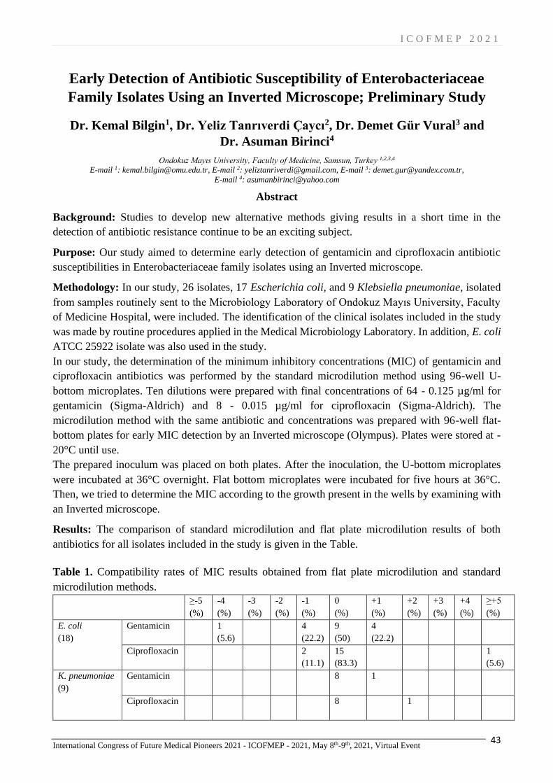
I C O F M E P 2 0 2 1
International Congress of Future Medical Pioneers 2021 - ICOFMEP - 2021, May 8th-9th, 2021, Virtual Event 43
Early Detection of Antibiotic Susceptibility of Enterobacteriaceae
Family Isolates Using an Inverted Microscope; Preliminary Study
Dr. Kemal Bilgin1, Dr. Yeliz Tanrıverdi Çaycı2, Dr. Demet Gür Vural3 and
Dr. Asuman Birinci4
Ondokuz Mayıs University, Faculty of Medicine, Samsun, Turkey 1,2,3,4
E-mail 1: [email protected], E-mail 2: [email protected], E-mail 3: [email protected],
E-mail 4: [email protected]
Abstract
Background: Studies to develop new alternative methods giving results in a short time in the
detection of antibiotic resistance continue to be an exciting subject.
Purpose: Our study aimed to determine early detection of gentamicin and ciprofloxacin antibiotic
susceptibilities in Enterobacteriaceae family isolates using an Inverted microscope.
Methodology: In our study, 26 isolates, 17 Escherichia coli, and 9 Klebsiella pneumoniae, isolated
from samples routinely sent to the Microbiology Laboratory of Ondokuz Mayıs University, Faculty
of Medicine Hospital, were included. The identification of the clinical isolates included in the study
was made by routine procedures applied in the Medical Microbiology Laboratory. In addition, E. coli
ATCC 25922 isolate was also used in the study.
In our study, the determination of the minimum inhibitory concentrations (MIC) of gentamicin and
ciprofloxacin antibiotics was performed by the standard microdilution method using 96-well U-
bottom microplates. Ten dilutions were prepared with final concentrations of 64 - 0.125 µg/ml for
gentamicin (Sigma-Aldrich) and 8 - 0.015 µg/ml for ciprofloxacin (Sigma-Aldrich). The
microdilution method with the same antibiotic and concentrations was prepared with 96-well flat-
bottom plates for early MIC detection by an Inverted microscope (Olympus). Plates were stored at -
20°C until use.
The prepared inoculum was placed on both plates. After the inoculation, the U-bottom microplates
were incubated at 36°C overnight. Flat bottom microplates were incubated for five hours at 36°C.
Then, we tried to determine the MIC according to the growth present in the wells by examining with
an Inverted microscope.
Results: The comparison of standard microdilution and flat plate microdilution results of both
antibiotics for all isolates included in the study is given in the Table.
Table 1. Compatibility rates of MIC results obtained from flat plate microdilution and standard
microdilution methods.
≥-5
(%)
-4
(%)
-3
(%)
-2
(%)
-1
(%)
0
(%)
+1
(%)
+2
(%)
+3
(%)
+4
(%)
≥+5
(%)
E. coli
(18)
Gentamicin 1
(5.6)
4
(22.2)
9
(50)
4
(22.2)
Ciprofloxacin 2
(11.1)
15
(83.3)
1
(5.6)
K. pneumoniae
(9)
Gentamicin 8
1
Ciprofloxacin 8
1

I C O F M E P 2 0 2 1
International Congress of Future Medical Pioneers 2021 - ICOFMEP - 2021, May 8th-9th, 2021, Virtual Event 44
Conclusion: In E. coli isolates, for gentamicin, 9 isolates fully compatible, 4 isolates with -1 dilution
difference and 4 isolates with +1 dilution difference; For ciprofloxacin, 15 isolates fully compatible
and 2 isolates with -1 dilution difference were detected. In K. pneumoniae isolates, for gentamicin, 8
isolates fully compatible, and 1 isolates with +1 dilution difference; For ciprofloxacin, 8 isolates fully
compatible were determined. The method's applicability is not yet clear due to the low number of
microorganisms included in our preliminary study. Therefore, to understand the applicability of the
method, comparative new studies with standard methods using a large number of Enterobacteriaceae
isolates with the use of different antibiotics are needed.
Keywords: E. coli; K. pneumoniae; MIC; Inverted microscope

I C O F M E P 2 0 2 1
International Congress of Future Medical Pioneers 2021 - ICOFMEP - 2021, May 8th-9th, 2021, Virtual Event 45
Optimization Study of Detecting Antibiotic Sensitivity of
Pseudomonas aeruginosa Using an Inverted Microscope
Dr. Kemal Bilgin
Ondokuz Mayıs University, Faculty of Medicine, Samsun, Turkey
E-mail: [email protected]
Abstract
Background: Although there are standard methods for detecting antibiotic resistance, studies
continue on alternative methods that resulted in a shorter time.
Purpose: Our study investigated the detection hour of the antibiotic sensitivity of Pseudomonas
aeruginosa to gentamicin and ciprofloxacin by using an inverted microscope.
Methodology: P. aeruginosa ATCC 27853 standard strain was used as an isolate in our study.
In our study, the determination of the minimum inhibitory concentrations (MIC) of gentamicin
(Sigma-Aldrich) and ciprofloxacin (Sigma-Aldrich) antibiotics was done by microdilution method
using 96-well U-bottom microplates. Ten dilutions were prepared with final concentrations of 64 -
0.125 µg/ml for gentamicin and 8 - 0.015 µg/ml for ciprofloxacin. The microdilution method was
prepared in 96-well flat-bottom plates, with the same antibiotic and concentrations for optimization
trials of early MIC detection by Inverted microscope (Olympus).
The prepared inoculum was applied in the same shape and amount on both plates. After the
inoculation, the U-bottom microplates were incubated at 36°C overnight. Flat bottom microplates
were incubated at 36°C, and the plates were examined with an Inverted microscope every hour for
eight hours, and MIC determination was attempted according to the presence of growth in the wells.
Then the flat bottom microplates were incubated overnight, and the MIC value was determined
depending on the color change due to the pigments created by P. aeruginosa.
Results: The flat plate results were compared with the standard microdilution results and determined
as equal, one-fold up or down concentrations.
Table1. Comparison of MIC results by microdilution and inverted microscope.
Trial Antibiotic MIC Hour
1 2 3 4 5 6 7 8 24*
1 Gentamycin 2 - - - 2 1 1 1 1 2
Ciprofloxacin 0.25 - - - 0,5 0,5 0,25 0,5 0,25 0,5
2 Gentamycin 2 - - - 2 1 1 1 1 2
Ciprofloxacin 0,25 - - - 0,25 0,25 0,25 0,25 0,25 0,25
3 Gentamycin 2 - - - - 1 1 1 1 2
Ciprofloxacin 0,25 - - - 0,25 0,25 0,25 0,25 0,25 0,25
*: MIC determination by color change due to pigment formation.
Conclusion: In our study, no bacteria were found in the wells for the first three hours. Bacteria could
be seen at the bottom of the wells at the fourth hour, except for the gentamicin antibiotic of the third
trial. However, a clear image could be taken at the seventh and eighth hours. The method's
applicability is not clear yet, since clinical isolates were not included in our optimization study. In
order to understand the applicability of the method, comparative studies with standard methods using

I C O F M E P 2 0 2 1
International Congress of Future Medical Pioneers 2021 - ICOFMEP - 2021, May 8th-9th, 2021, Virtual Event 46
a large number of P. aeruginosa isolates obtained from clinical samples with the use of different
antibiotics are needed.
Keywords: Pseudomonas; aeruginosa; MIC; Inverted microscope

I C O F M E P 2 0 2 1
International Congress of Future Medical Pioneers 2021 - ICOFMEP - 2021, May 8th-9th, 2021, Virtual Event 47
The distribution of Enterobacteriales isolated from the urine samples
of children, and the evaluation of antimicrobial susceptibility
Dr. Yeliz Tanrıverdi Çaycı1, Dr. Gülşah Karacan2, Dr. Moein Yoosefi3,
Dr. Kemal Bilgin4, Dr. Demet Gür Vural5 and Dr. Asuman Birinci6
Ondokuz Mayıs University, Faculty of Medicine, Samsun, Turkey
E-mail 1: [email protected], E-mail 2: [email protected], E-mail 3: [email protected],
E-mail 4: [email protected], E-mail 5: [email protected], E-mail 6: [email protected]
Abstract
Background: Antimicrobial resistance has become a serious problem as a result of extensive
antibiotic comsumption.
Purpose: In this study we aimed to determine the bacteria grown in urine cultures of children and
antimicrobial resistance rates in our region.
Methodology: The results of urine cultures that sent from Department of Pediatrics between
01.01.2015 and 31.12.2020 to the Microbiology Laboratory, were evaluated retrospectively. The
identification of the bacterial growth was tested in Vitek MS (Biomeriux, France) and antimicrobial
susceptibility of the isolates were tested in Vitek2 Compakt (Biomeriux, France).
Results: Total of 5511 urine culture samples were evaluated. The most frequently isolated bacteria
was E. coli 67.5% and followed by Klebsiella spp 21.1%, Proteus spp 4.81%. The highest resistance
rates were determined against ampicillin in E. coli and Klebsiella spp isolates (78.96%, 99.91%). The
lowest resistance rate was determined against imipenem and meropenem for E. coli (0.12%, 0.15%)
isolates and amikacin and nitrofrontain in Klebsiella spp isolates (6.10%, 2.46%).
Conclusion: Order to provide effective treatment in urinary tract infections and to minimize
morbidity and long-term complications, the frequency of the pathogens causing urinary tract
infections, local or regional antibiotic resistance should be known and empirical treatment should be
arranged accordingly.
Table 1. The antimicrobial resistance rate of Enterobactericeae isolates.
E.
coli
Klebsie
lla
Enteroba
cter
Prote
us
Citroba
cter
Serrat
ia
Providen
cia
Morga
nella
Raotuel
la
Salmon
ella
Ampicilin 78.9
6
99.91 99.96 40.28 100 100 100 100 100 20
Amoxisilin/
clavulonika
cid
15.2
1
19.34 39.21 3.80 20.48 25.04 0 33.75 0 0
Cefuroxim 42.9
6
63.11 54.06 13.30 33.28 81.38 66.68 82.50 0 0
Cefepim 5.94 14.18 8.16 4.56 10.24 0 0 7.50 0 0
Ceftriaxone 39.4
5
60.96 55.08 9.12 30.72 15.65 50 41.25 0 0
Ceftazidim 35.6
4
53.74 52.53 8.36 28.16 12.52 66.68 37.50 0 0
Ciprofloxac
in
23.8
5
27.94 10.71 11.40 12.80 0 50 27.50 0 20
Ertapenem 1.17 19.51 17.34 3.42 7.68 9.39 50 1.25 0 0
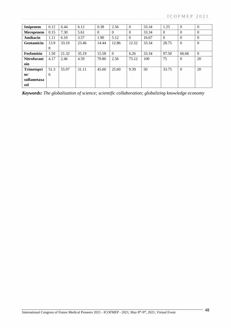
I C O F M E P 2 0 2 1
International Congress of Future Medical Pioneers 2021 - ICOFMEP - 2021, May 8th-9th, 2021, Virtual Event 48
Imipenem 0.12 6.44 6.12 0.38 2.56 0 33.34 1.25 0 0
Meropenem 0.15 7.30 5.61 0 0 0 33.34 0 0 0
Amikacin 1.11 6.10 3.57 1.90 5.12 0 16.67 0 0 0
Gentamicin 13.9
8
33.10 23.46 14.44 12.86 12.52 33.34 28.75 0 0
Fosfomisin 1.50 21.32 35.19 15.58 0 6.26 33.34 97.50 66.66 0
Nitrofurant
oin
4.17 2.46 4.59 79.80 2.56 75.12 100 75 0 20
Trimetopri
m/
sulfametaxa
zol
51.3
6
55.97 31.11 45.60 25.60 9.39 50 33.75 0 20
Keywords: The globalization of science; scientific collaboration; globalizing knowledge economy
I C O F M E P 2 0 2 1
International Congress of Future Medical Pioneers 2021 - ICOFMEP - 2021, May 8th-9th, 2021, Virtual Event 49
Future Expectations in ART
Assoc. Prof. İlay Gözükara1 and Prof. M. Turan Çetin2
Private Prof.Dr.Turan Cetın IVF Centre, Turkey1,2
E-mail 1: [email protected], E-mail 2: [email protected]
Abstract
There are some troubles during In Vitro Fertilisation (IVF) treatment even though advances. Embryo
selection, advanced maternal age and fertility preservation are most common ones. Firstly, the
selection of viable embryo remains a principle challenge of IVF in order to predict live birth. Artificial
intelligence could provide benefit in the future. Dataset based on time-lapse images combined with
embryo characteristics may provide the selection of accurate embryo. Left over products of IVF for
their contents in proteins, metabolites, nucleic acid and -omics technologies may also help embryo
selection and growing that will likely change the current IVF procedures. The laboratory environment
may be completely identical to the in vivo environment, and as a result, the fetus will be grown in
vitro until birth in the future. Secondly advanced maternal age is a critical clinical issue in IVF cycles.
Emerging therapeutic approaches have been proposed for these patients. Germ line engineering could
be considered to restore the competence of aged oocytes. Chromosome therapy is another promising
one. In the future such practice might find direct application in the treatment of chromosomal
disorders but might also apply to the correction of aneuploid germ cells and embryos in IVF. Beside
chromosome therapy it may be possible to generate new gamet cells from stem cell in the future. It
was already reported that murine induced pluripotent stem cells were differentiated into functional
oocytes in the presence of specific growth-factors. Thirdly some future fertility preservation methods
can be needed like some new gonadoprotective agents, also some new drugs to improve tissue
grafting. Artificial ovaries containing isolated follicles and culturing primordial follicles to mature
oocyte might serve as a revolutionary method without the danger of cancer reseeding. In conclusion
we may not see any pregnant women in the future even may not see any children with syndrome and
congenital diseases in the future because of some advanced technologies.
Keywords: Germ line engineering; chromosome therapy; IVF; gamet cells.

I C O F M E P 2 0 2 1
International Congress of Future Medical Pioneers 2021 - ICOFMEP - 2021, May 8th-9th, 2021, Virtual Event 50
Can We Consider Embryos Solely as Biological Material?
Maide Barış
Istanbul Health and Technology University, Department of Medical History & Ethics, Faculty of Medicine, Istanbul, Turkey
E-mail: [email protected]
Abstract
Traditionally, bioethical discussions about embryo research tend to resort to moral status arguments.
Pro-embryo research argumentators in bioethics who embrace what we call “none-approach” use
“psychological arguments” or “physiological arguments” to claim that embryos do not have any
moral status. On the other hand, major anti-embryo research argumentators usually adopt what we
call “all-approach” use “development-based moral arguments” and “metaphysical moral arguments”
to defend that embryos have moral status. This paper firstly presents these moral approaches briefly,
maintaining that the “moral status” the approach does not help in practice, especially regarding
interventions in preimplantation-stage embryos in the laboratory. While embracing and respecting
great moral diversity of beliefs about the embryo, this paper suggests that there should be a practice-
focused and less counter-intuitive moral stance somewhere in between “embryos are complete
humans who have the full moral status” and “embryos are nothing but accumulations of cells.”
Secondly, we will argue that although embryos are not entitled to full moral status as much as a
paradigm human does, they are still worthy of moral respect and cannot be considered solely as
biological materials because (i) they have inherent potential to become one of us; (ii) they are human
organisms in a biological sense and (iii) they are soon to be considered as patients in near future,
rather than only as biological research entities, at a time when Nobel-winning CRISPR/Cas9-based
germline genetic intervention is waiting at the doors of reproductive clinics. Finally, we will maintain
that the statement “embryos are worthy of moral respect” does not say anything about the post-
implantation stage, especially regarding abortion. It only contends that because embryos are worthy
of moral respect and to be considered patients and subjected to genetic intervention in a petri dish,
then they need to be handled by a physician/ specialist who is trained microsurgery to deal with
embryos: A clinical embryologist.
Keywords: Embryo Research; Moral Status; Moral Respect; Bioethics.

I C O F M E P 2 0 2 1
International Congress of Future Medical Pioneers 2021 - ICOFMEP - 2021, May 8th-9th, 2021, Virtual Event 51
Common Chest CT Findings in 100 COVID-19 Patients Followed for
Pneumonia
Dr. Mehmet Akçiçek
Malatya Turgut Özal University, Faculty of Medicine, Malatya Training and Research Hospital, Radiology Department,
Malatya/Turkey
E-mail: [email protected]
Abstract
Background: CT has become an important imaging method in the diagnosis and treatment of patients
with viral pneumonia. The use of low-dose CT scanning for the diagnosis of viral pneumonia
optimizes patient management by providing rapid treatment of patients suspected of COVID-19
infection. As shown in previous studies; chest CT images may show different imaging features in
COVID-19 patients depending on the period and severity of the disease.
Purpose: In this study, we investigated the common CT imaging features and changes of COVID-19
in 100 pneumonia patients, who were confirmed by RT-PCR and underwent control CT examination
during follow-up.
Method: The patients who admitted in July, August and September 2020 when the highest rate of
patient admission and hospitalization in our hospital during the COVID-19 pandemic and whose RT-
PCR test positivity was confirmed has been scanned retrospectively. Patients wh8om was detected
pneumonia in chest CT examined at their first visit and a second CT performed in the 5-14 day period
in their follow-up, were included. A total of 100 patients, 61 of whom are male, between the ages of
25-90 were identified. CT examinations were performed on 16 and 128 MDCT devices in low dose.
Images were transferred to the workstation and analyzed in the PACS system. The findings of the
first and second CT of each patient were defined according to the Fleishner community and literature
information.
Results: The most common findings in the first CT were GGO (Ground glass opacity) (68%),
consolidation (46%), nodule (31%) and reticulation (28%). Findings other than these are as follows;
airway changes (17%), vascular enlargement (12%), subpleural line (8%), halo (2%), reverse halo
(2%), pleural effusion. 94% of the lesions were peripheral and 92% were multifocal. In the control
CT examinations performed within 4-15 days, the most common findings were GGO (70%),
consolidation (70%), 65% reticulation, 31% CPP, 33% airway changes. Vascular enlargement in
27%, subpleural line in 22%, subleural fibrotic strip in 13%, nodule in 13%, pleural effusion in 6%,
reverse halo findings in 4%, and pericardial effusion in 1% of the patients were found. Pleural
thickening (1%) and lymphadenopathy (11%), which were also detected in the first examinations
were detected. 78% of the lesions were peripheral, 22% were peripheral and centrally located, and
99% were multifocal.
Conclusion: CT findings of COVID-19 pneumonia resemble those of many other infectious and non-
infectious diseases, especially viral pneumonia types. These findings may vary depending on the
disease phase during follow-up, individual characteristics of the patient, underlying diseases and
treatment interventions.
Keywords: COVID-19, Chest, CT, Pneumonia

I C O F M E P 2 0 2 1
International Congress of Future Medical Pioneers 2021 - ICOFMEP - 2021, May 8th-9th, 2021, Virtual Event 52
Does COVID-19 infection trigger the formation of Anti-nuclear
Antibodies?
Dr. Melek Bilgin1 and Dr. Eşe başbulut2
Health Sciences University, Samsun, Training and Research Hospital, Medical Microbiology Department, Samsun,
Turkey 1,2
E-mail 1: [email protected], E-mail 2: [email protected]
Abstract
Aim: Increasing evidence suggests that autoimmunity may play a role in the pathophysiology of
SARS-CoV-2 infection during both the acute and ‘long COVID’ phases of disease. However the
long-term consequences of COVID-19 and whether it will trigger autoimmunity in the future are
curious and require further research. There is insufficient knowledge on the assessment of
autoimmune antibodies in convalescent SARS-CoV-2 patients. The aim of this study is to evaluate
the presence of antinükleer antibodies (ANAs) in convalescent SARS-CoV-2 patients and to compare
with ANA results before the onset of COVID-19.
Material and Methods: 23 healthcare workers with Covid 19 infection (SARS CoV-2 PCR test
positive) whose ANA test results were known and reported before the Covid 19 infection were
included in this study. ANA tests were studied from the serum of participants at least 3 months after
Covid-19 infection.
Diagnosis of COVID-19 infection was performed by a RT-qPCR method. Bio-Speedy, SARSCoV-2
Double Gene RT-qPCR Detection Kit (Bioeksen, İstanbul, Türkiye) were used for detection of virus
RNA. ANA positivity and patterns were assessed by indirect immune-fluorescence on Hep-2 cells
(Euroimmun, Luebeck, Germany) using standard techniques.
Results: The study included 23 people, 19 women and four men, with a mean age of 45. Differences
were found in the ANA IFA pattern in 3 of 23 patients after Covid-19 infection compared to pre-
infection. In the first case, the ANA IFA pattern before Covid-19 was speckled weakly positive, while
the ANA IFA result was detected as anti-nuclear membrane positive (++) 3 months after infection.
Thereupon, anti-mitochondrial antibody (AMA), anti-smooth muscle antibody (ASMA), and anti-
LKM were studied for autoimmune liver disease in the patient and found as negative. Also, RNP /
Sm, Sm, SS-A, Ro 52, SS-B, Scl-70, PM-Scl, Jo-1, CENP B, PCNA, dsDNA, nucleosomes, histones,
rib P-prot., AMA-M2, and DFS70 antibodies were studied by immunoblot method, and anti-RNP /
Sm were detected positive. While the second case was chromosomal granular (GGK) positive before
infection, ANA IFA was found homogeneous positive, and cytoplasm speckled positive after
infection. Then Immunoblot ANA wide panel was studied in the case, and Scl-70, DFS70, dsDNA
found positive. In the third case, while the previous result was negative, it was determined as granular
weak positive after infection. In this case Ena blot result was evaluated as negative. One year before
they had the Covid-19 infection, all 3 cases had negative Ena blot results.
Conclusion: As a conclusion, our study revealed that Covid-19 infection can trigger autoimmunity,
and patients should be followed up for a long time after Covid-19 infection. Further studies with
larger patient series are needed to better define the clinical significance of auto-antibody positivity
and to understand the possible role of Covid-19 in autoimmune diseases.
Keywords: SARS-CoV-2; COVID-19; Anti-nuclear Antibodies; Autoimmunity

I C O F M E P 2 0 2 1
International Congress of Future Medical Pioneers 2021 - ICOFMEP - 2021, May 8th-9th, 2021, Virtual Event 53
Guillain Barre Syndrome Induced by Covid 19: Discussion with a
Case
Süsen Banazılı, MD1 and Asst. Prof. Mesut Öterkuş2
Malatya Training and Research Hospital, Anesthesiology and Reanimation Clinic 1
Malatya Turgut Özal University Faculty of Medicine Department of Anesthesiology and Reanimation 2
E-mail 1: [email protected], E-mail 2: [email protected]
Abstract
Covid 19, which is among the most important health problems of today, affects many organs and
systems. The nervous system is one of them. Many complications such as stroke, encephalitis,
necrotizing encephalitis, and Guillain Barre syndrome (GBS) have been reported in the literatüre.
GBS is an autoimmune-induced demialysing polyneuropathy. It usually occurs following a recent
infection. Covid 19 is one of these infections. The main mechanism is thought to be T cell activation
or similarity of virus proteins to nerve cell protein. The most effective treatments are immunotherapy
(plasma exchange, IVig) and physiotherapy. It leaves permanent neurological sequelae in
approximately 20% of patients.
We aimed to present a 51-year-old male patient who developed GBS. Our patient did not have a
recent history of any infection or neurological deficit other than covid 19 infection. Our patient was
intubated electively due to increased respiratory distress and with the improvement of respiratory
parameters, the patient was extubated decreased oxygen saturation during his follow-up and treatment
in the intensive care unit. The patient was extubated as the respiratory parameters improved after the
treatment. In his examination after extubation, biletareal lower extremity and loss of strength and
reflexes in the left upper extremity were observed. GBS was considered in the foreground. Since the
EMG was also compatible, IVIG (0.4g / kg) treatment was started for 5 days. Physiotherapy was
started on the patient in consultation with a physical therapy and rehabilitation specialist. After 4
weeks, the patient started walking with support. n literature,
GBS after covid 19 is usually a case report. In a study conducted in Italy; showed that GBS
increased 2.6 times during the pandemic period of covid 19. As a result, It should be kept in mind
that GBS can also appear as a Covid 19 complication and early treatment increases the chance of
success.
Keywords: Covid-19, Autoimmunty, Guillain Barre syndrome
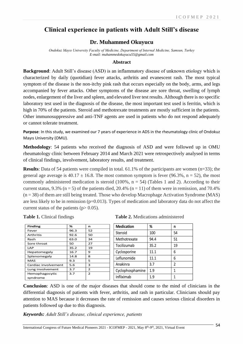
I C O F M E P 2 0 2 1
International Congress of Future Medical Pioneers 2021 - ICOFMEP - 2021, May 8th-9th, 2021, Virtual Event 54
Clinical experience in patients with Adult Still’s disease
Dr. Muhammed Okuyucu
Ondokuz Mayıs University Faculty of Medicine, Department of Internal Medicine, Samsun, Turkey
E-mail: [email protected]
Abstract
Background: Adult Still’s disease (ASD) is an inflammatory disease of unknown etiology which is
characterized by daily (quotidian) fever attacks, arthritis and evanescent rash. The most typical
symptom of the disease is the non-itchy pink rash that occurs especially on the body, arms, and legs
accompanied by fever attacks. Other symptoms of the disease are sore throat, swelling of lymph
nodes, enlargement of the liver and spleen, and elevated liver test results. Although there is no specific
laboratory test used in the diagnosis of the disease, the most important test used is ferritin, which is
high in 70% of the patients. Steroid and methotrexate treatments are mostly sufficient in the patients.
Other immunosuppressive and anti-TNF agents are used in patients who do not respond adequately
or cannot tolerate treatment.
Purpose: In this study, we examined our 7 years of experience in ADS in the rheumatology clinic of Ondokuz
Mayıs University (OMU).
Methodology: 54 patients who received the diagnosis of ASD and were followed up in OMU
rheumatology clinic between February 2014 and March 2021 were retrospectively analysed in terms
of clinical findings, involvement, laboratory results, and treatment.
Results: Data of 54 patients were compiled in total. 61.1% of the participants are women (n=33); the
general age average is 40.17 ± 16.8. The most common symptom is fever (96.3%, n = 52), the most
commonly administered medication is steroid (100%, n = 54) (Tables 1 and 2). According to their
current status, 9.3% (n = 5) of the patients died, 20.4% (n = 11) of them were in remission, and 70.4%
(n = 38) of them are still being treated. Those who develop Macrophage Activation Syndrome (MAS)
are less likely to be in remission (p=0.013). Types of medication and laboratory data do not affect the
current status of the patients (p> 0.05).
Table 1. Clinical findings Table 2. Medications administered
Conclusion: ASD is one of the major diseases that should come to the mind of clinicians in the
differential diagnosis of patients with fever, arthritis, and rash in particular. Clinicians should pay
attention to MAS because it decreases the rate of remission and causes serious clinical disorders in
patients followed up due to this diagnosis.
Keywords: Adult Still’s disease, clinical experience, patients

I C O F M E P 2 0 2 1
International Congress of Future Medical Pioneers 2021 - ICOFMEP - 2021, May 8th-9th, 2021, Virtual Event 55
Distal metatarsal Chevron osteotomy + modified McBride method in
hallux valgus, short term 1-3 year results
Dr. Muhammet Salih Ayas
Republic of Turkey Ministry Of Health, Erzurum Regional Education and Research Hospital, Erzurum/Turkey
E-mail: [email protected]
Abstract
Background: Short-term efficacy of distal metatarsal Chevron osteotomy + modified McBride
method in hallux valgus.
Purpose: In this study, the results of distal metatarsal Chevron osteotomy + modified McBride
method in patients with a diagnosis of mild disease (HVA ≤ 40, IMA <13) hallux valgus were
evaluated.
Methodology: Twenty-six feet of 22 patients with hallux valgus deformity, who were operated
between 2017 and 2020 in Erzurum Regional Training and Research Hospital, were operated with
distal metatarsal Chevron osteotomy + modified McBride method. Radiological, functional and pain
evaluations were examined.
Results: Preoperative and postoperative hallux valgus angle and 1.-2. There was a statistically
significant difference between the intermetatarsal angle values (p <0.001 for both values), and no
statistically significant difference was found between the 1st distal metatarsal articular angle values
(p> 0.5). The average visual analog scale values, which were 5.6 ± 1.0 before the operation, decreased
to 1.4 ± 1.0 after the follow-up and this result was found statistically (p <0.001). When the results of
the control American Orthopedic Foot-Ankle Association Score (86.2 ± 8.2) were compared with the
preoperative values (34.2 ± 14.8), a statistically significant difference was found (p <0.001).
Conclusion: In conclusion, distal metatarsal Chevron osteotomy + modified McBride operation is an
effective and safe technique in the surgical treatment of hallux valgus. Successful results have been
demonstrated in reducing pain and correcting radiological and cosmetic deformity.
Keywords: Hallux valgus, Chevron, Modified McBride, Osteotom

I C O F M E P 2 0 2 1
International Congress of Future Medical Pioneers 2021 - ICOFMEP - 2021, May 8th-9th, 2021, Virtual Event 56
Effects of Avanafil, A Phofodiesterase Type 5 (Pde-5) Inhibitor, on
A549 Lung Cancer Cell Proliferation
Asst. Prof. Muhammet Çelik
Atatürk University, Faculty of Medicine, Department of Medical Biochemistry, Erzurum, Turkey
E-mail: [email protected]
Abstract
Objective: Cyclic nucleotide phosphodiesterase enzymes (PDEs) are a family of super enzymes
found in all cell types and tissues. Different enzymes are found in this enzyme family, from one to
eleven (PDE1-PDE11). The functions of PDE enzymes found in different tissues of the body are, of
course, different. The contribution of PDE iso-enzymes to metabolism and their roles in
physiopathological events has attracted attention. These enzymes play a role in the pathophysiology
of many diseases such as inflammation, neurodegeneration, cancer, immunomodulation. With the
studies in recent years, new PDE inhibitors have been introduced to the market. Studies have shown
the significant effects of sildenafil, a PDE-5 inhibitor, especially on lung cancer. Many different
mechanisms play a role in the anticancer activity of PDE-5 inhibitors. Avanafil is also a PDE-5
inhibitor, and its effect is much higher than sildenafil. In our study, the effect of Avanafil on the
viability of A549 lung cancer cells was investigated.
Methods: We investigated the dose and time-dependent effects of Avanafil on cell proliferation in
A549 cells. For this aim, different concentrations of Avanafil (from 1 µM to 128 µM) were added
onto A549 cells, and viability and proliferation were evaluated with MTT test at 24 and 48 hours after
incubation.
Results: Avanafil has been studied with concentrations of 1-2-4-8-16-32-64 and 128 µM. When the
cell viability tests were examined at 24th and 48th hours, it was observed that all doses of Avanafil
reduced cell division in 24 hours, and the best effect was at 128 µM concentration of Avanafil. When
the 48th hour was examined, it was shown that the effect continued, especially the anticancer activity
of Avanafil at 128 µM concentration remained very high and significantly reduced cancer cell
proliferation.
Conclusion: Our findings showed that Avanafil, like other PDE5 inhibitors, inhibited the
proliferation in cancer cells. Studies have shown that PDE5 is expressed in colon, prostate, breast,
and pancreatic cancers. These results show that PDE5 enzymes are effective in carcinogenesis. PDE5
enzyme inhibition has become an important target in cancer prevention and treatment. Intracellular
apoptosis and necrosis pathways induced by PDE5 inhibition may be responsible for the anticancer
action mechanism of Avanafil. However, additional laboratory studies are needed to elucidate these
pathways. Our study concluded that Avanafil emerges as a new therapeutic target in lung cancer
patients, although it is outside of the current indications for use. The shorter duration of action
compared to sildenafil will also reduce its unwanted side effects and facilitate dose control in cancer
treatment.
Keywords: Lung cancer; Phosphodiesterase enzymes; PDE-5; Avanafil; A549 cell line.

I C O F M E P 2 0 2 1
International Congress of Future Medical Pioneers 2021 - ICOFMEP - 2021, May 8th-9th, 2021, Virtual Event 57
Comparison of posterolateral approach and direct reduction
posterior-anterior screw fixation method versus indirect reduction
anterior-posterior screw fixation method for posterior malleolus
fixation in ankle trimalleolar fractures
Muhammet Kalkışım, MD1 and Kerim Öner, PhD, MD2
Surgeon, Department of Orthopaedic Surgery, Sorgun Public Hospital, Yozgat, Turkey 1
Assistant Professor, Department of Orthopaedic Surgery, Karadeniz Technical University School of Medicine, Trabzon, Turkey 2
E-mail 1: [email protected], E-mail 2: [email protected]
Abstract
Background: A consensus on how posterior malleolar fragments in trimalleolar fractures should be
reduced and fixated still lacks. Anatomic reduction and rigid fixation are important in the treatment
of posterior fragment in trimalleolar fractures.
Purpose: In this study we aimed to compare direct reduction (DR) using posterolateral approach and
the posterior-anterior (P-A) screw fixation method versus indirect reduction (IR) with anterior-
posterior (A-P) screw fixation method.
Methodology: In our study, patients with ankle trimalleolar fracture were evaluated retrospectively.
This comparative study reviewed a case series from 2017 to 2020. Patients with at least 1 year
postoperative follow-up were included in the study. Patients were categorized into two groups. The
first group consisted of patients who underwent fixation with direct reduction (DR) and PA screws,
and the second group consisted of patients who underwent fixation with indirect reduction (IR) and
AP screws. Demographic datas, American Ankle Society scores (AOFAS), ankle range of motions
(ROM), fracture types (according to Lauge-Hansen classification), the size of posterior malleolar
fragment, complications and functional bone healing time were compared in both groups.
Postoperative X-rays were performed to determine the residual step-off in the articular reduction. The
degree of osteoarthritis was evaluated by final follow up controls with x-rays.
Results: 16 patients (9 males, 7 females) in group DR and 12 patients (6 males, 6 females) in IR
group were recruited. The mean age in group DR were 41.6 (20-56) while 37.1 (21-53) were in group
IR. There was no significant difference between the two groups in terms of complication rates,
functional bone healing times, follow-up times, fracture types, amount of intra-articular step-off and
ROM (p> 0.05). The AOFAS scores were significantly better in the IR group compared to the DR
group (p> 0.05). None of the complications such as fixation failure, infection, delayed union were
observed in both groups.
Conclusion: The results are satisfied both in two different approaches for fixation the posterior
malleolus in trimalleolar fractures. However, functional results are better in the indirect reduction (IR
group) method. We attribute the better functional results in indirect reduction and anterior-posterior
screw fixation method because of the less invasive approach.
Keywords: Posterior malleolar fractures, Trimalleolar fractures, Posterolateral approach, Screw
fixation

I C O F M E P 2 0 2 1
International Congress of Future Medical Pioneers 2021 - ICOFMEP - 2021, May 8th-9th, 2021, Virtual Event 58
Covid-19 and Comorbidities
Dr. Mukadder Erdem1, Recai Aci2, Assoc. Prof. Dr. Mahcube Çubukçu3, Adem
Keskin, PhD4, Dr. Eda Türe5 and Dr. Ebru Ulaş6
Samsun Training and Research Hospital Biochemistry 1,2,6
Samsun Training and Research Hospital Family Medicine 3,5
Aydın Adnan Menderes University Biochemistry 4
E-mail 1: [email protected], E-mail 2: [email protected], E-mail 3: [email protected],
E-mail 4: [email protected], E-mail 5: eda.tü[email protected], E-mail 6: [email protected]
Abstract
Objectives: The pandemic situation with the emergence of severe acute respiratory syndrome
coronavirus 2 (SARS-CoV-2) from China has endangered human lives. Corona virus disease 2019
(COVID-19) is presented with asymptomatic, mild, or severe pneumonia-like symptoms. COVID-19
patients with diabetes, chronic obstructive pulmonary disease (COPD), cardiovascular diseases
(CVD), hypertension, malignancies, HIV, and other comorbidities could develop a life-threatening
situation. In this study, the files of patients with COVID-19(+) who were followed up in the service
and intensive care unit were retrospectively scanned, and their comorbid disease status was examined.
This study aimed to compare with some biochemical and hematological parameters in terms of the
mortality rates, taking into account the length of stay in the hospital of Covid 19 patients with
comorbid and non-comorbid.
Methods: A total of 44 patients with COVID-19 in the intensive care unit and 71 patients in service
were included in the study. We performed correlation analysis between whole blood parameters,
important biomarkers for prognosis of the disease in the inflammatory process and hospitalization
day, and mortality. We also evaluated the effects of these parameters of comorbidity diseases.
Results: When looking at the correlation of biochemical and hematological parameters of allCOVID-
19 (+) patients hospitalized in both the service and the intensive care unit with the day of
hospitalization; it was positively correlated with PT, INR, CRP, FERRITINE, PCT, ESR, D-DIMER,
WBC, NEUTROPHIL, MLR, and NLR parameters of the day of hospitalization, while it was
negatively correlated with HGB and LYMPHOCYTE parameters (p<0.01). In addition, Ex/discharge
status was positively correlated with LNF (p<0.01), PLT (p<0.01) parameters, while it was negatively
correlated with PT (p<0.05), INR (p<0.05), CRP (p<0.01), Ferritin (p<0.01), PCT (p<0.01), ESR
(p<0.01), D-Dimer (p<0.01), WBC (p<0.01), NTR (p<0.01), MLR (p<0.01), NLR (p<0.01) and
Troponin-I (p<0.01) parameters as shown. Also, out of 115 COVID-19 (+) patients participating in
this study, 91 comorbid diseases were detected, while 24 of them did not. This comorbid disease was
detected in 22 patients with DM (24.18%), 21 with hypertension (23.08%), 11 with CVD (12.09%),
9 with CRF(Chronic Renal Failure) (9.89%), 6 in COPD (6.59%), 15 of them had pneumonia
(16.48%), and 7 had cancer (7.69%). Besides, out of 115 COVID-19 (+) patients participating in this
study, all 28 patients who died were found to have comorbid diseases. It was determined that DMD
in 5 (17.86%), hypertension in 4 (14.29%), CVD in 6 (21.43%), CRF in 3 (10.71%), COPD in 2
(%7.14), Pneumonia in 4 (14.29%), cancer in 4 (14.29%) patients with Exitus.
Conclusion: It has been determined that whole blood and essential parameters in the inflammatory
process correlated with the length of stay and mortality, these parameter levels varied in COVID-19
patients with comorbidity and lead to an increase in the size of stay and mortality rates.
Keywords: Covid-19, Comorbidity, Hospitalization day, Mortality.

I C O F M E P 2 0 2 1
International Congress of Future Medical Pioneers 2021 - ICOFMEP - 2021, May 8th-9th, 2021, Virtual Event 59
A Rare Form of Liver Failure and Immune Deficiency: IFNGR1 gene
Mutation
Murat Cag, PhD, MD
Strasbourg University, Hepatopancreatic Biliary Surgery Unit, France
Memorial Bahcelievler Hospital, General Surgery Department, Istanbul Turkey
E-mail: [email protected]
Abstract
Immunodeficiency-27A is a disorder caused by mutations in the IFNGR1 gene that is inherited
autosomal recessively. Patients with complete IFNGR1 deficiency have a severe clinical phenotype
characterized by early and often fatal mycobacterial infections. Thus, the disorder can be categorized
as a form of mendelian susceptibility to mycobacterial disease (MSMD). Resistant mycobacterial
infections can begin from childhood. Salmonella infections and diarrhea can be seen in 5% of the
patients. Index case applied to our clinic as the 3rd living child of 3rd degree relatives parents. It was
learned that there was no other affected individual in the family. From her history, it was learned that
there was no significant disease in the prenatal period and until his second year of age, but then
gradually swelling in the abdomen, uncontrollable diarrhea, weight loss, and she had received 4-way
tuberculosis treatment for the last 3 months. Laboratory tests revealed impaired liver function, high
bilirubin levels and pancytopenia, very low total protein, albumin, Na and Mg values, and high CRP.
In the abdominal ultrasound, liver and spleen were larger than normal and heterogeneous, and
congestion in the portal circulation was observed. This mutation is known as Immunodeficiency Type
27 A and is one of the ultra-rare forms of immunodeficiency. As in our patient, resistant mycobacterial
and Salmonella infections are usually seen and are usually fatal. This Syndrome causing liver and
immunodeficiency was presented as an ultra rare Syndromic case.
Keywords: Liver failure, immune deficiency, IFNGR1, whole exome sequencing, WES

I C O F M E P 2 0 2 1
International Congress of Future Medical Pioneers 2021 - ICOFMEP - 2021, May 8th-9th, 2021, Virtual Event 60
Commitment to the medical profession and individual competitiveness
of Medical Faculty students in Turkey
Asst. Prof. Mustafa Bayraktar
Atatürk University, Faculty of Medicine, Department of Family Medicine, Erzurum, Turkey
E-mail: [email protected]
Abstract
Objective: To investigate the individual competitiveness and commitment to the medical profession
of first year medical students in Erzurum, Turkey.
Methods: Students starting the first year of the Faculty of Medicine were asked to answer the
questions on the five-point Likert scale between "strongly disagree" and "strongly agree" with the
online questionnaire method. The questionnaire included "Medical Faculty Students' Commitment to
the Medical Profession Scale" consisting of nine questions and the "Revised Individual
Competitiveness Scale" consisting of 11 questions with reversely coded three questions. The data
obtained were statistically analyzed using Student t-test, one-way ANOVA and Pearson correlation
tests.
Results: Out of 517 people who accepted to participate in the study and answered the questionnaire,
151 questionnaires with incomplete, missing or duplicate results were excluded from the study. Of
the 366 participants included in the study, 54.1% (n = 198) were female, 45.9% (n = 168) were male;
Their average age was 19.1 ± 1.3 years. The average individual competition scale score of the
participants was 33.21 ± 3.38; The scale of commitment to the medical profession was found to be
37.16 ± 5.89. While the commitment score of female participants to the profession was 38.63 ± 4.97,
it was found to be 35.44 ± 6.41 for men, and a statistically significant difference was found (p <0.001).
While the individual competitiveness scale score of the female and male participants were 33.17 ±
3.38, and 33.24 ± 3.39, respectively, no statistically significant difference was found (p = 0.839).
There was no statistically significant difference between the scores of both scales in terms of age (p>
0.05). A weak positive correlation was found between the individual competition scale score of the
participants and their commitment to the profession (p = 0.030; r = 0.114).
Conclusion: In this study, for the first time in the literature, the individual competitiveness and
commitment to the medical profession of the Medical Faculty students in Turkey were investigated.
It was found that there was a positive correlation between the scores of the individual competitiveness
and commitment to the medical profession, and female physician candidates were found to be more
dedicated to the medicine profession than males.
Keywords: Individual Competitiveness Index; Commitment to Profession of Medicine; Medical
students.

I C O F M E P 2 0 2 1
International Congress of Future Medical Pioneers 2021 - ICOFMEP - 2021, May 8th-9th, 2021, Virtual Event 61
Evaluation of staging 18F FDG PET/CT images with conventional and
volumetric data in patients diagnosed with rectal cancer: Do the
characteristics of the primary tumor provide information about the
metastatic potential of the tumor?
Dr. Nazlı Pınar Karahan Şen1 and Prof. Gamze Çapa Kaya2
Dokuz Eylul University, Faculty of Medicine Department of Nuclear Medicine, Izmir, Turkey 1,2
E-mail 1: [email protected], E-mail 2: [email protected]
Abstract
Objective: In 18F FDG PET/CT imaging, in addition to the conventional standard uptake value
(SUV) parameters of the tumor tissue, metabolic tumor volume (MTV) and tumor lesion glycolysis
(TLG) values can be obtained. In this study, we investigated the relationship between the
conventional and volumetric characteristics of the primary tumor obtained from the staging F-18 FDG
PET/CT images and the metastatic potential of the primary tumor at the time of diagnosis in patients
diagnosed with rectal cancers.
Method: Fifty-seven patients who were diagnosed with rectal cancer and underwent 18F FDG
PET/CT for staging between January 2012 and January 2021 were included in our study. Primary
tumor, regional lymph node metastasis, distant metastasis and KRAS mutation were examined.
Conventional and volumetric data of the tumor were obtained by drawing the area of interest (ROI)
of the primary tumors from PET/CT images. In order to prevent manual error, the area of interest was
drawn in the tumor area with a minimum SUV value of 2 and above. SUV values (minimum,
maximum, standard, mean, peak) and metabolic data (MTV and TLG) of the primary tumor within
the drawn ROI were obtained. Patients were evaluated in two different groups as KRAS mutant and
wild-type. A statistically significant difference in terms of SUV, TLG and MTV values in patients
with or without lymph node metastasis in PET/CT and patients with distant metastasis and no
metastasis was investigated by Mann Whitney U test. P <0.05 was considered significant. Receiver
Operating Curve (ROC) analysis was performed for the significant features.
Results: While 26 of the patients were KRAS mutant, 31 patients were wild-type. Lymph node
metastasis was found in 66.7% of the patients, and distant metastasis was found in 61.4%. There was
no significant difference in lymph node metastasis and distant metastasis levels between patients with
and without KRAS mutation (p> 0.05). In KRAS mutant patients, no significant difference was found
between the conventional and volumetric characteristics of the primary tumor and the observation of
lymph node and distant metastasis. However, TLG and MTV values of the primary tumor were
significantly higher in patients with lymph node (AUC: 0.746, 0.737; p=0.023, p=0.029), and distant
metastasis (AUC: 0.718, 0.732; p=0.048, p=0.035), in patients with KRAS wild-type.
Conclusion: Although rates of local and distant metastasis at the time of diagnosis are similar in
patients diagnosed with KRAS mutant and wild-type rectal cancer, in this study, it was observed that
the high TLG and MTV values of the primary tumor in KRAS wild-type patients were associated
with lymph node and distant metastasis at the time of diagnosis. However, in KRAS mutant patients,
there was no relationship between conventional and volumetric features and metastasis at the time of
diagnosis.
Keywords: 18F-FDG PET-CT, MTV, Rectal cancer, KRAS.

I C O F M E P 2 0 2 1
International Congress of Future Medical Pioneers 2021 - ICOFMEP - 2021, May 8th-9th, 2021, Virtual Event 62
Is the Ganglion Cell Layer Thickness more sensitive marker than
retinal nerve fibre layer thickness in Multiple Sclerosis Patients?
Dr. Nuray Can Usta1 and Dr. Betül Önal Günay2
Health Sciences University, Trabzon Training and Research Hospital, Neurology Clinic, Trabzon, Turkey 1
Health Sciences University, Trabzon Training and Research Hospital, Eye Clinic, Trabzon, Turkey 2
E-mail 1: [email protected], E-mail 2: [email protected]
Abstract
Background: Optical coherence tomography (OCT) can be used in the follow-up of Multiple
Sclerosis (MS), which is the most common cause of non-traumatic disability in young adults.
Purpose: It was aimed to evaluate the OCT findings of Multiple Sclerosis patients who applied to
the MS outpatient tertiary referral hospital clinic in April 2021 and those in the healthy control group.
Methodology: Patients who applied to the MS outpatient clinic of the tertiary referral hospital
between April 1, 2021 and April 28, 2021 were included in the study. The exclusion criteria were
patients who failed to comply with the OCT examination and patients who did not accept the tests.
Age-matched healthy individuals were selected as control group. The patients underwent a
comprehensive ophthalmological and neurological examination and OCT parameters were compared.
Results: A total of 34, including 12 MS patients and 22 healthy controls, were included in the study.
Three of the patients were male and 9 of them were female in MS. The mean age of MS patients was
36.5±11.6 (19-65), and the mean age of the control group was 35.5±7.2 and there was no significant
difference in terms of age (p=0.79) and gender (p=0.8). There was no significant difference between
patients who had previously optic neuropathy and had not (p<0.05). Compared to the control group,
there was no significant difference in Retinal Nerve Fibre Layer (RNFL) thickness on all quadrants
in MS patients. Compared to macular thickness (MT), all quadrants except central MT were
significantly lower in MS patients [ central MT in MS versus control group (258.9±19.6 versus
266.9±15.4; p=0.21), superior MT (332.3±17.9 versus 350.8±8; p=0.007), temporal MT (314.9±16.2
versus 335.1±11.2; p=0.001), inferior MT (330.7±20.3 versus 346.7±11.6; p=0.01), nazal MT
(335.1±20.7 versus 350.7±13.1;p=0.02)]. Furthermore in correlation with central MT, thinning was
found in the ganglion cell layer (GCL) in all areas except the central region compared to the control
group [central GCL (13.6±3.1 versus 14.8±3.0; p=0.45 ), superior GCL (48.9±6.6 versus 55.4±3.3
p=0.006), temporal GCL (42.4±7.6 versus 42.4±7.6; p=0.008), inferior GCL (49.1±5.5 versus
55.6±3.4; p=0.001), nasal GCL (48.6±5.8 versus 55.0±3.9; p=0.005)]
Conclusion: In MS patients, in the ophthalmology department, patients are generally evaluated and
followed up with RNFL thickness map. In our study, it was found that there was no significant
difference in RNFL thickness map values in MS patients compared to healthy controls, but there was
a significant difference in the GCL. It was also thought that the decrease in central MT might be
related with thinning in the GCL. Therefore, GCL may be a more sensitive surrogate marker in the
follow up of MS patients. Prospective studies involving a greater number of patients are needed to
ascertain the relationship between GCL thickness and MS.
Keywords: Ganglion Cell Layer, Multiple Sclerosis, Optic Cohorence Tomography, Retinal Nerve
Fibre Layer

I C O F M E P 2 0 2 1
International Congress of Future Medical Pioneers 2021 - ICOFMEP - 2021, May 8th-9th, 2021, Virtual Event 63
How can the ocular surface cells be evaluated histologically with the
impression cytology method?
Dr. İrem İnanç1, Dr. Burcu Kazancı2, Dr. Fatma Çorak Eroğlu3 and
Prof. Bizden Sabuncuoğlu 4
Ankara University, Faculty of Medicine, Department of Histology and Embryology, Ankara, Turkey 1,4
Ulucanlar Eye Research and Education Hospital, Department of Opthalmology, Ankara, Turkey 2,3
E-mail 1: [email protected], E-mail 2: [email protected], E-mail 3: [email protected],
E-mail 4: [email protected]
Abstract
Aim: Impression cytology refers to the contact of filters such as cellulose acetate, polycarbonate,
nitrocellulose or polyethersulfone to the ocular surface and taking cells from this area. It is possible
to obtain superficial tissues using a non-invasive impression cytology technique without a biopsy to
evaluate ocular surface diseases. In this study we aimed to evaluate the samples obtained from
volunteers with the impression cytology technique in terms of histology.
Method: Samples were removed from the conjunctiva by rubbing on a filter paper with a diameter
of 5x10mm from 20 volunteers. The samples were fixed in 96% alcohol. The cells on the filter paper
were stained with Periodic acid-Schiff (PAS) and Hematoxylin. Goblet cells stained with PAS and
epitelial cells stained with Hematoxylin. After fixation staining procedure includes following steps:
1. Washing with distilled water(3-5min.), 2. Administration of Periodic Acid(5min.), 3. Rinse in
distilled water , 4. Administration of Schiff reagent(5-15min.), 5. Washing in tap water(5min.), 6.
Staining with Hematoxylin(1min.), 7. Washing with distilled water(2-3min.), 8. Transition through
96% and 100% alcohol series (1min.for each) and making transparent in xylol (2-3min.), 9. Taking
the filter paper on a slide and covering it with coverslip using entellan.
Results: We grouped cells in line with the images we obtained.
According to the epithelial cell appearance:
1. Group: The cells have round shape, the nucleus are large and cover the cytoplasm to a large extent.
2. Group: The cells have polygonal shape and the nucleus are smaller than first group.
3. Group: The cells have polygonal shape and pycnotic nucleus.
According to the Goblet cell appearance:
1. Group: Goblet cells are in round or oval shape. They stained with PAS in large numbers.
2. Group: Goblet cells are in round or oval shape. They are in few numbers.
3. Group: There is no Goblet cells.
4. Group: There is abundant content stained with PAS that is thought to be Goblet cell secretions.
5. Group: There is few content stained with PAS that is thought to be Goblet cell secretions.
Conclusion: In this study, when comparing the anamnesis received from patients and examining the
ocular surface we determined that various ocular surface irritation findings were found higher in those
with polygonal shape of epithelial cells, smaller or pycnotic nuclei and low numbers of goblet
cells.publication since 2016. This increase in research has built a worldwide network of scientists
participating in intra-national and international collaboration.
Keywords: impression cytology, ocular surface, conjunctiva epitelium, Goblet cell

I C O F M E P 2 0 2 1
International Congress of Future Medical Pioneers 2021 - ICOFMEP - 2021, May 8th-9th, 2021, Virtual Event 64
Does the tissue morphology change during the development of the age-
related metabolic syndrome?
Mihriban Alemdar, M.Sc1, Dr. İrem İnanç2, Assoc. Prof. Deniz Billur3 and
Prof. Dr. Nuray Yazıhan 4
Ankara University, Institute of Health Sciences, Interdisciplinary Food, Metabolism and Clinical Nutrition Department, Ankara,
Turkey 1
Ankara University, Faculty of Medicine, Department of Histology and Embryology, Ankara, Turkey 2,3
Ankara University, Faculty of Medicine, Internal Medicine, Department of Pathophysiology, Ankara, Turkey 4
E-mail 1: [email protected], E-mail2: [email protected], E-mail 3: [email protected],
E-mail 4: [email protected]
Abstract
Aim: Studies show that the prevalence of metabolic syndrome (MetS), which is progress with
abdominal obesity, insulin resistance, glucose intolerance, hypertension and dyslipidemia increases
in the elderly population. Changes are occurred in the immune system with aging, deficiencies in the
immunological response to aging-related pathogens are observed. Elderly population in the world and
Turkey and the prevalence of diseases in the elderly population increased, it is thought that the effect
of metabolic syndrome development process of developing immune deficiency with aging. Based on
this, we aimed to evaluate the liver and kidney tissues morphologically in rats with adult, aged and
age-related metabolic syndrome in this study.
Method: In this study, we used adult age (6-month) and aged (24-month) Wistar-type male rats. Oral
Glucose Tolerance Test used to evaluate insülin resistance and blood glucose levels above 100 mg/dl
were accepted as the metabolic syndrome group. In this context; adult , aged MetS(-) and aged
MetS(+) groups were formed in our study. At the end of the experiment, the rats liver and kidney
tissues were removed under anesthesia and fixed in a 10% phosphate buffered formalin solution for
72 hours. Paraffin blocks were obtained by routine histological tissue processing method and then 5
µm thick sections were taken with Leica RM 2125RT model sliding microtome. Hematoxylin-Eosin
and Mallory Azan (MA) stainings were applied to the sections.
Results: In adult group, classical liver lobules were observed in the usual appearance. In aged MetS
(-) and aged MetS (+) groups, collagen fibers increase was observed around the vena centralis and
sinusoid structures and the sinusoids were also enlarged. In the portal area; enlargement of bile ducts,
an increase in the number of hepatocytes containing pycnotic nuclei, congestion and local
mononuclear cell infiltration were observed especially in the aged MetS (+) group. When the aged
MetS (-) and aged MetS (+) groups were compared with the adult group, shrinkage and
fragmentations were observed in the kidney glomerular structure and expansion in the Bowman’s
space was also determined. The increase in the collagen fibers with MA staining in the glomerular
capillaries was evaluated as thickening in the glomerular basement membrane. In the same sections,
collagen increase was observed in the parietal layer of the Bowman capsule. In addition, an increase
in inflammation was observed in the aged MetS (+) group.
Conclusion: In aged groups; liver and kidney tissue morphology differed from the adult groups.
Edematous areas, congestion, mononuclear cellular infiltration, and collagen fibers were significantly
increased in both aged groups. Especially the cell inflammation was distinctive in the aged MetS (+)
groups. For this reason, we aimed to evaluate the phenomenon of inflammation in terms of
macrophage polarization in our future studies.
Keywords: Aging, Metabolic Syndrome, Liver, Kidney, Morphology.
I C O F M E P 2 0 2 1
International Congress of Future Medical Pioneers 2021 - ICOFMEP - 2021, May 8th-9th, 2021, Virtual Event 65
Histological evaluation of the effectiveness of different microorganism
types forming infections in the abscess formation
Dr. Mehmet Batu Ertan1, Dr. İrem İnanç2, Dr. Mehmet Yağız Ayduğan3,
Assoc. Prof. Ebru Evren 4, Prof. Esra Erdemli5 and Prof. Bülent Erdemli6
Ankara University, Faculty of Medicine, Department of Orthopedics and Traumatology, Ankara, Turkey1,3,6
Ankara University, Faculty of Medicine, Department of Histology and Embryology, Ankara, Turkey2,5
Ankara University, Faculty of Medicine, Department of Medical Microbiology, Ankara, Turkey4
E-mail 1: [email protected], E-mail 2: [email protected], E-mail 3: [email protected], E-mail 4:
[email protected], E-mail 5: [email protected], E-mail 6: [email protected]
Abstract
Aim: Total knee and hip arthroplasties are safe and effective surgical procedures. It decreases the
patients’ complaints, restores movement functions and improves quality of life, especially in the
elderly population. The risky side of this process is that microorganisms around the prosthesis
accumulate and create a biofilm over time and in the end they can cause infection. Despite the
increased interest and developments in the diagnosis and treatment of prosthetic joint infection, this
topic is still a mystery. In this study, we designed a method similar surgical procedure mentioned
above on a rat model and we aimed to determine if there is any difference between the types of
microorganisms in biofilm formation histologically.
Method: 6 standard titanium alloy (Ti6Al4V) discs were placed in pouch regions formed in the
interscapular regions of the experimental animals and each pouch were infected with microorganisms
(except group 1) such as staphylococcus epidermidis (group 2), staphylococcus aureus (group 3),
pseudomonas aeruginosa (group 4) and candida albicans (group 5). The concentration of the each
microorganism was 1ml of 1.5x10⁷cfu/ml. In the 2nd, 4th and 6th weeks of the study, 2 discs were
removed from each group of animals and examined in the scanning electron microscope. When the
experimental procedure was completed at the 6th week, the rats were sacrificed, the abscess tissues
were taken in 10% buffered formaline, and paraffin blocks obtained by the routine histological
processing method. Sections in 4µm thickness were taken and the cyst area was evaluated with
Hematoxylin&Eosin staining. The spread and involvement of these microorganisms on titanium discs
in in vitro conditions were investigated with scanning electron microscope.
Results: When compared with the control group, the microorganisms development in in vitro
conditions on titanium discs determined with the scanning electron microscopy. Except the control
group intensive inflammation seen in all groups. Consequently, there was excessive amount of
macrophages and leukocyte serial cells. Inflamatuar cell infiltration, vessel formation and edema
areas that indicates inflammation was determined with Hematoxylin&Eosin staining. Furthermore,
significant connective tissue increasing was observed.
Conclusion: We determined intense inflammation in all groups where microorganisms applied with
this experimental model and we also detected effective defence cells against inflammation. Although
there was no difference between the groups, the tissue regeneration has better morphology with
Hematoxylin&Eosin staining in staphylococcus aureus treated groups. Therefore, the molecular
mechanisms underlying the stages of inflammation and tissue regeneration is planned with our future
studies.
Keywords: Arthroplasty surgery, infection, microorganisms, inflammation, histology

I C O F M E P 2 0 2 1
International Congress of Future Medical Pioneers 2021 - ICOFMEP - 2021, May 8th-9th, 2021, Virtual Event 66
Ultrastructural examination of anterior capsule samples taken from
patients with congenital or juvenile cataract cases
Dr. İrem İnanç1, Dr. Pınar Bingöl Kızıltunç2, Dr. Ferhad Özer3,
Prof. Huban Atilla4, and Prof. Belgin Can5
Ankara University, Faculty of Medicine, Department of Histology and Embryology, Ankara, Turkey 1,5
Ankara University, Faculty of Medicine, Department of Ophthalmology, Ankara, Turkey 2,3,4 E-mail 1: [email protected], E-mail 2: [email protected], E-mail 3: [email protected], E-mail 4:
[email protected], E-mail 5: [email protected]
Abstract
Aim: Cataract is the loss of transparency of the lens and it is mainly a pathology in elder population.
However, it can be seen in congenital and juvenile forms. In this study we aimed to examine the
histopathological changes occured in the anterior lens capsule samples from congenital or juvenil
cataract patients.
Method: Tissue samples taken from 10 patients were fixed in 2.5% glutaraldehyde with 0.1 M
phosphate buffer at +4 °C for 2-4 hours. Samples washed in 0.1 M phosphate buffer and there after
treated with osmium tetraoxide for 2 hours. Block staining was performed with uranyl acetate. For
dehydration, samples were passed through the alcohol series and were embedded in Araldite 6005
material. Semi-thin sections in 800 nm thickness taken in Leica ultracut R ultramicrotome were
stained with toluidine blue-azur II and sections in 60 nm thickness were stained with uranyl acetate
and lead citrate. Sections examined and viewed with LEO 906 E transmission electron microscope.
Results: At the electron microscopic level, vacuolized areas appeared between capsule and
subcapsular epithelium, whereas in some of the samples the lens capsule and the subcapsular
epithelium were observed in normal formation. The regular subcapsular epithelium is composed of a
single layer of cuboidal cells with round nuclei. In our samples accompanying to the changes of the
cellular shapes, different types of nuclei such as flat, oval forms were observed in the subcapsular
epithelium. In some of the cases the normal structure of the single-layer epithelium was replaced by
the multi-layer epithelial form. The cytoplasmic organelles were degenerated in most of the epithelial
cells and the most prominent damage was determined in mitochondria which showed abnormal
structure. They were larger in size and loss of cristae was evident. Within all samples only one case
had connective tissue instead of epithelial cells.
Conclusion: In this study, vacuolized areas were observed in the lens anterior capsule tissue of both
congenital and juvenile cataracts. Especially different forms of epithelium and degeneration of
organelles were significant in juvenile cataract. We observed these changes in all cataract cases. On
case basis, the ultrastructral changes were not specific to define the diseases with the electron
microscope. The number of patients need to be increased to get significant results.
Keywords: Congenital and juvenile cataract, anterior lens capsule, electron microscopy.

I C O F M E P 2 0 2 1
International Congress of Future Medical Pioneers 2021 - ICOFMEP - 2021, May 8th-9th, 2021, Virtual Event 67
The Psychosocial Effects of The Covid-19 Pandemic on Adolescents at
A Private High School in Istanbul
Dr. Gokce Hazar Otcu1, Aydin Arman Canbaz2, Sarp Esen3, and
Assoc. Prof. Meryem Merve Oren4
Istanbul University Istanbul Faculty of Medicine, Department of Public Health, Istanbul, Turkey 1,4
High school students, GIB program, Istanbul, Turkey ,2,3
E-mail 1: [email protected], E-mail 2: [email protected], E-mail 3: [email protected],
E-mail 4: [email protected]
Abstract
Many countries have closed schools in order to prevent the spread of COVID-19. As a result,
adolescents have become removed from the peer and teacher interactions of school life and/or become
socially isolated. The aim of this cross-sectional study was to determine the psychosocial effects of
the COVID-19 pandemic on students attending a private high school in Istanbul in the 2020-2021
academic year.
Between November 1, 2020 and February 1, 2021, participants completed an online questionnaire
including the COVID-19 Phobia Scale (C19P-S), Trait Anxiety Inventory (TAI), and questions about
their sociodemographic characteristics, COVID-19 prevention measures taken, and pandemic-related
life changes. The minimum sample number was calculated as 199 students, and 92% (n=183) of this
target was achieved.
Of the participants, 144 (78.7%) stated that they had very good knowledge of how to prevent the
spread of COVID-19, 181 (98.9%) reported wearing face masks for protection, 159 (86.9%) washed
their hands for at least 20 seconds, and 140 (76.5%) followed social distancing rules. The most
common changes in their lives during the pandemic were increased use of digital devices, feeling
socially isolated, and lethargy and laziness due to staying at home for a prolonged period. Based on
the group medians, 39 (21.3%) of all participants were found to have COVID-19 phobia, with phobia
in the psychological, somatic, social, and economic domains in 74 (40.4%), 57 (31.1%), 66 (36.1%),
and 47 (25.7%) of participants, respectively. Based on the median TAI score, 55 (30.1%) of the
participants had anxiety.
Adolescents are a vulnerable group; to protect their mental health during the pandemic, risks must
be reduced and preventive mental health interventions increased. Parents should provide guidance to
adolescents in order to provide access to sources of accurate information, use the internet to support
individual and social development, and create alternative ways for peer interaction to reduce isolation
and loneliness.
Keywords: Adolescent; COVID-19

I C O F M E P 2 0 2 1
International Congress of Future Medical Pioneers 2021 - ICOFMEP - 2021, May 8th-9th, 2021, Virtual Event 68
Evaluation of the effect of mesenchymal stem cell administration on
prognosis in critical COVID-19 patients in the intensive care unit
Dr. Seyfi Kartal
Health Sciences University, Trabzon Kanuni Training and Research Hospital, Anesthesiology and Reanimation Clinic, Trabzon,
Turkey
Email: [email protected]
Abstract
Background: In the literature, there are several publications regarding the use of Mesenchymal stem
cells for acute respiratory distress syndrome (ARDS) related to COVID, there is no large-scale
research about its use in critical cases.
Purpose: In this study, it was aimed to evaluate the effectiveness of MSC application in critical
COVID-19 patients who were treated in intensive care unit (ICU)
Methodology: All patients included in the study were routinely treated according to the COVID-19
treatment algorithm of the Turkish Ministry of Health; (1) oxygen supplements, (2) antiviral agents,
(3) antibiotic agents, (4) glucocorticoid agents.
After 7-10 days of routine treatment patients were included; increase in oxygen demand while under
treatment and rapid progression on radiological imaging.
MSC for patients was calculated as a single dose, 1x10⁶ cells per kilogram as well as with routine
treatment. MSC was infused intravenously in 100 ml normal saline for 40 min. Demographic data
and various data of the patients were evaluated.
Results: In this study, we evaluated 81 ICU patients. There were 49 patients in the mortal group and
32 patients in the survival group in the study. The mean age of mortal group was 61.35 ± 12.59, and
the mean age of the survival group was 51.19 ± 12.62 and there was no significant difference between
the age groups (p> 0.05). There was no significant difference between the age groups in terms of
Bady Mass Index, Acute Physiology and Chronic Health Evaluation-II and Modified Charlson
Comorbidity Index (p> 0.05). Mortality was higher in patients who received invasive mechanical
ventilator therapy at the time of MSC application (p< 0,001).
Compared to MSC treatmend day, partial pressure of arterial oxygen: percentage of inspired
oxygen (PaO2/FiO2) ratio was higher in the survival group (59,52±23,32 and respectively 80;
47±29,62, 92,47±27,12, 115,33;±34,1) compared to mortal group (69,11±22,79 and respectively
78,53±29,3, 88,07±27,3, 93,02±33,8)at days 3, 7, 14 days (p< 0,005). There was no difference
between the groups in terms of total days of stay in ICU, total days of hospitalization and days stay
in ICU after application (p>0,05).
Conclusion: ARDS develops in COVID patients due to uncontrolled inflammation and alveolar
membrane damage due to cytokine storm. MSC has anti-inflammatory, immunomodulatory, anti-
fibrotic, high proliferation, regeneration and tissue repair effects, as well as cleaning pathogens and
reducing the damage they cause. After a single dose injection of MSC, pulmonary function and
patients' symptoms (PaO2 / FiO2 ratio) improved. This increase is not significant enough in all
patients. We are of the opinion that MSC treatment has a positive effect on the early stage of the
cytokine storm, before the initiation of invasive mechanical ventilation treatment, and reduces the
mortality. Further detail research is needed on the subject.
Keywords: COVID-19, stem cell, intensive care unit

I C O F M E P 2 0 2 1
International Congress of Future Medical Pioneers 2021 - ICOFMEP - 2021, May 8th-9th, 2021, Virtual Event 69
Polymicrobial Sepsis Causes Edema and Neuronal Damage in Rat
Cerebral Cortex; an Experimental Study
Asst. Prof. Songül Doğanay1 and Asst. Prof. Özcan Budak2
Sakarya University, Department of Physiology, Faculty of Medicine, Sakarya, Turkey 1
Sakarya University, Department of Histology and Embryology, Faculty of Medicine, Sakarya, Turkey 2
E-mail 1: [email protected], E-mail 2: [email protected]
Abstract
Objective: Inflammation is thought to play an important role in the pathogenesis of many
neurological diseases in recent years. Neuroinflammation is a common finding in acute neurological
events such as stroke, trauma and infection, as well as chronic neurodegenerative diseases such as
Alzheimer's, Parkinson's, and multiple sclerosis. Sepsis defined as an inflammatory response, and it
involves organ systems remote from the locus of the initial infectious insult. Perforation in the
intestinal barrier plays an important role in multiple organ failure due to sepsis. In line with this
information, the aim of our study was to investigate the damage to the brain tissue due to
polymicrobial sepsis.
Materials and Methods: Experimental animals (Wistar-albino rats; n = 14) were divided into 2
groups as sham (control) and sepsis group (CLP). Sepsis was induced through the cecal ligation and
perforation method (CLP). Twenty-two hours after surgery, brain tissues were taken for histological
evaluation. The sections taken from paraffin blocks of brain tissues were stained with hematoxylin
and eosin (H&E) and examined histopathologically under a light microscope.
Results: In the histomorphological evaluation performed in the experimental groups, serious damage
was observed in the cerebral cortex of septic rats. We observed that the cerebral cortex areas of the
rats in the control group had regular morphology. However, in CLP group rats, most of the neurons
were shrunken and scattered in the cerebrum. Severe degeneration, perineuronal and perivascular
edema and inflammatory cell infiltration were observed in neurons with pycnotic nuclei. These
changes were found to be statistically significant when compared to the control group (p <0.001).
Conclusion: According to the results of our study, polymicrobial sepsis causes serious damage to the
cerebral cortex.
Keywords: Brain injury, cerebral cortex, neuronal degeneration, sepsis.

I C O F M E P 2 0 2 1
International Congress of Future Medical Pioneers 2021 - ICOFMEP - 2021, May 8th-9th, 2021, Virtual Event 70
Pediatric Endocrinology and Syrian Refugee Children: A Photo We
Want to Look Together
Dr. Tuğba Kontbay1 and Dr. Müge Atar 2
Şanlıurfa Training and Research Hospital 1,2
E-mail 1: [email protected] , E-mail 2: [email protected]
Abstract
Aim: Increasing number of refugees take concern of health workers, and lead to identify health
challenges in refugee population. Along with the follow-up characteristics of the children under the
current life conditions, the evaluation of the newly diagnosed children in our country is important for
the future health goals and plans.
In this study, we aimed to determine the etiological distribution of refugee children who applied
to our outpatient clinic.
Material-Methods: Refugee children admitted to Pediatric Endocrinology outpatient clinic during
July-December 2019 was evaluated in this cross-sectional study. Hospitalized patient were excluded.
The cases were grouped according to their age, gender, anthropometric measurements, reasons for
admission, diagnosis whether they had a chronic disease at the time of admission, and the patients
were photographed in terms of endocrine diseases.
Results: The mean age of the 414 patients were 8.13 ± 5.1 years. 62.5% of patients were applied due
to short stature while remaining complaints were associated with thyroid disease (20.1%), diabetes
mellitus (5.3%), pubertal disorders (2.7%), obesity (2.7%), rickets (2.4%), and the others (4.4%).
After pediatric endocrinology evaluation, the patients diagnosed with short stature (33.9%),
malnutrition (17%), congenital hypothyroidism (6.1%), diabetes mellitus (5.1%), subclinical
hypothyroidism (3.4%), obesity (3.1%), calciopenic rickets (2.4%), Hashimoto thyroiditis (2.2%),
precocious puberty (1.2%), others (6.5%) while 17.7% of the patients were healthy.
Conclusion: The evaluation of refugee children who have been diagnosed in their country or newly
diagnosed in our country is very important for the world health goals. This study is designed to raise
awareness on a serious condition.
Keywords: Refugee Children; Pediatric Endocrinology; etiological distribution
I C O F M E P 2 0 2 1
International Congress of Future Medical Pioneers 2021 - ICOFMEP - 2021, May 8th-9th, 2021, Virtual Event 71
Methanol Intoxication with Cerebral Hemorrhage: A Case Study
Dr. Tuğçehan Sezer Akman 1, Dr. Hale Kefeli Çelik 2 and
Assoc. Prof. Dr. Ahmet Şen 3
Health Sciences University, Anesthesiology and Reanimation, Samsun Training and Research Hospital, Samsun, Turkey 1,2,3
E-mail 1: [email protected], E-mail 2: [email protected], E-mail 3: [email protected]
Abstract
Methyl alcohol (methanol, wood alcohol) is the simplest alcohol used in manufacture of various
laboratory and industrial products. Methanol intoxication is a serious case potentially ending up with
mortality in case of late diagnosis and treatment. In present case, 65 years old male patient was
brought to emergency service with the complaints of confusion and epigastric pain. The patient with
a learned cologne drinking history had deep metabolic acidosis. The patient’s history and current
metabolic table bring into mind the methanol intoxication. Intravenous sodium bicarbonate, ethanol
and 5% dextrose were administered. Cardiopulmonary arrest was developed, resuscitation was
applied to patient and then hemodialysis was applied accompanied with inotropic support. Following
the dialysis, the patient was treated in an intensive care unit in intubated and sedated fashion.
Following the termination of sedation of the patient with improved metabolic table, Glasgow coma
score (GCS) remained low and tracheotomy was opened and follow ups continued under ventilator
support. In brain BT taken in 15th day of hospitalization, bilateral intracerebral hemorrhage areas
were seen and the patient was lost in 20th day of hospitalization. Accompanied with the present case,
the objective of the study was to evaluate diagnosis, treatment and follow up of methanol intoxication.
Keywords: Methyl Alcohol; Poisoning; Metabolic Acidosis; Intoxication.
I C O F M E P 2 0 2 1
International Congress of Future Medical Pioneers 2021 - ICOFMEP - 2021, May 8th-9th, 2021, Virtual Event 72
Is There a Relationship between Hip Fracture Surgical Treatment and
Devoloping Dementia? One Year- Prospective Study
Dr. Tuğrul Ergün
Ministry of Health Mardin State Hospital, Mardin, Turkey
E-mail: [email protected]
Abstract
Background: Hip fractures are common pathologies with high mortality and morbidity in the elderly
people (1). Its treatment can be performed with different surgical options for early weight bearing
and mobilization (2). Many studies have shown that patients with hip fractures are susceptible to
developing dementia in their long-term follow-up (3).
Purpose: In this study, we aimed to investigate the relationship between two different surgical
techniques which are commonly applied in the treatment of hip fractures and the development of
dementia.
Methodology: Patients who underwent orthopedic surgery for hip fractures were included in the
study. Patients with advanced dementia and who could not be followed up postoperatively period
were excluded from the study. The demographic characteristics of the patients (age, gender), the
surgical technique [Hemiarthplasty and Proximal Femoral Nail (PFN)] and the Mini-Mental State
Examination (MMSE) test that was used to evaluate the patients for dementia during their stay in the
hospital and 12 months after the operation were recorded. The Mann-Whitney U test was used to
analyze the relationship between two different surgical techniques and patient´s dementia.
Results: 31 patients were included in the study. The mean follow-up time period was 15 (13-23)
months. While 61.3% (n = 19) of the patients were female, the mean age was 77.1 ± 9 years. 54.8%
(n = 17) of the patients were treated with PFN. Firstly, the mean MMSE value was 17.7 ± 1 in the
hemiarthroplasty group and it was 17.8 ± 1 in the PFN group. After 1 year, while the mean MMSE
value in the PFN group was 16.4 ± 1, it was 13.7 ± 1 in the hemiarthroplasty group. While a decrease
was observed in the mean MMSE values of both groups, this decrease was observed more than in the
hemiarthroplasty group compared to the PFN group. (p <0.5).
Conclusion: The patient group who underwent hemiarthroplasty in the treatment of hip fractures
should be followed closely for the development of dementia, and minimally invasive surgical
techniques may be considered in the hip fracture treatment.
Keywords: Hip Fracture, Dementia, Hemiarthroplasty, Proximal Femoral Nail (PFN).

I C O F M E P 2 0 2 1
International Congress of Future Medical Pioneers 2021 - ICOFMEP - 2021, May 8th-9th, 2021, Virtual Event 73
Effects of Virtual Reality on Stress Management of Disasters
Volkan Ülker, MD, PhD 1, Assoc. Prof. Bedia Gülen2 and Prof. Yusuf Yürümez3
Sakarya University Training and Research Hospital, Clinics of Emergency Services, Sakarya, Turkey 1
Bezmialem Foundation University, Institutes of Health Sciences, Department of Disaster Medicine, Istanbul, Turkey 1
Medipol University Medical Faculty, Department of Emergency Medicine, Istanbul, Turkey 2
Sakarya University Medical Faculty, Department of Emergency Medicine, Sakarya, Turkey 3
E-mail 1: [email protected], E-mail 2: [email protected], E-mail 3: [email protected]
Abstract
Introduction: Technology-supported applications used in healthcare services are effective in
increasing service efficiency and improving quality, providing fewer errors, while also allowing
patients to recover in a shorter time. Virtual reality (VR) applications one of these technologies. VR
presents a new human-computer interaction paradigm where users are not external observers on a
computer screen but are active participants in a computer-generated three-dimensional virtual world.
In this study, it is aimed to use VR in medical triage and to evaluate whether it will provide benefits
in terms of stress management for the Healthcare Personnel (HCP) that performs at the scene in
disasters, and this report covers the first data obtained from the study.
Materials and Method: This study was aimed to be carried out between 01.01.2020-01.01.2021,
with a total of 135 participants within Disaster Medicine Department of Bezmialem University.
Before the study, permission was obtained from the local ethics committee (Date: 10.09.2019,
Decision No: 16/327). The population of the study was orientated from doctors and nurses of
Emergency Clinics of Sakarya University Training and Research Hospital and senior students of
Sakarya University Faculty of Medicine. Theoretical and video-supported practical lessons about
field management, stress management, triage, life-saving interventions and the structure of field
hospitals in disasters were given through participants and the stress levels of the participants were
measured with Burnout Measure Short Version (BMSV), Perceived Stress Scale (PSS) and Beck
Anxiety Inventory (BAI). Then, VR was made about a disaster caused by chemical explosions and
participants were asked to triage for the victims. After VR, the stress levels of the participants were
again determined with BAI. The data obtained were uploaded to the IBM STATISTICS SPSS
computer system and analyzed. As a result of the analysis, values below 0.05 were accepted as
significant.
Result: According to the first findings obtained from the research, 52 of the participants (n: 80) were
women and their mean age was 23.78. The most prominent findings of the participants according to
the BMSV are; he was exhausted, insecure and depressed. According to PSS; routine daily
difficulties, personal problems, and the desire to succeed are the most prominent findings. While the
proportion of participants who had no anxiety in terms of BAI before the training was 30% and the
rate of severe anxiety was 12.5%. The proportion of no anxiety after the application was 30%, while
severe anxiety rate was 16.3%.
Conclusion: We believe that VR practices for HCP responding to disasters did not cause any
statistical change in the assessment of stress management at the time of the incident with BAI, but
this result may be due to the initial limited number of participants.
Keywords: Disaster, Stress, Triage, Virtual Reality

I C O F M E P 2 0 2 1
International Congress of Future Medical Pioneers 2021 - ICOFMEP - 2021, May 8th-9th, 2021, Virtual Event 74
Two siblings with Hyper IgE Syndrome diagnosed by Exome
Sequence; DOCK8 mutation
Asist. Prof. Yeşim Özdemir
Uskudar University Medical Faculty, Medical Genetics Department,Istanbul Turkey
Memorial Sisli Hospital, Genetic Diagnosis Center, Istanbul, Turkey
E-mail: [email protected], [email protected]
Abstract
Hyper IgE Syndrome is a syndrome characterized by primary immunodeficiency, eosinophilia,
recurrent Staphylococcal infections,recurrent lung infections, eczematous dermatitis, dental and
skeletal system abnormalities. While the autosomal dominant form occurs with heterozygous
mutations in the STAT3 gene, the autosomal recessive form is caused by homozygous or compound
heterozygous mutations in the DOCK8 gene. Autosomal recessive form is associated with severe
eczematous and viral infections and can lead to skin cancers and premature death.
6- and 8-years old brothers presented to our clinic with complaints of severe immunodeficiency,
eczematous lesions, growth and developmental retardation. Physical examinations showed severe
thickening and roughening of t he skin, diffuse dental caries and loss of enamel, candidiasis and
aphthous lesions in the oral mucosa, perianal abscess, especially in the scrotum skin, in the big one.
There was diffuse infiltration and consolidation in the lung and pleural effusion. One of the siblings
had minor fatty liver and enlargement. Leukocytosis and eosinophilia were dominant in their
peripheral blood smear. In the family history, it was learned that the mother and father were third
degree relatives. Dual Exome Sequence analysis performed for etiology and deletion in the DOCK8
gene covering the 46th exon was detected in both siblings. This deletion was confirmed by MLPA
method. While the children were followed up in the Pediatric Hematology-Oncology Clinic,
preimplantation Genetic Diagnosis was recommended to the family for their next childs. Children
with this mutation, which was previously published only once in the literature, were also presented
as rare cases.
Keywords: Hiper IgE, DOCK8, immune deficiency, whole exome sequencing, WES

I C O F M E P 2 0 2 1
International Congress of Future Medical Pioneers 2021 - ICOFMEP - 2021, May 8th-9th, 2021, Virtual Event 75
Evaluation of protective effects of Ghrelin on Gastric tissue of
ovariectomized rats
Dr. Özlem Tuğçe Çilingir-Kaya
Marmara University, School of Medicine, Histology and Embryology Department, İstanbul, Turkey
E-mail: [email protected]
Abstract
Background: Menopause is a significant event that effects women body physiologically. Whereas
some pathophysiological effects occur before menopause, some of them occur after menopause
named as postmenopausal syndrome. Many studies showed that the gastric inflammation is increased
after menopause. Ovariectomy is one of the most commonly used animal models to mimic the human
menopause.
Purpose: In the present study we aimed to evaluate possible protective effects of ghrelin on gastric
tissue in ovariectomized (OVT) rat at light and electron microscopic levels.
Methodology: Under ketamine-chlorpromazine anesthesia (100 mg/kg, 0.75 mg/kg), Sprague-
Dawley rats (n=12) underwent bilateral OVT, while control group had sham-surgery (n=6). Four
weeks after surgery, half of OVT rats were treated intraperitoneally with ghrelin (1 mg/kg/week) for
4 weeks, while others were not treated. Rats were euthanized by cardiac puncture at the end of 8th
weeks, and serum levels of estradiol were measured. Gastric tissue was also analyzed with light and
scanning electron microscopy. For this purposes, part of tissues were fixed in formaldehyde and
underwent routine histological assessments for light microscopic examinations whereas the
remaining part of tissues were fixed in glutaraldehyde and coated with gold for scanning electron
microscopic investigations. Then all tissues from the different experimental groups were evaluated in
terms of histological alterations.
Results: Increased body weights in OVT rats (p<0.001) recorded at the end of 2 months was not
changed with ghrelin. Serum estradiol levels were reduced (p<0.05) verifying altered gonadal
hormone status. Apical mucus cells and gastric pits were observed in regular morphology in sham
group while vascular dilatation and degeneration was observed in OVT rats, whereas in the ghrelin
group it was generally observed similar to sham group.
Conclusion: In conclusion, mild gastric inflammation in early post-menopausal period appears to be
attenuated by ghrelin treatment, and it is required further investigations.
Keywords: Ghrelin, postmenopausal, gastric Inflammation, light microscopy, electron microscopy.
I C O F M E P 2 0 2 1
International Congress of Future Medical Pioneers 2021 - ICOFMEP - 2021, May 8th-9th, 2021, Virtual Event 76
Effect of Chronic Alcohol Abuse on the Spectroscopically Determined
Blood Secondary Structure of Proteins and Lipid Balance
Zozan Güleken, PhD
Üsküdar University, Department of Physiology, Medical Faculty, Istanbul, Turkey
E-mail: [email protected]
ORCID: https://orcid.org/0000-0002-4136-4447
Abstract
Alcohol abuse is the most common and costly form of drug abuse. The disease is a major contributing
factor leads to many disease categories. The main purpose of this study is to identify the effect of
chronic alcohol consumptions in the secondary structure of proteins and lipid balance in the blood
samples of the patients. To identify the changes on blood parameters, we measured lipid peroxidation
and oxidative load of the alcohol abuse patients (n=20) and healthy subjects (n=20). We measured
malondialdehyde (MDA), glutathione (GSH), superoxide dismutase (SOD) and catalase (CAT) level
to evaluate oxidative load. Additionally, to see structural changes, we used Fourier InfraRed
Spectroscopy (FTIR) tool in the regions of 2nd derivative data between 1250 and 1300 cm -1, 1500-
1700 cm -1 and between 1600-1700 cm -1. Our results showed that there was an elevated oxidative
load with high MDA level (72.13±4.01) compared to the control (25.17±6.13) group (p<0.01). GSH
(41.12±7.03) level were elevated compared to the control (7.01 ±3.06) group (p<0.05) too. SOD level
was 672.11±56.7 for alcohol abuse patients and 1509.18 ± 78.02 for healthy control (p<0.001).
Finally, CAT level was 896.01±40.38 with mean± SD for alcohol abuse patients and 1357±188.96
for healthy control group (p<0.05). FTIR analysis showed that there is an important structural change
in the regions of the peaks corresponding to functional groups building nucleic acid, carbohydrates,
proteins and lipids structures. Furthermore, asymmetric vibrations of PO2 groups from nucleic acids
(1234 cm-1) and CH2 as well as C=O stretching vibrations of COO- groups (1401 cm-1) and
scissoring vibrations of CH2 groups from carbohydrates and proteins were observed in FTIR spectra
of blood samples. Amide II and amide I, vibrations were placed at 1600-1700 cm -1 regions, while
vibrations of lipids functional groups were noticed respectively. When we compared amide I region
obtained for control and alcohol abuse groups, different structure of 1600 cm-1 – 1700 cm-1 range,
were observed. Consequently, different number of fitted curves, was noticed. These curves were
originated from secondary structure of proteins: α-helix and β-sheet. Our results showed that
contrarily to laboratory report’s Chronic consumption of alcohol causes oxidative load and changes
on the structure of blood proteins. Spectroscopic analysis and biochemical analysis are important
tools for the diagnosis of the pathophysiologic process of the disease in the level of the changes on
biomacromolecules and lipids.
Keywords: Alcohol abuse; Physiopathology; Oxidative status; Diagnostic techniques
I C O F M E P 2 0 2 1
International Congress of Future Medical Pioneers 2021 - ICOFMEP - 2021, May 8th-9th, 2021, Virtual Event 77
Effects of Rosmarinic Acid Against Nephrotoxicity Induced by
Cyclophosphamide in Rats
Dilan Çetinavcı, MD1, Prof. Engin Yenilmez2, Ayşe Firuze Bıyık, MD3,
Prof. Ahmet Alver4, Neslihan Sağlam, R.A5
Mugla Training and Research Hospital, Mugla, Turkey 1
Karadeniz Technical University Faculty of Medicine, Histology and Embryology, Trabzon, Turkey 2, 3
Karadeniz Technical University Faculty of Medicine, Medical Biochemistry, Trabzon, Turkey 4, 5
E-mail 1: [email protected]
Abstract
In this study, the protective effect of rosmarinic acid (RA) against renal damage caused by
cyclophosphamide (CP) was investigated. For this purpose, histopathological and biochemical
evaluations were made.
18 Spraque Dawley male rats were randomly divided into three groups: Sham, CP and CP+RA.
Sham group was given physiologic serum (PS) by gavage for 8 days from the 1st day of the
experiment and PS was administered intraperitoneally on the 8th day of the experiment. CP group
was given PS by gavage from the 1st day to the 8th day and a single dose of 200 mg/kg CP was
administered on the 8th day of the experiment intraperitoneally. The CP+RA group was given 100
mg/kg RA by gavage for 8 days from first day and 200 mg/kg CP intraperitoneally on the 8th day.
On the 9th day of the experiment, the rats were sacrificed and the blood of the rats was taken into the
blood tubes. Half of each left kidney removed was taken into a 10% formalin solution for fixation
and histological evaluations. Kidney tissue samples were stained using hematoxylin and eosin (H&E)
and periodic acid-Schiff (PAS) staining methods for light microscopic evaluation. Tissue MDA,
SOD, CAT assessments were made from the other half of the left kidney. BUN and creatinine in
blood serum samples were measured.
In histopathological evaluations, tubular atrophy, glomerular damage, vascular congestion,
interstitial inflammation, epithelial vacuolization, desquamation in tubular epithelium, leukocyte
infiltration and hyaline material accumulation was observed in the kidney sections of the CP group.
These disorders were significantly reduced in the CP+RA group compared to the CP group. In PAS
stained sections, histopathological findings such as widespread shedding, vacuolization were detected
in the tubular epithelium and disruption of brush border and basal membrane were observed in CP
group. These disorders decreased in CP+RA group compared to CP group. Increased BUN value in
CP group decreased in CP+RA group; whereas decreased SOD value in CP group increased in
CP+RA group.
Our results show that CP causes histopathological damage in the rat kidney and that RA treatment
relatively corrects these disorders caused by CP.
Keywords: Rosmarinic acid, Cyclophosphamide, Toxicity, Kidney, Oxidative stress

I C O F M E P 2 0 2 1
International Congress of Future Medical Pioneers 2021 - ICOFMEP - 2021, May 8th-9th, 2021, Virtual Event 78
Brucellosis Mimicking Covid-19: A Point of View on Differential
Diagnosis in Patients with Fever, Dry Cough, Arthralgia, and
Hepatosplenomegaly
Selim Görgün 1, Gültekin Ozan Küçük 2
Department of Microbiology, University of Health Sciences, Samsun Education and Research Hospital, Samsun, Turkey 1
Department of General Surgery, University of Health Sciences, Samsun Education and Research Hospital, Samsun, Turkey 2
E-mail 2: [email protected]
ORCID 2: 0000-0002-6415-0900
Abstract
The novel Coronavirus disease 2019 (COVID-19), caused by severe acute respiratory syndrome
Coronavirus-2, emerged in China in late 2019, and a variety of clinical symptoms and signs
were reported following patients’ clinical presentation. By contrast, human Brucellosis is a
worldwide zoonosis that may present with general symptoms including fever, dry cough,
malaise, and arthralgia, making it indistinguishable from other causes of respiratory infection.
Here, an 18-year-old man who was hospitalized with a suspected COVID-19 infection, but
finally confirmed as having Brucellosis with positive blood culture for Brucella melitensis, is
presented. This case is a reminder for healthcare workers to consider the diagnosis of
Brucellosis in patients exhibiting febrile syndromes in endemic regions during the COVID-19
pandemic.
Keywords: Brucella melitensis; Brucellosis; Coronavirus disease 2019 (COVID-19)
Case Report
An 18-year-old male patient was admitted to the emergency department following a two-day
history of fever, cough, shortness of breath, and malaise. On admission, there was no confirmed
case of COVID-19 in his family. The patient had a history of operation due to urolithiasis,
which was performed 10 years ago. In the emergency clinic, his workup included a
computerized chest tomography (CT) scan with a result of no finding suggesting COVID-19
infection. On admission, his body temperature was 37.5 °C. Laboratory results from the
emergency department showed a normal white blood cell count (4.5 x 10⁹/L), a hemoglobin
level of 12.4 g/dl, a low platelets count (89.000 per mm3), elevated transaminases (Aspartate
aminotransferase level of 133 U/L, alanine aminotransferase level of 100 U/L), and a high c-
reactive protein concentration (66.54 mg/L). After an initial evaluation, he was suspected of
having a COVID-19 and was hospitalized on account of his high body temperature, dry cough,
and dyspnea. In the clinic, a reverse transcription real-time fluorescence polymerase chain
reaction (RT-PCR) with nasopharyngeal swab culture was taken, and the drugs
hydroxychloroquine (400 mg/2 times on Day 1 and then 200 mg/2 times per day for 4 days)
and azithromycin (500 mg/2 times on Day 1 and then 250 mg/2 times per day for 4 days) were
administered as empirical treatments.
On the second day of hospitalization, his fever was measured at 39.6°C, and a blood culture
was sent to the microbiology laboratory. The first swab culture RT-PCR result for COVID-19
infection was found negative, and a second swab culture was taken. The second swab culture
for COVID -19 reported a negative result, too.

I C O F M E P 2 0 2 1
International Congress of Future Medical Pioneers 2021 - ICOFMEP - 2021, May 8th-9th, 2021, Virtual Event 79
On the fourth day of hospitalization, the patient complained of pain in his left knee.
Paracetamol 3x500 mg was added to his treatment. After five days of hospitalization, he was
discharged with advice to quarantine at home for l4 days. At the time of discharge, his
temperature and other vitals were normal. His respiratory symptoms and pain in the lower
extremities had improved. Two days later, after his discharge, the blood culture reported
Brucella melitensis. Further, the radiologic images, including the thorax CT, was re-assessed,
and hepatosplenomegaly was detected in lower sections of the thorax CT. Brucellosis was
diagnosed based on clinical, laboratory, and radiological findings and the patient’s history. The
patient was re-called for further clinical evaluation and treatment.
Acknowledgement
Selim Görgün and Gültekin Ozan Küçük contributed equally to this work.

I C O F M E P 2 0 2 1
International Congress of Future Medical Pioneers 2021 - ICOFMEP - 2021, May 8th-9th, 2021, Virtual Event 80
Is There a Relationship Between Hip Fracture Surgical Treatment
and Devoloping Dementia? One Year- Prospective Study
Dr. Tuğrul Ergün
Ministry Of Health Mardin State Hospital, Mardin,Turkey
E-mail: [email protected]
ORCID: https://orcid.org/0000-0002-5615-3394
Abstract
Background: Hip fractures are common pathologies with high mortality and morbidity in the elderly
people (1). Its treatment can be performed with different surgical options for early weight bearing
and mobilization (2). Many studies have shown that patients with hip fractures are susceptible to
developing dementia in their long-term follow-up (3).
Purpose: In this study, we aimed to investigate the relationship between two different surgical
techniques which are commonly applied in the treatment of hip fractures and the development of
dementia.
Methodology: Patients who underwent orthopedic surgery for hip fractures were included in the
study. Patients with advanced dementia and who could not be followed up postoperatively period
were excluded from the study. The demographic characteristics of the patients (age, gender), the
surgical technique [Hemiarthplasty and Proximal Femoral Nail (PFN)] and the Mini-Mental State
Examination (MMSE) test that was used to evaluate the patients for dementia during their stay in the
hospital and 12 months after the operation were recorded. The Mann-Whitney U test was used to
analyze the relationship between two different surgical techniques and patient´s dementia.
Results: 31 patients were included in the study. The mean follow-up time period was 15 (13-23)
months. While 61.3% (n = 19) of the patients were female, the mean age was 77.1 ± 9 years. 54.8%
(n = 17) of the patients were treated with PFN. Firstly, the mean MMSE value was 17.7 ± 1 in the
hemiarthroplasty group and it was 17.8 ± 1 in the PFN group. After 1 year, while the mean MMSE
value in the PFN group was 16.4 ± 1, it was 13.7 ± 1 in the hemiarthroplasty group. While a decrease
was observed in the mean MMSE values of both groups, this decrease was observed more than in the
hemiarthroplasty group compared to the PFN group. (p<0.5).
Conclusion: The patient group who underwent hemiarthroplasty in the treatment of hip fractures
should be followed closely for the development of dementia, and minimally invasive surgical
techniques may be considered in the hip fracture treatment.
Keywords: Hip Fracture, Dementia, Hemiarthroplasty, Proximal Femoral Nail (PFN).
References
1. Guzon-Illescas O, Perez Fernandez E, Crespí Villarias N, Quirós Donate FJ, Peña M, Alonso-Blas
C, García-Vadillo A, Mazzucchelli R. Mortality after osteoporotic hip fracture: incidence,
trends, and associated factors. J Orthop Surg Res. 2019 Jul 4;14(1):203. doi: 10.1186/s13018-
019-1226-6. PMID: 31272470; PMCID: PMC6610901.
2. Palm H. Hip Fracture: The Choice of Surgery. 2020 Aug 21. In: Falaschi P, Marsh D, editors.
Orthogeriatrics: The Management of Older Patients with Fragility Fractures [Internet]. Cham
(CH): Springer; 2021. Chapter 9. PMID: 33347220.
3. Olofsson B, Persson M, Bellelli G, Morandi A, Gustafson Y, Stenvall M. Development of dementia
in patients with femoral neck fracture who experience postoperative delirium-A three-year
follow-up study. Int J Geriatr Psychiatry. 2018 Apr;33(4):623-632. doi: 10.1002/gps.4832.
Epub 2018 Jan 2. PMID: 29292537.

I C O F M E P 2 0 2 1
International Congress of Future Medical Pioneers 2021 - ICOFMEP - 2021, May 8th-9th, 2021, Virtual Event 81
Comparison of the VITEK-2 Automated System and the Double Disk
Synergy Test for the Detection of Expanded Spectrum Beta-
Lactamase (ESBL) in Escherichia Coli and Klebsiella Pneumoniae
Strains
Dr. Hacer İşler
Health Sciences University Samsun Training and Research Hospital University, Department of Microbiology, Samsun, Turkey.
E-mail: [email protected]
Abstract
Aim: The National Committee for Clinical Laboratory Standards (NCCL) and The European Committee on
Antimicrobial Susceptibility Testing (EUCAST) in their revised documents do not routinely recommend
investigating the presence of extended-spectrum beta-lactamase (ESBL). However, it still needs to be
investigated to provide infection control using correct and rational antibiotics. In this study, we aimed to
determine the ESBL rates of Escherichia coli (E. coli) and Klebsiella spp. strains, which were causative agents
of urinary tract infection (UTI) in outpatient clinics and clinics in our hospital. Then we investigated the
compatibility of the VITEK2 (bioMérieux, France) device- a fully automated system- with the Double Disc
Synergy (DDS) test and the Combined Disc (CD) test, which can be performed in any microbiology laboratory
in detecting this resistance.
Currently, E. coli and Klebsiella spp. are the most common cause of community and hospital-acquired urinary
tract infections (HAUTI). Increasing resistance to beta-lactam antibiotics and third generation cephalosporins
is observed, especially in hospital-acquired infections. Plasmid-derived beta-lactamases (TEM-1, TEM-2, and
SHV) are enzymes that can hydrolyze penicillins and first-generation cephalosporins. Then, as a result of
mutations in the genes encoding these enzymes, enzymes called ESBL that can hydrolyze the last generation
cephalosporins have been formed. ESBLs cause resistance development to broad-spectrum penicillins,
monobactams, and 3rd generation cephalosporins. This kind of resistance cannot always be identified in
routine antibiotic susceptibility tests, causing treatment failures.
ESBL positivity and negativity were detected with VITEK 2 in Escherichia coli (E. coli), and Klebsiella
spp. strains isolated from patient urine samples from our hospital's outpatient clinics and clinics. Then
randomly selected 80 clinical and 80 outpatient clinic strains, a total of 320 strains were included in the study.
In the study, ESBL detection of these strains was made with double-disk synergy test (DDST) and
combined disk method (CDM) -CLSI's phenotypic ESBL detection technique.
In 6 E. coli and 2 Klebsiella spp. strains, ESBL-production was positive with Vitek2 and CDM but negative
with DDST. When the disk's inter distance was reduced from 25 mm to 20 mm, positivity was observed in
those strains too. The same results were obtained when these strains were rerun with both tests. There were no
strains found negative with Vitek 2 but positive with DDST. The compatibility of both methods with VITEK
2 in determining ESBL positivity and negativity was defined 100%.
Keywords: Gram-negative bacteria; ESBL; VITEK 2; Double disk synergy test

I C O F M E P 2 0 2 1
International Congress of Future Medical Pioneers 2021 - ICOFMEP - 2021, May 8th-9th, 2021, Virtual Event 82
What the future holds? A Sneak Peek from Obstetrics, Gynecology
and Reproductive Medicine Perspective
Dr. Sabri Berkem Okten
Acibadem Health Group, Department of Obstetrics and Gynecology Istanbul / Turkey
E-mail 1: [email protected]
Abstract
Today the techology grows more exponentially than ever. Every branch of science – like computing,
engineering, medicine - are nested within each other and progress together which in turn shape our future.
Preventive medicine will be a keystone in the future which will avoid the costly burden of diseases. The
improving knowledge on genomics and epigenomics will lead to the ability to design patient specific tailored
therapies and also will help identify patients who should receive more timely interventions that are designed
specifically for their unique genome. The advancing technology on minimal invasive surgery supported by
augmented reality will help surgeons become more efficient during surgeries as it will help surgeons to avoid
injuring critical areas such as vessels and nerves or to highlight the location and the margins of a tumour during
its resection for an optimal surgery.
In obstetrics, artificial intelligence (AI) guided portable ultrasound devices will enable people to perform
self-examination in remote areas where it is hard to reach a healthcare center. These instruments will instantly
share the data with the health care provider and inform both the patient and the provider if there is an emergency
situation which may save the life of the mother or the fetus. In the future, with the development of artificial
wombs – fluid filled devices with umbilical cord like structures providing fresh blood and nutrients to the fetus
while keeping the environment at the right temperature, letting the fetus “breath” through fluids, move and
sleep - extremely premature babies, meaning born before 23-24 weeks of gestation will have chance to
maintain fetal maturation outside of the mother’s uterus and to survive without any risks of developing long-
term medical issues later in their lives.
In reproductive medicine, AI is already being used in pre-implantation genetic testing, predicting blastocyst
quality based on static or time-lapse embryo images with high accuracy in individual patients and selecting the
embryos most likely to give rise to successful pregnancies. It’s not at its best right now and there need to be
more data to be added in its data pool. As these data are gathered, with its unsupervised learning and utilizing
uncategorized data sets to find hidden patterns or grouping data abilities, AI may take the full control to select
the best quality embryos and sperms in infertility treatments.
The aim of this review is to highlight the current situation and what improvements / innovations we may
vitness in near future in obstetrics/ gynecology and reproductive medicine area.
Keywords: Future, reproductive medicine, obstetrics, gynecology