abdominal pain in a patient with sickle cell disease with
TRANSCRIPT

267
Case Report / Olgu Sunumu
Abdominal pain in a patient with sickle cell disease with multiple complicationsÇoklu komplikasyonlarla birlikte orak hücre hastalığı olan bir hastada karın ağrısı
Sanaz Mehrabani, Ahmad Tammadoni, Soheil Osia
Health Research Institute, Non-Communicable Pediatric Diseases Research Center, Babol, Iran
Cite this article as: Mehrabani S, Tammadoni A, Osia S. Abdominal pain in a patient with sickle cell disease with multiple complications. Turk Pediatri Ars 2019; 54(4): 267–71.
AbstractSickle cell disease is an inherited autosomal recessive hemoglobinopathy. Acute abdominal pain is the cause of hospital-ization in 10% of patients with sickle cell disease and usually occurs during vaso-occlusion or distal tissue ischemia. Determining the etiology of abdominal pain is very difficult in these patients because it is associated with several rare diagnoses, such as pancreatitis and splenic abscess in some patients. We represent a 14-year-old boy with sickle cell disease who was hospitalized due to acute abdominal pain and indicated multiple and scarce disturbances in the spleen and hepatobiliary system.
Keywords: Abdominal pain; cholelithiasis; pancreatitis; sickle cell disease; splenic abscess
ÖzOrak hücre hastalığı, otozomal resesif kalıtım geçişli bir hemoglobi-nopatidir. Orak hücre hastalığı olan hastaların %10’unda hastaneye yatış nedeni akut karın ağrısıdır ve bu durum genellikle vazo-okluzif tıkanma ya da enfarktif kriz, distal doku iskemisi esnasında ortaya çıkar. Bu hastalarda, karın ağrısının etiyolojisinin tanısı çok zordur, Çünkü bazı hastalarda pankreatit ve dalak absesi gibi bazı nadir du-rumlarla ilişkili olabilir. Burada, akut karın ağrısı nedeni ile yatırılan, orak hücre hastalığı olan ve dalak ve hepatobiliyer sistemde nadir görülen çoklu bozukluklar gösteren 14 yaşında bir erkek çocuğunu sunuyoruz.
Anahtar sözcükler: Dalak absesi; karın ağrısı; kolelitiazis; orak hücre hastalığı; pankreatitis
Corresponding Author / Sorumlu Yazar: Sanaz Mehrabani E-mail / E-posta: [email protected] / Geliş Tarihi: 17.12.2017 Accepted / Kabul Tarihi: 27.07.2018©Copyright 2019 by Turkish Pediatric Association - Available online at www.turkpediatriarsivi.com©Telif Hakkı 2019 Türk Pediatri Kurumu Dernegi - Makale metnine www.turkpediatriarsivi.com web adresinden ulasılabilir.DOI: 10.14744/TurkPediatriArs.2018.05668OPEN ACCESS This work is licensed under a Creative Commons Attribution-NonCommercial 4.0 International License.
Introduction
Sickle cell disease (SCD) is an autosomal recessive ab-normality of the β-globin chain of hemoglobin; its most common clinical manifestation is anemia due to chronic hemolysis (1).
Sickle cell disease can affect any part of the body. Nearly 10% of patients with SCD are hospitalized due to acute abdominal pain. It usually occurs during vaso-occlusion or distal tissue ischemia (2, 3).
The common causes of abdominal pain include bone marrow hyperplasia, mesenteric and retroperitoneal lym-phadenitis, ischemia and infarction of the abdominal or-
gans such as the spleen, liver, small intestine, colon, and pancreas. Other causes include viral hepatitis, acute chole-cystitis, cholelithiasis, and peptic ulcers (4). Available treat-ments include transfusions, hydroxycarbamide, stem cell transplantation, and several new therapeutic options such as gene therapy and gene editing in development (5, 6).
This article describes a case of acute abdominal pain in a 14-year-old boy with SCD who presented with multiple and scarce disturbances in the spleen and hepatopancre-atobiliary system.
Case
A 14-year-old boy, known to have sickle cell anemia, was

Mehrabani et al. Abdominal pain in sickle cell disease
268
Turk Pediatri Ars 2019; 54(4): 267–71
admitted with a history of abdominal pain and four vom-iting episodes, which were non-bilious and non-bloody. He had no recent illness and no history of fever or chills and trauma. His medical history indicated that he was di-agnosed as having sickle cell anemia and was put on he-moglobin electrophoresis at the age of 6 months, and he received his last transfusion 2 years prior to the study. His family history indicated that his brother also had SCD.
On physical examination, constant and non-positional abdominal pain, not aggravated by food, was located in the left upper quadrant (LUQ) and epigastric area. The ab-domen was not distended, and no mass could be felt. The other physical examination findings were unremarkable.
His vital signs were as follows: temperature 37.6°C, pulse rate 100 beats/minute, blood pressure 80/50 mm Hg, and respiratory rate 20 breaths/minute. At the time of admis-sion, the patient’s complete blood count findings were as follows: white blood cell (WBC), 18 000/mm3 with 85% polymorphonuclear cells and 13% lymphocytes; hemo-globin (Hgb), 7.9 g/dL; platelets, 140 000/mm3; and red blood celld, 1 900 000/mm3.
He was then admitted to the hospital, and an abdomi-nal ultrasound was performed in which cholelithiasis was revealed. During the next few days, his abdominal pain persisted, and the patient gradually became febrile and dyspneic; thus, imaging studies were conducted.
Chest X-ray showed elevation of the left side of the di-aphragm (Fig. 1).
Based on these findings and the patient’s symptoms, emergency abdominal ultrasound revealed pancreatitis, cholelithiasis with several stones, and enlarged spleen in which a mass was observed on one-third of its lower part, which was suggestive of splenic infarction, inflammatory mass, and/or tumoral lesions. Free fluid was observed in the pelvic and peritoneal cavities. Doppler ultrasound re-vealed splenomegaly and non-visible vessels in the spleen parenchyma.
A computed tomography (CT) scan with oral and in-travenous (IV) contrasts of the abdominopelvic region showed splenic abscess and splenomegaly associated with infarction. Further, free fluid was observed in the pleural cavity (Fig. 2).
The laboratory values were as follows: WBC, 21,200/mm3; neutrophils, 82%; lymphocytes, 16%; monocytes, 2%; Hgb, 7.2 mg/dL; C-reactive protein (CRP), 123 mg/dL; lipase, 395 U/L; gamma-glutamyl transferase (GGT),
48 U/L; albumin, 3.42 g/dL; erythrocyte sedimentation rate (ESR), 21 mm/h; total bilirubin, 3.1 mg/dL; indirect bilirubin, 0.5 mg/dL; sodium (Na), 125 meq/L; and BS, 147 mg/dL. The urine and blood culture findings were negative, and vein blood gas (VBG) test findings were as follows: pH, 7.34; PCO2, 53 mm Hg; PO2, 44 mmHg; and HCO3, 19 mmol/L. The other laboratory data, such as the Serum glutamic oxaloacetic transaminase (SGOT), serum glutamic pyruvic transaminase (SGPT), phosphorus (P), potassium (K), cholesterol, triglycerols (TG), amylase, blood urea nitrogen (BUN), and creatinine (Cr) levels, were within the normal limits.
The patient was initially managed conservatively with analgesic administration, IV fluid infusion, and oral nu-trition, as tolerated. With the presentation of symptoms such as fever, abdominal tenderness, and guarding be-
Figure 1. Chest X-ray showed left diaphragm elevation
Figure 2. Abdominal CT scan showed infarcts and abscess regions in spleen with splenomegaly
Mehrabani et al. Abdominal pain in sickle cell disease
269
Turk Pediatri Ars 2019; 54(4): 267–71
havior, and laboratory and imaging assay findings, he was treated with IV broad spectrum antibiotherapy, blood transfusion, and total parenteral nutrition; splenectomy and cholecystectomy were considered (Fig. 3). Nine days after admission and the patient becoming stabilized, he was transferred to the surgical unit.
During the surgery, a midline skin incision from the subxiphoid to the subumbilical region was performed. In the peritoneal cavity, multiple severe adhesions were noted between the enlarged spleen and the omentum, transverse colon, and stomach. After adhesion excision, splenectomy was performed. The gallbladder serosal sur-face was also incised to retract it in the cephalic direction to explore Calot’s triangle. Cholecystectomy was com-pleted. Broad spectrum antibiotic therapy was provided, and packed red cells and fresh frozen plasma (FFP) were transfused. Oral nourishment was established. Eight days after surgery, the patient was discharged without com-plications and followed up in the outpatient clinic. The pathology was reported as splenic necrosis and abscess.
Informed consent was obtained from the patient and his parents who participated in this study.
Discussion
Sickle cell disease is one of the common inherited hemoglobinopathies, which results from a single change in one amino acid, valine, instead of glutamic acid (7). Many patients with SCD sometimes experience acute ab-dominal pain with fever, leukocytosis, and mild jaundice (4); these findings serve as difficult diagnostic and man-agement problems, because the clinical presentation may mimic an acute surgical condition (8).
When patients with known SCD present with symptoms of abdominal pain, the characteristics of the symptoms, precipitating events, WBC count, bilirubin level, and fever should be compared with those in previous crises. A de-viation from previous patterns suggests an illness caused by problems other than sickle cell crisis (3).
Pancreatitis in children with SCD is a complication of cholelithiasis (9). Cholelithiasis is one of the most common manifestations of SCD in the hepatobiliary system (2). The prevalence is high in patients with SCD and increases with age (4). It is observed in 26–58% of these patients (1) as a result of elevated serum biliru-bin levels due to erythrocyte hemolysis, and bilirubin is in the form of its calcium salt (1, 10). Patients with cholelithiasis can be symptomatic or asymptomatic (2, 6, 10). Abdominal pain is the most common finding in symptomatic patients with cholelithiasis, which might localize in the epigastric area or RUQ (10). Cholelithiasis can cause cholecystitis, choledocholithiasis, cholangitis, and pancreatitis, yielding high morbidity rates in pa-tients with SCD (2).
Although pancreatitis during childhood is not common, the high incidence of cholelithiasis in patients with SCD should be considered in the differential diagnosis of SCD with abdominal pain (4, 9). Cholecystectomy is an appro-priate treatment in symptomatic patients (2). However, the general condition of our patient worsened. Thus, it was necessary to evaluate more and pancreatitis was de-termined. Pancreatitis is treated conservatively with anal-gesic administration, antibiotherapy, intravenous fluid infusion, and sometimes blood transfusion (9). We also treated our patient in this manner.
Splenic abscess and infarction were also observed in this case. One of the earliest affected organs in SCD is the spleen (7). It is commonly enlarged during the first decade of life, but undergoes progressive atrophy there-after as a result of multiple splenic infarctions leading to autosplenectomy (11). However, this does not always occur, and splenomegaly sometimes persists in an older age group (7, 11), making this group vulnerable to devel-oping splenic complications necessitating splenectomy, such as splenic abscess and massive splenic infarction (7, 11). Splenic abscess occurs in 0.14–0.7% of necropsy spec-imens. It is very rare in patients with SCD (12). The early development of functional asplenia makes these patients prone to systemic infections and predisposes patients with SCD to the development of splenic abscess in the presence of splenic infarction (12). In a review of 173 pa-tients with splenic abscess, 12% of the causes were related to hemoglobinopathies (7, 12).
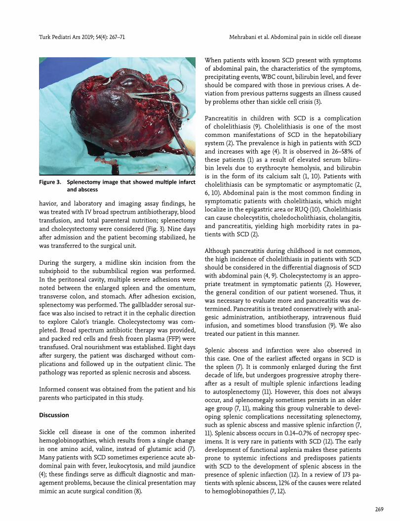
Figure 3. Splenectomy image that showed multiple infarct and abscess

Mehrabani et al. Abdominal pain in sickle cell disease
270
Turk Pediatri Ars 2019; 54(4): 267–71
The diagnosis of splenic abscess is very difficult in chil-dren who frequently present with fever because of other infective reasons and abdominal pain, which is com-monly associated with vaso-occlusive crisis in SCD (12). In a review of 10 patients with splenic abscess and SCD, all presented with fever and abdominal pain (12); however, in our patient, who initially presented with abdominal pain and vomiting without fever, infective causes were less possible. Based on the first ultrasound, only cholelithia-sis and splenomegaly were reported; however, the patient gradually became febrile, and the other diagnosis was made with the presence of persistent leukocytosis. The increased CRP level, high lipase level, and the findings of the second ultrasound indicated pancreatitis; thus, appro-priate treatment was provided. However, as the patient’s general condition worsened in addition to the presence of respiratory distress, the results of the chest X-ray, and tenderness in the LUQ, inflammatory lesions based on the second ultrasound became more possible.
Ahmed et al. (12) reported that splenic abscess should be considered when fever, abdominal pain, and tenderness and enlarged spleen were observed in patients with SCD. Thus, it was necessary to perform other imaging evalua-tions to confirm the diagnosis; a CT scan was performed because inflammation and infarction in the spleen were probable on the second ultrasound. Patients should be evaluated using CT scans if there are doubts with ultra-sound findings because CT is more reliable (7, 12). In our patient, abdominal CT revealed splenic abscess. Some authors recommended aspiration under ultrasound guidance to distinguish between splenic infarction and abscess; however, we performed Doppler ultrasound in which splenic infarction was found (12).
There are two treatment options for splenic abscess: splenectomy and abscess drainage. It is suggested that the treatment of choice for splenic abscess in patients with SCD is splenectomy and antibiotherapy (7, 12) be-cause the mortality rate is higher with percutaneous ab-scess drainage than with splenectomy (12). Our patient was treated with splenectomy. Laparoscopic surgery is mostly performed for cholelithiasis (1) but our patient underwent laparotomy because of the pancreatitis and splenic abscess.
The repeat abdominal ultrasound one week after surgery showed normal pancreas, liver, and biliary tree. Hydrox-yurea was administered daily for the patient. In the follow-up, there was no iron overload, there was no recurrence of abdominal pain attacks, and monitoring was performed as follows; CBC monthly, ferritin every 3 months, cardiac and liver MRI annually (5, 6).
In patients with SCD and acute abdominal pain, vaso-oc-clusive crisis consequences should be considered in mul-tiple gastrointestinal and hepatobiliary organs, especially in the spleen. Furthermore, spleen abscess may occur.
Informed Consent: Verbal informed consent was obtained from patients’ parents who participated in this case.
Peer-review: Externally peer-reviewed.
Author Contributions: Concept - M.S., T.A., O.S.; Design - M.S., T.A., O.S.; Supervision - M.S.; Materials - M.S., T.A., O.S.; Data Collection and/or Processing - M.S., T.A., O.S.; Analysis and/or Interpretation - M.S., T.A., O.S.; Litera-ture Review - M.S.; Writing - M.S.; Critical Review - M.S., T.A., O.S.
Conflict of Interest: No conflict of interest was declared by the authors.
Financial Disclosure: The authors declared that this study has received no financial support.
Hasta Onamı: Sözlü bilgilendirilmiş onam, bu olguya katılan hastaların ebeveynlerinden alınmıştır.
Hakem Değerlendirmesi: Dış bağımsız.
Yazar Katkıları: Fikir - M.S., T.A., O.S.; Tasarım - M.S., T.A.; Denetleme - M.S.; Malzemeler - M.S., T.A., O.S.; Veri To-planması ve/veya İşlemesi - M.S., T.A., O.S.; Analiz ve/veya Yorum - M.S., T.A., O.S.; Literatür Taraması - M.S.; Yazıyı Yazan - M.S.; Eleştirel İnceleme - M.S., T.A., O.S.
Çıkar Çatışması: Yazarlar çıkar çatışması bildirmemişlerdir.
Mali Destek: Yazarlar bu çalışma için mali destek almadık-larını beyan etmişlerdir.
References1. Ebert EC, Nagar M, Hagspiel KD. Gastrointestinal and
Hepatic Complications of Sickle Cell Disease. Clin Gas-troenterol Hepatol 2010; 8: 483−9. [CrossRef ]
2. Gumiero APDS, Bellomo-Brandao MA, Costa-Pinto EAL. Gallstones in children with Sickle Cell Disease followed up at a Brazilian hematology center. Arq Gastroenterol 2008; 45: 313−8. [CrossRef ]
3. Kudsk KA, Tranbaugh RF, Sheldon GF. Acute surgical ill-ness in patients with sickle cell anemia. Am J Surg 1981; 142: 113−7. [CrossRef ]
4. Crastnopol P, Stewart CF. Acute Abdominal Manifesta-tions in Sickle Cell Disease. Arch Surg 1994; 48: 123−5.
5. Al-Salem A. Splenic Complications of Sickle Cell Anemia and the Role of Splenectomy. ISRN Hematol 2011; 2011: 864257. [CrossRef ]
6. Lee MG. Abdominal Pain in Sickle Cell Anaemia. Trop

Mehrabani et al. Abdominal pain in sickle cell disease
271
Turk Pediatri Ars 2019; 54(4): 267–71
Doct 1989; 19: 177−8. [CrossRef ]
7. Buntain WL, Wood JB, Woolley MM. Pancreatitis in childhood. J Pediatr Surg 1978; 13: 143−9. [CrossRef ]
8. Al-Salem A, Qaisaruddin S, Jam’a A, Al-Kalaf J, El-Bashier M. Splenic Abscess and Sickle Cell Disease. Am J Hema-tol 1998; 58: 100−4. [CrossRef ]
9. Amoako MO, Casella JF, Strouse JJ. High Rates of Recur-rent Biliary Tract Obstruction in children with Sickle Cell Disease. Pediatr Blood Cancer 2013; 60: 650−2. [CrossRef ]
10. Ware RE, de Montalembert M, Tshilolo L, Abboud MR. Sickle cell disease. Lancet 2017; 390: 311−23. [CrossRef ]
11. Powars DR, Chan LS, Hiti A, Ramicone E, Johnson C. Outcome of sickle cell anemia: a 4-decade observational study of 1056 patients. Medicine (Baltimore) 2005; 84: 363−76. [CrossRef ]
12. Al-Salem AH. Massive Splenic Infraction in children with sickle cell anemia and the role of splenectomy. Pediatr Surg Int 2013; 29: 281−5. [CrossRef ]