aa, structure function, hair isorders george … structure function, hair isorders george...
TRANSCRIPT

AAD, Structure & Function, Hair Disorders George Cotsarelis, M.D.
Page 1
Introduction to the hair follicle The functional end product of hair follicle proliferation and differentiation is the hair shaft. The hair shaft and surrounding root sheaths are derived from epithelial cells, while the dermal papilla, which is also required for hair growth, is of mesenchymal origin (Fig. 1). The formation of hair follicles takes place during embryogenesis, and no new hair follicles form after birth under homeostatic conditions. However, the character of individual follicles can change drastically over time. Thicker and darker hairs replace fine lightly pigmented hairs in the beard at puberty. Conversely, follicles that produce thick scalp hairs on the scalp miniaturize and generate fine small hairs later in life. Paradoxically, both of these processes occur in response to the hormone testosterone. The hair follicle remodels during cyclical periods of growth (anagen), regression (catagen), rest (telogen) and shedding (exogen) (Fig. 1). During catagen, the lower follicle degenerates through apoptosis of its epithelial components. Regeneration requires the transient activation of slowly cycling epithelial stem cells located in the permanent, bulge region of the follicle. Stem cell progeny form a new follicle matrix during early anagen, and the hair shaft and inner root sheath are derived from these relatively undifferentiated cells. The size and length of the hair shaft correspond to the size of the hair follicle and to the duration of anagen, respectively. These characteristics vary considerably with body site, and change as a result of disease. Pigmentation of the hair shaft depends on hair follicle melanocytes, which reside in the hair follicle bulb and deposit melanin into the growing hair shaft.
ANAGEN II
Signal from dermal papilla to epithelium:
FGF7?
Signal fromepithelium to
dermal papilla:WNT
Epithelialproliferation:
SHH
CATAGEN
Factors requiredfor normal catagen:
FGF5TGFβ1
HRVDR
ANAGEN IV / EXOGEN
Differentiation ofinner root sheath:
GATA3
Factors that causeexogen:
?
IRS
M
HS
KZ
TELOGEN
Signal from dermalpapilla to epithelial
stem cells:?
Factors required foranagen onset:
STAT3RXR
S
B
CHORS
E
DPCTS
ANAGEN I ANAGEN VI
Differentiation ofhair shaft:
WNTBMP
NOTCHFOXN1
HOXC13
C
Figure 1. Factors regulating hair growth and control of the hair follicle cycle. At anagen onset (anagen I) an unknown signal from the dermal papilla is thought to direct transient proliferation of stem cells in the bulge (green arrow). During anagen, signals from the dermal papilla regulate the proliferation of hair matrix cells, while epithelial cells maintain the inductive properties of the dermal papilla (green arrows in base of anagen II follicle). A new hair shaft is produced during anagen, and the old hair is released from the follicle as the new shaft develops (anagen IV / exogen). Lateral signaling between differentiating cells may maintain the separate pathways of differentiation of hair shaft and inner root sheath cells during anagen. During catagen the lower two thirds of the epithelial follicle are destroyed but the dermal papilla remains associated with the regressing follicle. The hair develops a club structure at its proximal end, which retains the hair in the follicle. The follicle then enters the resting, telogen, phase until a new growth cycle is initiated. E, epidermis; CH, club hair; ORS, outer root sheath; CTS, connective tissue sheath; DP, dermal papilla; B, bulge; S, sebaceous gland; HS, hair shaft; KZ, keratogenous zone (differentiating cells); M, matrix; IRS, inner root sheath; C, club.

AAD, Structure & Function, Hair Disorders George Cotsarelis, M.D.
Page 2
AlopeciasDermatologists classify alopecia into scarring (cicatricial) and non-scarring (non-cicatricial) types.
The etiology of cicatricial alopecias is poorly understood, but destruction of the sebaceous gland and/or the stem cells in the bulge may contribute to these disorders. Almost all non-cicatricial alopecias can be thought of as defects in hair follicle cycling; therefore, understanding the hair follicle cycle is critical to understanding alopecia and its potential treatments. Hair follicles traverse through stages of growth (anagen), degeneration (catagen) and rest (telogen). The duration of anagen determines the length of the hair. Controlling transitions between phases and the morphogenesis of the follicle at anagen onset are keys to treating alopecia. Over the last decade, we have developed an understanding of the molecular pathways involved in hair follicle cycling, and these may hold the key to future treatments (for reviews, see (1-3)).
Cicatricial AlopeciasPossible cellular targets in cicatricial alopecia include the sebaceous gland (4) or stem cells in the
hair follicle bulge (1). Inflammation generally involves these structures in lichen planopilaris, discoid lupus erythematosus and graft vs. host disease. These areas are spared in non-cicatricial alopecias such as alopecia areata.The inciting events in cicatricial alopecia are unknown, but could involve aberrant functioning of the sebaceous gland, which appears necessary for normal detachment of the inner root sheath from the hair shaft causing follicle rupture followed by an abnormal fibrotic wound healing process that leads to deletion of the follicle.
Androgenetic alopecia (AGA) In AGA, hair follicles located in specific patterns over the male and female scalp diminish in size over time until they produce effete and cosmetically insignificant hairs (Fig. 2). Testosterone is required, along with a genetic predisposition, for androgenetic alopecia to develop in men. In women, there is no consensus on whether pattern hair loss is truly androgen-dependent, although both male- and
ANAGEN VI EXOGEN /LAG PHASE
ANAGEN II ANAGEN IV ANAGEN VICATAGEN TELOGEN
E
DPM
KZ
IRS
HS
BS
ORSCH
FT
CTS
Figure 2. Miniaturization of hair follicles in androgenetic alopecia. As the follicle regresses during catagen, it is trailed by a collagenous fibrous tract. Following telogen, follicles undergoing miniaturization enter an abnormal, prolonged lag phase, during which the hair shaft is shed. The fibrous tract becomes abnormally thick and may impede subsequent growth of the follicle. E, epidermis; ORS, outer root sheath; DP, dermal papilla; M, matrix; KZ, keratogenous zone; IRS, inner root sheath; HS, hair shaft; CTS, connective tissue sheath; B, bulge; S, sebaceous gland; CH, club hair; FT, fibrous tract.

AAD, Structure & Function, Hair Disorders George Cotsarelis, M.D.
Page 3
female-pattern alopecia result in a decrease in hair follicle size accompanied by a decrease in the duration of anagen and an increase in the percentage of hair follicles in telogen. In addition, several months can transpire between hair shedding and regrowth, a lag period that is absent or fleeting in normal individuals (5) (Fig. 2). These changes result in very short hairs and follicles devoid of hair shafts. Miniaturized hairs also lack pigmentation. In advanced androgenetic alopecia some follicles permanently “drop-out” and develop fibrous tracts (Fig. 2).
The miniaturized hair follicle of AGA has a drastically shortened anagen stage. As the miniaturizing hair follicle enters anagen during each new hair cycle, the new lower follicle is smaller than its predecessor. The signals causing miniaturization likely occur sometime between the end of the previous anagen and the beginning of the new anagen. In the miniaturizing hair follicle, each new hair cycle progressively shortens. The resulting hair shafts become smaller and smaller, both in length and width, so eventually the hair is unapparent clinically. The shortening of anagen results in a greater percentage of follicles in telogen. Thus, miniaturization can be thought of as a defect in hair follicle cycling.
Based on this knowledge, the goals for treating androgenetic alopecia include prolonging anagen, converting telogen follicles to anagen, reversing miniaturization and possibly generating new follicles. What do we know about molecules that may do this? Much information derives from gene knockout and transgenic studies in mice (see Tables at end). Blocking the effects of testosterone and its more active metabolite, dihydrotestosterone, through administration of finasteride accomplishes some of these goals and clearly benefits patients with early androgenetic alopecia; however, even drastic forms of testosterone reduction (e.g. castration) do not result in appreciable reversal of miniaturization.
Telogen effluvium Telogen is a heterogeneous state. Approximately 10% of scalp follicles are normally in telogen (10,000 follicles). We lose ~100 hairs per day, so only 1% of the telogen hairs are shed each day. Stenn coined the term “exogen”for the shedding stage. (6) There are several different mechanisms that can lead to excessive hair shedding (increased exogen). Headington (7) divided telogen effluvium into five different types based on these mechanisms (I have changed the terminology slightly, but see his paper for detailed discussions of these):
Premature Telogen: occurs when anagen follicles are pushed into telogen prematurely, which leads to synchronized exogen. Precipitating events include fevers and some medications.
Delayed Telogen: follicles stay in anagen (anagen is prolonged) and anagen follicles accumulate until they synchronously enter telogen and then exogen. Pregnancy causes anagen accumulation and delivery triggers telogen and synchronized exogen. Oral contraceptives might also mimic this.
Premature Exogen: telogen duration shortens and follicles enter exogen synchronously. Minoxidil and isotretinoin can trigger exogen.
Chronically shortened anagen: shedding is constant due to a higher percentage of follicles in telogen and exogen. This may be the mechanism for “chronic telogen effluvium”.
Prolonged Telogen: follicles stay in telogen for long periods (perhaps due to changes in the light cycle from seasonal variations). Eventually, follicles synchronously enter anagen and the club hairs are shed.
Treatment for telogen effluvium also depends on controlling transitions between stages of the hair cycle. In particular, modulation of exogen could control hair shedding, although no treatment is known to do this. Prolongation of anagen or prevention of catagen would be beneficial since this would obviously prevent telogen and subsequent exogen.
Chemotherapy disrupts proliferation of the matrix keratinocytes in the anagen bulb that produce the hair shaft (Fig. 2). This forces anagen follicles to enter a dystrophic catagen stage, in which the integrity of the hair shaft is compromised and the hair then breaks and falls out. Since over 90% of scalp follicles are in anagen at any one time, these hairs are rapidly lost after chemotherapy, and thus the alopecia is rapid and extensive. Hair loss is one of the most feared side effects of chemotherapy among patients with cancer. However, hair lost

AAD, Structure & Function, Hair Disorders George Cotsarelis, M.D.
Page 4
following chemotherapy does eventually re-grow, presumably because the slowly cycling follicular stem cells are unaffected by chemotherapy and generate a new hair follicle and hair.
Botchkarev et al. showed that p53 is necessary for the development of chemotherapy-induced alopecia, as p53 knockout mice treated with chemotherapeutic agents remarkably do not lose their hair (8). Since chemical inhibitors of p53 already exist, testing their efficacy for local inhibition of p53 in hair follicles provides an potential possibility for preventative therapy.
Alopecia areata is an autoimmune disorder in which cells of the anagen hair bulb cells are attacked by lymphocytes. In a process similar to that following chemotherapy, anagen follicles enter dystrophic catagen and the hair shaft breaks off. Gilhar demonstrated that alopecia areata is a T-cell mediated disease by injecting T-cells from patients into skin grafted to immunodeficient mice. This reproduces the peri-bulbar inflammation and hair loss. Possible targets of the immune attack include matrix keratinocytes, dermal papilla cells and melanocytes. A genetic predisposition for this disease was supported by HLA linkage studies for many years. Candidate genes identified in a genome-wide association study include genes controlling regulatory T cells (Tregs) as well as genes expressed within the hair follicle (9). The hair loss is reversible if patients are treated with immunosuppression, even after years without hair, confirming the non-scarring nature of the inflammatory process, which spares the stem cell rich bulge area.
The development of effective therapies for alopecia, including the ability to block the effects of testosterone in androgenetic alopecia, to recreate hair follicles in patients suffering from advanced androgenetic or inflammatory alopecia, to inhibit exogen in telogen effluvium, and to identify the primary follicular target antigens in inflammatory alopecias, will clearly require a thorough understanding of the molecular processes involved in hair follicle morphogenesis, hair shaft production, and control of the various phases of the hair growth cycle
Selected Molecules Involved in Hair Follicle Cycling and Morphogenesis
Molecules important for transition from anagen to telogen:
Fibroblast Growth Factor 5 (FGF-5): mutations lead to angora phenotype. Hair is ~30-50% longer than normal. Anagen is prolonged. (10)
Hairless: mutations cause atrichia. Follicular development is normal but the follicle falls apart during catagen. The dermal papilla never moves upward and cycling ceases. (11, 12)
Vitamin D Receptor: same phenotype as hairless (13).
Molecules important for transition from telogen to anagen:
Sonic hedgehog (SHH): injection of sonic hedgehog (in adenoviral vector) causes telogen follicles to enter anagen. (14)
Patched: receptor for SHHß-catenin: activation of this pathway in the epithelium induces anagen onset. It’s not clear that this
pathway in involved in normal anagen onset. (15)
Molecules controlling size of follicle:
SHH: knockout mice have diminutive hair follicles. (16, 17)EDAR: a gene variant accounts for larger hair in Asians (21)

AAD, Structure & Function, Hair Disorders George Cotsarelis, M.D.
Page 5
Molecules involved in follicle morphogenesis (neo-genesis)
ß-catenin: excess activated beta-catenin in epidermis results in new hair follicle formation. (18)Noggin: excess noggin accelerates hair follicle morphogenesis. (19)Bone Morphogenetic Proteins (BMPs), Fibroblast Growth Factors (FGFs),TGFß and receptor, Ectodermal dysplasin and its receptor are all involved in hair follicle morphogenesis
but their role in adult hair follicle cycling is not clear.Laminin 10: mice lacking laminin 10 do not develop follicles. Exogenous laminin 10 can partially
restore hair growth (20).Molecules involved in hair shaft differentiation
Notch: may regulate cell-cell interactions between different follicle cell layers.WNT: present in hair shaft precursors, causes premature differentiation of hair shaft when over
expressed.Whn: regulates hair keratin expression, knockout mouse has nude phenotype.BMP: little to no hair shaft differentiation in BMP receptor knockout mice.
Defects of hair shaft (selected):
Monilethrix: mutated hair keratin (hHb6) in cortex causes alternating thick and thin areas in hair shaft (beaded hair).
Netherton’s syndrome: trichorhexis invaginatum (bamboo hair) caused by mutations in a serine-protease inhibitor.
Pili annulati: banded hair, unknown mutation causes defect in hair cortex.Menkes’ Disease: twisted hair (pili torti) caused by defects in copper transporter.Uncombable hair syndrome: unknown mutation causes triangle-shaped hair.Hereditary Mucoepithelial Dysplasia mutation unknown, causes erythematous gums, keratosis pilaris,
episodic hair loss.Naxos syndrome: caused by mutations in plackoglobin, results in tightly curled “wooly” hair,
hyperkeratosis of palms and soles and cardiac arrhythmias.
References
1. Cotsarelis, G., and Millar, S.E. 2001. Towards a molecular understanding of hair loss and its treatment. Mol Med Today 7:293-301.
2. Paus, R., and Cotsarelis, G. 1999. The biology of hair follicles. New England Journal of Medicine 341:491-497.
3. Stenn, K.S., and Cotsarelis, G. 2005. Bioengineering the hair follicle: fringe benefits of stem cell technology. Curr Opin Biotechnol 16:493-497.
4. Zheng, Y., Eilertsen, K.J., Ge, L., Zhang, L., Sundberg, J.P., Prouty, S.M., Stenn, K.S., and Parimoo, S. 1999. Scd1 is expressed in sebaceous glands and is disrupted in the asebia mouse. Nature Genetics 23:268-270.
5. Guarrera, M., and Rebora, A. 1996. Anagen hairs may fail to replace telogen hairs in early androgenic female alopecia. Dermatology 192:28-31.
6. Milner, Y., Sudnik, J., Filippi, M., Kizoulis, M., Kashgarian, M., and Stenn, K. 2002. Exogen, shedding phase of the hair growth cycle: characterization of a mouse model. J Invest Dermatol 119:639-644.

AAD, Structure & Function, Hair Disorders George Cotsarelis, M.D.
Page 6
7. Headington, J.T. 1993. Telogen effluvium. New concepts and review. Archives of Dermatology 129:356-363.
8. Botchkarev, V.A., Komarova, E.A., Siebenhaar, F., Botchkareva, N.V., Komarov, P.G., Maurer, M., Gilchrest, B.A., and Gudkov, A.V. 2000. p53 is essential for chemotherapy-induced hair loss. Cancer Res 60:5002-5006.
9. Petukhova1, L., Duvic, M., Hordinsky, M., Norris, D., Price, V. et al. 2010. Genome-wide association study in alopecia areata implicates both innate and adaptive immunity. Nature. 466: 113-118.
10. Herbert, J., Rosenquist, T., J., G., and Martin, G. 1994. FGF-5 as a regulator of the hair growth cycle: evidence from targeted and spontaneous mutations. Cell 78:1017-1025.
11. Ahmad, W., Faiyaz ul Haque, M., Brancolini, V., Tsou, H.C., ul Haque, S., Lam, H., Aita, V.M., Owen, J., deBlaquiere, M., Frank, J., et al. 1998. Alopecia universalis associated with a mutation in the human hairless gene. Science 279:720-724.
12. Cotsarelis, G., Sun, T.T., and Lavker, R.M. 1990. Label-retaining cells reside in the bulge area of pilosebaceous unit: implications for follicular stem cells, hair cycle, and skin carcinogenesis. Cell 61:1329-1337.
13. Miller, J., Djabali, K., Chen, T., Liu, Y., Ioffreda, M., Lyle, S., Christiano, A.M., Holick, M., and Cotsarelis, G. 2001. Atrichia caused by mutations in the vitamin D receptor gene is a phenocopy of generalized atrichia caused by mutations in the hairless gene. Journal of Investigative Dermatology 117:612-617.
14. Sato, N., Leopold, P.L., and Crystal, R.G. 1999. Induction of the hair growth phase in postnatal mice by localized transient expression of Sonic hedgehog. Journal of Clinical Investigation 104:855-864.
15. Van Mater, D., Kolligs, F.T., Dlugosz, A.A., and Fearon, E.R. 2003. Transient activation of beta -catenin signaling in cutaneous keratinocytes is sufficient to trigger the active growth phase of the hair cycle in mice. Genes Dev 17:1219-1224.
16. Chiang, C., Swan, R.Z., Grachtchouk, M., Bolinger, M., Litingtung, Y., Robertson, E.K., Cooper, M.K., Gaffield, W., Westphal, H., Beachy, P.A., et al. 1999. Essential role for Sonic hedgehog during hair follicle morphogenesis. Developmental Biology (Orlando) 205:1-9.
17. St-Jacques, B., Dassule, H.R., Karavanova, I., Botchkarev, V.A., Li, J., Danielian, P.S., McMahon, J.A., Lewis, P.M., Paus, R., and McMahon, A.P. 1998. Sonic hedgehog signaling is essential for hair development. Curr Biol 8:1058-1068.
18. Gat, U., DasGupta, R., Degenstein, L., and Fuchs, E. 1998. De Novo hair follicle morphogenesis and hair tumors in mice expressing a truncated beta-catenin in skin. Cell 95:605-614.
19. Botchkarev, V.A., Botchkareva, N.V., Roth, W., Nakamura, M., Chen, L.H., Herzog, W., Lindner, G., McMahon, J.A., Peters, C., Lauster, R., et al. 1999. Noggin is a mesenchymally derived stimulator of hair-follicle induction. Nature Cell Biology 1:158-164.
20. Li, J., Tzu, J., Chen, Y., Zhang, Y.P., Nguyen, N.T., Gao, J., Bradley, M., Keene, D.R., Oro, A.E., Miner, J.H., et al. 2003. Laminin-10 is crucial for hair morphogenesis. Embo J 22:2400-2410.
21. Kamberov, Yana G. et al. “Modeling Recent Human Evolution in Mice by Expression of a Selected EDAR Variant.” Cell 152.4 (2013): 691–702. PMC. Web. 18 Mar. 2015.

2/27/17
1
Hair Follicle Biology & Diseases
George Cotsarelis, M.D.Perelman School of Medicine
University of PennsylvaniaStructure and Function, March 6, 2016
DISCLOSURE OF RELATIONSHIPS WITH INDUSTRY
George Cotsarelis, M.D.Structure and Function, March 6, 2016
Follica: SABAllergan: SRAJ&J: SRALilly: ConsultantCassiopea: Consultant
Goals• Understand how changes in hair
follicle cycle cause common alopecias• Foundation for understanding new
findings in hair research• Review (some) important genes
involved in hair growth and development
J. Invest Dermatol July, 2006
Hair Follicle Cycle Concept
• Hair follicle stem cells are located in the bulge: implications for cicatricial vs. non-cicatricial alopecia.
Types of Alopecia
Alopecia AreataLichen planopilaris
Role of Bulge in Alopecia
Scarring Alopecia: LPP, DLE, GVH
Non-Scarring Alopecia:Alopecia Areata
Pathogenesis of Cicatricial Alopecia
• sebaceous gland dysfunction?
• immune dysfunction?
• trauma, infection?
• aberrant structural proteins?
• aberrant fibrotic response?
Pathogenesis of Androgenetic Alopecia
• miniaturization of follicles
• decreased anagen duration
• increased percentage of follicles in telogen
• loss of follicles

2/27/17
2
Androgenetic Alopecia
TerminalMiniaturized
T 5-alpha
reductase type II
Finasteride
( – )
DHT
Switch in Hair Follicle Phenotype Switch in Hair Follicle Phenotype
Lanugo to vellus: 3rd trimester/infancy, trunkVellus to terminal: adolescence, male beardTerminal to vellus: adult, balding scalp
Types of Hair Follicles: Duration of Anagen Determines Hair Length
Terminal vs. Lanugo vs. Vellus vs. MiniaturizedAnagen Duration
Terminal yearsLanugo months Vellus weeksMiniaturized days to weeks
Ambras SyndromeCongenital hypertrichosis
Human Ambras syndrome caused by mutation of TRPS1
Fantauzzo et al. (2008) Human Mol. Genet. 17 3539-3551
Duration of Anagen Determines Hair Length
Eyelash Trichomegaly Angora mouse
FGF-5 knock-out mouse
Duration of Anagen determines hair length (scalp hair grows ~ 0.37 mm/day or~ 6 inches/yr.)
6 yrs Role of FGF-5 in humans?
Anagen duration determines hair loss
No. of hairs lost per day
Duration of Anagen (days)
1000
1000
500
200
*modified from Headington, Archives of Derm, 1993

2/27/17
3
ANAGEN
TELOGEN
ANAGEN
TELOGEN
NORMAL ALOPECIA(androgenetic)
(Scalp) (Eyebrows)
• 100,000 hairs on scalp• ~7% in telogen• telogen hairs are shed• Why don’t we lose 7,000 hairs/day?
Telogen
• 100,000 hairs on scalp• ~7% in telogen• telogen hairs are shed• Why don’t we lose 7,000 hairs/day?
Telogen is a heterogeneous state
Telogen
Hair Follicle Cycle
ANAGEN(90,000)
CATAGEN
TELOGEN(7,000)
EXOGEN (70)
APM
ch
hs
bulge
bulb [
hs
DPDP
DP
Telogen Effluvium
5 types*:1. Premature Telogen2. Delayed Telogen3. Premature Exogen4. Shortened Anagen5. Prolonged Telogen
Documented
Speculative
*modified from Headington, Archives of Derm, 1993
Premature Telogenpremature Anagen to Telogen conversionperhaps most commondue to fever, meds, etc…
AnagenTelogenExogen
time
precipitating event
Prolonged anagen(anagen hair follicles accumulate)
Delivery
(Post-Partum Effluvium)
AnagenTelogenExogen
Pregnancy
AnagenTelogenExogen
precipitating event, e.g. minoxidil
Premature Exogen(rapid onset, club hairs shed
prematurely and synchronously)
Chronic Short Anagen(chronic telogen effluvium?)
No. of hairs lost per day
Duration of Anagen (days)
1000
1000
500
200

2/27/17
4
Prolonged Telogen(telogen follicles accumulate, then
enter exogen synchronously)
time
?precipitating factorschanges in light cycle from seasonal variationmalnutrition
Hair Follicle Cycle
ANAGEN(93,000)
CATAGEN
TELOGEN(7,000)
EXOGEN (70)
APM
ch
hs
bulge
bulb [
hs
DPDP
DP
Catagen Telogen
Hairless gene mutation
Alopecia Universalis Associated with a Mutation in the Human
Hairless GeneAhmad et al., Science.
1998;279:720-724.
Mouse lacking the Hairless gene
Atrichia with Papules- with Rickett’s, indicates vitamin D receptor mutation- without Rickett’s, indicates Hairless mutation
Mice lacking Vitamin D receptor
Miller et al. (2001) J. Invest Dermatol. 117 612-7
Hair Follicle Cycle
CATAGEN
TELOGEN"EXOGEN"
APM
ch
hs
bulge
bulb [DP
DP
DP
ANAGEN
“LAG PHASE”
Concept
• Anagen onset recapitulates hair follicle development (morphogenesis)
courtesy of Anj Dlugosz, Univ. of Michigan (Developmental Biology 205:1-9, 1999)
shh -/- mouse skin
2/27/17
5
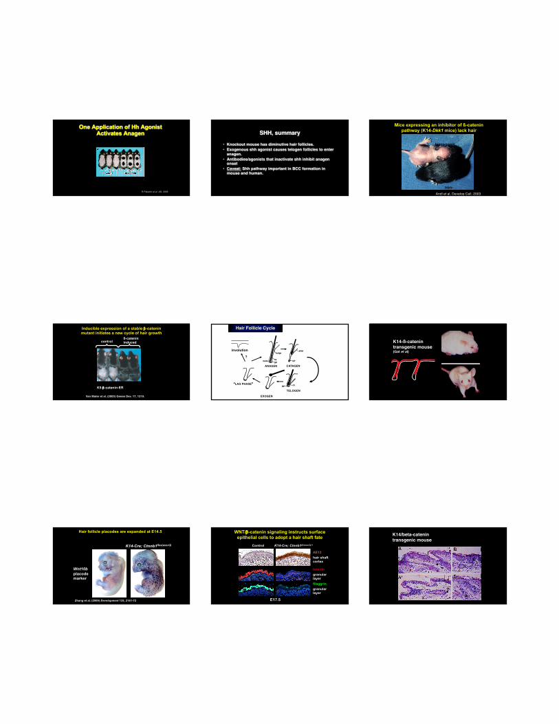
One Application of Hh Agonist Activates Anagen
R Paladini et al, JID, 2005
SHH, summary
• Knockout mouse has diminutive hair follicles.• Exogenous shh agonist causes telogen follicles to enter
anagen.• Antibodies/agonists that inactivate shh inhibit anagen
onset• Caveat: Shh pathway important in BCC formation in
mouse and human.
Mice expressing an inhibitor of ß-catenin pathway (K14-Dkk1 mice) lack hair
transgenic
non-transgenicAndl et al, Develop Cell, 2003
Inducible expression of a stable b-catenin mutant initiates a new cycle of hair growth
K5-b-catenin-ER transgenic
Van Mater et al. (2003) Genes Dev. 17, 1219.
controlß-catenin induced
Hair Follicle Cycle
CATAGEN
TELOGEN
EXOGEN
APM
ch
hs
bulge
bulb [DP
DP
DP
ANAGEN
“LAG PHASE”
involution
?
K14-ß-catenintransgenic mouse(Gat et al)
K14-Cre; Ctnnb1flox(exon3)
Control
Hair follicle placodes are expanded at E14.5
Wnt10bplacode marker
Zhang et al. (2009) Development 135, 2161-72
filaggringranular layer
loricringranular layer
E17.5
AE13hair shaft cortex
WNT/b-catenin signaling instructs surface epithelial cells to adopt a hair shaft fate
Control K14-Cre; Ctnnb1fl(exon3)/+
K14/beta-catenintransgenic mouse

2/27/17
6
K14/beta-catenin transgenic mouse skin forms tumors
Cadieu et al., Science, 2009
3 genes determine hair phenotype:FGF5-hair lengthR-spondin (wnt activator)-hair sizeKeratin71-curliness
EDAR mutation causes thicker hair in Asians
Kamberov YG, Wang S, Tan J, et al. Modeling recent human evolution in mice by expression of a selected EDAR variant. Cell. 2013;152(4):691-702.
Summary• Destruction of stem cells in bulge may
lead to cicatricial alopecias• Anagen duration determines hair
length• Hair cycle transitions important• Anagen onset recapitulates hair
follicle developmen• Specific genes control hair phenotype