a systems biology strategy to identify molecular ... · a systems biology strategy to identify...
TRANSCRIPT

A Systems Biology Strategy to IdentifyMolecular Mechanisms of Action andProtein Indicators of Traumatic BrainInjury
Chenggang Yu,1 Angela Boutt�e,2 Xueping Yu,1 Bhaskar Dutta,1 Jacob D. Feala,1
Kara Schmid,2 Jitendra Dave,2 Gregory J. Tawa,1 Anders Wallqvist,1 andJaques Reifman1*1Department of Defense Biotechnology High Performance Computing Software Applications Institute,Telemedicine and Advanced Technology Research Center, U.S. Army Medical Research and MaterielCommand, Fort Detrick, Maryland2Department of Brain Trauma Neuroprotection and Neurorestoration, Center for Military Psychiatry andNeuroscience, Walter Reed Army Institute of Research, Silver Spring, Maryland
The multifactorial nature of traumatic brain injury (TBI),especially the complex secondary tissue injury involvingintertwined networks of molecular pathways that mediatecellular behavior, has confounded attempts to elucidatethe pathology underlying the progression of TBI. Here,systems biology strategies are exploited to identify novelmolecular mechanisms and protein indicators of braininjury. To this end, we performed a meta-analysis of fourdistinct high-throughput gene expression studies involvingdifferent animal models of TBI. By using canonical path-ways and a large human protein-interaction network as ascaffold, we separately overlaid the gene expression datafrom each study to identify molecular signatures that wereconserved across the different studies. At 24 hr afterinjury, the significantly activated molecular signatureswere nonspecific to TBI, whereas the significantly sup-pressed molecular signatures were specific to the nervoussystem. In particular, we identified a suppressed subnet-work consisting of 58 highly interacting, coregulated pro-teins associated with synaptic function. We selected threeproteins from this subnetwork, postsynaptic density pro-tein 95, nitric oxide synthase 1, and disrupted in schizo-phrenia 1, and hypothesized that their abundance wouldbe significantly reduced after TBI. In a penetratingballistic-like brain injury rat model of severe TBI, Westernblot analysis confirmed our hypothesis. In addition, ouranalysis recovered 12 previously identified protein bio-markers of TBI. The results suggest that systems biologymay provide an efficient, high-yield approach to generatetestable hypotheses that can be experimentally validatedto identify novel mechanisms of action and molecularindicators of TBI. VC 2014 The Authors. Journal of Neuro-
science Research Published by Wiley Periodicals, Inc.
Key words: traumatic brain injury; systems biology;pathway analysis; protein–protein interaction networks;biomarkers
Combat injuries from the military conflicts in Iraqand Afghanistan as well as the suicides of recently retiredU.S. football players have brought to light the short- andlong-term consequences of traumatic brain injury (TBI;DeKosky et al., 2010; MacGregor et al., 2010). The pri-mary insult to the head leads to secondary tissue injury,which is manifested by both immediate and delayed neu-rological deficits and disease (DeKosky et al., 2010; Baughet al., 2012; Feala et al., 2013). The secondary injury pro-cess involves an intertwined cascade of evolving biochem-ical molecular interactions that mediate neuronal damageover hours to months after the initial trauma (Ulitsky andShamir, 2007; Greve and Zink, 2009; Feala et al., 2013).To date, much remains unknown about how thesemolecular responses to injury and their poorly understoodpathways are linked to clinical outcomes.
Systems biology provides an opportunity to helpshed light on the extremely complex, multifactorial natureof the TBI secondary injury. Unprecedented advance-ments in the ability to generate high-throughput, large-scale genomic and proteomic data have led to the
Contract grant sponsor: U.S. Department of Defense Medical Research
and Development Program; Contract grant number: D61_I_10_J6_126;
Contract grant sponsor: U.S. Army Network Science Initiative; Contract
grant sponsor: Combat Casualty Care Research Area Directorate of the
U.S. Army Medical Research and Materiel Command.
*Correspondence to: Jaques Reifman, PhD, Senior Research Scientist,
Director, Department of Defense Biotechnology High Performance
Computing Software Applications Institute, Telemedicine and Advanced
Technology Research Center, U.S. Army Medical Research and Materiel
Command, MCMR-TT 504 Scott Street, Fort Detrick, MD 21702.
E-mail: [email protected]
Received 2 June 2014; Revised 26 August 2014; Accepted 24 September
2014
Published online 14 November 2014 in Wiley Online Library
(wileyonlinelibrary.com). DOI: 10.1002/jnr.23503
VC 2014 The Authors. Journal of Neuroscience Research Published by Wiley Periodicals, Inc.This is an open access article under the terms of the Creative Commons Attribution-NonCommercial-NoDerivs License, which permits use anddistribution in any medium, provided the original work is properly cited, the use is non-commercial and no modifications or adaptations aremade.
Journal of Neuroscience Research 93:199–214 (2015)

abstraction, construction, and graphic representation ofbiological networks and the ability to integrate comple-mentary, disparate data sets in a systems-level analysis. Tothis end, systems biology allows for the holistic and sys-tematic analysis of experiment-specific, high-throughputgenomic and proteomic data within the context ofcondition-agnostic, canonical biological networks (Idekeret al., 2001). Ultimately, systems biology could be used togenerate experimentally testable hypotheses.
Shojo et al. (2010) recently integrated gene expres-sion microarray data from a fluid percussion injury (FPI)model of TBI in rats with molecular pathways to generatesystems-level hypotheses and to suggest causal temporalrelationships between inflammatory and apoptotic systemsduring the acute phase of TBI (<6 hr). These findings reaf-firmed the involvement of inflammatory and survival sig-naling pathways that were independently observed by adifferent group using a systems biology approach (Kobeissyet al., 2008). The authors also hypothesized pathway linksbetween TBI and synaptic plasticity. In another study, thefunction of proteins in molecular pathways was exploitedto rank order and down-select potential TBI biomarkersfrom a list of candidates (Mondello et al., 2011).
Recently, our group reviewed strategies and poten-tial opportunities for using systems biology to gain insightsinto the underlying molecular mechanisms of the TBIresponse and to identify novel protein indicators of braininjury (Feala et al., 2013). Through an illustrative set of32 proteins, we showed how to use biological networksas a scaffold and overlay protein data to achieve thesegoals. Here, we built on these concepts and performed ameta-analysis of four large-scale gene expression data setsfrom distinct murine models of TBI and integrated themwith biological networks to identify molecular mecha-nisms of action and novel protein indicators of TBI. Weobserved that, whereas the significantly activated biologi-cal networks were primarily associated with the immunesystem, the significantly suppressed networks were spe-cific to the nervous system, particularly synaptic function.Accordingly, we hypothesized that constituent proteinsfrom the suppressed networks were downregulated and
TBI specific. Experimental testing of three such proteinsby using a validated penetrating ballistic-like brain injury(PBBI) rat model of severe TBI (Williams et al., 2005)supported our hypothesis and systems biology strategy.
The strategy is based on the overarching hypothesisthat certain robust molecular signatures of TBI are con-served in a meta-analysis of distinct studies involving dif-ferent animal types, injury models, severity levels, andbrain tissues. Implicit in this overarching hypothesis aretwo component hypotheses. First, a systems approachintegrating gene expression data with biological networkinformation is more likely to reveal common responsemechanisms of TBI that are conserved across studies thanthe sole analysis of individual genes. Second, a systemsapproach, focused on coregulated and interconnectedproteins, is effective in recovering known and identifyingnovel molecular mechanisms of action of TBI.
MATERIALS AND METHODS
Data Collection and Processing
In our meta-analysis, we used four publicly available geneexpression microarray data sets from murine studies of moderateand severe TBI 24 hr after injury (Table I; Matzilevich et al.,2002; Natale et al., 2003; Babikian et al., 2010). These studiesincluded two different experimental models of TBI (controlledcortical impact [CCI] and FPI), two animal types (mouse andrat), and two tissue types (cortex and hippocampus). Each studyconsisted of two cohorts of animals, TBI-induced animals andsham animals (i.e., controls). We downloaded the raw geneexpression microarray data sets from the Gene ExpressionOmnibus repository (http://www.ncbi.nlm.nih.gov/geo/) inSeptember, 2011, and concurrently normalized the four datasets by using the robust multiarray average method imple-mented in the Bioconductor R-language suite of bioinformaticstools (Irizarry et al., 2003). This uniform standardization acrossthe studies ensured an unbiased analysis.
Systems Biology Strategy
Figure 1 illustrates the systems biology strategy. Westarted by performing computational analyses to generate
TABLE I. Summary of the Four TBI Gene Expression Data Sets
Data set name* M-CCI R-FPIm R-FPIs R-CCI
Reference Natale et al. (2003) Natale et al. (2003) Babikian et al. (2010) Matzilevich et al. (2002)
Model organism† Mouse (C57BL/6) Rat (Sprague-Dawley) Rat (Sprague-Dawley) Rat (Long-Evans)
TBI model CCI FPI FPI CCI
TBI severity Moderate Moderate Severe Severe
Tissue type Cortex Cortex Cortex Hippocampus
Time (hr after injury) 4, 8, 24, 72 0.5, 4, 8, 24, 72, 504 0.5, 4, 24 3, 24
Microarray platform 74Av2 U34A U34A U34A
Number of probes 12,488 8,799 8,799 8,799
Number of genes 7,934 4,554 4,554 4,554
Upregulated‡ 667 (8.4%) 397 (8.7%) 338 (7.4%) 314 (6.9%)
Downregulated 835 (10.5%) 388 (8.5%) 372 (8.2%) 296 (6.5%)
*M-CCI, mouse controlled cortical impact (CCI) model; R-FPIm, rat fluid percussion injury (FPI) model with moderate injury; R-FPIs, rat FPI
model with severe injury; R-CCI, rat CCI model.†All model organisms are male.‡Up-/downregulated genes were determined by using the RankProd method with a P< 0.05 cutoff (Hong et al., 2006).
200 Yu et al
Journal of Neuroscience Research

testable hypotheses regarding molecular mechanisms of actionand protein indicators of TBI, which were then experimentallytested in the laboratory. In the computational analyses, we pur-sued two parallel approaches for each of the four microarraydata sets. In the first approach, we separately integrated eachdata set with molecular pathways from a list of well-establishedcanonical signaling and disease pathways to identify those spe-cific to TBI 24 hr after injury. Similarly, in the secondapproach, we separately integrated each data set with a humanprotein–protein interaction (PPI) network to identify regions inthe network (modules and subnetworks) representing molecularmechanisms of action specific to TBI. To generate hypothesesabout molecular mechanisms and protein indicators of TBI, weconsidered the concordance (i.e., the conservation) of the
results across the data sets for each approach and between theapproaches.
Because the gene expression data sets consisted of genesfrom rats and mice, whereas the biological networks (molecularpathways and PPI network) were represented by human genesand human proteins, respectively, for analyses we mapped thedata to a common terminology, human genes, following theapproach proposed by Zhang et al. (2011). We assumed a one-to-one mapping between a protein and its coding gene (i.e., weignored protein isoforms and other translational modifications)and used Entrez Gene ID (http://www.ncbi.nlm.nih.gov/gene)to map genes and proteins, which henceforth are used inter-changeably. To map genes from rat and mouse to human, weused orthology tools (Eppig et al., 2012; C. Yu et al., 2012).
Fig. 1. Illustration of the systems biology strategy, in which we usedcomputational analysis to generate testable hypotheses that can beexperimentally validated in the laboratory. We started by perform-ing a meta-analysis of four gene expression data sets and separatelyoverlaying each data set onto two types of biological networks,canonical molecular pathways and PPI networks. In these analyses,we considered up- and downregulated genes separately because pre-vious findings support the hypothesis that biological processes arecharacterized by interacting, coregulated proteins. Next, we identi-
fied statistically significant molecular mechanisms of action and pro-tein indicators of TBI that were conserved across the studies andanalyses. Finally, we experimentally tested protein indicators of TBIwith an in vivo animal model. The right side of the figure illustratesthe construction of a PPI subnetwork (shaded area) formed by threeproteins (red circles, denoting module centers C1, C2, and C3 fromthree protein modules) and three other proteins (green circles) fromthese modules. KEGG, Kyoto Encyclopedia of Genes andGenomes.
Systems Biology for Characterizing TBI 201
Journal of Neuroscience Research

Pathway Enrichment Analysis
Canonical molecular pathways provide “wiring diagrams”describing how gene products and other biomolecules interact,relate, and regulate each other to perform particular biologicalfunctions. Molecular pathways are often constructed by manualcuration of literature data, which are then compiled into largedatabases. We collected 143 nonmetabolic canonical signalingand disease pathways representing 4,672 proteins from theKyoto Encyclopedia of Genes and Genomes (KEGG; Kanehisaet al., 2008; http://www.genome.jp/kegg/pathway.html),downloaded in December, 2011. KEGG, one of the largest andmost widely used publicly available pathway databases, anno-tates pathways by using biological functional categories, such asnervous system, immune system, cell growth and death, andinfectious diseases, which we used to identify molecular mecha-nisms specific to TBI. This was achieved by identifying path-ways that were significantly regulated by TBI, i.e., pathwaysthat were “enriched” with differentially expressed genes afterbrain injury, and associating their annotated biological func-tional category with brain injury.
To identify molecular pathways significantly regulated(activated by upregulated genes or suppressed by downregulatedgenes) at 24 hr after brain injury, we separately integrated theKEGG pathways with each of the four gene expression data setsby using the recently developed PathNet algorithm (Duttaet al., 2012). PathNet performs an extension of hypergeometricstatistical test (Breitling et al., 2004), which identifies signifi-cantly regulated pathways by assessing whether the number ofdifferentially expressed genes in the pathway is statistically sig-nificantly higher than would be expected by chance. However,unlike the hypergeometric test, which treats pathways as unor-dered collections of genes, PathNet capitalizes on the wiringpattern, or connectivity, among the genes in and between path-ways to determine a pathway’s significance within the contextof the gene expression data.
PathNet separately rank ordered the association of the143 signaling and disease pathways with TBI for each of thefour data sets. We used the rank product method, a rank-based,nonparametric statistical test implemented in the RankProdroutine of Bioconductor (Hong et al., 2006), to compute thestatistical significance of the differential expression of the genesin each data set. The rank product method has been shown toproduce differentially expressed gene lists that are more repro-ducible across laboratories and experimental models than othermethods, especially when a small number of samples is available(Shi et al., 2006). We separately identified significantly activated(upregulated) pathways and significantly suppressed (downregu-lated) pathways by using a false discovery rate (FDR)-correctedP value of 0.05 as the statistical significance cutoff for bothgenes and pathways.
PPI Network Analysis
High- and low-throughput experimental assays are avail-able to screen physical interactions among proteins within andbetween species (Rual et al., 2005; Konig et al., 2008). Forinterpretation and analysis, these interactions are often repre-sented as PPI networks, in which network nodes correspond toproteins and edges between nodes represent the observed pro-
tein interactions. We constructed a comprehensive human PPInetwork (consisting of 74,376 physical PPIs among 11,789human proteins) by aggregating experimentally obtained pro-tein interaction data from nine publicly available databases (X.Yu et al., 2012), the Biomolecular Interaction Network Data-base (Bader et al., 2003), the Biological General Repository forInteraction Data Sets (Stark et al., 2006), the Database of Inter-acting Proteins (Salwinski et al., 2004), the Human ProteinReference Database (Peri et al., 2003), IntAct (Aranda et al.,2010), the Molecular Interaction database (Chatr-aryamontriet al., 2007), the mammalian PPI database of the Munich Infor-mation Center on Protein Sequences (Pagel et al., 2005),PDZBase (a PPI database for PDZ domains; Beuming et al.,2005), and Reactome (Vastrik et al., 2007). The databases weredownloaded from their corresponding web sites in October,2011.
To identify regions in the human PPI network that weresignificantly regulated (activated or suppressed) at 24 hr afterbrain injury, we separately integrated each of the four geneexpression data sets with the PPI network. In particular, wesearched for regions in the network where groups of connectedproteins were coregulated, i.e., where groups of proteins tendedto be either up- or downregulated together. We analyzed twotypes of PPI network regions, modules and subnetworks (con-structed by combining a subset of proteins from the modules).
Module identification. For each protein in the PPInetwork, we defined a module as a set of connected proteinsconsisting of that protein (or module center Ci in Fig. 1) and itsdirectly interacting protein partners (black circles connected toCi). To identify TBI-specific, coregulated modules, we fol-lowed a statistical randomization procedure similar to that inour previous work detailed by Yu et al. (2011). Briefly, weidentified TBI-specific modules that were either significantlyactivated (upregulated) or significantly suppressed (downregu-lated) by first computing the aggregate gene expression value ofthe constituent proteins in each module. Then, we randomlyswitched the gene expression level of each gene in all samplesand recomputed the aggregate gene expression value for eachmodule. We repeated this randomization 100 times and identi-fied significantly activated modules (significantly suppressedmodules). This was achieved by identifying those moduleswhose aggregate expression levels were larger (smaller) than95% of their randomized aggregate expression levels.
Subnetwork identification. To identify larger regionsof the PPI network from which to infer novel molecular mech-anisms of action, we separately constructed activated and sup-pressed subnetworks from the corresponding statisticallysignificant modules. In constructing subnetworks, we attemptedto retain topologically important proteins from modules thatwere statistically significant in two or more of the four studies(i.e., modules whose significance was preserved across studies).A protein’s topological importance considered the number ofits interactions with other proteins, which we assumed to implyfunctional importance, and was used later in the downselectionof protein indicators. Accordingly, we constructed interacting,coregulated subnetworks by retaining the module centers of theconserved modules and all other proteins in these modules thatinteracted with at least one other module center. Figure 1 pro-vides an example of such a subnetwork (shaded area at right in
202 Yu et al
Journal of Neuroscience Research

the figure), constructed from three module centers (C1, C2, andC3) and three other proteins (green circles) that interacted withat least two module centers.
Subnetwork temporal profile. By using the differenttime points for which gene expression data were available(Table I), we calculated the temporal gene expression profile ofa subnetwork as a function of time. At each time point, wecomputed the aggregate gene expression value of the constitu-ent genes in the subnetwork and normalized the results suchthat zero represented no gene expression change, positive valuesindicated subnetwork activation, and negative values indicatedsubnetwork suppression.
Function enrichment analysis. Similarly to the path-way analysis, we also performed enrichment analysis (with thehypergeometric test) for the significantly regulated PPI modulesand subnetworks. The analysis determined whether the numberof proteins associated with a particular biological function in amodule (or in a subnetwork) was significantly higher thanwould be expected by chance. However, because these net-work regions represent uncharacterized biological functions(unlike pathways), we used the web service resources of theDatabase for Annotation, Visualization, and Integrated Discov-ery (DAVID; version 6.7; http://david.abcc.ncifcrf.gov; Huanget al., 2009) to associate modules and subnetworks with biolog-ical functions in the form of disease, tissue, and gene ontology(GO) terms (Ashburner et al., 2000). We accessed DAVID inFebruary, 2012, and used a significance cutoff of 0.05 (FDR-corrected P).
Known Biomarker Candidates and Downselection ofProtein Indicators
We used a list of 32 previously identified TBI protein bio-marker candidates (see Table I; Supp. Table S1 in Feala et al.,2013) to assess the ability of the significantly regulated pathwaysand subnetworks to recover such proteins. Some of these bio-marker candidates have garnered multiple literature citations, areassociated with TBI through diverse biological roles reported inclinical and laboratory studies, and have been found to beexpressed in serum, cerebrospinal fluid, and brain tissue.
The molecular pathways and PPI network analyses gener-ated distinct evidence for implicating protein indicators with braininjury 24 hr after the insult. Hence, to identify potential proteinindicators of TBI, we considered a protein’s 1) topological impor-tance in a subnetwork, 2) presence in a significantly regulatedpathway, 3) association with neurological diseases, 4) functionalimportance in the central nervous system, and 5) literaturereview. Subsequently, we experimentally validated a subset of theidentified protein indicators in an in vivo animal model of TBI.
Experimental Animal Model Validation
Animals and surgical procedures. Male Sprague-Dawley rats weighing 250–300 g (Charles River Laboratories,Raleigh, NC) were used for these studies and housed individu-ally under a normal 12-hr light/dark cycle (lights on at 6:00AM). For PBBI and sham surgery, animals were anesthetizedwith 5% isoflurane delivered in oxygen. The body temperatureof each animal was maintained at 37�C with a heating blanket(Harvard Apparatus, Holliston, MA). Prior to biospecimen col-
lection, animals were anesthetized with 70 mg/kg ketamineand 6 mg/kg xylaxine. Facilities at the Walter Reed ArmyInstitute of Research (WRAIR) are accredited by the Associa-tion for Assessment and Accreditation of Laboratory AnimalCare International. The experimental procedures wereapproved by the WRAIR Animal Care and Use Committee.Research was conducted in compliance with the Animal Wel-fare Act and other federal statutes and regulations relating toanimals and experiments involving animals and adhered to prin-ciples stated in the Guide for the care and use of laboratory animals(National Research Council Publication, 2011 edition).
PBBI model. We separated the six rats into twoequally sized groups and applied a PBBI (as previously describedby Williams et al. [2006a,2006b]) to one group and a shamoperation to the other group. Experimental PBBI has beenextensively characterized and reproduces the temporary cavityleft in the brain to mimic the ballistic nature of a high-velocitybullet wound. Briefly, a 10% unilateral frontal PBBI wasinduced in rats by stereotaxic insertion of a specially designedprobe into the right hemisphere of the brain. The probe wasinserted through a cranial window over the frontal cortex, andrapid inflation/deflation of the water-filled balloon was used tocreate a temporary cavity in the cerebrum. Sham rats receivedidentical surgical procedures without balloon expansion. Inboth groups, animals were sacrificed 24 hr after surgery. A 2-mm slice (6 mm from bregma) of contralateral tissue and ipsilat-eral tissue was flash frozen in liquid nitrogen and stored at280�C until use (three animals each for PBBI and sham).
Western blot analysis. For Western blotting, wethawed ipsilateral and contralateral tissues on ice and sonicatedthem in 13 radioimmunoprecipitation assay buffer (Millipore,Billerica, MA) supplemented with a protease/phosphataseinhibitor mix (Thermo Fisher Scientific, Rockford, IL). Lysatewas then centrifuged at 10,000g for 20 min, and the supernatantwas stored. We determined protein concentrations with thebicinchoninic acid assay (Thermo Fisher Scientific, Waltham,MA). After normalization, 10 mg of total protein from eachsample was separated with 4–12% NuPAGE gels (Life Technol-ogies, Grand Island, NY) and transferred to polyvinylidenedifluoride membranes. We probed blots with antibodies forparticular proteins (Abcam, Cambridge, MA; Cell SignalingTechnology, Danvers, MA), and we performed densitometryand background subtraction with an LAS 4000 in ImageQuantTL v 7.0 (GE Healthcare, Piscataway, NJ).
RESULTS
Significant Pathways Conserved Across Data sets
In separate analyses of up- and downregulated genesfor each of the four gene expression data sets, we identi-fied a total of 61 significantly activated pathways and 36significantly suppressed pathways. Among these, 14 (23%)activated pathways and 19 (53%) suppressed pathwayswere conserved in three or more data sets (Fig. 2). Thesefractions of conserved pathways across the studies weremuch higher than the fractions obtained in a separateanalysis in which we combined the up- and downregu-lated genes together (only one of 38 significant pathways[2.6%] was conserved in three data sets).
Systems Biology for Characterizing TBI 203
Journal of Neuroscience Research

Figure 2 lists the 33 conserved pathways, whichshow a sharp contrast in the functional categories betweenthe activated (left column) and suppressed (right column)pathways. Ten (71%) of the fourteen activated pathwayswere related to immune response or infectious diseases,and seven (37%) of the 19 suppressed pathways wererelated to the nervous system. Our analysis captured seven(78%) of nine nervous system pathways represented in theKEGG database, with five pathways conserved in all fourdata sets and two pathways conserved in three data sets.Conversely, no activated pathway was related to the nerv-ous system, and no suppressed pathway was related toimmune response or infectious diseases.
Significant PPI Modules Conserved Across DataSets
We identified a total of 226 significantly activatedPPI modules, of which 27 (12%, consisting of 2,119 pro-teins) were conserved in two or more data sets (Fig. 3).
Similarly, we identified 53 significantly suppressed PPImodules, of which six (11%, consisting of 296 proteins)were conserved in two or more data sets (Figs. 3, 4A).None of the 27 activated modules and only half of the sup-pressed modules were significant in the R-CCI data set.This finding was consistent with the results of the pathwayanalysis described above, in which the R-CCI study iden-tified the lowest number of significant pathways (Fig. 2).
To determine the biological functions associatedwith the conserved PPI modules, we first characterizedthe functions of the module center proteins. Among theactivated modules, over 50% were associated withimmune response or apoptosis, whereas all center proteinsfor the suppressed modules were associated with synapticfunction (Fig. 3). We then extended the analysis and char-acterized the entire set of constituent proteins of the con-served modules (2,119 for the activated modules and 296for the suppressed modules) in terms of four functionalcategories: GO biological process, GO cellular compo-nent, disease association, and tissue type. Table II shows
TABLE II. Enrichment Analysis of the Proteins in the 27 Activated Protein–Protein Interaction (PPI) Modules and the Six Suppressed
PPI Modules That Were Conserved in at Least Two of the Four Data Sets
Category
Activated modules* Suppressed modules/subnetwork†
Term
Protein modules‡
Term
Protein
Modules§ Subnetwork§
Count Percentage Count Percentage Count Percentage
GO biological process
Translational elongation 84 4.0 Transmission of nerve impulse 46 15.9 18 31.0
Regulation of cellular protein metabolic process 207 9.8 Synaptic transmission 43 14.9 17 29.3
Translation 140 6.7 Cell–cell signaling 56 19.4 20 34.5
Regulation of programmed cell death 282 13.4 Regulation of synaptic transmission 27 9.3 15 25.9
Regulation of cell death 282 13.4 Regulation of neurological system process 28 9.7 15 25.9
GO cellular component
Cytosol 504 24.0 Synapse 60 20.8 26 44.8
Cytosolic ribosome 68 3.2 Synapse part 48 16.6 23 39.7
Cytosolic part 93 4.4 Cytoskeletal part 82 28.4 25 43.1
Ribosomal subunit 76 3.6 Cytoskeleton 100 34.6 26 44.8
Ribosome 96 4.6 Postsynaptic membrane 34 11.8 17 29.3
Disease association
Lupus erythematosus 38 1.8 Schizophrenia 23 8.0 16 28.0
Breast cancer 101 4.8 Huntington’s disease 5 1.7 3†† 5.2
Colorectal cancer 75 3.6 Cognitive function 6 2.1 2†† 3.4
Crohn’s disease 28 1.3 Schizophrenia/bipolar disorder 4†† 1.4 3†† 5.2
Ovarian cancer 37 1.8 Bipolar disorder 9†† 3.1 2†† 3.4
Tissue type
Cajal-Retzius cell 115 5.5 Brain 191 66.1 50 86.2
Fetal brain cortex 115 5.5 Platelet 29 10.0 3†† 5.2
Epithelium 570 27.1 Fetal brain 31 10.7 10 17.2
B-cell lymphoma 68 3.2 Fetal brain cortex 16 5.5 3†† 5.2
Platelet 175 8.3 Epithelium 78 27.0 13†† 22.4
*Involving 2,119 proteins (2,100 annotated in DAVID).†Involving 296 proteins (289 annotated in DAVID).‡Number and fraction among the 2,100 proteins in the activated modules annotated by DAVID with a specific term. A protein may have multiple dis-
tinct annotations in a given category.§Number and fraction among the 289 proteins in the suppressed modules (or 58 proteins in the subnetwork) annotated by DAVID with a specific
term. A protein may have multiple distinct annotations in a given category.
Italic indicates terms associated with the nervous system.††Not statistically significant (P> 0.05); for all other entries P< 0.05.
204 Yu et al
Journal of Neuroscience Research

the five most enriched terms for each of the four catego-ries in order of decreasing statistical significance (exceptfor subnetwork entries). For the 2,119 proteins in the 27activated modules, the functional characterization wasnonspecific in each of the four categories, involving termsassociated with a whole host of biological functions,including cell death. Only two entries were associatedwith the central nervous system (tissue type: Cajal-Retzius cell and fetal brain cortex), but each had a lowprevalence (<6%). In sharp contrast, the 296 proteins inthe six suppressed modules were both strongly associatedwith and prevalent in neurological processes and cellularcomponents, neurological diseases, and brain tissues. Forexample, 21% of the proteins were associated with neuro-nal synapse and 66% of the proteins were expressed inbrain tissues.
Synaptic Subnetwork
Starting with the 296 proteins in the six suppressedmodules (Fig. 4A), we constructed a subnetwork byretaining the six protein module centers and 52 proteinsthat interacted with at least two module centers (Fig. 4B;Supp. Info. Tables I, II). When compared with the 296proteins, the subnetwork comprising these 58 proteins wasfurther enhanced with neurological-function-related anddisease-related proteins. For example, 45% (26 of 58 cor-responding to P< 2.0 3 10220) of the proteins in the sub-network were associated with synapses and 28% (16 of 58corresponding to P< 8.0 3 1023) were associated withschizophrenia, whereas the fractions of corresponding pro-teins in the six modules were 21% and 8%, respectively(Table II). In addition to the 26 proteins annotated as syn-apse, we found that the subnetwork also included three
Fig. 2. Significantly regulated pathways conserved in at least three of the four data sets.
Systems Biology for Characterizing TBI 205
Journal of Neuroscience Research

other proteins, disrupted in schizophrenia 1 (DISC1), syn-aptic Ras GTPase-activating protein 1, and discs large(Drosophila) homolog-associated protein 4 that play a sig-nificant role in synaptic function. Given the significantoverrepresentation of proteins associated with synapticfunction in this group of interacting, coregulated genes,we termed this suppressed network the synaptic subnetwork.
To investigate the functional characteristics of thesynaptic subnetwork further, we analyzed its temporalexpression changes from 30 min to 21 days after injury byusing the R-FPIm data set (Table I). Figure 5 shows thetemporal profile of the normalized aggregate gene expres-
sion values, indicating that the genes in the synaptic sub-network tended to be attenuated during the 21-dayperiod, reaching a nadir between 24 and 72 hr after injuryand eventually returning to baseline at 21 days. A similaranalysis with the more limited M-CCI data set (Table I)corroborated these results.
Recovering Previously Identified and InferringNovel TBI Protein Indicators
We also hypothesized that the systems biology strat-egy would be able to recover previously identified TBI
Fig. 3. Significantly regulated protein–protein interaction (PPI) modules conserved in at least two ofthe four data sets.
206 Yu et al
Journal of Neuroscience Research

biomarker candidates and infer novel protein indicators. Totest the former hypothesis, we investigated the occurrenceof the 32 proteins previously implicated with TBI in thesignificantly regulated pathways and subnetworks discussedabove. Among these 32 proteins, 30 were represented inthe human PPI network and 24 were annotated in theKEGG database. We located 12 (38%) of the 32 proteins inthe significantly regulated pathways and subnetworks con-served across the studies. Table III shows that we observedfive of the 12 proteins in both analyses, eight in the PPIsubnetworks (one suppressed and seven activated) and 10 inthe pathways (four suppressed and six activated; see Fig. 2).We believe that this was not a chance observation because,for example, the probability of recovering at least eight of32 proteins in an equally sized random PPI network is lessthan one in 5,000 (P< 2.0 3 1024).
We also used the systems biology strategy to generatetestable hypotheses and infer novel protein indicators of TBI.In particular, we focused our analysis on the constituent pro-teins of the synaptic subnetwork, considering not only theirtopological importance (which could lead to truly novel bio-markers) but also distinct complementary evidence, such astheir presence in significantly regulated pathways as well astheir biological function and association with neurologicalprocesses. After down-selection, we arrived at three potentialprotein indicators of TBI that we hypothesized to be down-regulated (Table III), postsynaptic density protein 95(PSD95), nitric oxide synthase 1 (NOS1), and DISC1.
Hypothesis Testing in an Animal Model of TBI
We tested the hypothesized protein indicators at 24hr after injury with a total of six rats separated into two
Fig. 4. A: The six suppressed PPI modules, each composed of a mod-ule center protein (red hexagon) and other proteins directly connectedto the module center, for a total of 296 interacting proteins. B: Syn-aptic subnetwork extracted from the six modules in A consists of 58proteins, including the six module center proteins and proteins that
interacted with two or more module centers (green circles in A).Forty-five percent (26 of 58, corresponding to P< 2.0 3 10220) ofthese proteins were associated with synapses and 28% (16 of 58, corre-sponding to P< 8.0 3 1023) with schizophrenia. *Proteins selectedfor experimental testing.
Systems Biology for Characterizing TBI 207
Journal of Neuroscience Research

groups, three subjected to 10% PBBI and three sham(controls), to determine whether abundance changesbetween the groups were suppressed as we had hypothe-
sized. For both ipsilateral and contralateral tissues, weobserved multiple immunoreactivity bands for DISC1(Fig. 6A), corresponding to multiple DISC1 isoforms ortheir complex. Because of the indistinguishable neuro-logical functions of these isoforms, their relative densitieswere compared as the aggregate of all detectable bands.Compared with sham, the abundance of DISC1decreased by 47% in the ipsilateral tissues (P< 0.001 forbetween-group differences) but showed no significantdifferences in the contralateral tissues. We observed asignificant decrease in the major isoform of PSD95 (95kDa band) in ipsilateral PBBI tissues (69%, P< 0.05)and a nonsignificant increase (50%, P> 0.05) in thecontralateral PBBI tissues (Fig. 6B). The protein’s low-molecular-weight fragments (38 kDa and 25 kDa) weredetectable only in the ipsilateral PBBI tissues. ForNOS1, we observed an isoform of 160 kDa (a isoform)in both tissues for the two conditions, which showed asignificant abundance decrease in the ipsilateral tissue(50%, P< 0.02) and the contralateral tissue (46%,P< 0.005). Another NOS1 isoform of 150 kDa (b iso-form) was observed only in the ipsilateral PBBI tissues(Fig. 6C).
In summary, we observed a statistically significant(P< 0.05) decrease in the abundance of each of the threeproteins (DISC1, PSD95, and NOS1) in ipsilateral PBBItissues (47%, 69%, and 50%, respectively) compared withipsilateral sham tissues. Thus, we conclude that thein vivo experimental results support our hypothesis.
Fig. 5. Temporal profile of the aggregate expression scores of the genesin the synaptic subnetwork (Fig. 4B) for data set R-FPIm in Table I.The gene expression was suppressed during the 21-day period, reachinga nadir between 24 hr and 72 hr after injury and eventually returning tobaseline at 21 days.
TABLE III. Previously Identified TBI Biomarker Candidates and Novel Protein Indicators Present in the Significant Subnetworks and
Pathways
Gene symbol Gene name Subnetwork* Pathway†
Previously identified candidates
MAPT Microtubule-associated protein tau§ # N/A‡
UCHL1 Ubiquitin C-terminal hydrolase " N/A
MBP Myelin basic protein " N/A
HSPA4 Heat shock protein 70 " Antigen processing and presentation
CYCS Cytochrome c " Small-cell lung cancer, pathways in cancer, toxoplasmosis,
p53 signaling pathway, legionellosis, tuberculosis,
influenza A, herpes simplex infection
BCL-2 B-cell CLL/lymphoma 2 " Cholinergic synapse
Toxoplasmosis, pathways in cancer, small cell lung cancer, tuberculosis
IL6 Interleukin 6 N/A NOD-like receptor signaling pathway, malaria, pathways in cancer
APP b-Amyloid (Ab) protein 42 N/A Serotonergic synapse
CASP7 Caspase-7 " Pertussis, legionellosis
CASP9 Caspase-9 " p53 Signaling pathway, toxoplasmosis, pathways in cancer,
small-cell lung cancer
BDKRB1 B1 bradykinin receptor N/A Calcium signaling pathway
BDKRB2 B2 bradykinin receptor N/A Calcium signaling pathway
Novel protein indicators
DISC1 Disrupted in schizophrenia 1 # N/A
PSD95†† Post synaptic density 95 # Glutamatergic synapse
NOS1 Nitric oxide synthase 1 # Long-term depression
*Significantly regulated subnetworks conserved in two or more databases (", activated subnetwork; #, suppressed subnetwork).†Significantly regulated Kyoto Encyclopedia of Genes and Genomes (KEGG) pathways from Figure 2 (italic indicates pathways associated with nervous
system).‡N/A, protein not present in significant pathway or subnetwork.§Tau protein is upregulated in the suppressed module.††Also known as discs large homolog 4 (DLG4).
208 Yu et al
Journal of Neuroscience Research

DISCUSSION
Our findings support the overarching hypothesis that cer-tain robust molecular signatures of TBI are conserved in asystems biology meta-analysis of distinct studies involvingdifferent rodents and animal injury models, severity levels,
and brain tissues. In particular, we showed that, by pro-jecting high-throughput, brain gene expression data at 24hr after TBI onto injury-independent biological networkscaffolds (canonical molecular pathways and human PPInetworks), we could delineate a subset of coregulatedprotein interactions and mechanisms of action associatedwith TBI that were preserved across studies. This preser-vation was also demonstrated in the animal study, whichconfirmed the downregulation of three hypothesized pro-tein indicators in spite of using a different model of injury(PBBI) from those in the meta-analysis.
Conserved Pathways
In our functional analyses of the conserved molecu-lar pathways, PPI modules, and subnetworks we foundthat the significantly activated (upregulated) responseswere nonspecific to TBI and were associated primarilywith the immune system, infectious diseases, and apopto-sis (Figs. 2, 3, Table II), each playing important roles inmediating and resolving inflammatory responses. In sharpcontrast, the significantly suppressed (downregulated)responses were associated with the nervous system in gen-eral and with synaptic function in particular. These find-ings are supported by previously observed experimentalstudies that showed an elevation of neuroinflammation inresponse to brain tissue damage (Shojo et al., 2010) and areduction of synapse density as a consequence of the dam-age (Matzilevich et al., 2002; Natale et al., 2003). In par-ticular, in a study of three TBI injury models, Rislinget al. (2011) found that enrichment analysis of differen-tially expressed genes 24 hr after injury also indicated adownregulation of genes involved in neurogenesis andsynaptic transmission.
Our pathway analysis showed that �70% of the acti-vated pathways were related to immune response andinfectious diseases (Fig. 2, left column). In fact, the sixactivated pathways conserved across all four gene expres-sion studies contain one smaller pathway, toll-like recep-tor signaling, which is part of the innate immune systemthat recognizes damaged-associated molecular patternsand triggers the inflammatory response after TBI (Huaet al., 2011). Acute inflammatory response after TBI lead-ing to neuroprotection and neurodegeneration is wellknown (Morganti-Kossmann et al., 2007; Blaylock andMaroon, 2011; Fahlenkamp et al., 2011), and it has beenreported with the upregulation of a variety of proteinsrelated to immunity and inflammation (Matzilevich et al.,2002).
Seven suppressed pathways were related to the nerv-ous system, with five regulating different neurotrans-mitters (Fig. 2, right column). This suggests a common,nonspecific synaptic degeneration mechanism, perhapsresulting from the elevation of intracellular calcium ionthat can occur through various processes and has beenlinked to TBI-induced neuronal loss and synaptic degen-eration (Young, 1992; Bezprozvanny and Hiesinger,2013). Nine of the twelve significantly suppressed path-ways not specific to the nervous system contain a small
Fig. 6. Western blot analyses of the three proteins hypothesized tohave a reduced abundance at 24 hr after PBBI compared with sham(craniotomy surgery without PBBI); n 5 3 animals for PBBI and shamunless otherwise noted. A: DISC1 in ipsilateral tissues shows reducedabundances (**P< 0.001). 1For ipsilateral and contralateral tissues,each bar in the graphs represents the sum of all bands on one gel lanefor one animal. B: PSD95 in ipsilateral tissues also shows reducedabundances compared with sham (*P< 0.05). Unexplained increasedabundances in contralateral tissues were not significant (P> 0.05). C:NOS1 shows reduced abundances in both ipsilateral tissues(*P< 0.02) and contralateral tissues (*P< 0.005). 2One PBBI sampleand one sham sample yielded no signal.
Systems Biology for Characterizing TBI 209
Journal of Neuroscience Research

calcium signaling pathway (Fig. 2, right column, regularfont), which supports our conjecture.
Conserved Modules and Synaptic Subnetwork
In contrast to pathway analysis, which is inherentlyunable to reveal new protein interactions and mechanismsof action, our analysis of a large human PPI network(consisting of 74,376 interactions among 11,789 proteins)revealed the structure and composition of groups of core-gulated interacting proteins. In agreement with the path-way analyses, we found that the conserved activatedprotein modules were associated with immune responseor apoptosis (Fig. 3, left column) or were nonspecific(Table II, left column). In sharp contrast, we found all sixsuppressed modules to be highly associated with synapticfunction (Fig. 3 and Table II, right columns). We notedthat the R-CCI study was less well conserved in both thepathway and the PPI analyses (Figs. 2, 3, respectively).We believe that this was because the R-CCI study, unlikethe other three, contained no replicate samples for eachcondition (Matzilevich et al., 2002), resulting in fewersignificant differentially expressed genes (Table I).
Construction of the synaptic subnetwork from the sixsuppressed modules (Figs. 3, 4) delineated a highly interact-ing, coregulated group of 58 proteins in which 45% wereassociated with synaptic function and 86% were expressedin the brain (Table II; Supp. Info. Tables I, II). These find-ings support our hypothesis and previous observations(Chuang et al., 2007; Yu et al., 2011; Zhang and Ouellette,2011) that proteins with similar abundances and functionstend to interact and that interacting proteins in a PPI net-work work together to produce a specific phenotype.Together with our recent observation that TBI biomarkercandidates are highly connected in a human PPI (Fealaet al., 2013), these findings allow us to speculate that someof these 58 proteins could serve as molecular indicators ofTBI. Although the structure and composition of this synap-tic subnetwork is likely to change with time after injury,cell type, and brain region, it allowed us to hypothesize thatthe downregulation of the constituent subnetwork proteinsis correlated with the density of neuronal synapses andreflects the structural degeneration of neuronal synapsesinduced by TBI. In addition, analysis of its temporal profile(Fig. 5) supports the notion that such a degeneration ofneuronal synapses is reversible. This plasticity is supportedby the work of Scheff et al. (2005), who observed synapto-genesis after TBI, and the recent findings of Gao et al.(2011), who showed that synaptic density is significantlyreduced at 72 hr after moderate TBI in mice and that thedegree of degeneration diminishes over time. The modestactivation of the subnetwork at 4 hr after injury (Fig. 5)possibly is due to the primary-injury-induced excessive glu-tamate release, which promotes the transcription of gluta-mate receptors (Wang et al., 2012) and the delayed synapticdegeneration (Gilman et al., 2003) manifested in the sup-pression of the synaptic network at 24 hr after injury.
We also hypothesized that the discovery of TBI-specific molecular mechanisms conserved across studies is
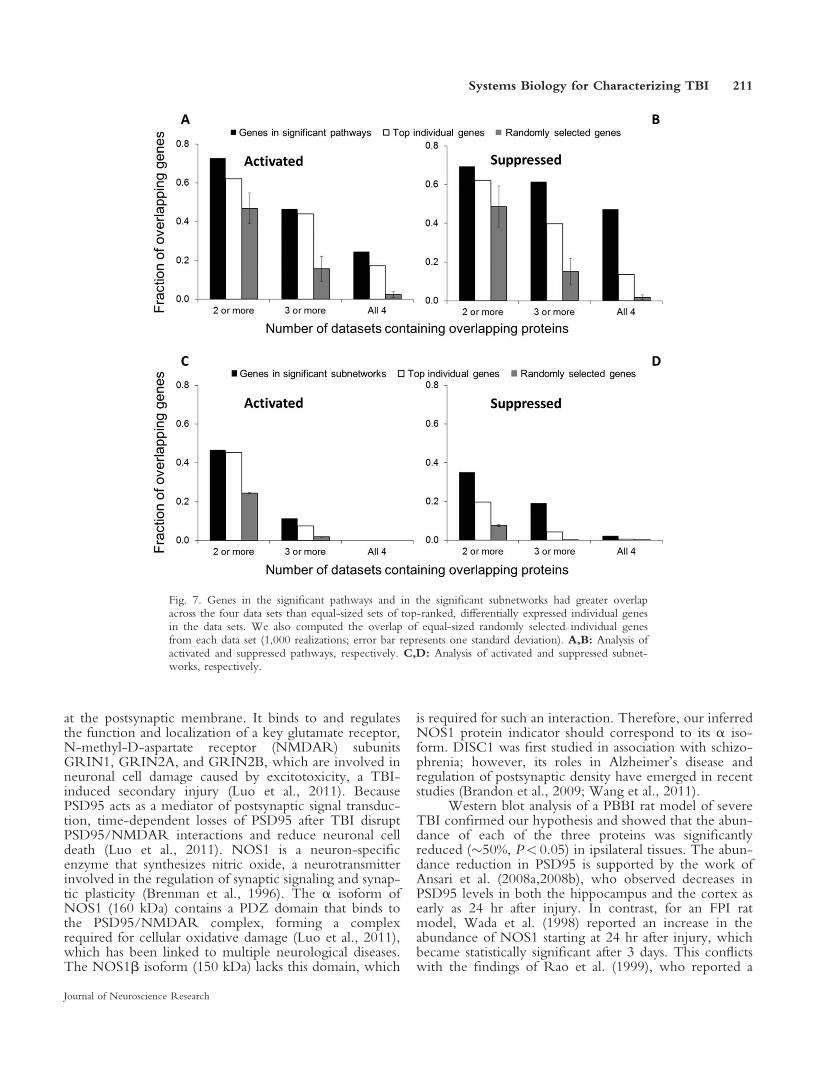
more likely to be reproducible by using a systems biologyapproach than by the sole analysis of individual genes. Totest this hypothesis, we computed the overlap of the con-stituent genes in the significant pathways and in the signif-icant subnetworks across the four studies and comparedthose with the overlap of equally sized sets of top-ranked,differentially expressed individual genes from the geneexpression studies. Figure 7 shows a consistent trend; theoverlap in the genes extracted from the pathways and sub-networks was consistently larger than the overlap fromthe individual genes in the experimental list. These resultssupport our hypothesis and hinge on the notion that, byprojecting TBI-specific, high-throughput gene expressiondata onto injury-agnostic biological networks and essen-tially integrating complementary and diverse molecularinformation, we are better able to filter out some of theinherent variability in gene transcription data and the dif-ferences in the experimental setups. Recent studiesinvolving comparisons across species and breast cancerdata sets support our findings (Chuang et al., 2007; Zin-man et al., 2011).
Recovering Previously Identified and InferringNovel TBI Protein Indicators
The ability to recover previously identified TBI bio-marker candidates provided reassurance that the systemsbiology strategy was also capable of inferring novel pro-tein indicators. From a list of 32 proteins previouslyimplicated with TBI, our analysis recovered 12 proteins(38%; Table III), including the microtubule-associatedprotein tau, which has been shown to be predictive ofclinical outcome and intracranial pressure after severe TBI(Zemlan et al., 2002; Liliang et al., 2010), and ubiquitinC-terminal hydrolase, which is currently undergoing clin-ical trials (Mondello et al., 2012).
Our investigation of the synaptic subnetwork alongwith distinct complementary evidence from the pathwayfunctional enrichment analyses led to the hypothesis thatthree proteins (PSD95, NOS1, and DISC1; see Table III)would be downregulated 24 hr after TBI. From a topolog-ical perspective, both PSD95 and DISC1 were modulecenter proteins (Fig. 2B). In addition, PSD95 formed ahub that interacted with as many as 39 proteins (67%) inthe synaptic subnetwork. NOS1 directly interacted withPSD95 and served as a bridge between PSD95 and anothermodule center, calcium/calmodulin-dependent proteinkinase type IIa, an enzyme involved in calcium signaling,which is crucial for regulating glutamatergic synapses.From biological and functional perspectives, all three pro-teins were linked to schizophrenia (Fig. 4B; Supp. Info.Table I) and have long been studied for their associationswith neurological diseases (Brenman et al., 1996; Takeu-chi et al., 1997; Heales et al., 1999; Deckel, 2001; Lawet al., 2001; Hodgkinson et al., 2004; Chubb et al., 2008;Brandon et al., 2009). However, to date, only PSD95 hasbeen considered in TBI (Luo et al., 2011).
PSD95 is a scaffolding protein whose most impor-tant function is to organize glutamate receptor complexes
210 Yu et al
Journal of Neuroscience Research
at the postsynaptic membrane. It binds to and regulatesthe function and localization of a key glutamate receptor,N-methyl-D-aspartate receptor (NMDAR) subunitsGRIN1, GRIN2A, and GRIN2B, which are involved inneuronal cell damage caused by excitotoxicity, a TBI-induced secondary injury (Luo et al., 2011). BecausePSD95 acts as a mediator of postsynaptic signal transduc-tion, time-dependent losses of PSD95 after TBI disruptPSD95/NMDAR interactions and reduce neuronal celldeath (Luo et al., 2011). NOS1 is a neuron-specificenzyme that synthesizes nitric oxide, a neurotransmitterinvolved in the regulation of synaptic signaling and synap-tic plasticity (Brenman et al., 1996). The a isoform ofNOS1 (160 kDa) contains a PDZ domain that binds tothe PSD95/NMDAR complex, forming a complexrequired for cellular oxidative damage (Luo et al., 2011),which has been linked to multiple neurological diseases.The NOS1b isoform (150 kDa) lacks this domain, which
is required for such an interaction. Therefore, our inferredNOS1 protein indicator should correspond to its a iso-form. DISC1 was first studied in association with schizo-phrenia; however, its roles in Alzheimer’s disease andregulation of postsynaptic density have emerged in recentstudies (Brandon et al., 2009; Wang et al., 2011).
Western blot analysis of a PBBI rat model of severeTBI confirmed our hypothesis and showed that the abun-dance of each of the three proteins was significantlyreduced (�50%, P< 0.05) in ipsilateral tissues. The abun-dance reduction in PSD95 is supported by the work ofAnsari et al. (2008a,2008b), who observed decreases inPSD95 levels in both the hippocampus and the cortex asearly as 24 hr after injury. In contrast, for an FPI ratmodel, Wada et al. (1998) reported an increase in theabundance of NOS1 starting at 24 hr after injury, whichbecame statistically significant after 3 days. This conflictswith the findings of Rao et al. (1999), who reported a
Fig. 7. Genes in the significant pathways and in the significant subnetworks had greater overlapacross the four data sets than equal-sized sets of top-ranked, differentially expressed individual genesin the data sets. We also computed the overlap of equal-sized randomly selected individual genesfrom each data set (1,000 realizations; error bar represents one standard deviation). A,B: Analysis ofactivated and suppressed pathways, respectively. C,D: Analysis of activated and suppressed subnet-works, respectively.
Systems Biology for Characterizing TBI 211
Journal of Neuroscience Research

significant increase in NOS1 as soon as 2 hr after CCIbrain injury. We could not find previously reportedresults for DISC1 within the context of neurotrauma.
These results have two overarching implications.First, they show that our analysis was able to identify sig-nificantly regulated proteins regardless of experimentalbrain-injury model; none of the four gene expression datasets used in our meta-analysis was derived with the PBBImodel used in the experimental testing (Table I). Thisfurther suggests that the systems biology strategy is robustand can detect molecular signatures of TBI independentof the mechanism of injury. Second, none of the protein-coding genes of PSD95, NOS1, and DISC1 was signifi-cantly differentially expressed in any of the four data sets(0.09<P< 0.59). In fact, DISC1 was not even includedin any of the data sets. This strongly suggests that,although genes might be unmeasured or nonsignificant inthe original gene expression data set, their significancemay emerge within the context of the connectivity infor-mation in biological networks.
Limitations
Our ability to perform a whole-genome analysis ofbrain injury in animal models was limited by multiple fac-tors. The publicly available gene expression data setscover only a partial number of genes (<8,000), brainregions, and time points after injury. In addition, the bio-logical networks cover only a fraction of the nearly20,000 human genes (�5,000 in the pathways and�12,000 in the human protein-interaction network), andtheir construction is biased toward well-studied genes andinteractions (X. Yu et al., 2012; Feala et al., 2013). Also,pathway analysis is inherently unable to reveal novel pro-tein interactions, and algorithms to extract new molecularmechanisms of action from protein interaction networksare still immature. Our ad hoc approach to identifyingprotein indicators of TBI could also be susceptible to sim-ilar limitations in the discovery of novel proteins, primar-ily because of our reliance on established associations withknown diseases and literature reviews (Kobeissy et al.,2006). We partially mitigated this by using the topologicalimportance of a protein in the network and its presencein a significantly regulated pathway in our selectionscheme, i.e., using information that is less susceptible tobiases.
CONCLUSIONS
Our findings show that certain molecular signatures ofTBI are conserved in a systems-level meta-analysis of fourdistinct gene expression studies involving different rat andmouse strains and different models of brain injury, sever-ity levels, and brain tissues. The significantly activatedmolecular signatures conserved at 24 hr after brain injurywere nonspecific to TBI. In stark contrast, the suppressedsignatures were specific to the nervous system and associ-ated primarily with synaptic function. We identified asuppressed synaptic subnetwork consisting of 58 highlyinteracting, coregulated proteins from which we hypothe-
sized three novel protein indicators that were either notmeasured or not statistically significant in the originalgene expression studies. We confirmed the hypothesis byWestern blot analysis. This demonstrates that the impor-tance of protein indicators can emerge within the contextof biological network information. Taken together, ourresults suggest that systems biology may provide an alter-native approach to generate testable hypotheses and iden-tify novel molecular mechanisms of action and proteinindicators of TBI.
ACKNOWLEDGMENTS
The opinions and assertions contained herein are the pri-vate views of the authors and are not to be construed asofficial or as reflecting the views of the U.S. Army or ofthe U.S. Department of Defense. This article has beenapproved for public release with unlimited distribution.The authors have no competing financial interests.
REFERENCES
Ansari MA, Roberts KN, Scheff SW. 2008a. Oxidative stress and modifi-
cation of synaptic proteins in hippocampus after traumatic brain injury.
Free Radic Biol Med 45:443–452.
Ansari MA, Roberts KN, Scheff SW. 2008b. A time course of
contusion-induced oxidative stress and synaptic proteins in cortex in a
rat model of TBI. J Neurotrauma 25:513–526.
Aranda B, Achuthan P, Alam-Faruque Y, Armean I, Bridge A, Derow
C, Feuermann M, Ghanbarian AT, Kerrien S, Khadake J, Kerssemakers
J, Leroy C, Menden M, Michaut M, Montecchi-Palazzi L, Neuhauser
SN, Orchard S, Perreau V, Roechert B, van Eijk K, Hermjakob H.
2010. The IntAct molecular interaction database in 2010. Nucleic Acids
Res 38:D525–D531.
Ashburner M, Ball CA, Blake JA, Botstein D, Butler H, Cherry JM,
Davis AP, Dolinski K, Dwight SS, Eppig JT, Harris MA, Hill DP,
Issel-Tarver L, Kasarskis A, Lewis S, Matese JC, Richardson JE,
Ringwald M, Rubin GM, Sherlock G. 2000. Gene ontology: tool for
the unification of biology. The Gene Ontology Consortium. Nat Genet
25:25–29.
Babikian T, Prins ML, Cai Y, Barkhoudarian G, Hartonian I, Hovda
DA, Giza CC. 2010. Molecular and physiological responses to juvenile
traumatic brain injury: focus on growth and metabolism. Dev Neurosci
32:431–441.
Bader GD, Betel D, Hogue CW. 2003. BIND: the Biomolecular Inter-
action Network Database. Nucleic Acids Res 31:248–250.
Baugh CM, Stamm JM, Riley DO, Gavett BE, Shenton ME, Lin A,
Nowinski CJ, Cantu RC, McKee AC, Stern RA. 2012. Chronic trau-
matic encephalopathy: neurodegeneration following repetitive concus-
sive and subconcussive brain trauma. Brain Imaging Behav 6:244–254.
Beuming T, Skrabanek L, Niv MY, Mukherjee P, Weinstein H. 2005.
PDZBase: a protein–protein interaction database for PDZ-domains.
Bioinformatics 21:827–828.
Bezprozvanny I, Hiesinger PR. 2013. The synaptic maintenance prob-
lem: membrane recycling, Ca21 homeostasis, and late onset degenera-
tion. Mol Neurodegener 8:23.
Blaylock RL, Maroon J. 2011. Immunoexcitotoxicity as a central mecha-
nism in chronic traumatic encephalopathy–a unifying hypothesis. Surg
Neurol Int 2:107.
Brandon NJ, Millar JK, Korth C, Sive H, Singh KK, Sawa A. 2009.
Understanding the role of DISC1 in psychiatric disease and during nor-
mal development. J Neurosci 29:12768–12775.
212 Yu et al
Journal of Neuroscience Research

Breitling R, Armengaud P, Amtmann A, Herzyk P. 2004. Rank prod-
ucts: a simple, yet powerful, new method to detect differentially regu-
lated genes in replicated microarray experiments. FEBS Lett 573:83–92.
Brenman JE, Chao DS, Gee SH, McGee AW, Craven SE, Santillano
DR, Wu Z, Huang F, Xia H, Peters MF, Froehner SC, Bredt DS.
1996. Interaction of nitric oxide synthase with the postsynaptic density
protein PSD-95 and alpha1-syntrophin mediated by PDZ domains.
Cell 84:757–767.
Chatr-aryamontri A, Ceol A, Palazzi LM, Nardelli G, Schneider MV,
Castagnoli L, Cesareni G. 2007. MINT: the Molecular INTeraction
database. Nucleic Acids Res 35:D572–D574.
Chuang HY, Lee E, Liu YT, Lee D, Ideker T. 2007. Network-based
classification of breast cancer metastasis. Mol Syst Biol 3:140.
Chubb JE, Bradshaw NJ, Soares DC, Porteous DJ, Millar JK. 2008. The
DISC locus in psychiatric illness. Mol Psychiatry 13:36–64.
Deckel AW. 2001. Nitric oxide and nitric oxide synthase in Hunting-
ton’s disease. J Neurosci Res 64:99–107.
DeKosky ST, Ikonomovic MD, Gandy S. 2010. Traumatic brain
injury—football, warfare, and long-term effects. N Engl J Med 363:
1293–1296.
Dutta B, Wallqvist A, Reifman J. 2012. PathNet: A tool for pathway
analysis using topological information. Source Code Biol Med 7:10.
Eppig JT, Blake JA, Bult CJ, Kadin JA, Richardson JE, Mouse Genome
Database Group. 2012. The Mouse Genome Database (MGD): com-
prehensive resource for genetics and genomics of the laboratory mouse.
Nucleic Acids Res 40:D881–D886.
Fahlenkamp AV, Coburn M, Czaplik M, Ryang YM, Kipp M, Rossaint
R, Beyer C. 2011. Expression analysis of the early chemokine response
4 hours after in vitro traumatic brain injury. Inflamm Res 60:379–387.
Feala JD, Abdulhameed MD, Yu C, Dutta B, Yu X, Schmid K, Dave J,
Tortella F, Reifman J. 2013. Systems biology approaches for discover-
ing biomarkers for traumatic brain injury. J Neurotrauma 30:1101–
1116.
Gao X, Deng P, Xu ZC, Chen J. 2011. Moderate traumatic brain injury
causes acute dendritic and synaptic degeneration in the hippocampal
dentate gyrus. PLoS One 6:e24566.
Gilman CP, Chan SL, Guo Z, Zhu X, Greig N, Mattson MP. 2003. p53
Is present in synapses where it mediates mitochondrial dysfunction and
synaptic degeneration in response to DNA damage and oxidative and
excitotoxic insults. Neuromol Med 3:159–172.
Greve MW, Zink BJ. 2009. Pathophysiology of traumatic brain injury.
Mt Sinai J Med 76:97–104.
Heales SJ, Bolanos JP, Stewart VC, Brookes PS, Land JM, Clark JB.
1999. Nitric oxide, mitochondria, and neurological disease. Biochim
Biophys Acta 1410:215–228.
Hodgkinson CA, Goldman D, Jaeger J, Persaud S, Kane JM, Lipsky RH,
Malhotra AK. 2004. Disrupted in schizophrenia 1 (DISC1): association
with schizophrenia, schizoaffective disorder, and bipolar disorder. Am J
Hum Genet 75:862–872.
Hong F, Breitling R, McEntee CW, Wittner BS, Nemhauser JL, Chory
J. 2006. RankProd: a bioconductor package for detecting differentially
expressed genes in meta-analysis. Bioinformatics 22:2825–2827.
Hua F, Wang J, Ishrat T, Wei W, Atif F, Sayeed I, Stein DG. 2011.
Genomic profile of toll-like receptor pathways in traumatically brain-
injured mice: effect of exogenous progesterone. J Neuroinflamm 8:42.
Huang DW, Sherman BT, Lempicki RA. 2009. Systematic and integra-
tive analysis of large gene lists using DAVID bioinformatics resources.
Nat Protoc 4:44–57.
Ideker T, Galitski T, Hood L. 2001. A new approach to decoding life:
systems biology. Annu Rev Genomics Hum Genet 2:343–372.
Irizarry RA, Hobbs B, Collin F, Beazer-Barclay YD, Antonellis KJ,
Scherf U, Speed TP. 2003. Exploration, normalization, and summaries
of high density oligonucleotide array probe level data. Biostatistics 4:
249–264.
Kanehisa M, Araki M, Goto S, Hattori M, Hirakawa M, Itoh M,
Katayama T, Kawashima S, Okuda S, Tokimatsu T, Yamanishi Y.
2008. KEGG for linking genomes to life and the environment. Nucleic
Acids Res 36:D480–D484.
Kobeissy FH, Ottens AK, Zhang Z, Liu MC, Denslow ND, Dave JR, Tortella
FC, Hayes RL, Wang KK. 2006. Novel differential neuroproteomics analysis
of traumatic brain injury in rats. Mol Cell Proteomics 5:1887–1898.
Kobeissy FH, Sadasivan S, Oli MW, Robinson G, Larner SF, Zhang Z,
Hayes RL, Wang KK. 2008. Neuroproteomics and systems biology-
based discovery of protein biomarkers for traumatic brain injury and
clinical validation. Proteomics Clin Appl 2:1467–1483.
Konig R, Zhou Y, Elleder D, Diamond TL, Bonamy GM, Irelan JT,
Chiang CY, Tu BP, De Jesus PD, Lilley CE, Seidel S, Opaluch AM,
Caldwell JS, Weitzman MD, Kuhen KL, Bandyopadhyay S, Ideker T,
Orth AP, Miraglia LJ, Bushman FD, Young JA, Chanda SK. 2008.
Global analysis of host–pathogen interactions that regulate early-stage
HIV-1 replication. Cell 135:49–60.
Law A, Gauthier S, Quirion R. 2001. Say NO to Alzheimer’s disease:
the putative links between nitric oxide and dementia of the Alzheimer’s
type. Brain Res Brain Res Rev 35:73–96.
Liliang PC, Liang CL, Weng HC, Lu K, Wang KW, Chen HJ, Chuang
JH. 2010. Tau proteins in serum predict outcome after severe traumatic
brain injury. J Surg Res 160:302–307.
Luo P, Fei F, Zhang L, Qu Y, Fei Z. 2011. The role of glutamate recep-
tors in traumatic brain injury: implications for postsynaptic density in
pathophysiology. Brain Res Bull 85:313–320.
MacGregor AJ, Shaffer RA, Dougherty AL, Galarneau MR, Raman R,
Baker DG, Lindsay SP, Golomb BA, Corson KS. 2010. Prevalence and
psychological correlates of traumatic brain injury in Operation Iraqi
Freedom. J Head Trauma Rehabil 25:1–8.
Matzilevich DA, Rall JM, Moore AN, Grill RJ, Dash PK. 2002. High-
density microarray analysis of hippocampal gene expression following
experimental brain injury. J Neurosci Res 67:646–663.
Mondello S, Muller U, Jeromin A, Streeter J, Hayes RL, Wang KK.
2011. Blood-based diagnostics of traumatic brain injuries. Expert Rev
Mol Diagn 11:65–78.
Mondello S, Linnet A, Buki A, Robicsek S, Gabrielli A, Tepas J, Papa L,
Brophy GM, Tortella F, Hayes RL, Wang KK. 2012. Clinical utility of
serum levels of ubiquitin C-terminal hydrolase as a biomarker for severe
traumatic brain injury. Neurosurgery 70:666–675.
Morganti-Kossmann MC, Satgunaseelan L, Bye N, Kossmann T. 2007.
Modulation of immune response by head injury. Injury 38:1392–1400.
Natale JE, Ahmed F, Cernak I, Stoica B, Faden AI. 2003. Gene expres-
sion profile changes are commonly modulated across models and species
after traumatic brain injury. J Neurotrauma 20:907–927.
Pagel P, Kovac S, Oesterheld M, Brauner B, Dunger-Kaltenbach I,
Frishman G, Montrone C, Mark P, Stumpflen V, Mewes HW, Ruepp
A, Frishman D. 2005. The MIPS mammalian protein–protein interac-
tion database. Bioinformatics 21:832–834.
Peri S, Navarro JD, Amanchy R, Kristiansen TZ, Jonnalagadda CK,
Surendranath V, Niranjan V, Muthusamy B, Gandhi TK, Gronborg M,
Ibarrola N, Deshpande N, Shanker K, Shivashankar HN, Rashmi BP,
Ramya MA, Zhao Z, Chandrika KN, Padma N, Harsha HC, Yatish
AJ, Kavitha MP, Menezes M, Choudhury DR, Suresh S, Ghosh N,
Saravana R, Chandran S, Krishna S, Joy M, Anand SK, Madavan V,
Joseph A, Wong GW, Schiemann WP, Constantinescu SN, Huang L,
Khosravi-Far R, Steen H, Tewari M, Ghaffari S, Blobe GC, Dang CV,
Garcia JG, Pevsner J, Jensen ON, Roepstorff P, Deshpande KS,
Chinnaiyan AM, Hamosh A, Chakravarti A, Pandey A. 2003. Develop-
ment of human protein reference database as an initial platform for
approaching systems biology in humans. Genome Res 13:2363–2371.
Rao VL, Dogan A, Bowen KK, Dempsey RJ. 1999. Traumatic injury to
rat brain upregulates neuronal nitric oxide synthase expression and L-
[3H]nitroarginine binding. J Neurotrauma 16:865–877.
Systems Biology for Characterizing TBI 213
Journal of Neuroscience Research

Risling M, Plantman S, Angeria M, Rostami E, Bellander BM,
Kirkegaard M, Arborelius U, Davidsson J. 2011. Mechanisms of blast
induced brain injuries, experimental studies in rats. Neuroimage
54(Suppl 1):S89–S97.
Rual JF, Venkatesan K, Hao T, Hirozane-Kishikawa T, Dricot A, Li N,
Berriz GF, Gibbons FD, Dreze M, Ayivi-Guedehoussou N, Klitgord
N, Simon C, Boxem M, Milstein S, Rosenberg J, Goldberg DS, Zhang
LV, Wong SL, Franklin G, Li S, Albala JS, Lim J, Fraughton C,
Llamosas E, Cevik S, Bex C, Lamesch P, Sikorski RS, Vandenhaute J,
Zoghbi HY, Smolyar A, Bosak S, Sequerra R, Doucette-Stamm L,
Cusick ME, Hill DE, Roth FP, Vidal M. 2005. Towards a proteome-
scale map of the human protein–protein interaction network. Nature
437:1173–1178.
Salwinski L, Miller CS, Smith AJ, Pettit FK, Bowie JU, Eisenberg D.
2004. The Database of Interacting Proteins: 2004 update. Nucleic Acids
Res 32:D449–D451.
Scheff SW, Price DA, Hicks RR, Baldwin SA, Robinson S, Brackney
C. 2005. Synaptogenesis in the hippocampal CA1 field following trau-
matic brain injury. J Neurotrauma 22:719–732.
Shi L, Reid LH, Jones WD, Shippy R, Warrington JA, Baker SC,
Collins PJ, de Longueville F, Kawasaki ES, Lee KY, Luo Y, Sun YA,
Willey JC, Setterquist RA, Fischer GM, Tong W, Dragan YP, Dix DJ,
Frueh FW, Goodsaid FM, Herman D, Jensen RV, Johnson CD,
Lobenhofer EK, Puri RK, Schrf U, Thierry-Mieg J, Wang C, Wilson
M, Wolber PK, Zhang L, Amur S, Bao W, Barbacioru CC, Lucas AB,
Bertholet V, Boysen C, Bromley B, Brown D, Brunner A, Canales R,
Cao XM, Cebula TA, Chen JJ, Cheng J, Chu TM, Chudin E, Corson
J, Corton JC, Croner LJ, Davies C, Davison TS, Delenstarr G, Deng
X, Dorris D, Eklund AC, Fan XH, Fang H, Fulmer-Smentek S,
Fuscoe JC, Gallagher K, Ge W, Guo L, Guo X, Hager J, Haje PK,
Han J, Han T, Harbottle HC, Harris SC, Hatchwell E, Hauser CA,
Hester S, Hong H, Hurban P, Jackson SA, Ji H, Knight CR, Kuo WP,
LeClerc JE, Levy S, Li QZ, Liu C, Liu Y, Lombardi MJ, Ma Y,
Magnuson SR, Maqsodi B, McDaniel T, Mei N, Myklebost O, Ning
B, Novoradovskaya N, Orr MS, Osborn TW, Papallo A, Patterson
TA, Perkins RG, Peters EH, Peterson R, Philips KL, Pine PS, Pusztai
L, Qian F, Ren H, Rosen M, Rosenzweig BA, Samaha RR, Schena
M, Schroth GP, Shchegrova S, Smith DD, Staedtler F, Su Z, Sun H,
Szallasi Z, Tezak Z, Thierry-Mieg D, Thompson KL, Tikhonova I,
Turpaz Y, Vallanat B, Van C, Walker SJ, Wang SJ, Wang Y,
Wolfinger R, Wong A, Wu J, Xiao C, Xie Q, Xu J, Yang W, Zhang
L, Zhong S, Zong Y, Slikker W Jr. 2006. The MicroArray Quality
Control (MAQC) project shows inter- and intraplatform reproducibility
of gene expression measurements. Nat Biotechnol 24:1151–1161.
Shojo H, Kaneko Y, Mabuchi T, Kibayashi K, Adachi N, Borlongan
CV. 2010. Genetic and histologic evidence implicates role of inflamma-
tion in traumatic brain injury-induced apoptosis in the rat cerebral cor-
tex following moderate fluid percussion injury. Neuroscience 171:
1273–1282.
Stark C, Breitkreutz BJ, Reguly T, Boucher L, Breitkreutz A, Tyers M.
2006. BioGRID: a general repository for interaction data sets. Nucleic
Acids Res 34:D535–D539.
Takeuchi M, Hata Y, Hirao K, Toyoda A, Irie M, Takai Y. 1997.
SAPAPs. A family of PSD-95/SAP90-associated proteins localized at
postsynaptic density. J Biol Chem 272:11943–11951.
Ulitsky I, Shamir R. 2007. Pathway redundancy and protein essentiality
revealed in the Saccharomyces cerevisiae interaction networks. Mol Syst
Biol 3:104.
Vastrik I, D’Eustachio P, Schmidt E, Gopinath G, Croft D, de Bono B,
Gillespie M, Jassal B, Lewis S, Matthews L, Wu G, Birney E, Stein L.
2007. Reactome: a knowledge base of biologic pathways and processes.
Genome Biol 8:R39.
Wada K, Chatzipanteli K, Kraydieh S, Busto R, Dietrich WD. 1998.
Inducible nitric oxide synthase expression after traumatic brain injury
and neuroprotection with aminoguanidine treatment in rats. Neurosur-
gery 43:1427–1436.
Wang JW, Wang HD, Zhong WZ, Li N, Cong ZX. 2012. Expression
and cell distribution of metabotropic glutamate receptor 5 in the rat
cortex following traumatic brain injury. Brain Res 1464:73–81.
Wang Q, Charych EI, Pulito VL, Lee JB, Graziane NM, Crozier RA,
Revilla-Sanchez R, Kelly MP, Dunlop AJ, Murdoch H, Taylor N, Xie
Y, Pausch M, Hayashi-Takagi A, Ishizuka K, Seshadri S, Bates B,
Kariya K, Sawa A, Weinberg RJ, Moss SJ, Houslay MD, Yan Z,
Brandon NJ. 2011. The psychiatric disease risk factors DISC1 and
TNIK interact to regulate synapse composition and function. Mol Psy-
chiatry 16:1006–1023.
Williams AJ, Hartings JA, Lu XC, Rolli ML, Dave JR, Tortella FC.
2005. Characterization of a new rat model of penetrating ballistic brain
injury. J Neurotrauma 22:313–331.
Williams AJ, Hartings JA, Lu XC, Rolli ML, Tortella FC. 2006a. Pene-
trating ballistic-like brain injury in the rat: differential time courses of
hemorrhage, cell death, inflammation, and remote degeneration.
J Neurotrauma 23:1828–1846.
Williams AJ, Ling GS, Tortella FC. 2006b. Severity level and injury track
determine outcome following a penetrating ballistic-like brain injury in
the rat. Neurosci Lett 408:183–188.
Young W. 1992. Role of calcium in central nervous system injuries.
J Neurotrauma 9(Suppl 1):S9–S25.
Yu C, Desai V, Cheng L, Reifman J. 2012. QuartetS-DB: a large-scale
orthology database for prokaryotes and eukaryotes inferred by evolu-
tionary evidence. BMC Bioinformatics 13:143.
Yu X, Ivanic J, Memisevic V, Wallqvist A, Reifman J. 2011. Categoriz-
ing biases in high-confidence high-throughput protein–protein interac-
tion data sets. Mol Cell Proteomics 10:M111.012500.
Yu X, Wallqvist A, Reifman J. 2012. Inferring high-confidence human
protein–protein interactions. BMC Bioinformatics 13:79.
Zemlan FP, Jauch EC, Mulchahey JJ, Gabbita SP, Rosenberg WS,
Speciale SG, Zuccarello M. 2002. C-tau biomarker of neuronal damage
in severe brain injured patients: association with elevated intracranial
pressure and clinical outcome. Brain Res 947:131–139.
Zhang J, Yang Y, Wang Y, Zhang J, Wang Z, Yin M, Shen X. 2011.
Identification of hub genes related to the recovery phase of irradiation
injury by microarray and integrated gene network analysis. PLoS One
6:e24680.
Zhang KX, Ouellette BF. 2011. CAERUS: predicting CAncER oUt-
comeS using relationship between protein structural information, pro-
tein networks, gene expression data, and mutation data. PLoS Comput
Biol 7:e1001114.
Zinman GE, Zhong S, Bar-Joseph Z. 2011. Biological interaction net-
works are conserved at the module level. BMC Syst Biol 5:134.
214 Yu et al
Journal of Neuroscience Research