a synthetic prestin reveals protein domains and molecular ... filea synthetic prestin reveals...
TRANSCRIPT

A synthetic prestin reveals protein domains andmolecular operation of outer hair cellpiezoelectricity
Thorsten J Schaechinger1, DmitryGorbunov2, Christian R Halaszovich2,Tobias Moser3, Sebastian Kugler4,Bernd Fakler1,5 and Dominik Oliver2,*1Institute of Physiology, University of Freiburg, Freiburg, Germany,2Department of Neurophysiology, Institute of Physiology andPathophysiology, Philipps-University, Marburg, Germany, 3InnerEarLab,Department of Otolaryngology and Center for Molecular Physiology ofthe Brain, University of Gottingen, Goettingen, Germany, 4Departmentof Neurology, University of Gottingen, Medical School, Gottingen,Germany and 5BIOSS Centre for Biological Signaling Studies, Universityof Freiburg, Freiburg, Germany
Prestin, a transporter-like protein of the SLC26A family,
acts as a piezoelectric transducer that mediates the fast
electromotility of outer hair cells required for cochlear
amplification and auditory acuity in mammals. Non-mam-
malian prestin orthologues are anion transporters without
piezoelectric activity. Here, we generated synthetic prestin
(SynPres), a chimera of mammalian and non-mammalian
prestin exhibiting both, piezoelectric properties and anion
transport. SynPres delineates two distinct domains in the
protein’s transmembrane core that are necessary and
sufficient for generating electromotility and associated
non-linear charge movement (NLC). Functional analysis
of SynPres showed that the amplitude of NLC and hence
electromotility are determined by the transport of mono-
valent anions. Thus, prestin-mediated electromotility is
a dual-step process: transport of anions by an alternate
access cycle, followed by an anion-dependent transition
generating electromotility. The findings define structural
and functional determinants of prestin’s piezoelectric acti-
vity and indicate that the electromechanical process
evolved from the ancestral transport mechanism.
The EMBO Journal advance online publication, 24 June 2011;
doi:10.1038/emboj.2011.202
Subject Categories: membranes & transport; neuroscience
Keywords: anion transport; cochlea; outer hair cell; prestin;
SLC26
Introduction
Hearing in vertebrates involves an active amplification pro-
cess in the auditory periphery that determines the exquisite
sensitivity and frequency selectivity (Dallos, 2008; Hudspeth,
2008). In mammals, this ‘cochlear amplification’ is provided
by the outer hair cells (OHCs), a distinct and structurally
specialized population of mechanosensory hair cells that
change their length in response to changes in membrane
potential (Brownell et al, 1985; Ashmore, 1987). These fast
voltage-dependent cellular length changes, termed electro-
motility, can operate at frequencies of at least 70 kHz (Frank
et al, 1999) (i.e. ultrasonic frequencies, e.g. in bats) and are
required for amplification of the sound-induced vibrations in
the cochlea (Dallos et al, 2008).
Mechanistically, the electromotility of OHCs is similar to
piezoelectric materials that change dimensions under the
influence of voltage (Ashmore, 2008). As the elementary
molecular motor generating electromotility, Dallos and co-
workers identified prestin (SLC26A5), a member of the SLC26
family of anion exchangers (Zheng et al, 2000). Prestin is
thought to act as an area motor by alternating between two
major conformations that occupy different cross-sectional
areas within the membrane and were, therefore, termed
‘long’ and ‘short states’ (Iwasa, 2001; Dallos and Fakler,
2002). Joint conformational transitions of the tightly packed
prestin motors in the plasma membrane of the OHC lead to
length changes of the whole cell (models reviewed by Dallos
and Fakler, 2002; Ashmore, 2008). The distribution between
long and short conformations is immediately voltage depen-
dent, which inevitably requires a mobile charged particle
acting as a voltage sensor (Bezanilla, 2008). In fact, fast
voltage-dependent charge movement is a hallmark of prestin
that can be experimentally assessed as a non-linear capaci-
tance (NLC) (Santos-Sacchi, 1991; Dallos and Fakler, 2002).
Because of its straight accessibility and precise quantification,
NLC is routinely used as a surrogate measure for electro-
motility (Figure 1A).
Aside from the basic biophysical characteristics, little is
known about structural and molecular details of prestin’s
function. We and others have previously demonstrated that
NLC, and hence electromotility, is critically dependent
on monovalent intracellular anions, in particular chloride
(Cl�) (Oliver et al, 2001; Rybalchenko and Santos-Sacchi,
2003, 2008; Santos-Sacchi et al, 2006), suggesting a mecha-
nistic link between electromotility and the anion transport
observed with related SLC26 transporters. In fact, analysis
of the phylogenetic relationship of vertebrate SLC26A5 ortho-
logues (Franchini and Elgoyhen, 2006; Okoruwa et al,
2008) indicated that mammalian prestin evolved from an
anion transporter present in pre-mammalian ancestors.
Thus, we recently found that non-mammalian SLC26A5
orthologues are bona fide electrogenic anion antiporters
(Schaechinger and Oliver, 2007), which, however, fail to
generate fast NLC and electromotility (Albert et al, 2007;
Tan et al, 2011). Vice versa, mammalian prestin lacks elec-
trically detectable transport activity (Schaechinger and
Oliver, 2007; Figure 1A and B).Received: 28 January 2011; accepted: 24 May 2011
*Corresponding author. Department of Neurophysiology, Instituteof Physiology and Pathophysiology, Philipps-University,Deutschhausstrasse 2, 35037 Marburg, Germany.Tel.: þ 49 6421 286 6444; Fax: þ 49 6421 286 2306;E-mail: [email protected]
The EMBO Journal (2011), 1–12 | & 2011 European Molecular Biology Organization | All Rights Reserved 0261-4189/11
www.embojournal.org
&2011 European Molecular Biology Organization The EMBO Journal
EMBO
THE
EMBOJOURNAL
THE
EMBOJOURNAL
1

In this study, we took advantage of the functional diver-
gence between mammalian and non-mammalian orthologues
for analysing prestin-mediated fast electromotility. Using
chimeras between rat (rPres) and zebrafish (zPres) ortho-
logues, we generated a synthetic prestin (SynPres) protein
that combines piezoelectric properties with robust electro-
genic anion transport and thus provides a novel experimental
paradigm for direct investigation of the mechanistic relation
between fast electromotility and anion transport.
Results
Structural determinants of electromotility—generation
of SynPres
For elucidating the protein domains that underlie electromo-
tility and NLC, its electrical signature, in mammalian prestin,
we generated chimeras between the prestin orthologues
of rat (rPres) and zebrafish (zPres). Both proteins are highly
homologous (see Supplementary Figure S1) and exhibit
identical overall topologies, but are largely different with
respect to their functional properties observed in cultured
CHO cells upon heterologous expression (Albert et al, 2007;
Schaechinger and Oliver, 2007). Thus, rPres displays robust
electromotility and NLC, but fails to transport anions across
the plasma membrane (Figure 1A); in contrast, zPres does
not show fast NLC, but operates as an electrogenic anion
transporter that effectively exchanges Cl� for divalent anions
such as oxalate, giving rise to large transport currents
(Figure 1B).
Initially, we switched the hydrophobic core region thought
to comprise 12 transmembrane domains (Oliver et al, 2001;
Zheng et al, 2001) between rPres and zPres (Figure 1, upper
panel). As shown in Figure 1C, the chimera (Chi1) placing the
core of rPres between the cytoplasmic N- and C-termini of
zPres lacked electrogenic anion transport activity, but dis-
played robust NLC with voltage-dependent characteristics
very similar to rPres. The voltage at maximal NLC (V1/2)
and the steepness of voltage dependence (a) obtained
from fits of the derivative of a Boltzmann function to the
data were V1/2¼�31.9±14.8 mV (mean±s.d., n¼ 11) and
�77.7±16.0 mV (n¼ 20), and a¼ 38.0±3.0 mV and 38.6±
2.6 mV, for Chi1 and rPres, respectively. Similarly, the inverse
chimera where the N- and C-termini of rPres flanked the core
region of zPres (revChi1) reproduced the properties of the
non-mammalian zPres as indicated by electrogenic transport
and complete lack of NLC (Figure 1D). These data indicated
that NLC and anion transport are mediated by the transmem-
brane core, while the cytoplasmic termini are not directly
involved.
Consequently, the next series of constructs probed distinct
domains within the transmembrane core for their functional
significance, with a first focus on its N-terminal end. This
region comprising the first two transmembrane helices
(amino acids (aa) 86–140 of the rPres sequence) exhibits
the highest degree of sequence identity across the SLC26
rPres
0.5 pF
–100 0 100
nA
–100 0 100
0.0
–0.2
–0.4
0.5 pF
–100 0 100
–100 0 100
zPres
0.2 pF
–100 0 100
–100 0 100
Chi1
Membrane potential (mV)
–100 0 100
0.5 pF
Membrane potential (mV)
–100 0 100
revChi1
NLC
Tra
nspo
rt
A B C D
Figure 1 Electromotility-associated NLC and transport are determined by the transmembrane core region of mammalian and non-mammalianprestin. (A–D) NLC and electrogenic anion transport recorded from whole-cell voltage-clamped CHO cells expressing rPres (A), zPres (B), Chi1(C), or revChi1 (D). Intracellular solution contained 160 mM Cl� for NLC recordings or 10 mM oxalate and 10 mM Cl� for transport currentmeasurements. Traces for NLC and transport are each representative for more than five experiments; grey trace in (B, transport measurement)is with oxalate omitted from the intracellular solution. Plasma membrane localization of the constructs is shown by representative confocalimages from CHO cells (lower panels; scale bar for all images, 10mm). Cartoons (upper panels) represent the suggested 12 TM topology ofprestin (Oliver et al, 2001; Zheng et al, 2001); for alternative topology model, see Navaratnam et al (2005); shaded domain indicates SLC26signature motif.
Molecular determinants of prestin functionTJ Schaechinger et al
The EMBO Journal &2011 European Molecular Biology Organization2

transporter family and includes the highly conserved ‘SLC26
transporters signature motif’ (residues 109–130; Prosite
PS01130; Mount and Romero, 2004). When we replaced this
segment in Chi1 with the respective zPres sequence (chimera
termed Chi2), NLC was completely abolished (Figure 2A),
suggesting that this domain may contain determinants
essential for rPres function. Sequence comparison between
rPres and zPres showed that only seven amino acids are
exchanged within this region, most of them being conser-
vative substitutions (Figure 2E). We next replaced each of
these residues in rPres individually by the respective amino
acid of zPres and measured the electromotility-related NLC.
As shown in Figure 2E, replacement of each of the four
positions within the signature motif (I121L, M122L, C124T,
R130K) did not substantially affect NLC, while mutating
each of the three remaining amino acid exchanges (L93M,
F101Y, P136T) shifted NLC towards depolarized potentials
by 460 mV (for values of V1/2 and a, see Supplementary
Table S1). Combining all three mutations completely abol-
ished NLC (Figure 2F), indicating that these three exchanges
can fully account for the non-NLC phenotype observed with
Chi2 (Figure 2A).
Despite its significance, however, the N-terminal end of the
core region is not an exclusive determinant of electromotility
Chi2
Membrane potential (mV)
–100 0 100
0.2 pF
revChi2 SynPres Chi3
–100 0 100 –100 0 100 –100 0 100
A B C D
E
rPres (L93M,F101Y,P136T)
0.2 pF
Membrane potential (mV)–100 0 100
86 140 SGISTGVLQLPQGLAFAMLAAVPPVFGLYSSFYPVIMYCFFGTSRHISIGPFAVI SGISTGVMQLPQGLAYAMLAAVPPVFGLYSSFYPVLLYTFFGTSKHISIGTFAVI
–100 0 100 0–40–80–120
V1/2 (mV)
P136T
R130K
C125T
M122L
I121L
F101Y
L93M
Membrane potential (mV)
rPreszPres
F
Figure 2 Identification of molecular determinants of NLC and electromotility. (A–D) The indicated prestin chimeras were expressed in CHOcells as C-terminal GFP fusions and subjected to capacitance measurements (middle panel, NLC traces representative for 5–9 cells with eachchimera). All constructs were properly targeted to the plasma membrane as shown by confocal images from representative CHO cells (lowerpanels; scale bar, 10mm). Note that NLC was only reconstituted into the zPres background in SynPres, where both domains aa 86–140(revChi2) and aa 381–438 (Chi3) were combined. (E) Sequence alignment of the region exchanged in Chi2 (indexing according to rPressequence). Non-conserved amino acids are shown in red, the SLC26 signature is boxed. Each of the non-conserved residues in rPres wasindividually mutated to the respective zPres residue. NLC traces are representative for the indicated mutants, the respective values for V1/2
(mean±s.d.) are given on the right. Solid line and shaded voltage range represent V1/2±s.d. obtained for rPres (n¼ 5–10 cells for each mutant;for details, see Supplementary Table 1). (F) NLC is completely abolished in the triple mutant L93M/F101Y/P136T, although the protein isrobustly targeted to the plasma membrane. P136T and the triple mutant were generated in the Chi1 background, because the equivalentmutants in rPres were not delivered to the plasma membrane.
Molecular determinants of prestin functionTJ Schaechinger et al
&2011 European Molecular Biology Organization The EMBO Journal 3
as revealed by construct revChi2 that placed this segment of
rPres into the zPres background. As illustrated by Figure 2B,
this domain swapping failed to confer NLC onto zPres
and, thus, indicated that the mammalian-specific residues
within this region, although necessary, are not sufficient for
generation of electromotility-associated NLC. We, therefore,
designed a series of additional chimeras where various
segments of the rPres core region were successively added
onto revChi2. These efforts finally identified another segment
comprising putative transmembrane domains 9 and 10 (aa
381–438 in the rPres sequence; Figure 2C; Supplementary
Figure S1). On its own, this region was also not sufficient to
introduce fast NLC into zPres (Chi3; Figure 2D). However, in
combination with the N-terminal segment around transmem-
brane domains 1 and 2, it generated robust NLC (Figure 2C).
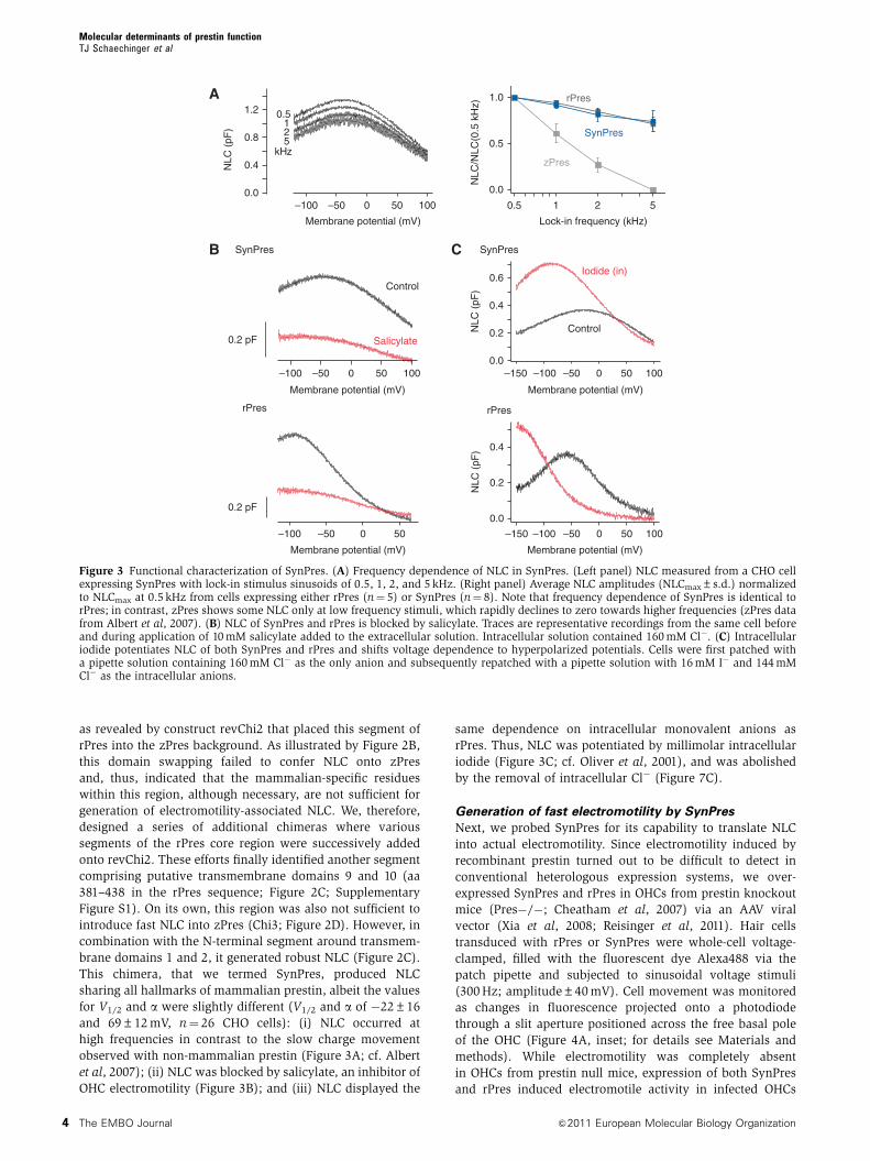
This chimera, that we termed SynPres, produced NLC
sharing all hallmarks of mammalian prestin, albeit the values
for V1/2 and a were slightly different (V1/2 and a of �22±16
and 69±12 mV, n¼ 26 CHO cells): (i) NLC occurred at
high frequencies in contrast to the slow charge movement
observed with non-mammalian prestin (Figure 3A; cf. Albert
et al, 2007); (ii) NLC was blocked by salicylate, an inhibitor of
OHC electromotility (Figure 3B); and (iii) NLC displayed the
same dependence on intracellular monovalent anions as
rPres. Thus, NLC was potentiated by millimolar intracellular
iodide (Figure 3C; cf. Oliver et al, 2001), and was abolished
by the removal of intracellular Cl� (Figure 7C).
Generation of fast electromotility by SynPres
Next, we probed SynPres for its capability to translate NLC
into actual electromotility. Since electromotility induced by
recombinant prestin turned out to be difficult to detect in
conventional heterologous expression systems, we over-
expressed SynPres and rPres in OHCs from prestin knockout
mice (Pres�/�; Cheatham et al, 2007) via an AAV viral
vector (Xia et al, 2008; Reisinger et al, 2011). Hair cells
transduced with rPres or SynPres were whole-cell voltage-
clamped, filled with the fluorescent dye Alexa488 via the
patch pipette and subjected to sinusoidal voltage stimuli
(300 Hz; amplitude±40 mV). Cell movement was monitored
as changes in fluorescence projected onto a photodiode
through a slit aperture positioned across the free basal pole
of the OHC (Figure 4A, inset; for details see Materials and
methods). While electromotility was completely absent
in OHCs from prestin null mice, expression of both SynPres
and rPres induced electromotile activity in infected OHCs
0.5 2 51
Lock-in frequency (kHz)
0.0
0.5
1.0
NLC
/NLC
(0.5
kH
z)
zPres
SynPres
rPres
0.0
0.4
0.8
1.2
NLC
(pF
)
–100 0 50–50 100
Membrane potential (mV)
5 21
0.5
kHz
A
0.2 pF
Control
Salicylate
–100 –50 500
0.2 pF0.0
0.4
0.2N
LC (
pF)
–100 500–50 100–150
Membrane potential (mV)
0.0
0.4
0.6
0.2NLC
(pF
)
Control
Iodide (in)
–100 0 50 50–50 100
Membrane potential (mV)
SynPres
Membrane potential (mV)
CB
rPres
–100 0–50 100–150
Membrane potential (mV)
rPres
SynPres
Figure 3 Functional characterization of SynPres. (A) Frequency dependence of NLC in SynPres. (Left panel) NLC measured from a CHO cellexpressing SynPres with lock-in stimulus sinusoids of 0.5, 1, 2, and 5 kHz. (Right panel) Average NLC amplitudes (NLCmax±s.d.) normalizedto NLCmax at 0.5 kHz from cells expressing either rPres (n¼ 5) or SynPres (n¼ 8). Note that frequency dependence of SynPres is identical torPres; in contrast, zPres shows some NLC only at low frequency stimuli, which rapidly declines to zero towards higher frequencies (zPres datafrom Albert et al, 2007). (B) NLC of SynPres and rPres is blocked by salicylate. Traces are representative recordings from the same cell beforeand during application of 10 mM salicylate added to the extracellular solution. Intracellular solution contained 160 mM Cl�. (C) Intracellulariodide potentiates NLC of both SynPres and rPres and shifts voltage dependence to hyperpolarized potentials. Cells were first patched witha pipette solution containing 160 mM Cl� as the only anion and subsequently repatched with a pipette solution with 16 mM I� and 144 mMCl� as the intracellular anions.
Molecular determinants of prestin functionTJ Schaechinger et al
The EMBO Journal &2011 European Molecular Biology Organization4

(Figure 4A and B). Electromotility recorded from SynPres-
transduced OHCs was reversibly inhibited by salicylate
(Figure 4C). The response amplitude of SynPres was mark-
edly smaller than that observed with rPres (or in wild-type
OHCs), most likely as a consequence of the considerably
lower expression level of SynPres (estimated from NLC
amplitudes; Supplementary Figure S2) and the less steep
voltage dependence.
Together, these experiments identified two structural
domains in the prestin core (termed NLC-domains 1 and 2
hereafter) that are both necessary and sufficient for the
generation of electromotility and NLC by an SLC26 transpor-
ter. Although distantly located in the primary sequence of
prestin, these two domains jointly convert the non-mamma-
lian zPres into a membrane motor with characteristics very
similar to those observed with mammalian prestin.
SynPres features both electromotility and transport
activity
Subsequent analysis showed that, despite the newly acquired
NLC and electromotility, SynPres retained electrogenic anion
transport activity, the hallmark of non-mammalian SLC26A5.
When the divalent oxalate (10 mM) was included in the patch
pipette (Schaechinger and Oliver, 2007), SynPres produced
robust inward currents that were reversibly inhibited by
salicylate (Figure 5A). At this steep outward concentration
gradient for oxalate and a 10:1 inward gradient for Cl�, the
transport current, defined as the salicylate-sensitive whole-
cell current, reversed at around 100 mV (Figure 5B), consis-
tent with the same 1:1 stoichiometric antiport of oxalate
versus chloride previously determined for non-mammalian
SLC26A5 (Schaechinger and Oliver, 2007). Thus, the anion
transport mechanism of zPres appeared largely unimpaired
by the introduction of the NLC-domains 1 and 2.
We probed involvement of both domains in anion trans-
port by a detailed examination of substrate specificity in the
chimeras containing one or both NLC-conferring domains
from mammalian prestin. To this end, the two known diva-
lent transport substrates, oxalate and sulphate (10 mM each),
were applied successively to cells expressing zPres or the
chimeras. For zPres, robust net outward currents resulting
from stoichiometric exchange of the divalent anion (inward)
against chloride (outward) (Schaechinger and Oliver, 2007)
were similar with oxalate and sulphate: the ratio of conduc-
tances (G(oxalate)/G(sulphate)) was 1.5 (Figure 5C and F),
indicating little selectively among both substrates. Strikingly,
insertion of NLC-domain 1 into zPres (revChi2) drama-
tically changed this ratio to 8.9 (Figure 5D and F), indicating
pronounced preference for oxalate over sulphate. In SynPres,
additionally containing NLC-domain 2 from rPres, the con-
ductance ratio was restored to 1.6 (Figure 5E and F). Thus,
both structural domains have substantial impact on ion
Pow
er s
pect
ral d
ensi
ty (
AU
)
200 300 400
WT Pres –/– Pres –/–
+ rPres-AAVPres –/–
+ SynPres-AAV
Frequency (Hz)
A
200 300 400 200 300 400 200 300 400
OHC
10–6
10–7
10–8
10–6
10–7
10–8
10–9
Pea
k P
SD
(A
U)
250 300 350
0
Control
Wash
Salicylate
Pow
er s
pect
ral d
ensi
ty (
AU
)Frequency (Hz)
CB Pres –/–
+ SynPres-AAV
WTPres –
/–
Pres –/–
+ rPres-A
AVPres –
/–
+ SynPres-A
AV
1×10⁻⁷
2×10⁻⁷
Figure 4 SynPres is an electromotility motor. (A) Representative evoked motility recordings from individual OHCs. Longitudinal movementsinduced by voltage changes were recorded from OHCs in organotypic cultures of the organ of Corti from wild-type mice, from prestin knockout(Pres�/�) mice, or from (Pres�/�) OHCs infected with AAV viral vectors encoding either rPres or SynPres. Cells were filled with Alexa488through the pipette and length changes were detected as changes in fluorescence projected onto a photodiode through a rectangular aperturepositioned across the basal end of the hair cell (inset). Motility was recorded in response to 300 Hz sinusoidal voltage commands (peak-to-peakamplitude 80 mV, holding potential �20 mV) and quantified as the power spectral density of fluorescence intensity. (B) Average electromotilitymeasured as peak power spectral density (PSD) at 300 Hz (±s.e.m.) from Presþ /þ OHCs, Pres�/� OHCs, and Pres�/� OHCs expressingeither rPres or SynPres (n¼ 18, 13, 9, and 6 cells, respectively). Data are shown with noise floor subtracted. (C) Extracellular application ofsalicylate (10 mM) reversibly inhibited evoked motility in OHCs expressing SynPres.
Molecular determinants of prestin functionTJ Schaechinger et al
&2011 European Molecular Biology Organization The EMBO Journal 5

selectivity of transport and appear to cooperate not only in
conferring NLC/electromotility but also in determining sub-
strate selectivity, most likely by establishing an anion-binding
site or part of the anion permeation pathway.
Interdependence of electromotility and transport
Notably, in the presence of the divalent transport substrate,
both NLC and transport current of SynPres could be recorded
simultaneously (Figure 6A). As illustrated in Figure 6B, the
amplitudes of the transport current and the NLC were closely
correlated across different cells, corroborating their simulta-
neous generation by SynPres. Given that in SynPres, NLC and
consequently electromotility operate while anions permeate
through the transport pathway, the finding of common struc-
tural determinants immediately raised the question of poten-
tial interaction and mechanistic relation of both processes.
With NLC and transport activity occurring simultaneously,
SynPres provided a novel experimental approach for analys-
ing such interaction. Thus, we probed for reciprocal inter-
ference of NLC and anion transport by measuring NLC in
SynPres with anion exchange activity switched either on
or off. For this purpose, NLC was measured from the same
cells expressing rPres or SynPres before (first whole-cell
recording; Figure 7A, inset) and after addition of the transport
substrate oxalate (10 mM) to the patch pipette (second
whole-cell recording); only the latter configuration promotes
–100 0 100
Membrane potential (mV)
–200
0
100
–100
Salicylate
Control
Cur
rent
(pA
)
20
–20
0
–40
–60
–80
–100Diff
eren
ce c
urre
nt (
pA)
–100 0 100
Membrane potential (mV)
A
DC
BSynPres
0
4
10
8
6
2
zPres
Oxalate
Control
–100 0 100
Membrane potential (mV)
Cur
rent
(pA
)
0
100
200
300
–100 0 100
Membrane potential (mV)
Cur
rent
(pA
)
0
100
200
revChi2
FE
–100 0 100
Membrane potential (mV)
Cur
rent
(pA
)
0
50
–50
100
150
SynPres
SynPres
revChi2
zPres
G(o
xala
te)/
G(s
ulph
ate)
Sulphate
Figure 5 Electrogenic transport of divalent anions by SynPres. (A) Transport currents recorded from a SynPres expressing CHO cell in thepresence of the divalent substrate oxalate (intracellular solution contained 10 mM oxalate2�, 10 mM Cl�, and 130 mM aspartate�). Oxalateoutward transport is readily identified by the positive reversal potential and the current inhibition by salicylate (10 mM). (B) Salicylate-sensitivetransport current isolated by subtraction from the data in (A). (C–E) Representative transport currents generated by zPres (C), revChi2 (D), andSynPres (E) in the presence of the divalent transport substrates oxalate (red) or sulphate (blue) applied extracellularly. Whole-cell currentsobtained from the same cells in the absence of divalent substrates are shown in black (control). (F) Ratios of transport conductance for bothsubstrates obtained from experiments as in (C–E). Slope conductance at 0 mV was measured after subtraction of background currents. Data aremean (±s.e.m.) of 9 (zPres), 8 (revChi2), and 9 (SynPres) cells.
Molecular determinants of prestin functionTJ Schaechinger et al
The EMBO Journal &2011 European Molecular Biology Organization6

Cl�/oxalate exchange activity that can be monitored as
a transport current simultaneous to NLC measurements.
This transport mode was the only one possible under the
experimental conditions, since Cl� and oxalate were the
only monovalent and divalent transport substrates present,
respectively (cf. Schaechinger and Oliver, 2007).
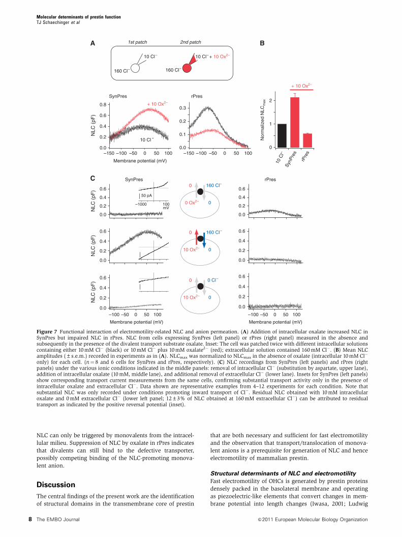
When anion transport was elicited by intracellular oxalate,
the amplitude of the NLC mediated by SynPres increased
by 2.14±0.46-fold (mean±s.d., N¼ 8 cells; Figure 7A and B)
compared with the zero transport condition before addition
of the divalent (intracellular and extracellular Cl�, 10 and
160 mM, respectively). In contrast, in mammalian prestin
lacking transport activity, addition of intracellular oxalate
markedly reduced NLC (to 0.58±0.06 of control, N¼ 6;
Figure 7A and B), despite the constant intracellular concen-
tration of Cl�. A similar reduction of NLC by intracellular
oxalate was observed with native prestin in rat OHCs
(Supplementary Figure S3). In either case, V1/2 was shifted
to slightly more positive potentials (by 34.1±11.9 mV and by
5.5±6.6 mV for recombinant SynPres and rPres, respectively;
Figure 7A). The enhancement of NLC exclusively in the
transport-competent SynPres suggested an influence of
anion transport on the generation of NLC.
Subsequent experiments that further investigated the
nature of the oxalate-induced increase in NLC amplitude
directly demonstrated that inward transport of the monova-
lent Cl� was the prerequisite for this striking phenomenon
observed with SynPres. These experiments were done in the
absence of intracellular Cl� (substitution by aspartate),
which fully abolished NLC (Figure 7C; upper panel), consis-
tent with previous observations on rPres (Oliver et al, 2001).
Loss of NLC indicated that binding of Cl� from the intra-
cellular face is required to support NLC in the absence of
anion exchange activity. However, robust NLC could be
measured with oxalate (10 mM) in the cytoplasm and, at
the same time, Cl� (160 mM) present at the extra-
cellular side of the membrane, which promoted inward
transport of the monovalent monitored as the transport
current (Figure 7C, middle left panel). When anion transport
was switched off by removal of one of the two substrates,
either Cl� from the extracellular milieu or oxalate from
the cytoplasm, NLC was no longer recorded (Figure 7C,
upper and lower left panels, respectively). Moreover, control
experiments with oxalate on both sides of the membrane
(160 mM extracellular, 10 mM intracellular) demonstrated
that the divalent per se was not sufficient to support NLC in
SynPres (data not shown). Finally, oxalate (10 mM) added
to the cytoplasm failed to trigger NLC in the transport-
incompetent rPres both in the absence and in the presence
of high concentrations of Cl� in the extracellular solution
(Figure 7C, right panel).
Several observations indicate that the observed potentiation
of NLC by anion transport resulted from a genuine interaction
of both processes rather than from an accumulation of Cl� at
the intracellular side. First, a two-fold increase was observed
even in the presence of 10 mM Cl� in the intracellular
medium (Figure 7A and B). Given the known sensitivity of
NLC for intracellular Cl� (Oliver et al, 2001), the cytoplasmic
concentration of Cl� would need to be elevated by 4100 mM
to double the NLC amplitude, which seems incompatible with
the ‘[Cl�] clamp’ by the whole-cell patch-clamp and the small
transport current. Second, the change of NLC (Figure 7C,
middle panel) occurred simultaneous (without any time lag)
with the change of the extracellular concentrations monitored
by transport current reversal, arguing against a requirement
for transport-dependent accumulation of intracellular Cl�.
In summary, these experiments indicated that generation
of the electromotility-related NLC in prestin is tightly linked
to anion transport. Specifically, NLC requires (i) monovalent
anions such as Cl� or other anion species (Rybalchenko and
Santos-Sacchi, 2008) that (ii) must be translocated to an
interaction/binding site within the protein. In SynPres, this
translocation may occur from either the extracellular
(through divalent/chloride electrogenic antiport) or the
intracellular side of the membrane (independent of divalent-
triggered transport), while in the transport-deprived rPres
B
Tra
nspo
rt c
urre
nt @
-100
mV
(pA
)
NLCmax (pF)
0.0 0.2 0.30.1 0.4 0.5
0
–50
–100
Control
Salicylate
0
50
100
150
–100 0 100–80
0
40
–40Cur
rent
(pA
)N
LC (
fF)
A
Membrane potential (mV)
Figure 6 Simultaneous anion transport and NLC in SynPres. (A) NLC of SynPres measured during anion transport. NLC and transport currentwere recorded simultaneously in the presence of intracellular oxalate (recording conditions as in Figure 5A) before and during applicationof 10 mM salicylate. (B) Correlation of NLC and transport current amplitudes obtained from 13 cells as in (A). Transport current wasdetermined as the salicylate-sensitive whole-cell current at �100 mV. Solid line represents a linear regression.
Molecular determinants of prestin functionTJ Schaechinger et al
&2011 European Molecular Biology Organization The EMBO Journal 7
NLC can only be triggered by monovalents from the intracel-
lular milieu. Suppression of NLC by oxalate in rPres indicates
that divalents can still bind to the defective transporter,
possibly competing binding of the NLC-promoting monova-
lent anion.
Discussion
The central findings of the present work are the identification
of structural domains in the transmembrane core of prestin
that are both necessary and sufficient for fast electromotility
and the observation that transport/translocation of monova-
lent anions is a prerequisite for generation of NLC and hence
electromotility of mammalian prestin.
Structural determinants of NLC and electromotility
Fast electromotility of OHCs is generated by prestin proteins
densely packed in the basolateral membrane and operating
as piezoelectric-like elements that convert changes in mem-
brane potential into length changes (Iwasa, 2001; Ludwig
10 Cl–
1st patch 2nd patch
160 Cl– 160 Cl–
0.0
0.2
0.4
0.6
0.8
NLC
(pF
)SynPres
–150 –100 –50 0 50 100
Membrane potential (mV)
–100 –50 0 50 100
Membrane potential (mV)
–100 –50 0 50 100
Membrane potential (mV)
+ 10 Ox2–
10 Cl -
0.0
0.1
0.2
0.3
rPres
–150
Nor
mal
ized
NLC
max
0
1
2
10 C
l–Sy
nPre
s
rPre
s
+ 10 Ox2–
BA
C
0.0
0.2
0.4
0.6
0.0
0.2
0.4
0.6
rPres
0.0
0.2
0.4
0.6
0.0
0.2
0.4
0.6
0
160 Cl–0
0 Ox2–
0.0
0.2
0.4
0.6
0.0
0.2
0.4
0.6
SynPres
NLC
(pF
)N
LC (
pF)
NLC
(pF
)
50 pA
–1000 100mV
0
160 Cl–0
10 Ox2–
0
0 Cl–0
10 Ox2–
10 Cl– + 10 Ox2–
100500–50–100
Figure 7 Functional interaction of electromotility-related NLC and anion permeation. (A) Addition of intracellular oxalate increased NLC inSynPres but impaired NLC in rPres. NLC from cells expressing SynPres (left panel) or rPres (right panel) measured in the absence andsubsequently in the presence of the divalent transport substrate oxalate. Inset: The cell was patched twice with different intracellular solutionscontaining either 10 mM Cl� (black) or 10 mM Cl� plus 10 mM oxalate2� (red); extracellular solution contained 160 mM Cl�. (B) Mean NLCamplitudes (±s.e.m.) recorded in experiments as in (A). NLCmax was normalized to NLCmax in the absence of oxalate (intracellular 10 mM Cl�
only) for each cell. (n¼ 8 and 6 cells for SynPres and rPres, respectively). (C) NLC recordings from SynPres (left panels) and rPres (rightpanels) under the various ionic conditions indicated in the middle panels: removal of intracellular Cl� (substitution by aspartate, upper lane),addition of intracellular oxalate (10 mM, middle lane), and additional removal of extracellular Cl� (lower lane). Insets for SynPres (left panels)show corresponding transport current measurements from the same cells, confirming substantial transport activity only in the presence ofintracellular oxalate and extracellular Cl�. Data shown are representative examples from 4–12 experiments for each condition. Note thatsubstantial NLC was only recorded under conditions promoting inward transport of Cl�. Residual NLC obtained with 10 mM intracellularoxalate and 0 mM extracellular Cl� (lower left panel; 12±3% of NLC obtained at 160 mM extracellular Cl�) can be attributed to residualtransport as indicated by the positive reversal potential (inset).
Molecular determinants of prestin functionTJ Schaechinger et al
The EMBO Journal &2011 European Molecular Biology Organization8

et al, 2001) and thereby produce the mechanical force crucial
for active cochlear amplification (Ashmore, 2008; Dallos,
2008; Dallos et al, 2008). Conversion between electrical and
mechanical energy by prestin is fully reciprocal (Iwasa, 2001;
Ludwig et al, 2001) and requires intracellular anions as an
extrinsic factor (Oliver et al, 2001; Rybalchenko and Santos-
Sacchi, 2008). How anions initiate the coupling of mechanical
and electrical action, however, has remained elusive, in
particular as structural information on the functionally im-
portant core region of prestin and of other SLC26 transporters
is largely missing. Despite some initial work towards respec-
tive structure–function analyses (Oliver et al, 2001;
Navaratnam et al, 2005; Rajagopalan et al, 2006; Bai et al,
2009; McGuire et al, 2010), no data are yet available that
firmly link distinct protein domains of prestin to particular
functions, such as voltage-sensing or motility-generating
transitions.
Using domain swapping between rPres and zPres together
with measurements of transport currents and capacitance we
identified structural elements required for piezoelectricity:
two distinct stretches in the rPres polypeptide, one compris-
ing transmembrane domains 1 and 2 and the adjacent linker
(aa 93–136; NLC-domain 1), the other consisting of putative
transmembrane domains 9 and 10 (aa 381–438; NLC-domain
2). Despite the strikingly large distance in primary sequence,
these two domains closely cooperate in function and effec-
tively combine to endow the exclusive transporter zPres with
NLC and fast electromotility (Figures 2–4). Moreover, func-
tion of SynPres, the ‘gain-of-function’ rPres–zPres chimera,
strongly suggests that the NLC-domains are placed right at
the interface between electromotility and anion transport and
that they may even form part of the anion permeation path-
way. Such a structural view is supported by the fact that
monovalent anions must be translocated into the protein
before NLC/electromotility can occur (Figure 7). In addition,
it is noteworthy that the NLC-domain 1 coincides with the
region of highest sequence conservation across the large
family of SLC26-related SulP transporters and includes the
SLC26A signature motif (PS01130) implicated in transport
function (Leves et al, 2008). Individual residues within
NLC-domain 1 that were found to be critical for electromo-
tility and NLC (L93, F101, and P136) are essentially conserved
among prestins from all mammalian clades (Supplementary
Figure S4), which share electromotility and NLC (Okoruwa
et al, 2008; Tan et al, 2011). Vice versa, across all non-
mammalian SLC26A5 sequences, these residues are also
highly conserved but distinct from the mammalian forms,
indicating a critical role of these residues for electromotility
in mammalian and for transport in non-mammalian prestins.
This observation further suggests that these amino acid
exchanges were key events in the evolutionary conversion
from a transporter to a piezoelectric protein and strongly
supports the important role of NLC-domain 1 for prestin
function.
It should be noted that additional regions of prestin are
probably involved in shaping the exact functional properties
of mammalian prestin, since voltage sensitivity of SynPres
differs substantially from native mammalian prestin.
The finding of two discrete domains closely cooperating in
driving translocation of ions is reminiscent of two bacterial
transporters for which high-resolution structural data are
available, the Naþ/Hþ antiporter NhaA (Hunte et al, 2005)
and the Cl�/Hþ antiporter Ec-ClC (Dutzler et al, 2002, 2003;
Accardi and Miller, 2004; Miller, 2006). In either case, the ion
pathway is built from a-helical segments (conventional and
discontinuous) separated in the respective primary sequence
by seven helical domains (helices IV and XI in NhaA or
segments E/F and M/N in Ec-ClC) and offers binding site(s)
for the respective substrates. In particular, in NhaA, these
ion-binding sites are right in the middle of the membrane;
binding of the charged substrates to these sites are thought to
induce movements of the helical domains, which in turn
promote substrate translocation according to an alternating-
access mechanism (Hunte et al, 2005).
Molecular model for generation of NLC and
electromotility
Based on the structural similarities with NhaA, an alternat-
ing-access mechanism may be envisaged as the structural
and functional principle behind the coupling of transport and
NLC/electromotility observed for rPres and SynPres
(Figure 7). As a straightforward explanation for changes in
NLC amplitude, the transport process may determine the
occupancy of a structural conformation (‘active state’) that
enables the voltage-fuelled rapid elongation/contraction tran-
sition generating NLC and fast electromotility. According to
our results, this active state corresponds to a conformation in
which a monovalent anion occupies a binding site within
prestin’s permeation pathway. In SynPres, this binding site is
reached either from the cytoplasm or from the extracellular
side of the membrane (through an alternating-access cycle
driven by divalent anions such as oxalate); in rPres, where
the transport cycle is constitutively blocked (most likely by
amino acid exchanges in the mammalian NLC-domains com-
pared with their non-mammalian counterparts), the mono-
valent anions can reach the intramolecular binding site from
the cytoplasm only (Figure 7).
Figure 8 illustrates an alternating-access model that can
account for the observed behaviour of both rPres and
SynPres; in fact, it displays some similarity with a scheme
proposed by Muallem and Ashmore (2006). Stoichiometric
antiport (as observed with SynPres) is realized by transitions
between the two major conformations with the substrate
binding site facing either the cytosol (Ei) or the extracellular
space (Eo); transitions between Ei and Eo only occur upon
substrate binding. The distribution between the various
states depends on transport rates and substrate concentra-
tions. In this model, the ‘active state’ that exclusively under-
goes the voltage-driven piezoelectric transition is assigned to
the Cl-bound state EoCl (highlighted in yellow; Figure 8),
consistent with the requirement of Cl� binding for generation
of NLC (Figure 7C; Oliver et al, 2001, 2006).
Qualitative inspection of the model readily reveals that in
SynPres, induction of transport by addition of the divalent
substrate leads to redistribution between states, including an
altered occupancy of the active state and thus altered NLC
amplitude. Of note, transitions within the transport cycle may
be much slower than the ultrafast conformational changes
that generate force and NLC without limiting the kinetics of
the latter, which are voltage-driven conformational changes
between sub-states of the ‘active state’ as depicted in
Figure 8. Specifically, binding of intracellular oxalate to Ei
drives additional prestin molecules into the active state, thus
increasing NLC consistent with experimental findings
Molecular determinants of prestin functionTJ Schaechinger et al
&2011 European Molecular Biology Organization The EMBO Journal 9

(Figure 7A). The reason for this is the increase in overall rates
towards the outward-facing conformations and the rapid
unidirectional transition from EoOx to the active state (in
the absence of extracellular divalent substrate). This effect is
particularly pronounced if the transition between EiOx and
EoOx is faster than the transition between EiCl and active
state. A detailed quantitative analysis of the model confirms
this conclusion (see Supplementary Figure S5).
Moreover, the model readily predicts the suppressive effect
of intracellular oxalate on NLC generated by rPres: because
NLC is completely independent of extracellular Cl� (Oliver
et al, 2001), dissociation from and binding of Cl� to the
outward-facing conformation must be impossible or greatly
disfavoured (shaded in Figure 8). This is also consistent with
the lack of transport activity in rPres (Schaechinger and
Oliver, 2007; Tan et al, 2011). Hence, when binding of
intracellular oxalate increases occupancy of EiOx, occupancy
of EoCl and consequently NLC must decrease, consistent with
the experimental findings shown in Figure 7A and B.
Furthermore, results obtained in the absence of intracel-
lular Cl� (Figure 7C) are in good agreement with the predic-
tions derived from the alternating-access transport model.
Briefly, loss of NLC upon removal of intracellular Cl� is
readily explained by accumulation of prestin in the Ei state
that cannot promote NLC. Addition of intracellular oxalate
leads to suppression of NLC in rPres via additional trapping
of prestin molecules in the EiOx state, whereas in SynPres,
intracellular oxalate allows for divalent/chloride antiport,
which runs exclusively clockwise in the absence of extra-
cellular oxalate and shuttles prestin molecules from Ei
towards the Eo conformations. As a consequence, occupancy
of the active state must increase, in agreement with the
observed increase in NLC amplitude. Removal of extracellular
Cl� interrupts transport since the transition from Eo to the
active state becomes zero. As this prevents refilling of the
active state, NLC should be abolished as indeed observed
experimentally.
Finally, the results presented in Figure 7C also exclude
an alternative allosteric mechanism that was suggested
previously for various intracellular anions (Rybalchenko
and Santos-Sacchi, 2008), namely potentiation of NLC by
intracellular oxalate through binding to a distinct site.
Thus, intracellular oxalate promoted NLC only in the pre-
sence of extracellular Cl�, but not when extracellular Cl� was
removed (Figure 7C) or when only oxalate was present at
both sides of the membrane.
In summary, the dependence of NLC on transport sub-
strates in both SynPres and mammalian prestin is fully
consistent with a model in which the NLC-generating transi-
tion is embedded within an alternating-access transport cycle.
Notably, the presented model does not make any assumption
about the nature of the NLC-generating transition; NLC
may derive from shuttling of Cl� through the electric field
(Oliver et al, 2001) or from movement of an intrinsic voltage
sensor (Bai et al, 2009) or a combination of both.
Materials and methods
Molecular biologycDNAs coding for native, mutant, or chimeric prestin proteins werederived from Rattus norvegicus and Danio rerio prestin cDNAs(GenBank accession No. NM_030840 and BC054604.1, respectively)by standard molecular biology techniques and cloned into pEGFP-N1 (Clontech) (rPres, Chi1, Chi2, revChi2, SynPres) or pEGFP-N3(zPres, Chi3), yielding C-terminal GFP fusion constructs. Allconstructs were verified by sequencing. For viral infection of OHCs,rPres and SynPres were subcloned into the viral vector AAV-HBA-EWB (Kugler et al, 2007).
ElectrophysiologycDNAs containing pEGFP plasmids were transfected into CHO cellsusing JetPEI transfection reagent (Polyplus, Illkirch, France).For electrophysiological experiments (24–48 h after transfection),cells with unequivocal and comparable membrane fluorescencewere selected. Whole-cell patch-clamp recordings were carried outat room temperature (20–221C) with EPC10 amplifiers (Heka,Lambrecht, Germany) controlled by Patchmaster software (Heka).
NLC. Whole-cell membrane capacitance (CM) was recorded usingthe sineþDC software lock-in function of Patchmaster. Frequencyof stimulus sinusoids was 2 kHz unless indicated otherwise.Voltage-dependent NLC was assessed by recording CM duringvoltage ramps (slope þ 0.2 to þ 0.56 V/s) as described previously(Oliver and Fakler, 1999; Schaechinger and Oliver, 2007) and plottedas a function of membrane potential (VM). Traces shown usuallyrepresent averages from 2 to 10 individual capacitance traces.
NLC was quantified by fitting the derivative of a first-orderBoltzmann function to the CM(VM) traces,
CMðVMÞ ¼ Clin þQmax
aeV�V1=2
a 1þ e�V�V1=2
a
� �2ð1Þ
where Clin is linear membrane capacitance, VM is membranepotential, Qmax is maximum voltage-sensor charge moved throughthe membrane electric field, V1/2 is voltage at half-maximalcharge transfer, and a is the slope factor of the voltage dependence.The amplitude of NLC was quantified as peak NLC, NLCmax¼CM
(V1/2)�Clin. As a measure of expression level, NLC was normalizedto linear membrane capacitance, which is proportional to plasmamembrane area: NLCrel¼NLCmax/Clin.
Transport. Electrogenic anion transport was measured as the ionictransport current in response to command voltage ramps (�100 to
Active state
EiOx Ei EiCl
V
EoEoOx
ChlorideOxalate
SynPres (only)
Intra
Extra
EoCl
Figure 8 Alternating-access model for operation of rPres andSynPres. Anion transport (in SynPres only) occurs through transi-tions between two major conformations, with the anion-binding siteexposed either to the cytosol (Ei) or to the extracellular side (Eo).Binding and dissociation of Cl� (blue) and oxalate (red) occurs fromboth major states; transitions between the Ei and Eo conformationsrequire substrate binding. SynPres may adopt all states shown,while for rPres only states in the non-shaded area are accessible.Specifically, binding and unbinding of Cl� to and from Eo isimpossible in rPres, as symbolized by the external barrier in thepermeation pathway. NLC is generated exclusively by prestinmolecules residing in the Cl�-bound EoCl state (‘active state’;yellow). More precisely, NLC arises from fast voltage-dependenttransitions between the states highlighted in yellow, for simplicitylumped into EoCl (V, membrane potential). Note, that the differencein extension between NLC-generating states is merely meant tosymbolize area motor activity and that assignment of extendedversus compact state is chosen arbitrarily.
Molecular determinants of prestin functionTJ Schaechinger et al
The EMBO Journal &2011 European Molecular Biology Organization10

þ 100 mV; 0.5 or 1 V/s) as described previously (Schaechinger andOliver, 2007).
Solutions. For NLC recordings, patch pipettes were filled withintracellular solution containing 160 mM CsCl. For experimentswith intracellular iodide, 15 mM CsI was substituted for an equalconcentration of CsCl. For transport or combined transport and NLCmeasurements, intracellular solution was (in mM) 10 CsCl, 10oxalate, 130 K-aspartate, pH 7.3 (KOH), or 10 CsCl, 150 K-aspartate.For measurements with nominally Cl�-free conditions, pipetteswere tip-filled with solution of either 160 mM K-aspartate or150 mM K-aspartate and 10 mM oxalate. Pipettes were back-filledwith Cl�-containing solutions to ensure stable electrode offset. Allpipette solutions additionally contained 1 mM Hepes, 1 mMK2EGTA, and pH was adjusted to 7.3 with KOH. Unless specifiedotherwise, extracellular solution was (in mM): 144 NaCl, 5.8 KCl,1.3 CaCl2, 0.9 MgCl2, 10 Hepes, 0.7 Na2HPO4, and 5.6 glucose, pH7.4 (NaOH). For inhibition of anion transport and NLC, 10 mMsodium salicylate was added. For exchange of extracellular Cl�,solutions were simplified to contain either (in mM) 160 KCl and 5Hepes, or 160 K-aspartate and 5 mM Hepes, pH 7.4 with KOH. Forextracellular application of divalent transport substrates solutionscontained either 10 mM Na2-oxalate or Na2SO4 and (in mM) 1 NaCl,149 Na-aspartate, 2 Mg-gluconate, 10 Hepes, pH 7.4 with NaOH.An agarose bridge was used as the bath electrode wheneverextracellular anions were exchanged.
Organotypic culture and viral transductionOrganotypic cultures of organs of Corti from mice at days 6–7 afterbirth were prepared and cultured as reported previously (Oliveret al, 1997). Mice were either homozygous prestin null mice(Cheatham et al, 2007) (Pres �/�) or control C57BL/6 mice (wt).AAV vectors of the hybrid serotype 1/2 were constructed essentiallyas described (Kugler et al, 2007; Reisinger et al, 2011). Expressionwas driven by the b-actin promoter. Recombinant AAVs werepurified by iodixanol gradient centrifugation and FPLC on heparinaffinity columns. Vectors were dialysed against PBS and storedat �801C in single use aliquots. Purity 499% was confirmed bySDS–PAGE and silver staining, genome titres were quantified byreal-time PCR.
Cultures from Pres�/� mice were infected with AAV vectorsencoding either rPres or SynPres (2ml per 0.3 ml well) in serum-freemedium at 1 day in vitro. Measurements were done after 6–8 daysin vitro (5–7 days after infection), when prestin expression levelshad reached saturation as determined from NLC recordings(unpublished results).
ElectromotilityFor NLC and electromotility measurements, OHCs were whole-cellpatch-clamped, using standard extracellular solution (see above)and an intracellular solution composed of (in mM): 150 NaCl,2.5 Na2ATP, 2 HEPES, 1 K2EGTA, 0.1 AlexaFluor488 hydrazide(Invitrogen), pH 7.3 (KOH), osmolality 290 mosm/kg. This solution
was found to minimize endogenous ionic currents and to supportrobust electromotile responses. The patch pipette was positionedwith an MM3A-LS piezoelectric manipulator (Kleindiek Nanotech-nik, Reutlingen, Germany) to exclude vibrational artifacts.
OHCs oriented with their longitudinal axis parallel to thecoverslip were patch-clamped with the pipette sealed onto thelateral membrane close to the cuticular plate after gently removingneighbouring supporting cells, allowing movement of the basal cellpole. Fluorescence from Alexa488-filled OHCs was obtained withwidefield fluorescence optics and a conventional EGFP filter set.Fluorescence from the basal pole of the cell was selectively projectedonto a photodiode through a rectangular aperture positioned acrossthe basal OHC pole (see Figure 4A, inset) by means of a Viewfinderphotometry device (Till Photonics, Grafelfing, Germany). OHC lengthchanges evoked by sinusoidal voltage commands (20 mV±40 mV,300 Hz), resulted in changes of fluorescence intensity. These fluores-cence changes recorded as the uncalibrated photodiode output wereused as the measure for electromotility. Electromotility was thenquantified by calculating power spectra from raw fluorescence datausing IgorPro software (IgorPro; Wavemetrics, Lake Oswego) and byanalysing power spectral density at the stimulus frequency (300 Hz).
Motility recordings from virus-infected OHCs were restricted tothose cells displaying a clearly discernable NLC that was inhibitedby salicylate (10 mM), indicating successful expression of exogen-ous prestin construct. Such salicylate-sensitive NLC was neverdetected in non-infected Pres�/� OHCs.
Confocal microscopyLive CHO cells transfected with GFP-fused prestin constructs wereimaged with a Zeiss LSM710 confocal microscope with GFPexcitation at 488 nm.
Supplementary dataSupplementary data are available at The EMBO Journal Online(http://www.embojournal.org).
Acknowledgements
We thank Anna Bulankina for initial characterization of the AAVvector and for sharing experimental protocols, Mary-Ann Cheathamand Peter Dallos for providing prestin null mice and Olga Ebers forexpert technical assistance.
Author contributions: TJS and DO designed and performed theexperiments and analysed the data; DG designed and performedthe experiments; CRH generated the mathematical model; SK andTM developed and generated viral vectors; DO and BF conceivedthe study and wrote the manuscript.
Conflict of interest
The authors declare that they have no conflict of interest.
References
Accardi A, Miller C (2004) Secondary active transport mediatedby a prokaryotic homologue of ClC Cl- channels. Nature 427:803–807
Albert JT, Winter H, Schaechinger TJ, Weber T, Wang X, He DZZ,Hendrich O, Geisler H-S, Zimmermann U, Oelmann K,Knipper M, Gopfert MC, Oliver D (2007) Voltage-sensitive prestinorthologue expressed in zebrafish hair cells. J Physiol 580:451–461
Ashmore J (2008) Cochlear outer hair cell motility. Physiol Rev 88:173–210
Ashmore JF (1987) A fast motile response in guinea-pig outer haircells: the cellular basis of the cochlear amplifier. J Physiol 388:323–347
Bai J-P, Surguchev A, Montoya S, Aronson PS, Santos-Sacchi J,Navaratnam D (2009) Prestin’s anion transport and voltage-sensing capabilities are independent. Biophys J 96: 3179–3186
Bezanilla F (2008) How membrane proteins sense voltage. Nat RevMol Cell Biol 9: 323–332
Brownell WE, Bader CR, Bertrand D, de Ribaupierre Y (1985)Evoked mechanical responses of isolated cochlear outer haircells. Science 227: 194–196
Cheatham MA, Zheng J, Huynh KH, Du GG, Edge RM, Anderson CT,Zuo J, Ryan AF, Dallos P (2007) Evaluation of an independentprestin mouse model derived from the 129S1 strain. AudiolNeurootol 12: 378–390
Dallos P (2008) Cochlear amplification, outer hair cells and prestin.Curr Opin Neurobiol 18: 370–376
Dallos P, Fakler B (2002) Prestin, a new type of motor protein.Nat Rev Mol Cell Biol 3: 104–111
Dallos P, Wu X, Cheatham MA, Gao J, Zheng J, Anderson CT, Jia S,Wang X, Cheng WHY, Sengupta S, He DZZ, Zuo J (2008) Prestin-based outer hair cell motility is necessary for mammalian co-chlear amplification. Neuron 58: 333–339
Dutzler R, Campbell EB, Cadene M, Chait BT, MacKinnon R (2002)X-ray structure of a ClC chloride channel at 3.0 A reveals themolecular basis of anion selectivity. Nature 415: 287–294
Molecular determinants of prestin functionTJ Schaechinger et al
&2011 European Molecular Biology Organization The EMBO Journal 11
Dutzler R, Campbell EB, MacKinnon R (2003) Gating the selectivityfilter in ClC chloride channels. Science 300: 108–112
Franchini LF, Elgoyhen AB (2006) Adaptive evolution in mamma-lian proteins involved in cochlear outer hair cell electromotility.Mol Phylogenet Evol 41: 622–635
Frank G, Hemmert W, Gummer AW (1999) Limiting dynamics ofhigh-frequency electromechanical transduction of outer hair cells.Proc Natl Acad Sci USA 96: 4420–4425
Hudspeth AJ (2008) Making an effort to listen: mechanical ampli-fication in the ear. Neuron 59: 530–545
Hunte C, Screpanti E, Venturi M, Rimon A, Padan E, Michel H(2005) Structure of a Na+/H+ antiporter and insights intomechanism of action and regulation by pH. Nature 435:1197–1202
Iwasa KH (2001) A two-state piezoelectric model for outer hair cellmotility. Biophys J 81: 2495–2506
Kugler S, Hahnewald R, Garrido M, Reiss J (2007) Long-term rescueof a lethal inherited disease by adeno-associated virus-mediatedgene transfer in a mouse model of molybdenum-cofactor defi-ciency. Am J Hum Genet 80: 291–297
Leves FP, Tierney ML, Howitt SM (2008) Polar residues in aconserved motif spanning helices 1 and 2 are functionallyimportant in the SulP transporter family. Int J Biochem Cell Biol40: 2596–2605
Ludwig J, Oliver D, Frank G, Klocker N, Gummer AW, Fakler B(2001) Reciprocal electromechanical properties of rat prestin: themotor molecule from rat outer hair cells. Proc Natl Acad Sci USA98: 4178–4183
McGuire RM, Liu H, Pereira FA, Raphael RM (2010) Cysteinemutagenesis reveals transmembrane residues associated withcharge translocation in prestin. J Biol Chem 285: 3103–3113
Miller C (2006) ClC chloride channels viewed through a transporterlens. Nature 440: 484–489
Mount DB, Romero MF (2004) The SLC26 gene family of multi-functional anion exchangers. Pflugers Arch 447: 710–721
Muallem D, Ashmore J (2006) An anion antiporter model of prestin,the outer hair cell motor protein. Biophys J 90: 4035–4045
Navaratnam D, Bai J-P, Samaranayake H, Santos-Sacchi J (2005)N-terminal-mediated homomultimerization of prestin, the outerhair cell motor protein. Biophys J 89: 3345–3352
Okoruwa OE, Weston MD, Sanjeevi DC, Millemon AR, Fritzsch B,Hallworth R, Beisel KW (2008) Evolutionary insights into theunique electromotility motor of mammalian outer hair cells. EvolDev 10: 300–315
Oliver D, Fakler B (1999) Expression density and functional char-acteristics of the outer hair cell motor protein are regulated duringpostnatal development in rat. J Physiol 519: 791–800
Oliver D, He DZ, Klocker N, Ludwig J, Schulte U, Waldegger S,Ruppersberg JP, Dallos P, Fakler B (2001) Intracellular anions asthe voltage sensor of prestin, the outer hair cell motor protein.Science 292: 2340–2343
Oliver D, Plinkert P, Zenner HP, Ruppersberg JP (1997) Sodiumcurrent expression during postnatal development of rat outer haircells. Pflugers Arch 434: 772–778
Oliver D, Schaechinger T, Fakler B (2006) Interaction of prestin(SLC26A5) with monovalent intracellular anions. Novartis FoundSymp 273: 244–253
Rajagopalan L, Patel N, Madabushi S, Goddard JA, Anjan V, Lin F,Shope C, Farrell B, Lichtarge O, Davidson AL, Brownell WE,Pereira FA (2006) Essential helix interactions in the anion trans-porter domain of prestin revealed by evolutionary trace analysis.J Neurosci 26: 12727–12734
Reisinger E, Bresee C, Neef J, Nair R, Reuter K, Bulankina A,Nouvian R, Koch M, Buckers J, Kastrup L, Roux I, Petit C, HellSW, Brose N, Rhee JS, Kugler S, Brigande JV, Moser T (2011)Probing the functional equivalence of otoferlin and synaptotag-min 1 in exocytosis. J Neurosci 31: 4886–4895
Rybalchenko V, Santos-Sacchi J (2003) Cl- flux through a non-selective, stretch-sensitive conductance influences the outer haircell motor of the guinea-pig. J Physiol 547: 873–891
Rybalchenko V, Santos-Sacchi J (2008) Anion control of voltagesensing by the motor protein prestin in outer hair cells. Biophys J95: 4439–4447
Santos-Sacchi J (1991) Reversible inhibition of voltage-dependentouter hair cell motility and capacitance. J Neurosci 11: 3096–3110
Santos-Sacchi J, Song L, Zheng J, Nuttall AL (2006) Control ofmammalian cochlear amplification by chloride anions. J Neurosci26: 3992–3998
Schaechinger TJ, Oliver D (2007) Nonmammalian orthologs ofprestin (SLC26A5) are electrogenic divalent/chloride anion ex-changers. Proc Natl Acad Sci USA 104: 7693–7698
Tan X, Pecka JL, Tang J, Okoruwa OE, Zhang Q, Beisel KW, He DZ(2011) From zebrafish to mammal: functional evolution of pres-tin, the motor protein of cochlear outer hair cells. J Neurophysiol105: 36–44
Xia A, Wooltorton J, Palmer D, Ng P, Pereira F, Eatock R, Oghalai J(2008) Functional prestin transduction of immature outer haircells from normal and prestin-null mice. JARO 9: 307–320
Zheng J, Long KB, Shen W, Madison LD, Dallos P (2001) Prestintopology: localization of protein epitopes in relation to the plasmamembrane. Neuroreport 12: 1929–1935
Zheng J, Shen W, He DZ, Long KB, Madison LD, Dallos P (2000)Prestin is the motor protein of cochlear outer hair cells. Nature405: 149–155
Molecular determinants of prestin functionTJ Schaechinger et al
The EMBO Journal &2011 European Molecular Biology Organization12