a study on mandibular vascular canals: the risk of
TRANSCRIPT

Western UniversityScholarship@Western
Masters of Clinical Anatomy Projects Anatomy and Cell Biology Department
2016
A Study on Mandibular Vascular Canals: The Riskof Hemorrhage During Implant SurgeryPrabhsimrat GillWestern University, [email protected]
Follow this and additional works at: https://ir.lib.uwo.ca/mcap
Part of the Anatomy Commons
Citation of this paper:Gill, Prabhsimrat, "A Study on Mandibular Vascular Canals: The Risk of Hemorrhage During Implant Surgery" (2016). Masters ofClinical Anatomy Projects. 12.https://ir.lib.uwo.ca/mcap/12

A Study on Mandibular Vascular Canals: The Risk of Hemorrhage During Implant Surgery
Project format: Monograph
by
Prabhsimrat Singh Gill
Graduate Program in Clinical Anatomy
A project submitted in partial fulfillment of the requirements for the degree of
Master of Science
The School of Graduate and Postdoctoral Studies The University of Western Ontario
London, Ontario, Canada
© Prabhsimrat Gill 2016

ii
Abstract
Dental implants are commonly used to replace missing teeth in the anterior mandible.
This area is vascularized by the sublingual artery, which can be damaged during the implant
procedure. This investigation aims to determine the variation in the sublingual artery and its
relevant lingual foramina, such that it can be avoided by dental surgeons in the future. Dry
mandibles (n=43) were acquired and the distance from the internal superior border of the
mandible to the lingual foramen was measured and compared to the total height of the anterior
mandible. 67% of foramina were found at a distance greater than 50% below the internal
superior border of the mandible, and there was a significant difference in the number of
foramina on the left half of the anterointerior mandible (2.3 ± 1.1) compared to the right half
(1.4 ± 1.1) (P=0.021). These results imply increased vascularization in the lower half of the
anterior mandible as well as on the left half of the same area. Dental surgeons should take extra
care when working in these areas in order to minimize risk of sublingual hemorrhaging
Keywords
Lingual Artery, Sublingual Artery, Lingual Foramen, Sublingual Hemorrhaging, Dental Implant

iii
Co-Authorship Statement (where applicable)
This thesis was written and completed by Prabhsimrat Singh Gill under the supervision of
Dr. Khadry Galil, Dr. Kat Willmore, Dr. Tim Wilson, and Dr. Hiran Perinpanayagam.
Experiments were designed and carried out jointly by Prabhsimrat Singh Gill and Dr. Khadry
Galil.

iv
Acknowledgments
I would like to take this time to thank some of the people who were vital to this project,
and without whom it could not have been done.
I’d like to begin by extending my gratitude to my committee. Dr. Khadry Galil, not just
for genesis of the project, but also for your continued support throughout the year. You were
always available to meet whenever I wasn’t sure about next steps and continually sent me
resources even without me asking for them. You have been an excellent supervisor throughout
the year and I’m very grateful for getting to work with you. I would also like to thank Dr. Hiran
Perinpanayagam for his very prompt and helpful responses whenever I had questions about any
aspect of the project. You were always willing to take time out of your day to help me when
needed and your knowledge and expertise in the field were very helpful. I’m glad you joined our
committee and believe any future students who have you as a mentor will be very fortunate.
Next, I would like to thank Dr. Tim Wilson. I knew from the very beginning that I wanted to
have you on the committee due to your willingness to help others and your positive, enthusiastic
attitude. You provided me with motivation whenever I wasn’t sure of my next steps, and I truly
appreciate it. Finally, I would like to thank Dr. Kat Willmore. You were always eager to assist
me in any way you could, and made it very clear to me from the beginning that you were
someone I could come to if I had any questions. Not only were you very helpful with your
extensive knowledge of the anatomy of the region, but you were also always present to motivate
me whenever I was unsure about myself or my work. Overall, you were all very helpful from
the beginning to the end, and if I could I would work with any of you again.
I would also like to thank Kevin Walker and Haley Linklater for acquiring specimens for
me, as well as keeping me company during the long hours of dissecting in the lab. Mark Abbot
was also very helpful in assisting with my measurements. Finally, I would like to thank my
Clinical Anatomy colleagues, who provided me with endless support through the last two years.

v
Table of Contents
Abstract ............................................................................................................................... ii
Co-Authorship Statement (where applicable) .................................................................... iii
Table of Contents ................................................................................................................ v
List of Tables .................................................................................................................... vii
List of Figures .................................................................................................................. viii
List of Abbreviations .......................................................................................................... x
List of Appendices ............................................................................................................. xi
Chapter 1 ............................................................................................................................. 1
1 Literature Review ........................................................................................................... 1
1.1 General Anatomy .................................................................................................... 1
1.2 The Lingual Foramen and other relevant foramina ................................................ 2
1.3 Vasculature ............................................................................................................. 3
1.4 The Floor of the Mouth ........................................................................................... 6
1.5 Dental Implants and Subsequent Hemorrhaging .................................................... 9
Chapter 2 ........................................................................................................................... 13
2 Introduction .................................................................................................................. 13
2.1 Background ........................................................................................................... 13
2.2 Objectives ............................................................................................................. 14
Chapter 3 ........................................................................................................................... 15
3 Methods ........................................................................................................................ 15
3.1 Subject Collection ................................................................................................. 15
3.2 Study Design ......................................................................................................... 16
3.2.1 Study of the Lingual Foramina ................................................................. 16
3.2.2 Study of the Submental and Sublingual Arteries – Bisected Heads ......... 17
vi
3.2.3 Study of the Submental and Sublingual Arteries – Whole Heads ............ 22
Chapter 4 ........................................................................................................................... 23
4 Results .......................................................................................................................... 23
4.1 Interrater Reliability .............................................................................................. 23
4.2 Lingual Foramina Characterization ...................................................................... 23
4.3 Accessory Lingual Foramina Characterization ..................................................... 27
Chapter 5 ........................................................................................................................... 30
5 Discussion .................................................................................................................... 30
5.1 Anatomical and Clinical Significance .................................................................. 30
5.2 Future Considerations ........................................................................................... 36
5.3 Limitations of the Study........................................................................................ 36
Bibliography ..................................................................................................................... 38
Appendix A ....................................................................................................................... 42
Appendix B - Permissions ................................................................................................ 43
Appendix C - Raw Data .................................................................................................... 44

vii
List of Tables
Table 4.3.1 Mean distances (mm) of the primary and accessory lingual foramina from the
alveolar and mental surfaces of the mandible ......................................................................... 28
viii
List of Figures
Figure 1.2.1 A Posterior view of the interforaminal region of the mandible. The Lingual
Foramen is indicated in the center by the black arrow ............................................................. 3
Figure 1.3.2 The Lingual Artery (LA) can be seen branching into its inferior sublingual
branch (SL) and superior deep lingual branch (DL). The Genioglossus (GG) and anterior
mandible (AM) are also labelled............................................................................................... 4
Figure 1.4.1 The sublingual space can be seen highlighted in yellow, superior to the
mylohyoid muscle (MH). geniohyoid (GH), genioglossus (GG), and the anterior mandible
(AM) are also depicted. The submental space would be found inferior to the mylohyoid
muscle ....................................................................................................................................... 8
Figure 1.4.2 The mylohyoid boutonniere can be seen as a space in the mylohyoid muscle
(MH). In this scenario, the sublingual gland can be seen passing through the boutonnierre,
thus leaving its usual position deep to the muscle. ................................................................... 8
Figure 1.5.1 A patient who had severe hemorrhaging following a dental implant procedure. A
tracheotomy was performed to secure the airway before surgery .......................................... 10
Figure 3.2.1 A probe is being inserted into the space between the hyoglossus (HG) and
genioglossus (GG). The lingual mucosa (LM) had to be removed before this region could be
accessed. The hyoglossus will be cut superior to the probe and reflected to access the lingual
artery deep to it. ...................................................................................................................... 18
Figure 3.2.2 The hyoglossus (HG) has been reflected laterally to expose the lingual artery
(LA) underneath. ..................................................................................................................... 19
Figure 3.2.3 A full dissection of the sublingual artery (SA) travelling to the lingual foramen
in the anterior mandible. The deep lingual (DL) artery can also be seen supplying the tongue
(top). Note that the genioglossus has been stripped away from the mandible to expose the
foramen. The primary lingual artery (LA) as well as the anterior mandible (AM) have also
been labelled. .......................................................................................................................... 20

ix
Figure 3.2.4 An enlarged submental artery (indicated in yellow) taking the place of the
sublingual artery and entering the lingual foramen. The sublingual gland (SG), mylohyoid
muscle (MH) and anterior mandible (AM) are all labeled for orientation purposes. ............. 21
Figure 3.2.5 An inferior view of the mandible in which the skin, fascia, and musculature has
been removed. A Sublingual Artery can be seen travelling towards the anterior mandible
(AM) ....................................................................................................................................... 22
Figure 4.2.1 Histogram depicting the variation in positioning of the lingual foramen along the
lingual surface of the mandible in relation to the alveolar surface of the bone. 39 specimens
were measured with the mean distance from the alveolar surface being 16.17 ± 3.72 mm with
a range of 9.27 mm – 24.33 mm. ............................................................................................ 24
Figure 4.2.2 Histogram depicting the variation in positioning of the lingual foramen along the
lingual surface of the mandible in relation to the mental surface of the bone. 39 specimens
were measured with the mean distance from the mental surface being 11.61 ± 2.36 mm with
a range of 5.22 mm – 15.62 mm. ............................................................................................ 25
Figure 4.2.3 Histogram depicting the position of the lingual foramen on the lingual surface of
the mandible as a ratio between D1 and the total height of the mandible at its lingual surface.
39 specimens were measured with the mean distance from the superior alveolar surface being
0.58 ± 0.09 with a range of 0.42 – 0.81. ................................................................................. 26
Figure 4.3.1 Mean distances of various accessory lingual foramina plotted against the
primary lingual foramen (LF). The accessory foramina examined were the left lingual
foramen (LLF), right lingual foramen (RLF), superior lingual foramen (SLF), and inferior
lingual foramen (ILF). Error bars represent a 95% Confidence Interval ................................ 29
Figure 0.1 Images from three different mandibular specimens showing presence of dental
facets on the surface of teeth (A), furcation involvement (B), and severe bone loss around
anterior teeth (C). These factors are typically present in older populations. .......................... 35

x
List of Abbreviations
LA – Lingual Artery
SL – Sublingual Artery
DL – Deep Lingual Artery
GG – Genioglossus Muscle
MH – Mylohyoid Muscle
GH – Geniohyoid Muscle
AM – Anterior Mandible
LF – Lingual Foramen
ALF – Accessory Lingual Foramina
RLF – Right Lingual Foramen
LLF - Left Lingual Foramen
SLF – Superior Lingual Foramen
ILF – Inferior Lingual Foramen

xi
List of Appendices (where applicable)
Appendix A ............................................................................................................................. 42
Appendix B - Permissions ...................................................................................................... 43
Appendix C - Raw Data .......................................................................................................... 44

1
Chapter 1
1 Literature Review
1.1 General Anatomy of the Mandible
The mandible is the lower jaw bone and helps define the antero-inferior border of the
mouth. The lower row of teeth is attached to the mandible via alveolar processes, which are
thickened ridges of bone in the mandible that hold the teeth in place. The mandible is in
close relation with the maxilla, which is the bone forming the superior portion of your
mouth. This bone has its own alveolar processes as well as teeth, and together these
structures allow for chewing of food. The mandible articulates with the temporal bone at the
Temporo-Mandibular joint (TMJ). This joint is surrounded by a capsule and has an articular
disc which divides the joint into an upper and lower half. The upper half of the joint allows
for translational movement (forward and back) whiles the lower joint allows for rotational
movement (initial movement when opening jaw). During development, the mandible
actually begins as two halves but fuses at the midline to form one single bone (Moore,
DalleyII, & Agur, 2014). The mandible consists of a body where the alveolar processes and
teeth insert into. This body curves upwards posteriorly to form the ramus, which ultimately
terminates in two large protuberances. The anterior and posterior protuberances are known
as the coronoid process and the condyle of the mandible respectively. The condyle is what
articulates with the temporal bone to form the TMJ.
The mandible has a couple of important foramen through which blood vessels and
nerves can enter or leave. The mandibular foramen is found on the medial side of the
mandible where the body of the mandible angles upwards to form the ramus, and forms the
mandibular canal which exits as the mental foramen. The contents of this canal include the
inferior alveolar artery, inferior alveolar vein, and inferior alveolar nerve. The inferior
alveolar nerve is a branch off the mandibular nerve which comes off of cranial nerve V
(trigeminal nerve). This nerve gives off the nerve to mylohyoid before entering the
mandibular canal and supplying sensory information to the lower set of teeth. Before
terminating, it gives off the mental nerve which will exit the mandible via the mental

2
foramen and supply the skin of the chin and lower lip (Greenstein & Tarnow, 2006). The
inferior alveolar nerve has the possibility of being damaged during dental implant surgery.
However, extensive studies have been done on its variation in position and how to avoid
hitting this nerve during surgery, particularly as it branches into the mental nerve (Ivey,
Wilson, Merrifield, Shimizu, & Galil, 2014). The main variation found is that the inferior
alveolar nerve can undergo an “anterior loop” in which it extends anteriorly from the mental
foramen. The mean length of this loop was 6.95 mm in length (Greenstein & Tarnow, 2006)
and its presence made it easier to damage the nerve during implant surgery.
The inferior alveolar artery is a branch off the maxillary artery, which in turn is a
branch of the external carotid artery. This artery’s distribution follows that of the
corresponding nerve, as outlined above. The inferior alveolar artery will enter the
mandibular foramen and travel through the incisive canal until it anastomoses with its
counterpart on the other side via the incisor branch, which also supplies the bottom row of
teeth as it moves anteriorly (Flanagan, 2003). As this vessel travels through the mandible, it
lets off several branches that travel through what are known as nutrient canals. These
arteries travel with branches from the inferior alveolar nerve to supply the mandibular bone
(Ogawa, Fukuta, Nakasato, & Nakasato, 2016). Once the inferior alveolar artery leaves the
mandible and becomes the mental artery via the mental foramen, it will supply the chin and
bottom lip and will anastomose with the submental and inferior labial arteries. This artery
also has been known to anastomose with the submental and sublingual artery within the
mandible (Kawai & al, 2006). Presently, there does not seem to be much if any literature on
the effects of dental implant surgery on the inferior alveolar artery. However, it has been
mentioned that this surgery can affect the artery indirectly by damaging one of its
anastomoses (Flanagan, 2003).
1.2 The Lingual Foramen and other relevant foramina
The lingual foramen is a foramen found at the midline of the mandibular body on the
lingual side, typically level with the genial bodies (McDonell, Nouri, & Todd, 1994) (Figure
1.2.1). Variations of this foramen have been found and it is not uncommon to find multiple
lingual foramina in a mandible. These variations were classified as either superior or inferior
genial spinal foramina depending on their location relative to the genial spine (Bernardi &

3
Rastelli, 2014). The main vessel traveling through this foramen is the sublingual artery
which branches off the lingual artery and supplies the floor of the mouth as well as the
mandible. However, the contents of the foramen vary based on its position. A lingual
foramen higher than normal may contain branches of the main lingual artery, whereas a
lower lingual foramen is more likely to have the actual sublingual artery travelling through it
(Bernardi & Rastelli, 2014). The anterior mandible was thought to be a safe place for
surgery, but with the discovery of this foramen and its contents it is clear surgeons must be
careful working in this area.
1.3 Vasculature of the Mandible
The sublingual artery is a blood vessel that arises from the lingual artery, which in
turn comes off of the external carotid artery. The lingual artery is usually the third branch
coming off the external carotid, and arises close to the hyoid bone (Niamtu, 2001). The
artery is characterized by its inferior loop before moving superior and anteriorly into the
tongue. The lingual artery then passes under the hyoglossus muscle and splits at its anterior
border to form the sublingual and deep lingual arteries (Figure 1.3.1). The deep lingual
Figure 1.2.1 A Posterior view of the
interforaminal region of the
mandible. The Lingual Foramen is
indicated in the center by the black
arrow

4
artery travels more superiorly to supply the tongue whereas the sublingual artery tends to
travel more inferiorly and let off branches supplying not only the tongue, but the floor of the
mouth and the mandible as well (Bavitz & Harn, 1994). This supply of the mandible is done
via the lingual foramen (Bernardi & Rastelli, 2014). It is worth noting that the lingual artery
seems to be resistant to atherosclerosis and therefore maintains a strong blood flow even in
aged patients (Flanagan, 2003). As a result damage to this artery may be more serious than
most.
The venous drainage of the mouth and tongue do not seem to be well researched in
the literature. There are three major veins that drain the floor of the mouth and tongue. The
sublingual vein, the deep lingual vein, and the dorsal lingual veins. These three have been
collectively dubbed the “lingual veins” and will drain into the internal jugular vein. The
sublingual veins drain the floor of the mouth whereas the deep and dorsal lingual veins drain
DL
GG
AM
Figure 1.3.1 The Lingual Artery (LA) can be seen branching into its
inferior sublingual branch (SL) and superior deep lingual branch (DL).
The Genioglossus (GG) and anterior mandible (AM) are also labelled
DL
LA SL
GG

5
the tongue. The latter two veins typically follow the lingual and deep lingual artery, however
they are usually found superficial to Hyoglossus rather than deep to it.
Other vessels have been found to supply the oral cavity as well. The inferior alveolar
artery arises as a branch of the maxillary artery, which is one of the last branches off the
external carotid. It is known to enter the mandible via the mandibular foramen and supply
the bone as well as the teeth (Castelli, 1962). The submental artery will also supply the oral
cavity. The fourth branch of the external carotid is known as the facial artery. This artery lets
off the submental artery as it passes through the submandibular gland (Faltaous & Yetman,
1996). This artery will pass over the mylohyoid and supply the floor of the mouth as well as
skin in the submental area (Faltaous & Yetman, 1996). In several cases (53% of donors in
one study), the sublingual artery was either not present or very small. In these cases, the
submental artery would enlarge and perforate the mylohyoid (Bavitz & Harn, 1994) (Figure
1.3.2). Therefore, it is important to note the presence of this artery in cases where the
sublingual artery is missing.
This presents an interesting dilemma for dental surgeons who notice a bleeding
artery. A feature of arteries is that when they are severed, they tend to retract into the tissue,
occasionally causing them to no longer be visible. As a result, a dental surgeon is presented
with a growing hemorrhage for which they have no visible source (Gakonyo, Butt,
Mwachaka, & Wagaiyu, 2015). The natural course of action would be to ligate the source of
the affected artery. In this case, that artery is thought to be the lingual artery. However, if the
sublingual artery is not present in an individual a ligation of the lingual artery will have no
effect, block off blood supply to other regions unnecessarily, and waste time in an already
delicate situation. The same situation is true if the artery entering the lingual foramen does
not have a contribution from the sublingual artery, which occurs in approximately 17.6% of
individuals. In these situations, the submental artery which has been known to replace the
sublingual artery in the floor of the mouth when it is missing will also enter the lingual
foramen to supply the mandible (Gakonyo et al., 2015). Damage to this vessel would
therefore necessitate ligation of the facial artery; however this is not common knowledge
and is typically not done in time to prevent surgical intervention. It is worth noting that in
8.9% of individuals the vessel entering the lingual foramen is an anastomoses of both the

6
sublingual and submental vessels, and therefore a dual ligation would be required to
completely stop bleeding.
1.4 The Floor of the Mouth
The floor of the mouth is comprised of several fascial spaces, as well as various
muscles that allow for stability in this region. Two very important spaces that contain the
blood released from severed vessels during dental implant surgery are the submental and
sublingual spaces.
A
B
Figure 1.3.2 In this dissection, the Lingual Artery (A) has branched, but
both branches are supplying the tongue and therefore the sublingual
branch to the floor of the mouth is not present. In this situation, a branch
of the submental artery (B) has enlarged and perforated the mylohyoid to
supply the floor of the mouth via various visible accessory branches. It
can also be seen entering the lingual foramen and supplying the anterior
mandible (AM)
AM
Superior
Inferior
An
terior P
ost
eri
or

7
The sublingual space lies on either side of the tongue. Its borders consist of the
mylohyoid muscle inferiorly, the mucosa and floor of the mouth superiorly, and the
mandible anteriorly (La, 2011) (Figure 1.4.1). It is a bilateral space which consists of various
structures, including but not limited to the lingual artery, lingual nerve, hypoglossal nerve,
and glossopharyngeal nerve. Since the sublingual artery is not mentioned in literature often,
it is not mentioned as being a component of the sublingual space. However, since the
mandible composes the anterior border of the sublingual space and the sublingual space is
the major space that swells when the sublingual artery is damaged, it can be reasoned that
this artery also lies within the space. Interestingly, damage to the sublingual artery will cause
hemorrhaging that can leave the sublingual space and enter the submandibular space
(Tomljenovic, Herrmann, Filippi, & Kühl, 2015). This is likely due to a defect in the
mylohyoid muscle that is present in approximately 77% of individuals known as a
“boutonnierre” (White, Davidson, Harnsberger, Haller, & Kamya, 2001). The mylohyoid is
typically the border between the sublingual and submandibular spaces but with this defect
those spaces become continuous, resulting in a less contained and more difficult to manage
bleed (Figure 1.4.2). The 2 spaces are sometimes collectively known as the submandibular
space, with the sublingual space being called the “supramylohyoid portion of the
submandibular space” and the submental space being called the “inframylohyoid portion of
the submandibular space” (Hartmann, 1999)
The borders of the submental space consist of the hyoid bone inferiorly, the
mandible superiorly, and the anterior bellies of the 2 digastric muscles laterally. Some of its
contents include the mylohyoid nerve and vessels, submental branch of the facial artery, and
the facial vein (Ural et al., 2011). Unlike swelling in the sublingual space, which is more
visible in the mouth due to the noticeable swelling of the tongue, the submental swelling can
be seen externally and is therefore a good clinical indicator for a dental surgeon.

8
Figure 1.4.1 The sublingual space can be seen highlighted in yellow, superior to the
mylohyoid muscle (MH). geniohyoid (GH), genioglossus (GG), and the anterior
mandible (AM) are also depicted. The submental space would be found inferior to the
mylohyoid muscle
Figure 1.4.2 The mylohyoid boutonniere can be seen as a space in the mylohyoid muscle
(MH). In this scenario, the sublingual gland can be seen passing through the
boutonnierre, thus leaving its usual position deep to the muscle.
MH
GG
GH
AM
Superior
Inferior
An
terior P
ost
eri
or

9
1.5 Dental Implants and Subsequent Hemorrhaging
Dental implants are commonly used for replacing missing or broken teeth and have a
high success rate (five year functional success rates of approximately 90%) (Brooks, 2000).
The need for dental implants is directly related to age, and as a result dental implant surgery
is becoming more popular as the proportion of the population over the age of 65 years
continues to rise (Misch, 2014). The implant itself is used to support other structures such as
crowns or dentures, and sufficient time is allowed for the implant to integrate itself into the
bone.
Because dental implants are inserted into the bone, risk of damaging vessels and
nerves is present. This damage is most common when an implant is inserted in the anterior
mandible, as this is where the sublingual, submental, and inferior alveolar arteries are known
to anastomose within the mandible (Castelli, 1962). In general, these vessels are well
defined and their paths are understood. However, hemorrhaging is likely to occur when
abnormal vessels or vessels that are not very well defined are present. A big contributor for
the seriousness of this condition is the weakness of the lingual floor. The floor is fairly lax
and easily expanded when filled with blood, resulting in a diffuse haemotoma that is spread
over a large area and is therefore more difficult to drain (Pigadas, Simoes, & Tuffin, 2009).
The hemorrhaging can happen at various depths and stages in the implant procedure.
Before the implant itself is placed, the area is typically probed to determine whether enough
bone is present for implant integration. During this procedure, bleeding can occur if an
artery is perforated (Mordenfield & Andersson, 1997). In the case of (Mordenfield &
Andersson, 1997), the bleeding was spotted immediately and the implant was put on hold
while the immediate concern was addressed. Another possibility is the hemorrhage occuring
after the placement has completed. In the case of (Felisati et al, 2012), the procedure was
completed and the dental surgeon did not observe any complications. The patient was asked
to stay in the waiting room for an observation period and it was during this time that slow
swelling of the sublingual and submandibular regions began, resulting in emergency
procedures. In both types of hemorrhaging, the first symptom is echymosis (discoloration
due to bleeding beneath the tissue) in the tongue and sublingual tissue due to the damaged
blood vessels. As the tongue begins to move upwards against the palate and oropharynx, the

10
dental surgeon must act to stem the bleeding. However, a major concern with this
hemorrhaging is that most dental surgeons do not have the tools to stop the bleeding as well
as drain the blood. The dental surgeon’s primary objective becomes securing the airway,
which can typically be done using a laryngeal mask (Niamtu, 2001). At this point, the
patient needs to be transferred to the hospital where the resources to stabilize the damaged
vessels are available (figure 1.5.1). Fortunately, the procedure is simple and involves tying
the bleeding artery and cleaning out any excess blood. Patient’s are generally discharged in
good health within a week. However, as this bleeding can lead to death by asphyxiation if
not treated in a timely manner, it is still an issue that needs to be addressed further in the
literature (Felisati et al, 2012).
There have been cases where a bleeding artery in this region was not life-
threatening. In the case of (Flanagan, 2003), the bleeding was detected after removal of the
old tooth but before placement of the implant. In this situation, a vasoconstricter as well as
compression of the affected vessel by a sponge tamponade were enough to stop the bleeding,
and this is the first course of action in this situation before the patient is sent to the hospital.
Figure 1.5.1 A patient who had severe hemorrhaging following a
dental implant procedure. A tracheotomy was performed to
secure the airway before surgery

11
1.6 Current Interventions
Because this is a problem that has been around for several years, dental surgeons
have done research to try to better understand ways they can avoid damaging these vessels
during surgery. The major issue is the variability of the sublingual artery and the foramen it
travels through, and so some dental surgeons have tried to visualize these structures before
surgery in order to avoid them.
Radiographs have become a popular method of imaging among dental surgeons due
to their ability to cover a wide portion of the jaw, allowing them to quickly locate abnormal
structures such as tumors, cavities, signs of bone loss, etc. They are also inexpensive, easy to
use, and the image is produced relatively quickly. As a result, studies were conducted to see
if radiography is a suitable candidate for detecting the lingual foramen. It was found through
a cadaveric study that the lingual foramen is found in 99% of the population (McDonnell,
Reza Nouri, & Todd, 1994). However, when radiographic imaging was used in the lingual
interforaminal region, the lingual foramen was not reliably visible, with visibility ranging
from 4.7% of patients (Jaju, Jaju, Agarwal, & Singh, 2014) up to 49% of patients
(McDonnell et al., 1994). Since a foraminal prevalence rate of 99% is known, the maximum
visibility rate of 49% found in the literature is not enough for radiographs to be considered a
reliable way to detect this foramen. There are a couple of reasons why this scan is not
effective. First, the cervical vertebrae are imaged clearly in radiography, and since they are
midline they obstruct the view of the lingual foramina (Figure 1.6.1). Another reason is the
orientation of the canals themselves. If the canals are not positioned parallel to the direction
of the x-ray beams, they will not be depicted (Jaju et al., 2014).
A more reliable method that has been proposed is using Cone-Beam Computed Tomography
(CBCT) to identify the lingual foramen. CBCT is an improvement over regular computed
tomography in that it has a fast scan time, lower radiation exposure for patients, and a higher
resolution (Scarfe, Farman, & Sukovic, 2006). In addition, CBCT imaging is able to take an
object and visualize it as a three dimensional image, whereas radiographs take three
dimensional objects and compress them into two dimensional images (Patel, Dawood, Pitt
Ford, & Whaites, 2007). CBCT is more reliable than radiography in that it has been found to

12
clearly image lingual foramen in about 75% of patients (Jaju & Jaju, 2011). However, there
are still two issues with this method of visualization. The first is that the 75% visualization
rate of the foramen, while significantly better than the 49% seen with radiographs, still
misses 24% of foramen. Additionally, all scans cost money which the patient must pay for if
uninsured. CBCT images can cost a patient anywhere from $150-$700. Therefore, many
patients may not be willing or able to pay the extra money to have the scan done, even if it
may result in a safer procedure. The goal then becomes finding an alternative to finding the
artery which is cost effective for the patient, but also provides the dental surgeon with
enough information such that they do not damage the vessel.
One physical landmark that has been proposed for use is the genial tubercles of the
mandible. As mentioned above, the lingual foramen was thought to be lined up between the
superior and inferior genial tubercles. Therefore, it was thought that these tubercles could be
used to approximate the location of the lingual foramen. However, several studies have
shown that the lingual foramen and its variations can be found above the genial tubercles
(Nagar, Bhardwaj, & Prakash, 2001) as well as below (McDonnell et al., 1994).
Unfortunately, the actual distance above and below the tubercles has not been measured, but
it is still evident that there is a large variation in the position of the lingual foramen with
respect to the genial tubercles, and therefore these structures are not reliable landmarks for
locating this foramen.

13
Chapter 2
2 Introduction
2.1 Background
Dental implant surgery has become an effective way of replacing teeth in patients
who have lost their natural teeth for a variety of reasons. The procedure involves inserting
titanium screws into the bone of the mandible, onto which an artificial tooth or crown can be
mounted. The screws are so effective because the titanium will incorporate itself into the
bone over time, allowing for artificial teeth that are stable and do not put any significant
mechanical strain on the mandibular bone. These implants are most often done in the
anterior mandibular interforaminal region, defined as the region that spans between the two
mental foramina. While this region was originally thought to be a relatively safe place for
surgical procedure, it has recently been shown that there are extensive anastomoses of blood
vessels in the anterior mandible formed by branches of the inferior alveolar artery, the
submental artery, and sublingual artery. Damage of these arteries either within the
mandibular bone, or in the soft tissue as a result of a dental drill perforating the bone can
cause severe hemorrhaging in the patient and require surgical intervention.
The most common cause for this abnormal bleed is damage to the sublingual artery,
a vessel that branches from the lingual artery and supplies the floor of the mouth, before
ultimately entering the lingual aspect of the anterior mandible via the lingual foramen. The
variation in this artery and its associated foramina is not well documented in the literature
and as a result many dental surgeons are not aware of its presence until it has already been
damaged. This hemorrhaging can also occur as a result of damage to the submental artery,
which replaces the sublingual artery in 50% of individuals. However, damage to this vessel
is less common.
The purpose of this study is to examine the anatomical relationship between the
sublingual vessel and its associated foramina to better understand why this vessel is being
damaged during surgical procedure and how this damage can be prevented.

14
2.2 Objectives
1. Establish the location and size of the primary lingual foramen as well as any
accessory foramina in the interforaminal region of the mandible
2. Observe the sublingual artery’s path into the mandible and how this path may result
in damage during implant surgery
3. Characterize variations in both the sublingual artery and lingual foramina as they
relate to the mandible

15
Chapter 3
3 Methods
3.1 Subject Collection
For the purpose of this study, 43 dry mandibles and 12 embalmed heads were
obtained from the cadaver lab at the University of Western Ontario, London, ON, Canada.
Use of cadaveric material is in accordance with the Anatomy Act of Ontario and Western’s
Committee for Cadaveric Use in Research. The mandibles were stored in their own separate
boxes along with their corresponding skull. The embalmed heads were separated from their
donor bodies and kept in sealed, individual bags and periodically soaked with Detol to avoid
dessication. Ten of the heads were bisected because of previous use in a medical school
anatomy dissection lab. Mean age of cadaveric specimens was 83 years ± 6.99, with 36.36%
being male and 63.64% being female. Individual donor information can be found in
Appendix C. Unfortunately, the age and sex of the dry mandible donors has been lost over
time and could not be retrieved. Both the dry mandibles and the cadaveric specimens were
included in the study regardless of their state of dentition.
16
3.2 Study Design
3.2.1 Study of the Lingual Foramina
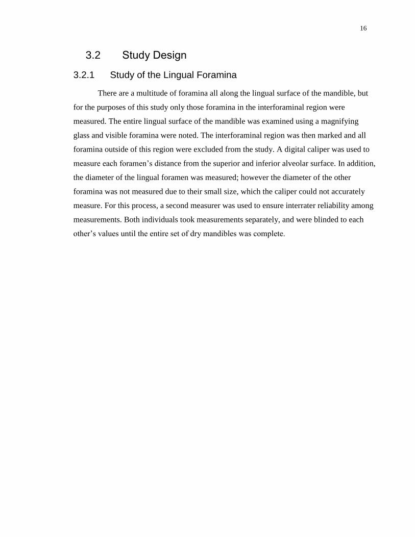
There are a multitude of foramina all along the lingual surface of the mandible, but
for the purposes of this study only those foramina in the interforaminal region were
measured. The entire lingual surface of the mandible was examined using a magnifying
glass and visible foramina were noted. The interforaminal region was then marked and all
foramina outside of this region were excluded from the study. A digital caliper was used to
measure each foramen’s distance from the superior and inferior alveolar surface. In addition,
the diameter of the lingual foramen was measured; however the diameter of the other
foramina was not measured due to their small size, which the caliper could not accurately
measure. For this process, a second measurer was used to ensure interrater reliability among
measurements. Both individuals took measurements separately, and were blinded to each
other’s values until the entire set of dry mandibles was complete.

17
3.2.2 Study of the Submental and Sublingual Arteries – Bisected Heads
The bisected tongues were pulled medially away from the vestibule of the mouth
to expose their lateral surfaces. The mucosa along this surface was stripped away
delicately using a probe to expose the muscle fibers of the tongue underneath. The hyoid
bone was then used as a landmark to locate the hyoglossus muscle, running superiorly
from the bone and eventually merging with the muscular fibers of the genioglossus
muscle. The muscle is typically found slightly superficial to genioglossus, and therefore a
probe was effective in separating the muscles from one another (figure 3.2.1). Once
isolated, the hyoglossus was cut at its superior border and reflected laterally. The lingual
artery is typically found underneath this muscle, however the depth varies and dissection
of the genioglossus may be required in order to locate the vessel (figure 3.2.2). Once
isolated, the sublingual branch was located and this vessel was followed anteriorly in
order to fully appreciate its branching into the floor of the mouth and mandible (figure
3.2.3). At the lingual foramen where the vessel enters the mandible, the genioglossus had
to be scraped from its origin at the genial tubercles in order to fully visualize the vessel.
The deep lingual artery was also typically identified where the lingual artery bifurcated,
but this branch was not followed anteriorly.
The submental artery was also isolated in the cadaveric specimens. Initially, the
facial artery was identified by reflecting the skin and fascia of the face. This vessel was
found crossing over the mandible, and was typically anterior to the facial vein. As this
vessel crossed the mandible, the submental artery was located as a branch moving
anteriorly and superficial to the mandible. This vessel would then enter the mandible via
unnamed foramina on the anterior surface of the chin, below the mental foramen. As
mentioned previously, the submental artery has an accessory branch which typically
enlarges when the sublingual artery is absent or insignificant. In these specimens, an
enlarged branch proximal to where the submental artery entered the anterior mandible
was located and followed superiorly as it pierced the mylohyoid muscle. This branch was
followed into the oral cavity until it reached the lingual foramen (figure 3.2.4). The
mylohyoid was not damaged during this process as it seems to be a valuable landmark to
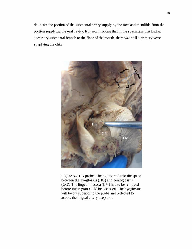
18
delineate the portion of the submental artery supplying the face and mandible from the
portion supplying the oral cavity. It is worth noting that in the specimens that had an
accessory submental branch to the floor of the mouth, there was still a primary vessel
supplying the chin.
LM
HG
GG
Figure 3.2.1 A probe is being inserted into the space
between the hyoglossus (HG) and genioglossus
(GG). The lingual mucosa (LM) had to be removed
before this region could be accessed. The hyoglossus
will be cut superior to the probe and reflected to
access the lingual artery deep to it.

19
HG
GG
LA
Figure 3.2.2 The hyoglossus (HG) has been reflected
laterally to expose the lingual artery (LA) underneath.
Superior
Inferior
Po
sterio
r
An
teri
or

20
Figure 3.2.3 A full dissection of the sublingual artery (SA) travelling to
the lingual foramen in the anterior mandible. The deep lingual (DL) artery
can also be seen supplying the tongue (top). Note that the genioglossus
has been stripped away from the mandible to expose the foramen. The
primary lingual artery (LA) as well as the anterior mandible (AM) have
also been labelled.
DL
SL LA
AM
AM
SL
Superior
Inferior
Po
sterio
r
An
teri
or

21
Figure 3.2.4 An enlarged submental artery (indicated in yellow) taking the place of the
sublingual artery and entering the lingual foramen. The sublingual gland (SG),
mylohyoid muscle (MH) and anterior mandible (AM) are all labeled for orientation
purposes.
GG
AM
Superior
Inferior
An
terio
r Po
ster
ior

22
3.2.3 Study of the Submental and Sublingual Arteries – Whole Heads
For this portion of the study, an inferior approach was taken to identify the
sublingual artery. An incision was made just below the mandible inferior to the ear on
either side of the specimen and continued medially, with the two incisions coming
together at the mandibular midline. An incision was then made inferiorly down the
midline of the neck, and the skin was removed to expose the underlying musculature and
fascia. The fascia was removed, along with the anterior bellies of the digastric muscles,
The mylohyoid was examined for evidence of a penetrating submental artery before
being carefully removed. The interior mandible was then examined for evidence of a
sublingual artery branch on either side (Figure 3.2.5).
Figure 3.2.5 An inferior view of the mandible in which the skin, fascia, and musculature
has been removed. A Sublingual Artery can be seen travelling towards the anterior
mandible (AM)
AM
Anterior
Posterior

23
Chapter 4
4 Results
4.1 Interrater Reliability
The interrater reliability was measured using the intraclass correlation coefficient
in order to determine whether the values of both raters agreed with one another using
IBM SPSS statistics version 23. The 95% confidence interval was 0.938-0.984, indicating
a high degree of agreement between raters.
4.2 Lingual Foramen Characterization
Of the 43 mandibles examined, four were excluded from this portion due to the
lack of any apparent lingual foramen, resulting in a lingual foramen prevalence of 90.7%.
First, the distance from the superior alveolar surface of the lingual side of the mandibular
bone (henceforth known as D1) was measured in millimeters and recorded as a histogram
in order to identify patterns among the foramina and their positions (figure 4.2.1).

24
Figure 4.2.1 Histogram depicting the variation in positioning of the lingual foramen
along the lingual surface of the mandible in relation to the alveolar surface of the bone.
39 specimens were measured with the mean distance from the alveolar surface being
16.17 ± 3.72 mm with a range of 9.27 mm – 24.33 mm.

25
The distance of the lingual foramen from the mental surface of the lingual aspect
of the mandible was also measured (henceforth known as D2) and expressed in a fashion
similar to figure 4.2.1 (Figure 4.2.2).
Figure 4.2.2 Histogram depicting the variation in positioning of the lingual foramen
along the lingual surface of the mandible in relation to the mental surface of the bone. 39
specimens were measured with the mean distance from the mental surface being 11.61 ±
2.36 mm with a range of 5.22 mm – 15.62 mm.
26
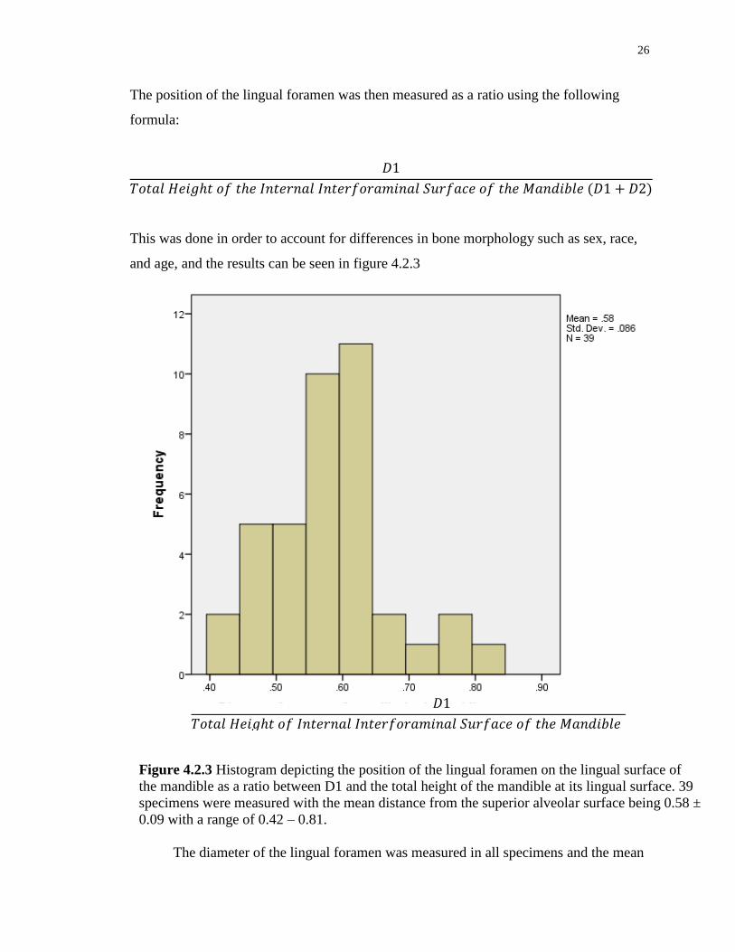
The position of the lingual foramen was then measured as a ratio using the following
formula:
This was done in order to account for differences in bone morphology such as sex, race,
and age, and the results can be seen in figure 4.2.3
The diameter of the lingual foramen was measured in all specimens and the mean
𝐷
𝑇𝑜𝑡𝑎𝑙 𝐻𝑒𝑖𝑔 𝑡 𝑜𝑓 𝐼𝑛𝑡𝑒𝑟𝑛𝑎𝑙 𝐼𝑛𝑡𝑒𝑟𝑓𝑜𝑟𝑎𝑚𝑖𝑛𝑎𝑙 𝑆𝑢𝑟𝑓𝑎𝑐𝑒 𝑜𝑓 𝑡 𝑒 𝑀𝑎𝑛𝑑𝑖𝑏𝑙𝑒 𝐷 𝐷
Figure 4.2.3 Histogram depicting the position of the lingual foramen on the lingual surface of
the mandible as a ratio between D1 and the total height of the mandible at its lingual surface. 39
specimens were measured with the mean distance from the superior alveolar surface being 0.58 ±
0.09 with a range of 0.42 – 0.81.

27
diameter was found to be 0.946mm ± 0.287mm with a range of 0.44 mm – 1.75 mm. We
then wanted to see if the position of the lingual foramen affected its diameter, since this
has been suggested in past literature. To do this, the specimens were split based on their
position on the mandible, with group 1 including those specimens that were found at a
point equal to or greater than the mean D1/total height ratio of 0.58 and group two
including those found at a point below this ratio. The mean diameter of foramina in group
1 was 0.933mm ± 0.256mm. The mean diameter of foramina in group two was 0.980mm
± 0.346mm. An Independent Samples t-test was performed on these two groups, and no
significant difference was found between the diameters of lingual foramina when
accounting for their height on the lingual mandibular surface (P=0.243).
4.3 Accessory Lingual Foramina Characterization
The positioning of the accessory lingual foramina (ALF) on the mandible was
examined similarly to how the primary lingual foramen’s position was evaluated. It was
found that the dry mandibles had, on average, 1.91 ± 0.84 accessory foramina directly
surrounding the lingual foramen. Of the 43 mandibles; 2/43 (5%) had no foramina, 11/43
(25.6%) had one foramen, 19/43 (44.2%) had two foramina, and 11/43 (25.6%) had three
foramina. None of the mandibles examined had more than three foramina that were
identifiable. A right lingual foramen was found in 22/43 (51.2%) of specimens, a left
lingual foramen was found in 24/43 (55.8%) of specimens, a superior lingual foramen
was found in 5/43 (11.6%) of specimens, and an inferior lingual foramen was found in
31/43 (72.1%) of specimens.
The primary statistics of interest for the different foramina were their distances
from the alveolar and mental surfaces of the mandibular bone. The average distance from
both the alveolar and mental surfaces of each accessory foramen was obtained,
documented in Table 4.3.1, and plotted on a graph against the primary lingual foramen,
as seen in figure 4.3.1.

28
Table 4.3.1 Mean distances (mm) of the primary and accessory lingual foramina from the
alveolar and mental surfaces of the mandible
A one-way ANOVA test was done to compare the mandibular location of all four
types of accessory foramina to each other as well as to the primary lingual foramen. It
was found that there was no significant difference between the lingual foramen and the
superior lingual foramen (P=0.619), however there was a significant difference in the
position of the lingual foramen and the inferior lingual foramen (P>0.0001), the right
lingual foramen (P=0.000026), and the left lingual foramen (P=0.010). There was an
expected difference between the overall positions of the superior lingual foramen and
inferior lingual foramen (P=0.000001). However, there was no significant difference
between the positions of the left and right lingual foramina (P=0.546). Both of these
foramina were typically found below the level of the primary lingual foramen, with the
left lingual foramen’s height being a mean distance of 3.46 ± 1.06 mm below the lingual
foramen, and the right lingual foramen’s height being a mean distance of 5.26 ± 1.26 mm
below the lingual foramen. The right lingual foramen was the only foramen that didn’t
have a significantly different position from the inferior lingual foramen (P=0.089), with
the right lingual foramen on average being 2.83 ± 1.11 mm above the inferior lingual
foramen.
Distance from Alveolar
Surface (mm)
Distance From Mental
Surface (mm)
Lingual Foramen 16.13 ± 3.58 11.61 ± 2.36
Right Lingual Foramen 21.38 ± 4.18 6.63 ± 2.39
Left Lingual Foramen 19.58 ± 4.42 7.75 ± 2.42
Superior Lingual Foramen 13.44 ± 3.84 14.33 ± 1.78
Inferior Lingual Foramen 24.21 ± 4.02 4.40 ± 2.26

29
Figure 4.3.1 Mean distances of various accessory lingual foramina plotted against the primary lingual
foramen (LF). The accessory foramina examined were the left lingual foramen (LLF), right lingual
foramen (RLF), superior lingual foramen (SLF), and inferior lingual foramen (ILF). Error bars
represent a 95% Confidence Interval
30
Chapter 5
5 Discussion
5.1 Anatomical and Clinical Significance
The prevalence of lingual foramen in the dry mandibles was found to be 39/44
(90.7%). In the literature, the prevalence of this foramen has been fairly consistent, with
values such as 96% (Salinas-Goodier et al., 2015), 99% (McDonnell et al., 1994), and
100% (Sheikhi, Mosavat, & Ahmadi, 2012) being reported. The value that was found was
slightly lower than these reported values. This could be due to our lower sample size,
with some studies such as Mcdonnell et al examining 314 mandibles. Unfortunately,
more mandibles were not able to be measured due to a lack of resources. Another
possible reason for this decreased foraminal prevalence is a difference in race between
the specimens. A study done in an Indian population did a gross examination of the
lingual foramen in a manner similar to our study and found a prevalence of only 72.45%
(Nagar et al., 2001). This is significantly lower than the studies listed above, which tested
mandibles without factoring in race. One drawback to our study is the loss of any
identifying information on the mandible specimens, and therefore it is not possible to
look at the data based on factors such as race and sex.
The mean distance of the lingual foramen from the mental surface of the mandible
was found to be 11.61mm ± 2.36. There are a couple of papers in the literature that have
also examined the lingual foramen’s mean distance from the inferior surface of the
mandible. These distances include 12.5 ± 2.1 mm (Rosano, Taschieri, Gaudy, Testori, &
Del Fabbro, 2009) and 19 ± 4.29 mm (Kilic et al., 2014). Our measurements agree with
the former paper but differ from the latter by 3.13 standard deviations. One difference is
that our study and the Rosano study used gross observation, whereas the Kilic study used
CT examination to determine their measurements. However, nothing was found in the
literature to suggest that a different method of measurement would result in changes in
the foraminal height.

31
The mean distance of the lingual foramen from the alveolar surface of the
mandible was found to be 16.17 ± 3.72 mm, with a range of 9.27 mm – 24.33 mm.
Because dental implants are inserted superiorly from the alveolar surface, it was felt this
was a very important distance to interpret in order to determine why dental implants will
damage the sublingual artery. Most hemorrhaging incidents in this region occur due to
the implant or implant drill perforating the bone near the site of the lingual foramen,
resulting in injury to the sublingual or accessory submental artery and heavy bleeding.
Therefore, it seemed important to determine the depth at which surgeons are inserting
these drills and see how it relates to the average height of the foramen found in our study.
A study done in 2010 looked to determine the length and diameter of implants surgeons
use and their typical failure rates. This was done by looking at 1,649 patients who
received implants over an 8 year period – from 1996-2004. The study showed that of all
the combinations of length and positioning of implants possible, short implants placed in
the anterior mandible had the greatest chance of early failure (Olate, Lyrio, de Moraes,
Mazzonetto, & Moreira, 2010). The fact that there were significantly fewer short
implants placed overall (10.7%) compared to medium and long implants (43.4% and
45.9% respectively) implies that surgeons understand this increased failure rate in shorter
implants and try to avoid them when possible. However, there seems to be a lack of
discrimination between the medium and long implants, likely due to their similar failure
rates of 3.0% and 3.4% respectively. It is worth noting that short implants were defined
as those ranging from 6-9mm, medium from 10-12mm, and long from 13-18mm. In our
study, only 5/39 (12.8%) lingual foramina were found at a height of 12mm or less from
the superior alveolar border, with the other 34/39 (87.2%) being found at a distance
greater than this. Due to the work done by Olate et al, combined with the data found in
this project, it may be stated that dental surgeons should prioritize using medium length
implants over long ones, since they have a similar rate of success but are much less likely
to come in close proximity to the lingual foramen in most patients.

32
Another factor that would affect the rate and severity of hemorrhaging during
dental implant surgery would be the size of the lingual foramen. The mean vertical
diameter of the lingual foramen in our study was found to be 0.946 ± 0.287mm with a
range of 0.44 mm – 1.75 mm. This value is in agreement with studies done by Ogawa et
al, 2016, (0.9 ± 0.4 mm) and Wang, Ju, Pan, & Chan, 2015, (0.61 ± 0.33 mm).
It has been found that the diameter of the lingual foramen directly relates to the
size of the vessel entering it (Kilic et al., 2014), and therefore a larger foramen should
carry a larger vessel, implying a greater severity of hemorrhage if damaged. Past
literature has shown that as a lingual foramen gets closer to the base of the mandible, it is
more likely to contain the primary sublingual artery. Inversely, a higher lingual foramen
is more likely to simply contain branches of the sublingual artery (Bernardi, Rastelli,
Leuter, Gatto, & Continenza, 2014). This research would imply that as the lingual
foramen travels inferiorly down the lingual mandibular surface, it would get larger. This
was tested this by splitting our sample into two groups at the mean lingual foraminal
distance ratio (0.58), and conducted a t-test to see if there was a difference in diameter.
Interestingly, a significant difference was found between the diameters of the lingual
foramina and their positions on the mandible (P=0.243).
A literature review of various sublingual hemorrhages found a large variance in
when these hemorrhages are detected, ranging from immediately during implant surgery
up to 7 hours after (Tomljenovic et al., 2015). Unfortunately, the artery that was damaged
and its corresponding foramen’s location were not often classified in these case reports.
Therefore, it is difficult to say whether the more severe and immediate bleeds were due to
a larger artery and foramen or other factors such as degree of damage to the vessel. That
being said, our research implies that more severe bleeds do not necessarily correspond to
a deeper artery, since no connection was found between foraminal depth and diameter.

33
The final aspect of the dry mandibles that was examined to determine potential
causes of sublingual hemorrhaging was the frequency and symmetry of the Accessory
Lingual Foramina. The only named foramina on the mandible are the mandibular,
lingual, and mental foramina, with all other foramina being considered “accessory”
(Gupta, Singh, & Soni, 2013). While there is a substantial amount of information in the
literature on the accessory foramina surrounding the mental and mandibular foramina, the
lingual accessory foramina remain relatively unstudied. (Gupta et al., 2013) looked at 50
dry mandibles of Indian descent and classified the accessory foramina, but only a
numerical count of these foramina was done without any measurements of their distance
from any mandibular landmark. Their study found that 94% of mandibles had at least one
accessory foramen on the interforaminal region of the lingual surface, thus agreeing with
our study’s accessory foramina prevalence of 95%. These results are corroborated by
studies done by (Murlimanju et al., 2012) and (Salinas-Goodier et al., 2015), who
counted accessory lingual foramina prevalence rates of 96% and 100% respectively.
The fact that only 0-5% of mandibles do not have any accessory lingual
foramina, as shown in our study as well as past studies, shows the importance of these
foramina and their associated structures. However, beyond characterizing the quantity of
these foramina on the lingual surface of the mandible, the literature is sparse with detail
about where these foramina are specifically located. Therefore, the heights of the
accessory lingual foramina along the lingual surface of the mandible were characterized
similar to the primary lingual foramen. The primary motivation for doing this was to
evaluate whether the branches of the sublingual artery that enter these accessory foramina
were also at risk of damage during dental implant surgery.
Normally, the superior lingual foramen would be considered most at risk of
damage due to its close proximity to the alveolar surface of the mandible, where dental
implants are placed. However, due to its relatively low rate of occurrence (11.6%), it is
not likely to pose a significant problem for dental surgeons doing surgery in the
mandible. Looking at the superior lingual foramen, it has a mean distance of 24.21 ± 4.02
mm from the alveolar surface. As discussed before, the mean length of even “long”
implants is only 13-18 mm (Olate et al., 2010), and therefore it is not likely that the

34
vascular contents of the superior lingual foramen would be at risk of damage during
dental implant surgery.
The right and left lingual foramina were compared to one another to examine whether
or not the lateral branches of the sublingual artery travel to the same relative location on
the lingual surface of the mandible. It was found that the average distance of the left
lingual foramen from the alveolar surface of the mandible was 19.58 ± 4.42, and the
average distance of the right lingual foramen from the same surface was 21.38 ± 4.18. A
Tukey Post Hoc test was performed and the two distances were found to not be
statistically different from one another (P=0.546). Interestingly, there was a significant
difference between the distances of the left lingual foramen and superior lingual foramen
(P=0.000394), but not between the right lingual foramen and superior lingual foramen
(P=0.089). This implies that on average, the right lingual foramen will be found slightly
lower than the left lingual foramen, making it less likely to be damaged by a dental
implant.
The symmetry of the accessory foramina was evaluated to determine relative
vascularization of the left and right halves of the interforaminal region. The left half of
the interforaminal region was found to have a greater number of foramina on average
than the right half (P=0.021). This implies a greater level of vasculature on the left half of
the mandible. This data, combined with the closer proximity of the left lingual foramen to
the alveolar surface of the mandible, implies that the left half of the anterior mandible is
riskier to operate on, and surgeons should take care when placing implants in this region.
A final observation made about the accessory foramina was their relation to the age of
the mandible specimens. It has been shown that there is a positive correlation between
hypertension (which is correlated to age) and the number of accessory lingual foramina
found in patients (Yilmaz, Akgül, Akgül, Dagistanli, & Çakur, 2003). As stated
previously, the age of our dry mandible specimens was not readily available, but past
literature has shown that mandibular age can be approximated within two-three years by
looking at the number of dental facets (Kim, Kho, & Lee, 2000). By using this paper,
along with the guidance of a Periodontist and Endodontist (Dr. Khadry Galil and Dr.

35
Hiran Perinpanayagam respectively), the relative age of these specimens was estimated
based on the level of dental attrition, the presence of furcation involvements (bone loss
where the tooth root branches), visibility of cementum, and rounded mandibular condyles
which imply significant wear over time (figure 5.1.1). Combining these factors, it is
believed that all mandibles involved in this study had a minimum age of 50 years old,
with many specimens being as old as 70 years of age.
A B
C
Figure 0.1 Images from three different mandibular specimens
showing presence of dental facets on the surface of teeth (A),
furcation involvement (B), and severe bone loss around anterior
teeth (C). These factors are typically present in older populations.

36
This study found 4.23 ±1.84 foramina per mandible, and our population seems to
have an average age above 50 years old. Future studies may wish to examine younger
specimens and attempt to determine the difference in prevalence of foramina between our
two groups.
5.2 Future Considerations
Future studies can aim to use this information and apply it to live patients. Studies
have been done to investigate different methods of imaging and how they can be used to
visualize the lingual foramina (Lustig, London, Dor, & Yanko, 2003). CBCT has been
shown to be the only adequate imaging technique for seeing this foramen, and future
studies may aim to see if variations in foramina in live patients with CBCT imaging
match those seen in this study. In addition, future studies may wish to use imaging to
observe the canals in the mandible and follow them to see if there is a pattern in their
pathway, and how this pathway may affect damage of the contained vessels via dental
implants.
In addition, future studies may wish to take a look at the sublingual artery in
whole heads. Due to the bisected nature of our specimens, an adequate number of
sublingual branches were not observed, leading to a lack of results for that portion of the
study. The size and branching pattern of this artery should be examined in order to better
understand how to avoid damaging it. In addition, the sublingual artery’s size should be
compared to the accessory submental branch that replaces it in its absence, in order to
determine if there is a significant difference, and a subsequent change in hemorrhage risk
based on which vessel is present in a patient.
5.3 Limitations of the Study
One major limitation of this study was the low number of cadaveric specimens for
dissection. Western University’s body bequeathal program allows individuals to donate
their body for various purposes including research and education. However, because
these donors are limited and they have various purposes, only a limited number were
obtained for the purposes of this study. In addition, because these specimens were

37
initially used as an educational resource for those students enrolled in the Schulich
School of medicine and dental surgery, they were bisected. This became an issue when
the sublingual artery and lingual foramina were examined on these specimens, since they
are located on the midline of the anterior mandible, and were therefore mostly damaged.

38
Bibliography
Bernardi, S., Rastelli, C., Leuter, C., Gatto, R., & Continenza, M. A. (2014). Anterior
Mandibular Lingual Foramina : An In Vivo Investigation, 2014.
Gakonyo, J., Butt, F., Mwachaka, P., & Wagaiyu, E. (2015). Arterial blood supply
variation in the anterior midline mandible: Significance to dental implantology.
International Journal of Implant Dental surgeonry, 1(1), 24.
http://doi.org/10.1186/s40729-015-0026-y
Gupta, S., Singh, P., & Soni, A. (2013). Morphological study of accessory foramina in
mandible and its clinical implication. Indian Journal of Oral Sciences, 4(1), 12.
http://doi.org/10.4103/0976-6944.118512
Hartmann, R. (1999). Ludwig’s Angina in Children - American Family Physician.
Retrieved April 7, 2016, from http://www.aafp.org/afp/1999/0701/p109.html
Ivey, D., Wilson, T., Merrifield, P., Shimizu, M., & Galil, K. (2014). A study of the
mandibular incisive nerve and possible causes of altered sensation following
maxillofacial surgery (922.5). The FASEB Journal , 28 (1 Supplement ). Retrieved
from http://www.fasebj.org/content/28/1_Supplement/922.5.abstract
Jaju, P., Jaju, S., Agarwal, R., & Singh, N. (2014). Detection of anatomical variations in
mandible by panoramic radiography. Journal of Cranio-Maxillary Diseases, 3(2),
95. http://doi.org/10.4103/2278-9588.138221
Kilic, E., Doganay, S., Ulu, M., ??elebi, N., Yikilmaz, A., & Alkan, A. (2014).
Determination of lingual vascular canals in the interforaminal region before implant
surgery to prevent life-threatening bleeding complications. Clinical Oral Implants
Research, 25(2), 2012–2015. http://doi.org/10.1111/clr.12065
Kim, Y. K., Kho, H. S., & Lee, K. H. (2000). Age estimation by occlusal tooth wear. J
Forensic Sci, 45(2), 303–9.

39
La, S. J. (2011). NEUROLOGIC/HEAD AND NECK IMAGING Imaging the Floor of
the Mouth and the Sublingual Space 1 ONLINE-ONLY CME Recipient of a Cer-
tificate of Merit, 1215–1231. http://doi.org/10.1148/rg.315105062
Lustig, J. P., London, D., Dor, B. L., & Yanko, R. (2003). Ultrasound identification and
quantitative measurement of blood supply to the anterior part of the mandible. Oral
Surgery, Oral Medicine, Oral Pathology, Oral Radiology, and Endodontics, 96(5),
625–629. http://doi.org/10.1016/j.tripleo.2003.08.015
McDonnell, D., Reza Nouri, M., & Todd, M. E. (1994). The mandibular lingual foramen:
a consistent arterial foramen in the middle of the mandible. Journal of Anatomy, 184
( Pt 2, 363–369.
Moore, K. L., DalleyII, A. F., & Agur, A. M. R. (2014). Clinically Oriented Anatomy, 7th
EditionMD. Clinical Anatomy.
Murlimanju, B. V, Prakash, K. G., Samiullah, D., Prabhu, L. V, Pai, M. M., Vadgaonkar,
R., & Rai, R. (2012). Accessory neurovascular foramina on the lingual surface of
mandible: incidence, topography, and clinical implications. Indian Journal of Dental
Research: Official Publication of Indian Society for Dental Research, 23(3), 433.
http://doi.org/10.4103/0970-9290.102252
Nagar, M., Bhardwaj, R., & Prakash, R. (2001). Accessory Lingual Foramen in Adult
Indian Mandibles, 50(1), 13–14.
Ogawa, A., Fukuta, Y., Nakasato, H., & Nakasato, S. (2016). Cone beam computed
tomographic evaluation of nutrient canals and foramina in the anterior region of the
mandible. Surgical and Radiologic Anatomy. http://doi.org/10.1007/s00276-016-
1664-3
Olate, S., Lyrio, M. C. N., de Moraes, M., Mazzonetto, R., & Moreira, R. W. F. (2010).
Influence of Diameter and Length of Implant on Early Dental Implant Failure.
Journal of Oral and Maxillofacial Surgery, 68(2), 414–419.
http://doi.org/10.1016/j.joms.2009.10.002

40
Patel, S., Dawood, A., Pitt Ford, T., & Whaites, E. (2007). The potential applications of
cone beam computed tomography in the management of endodontic problems.
International Endodontic Journal, 40(10), 818–830. http://doi.org/10.1111/j.1365-
2591.2007.01299.x
Pigadas, N., Simoes, P., & Tuffin, J. R. (2009). Massive sublingual haematoma following
osseo-integrated implant placement in the anterior mandible. British Dental Journal,
206(2), 67–8. http://doi.org/10.1038/sj.bdj.2009.2
Rosano, G., Taschieri, S., Gaudy, J. F., Testori, T., & Del Fabbro, M. (2009). Anatomic
assessment of the anterior mandible and relative hemorrhage risk in implant dental
surgeonry: A cadaveric study. Clinical Oral Implants Research, 20(8), 791–795.
http://doi.org/10.1111/j.1600-0501.2009.01713.x
Salinas-Goodier, C., Manchón, Á., Rojo, R., Coquerelle, M., Sammartino, G., & Prados-
Frutos, J. C. (2015). Prevalence and location of accessory foramina in the human
mandible. Oral Radiology. http://doi.org/10.1007/s11282-015-0212-x
Sheikhi, M., Mosavat, F., & Ahmadi, A. (2012). Assessing the anatomical variations of
lingual foramen and its bony canals with CBCT taken from 102 patients in Isfahan.
Dental Research Journal, 9(Suppl 1), S45–51. Retrieved from
http://www.pubmedcentral.nih.gov/articlerender.fcgi?artid=3692199&tool=pmcentr
ez&rendertype=abstract
Tomljenovic, B., Herrmann, S., Filippi, A., & Kühl, S. (2015). Life-threatening
hemorrhage associated with dental implant surgery: A review of the literature.
Clinical Oral Implants Research. http://doi.org/10.1111/clr.12685
Ural, A., Imamoğlu, M., Umit Işık, A., Bahadır, O., Bektaş, D., Cobanoğlu, B., &
Cobanoğlu, U. (2011). Neck masses confined to the submental space: our experience
with 24 cases. Ear, Nose, & Throat Journal, 90(11), 538–40. Retrieved from
http://www.ncbi.nlm.nih.gov/pubmed/22109923
Wang, Y. M., Ju, Y. R., Pan, W. L., & Chan, C. P. (2015). Evaluation of location and

41
dimensions of mandibular lingual canals: A cone beam computed tomography study.
International Journal of Oral and Maxillofacial Surgery, (Mlc), 1197–1203.
http://doi.org/10.1016/j.ijom.2015.03.014
White, D. K., Davidson, H. C., Harnsberger, H. R., Haller, J., & Kamya, A. (2001).
Accessory salivary tissue in the mylohyoid boutonniere: A clinical and radiologic
pseudolesion of the oral cavity. American Journal of Neuroradiology, 22(2), 406–
412.
Yilmaz, A., Akgül, N., Akgül, H., Dagistanli, S., & Çakur, B. (2003). Relationship
between Mandibular Nutrient Canals and Hypertension. Journal of International
Medical Research, 31(2), 123–125. http://doi.org/10.1177/147323000303100208

42
Appendix A
I grant, on behalf of UWO, the non-exclusive right to distribute my submission
publicly as part of the University of Western Ontario Institutional Repository,
Scholarship@Western.

43
Appendix B – Permissions
Permission for Figure 1.4.2
License Number 3852130817507
License date Apr 18, 2016
Licensed content publisher Elsevier
Licensed content publication Oral Surgery, Oral Medicine, Oral Pathology
Licensed content title Sublingual gland herniation through the mylohyoid muscle
Licensed content author Hilel Nathan,Ephraim Luchansky
Licensed content date January 1985
Licensed content volume number
59
Licensed content issue number 1
Number of pages 3
Type of Use reuse in a thesis/dissertation
Portion figures/tables/illustrations
Number of figures/tables/illustrations
1
Format electronic
Are you the author of this Elsevier article?
No
Will you be translating? No
Order reference number 001
Original figure numbers Figure 1
Title of your thesis/dissertation
Hemorrhage Secondary to Interforaminal Implant Surgery: Anatomical Considerations
Expected completion date Apr 2016
Estimated size (number of pages)
60
Elsevier VAT number GB 494 6272 12
Permissions price 0.00 USD
VAT/Local Sales Tax 0.00 USD / 0.00 GBP
Total 0.00 USD

44
Permission for Figure 1.5.1

45
Appendix C - Raw Data
Table C1. Descriptive statistics for the Lingual and Accessory Lingual Foramina’s distance
from the alveolar mandibular surface – One way ANOVA
Descriptives
Distance
N Mean Std. Deviation Std. Error
95% Confidence Interval for
Mean
Minimum Maximum Lower Bound Upper Bound
RLF 22 21.3845 4.18337 .89190 19.5297 23.2393 13.90 28.72
LLF 24 19.5838 4.42116 .90247 17.7169 21.4506 11.94 30.70
LF 39 16.1263 3.58403 .57390 14.9645 17.2881 9.27 24.33
SLF 5 13.4420 3.84169 1.71806 8.6719 18.2121 10.28 18.09
ILF 31 24.2097 4.01821 .72169 22.7358 25.6836 15.35 31.54
Total 121 19.7281 5.19265 .47206 18.7935 20.6628 9.27 31.54
Table C2. Post Hoc test for significance for the Lingual and Accessory Lingual Foramina’s
distance from the alveolar mandibular surface – One way ANOVA

46
Table C3. Descriptive statistics for the Lingual and Accessory Lingual Foramina’s distance
from the mental mandibular surface – One way ANOVA
Descriptives
Distance_Mental
N Mean Std. Deviation Std. Error
95% Confidence Interval for Mean
Minimum Maximum Lower Bound Upper Bound
LF 39 11.6050 2.36120 .37809 10.8396 12.3704 5.22 16.76
LLF 24 7.7454 2.42493 .49499 6.7215 8.7694 4.26 12.95
RLF 22 6.6268 2.39340 .51027 5.5656 7.6880 3.17 13.79
SLF 5 14.3300 1.78227 .79706 12.1170 16.5430 11.64 16.57
ILF 31 4.4048 2.26144 .40617 3.5753 5.2343 1.15 9.55
Total 121 8.2023 3.85086 .35008 7.5091 8.8954 1.15 16.76
Table C4. Post Hoc test for significance for the Lingual and Accessory Lingual Foramina’s
distance from the mental mandibular surface – One way ANOVA

47
Table C5. Description of Subjects (n=12)
Dissection Status Sex Age Cause of Death
Bisected
1776 Female 90 Myocardial Infarction,
Atrial Fibrillation
1807 Male 78 Interstitial Lung Disease
1741 Female 92 Myocardial Infarction
1758 Female 90 Multi organ failure,
Anemia,
Thrombocytopenia.
1765 Female 75 Breast Cancer
1754 Female 85 Metastatic Adenocarcinoma
of lung
1770 Male 72 Metastatic esophageal
cancer
1766 Female 77 Cerebrovascular accident
1793 Male 90 Carcinoma with a primary
unknown origin, Atrial
Fibrillation
Whole Heads
1656 Male 80 Arrythmia, C.A.D, COPD,
Dementia
1717 Female 84 Myocardial Infarction,
Stroke

48
Prabhsimrat Singh Gill
13 Neopolitan Road, Brampton, Ontario. L6Y 5N9
(416)-805-3111
Education
Western University (2010-2014) o Bachelor of Medical Sciences (BMSc.)
Honors double major in Medical Science and Pharmacology
o Dean’s Honor List 2012, 2013, 2014
o Transcript available upon request
Western University (2014-2016)
o Masters of Science (M.Sc) in Clinical Anatomy
o 2 year masters with one year of courses based on learning advanced principles of
anatomy, histology, and teaching and a one year research project
Research Experience
Undergraduate Research Project
o Worked in a genetic ecology lab examining genetic factors behind the migration
pattern of a species of swamp fly
o Learned how to manage my time effectively due to balancing this research with
my coursework at the time
Graduate Research Project
o Did a one year project looking at blood supply to the anterior mandible and its
anatomical variation
o Developed skills such as team work and problem solving as I worked with a
supervisory committee to complete this project
o Successfully defended April 29th 2016
Teaching Experience
Guest Lectures
ANATCELL 3309 – Histology of the heart and cardiovascular system guest lab talk
Led 2 lab talks on the histology of the cardiovascular system to approximately 200 3rd year undergraduate students
Discussed the abovementioned system as it relates to its physiology, using relevant clinical examples when possible
Spent the remaining 2 hour lab session assisting students in conducting the lab itself
Graduate Teaching Assistant
Anatomy for Medical Students (September 2015-April 2016)

49
Facilitated 4 groups of 6 first and second year medical students through their weekly anatomy labs
Used clinical cases to appeal to their passion for medical education in an effort to help them learn anatomy effectively
DENTISTRY 5160 – Systemic Anatomy and Histology (September 2015 – December 2015)
Led small groups of 15 first year dental students through various systems of the human body using pre-dissected prosections
Used microscope slides to relate the anatomical function of the body to its histological appearance
Helped create and administer bell-ringers and exams to evaluate the students’ progress throughout the year
DENTISTRY 5185 – Core Biology (September 2015 – November 2015)
Introduced first year dental students to basic anatomical and biological concepts through the use of prosections
Guided them by asking questions, facilitating discussion, and allowing them to explore specimens at their own pace
DENTISTRY 5186 – Head and Neck Anatomy (October 2015 – February 2016)
Led small groups of 10 first year dental students through an extensive head and neck anatomy course
Taught them anatomy using prosections and related the anatomy to various clinical cases that they are likely to see throughout their dental careers
ANATCELL 3309 – Mammalian Histology (face to face) (September 2014 - December 2014)
Facilitated students as they observed histological specimens under a microscope and completed assignments related to what they saw
Marked quizzes and assignments, proctored examinations, and provided feedback to students in an effort to better their understanding of the field of histology
HEALTSCI 2330B – Anatomy for Nursing students (January 2015 – April 2015)
Created and led tutorial sessions for students to build upon the content they learned in lecture
Combined anatomical information with clinically relevant cases that they will see throughout their career as nurses
Marked exams and assignments, providing constructive criticism where necessary to improve their anatomical skills
ANATCELL 3309 – Mammalian Histology (online) (May 2015 – August 2015)
Created and led online tutorial sessions to help students expand the histological knowledge they learned in lecture
Employed the use of online tools such as interactive quizzes and online forums as a way to keep the students engaged
Marked exams and assignments, providing feedback to students in order to expand their knowledge in the field of anatomy