a study on c reactive protein - morbidity predictor in
TRANSCRIPT
Brinda.J et al JMSCR Volume Issue 02 February 2017 Page 17650
JMSCR Vol||05||Issue||02||Pages 17650-17663||February 2017
A Study on C Reactive Protein - Morbidity Predictor in Ischaemic Stroke
Authors
Brinda.J1, John Christopher Ponnaiyan
2, Suresh Kumar
3, Manivel Ganesan
4
1Assistant Professor, Dept of General Medicine, Kanyakumari Government Medical College, Kanyakumari
2Associate Professor, Dept of General Medicine, Kanyakumari Government Medical College, Kanyakumari
3Post Graduate, Dept of General Medicine, Kanyakumari Government Medical College, Kanyakumari
4Intern, Dept of General Medicine, Kanyakumari Government Medical College, Kanyakumari
Corresponding Author
P. Suresh Kumar
Post Graduate, Department of General Medicine,
Kanyakumari Government Medical College, Kanyakumari
Abstract
Background: With an annual incidence of 0.2 to 2.5 per 1000 population, CVA is an important health
problem worldwide. Though ischaemic CVA is one of the leading causes for death and disability, parameters
for predicting, long term outcome in such patients have not been clearly delineated, especially in the Indian
context. Various studies proved that C-reactive protein at admission was found to be a predictor of
functional disability in ischaemic CVA. Inflammation regulates the production of the acute phase proteins
such as c-reactive protein (CRP), fibrinogen and serum amyloid A. The serum concentration of CRP can
increase >1000 fold upon inflammation and with a half life of 19 hrs, CRP is a very stable marker of the
inflammatory process. Most recent studies report that CRP is an independent predictor of risk of
atherosclerosis, cardiovascular events, atherothrombosis, hypertension and myocardial infarction.
Materials and Methods: The present study is a prospective study and was conducted on 49 patients in the
Department of General Medicine, Kanyakumari Government Medical college from January 2016 to January
2016. Various cerebrovascular accident cases admitted in our hospitals were clinically evaluated and
diagnosis were confirmed radiologically. All cases were subjected to routine blood investigations along with
acute phase proteins (c-reactive protein) on the day of admission and after 4 weeks. In all 49 cases, informed
consent obtained from their guardians.
Results: Out of total 49 cases studied, 18 Cases were females & 31 cases were males. All the cases were
grouped into 3 categories by Barthels Index accordingly. Patients with high C- reactive protein levels on the
day of admission had more severe deficits and poor prognosis after 4 weeks than compared with other
patients with normal level on the day of admission and better prognosis.
www.jmscr.igmpublication.org
Impact Factor 5.84
Index Copernicus Value: 83.27
ISSN (e)-2347-176x ISSN (p) 2455-0450
DOI: https://dx.doi.org/10.18535/jmscr/v5i2.74
Brinda.J et al JMSCR Volume Issue 02 February 2017 Page 17651
JMSCR Vol||05||Issue||02||Pages 17650-17663||February 2017
INTRODUCTION
Of several inflammatory markers studies, CRP
emerged as the most powerful inflammatory
predictor of future cardiovascular and cerebro
vascular risk. Also patient with elevated CRP
levels within 72hrs of stroke have an increased
risk of mortality. CRP in ischaemic stroke predicts
outcome and identifies patients who are at risk for
future vascular events and early mortality8. CRP
has also been found to be elevated in patients with
ischaemic stroke, correlating with the size of the
infarct as evidenced by CT scan.
Atherosclerosis is a multi factorial disease, driven
by inflammatory reactions10
. The process of
inflammation also contributes to the pathogenesis
of acute thrombotic events. CRP is an acute phase
protein and its concentration in serum reflects the
inflammatory condition by the patient. Levels of
CRP are consistently associated with cardiovas-
cular disease and predict myocardial infarctions
and stroke. Thus, CRP is useful and a reliable
predictor of cerebro vascular events.
MATERIALS AND METHODS
The study of CRP in ischaemic stroke was carried
out in the Department of General Medicine,
Kanyakumari Government Medical College. This
is an Observational prospective hospital based
type of study. The study period was from Jan
2016 to Jan 2017
INCLUSIONAL CRITERIA
1. Stroke as defined by WHO, is a rapidly
developing clinical signs of focal (at times
global) disturbances of cerebral function
lasting more than 24 hours or leading to
death, with no apparent cause other than
that of vascular origin.
2. All patients with CT proven case of
ischaemic stroke.
3. First episodes of ischaemic stroke.
4. Do not satisfy any of exclusion criteria.
EXCLUSION CRITERIA
1. Age >75 or <15 years.
2. Patients with TIA.
3. Patient with previous H/o stroke, TIA.
4. Patients with haemorrhagic stroke, tumour,
sub arachnoid haemorrhage.
5. Patients with head injury within 3 months.
6. CT Negative stroke.
7. Patient who reserved aspirin treatment
outside
8. Patient with H/o hypertension, diabetis,
heart disease, collagen disorders, hyperli-
pidemia, T.B, arteritis were excluded.
9. Smokers were excluded
10. Patients with obesity (BMI >30 kg/M2)
11. Patients with major renal, hepatic,
cancerous disease.
12. Patients with meningitis, brain abscess, or
any chronic infection that affected CRP
value.
13. Those with signs and symptoms of
potential clinical infection during the last 4
weeks before stroke.
14. Those with signs and clinical evidence of
acquired in hospital infection.
After obtaining verbal consent from either patient
or relatives, all patients in the study group were
evaluated by complete medical history, full
neurological examination, standardized blood tests
and imaging studies. Clinical history was recorded
from either the patient or his/her relatives. Special
emphasis was given to presenting complaint,
mode of onset, presence or absence of seizures,
loss of conciousness. headache, vomiting etc.
Presence or absence of risk factor, for stroke was
also noted. Past history of TIA, hypertension,
diabetis, coronary arterial disease, rheumatic heart
disease collagen disease, tuberculous etc were
carefully sought. Personal history regarding
dietary habits, smoking, alcoholic status were
noted.
Apart from routine observations, markers of
atherosclerosis like carotid arteries, status of
peripheral vessels, carotid thrill and B.P were
noted
A detailed clinical profile was obtained
Brinda.J et al JMSCR Volume Issue 02 February 2017 Page 17652
JMSCR Vol||05||Issue||02||Pages 17650-17663||February 2017
Neurological deficits such as aphasia, cranial
Nerve palsies, limb weakness, sensory impair-
ment, •cerebellar dysfunction, conjugate gaze
deviation and hemianopia were elicited by a
standard comprehensive bedside neurological
examination. Functional score was assessed using
Barthel index. CRP was measured by Nephelom-
etric method. Patients were reassessed on the 5th
day and condition reviewed. Proper nuring care
and physiotherapy were explained to the relatives,
caregivers and whenever possible to the patients.
Third evaluation was at 4th
week of follow up.
Improvement was objectively assessed by
determining the functional status using Barthel
index. Doubts and apprehensions of the relatives,
caregivers and patients were addressed and
cleared. Importance of nursing care and
physiotherapy were re-emphasized and absence of
confidence and hope were instilled. According to
the Barthel index4, patients were divided into 3
groups.
Barthel index <41 severely disabled.
Barthel index 4 1 - 6 0 moderately disabled.
Barthel index >60 mildly disabled.
Detailed analysis of date was performed.
Univariate analysis was done by chi.square test
and multivariate analysis by logistic regression.
BARTHEL INDEX OF ACTIVITY OF DAILY LIVING 1 Feeding 0 = Independent .
5 = Needs help (i.e) forcutting .
0 = Inferior performance
2. Bathing 5 = Performs without assistance
0 = Inferior performance
5 = Washes face, combs hair, brushes teeth
3 Personal toilet 0 = Inferior performance
0 = Inferior performance
10 =Independent
5 = Needs help
4. Dressing 0 = Inferior performance
10 = No accidents
5 = Occasional accidents
5 Bowel control 0 = Inferior performance
10 = No accidents
5 = Occasional accidents
6 Bladder control 0 = Inferior performance
10 = Independent with toilet or bed pan
5 = Needs help for balance
7. Toilet transfer 0 = Inferior performance
15 = Independent.
10 = Minimum assistance
5 = Able to sit, Needs assistance to transfer
8. Chair/ bed transfers 0 = Inferior performance
15 = Independent for 50 yards
10 = With help for 50yards
5 = Independent with wheelchair for 50 yards,
only if unable to walk
9. Ambulation 0 = Inferior performance
10 = Independent
5 = Needs help or supervision
10. Stair climbing 0 = Inferior performance
Maximum disability: = 0
Minimum disability: = 100
OBSERVATION AND RESULTS 49 patients satisfied all the above criteria were
included .31 were Males & 19 were Females. One
patient died.
The following observations were made out of the
49 patients,
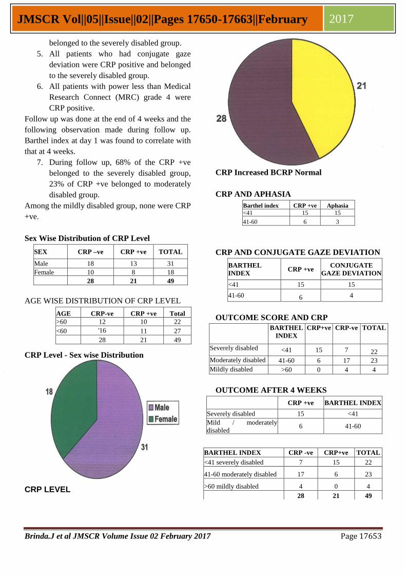
21 patients had an abnormal increased CRP and
28 patient had normal level.
1. Among patient with positive CRP 13 were
male and eight were female CRP level
status positive in 21 patients (ie) 42.85%
CRP level positive in males 26.5%
Percentage positive female is 16.32%
For the above date we used chi-square test for the
independence of association. The hypothesis
shows no association between the 2 groups. So,
sex does not influence the CRP of the patient
since ch.sq test X2 = .0292 P value >.05.
2. There is no association between age of
patient and CRP Ch.sq test X2 = .109
P value >.05
So age factor does not influence the group pattern
(CRP +ve)
3. Severe disability is more in CRP +ve
positive compared to CRP negative group.
Barthel index <41 in severely disabled
group.
4. All patients who had aphasia at the time of
admission were CRP positive and
Brinda.J et al JMSCR Volume Issue 02 February 2017 Page 17653
JMSCR Vol||05||Issue||02||Pages 17650-17663||February 2017
belonged to the severely disabled group.
5. All patients who had conjugate gaze
deviation were CRP positive and belonged
to the severely disabled group.
6. All patients with power less than Medical
Research Connect (MRC) grade 4 were
CRP positive.
Follow up was done at the end of 4 weeks and the
following observation made during follow up.
Barthel index at day 1 was found to correlate with
that at 4 weeks.
7. During follow up, 68% of the CRP +ve
belonged to the severely disabled group,
23% of CRP +ve belonged to moderately
disabled group.
Among the mildly disabled group, none were CRP
+ve.
Sex Wise Distribution of CRP Level
AGE WISE DISTRIBUTION OF CRP LEVEL
AGE CRP-ve CRP +ve Total
>60 12 10 22
<60 '16 11 27
28 21 49
CRP Level - Sex wise Distribution
CRP LEVEL
CRP Increased BCRP Normal
CRP AND APHASIA
Barthel index CRP +ve Aphasia
<41 15 15
41-60 6 3
CRP AND CONJUGATE GAZE DEVIATION
BARTHEL
INDEX CRP +ve
CONJUGATE
GAZE DEVIATION
<41 15 15
41-60 6 4
OUTCOME SCORE AND CRP
BARTHEL
INDEX
CRP+ve CRP-ve TOTAL
Severely disabled <41 15 7 22
Moderately disabled 41-60 6 17 23
Mildly disabled >60 0 4 4
OUTCOME AFTER 4 WEEKS
CRP +ve BARTHEL INDEX
Severely disabled 15 <41
Mild / moderately
disabled 6 41-60
SEX CRP –ve CRP +ve TOTAL
Male 18 13 31
Female 10 8 18 28 21 49
BARTHEL INDEX CRP -ve CRP+ve TOTAL
<41 severely disabled 7 15 22
41-60 moderately disabled 17 6 23
>60 mildly disabled 4 0 4
28 21 49
Brinda.J et al JMSCR Volume Issue 02 February 2017 Page 17654
JMSCR Vol||05||Issue||02||Pages 17650-17663||February 2017
30
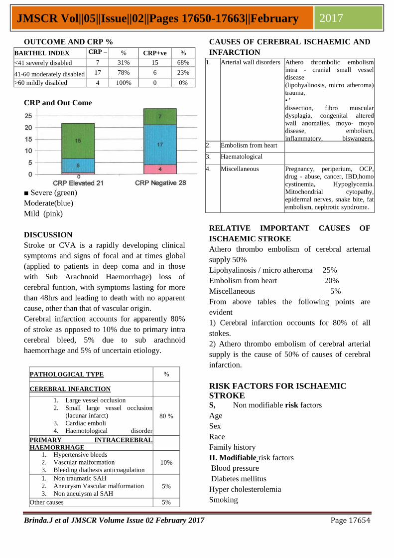
OUTCOME AND CRP %
BARTHEL INDEX CRP –
ve % CRP+ve %
<41 severely disabled 7 31% 15 68%
41-60 moderately disabled 17 78% 6 23%
>60 mildly disabled 4 100% 0 0%
CRP and Out Come
■ Severe (green)
Moderate(blue)
Mild (pink)
DISCUSSION
Stroke or CVA is a rapidly developing clinical
symptoms and signs of focal and at times global
(applied to patients in deep coma and in those
with Sub Arachnoid Haemorrhage) loss of
cerebral funtion, with symptoms lasting for more
than 48hrs and leading to death with no apparent
cause, other than that of vascular origin.
Cerebral infarction accounts for apparently 80%
of stroke as opposed to 10% due to primary intra
cerebral bleed, 5% due to sub arachnoid
haemorrhage and 5% of uncertain etiology.
PATHOLOGICAL TYPE %
CEREBRAL INFARCTION
1. Large vessel occlusion
2. Small large vessel occlusion
(lacunar infarct)
3. Cardiac emboli
4. Haemotological disorder
vasculities
5. Vasculopathy
80 %
PRIMARY INTRACEREBRAL
HAEMORRHAGE
1. Hypertensive bleeds
2. Vascular malformation
3. Bleeding diathesis anticoagulation 10%
1. Non traumatic SAH
2. Aneurysm Vascular malformation
3. Non aneuiysm al SAH 5%
Other causes 5%
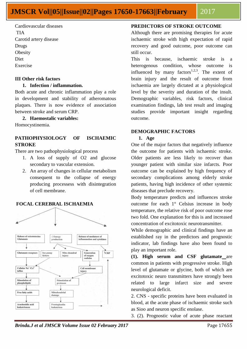
CAUSES OF CEREBRAL ISCHAEMIC AND
INFARCTION
1. Arterial wall disorders Athero thrombolic embolism
intra - cranial small vessel
disease
(lipohyalinosis, micro atheroma)
trauma,
• '
dissection, fibro muscular
dysplagia, congenital altered
wall anomalies, moyo- moyo
disease, embolism,
inflammatory, biswangers,
irradiations, infections. 2. Embolism from heart
3. Haematological
4. Miscellaneous Pregnancy, periperium, OCP,
drug - abuse, cancer, IBD,homo
cystinemia, Hypoglycemia.
Mitochondrial cytopathy,
epidermal nerves, snake bite, fat
embolism, nephrotic syndrome.
RELATIVE IMPORTANT CAUSES OF
ISCHAEMIC STROKE
Athero thrombo embolism of cerebral arternal
supply 50%
Lipohyalinosis / micro atheroma 25%
Embolism from heart 20%
Miscellaneous 5%
From above tables the following points are
evident
1) Cerebral infarction occounts for 80% of all
stokes.
2) Athero thrombo embolism of cerebral arterial
supply is the cause of 50% of causes of cerebral
infarction.
RISK FACTORS FOR ISCHAEMIC STROKE S, Non modifiable risk factors
Age
Sex
Race
Family history
II. Modifiable risk factors
Blood pressure
Diabetes mellitus
Hyper cholesterolemia
Smoking
Brinda.J et al JMSCR Volume Issue 02 February 2017 Page 17655
JMSCR Vol||05||Issue||02||Pages 17650-17663||February 2017
Cardiovascular diseases
TIA
Carotid artery disease
Drugs
Obesity
Diet
Exercise
III Other risk factors
1. Infection / inflammation.
Both acute and chronic inflammation play a role
in development and stability of atheromatous
plaques. There is now evidence of association
between stroke and serum CRP.
2. Haemostalic variables:
Homocystinemia.
PATHOPHYSIOLOGY OF ISCHAEMIC
STROKE
There are two pathophysiological process
1. A loss of supply of O2 and glucose
secondary to vascular extension.
2. An array of changes in cellular metabolism
consequent to the collapse of energy
producing processess with disintegration
of cell membrane.
FOCAL CEREBRAL ISCHAEMIA
PREDICTORS OF STROKE OUTCOME
Although there are promising therapies for acute
ischaemic stroke with high expectation of rapid
recovery and good outcome, poor outcome can
still occur.
This is because, ischaemic stroke is a
heterogenous condition, whose outcome is
influenced by many factors1,2,3
. The extent of
brain injury and the result of outcome from
ischaemia are largely dictated at a physiological
level by the severity and duration of the insult.
Demographic variables, risk factors, clinical
examination findings, lab test result and imaging
studies provide important insight regarding
outcome.
DEMOGRAPHIC FACTORS
1. Age
One of the major factors that negatively influence
the outcome for patients with ischaemic stroke.
Older patients are less likely to recover than
younger patient with similar size infarcts. Poor
outcome can be explained by high frequency of
secondary complications among elderly stroke
patients, having high incidence of other systemic
diseases that preclude recovery.
Body temperature predicts and influences stroke
outcome for each 1° Celsius increase in body
temperature, the relative risk of poor outcome rose
two fold. One explanation for this is and increased
concentration of excitotoxic neurotransmitter.
While demographic and clinical findings have an
established ray in the predictors and prognostic
indicator, lab findings have also been found to
play an important role.
(1). High serum and CSF glutamate are
common in patients with progressive stroke. High
level of glutamate or glycine, both of which are
excitotoxic neuro transmitters have strongly been
related to large infarct size and severe
neurological deficit.
2. CNS - specific proteins have been evaluated in
blood, at the acute phase of ischaemic stroke such
as Sioo and neuron specific enolase.
3. (2). Prognostic value of acute phase reactant
Brinda.J et al JMSCR Volume Issue 02 February 2017 Page 17656
JMSCR Vol||05||Issue||02||Pages 17650-17663||February 2017
such as ESR and CRP
Two inflammatory markers also provide
prognostic information.
1) . ESR erythrocyte sedimentation rate
2) . CRP
Some studies have shown male sex associated
with poorer outcome, while some other studies
have shown no difference. Probably hormonal
protection could be the reason as experiments on
amimals showed improving stroke.
5. Cerebro vascular risk factors:
Previous stroke and AF. These are 2 major risk
factors. Previous H/o stroke has been consistently
associated with higher likelihood of death or
dependence probably due to lower pre stroke level
of function and more advanced cardio - vascular
disease. Stroke in patients with atrial fibrillation
are usually more severe, more disability and
associated with high mortality.
6). Clinical findings :
Level of consiousness and gaze deviation Initial
level of consiousness is an important predictor
with decreased level of consiousness predicting
poor outcome. Presence of gaze deviation is
generally associated with poorer outcome. ’
Abnormal B.P may influence outcome. Clinical
studies of B.P reduction have shown a decrease in
cerebral blood flow to infarction area. On the
other hand, highly elevated BP has adverse long
term effects in blood - Brain barrier. Hypertension
has been found to play a role in haemorrhagic
transformation.
ESR: Erythrocyte sedimentation rate
independently predicted short term outcome with
increased level predicting poor outcome.
CRP: An acute phase reactant has been found to
predict outcome. CRP concentrations measured in
72hrs of stroke, independently predicted survival
after ischaemic stroke.
Inflammation plays an important role in the
initiation and progression of atherosclerosis and
systemic blood markers of inflammation (i.e)
acute phase reactants such as CRP and fibrinogen
have emerged as powerful predictors of coronary
and cerebrovascular events1.
The present study is a longitudinal hospital based
study focusing on the prognostic factors following
ischaemic CVA and includes follow up at 4
weeks. Haemorrhagic stroke was not included to
maintain homogeneity in the sample population.
Patients with clinical evidence of systemic
infection or inflammation were dropped to
exclude the other causes for elevated CRP. The
inclusion and exclusion criteria as well as dilution
of follow up were chosen based on similar studies
done from other centers in the world. Follow up
was achieved after 4 weeks.
III. CRP:
CRP is one of the substances, present in the
atherosclerotic lesion, more specifically in the
vascular intima, where it co-localizes with
monocytes, monocyte derived macroplaques and
lipoproteins. This localization makes a direct
contribution to the atherosclerotic process
possible15
.
CRP is a phylogenitically, highly conserved
plasma protein with homologues in vertebrates
and many invertebrates, that is part of the
systemic response to inflmmation5.
It is an acute phase protein and a member of the
family of pentraxins, CRP was originally observed
to 1930 in the plasma of patients with acute
infections, where it reacts with the
C.polysaccharide of pneumococcus5.
The major part of the CRP present in the plasma
comes from liver, where the synthesis of CRP is
mainly regulated by IL- 6, which in turn is
unregulated by other inflammatory cytokines such
as IL-1 and TNFa. Small amounts of CRP can
also be produced locally. CRP has been detected
on the surface of about 4% of normal blood
lymphocytes and CRP can be produced locally in
the atherosclerotic lesions by smooth muscle cells
and monocytic cells14
.
The structure of CRP is important for its stability
and for the execution of its function. CRP is
composed of five identical 21,500 Da subunits.
Upon dissociation of its pentameric structure,
CRP subunits undergo a spontaneous and
irreversible confirmational change. The loss of the
Brinda.J et al JMSCR Volume Issue 02 February 2017 Page 17657
JMSCR Vol||05||Issue||02||Pages 17650-17663||February 2017
pentameric structure of CRP results in modified or
monomeric CRP (MCRP) which is a naturally
occurring form of CRP and is tissue based rather
than serum based molecule.
MCRP is less soluble than CRP and tends to
aggregate and it has been described to induce
MRNA of chemokines and expression of adhesion
molecules in human cultured coronary artery
endothelial cells (HCAECS). Thus next to
circulating native pentameric CRP, MCRP can
also promote a pro inflammatory phenotype and
exert atherogenic effects in human endothelial
cells, although it may be in less potent manner
that native CRP.
The assumption that CRP is a casual factor in the
development of the atherosclerotic lesion is based
on its rapid accessibility to the plaque, localization
in the plaque and the results of in vitro studies in
which CRP has been demonstrated to actively
contribute to inflammatory process, further more
the specific interaction of CRP with complement
factors, cell receptors, lipids and other
inflammatory mediators occurs the possibility of
CRP being directly involved atherosclerosis.
V. EFFECTS OF CRP ON
ATHEROSCLEROSIS:
Inflammatory mechanisms play a central role in
all phases of atheroslcerosis from the intial
recruitment of circulating leukocytes from the
arterial wall to the rupture which results in the
clinical manifestation of the disease. CRP may be
involved in each of these stages by the following
processors.
(a) COMPLEMENT ACTIVATION;
Activation of the classical pathway of the
complement system is a well known and direct
biological function of CRP, Via this action, CRP
directly amplifies and facilities innate immunity.
CRP & co localizes with C5-C9, the membrane
attack complex of complement. Activation of this
Membrance attack complex (MAC) is initiated by
direct binding of CRP to Clq also present in the
atherosclerotic lesion and characterized by
elevated level of complement Csa6. Csa itself
exerts protect chemotactic and pro inflammatory
effects and its plasma level have been associated
with increased cardiovascular risk in patients with
advanced atherosclerosis.
(b) INTERACTION WITH CELL SURFACE
RECEPTORS:
The close proximity of CRP, to monocytic cells in
the arterial intima attenuates its possibilities for a
direct contribution to the progression of
atherosclerosis. The observation that CRP is
localized between monocytes underlines the
possibility of a direct interaction of CRP with
these cells and with monocyte - derived
macrophages via binding to a specific receptor
CRP binds to several receptors on human
monocytes to FcRy II a (CD32) with high affinity
and FeRyl (CD64) with lower affinity increasing
phagocytosis and the release of inflammatory
cytokines.
THROMBOSIS:
Thrombosis contributes to the progression of the
atherosclerotic lesion and to the precipitation of
the cardiovascular event. Direct action of CRP
which contribute to the indiction of a
prothrombotic state may be the enhancement of
the procoagulant activity on the reduction of
fibrinolysis. CRP has been suggested to induce a
prothrombolic state(via) induction of tissue factor
expression in human monocytes out only in the
presence of and through direction with other blood
cells such as T.lymphocytes, B.lymphocytes and
V.K.cells.
CELLULAR MODIFICATION, RECRUIT-
MENT AND ACTIVATION
CRP contributes to an arterial pro inflammatory
and proatherosclerotic phenotype by directly
upregulating adhesion molecules and chemo
.attractant chemokines in endothelial cells,
vascular SMCs and monocytic cells. On the
endothelial cells surface, expression of adhesion
molecules in the CD40- CD4oligand (CD40L or
CD 134) interaction. Like CRP the amount of
soluble CD40 increases during inflammation and
in the atherosclerotic lesion. Therefore CD40L has
been suggested to be a marker for inflammation
and involved in risk of cardiovascular events as

Brinda.J et al JMSCR Volume Issue 02 February 2017 Page 17658
JMSCR Vol||05||Issue||02||Pages 17650-17663||February 2017
well. CD40L is shed into the vasculature. Elevated
levels of this soluble CD40L (SCD40L) identities
patients with acute coronary syndromes, at
increased risk of recurrent MI and death, CRP
upregulates the cell surface expression of CD40
and CD40L.
NITRIC OXIDE EXPRESSION
CRP has been described to decrease the
expression and bio activity of endothelial nitric
oxide synthase (CNOS or NOS3), which results in
reduced bioavailability of Nitric monoxide (NO)
and a subsequent effect of vaso dilatation. CRP
contributes to a proatherogenic and prothrombolic
state by decreasing the release of NO and of the
vaso dilatation and inhibitor of platelet
aggregation (prostacyctin 10 GI2) through directly
increasing both superoxide and inducible No
synthase such as ICAM-1, VCAM-1 and
E.selection is upregulated by CRP, via these
processors, CRP induces platelet adhesion to
endothelial cells. CRP also appears to be involved
in the infiltration of monocytes into the vessel
wall and other subsequent development into foam
cells, CRP is chemotactic for human blood
monocytes.
EXPRESSION OF INFLAMMATORY
MEDIATORS CYTOKINES CHEMOKINES
AND ADHESIVE MOLECULE
CRP induces inflammatory cytokines in a dose
dependent way CRP induced release of
interleukin-6, interleukin-1, and TNFa all 3
cytokines were detected 4hrs after CRP elevation
with maximal level of TNFa at 8hrs and IL-1 and
IL-6 at 16hrs. CRP increases IL-8 protein and
MRNA expression in a time and dose dependent
manner (via) specific up regulation of NF KB
activity.
In atherosclerotic lesions, CRP directly
upregulates MRNA expression of the macrophage
markers CDIIb and HLA DR as well as their
protein products.
Another mechanism by which CRP influences the
development and maintenance of the
atherosclerotic lesion is its direct involvement.
APOPTOSIS
CRP is directly involved in the process of
apoptosis. It binds to apoptotic cells in a Ca2+
dependent manner and augments the classical
pathway of compliment activation, but protects
the cells from assembling the terminal
complement components (C5-C9)13
. Further CRP
enhances opsonization and phagocytosis of
apoptotic cells. CRP plays an essential role in
induced apoptosis of vascular smooth muscles.
CRP also binds to phosphatidyl cholines, by
which it participates directly in activation of
macrophages and neutrophils in the clearance of
apoptotic and necrotic cells.
LIPID
Interaction between lipids and CRP is divers. It
has been suggested that CRP could be the factor
that links lipoprotein deposition and complement
activation in atherosclerotic plaques. The majority
of sub endothelial foam cells show positive
staining for CRP. High levels of HDL are
atheroprotective since HDL is involved in
transporting cholesterol from periphery to the
liver, HDL might also protect the endothelium,
since CRP induced up regulation of inflammatory
adhesion molecules in HUVECS (HUman
Vascular Endothelial Cells) was completely
blocked by HDL. So HDL neutralizes CRP
induced pro.inflammatory activity HDL also
inhibits atherosclerosis through prevention of
oxidation of LDL.
Thus CRP plays role in complement activation,
cell adhesion and recruitment, thrombosis, the
expression of regulatory cytokines, apoptosis and
lipids. All these mechanism are part of or are
compromised by the process of inflammation.
CRP may thus contribute to the development of
atherosclerotic lesion (Via) direct pro-
inflammatory effects.
CRP may be a casual factor as well as a marker
for inflammation, depending on the concentration.
This concentration of CRP depends on rates of
production and clearance. The fact that CRP is
avery stable protein which is not consumed to a
significant concentration, in any process and the
Brinda.J et al JMSCR Volume Issue 02 February 2017 Page 17659
JMSCR Vol||05||Issue||02||Pages 17650-17663||February 2017
clearance of which is not influenced, by any
known condition is in agreement with its
functioning as a casual factor attempting to
prolong the stability of atherosclerotic lesion.
FRAMINGHAM STUDY!
To address the issue of baseline CRP level and
risk of subsequent stroke events the measurement
of CRP was done in member of Framingham
study, Original cohort, who were free of stroke or
TIA at the time of 1980 to 1982 clinical
examination and related the baseline CRP plasma
concentration to the incident of first stroke or TIA
in there subjects during 12-14 years follow up.
Men had twice the risk of ischaemic stroke and
women with highest CRP level had 5 fold increase
in the risk of any vascular event and 7 fold
increase in the risk of combined outcome of
myocardial infarction and stroke. The data derived
from the outcome of this study demonstrated a
graded increase in the incidence of ischaemic
stroke and TIA with increased level of CRP.
CRP levels are known to be greater in smokers.
Obese individuals with (BMI >130% of ideal),
individuals with abnormal fibrinolytic activity
(plasmin - antiplasmin complex) and individuals
with subclinical atherosclerosis). All of these
afore mentioned are individual risk factor of
adverse cerebro vascular or cardiovascular events.
But in a trend analysis conducted as a part of this
study, showed the relationship between the
increased incidence of stroke and TIA with
increased level of CRP persisted even after,
adjusting for a number of potential confounders
including smoking systolic BP, total and HDL
cholesterol and diabetics.
Elevated CRP are not disease specific, but are
sensitive markers produced in response to tissue
injury, infective agents immunologic stimuli and
inflammation cytokines such as IL-6, IL-1 and
TNF aare highly correlated with CRP and their
function5. Inflammation not only appears to be a
response to the underlying atherosclerotic disease
process, but also be an integral part of it. This is
consistent with beneficial effects of anti
inflammatory agents such as aspirin in reducing
the risk of both cardiovascular or cerebro vascular
events. All these date support the view that CRP
as a marker of low level inflammation, predicts an
increased risk of atherosclerosis.
In the Framingham study, the data were obtained
in a elderly cohort of men and women and led to
the conclusion that elevated level of CRP
significantly predicted greater risk of ischaemic
stroke or TIA in elderly men and women7.
PROGNOSTIC INFLUENCE OF INCRE-
ASED CRP AFTER FIRST EVER
ISCHAEMIC STROKE
In their study Ridler and colleagues found an
association between evidence of inflammation
after myocardial infarction and increased risk of
recurrent cerebral events.
This encouraged Mario-Di Napoli and Co to
conduct a study on the role of CRP level in short
term prognosis after first ever ischaemic stroke.
About 30 ischaemic stroke patients of either
gender (combined) with the age group 49-90 yrs
were studied within 4 weeks of the occurrence of
the first ever CVA ischaemic event1,2,3
. No
patients with evidence of acute infection were
included in the series CRP was collected with in a
medium of 14days from stroke event. It was found
that patients with highest CRP level >5mg/dl at
study entry died or had severe complication after
stroke such as pulmonary embolium or had no
evidence of recovery during the 2 months follow
up.
This study concluded that CRP was increased in
patients with cerebral ischaemia, the higher levels,
correlating with significant neurological deficit
and relevant disability and appear to provide
additional information regarding prognosis after
ischaemic stroke11
, as it appear to do after
myocardial infarction.
PROGNOSTIC INFLUENCE OF
INCREASED CRP AND FIBRINOGEN IN
ISCHAEMIC STROKED)
Mario Di Napoli and Co also did a study to
Brinda.J et al JMSCR Volume Issue 02 February 2017 Page 17660
JMSCR Vol||05||Issue||02||Pages 17650-17663||February 2017
investigate and compare the one year prognostic
influence of fibrinogen and CRP on the outcome
of ischaemic stroke. This led to two conclusions
Increased level of CRP are associated with worse
outcome in patients with ischaemic stroke and
Increased risk associated with elevated CRP is
independent of the prognostic influence of
fibrinogen.
INFLAMMATION AND STROKE
All 85 year old inhabitants of Leiden Netherlands
were visited at their place of residence (response
rate was 87%). Production levels of the anti
inflammatory cytokines IL-10 were assessed in a
whole blood assay, whereby lipopolyacchande
was used as a stimulation. Plasma concentrations
of CRP were also used as a marker of
inflammation. A history of stroke was obtained at
baseline (prevalence 10%). The number of fatal
strokes was prospectively obtained for a median
follow up of 2.6 years (incidence 1.82 per 100
person yearial rule). Subjects with a history of
stroke had significantly lower median IL-10
production levels at baseline than subjects without
stroke. They also had higher median CRP
concentrations.
Low IL-10 production level and high plasma CRP
concentrations are associated with an increased
risk of stroke.
C.REACTIVE PROTEIN AND OUTCOME
AFTER FIRST EVER ISCHAEMIC
STROKE8
In study by Dr.Keith W.Muir, Department of
Neurology, institute of Neurological sciences,
southern general hospital Glasgow. Patients
admitted to an acute stroke unit serving a
catchment population of 226000 were studied.
Survival time and cause of death for up to 4 years
after the index stroke were determined and related
to CRP concentration within 72 hours of stroke
and known prognostic variables by a COX
proportional hazards regression model. Ischaemic
stroke was diagnosed in 228 of 283 consecutive
admissions. Median follow up was 959 days
Geometric mean CRP concentration was
lO.lmg/L. Survival in those with CRP > lO.lmg/L
was significantly worse than with CRP < 10.1.
Higher CRP concentration was an independent
predictor of mortality together with age and stroke
severity on the National Institute of Health
strokescale12
.
CRP MORBIDITY PREDICTOR IN
ISCHAEMIC STROKE
A prospective hospital - based study of 105
patients of ischaemic stroke was conducted in
Department of Neurology by Dr.H.Npanicker, M-
Thomes and Co Focal neurological deficits and
functional score was assessed and CRP was
measured. A follow up was done at 5 days and at
6 months and outcome variable was the functional
status at 6 months using. Barthel index by
activities of daily living. Accordingly, patients
were grouped into three -p arthel index <41
severely disabled, Barthel index 41-60 moderately
disabled, and Barthel index >60 mildly disabled .
The results were if at admission if upper limb
power was less than MRC grade 4 or aphasia was
present or CRP assay was positive, then at 6
months there patients belonged to the severely
disabled group. If upper limb of lower limb power
was greater than MRC grade 3 or there was no
aphasia or conjuate gaze deviation or CRP array
was negative, these patients most likely belonged
to the mildly disabled group at 6 month.
Conclusion was patient can be stratified according
to the predicted prognosis9.
CRP AND INFARCT SIZE
In patient with ischaemic stroke, the extent of
necrosis is the main, though not the only
determinant of prognosis studies showed that CRP
concentration were increased (>5mg/dL) in
patients with larger infarct size and worse
outcome. Smaller increase were reported with
small infarcts.
The strong association between infection and ted
CRP concentration may result from accurate
quantification of cerebral infarction by CT and
Brinda.J et al JMSCR Volume Issue 02 February 2017 Page 17661
JMSCR Vol||05||Issue||02||Pages 17650-17663||February 2017
from variable intensity of acute phase response to
inflammatory stimuli ( in this case, the extent of
cerebral infarction). The possibility was suggested
by the observation that 24 hours concentration
were much higher in patients with previously
raised level.
If the intensity of the acute phase response was
not proportional to the intensity of the
inflammatory stimulus, the variable increase in
CRP concentration may not just be consequence
of later recanalisation or persistent occlusion of
infarct related artery. Thus the prognostic
importance of the 24 hours concentration may be
related partly to the extent of necrosis and partly
to the unknown individual determination of the
intensity of acute phase response. CRP might thus
indicate the inflammatory status during the acute
phase of ischaemic stroke and might aid in the
current challenge proved in secondary
prevention11
.
Activities of daily living reported after 4 weeks
have been shown to correlate highly with those
measured from direct examination. This novel
method ensured a good follow up.
Significant changes in the disability pattern were
not expected during the immediate post stroke
period. Hence 4 weeks was chosen as follow up
period. By 6 months the long term disability
pattern was the same as that of 4 weeks.
CRP is the prototype of the acute phase reactants
as it shows earliest and maximum elevation in
inflammation. It is secreted by the liver in
response to a variety of inflammatory cytokines
and level rises following trauma, inflammation
and infection. While CRP is a well known
prognostic factor following coronary events,
association with CVA is not well delineated.
C.Reactive protein may be elevated following
ischaemic stroke, because of inflammation
consequent to cerebral infarction, inflammation,
consequent to the unstable athersclerotic plaque
and complications secondary to stroke. Latex
agglution can also be used for CRP estimation,
instead of Nephelometry, because of easy
availability, simple, cost effective. Thus prognosis
following ischaemic stroke can be determined
without much burden on the existing
infrastructure of the health care system. Serial
measurement of CRP was not within the scope of
this study, but may be planned in future studies.
The present study did not include factors that
could modify the end point such as compliance to
treatment and occurrence of co. morbidities. But
this has not been taken into consideration in
previous studies such as Muir et al and hence the
results are comparable. Upper limb power and
aphasia was found to be important prognostic
factor.
The findings of this study are significant for
several reasons.
C.Reactive protein level at admission was found
to be a predictor of functional disability in
ischaemic CVA. In the study by Muir et all, a
prospective observational study, based in a
University Hospital, Acute stroke unit, serving a
population of 2,60,000 survival time and cause of
death for up to 4 years after the index stroke were
determined and related to CRP concentration
within 72 hours of stroke and known prognostic
variables by COX proportional hazards
regression. Ischaemic stroke was diagnosed in 228
of 283 admission. Median follow up was 959
days. Geometric mean CRP concentration was
lO.lmg/L. Survival in those with CRP > 10.1
mg/L was significantly more than in those with
CRP <10.1 mg/L. Higher CRP was an
independent predictor of mortality, together with
age and stroke severity on the National Institutes
of Health stroke scale. Cardiovascular disease,
accounted for 76% of deaths in those with CRP
>10.1 mg/L and 63% of death in those with CRP
<10.1mg/L. The conclusion was CRP
concentration is an independent predictor of
survival after ischaemic stroke. Unlike in the
previous study by Muir et al, where end point
taken was mortality, the present study has studied
morbidity (functional status) and hence one step
forward. The follow up generated an enthusiastic
response. The relatives, caregivers and patients
were immensely benefited. Similar study was
Brinda.J et al JMSCR Volume Issue 02 February 2017 Page 17662
JMSCR Vol||05||Issue||02||Pages 17650-17663||February 2017
conducted in Dept of Neurology Sree Chitra
Thirunal Institute of Medical sciences by
Dr.Jalesh N Panicker, M.Thomas, where Barthel
index at day 1 was found to correlate with that of
6 months, correlation coefficient being 63%.
Cerebro vascular accident is an important health
problem and is one of the leading cause for
morbidity and mortality. The variables found in
this study can be used to predict prognosis even at
the peripheral hospitals. This helps in stratification
of patients depending on the likely outcome and
will help the treating physician and the relatives.
Though intensive and scientific physiotherapy and
rehabilitation can be planned for all patients with
ischaemic CVA, the protocol is to be strictly
adhered to in patients predicted to have worse
prognosis. So that they receive maximum support
and assistance. Prognosis can be discussed with
caregivers and relatives and the social implication
of the illness can be addressed before the patient is
sent home. The relationship between CRP and
cerebro vascular disease has a bearing on newer
treatment modalities of the future. As a subset of
patients with stroke has elevated CRP, the role of
anti inflammatory agents and antibiotics in the
acute management of such patients is to be
addressed. As elevated CRP is an index of
increased risk for cardio vascular disease, these
patients can be targeted with more aggressive,
conventional therapy, or new therapies for plaque
stabilisations. Many trials are in progress, and are
needed to determine, if patients should be treated
on the basis of elevated CRP alone.
CONCLUSION
CRP is increased in a significant fraction of
ischaemic stroke. Increase in CRP is independent
of age & sex. Patient with increased CRP had
invariably more deficit during admission. Patients
with low CRP had mild deficit during admission.
Patients with low CRP had good prognostic
outcome 4 weeks after onset of stroke. Patient
with increased CRP had severe disability when
compared with patients with normal levels, 4
weeks after the onset of stroke.
REFERENCE
1. Di Napoli M, Di Giantilippa a, sollecito A
Bocala V - CRP and outcome after first
ever stroke 31(1) 238 - 9 Jan 2001
2. Di Napoli M, Di Gianfillippo G, Bocala V
C.Reactive protein after first ever
ischaemic stroke Neurology 1999, 52 : A
151 - A 152
3. Muir K.N., Weir CJ Alwan W, squire IB,
Lees KR, C.Reactive protein and outcome
after ischaemic stroke : Stroke 1999 ; 30 :
981 -985
4. Mahoney FI, Barthel DW, functional
evaluation, the Barthel Index Md state
Med J 1965 : 14 : 61 - 65
5. Grace A.J. Infection, Inflammation and
cerebro vascular Ischaemic Neurology
1997 : 49 (suppl 4): S 47 - S 51
6. Volanakis JE complement activation by
C.Reactive protein complexes.
7. Plasma concentration of C.Reactive
protein and risk of Ischaemic stroke and
Transient Ischaemic attach. The
Framingham study Natalia S.Rost, Philip
A Wolf, Carlas S.kase stroke 2001 : 32 :
2575 - 2579.
8. Morbidity predictors in ischemic stroke J
N Panicker, M.Thomas, K.Parithran
D.Nair, P.S.Sarma Neurology India Vol 51
No 1 Jan - mar 2003 pp 49 -51
9. Wide Range C Reactive Protein efficacy in
acute ischaemic stroke patients S.Shenhar
- Tsartaty, E.Ben Assayag, I.Bova. Acta
Neurol Scand 2006 : 114 : 29 - 32.
10. CHAMORRO A. Role of inflammation in
stroke and athero thrombosis.Cerebro
vascular diseases 2004 : 17 (Suppl 3) 1-5
11. Winbeck K, Poppert H, Etgen T, Cornard
B, Sander B, Prognostic relevance of early
serial C Reactive protein measurement
after first ischaemic stroke 2002 ; 33 ;
2459-64.
12. CRP in cerebro vascular events, Carnova
CR, Courtin C Rein hart W.H. Atherosc-
lerosis 1999 Nov 1 : 147(1) : 49 -53.
Brinda.J et al JMSCR Volume Issue 02 February 2017 Page 17663
JMSCR Vol||05||Issue||02||Pages 17650-17663||February 2017
13. Complement C3 and C.Reactive protein
are elevated in South Asians Independent
of a family History of stroke. Somane R,
Grant PJ, Kain K stroke 2006 Jun 29.
14. Zwaka T, Homback V, Torzewski J
C.Reactive protein mediated low density
lipo protein up take by macrophages.
Implications for atherosclerosis circulation
2001 : 103 : 1 1 9 4 - 1197.
15. C Reactive Protein, carotid intima media
thickness and incidence of ischaemic
stroke in the elderly the cardiovascular
Healthy study circulation 2003 Jul 15:
108(2) 166-70.