a study of the temporary adhesion of the podia in …biochemical composition, immunocytochemistry,...
TRANSCRIPT

2383The Journal of Experimental Biology 201, 2383–2395 (1998)Printed in Great Britain © The Company of Biologists Limited 1998JEB1551
A STUDY OF THE TEMPORARY ADHESION OF THE PODIA IN THE SEA STARASTERIAS RUBENS(ECHINODERMATA, ASTEROIDEA) THROUGH THEIR
FOOTPRINTS
PATRICK FLAMMANG1,*, ALAIN MICHEL 2, ANNE VAN CAUWENBERGE3, HENRI ALEXANDRE3 AND
MICHEL JANGOUX1,4
1Laboratoire de Biologie Marine, 2Laboratoire de Chimie Biologique, 3Laboratoire de Biologie et d’Embryologie,Université de Mons-Hainaut, 20 Place du Parc, B-7000 Mons, Belgium and 4Laboratoire de Biologie Marine (CP
160/15), Université Libre de Bruxelles, 50 Avenue F. D. Roosevelt, B-1050 Brussels, Belgium*e-mail: [email protected]
Accepted 1 June; published on WWW 27 July 1998
e,a,
Sea stars are able to make firm but temporaryattachments to various substrata owing to secretionsreleased by their podia. A duo-glandular model has beenproposed in which an adhesive material is released by twotypes of non-ciliated secretory (NCS1 and NCS2) cells anda de-adhesive material is released by ciliated secretory (CS)cells. The chemical composition of these materials and theway in which they function have been investigated bystudying the adhesive footprints left by the asteroids eachtime they adhere to a substratum. The footprints of Asteriasrubensconsist of a sponge-like material deposited as a thinlayer on the substratum. Inorganic residues apart, thismaterial is made up mainly of proteins and carbohydrates.The protein moiety contains significant amounts of bothcharged (especially acidic) and uncharged polar residues aswell as half-cystine. The carbohydrate moiety is also acidic,comprising both uronic acids and sulphate groups.
Polyclonal antibodies have been raised against footprintmaterial and were used to locate the origin of footprintconstituents in the podia. Extensive immunoreactivity wasdetected in the secretory granules of both NCS1 and NCS2cells, suggesting that their secretions together make up thebulk of the adhesive material. No immunoreactivity wasdetected in the secretory granules of CS cells, and the onlyother structure strongly labelled was the outermost layerof the cuticle, the fuzzy coat. This pattern ofimmunoreactivity suggests that the secretions of CS cellsare not incorporated into the footprints, but instead mightfunction to jettison the fuzzy coat, thereby allowing thepodium to detach.
Key words: temporary adhesion, podium, footprint, ultrastructurbiochemical composition, immunocytochemistry, EchinodermatAsteroidea, Asterias rubens.
Summary
stivere
,henyn.re,g
ofa,nddarby
Many marine benthic organisms are equipped with adhesorgans, the secretions of which allow them to attach to substratum. Three types of adhesion may be distinguishedpermanent adhesion involving the secretion of a cement (the attachment of barnacles on rocks); (2) transitory adhespermitting simultaneous adhesion and movement along substratum (e.g. the foot secretions of gastropod molluscs);(3) temporary adhesion allowing an organism to attach stronbut momentarily to the substratum (e.g. the adhesion echinoderm podia) (Walker, 1987; Tyler, 1988; Flamman1996).
Although adhesion mechanisms are beginning to deciphered in a few sessile organisms, including the bysattachment of bivalve molluscs (Waite, 1983, 1992) and cement attachment of cirripede crustaceans (Yule and Wal1987; Kamino et al.1996), they essentially remain poorlyunderstood. This is especially true for temporary adhesi
Introduction
ivethe: (1)e.g.ionthe andglyofg,
besal
theker,
on,
which generally relies on duo-gland organs requiring at leatwo antagonistic secretions, one adhesive and one de-adhes(Tyler, 1988). Moreover, these duo-gland adhesive organs afound very frequently in microscopic invertebrates inhabitingthe interstitial environment, e.g. turbellarians, gastrotrichsnematodes and polychaetes (Tyler, 1988). The small size of tduo-gland organs in these organisms has precluded abiomechanical or biochemical studies on temporary adhesioThe study of macroinvertebrate adhesive systems is, therefoa necessary and more promising way of understandintemporary adhesion.
Hermans (1983) has argued that the adhesive systemsechinoderms, which are usually associated with the podioperate with the functions of adhesion and de-adhesion, athat two or more glands are involved. Accordingly, he appliethe name ‘duo-gland’ to these systems. The duo-glandulnature of echinoderm podia has since been corroborated

2384
1lesnd
’s
otaleut
, 5010ird
ing.forearse of al
ll,.
ing;
C. fors
redg
edeial.
in
ntn
ee by
hee
ht.
P. FLAMMANG AND OTHERS
several morphological studies showing that the podia alwenclose two types of secretory cells: non-ciliated secret(NCS) cells, presumably releasing an adhesive material, ciliated secretory (CS) cells, presumably releasing a adhesive material (for a review, see Flammang, 199However, the chemical compositions of these materials andway in which the duo-gland adhesive systems operate arelargely unknown.
The aim of the present work was to investigate temporadhesion of the podia in the sea star Asterias rubensbystudying their footprints. Footprints are deposited each tipodia adhere to a substratum (Chaet, 1965; Thomas Hermans, 1985; Flammang et al.1994). They appear to consismainly of adhesive secretions, although they may also conde-adhesive secretions and/or material from the cuti(Flammang, 1996). In the present study, footprints weanalysed in terms of their fine structure and gross chemcomposition. Moreover, the cellular origin of footprinconstituents in the podia was investigated bimmunohistochemistry and immunocytochemistry using neantisera raised against footprint material.
Materials and methodsCollection and maintenance of sea stars
Individuals of Asterias rubensL. were collected intertidallyin Audresselles (Pas-de-Calais, France). They were transpoto the Marine Biology Laboratory of the University of Monswhere they were kept in a marine aquarium with closcirculation (13 °C, 33 ‰ salinity) and fed mussels (MytilusedulisL.). Footprints were collected primarily during the firs2 or 3 days after the arrival of the sea stars. Indeed, althothe asteroids survived for several weeks, their ability produce abundant footprints appeared to decrease quiwhen they were held in captivity.
Scanning electron microscopy
Footprints were prepared as follows. Podia were allowedadhere firmly to clean aluminium stubs. After the podia hdetached themselves, the footprints were briefly rinseddistilled water, frozen in liquid nitrogen and freeze-dried. Thwere then coated with gold in a sputter-coater and obserwith a JEOL JSM-6100 scanning electron microscope.
Collection of footprint material
Sea star footprints were obtained by allowing individualswalk across and/or attach to the bottom of clean glass Pdishes (diameter 15 cm) filled with artificial sea water (ASWThe asteroids and the ASW were renewed every hour for after which the Petri dishes were thoroughly rinsed in distillwater and placed in a freeze-dryer. The lyophilized footprmaterial was then scraped off using a glass knife and store−20 °C. This method allows the collection of 50–300µg of ared-brown powder per Petri dish. Attempts made to recowet footprint material all failed. However, after scraping, thmaterial appears as sticky, red-brown filaments.
aysoryandde-6). the still
ary
meandttainclere
icaltyw
rted,ed
tughto
ckly
toad ineyved
toetri).8 h,edintd at
veris
Antiserum production and characterisationPolyclonal antisera were raised in two female rabbits (R
and R2) to the footprint material. The immunisation scheduwas as follows. Approximately 1 mg of footprint material wasuspended in 0.75 ml of phosphate-buffered saline (PBS) aemulsified with an equal volume of complete Freundadjuvant (Difco, Detroit, MI, USA). The rabbits were theninjected subcutaneously at several sites in the back, with a tvolume of 0.5 ml of the emulsion to R1 and 1 ml of themulsion to R2. Three boosts were prepared identically, bwith incomplete Freund’s adjuvant (Difco, Detroit, MI, USA)and were administered, respectively, 20 days, 35 days anddays after the first injection. Antisera were harvested twice: days after the second boost (AS1) and 20 days after the thboost (AS2). Pre-immune sera (PS) were obtained by bleedthe rabbits just before the first injection of footprint material
The different antisera and pre-immune sera were tested specific footprint-binding activity using a fluorescencimmunoassay. Footprints were obtained by allowing sea stto walk across clean glass microscope slides. The presencfootprints was checked by soaking a few slides for 1 min in0.05 % solution (in distilled water) of the cationic dye CrystaViolet (Flammang et al.1994). The other slides were pre-incubated for 30 min in 10 % normal goat serum (BioCeCardiff, UK) in PBS to block non-specific antigenic sitesAntisera and pre-immune sera, diluted 1:100 in PBS contain1 % Tween 20 and 3 % bovine serum albumin (BSAPBS–Tween–BSA1), were then applied overnight at 4 °After several washes in PBS, the sections were incubated1 h in FITC-conjugated swine anti-rabbit immunoglobulin(DAKO A/S, Glostrup, Denmark) diluted 1:50 inPBS–Tween–BSA1. After a final wash in PBS, they wemounted in Vectashield (Vector, Burlingame, CA, USA) anobserved with a Leica TCS 4D confocal laser scanninmicroscope.
An enzyme-linked immunosorbent assay (ELISA) was usfor the comparison of specific antibody titres between thdifferent antisera and pre-immune sera. The footprint materwas digested with trypsin under the following conditionsApproximately 1 mg of footprint material was suspended 2 ml of 0.1 mol l−1 sodium bicarbonate, pH 8.2, at aprotease:protein ratio of approximately 1:100, under constastirring at 22–24 °C for 180 min. The progress of the digestiowas monitored at 30 min intervals by centrifuging thsuspension at 3000g for 5 min and measuring the absorbancof the supernatant at 280 nm. The digestion was terminatedcentrifuging the suspension at 3000g for 15 min and freezingthe supernatant. The ELISA was carried out according to tfollowing procedure at 37 °C. The soluble fraction of thtrypsinised footprint material, diluted 25 times in 0.1 mol l−1
carbonate/bicarbonate buffer, pH 9.2, was loaded (200µl perwell) into 96-well microtitration plates (Nunc MaxisorbImmunoplates, Rochester, NY, USA) and incubated overnigThe plates were washed several times with 300µl of PBScontaining 0.05 % Tween 20 and 0.1 % BSA(PBS–Tween–BSA2) per well. Each antiserum (200µl) and

2385Sea star adhesive footprints
e)ra
s
ere
reved
as
idstheassly,ingme
, as
natebyns
ashs the
hasd,ithe
eof
ork
ce.
t,ot
each pre-immune serum (200µl) (serial dilution in PBS) wasthen added and incubated for 1 h. After a second serieswashes with PBS–Tween–BSA2, 200µl of horseradish-peroxidase-conjugated goat anti-rabbit immunoglobul(DAKO A/S, Glostrup, Denmark) diluted 1:1000 in the sambuffer was added and incubated for an additional hour. Finathe plates were washed thoroughly with PBS–Tween–BSAand 200µl of the enzyme substrate (a solution of 0.04 % o-phenylenediamine in phosphate/citrate buffer, pH 5.containing 0.012 % H2O2) was added. After 10 min at roomtemperature (22–24 °C), the reaction was stopped by addition of 50µl of 2.5 mol l−1 H2SO4, and the plates were readat 492 nm using a Titertek Multiskan plate reader.
Immunohistochemistry and immunocytochemistry
For light microscopy, podia were fixed in 4 %paraformaldehyde in PBS (pH 7.4) for 2 h at room temperatuand then rinsed in the same buffer for 30 min. They weembedded using a routine method in paraffin wax, sectionat 8µm and mounted on clean glass slides. The sections wpermeabilised in PBS with 0.25 % Triton X-100 for 1 h, anpre-incubated for 30 min in 10 % normal goat serum in PBPrimary antisera, diluted 1:100 in PBS–Tween–BSA1, wethen applied overnight at 4 °C. After several washes in PBthe sections were incubated for 1 h in FITC-conjugatswine anti-rabbit immunoglobulin diluted 1:50 inPBS–Tween–BSA1. To make the tissue identification easpropidium iodide (Calbiochem, La Jolla, CA, USA) was addeto the secondary antibody solution at a final concentration5µg ml−1 to stain the nuclei. Following five washes in PBS, thsections were mounted and observed with the confocal lascanning microscope.
For transmission electron microscopy, podia were fixed 3 % glutaraldehyde in cacodylate buffer (0.1 mol l−1, pH 7.8)for 3 h at 4 °C, rinsed in cacodylate buffer, and post-fixed f1 h in 1 % osmium tetroxide in the same buffer. Some podwere fixed according to the same protocol but with the additiof 0.05 % Ruthenium Red to all the solutions (Luft, 1971). Thtechnique allows a better preservation of the cuticle as welof the secreted material, when present (Flammang et al.1994).After a final wash in buffer, the podia were dehydrated graded ethanol and embedded in Spurr’s resin. Ultrathsections (40–70 nm) were cut, using a Leica UCultramicrotome equipped with a diamond knife, and collecton gold grids. The sections were blocked with 10 % normgoat serum in PBS for 30 min and incubated overnight at 4with the primary antisera diluted 1:500 in PBS–Tween–BSAAfter rinsing in PBS, the sections were immunogold-stainfor 1 h at room temperature in goat anti-rabbit immunoglobuconjugated to 15 nm gold particles (BioCell, Cardiff, UKdiluted 1:100 in PBS–Tween–BSA1. Following severawashes in PBS, and then distilled water, they were furthstained with aqueous uranyl acetate and lead citrate observed using a Zeiss EM 10 transmission electrmicroscope.
For both light and transmission electron microscopy, tw
of
inelly,2,
0,
the
rereederedS.reS,
ed
ier,d
ofeser
in
oria
onisl as
ininTedal°C1.edlin)ler
andon
o
types of control were carried out: (1) substitution of thprimary antiserum with PBS–Tween–BSA1 and (2substitution of the primary antiserum with the pre-immune sediluted 1:100 in PBS–Tween–BSA1.
Analytical methods
All analyses were performed on duplicate sample(0.5–1 mg each) of freeze-dried footprint material.
For amino acid and hexosamine analysis, samples wsuspended in 6 mol l−1 HCl and hydrolysed under vacuum insealed tubes for 24 h at 110 °C. The hydrolysates weevaporated to dryness, and the residual solids were dissolin citrate buffer (0.2 mol l−1, pH 2.2). Amino acid, glucosamineand galactosamine concentrations were measured onBeckman 120C amino acid analyser. Total protein waestimated by adding the masses of the individual amino accalculated from the amino acid analysis and expressing total mass of the amino acids as a percentage of the dry mof the sample. Total hexosamine was estimated similarcorrecting the masses from standard curves constructed usglucosamine and galactosamine subjected to exactly the sahydrolytic conditions as the footprint samples.
Neutral sugars were determined by the anthrone methoddescribed by Jermyn (1975). D-Glucose was used as thestandard. Uronic acids were determined according to the m-hydroxydiphenyl method of Blumenkrantz and Asboe-Hanse(1973) using glucuronic acid as the standard. Total sulphwas assayed by the benzidine method, as modified Antonopoulos (1962). The lipid determination was based othe method of Holland and Gabbott (1971) using tripalmitin athe standard.
For inorganic residue analysis, samples were ashed inmuffle furnace for 3 h at 550 °C. The mass of the residual aexpressed as a percentage of the sample mass was taken atotal inorganic residue.
ResultsUltrastructure of the footprints
Asterias rubensfootprints are thin, disc-shaped films ofmaterial deposited on a substratum each time a podium adhered to it. These films, however, are not solihomogeneous layers, but instead are sponge-like wnumerous holes in the matrix (Fig. 1A). The thickness of thfilm may vary between different footprints or even within thsame footprint, presumably depending on the amount material secreted (see also Flammang et al. 1994). In thethinnest areas, the footprint material has the shape of a netwwith a mesh ranging from 3 to 10µm and with a wall thicknessof approximately 0.4µm (Fig. 1A). When more secretorymaterial is added, the footprint takes on a felt-like appearan
Antiserum characterisation
Preliminary solubility tests carried out on the footprinmaterial of A. rubensshowed that it was insoluble in sea waterdistilled water and salt solutions (e.g. buffers) (results n

2386
d,
gcdm
pue ad
toon
as ors indsalrs,
y aso).heg,
P. FLAMMANG AND OTHERS
shown). These solubility characteristics being incompatiwith quantitative assays for specific interactions, a qualitatfluorescence assay was used to evaluate the presencfootprint-binding antibodies in the antisera. Both immunisrabbits (R1 and R2) generated antibodies to footprint mateNon-fixed footprints freshly deposited on glass microscoslides were strongly labelled by the four antisera (e.g. Fig. 1but not by the pre-immune sera.
To estimate the antibody titre in each serum, footprmaterial was digested with trypsin to allow the collection osoluble fraction which could be used in an ELISA. Trypsin wused since it has previously been shown to remove footprfrom a substratum (Thomas and Hermans, 1985; Flamma1996). Moreover, owing to its specificity for cleavage aftlysine and arginine residues, it was expected that the pepgenerated would be large enough to be recognised byspecific antibodies. This was found to be the case, and sedilution curves were constructed (Fig. 2). For both R1 and there were no differences in the specific antibody levbetween the two successively collected antisera (AS1 AS2). The antisera from R2, however, appeared to hslightly higher titres than those from R1. This is not surprisisince R2 was immunised with twice the amount of footprmaterial as was used for R1.
Immunohistochemistry
The presence and location of footprint constituents in podia of A. rubenswas investigated by immunofluorescen
Fig. 1. Footprints of Asterias rubens. (A) Detail of a footprint fromimmunolabelled with R2AS1.
A
bleivee ofedrial.peB),
intf aasintsng,
ertides therum
R2,elsandavengint
thet
labelling using light microscopy. There was a strong anreproducible immunolabelling of podia with the antiserawhereas there was no labelling with the pre-immune sera.
The podia consist of an extensible cylindrical stem endindistally at a somewhat wider, flat and specialised dis(Fig. 3A). The stem allows the podium to lengthen, flex anretract, and the disc makes contact with the substratu(Flammang et al. 1994). The podia are hollow organsconsisting of a central, fluid-filled ambulacral lumensurrounded by quite a thin wall. The podium wall is made uof four tissue layers: an inner mesothelium, a connective tisslayer, a nerve plexus and an outer epidermis covered bycuticle (Fig. 3B). These layers are, however, organisedifferently according to whether they belong to the stem or the disc, those within the disc being specialised for adhesi(Flammang et al. 1994). Immunoreactivity (green labelling)was restricted to the epidermal layer; no immunoreactivity wdetected in the nerve plexus, in the connective tissue layerin the mesothelium (Fig. 4). Immunofluorescent labelling wastrongest at the level of the disc, being present extensivelyepidermal cell secretory granules (Fig. 4A–C). Labellegranules were particularly abundant in the apical and baparts of the disc epidermis, occurring as conspicuous clustewhereas the central part of the epidermis was occupied bthick layer of nuclei. In the stem, some secretory cells alenclosed immunoreactive secretory granules (Fig. 4DHowever, these granules differed from those of the disc in tshape, size and intensity of the immunofluorescent labellin
a scanning electron micrograph. (B) Freshly deposited, non-fixed footprint
B

2387Sea star adhesive footprints
d
dd
-chpexldal
tedas
yd
omely
ee
eredfe
y
izecell af
y inus
0.7
0.6
0.5
0.4
Abs
orba
nce
at 4
92nm
(ar
bitr
ary
units
)
1 2 3 4
log(serum dilution)
0.3
0.2
0.1
065
R2AS1
R2AS2R1AS1
R1AS2
R1PSR2PS
Fig. 2. Binding in enzyme-linked immunosorbent assay (ELISA)the antisera raised against footprint material of Asterias rubenswithpeptides generated by trypsin digestion of this material (see tR1PS, R1AS1, R1AS2, R2PS, R2AS1, R2AS2, pre-immune sefirst antiserum and second antiserum collected in rabbits R1 andrespectively.
which was much weaker in the former than in the latter (sFig. 4A). In both the disc and the stem, the only othimmunoreactive structure was the cuticle (Fig. 4A,D). Tasteroid cuticle is generally poorly preserved with aldehyfixatives (Holland and Nealson, 1978; McKenzie, 1988a), andthis may explain why it was no longer present on somicrographs (see Fig. 4B, for example). In the areas wherecuticle was preserved, e.g. in the wrinkles of the stemepidermis, at least two layers can be distinguished, outermost being the most strongly labelled (Fig. 4D).
Immunocytochemistry
To locate more precisely the footprint constituents in tepidermis of A. rubenspodia, immunogold labelling andtransmission electron microscopy were used. As in ligmicroscopy, the antisera were strongly immunoreactive, labelling the same structures, and there was no labelling wthe pre-immune sera.
A
Fig. 3. Schematic representation of a podium ofAsterias rubens. (A) External aspect of the distalportion; (B) longitudinal section through thisportion. CT, connective tissue layer; D, disc; E,epidermis; L, ambulacral lumen; M, mesothelium;NP, nerve plexus; S, stem.
eeerhede
me the
the
he
htallith
The disc epidermis is made up of five cell types: non-ciliatesecretory (NCS) cells of two different types (NCS1 andNCS2); ciliated secretory (CS) cells; non-secretory ciliate(NSC) cells; and support cells (Fig. 5A,B) (for a more detailedescription of the disc epidermis of the podia of A. rubens, seeFlammang et al.1994).
Non-ciliated secretory (NCS1 and NCS2) cells are flaskshaped, their enlarged cell bodies being located basally. Eacell body sends out a long apical process that reaches the aof the podium (Fig. 5A,B). The cytoplasm of both the celbody and the apical process is filled with densely packemembrane-bound secretory granules. At the end of the apicprocesses, the granules are extruded through a duct delimiby a ring of microvilli and opening onto the disc surface as pore. The secretory granules of NCS1 cells are ellipsoidapproximately 1µm long and 0.6µm in diameter. They havea complex ultrastructure. Most of their volume is occupied ba bundle of parallel rods. Each bundle of rods is surroundeby a ring of the same density as the rods and separated frthe granule membrane by a clear cortex (Fig. 6A). Thsecretory granules of NCS2 cells are spherical (approximate550 nm in diameter). They contain electron-lucent, finelygranular material surrounded by a clear cortex (Fig. 6B). Thsecretory granules of both NCS1 and NCS2 cells werstrongly immunoreactive (Fig. 6A,B), and gold particles wereobserved all over the granules, i.e. both over the core and ovthe cortex. However, the immunolabelling appeared to bfixation-dependent, the amount decreasing with improvefixation. This fixation artefact affected especially the core oNCS1 cell granules, sometimes giving the impression that thcortical regions of the granules were labelled preferentiall(Fig. 6A).
Ciliated secretory (CS) cells have the same shape and sas NCS cells. They possess an enlarged nucleus-containing body and a long and narrow apical process that ends withbulge just beneath the cuticle (Fig. 5). This bulge is devoid omicrovilli but bears a subcuticular cilium (Fig. 6B). The entirecytoplasm of the cell is filled with membrane-bound secretorgranules. These granules are spherical and 300–450 nmdiameter. They enclose an electron-dense homogeneomaterial surrounded by a thin clear cortex (Fig. 5). No
of
ext).rum, R2,
B
D
S
E
NP
CT
M
L

2388
r
l
P. FLAMMANG AND OTHERS
A B
C D
Fig. 4. Immunohistochemical location of footprint constituents in the podia of Asterias rubens(immunoreactive structures are labelled in greenwhile nuclei appear in red). (A) Longitudinal section through the distal part of a podium (labelled with R1AS1). (B) Longitudinal sectionthrough the apical part of the disc epidermis labelled with R1AS2. (C) Longitudinal section through the basal part of the disc epidermis labelledwith R1AS1. (D) Longitudinal section through the stem epidermis (labelled with R1AS1). CT, connective tissue layer; CU, cuticle; D, disc; E,epidermis; L, ambulacral lumen; M, mesothelium; NP, nerve plexus; S, stem; SG, secretory granules.
immunogold labelling was observed on the secretory granuof CS cells (Fig. 6B).
Non-secretory ciliated (NSC) cells are narrow and havecentrally located nucleus. Their characteristic feature is
les
a a
single short cilium whose apex protrudes into the outemedium (Fig. 5B). NSC cells did not contain anyimmunoreactive structure.
Support cells have a centrally located nucleus. Their apica

2389Sea star adhesive footprints
,1.3agse
68)
o).
reese
inryintItc) asst),icialafd
A B
Fig. 5. Ultrastructure of the disc epidermis in the podia of Asterias rubensfixed (A) without and (B) with Ruthenium Red. Arrowheads in Aindicate where the cuticle fuzzy coat should lie. BF, bundle of filaments; CPC, cuticle-protruding cilium; CS, ciliated secretory cell;CU, cuticle; FC, cuticle fuzzy coat; IL, cuticle inner layers; NCS1, type 1 non-ciliated secretory cell; NCS2, type 2 non-ciliated secretory cell;NSC, non-secretory ciliated cell; SC, support cell; V, vesicle.
surface bears numerous microvilli (Figs 5, 6A,B). Thecytoplasm encloses one longitudinal bundle of intermedifilaments that traverses the cell and joins its apical and bmembranes. It also contains several clear vesicles (ranfrom 100 to 200 nm in diameter), some of which weimmunolabelled (Fig. 6B).
A three-layered cuticle covers the epidermal cells of the d(Fig. 5). Its two innermost layers (approximately 200 nm thifor the proximal one and 350 nm thick for the distal one) between the microvilli of the epidermal cell, while thoutermost layer (approximately 200 nm thick) covers aembeds the tips of the microvilli (Figs 5B, 6C). The latter, tso-called fuzzy coat, is generally poorly preserved using classical transmission electron microscopy fixation (Fig. 5Aand special fixatives must be used to visualise it (compFig. 5A,B) (Holland and Nealson, 1978; McKenzie, 1988a).When preserved, however, the cuticle was stronimmunoreactive. The immunogold labelling was almoexclusively restricted to the fuzzy coat, the inner layers beonly lightly labelled (Fig. 6C).
Some podia, which were fixed while they were attacheda substratum, still had adhesive material bound to their disurface (Fig. 6C). This material, which appeared as electron-dense, compact fibrillar matrix, was strongimmunoreactive although, once again, the immunolabellseemed to be fixation-dependent.
At the level of the stem epidermis, only one cell type wfound to be immunoreactive. These cells are mucocyte-secretory cells which were also occasionally observed in
irateasalgingre
isccklieendhethe),
are
glysting
tostalanlying
aslikethe
disc epidermis. Their cytoplasm is packed with ovoidheterogeneous secretory granules measuring approximately µm×0.75µm (Fig. 6D). Only the contents of these granules, finely granular material of medium electron-density enclosinelectron-dense patches, were immunolabelled (Fig. 6D). Themucocytes are morphologically identical to the type Bmucocytes described by Souza Santos and Silva Sasso (19in the podial epidermis of the asteroid Asterina stellifera. Thestem cuticle was identical to the disc cuticle with respect tboth its organisation and its immunolabelling pattern (Fig. 6E
Biochemical composition of the footprints
The results of the chemical analyses of footprint material apresented in Table 1 together with the results from analyscarried out on the adhesive secretions of other marininvertebrates for comparative purposes. Total protecalculated from the amino acid analysis was 20.6 % of dmass, representing the main organic component of the footprmaterial. The amino acid composition is given in Table 2. shows significant amounts of both charged (especially acidiand uncharged polar residues (amounting to 52 % together)well as of half-cystine (3.2 %). The carbohydrate content wa8 % of dry mass (10.5 % if sulphates are taken into accounand the lipid content was 5.6 % of dry mass. The inorganresidue (ash) accounted for a large part of the footprint mater(40 % of dry mass). When combined, these values give recovery of approximately 75 %. Because of the insolubility othe footprint material, all the analyses had to be performeunder hydrolytic conditions. The relatively low recovery could

2390
thin tolls
ch
thethatso-)
a5;ee
P. FLAMMANG AND OTHERS
Table 1. Biochemical composition of the footprint material of Asterias rubens
Asterias rubens1 Patella vulgata2 Mytilus edulis3 Balanus crenatus4
(Echinodermata, Asteroidea) (Mollusca, Gastropoda) (Mollusca, Bivalvia) (Crustacea, Cirripedia)Footprints Pedal mucus Byssal attachment discs Basal cement
Total protein 20.6 32.8 99.4 84.43Total lipid 5.6 ND 8 0.69Total carbohydrate ND 12 0 1.05
Neutral sugars 3 2.11 ND NDAmino sugars 1.5 1.13 ND NDUronic acids 3.5 0.15 ND ND
Sulphates 2.5 16.8 ND NDInorganic residue 40 30–40 ND 4.18
For comparative purposes values are included for the adhesive secretions of other marine invertebrates.Results are expressed as percentages of dry mass (ND, not determined). 1Present work; 2Grenon and Walker (1980); 3Cook (1970); 4Walker (1972).
therefore be explained by losses or incomplete hydrolysis. Tinsolubility of the footprint material also precluded anfiltration or dialysis treatment prior to chemical analyses that the results will include values for any contaminaassociated with the footprint material.
DiscussionAdhesion is the joining together of two dissimila
materials, the adherends, with a sticky material, tadhesive. The surface properties of the adherends andchemical and physical properties of the adhesive determthe strength of adhesion (Waite, 1983). In adhesion of star podia, the adherends are the fuzzy coat (the outermlayer of the epidermal cuticle) on one side and a so
in,ikeis ofndednuale
entf the6;tt,
e
Table 2.Amino acid composition of the footprint material oAsterias rubensexpressed as residues of amino acid per 10
residues
Amino acid
Asx 118Thr 78Ser 76Glx 102Pro 61Gly 97Ala 62Cys/2 32Val 67Met 17Ileu 45Leu 61Tyr 27Phe 38Lys 56His 21Arg 41
heysont
rhe theinesea
ostlid
substratum on the other. The adhesive is present as a film between the cuticle and the substratum and appearsbe secreted by two types of non-ciliated secretory ce(NCS1 and NCS2 cells) (Flammang et al. 1994). Since thisadhesion is temporary, the podia are able to detavoluntarily from the substratum. The breaking of theadhesive bond always occurs between the surface of podium and the adhesive material. It has been suggested this breakage could be brought about by the release of a called ‘de-adhesive’ secretion by ciliated secretory (CScells (Flammang et al.1994). After detachment, the adhesivematerial remains strongly attached to the substratum asfootprint (Chaet, 1965; Thomas and Hermans, 198Flammang et al.1994; Flammang, 1996). The study of thesfootprints could therefore provide new insights into thtemporary adhesion of sea star podia.
Ultrastructure of the footprints
Footprints of A. rubensthat have been quickly frozen andlyophilised appear in scanning electron micrographs as thdisc-shaped films made up of a material having a sponge-lappearance, with numerous holes in the matrix. Thappearance is identical to that observed in light micrographsunfixed, stained footprints (Thomas and Hermans, 1985) ain scanning electron micrographs of footprints that havbeen chemically fixed, dehydrated and critical-point-drie(Flammang et al.1994), suggesting that the trabecular patteris not an artefact of preservation, but represents the actstructure of the footprint material. Interestingly, the adhesivsecretions of some marine invertebrates using permanadhesion also have a spongy organisation, as in the case obyssal plaque matrix of mussels (Benedict and Waite, 198Waite, 1986) and the basal cement of barnacles (Naldre1993).
Origin of footprint constituents in the podia
Although the footprints appear to consist mainly of adhesivsecretions (Flammang et al.1994), it is possible that they also
f00

2391Sea star adhesive footprints
nstdet
d
A B
C D E
Fig. 6. Immunocytochemical location of footprint constituents in the epidermis of the podia of Asterias rubens. (A,B) Apex of the discepidermis labelled with R1AS1 (arrows indicate immunoreactive vesicles in support cells). (C) Disc cuticle labelled with R1AS1 (fixative,buffer and post-fixative all include Ruthenium Red). (D) Cytoplasm of a stem mucocyte-like cell labelled with R2AS1. (E) Stem cuticlelabelled with R2AS1 (fixative, buffer and post-fixative all include Ruthenium Red). AM, adhesive material; CS, ciliated secretory cell; FC,cuticle fuzzy coat; IL, cuticle inner layers; MV, microvilli; NCS1, type 1 non-ciliated secretory cell; NCS2, type 2 non-ciliated secretory cell;SC, support cell; SCC, subcuticular cilium; SG, secretory granules.
enclose de-adhesive secretions, material from the cuticleeven general epidermal secretions. To address this issuehave raised four antisera to the footprint material of A. rubensand used them to locate the origin of footprint constituentsthe podia by taking advantage of the polyclonal character
or, we
in of
the antibodies generated. Indeed, being raised agaifootprints as a whole, these antibodies may react with a wivariety of the substances originally present in the footprinmaterial.
In sections of the podia, the immunoreactivity was restricte

2392
eldosnlythal
osee
lyntt
edty
tee
P. FLAMMANG AND OTHERS
Fig. 7. Diagrammatic representations of the different models proposed for the attachment (A) and detachment (B, ‘competition model’; C,‘enzyme model’) of a podium of Asterias rubensusing reconstructions of longitudinal sections through the disc epidermis (not to scale; see textfor detailed explanations). AM, adhesive material; CS, ciliated secretory cell; FC, cuticle fuzzy coat; FP, footprint; NCS1, type 1 non-ciliatedsecretory cell; NCS2, type 2 non-ciliated secretory cell; NSC, non-secretory ciliated cell; SC, support cell; SU, substratum.
to the epidermis and, although both the stem and the dcontained immunolabelled cells, the labelling was sevetimes stronger at the level of the disc. Most of it was localisin the secretory granules of the two types of non-ciliatsecretory cells (NCS1 and NCS2 cells), demonstrating ttheir secretions make up the bulk of the footprint materiConversely, no immunolabelling was found in the secretogranules of ciliated secretory (CS) cells. This could suggthat CS cells are not involved in adhesion or de-adhesionthat their secretions are not immunogenic or, more probabthat they are not incorporated into the footprints.
The only other strongly immunoreactive structure was tfuzzy coat, i.e. the outermost layer of the cuticle, indicatithat it is probably a relatively common constituent of thfootprint material. The presence of cuticular material in tfootprints could also explain the slight immunoreactivitobserved in support cells and type B mucocytes, two typescells apparently not involved in the adhesion mechanisIndeed, several studies have suggested that support cells cparticipate in the synthesis of the cuticle (Bargmann et al.
iscralededhatal.ryest, orly,
henge
hey ofm.ould
1962; Engster and Brown, 1972; Holland, 1984). Thimmunolabelled vesicles in the cytoplasm of these cells wouthen enclose fuzzy coat precursors. Similarly, Souza Santand Silva Sasso (1968) described type B mucocytes as the osecretory cells in sea star podia found in the epidermis of bothe stem and the disc; they concluded from their histochemicstudy that cuticular material originated mainly from thesecretory granules of these cells. Alternatively, it is alspossible that type B mucocytes secrete a general-purpomucus that could occasionally be incorporated into thfootprints.
Biochemical composition of the footprints
Inorganic residues apart, the footprints are made up mainof proteins, although carbohydrates and lipids are also presein significant amounts. The protein moiety contains significanamounts of both charged (especially acidic) and unchargpolar residues as well as half-cystine. The carbohydrate moieis likely to be acidic, comprising both uronic acids and sulphagroups. This gross composition is in accordance with th
2393Sea star adhesive footprints
t
n
tsem
toott,tosie,
ry
ive
atnfactdreofnd
rylly).aticilearedal
icgee,
byavebuthedey sntthatzy3; the).
results of dye-binding tests that have shown sea star footpto include both proteins and acid mucopolysaccharides (presence of lipids has never been investigated) (Chaet, 1Flammang et al. 1994). Since adhesive secretions appearmake up the bulk of the footprint material, the biochemiccomposition given in Table 1 probably reflects the compositof these secretions. Interpretation of the biochemicomposition, however, must take into account the fact thataddition to adhesive material, footprints also seem to concuticular material. Perpeet and Jangoux (1973) have publisa detailed histochemical study of the podia of A. rubens. Theirresults indicate that both the secretory granules of NCS cand the fuzzy coat consist of carboxylated and sulphated mucopolysaccharides as well as proteins. Lipids have not bdetected either in NCS cell granules or in the cuticle. Thhistochemical characteristics seem to be constant in all astespecies studied so far (Chaet and Philpott, 1964; Souza Saand Silva Sasso, 1968, 1970; Engster and Brown; 19Flammang et al. 1994). Unfortunately, no distinction can bmade between the adhesive secretion and the material fromfuzzy coat; both seem to involve protein and acidic glycansglycoproteins, proteoglycans or polysaccharide–protcomplexes. As for the lipid fraction detected in the footprinits origin is questionable. This fraction might not be detectaby classical histochemical stains or might come from tmembranes of the NCS cell secretory granules (thmembranes appear to be incorporated in the adhesive maP. Flammang, unpublished observations). It could also bcontaminant in the footprint material.
As there are no other studies published on the biochemcomposition of secretions used in temporary adhesion eitheechinoderms or in other phyla, the composition of tfootprints of A. rubenshas been compared with data obtainefor marine invertebrates using other types of adhesion: thcemented to the substratum with a solid adhesive (bivamolluscs and cirripede crustaceans) and those crawling athe substratum on a viscous film (gastropod mollus(Table 1). The permanent adhesives of mussels and barnaconsist almost exclusively of proteins, while the transitoadhesive of limpets is made up of an association of proteand glycans whose chemical composition resembles that ofootprint material of A. rubens. In the limpet Patella vulgata,the pedal mucus is a protein–carbohydrate complex with two moieties linked by electrovalent bonds, the carbohydrmoiety being in the form of sulphated acimucopolysaccharides (Grenon and Walker, 1980). It posseviscoelastic properties, and Grenon and Walker (1980) hproposed that it behaves like an elastic solid during adhesand like a viscous fluid during locomotion (see also Den1983). However, Smith (1991, 1992) showed that limpets muse different attachment mechanisms depending on whethey are moving or are stationary for a long time. The glulike attachment used when stationary gives far greater adhestrength to the limpet (Smith, 1991), but it also implies that,the end of the stationary period, the limpet must detach beit resumes crawling. This is close to temporary adhesion
rintsthe965; toal
ioncal, intainhed
ellsacideeneseroidntos72;e the as
eints,bleheesetrix;e a
icalr in
hed
oselve
longcs)clesryins
f the
theatedssesaveion
ny,aythere-sive atforeand
could explain the similarity between sea star and limpeadhesive secretions.
Implications for the duo-gland model of adhesion/de-adhesioof sea star podia
When integrated with the morphological informationalready available, the data obtained from the study of footpringive some new clues on how the duo-gland adhesive systof asteroid podia might function.
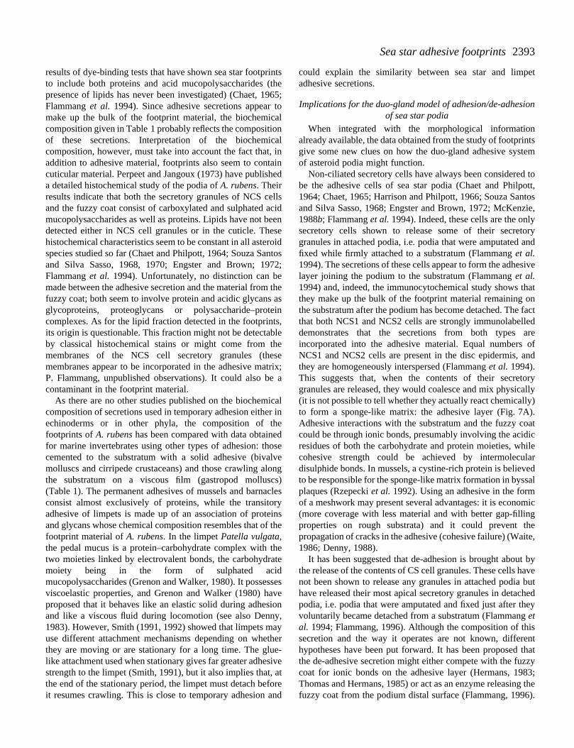
Non-ciliated secretory cells have always been consideredbe the adhesive cells of sea star podia (Chaet and Philp1964; Chaet, 1965; Harrison and Philpott, 1966; Souza Sanand Silva Sasso, 1968; Engster and Brown, 1972; McKenz1988b; Flammang et al.1994). Indeed, these cells are the onlysecretory cells shown to release some of their secretogranules in attached podia, i.e. podia that were amputated andfixed while firmly attached to a substratum (Flammang et al.1994). The secretions of these cells appear to form the adheslayer joining the podium to the substratum (Flammang et al.1994) and, indeed, the immunocytochemical study shows ththey make up the bulk of the footprint material remaining othe substratum after the podium has become detached. The that both NCS1 and NCS2 cells are strongly immunolabelledemonstrates that the secretions from both types aincorporated into the adhesive material. Equal numbers NCS1 and NCS2 cells are present in the disc epidermis, athey are homogeneously interspersed (Flammang et al.1994).This suggests that, when the contents of their secretogranules are released, they would coalesce and mix physica(it is not possible to tell whether they actually react chemicallyto form a sponge-like matrix: the adhesive layer (Fig. 7A)Adhesive interactions with the substratum and the fuzzy cocould be through ionic bonds, presumably involving the acidresidues of both the carbohydrate and protein moieties, whcohesive strength could be achieved by intermoleculdisulphide bonds. In mussels, a cystine-rich protein is believto be responsible for the sponge-like matrix formation in byssplaques (Rzepecki et al.1992). Using an adhesive in the formof a meshwork may present several advantages: it is econom(more coverage with less material and with better gap-fillinproperties on rough substrata) and it could prevent thpropagation of cracks in the adhesive (cohesive failure) (Wait1986; Denny, 1988).
It has been suggested that de-adhesion is brought aboutthe release of the contents of CS cell granules. These cells hnot been shown to release any granules in attached podia have released their most apical secretory granules in detacpodia, i.e. podia that were amputated and fixed just after thvoluntarily became detached from a substratum (Flammangetal. 1994; Flammang, 1996). Although the composition of thisecretion and the way it operates are not known, differehypotheses have been put forward. It has been proposed the de-adhesive secretion might either compete with the fuzcoat for ionic bonds on the adhesive layer (Hermans, 198Thomas and Hermans, 1985) or act as an enzyme releasingfuzzy coat from the podium distal surface (Flammang, 1996

2394
t
n
0.
rs
d
r
f
l
),
rIn
t.
P. FLAMMANG AND OTHERS
In the first hypothesis, one would expect to find the de-adhesecretion retained on the adhesive secretion within footprint, but there is no reason for cuticular material to present (competition model; Fig. 7B). Conversely, in thsecond hypothesis, the fuzzy coat would remain attached toadhesive secretion, while the de-adhesive secretion (perhasoluble enzyme) would fail to be incorporated into the footpr(enzyme model; Fig. 7C). The immunocytochemical dapresented in this report support the second hypothesis, wis further supported by the ultrastructural evidence showthat the CS cells release their secretion just under the cuand that the fuzzy coat can no longer be distinguishedtransmission electron micrographs of detached po(Flammang et al.1994; Flammang, 1996).
Future studies should allow the isolation and purificationadhesive molecules from either the footprints or the podiataking advantage of the new antisera raised against footpmaterial. The elucidation of their structures and physicchemical characteristics, together with the morphological dashould provide the necessary basis for understanding howstar adhesive systems are used in temporary adhesion.
We are grateful to Professor J. H. Waite for his criticreading of the manuscript. We also thank Professor J.Heuson-Stiennon for the use of the transmission electmicroscope and J.-P. Dardenne, G. Laurent and P. Postiautechnical assistance. P.F. is Senior Research Assistant oNational Fund for Scientific Research of Belgium (NFSRThis work was supported by grants from the NFSR Professor M. Jangoux (no. 9.4592.96 – Credit aChercheurs) and to Professor H. Alexandre (no. 9.4589.9Loterie nationale). This study is a contribution of the ‘CentInteruniversitaire de Biologie Marine’ (CIBIM).
ReferencesANTONOPOULOS, C. A. (1962). A modification for the determination
of sulphate in mucopolysaccharides by the benzidine method. Achem. scand.16, 1521–1522.
BARGMANN, W., HARNACK, M. AND JACOB, K. (1962). Uber denFeinbau des Nervensystem des Seesternes (Asterias rubensL.). EinBeitrag zur Vergleichenden Morphologie der Glia. Z. Zellforscmikrosk. Anat. 56, 573–594.
BENEDICT, C. V. AND WAITE, J. H. (1986). Composition andultrastructure of the byssus of Mytilus edulis. J. Morph.189,261–270.
BLUMENKRANTZ, N. AND ASBOE-HANSEN, G. (1973). New method forquantitative determination of uronic acids. Analyt. Biochem.55,484–489.
CHAET, A. B. (1965). Invertebrates adhering surfaces: Secretionsthe starfish, Asterias forbesiand the coelenterate, Hydra pirardiAnn. N.Y. Acad. Sci.118, 921–929.
CHAET, A. B. AND PHILPOTT, D. E. (1964). A new subcellular particlesecreted by the starfish. J. Ultrastruct. Res.11, 354–362.
COOK, M. (1970). Composition of mussel and barnacle deposits atattachment interface. In Adhesion in Biological Systems(ed. R. S.Manly), pp. 139–150. New York: Academic Press.
sivethebee theps aintta
hichingticle india
of byrinto-ta, sea
al A.ron for
f the).toux3 –re
cta
h.
of.
the
DENNY, M. (1983). Molecular biomechanics of molluscan mucoussecretions. In The Mollusca, vol. 1,Metabolic Biochemistry andMolecular Biomechanics(ed. P. W. Hochachka), pp. 431–465.New York: Academic Press.
DENNY, M. (1988). Biology and the Mechanics of the Wave SwepEnvironment. Princeton, NJ: Princeton University Press.
ENGSTER, M. S. AND BROWN, S. C. (1972). Histology andultrastructure of the tube foot epithelium in the phanerozoniastarfish, Astropecten. Tissue & Cell4, 503–518.
FLAMMANG , P. (1996). Adhesion in echinoderms. In EchinodermStudies, vol. 5 (ed. M. Jangoux and J. M. Lawrence), pp. 1–6Rotterdam: Balkema.
FLAMMANG , P., DEMEULENAERE, S. AND JANGOUX, M. (1994). ) Therole of podial secretions in adhesion in two species of sea sta(Echinodermata). Biol. Bull. mar. biol. Lab., Woods Hole187,35–47.
GRENON, J.-F. AND WALKER, G. (1980). Biochemical and rheologicalproperties of the pedal mucus of the limpet, Patella vulgataL.Comp. Biochem. Physiol. 66B, 451–458.
HARRISON, G. AND PHILPOTT, D. (1966). Subcellular particles inechinoderm tube feet. I. Class Asteroidea.J. Ultrastruct. Res.16,537–547.
HERMANS, C. O. (1983). The duo-gland adhesive system. Oceanogr.mar. Biol. A. Rev.21, 281–339.
HOLLAND, D. L. AND GABBOTT, P. A. (1971). A micro-analyticalscheme for the determination of protein, carbohydrate, lipid anRNA levels in marine invertebrate larvae. J. mar. biol. Ass. U.K.51, 659–668.
HOLLAND, N. D. (1984). Echinodermata: epidermal cells. In Biologyof the Integument, vol. 1, Invertebrates(ed. J. Bereiter-Hahn, A. G.Matoltsy and K. S. Richards), pp. 756–774. Berlin: SpringeVerlag.
HOLLAND, N. D. AND NEALSON, K. H. (1978). The fine structure ofthe echinoderm cuticle and the subcuticular bacteria oechinoderms. Acta zool. 59, 169–185.
JERMYN, M. A. (1975). Increasing the sensitivity of the anthronemethod for carbohydrate. Analyt. Biochem.68, 332–335.
KAMINO, K., ODO, S. AND MARUYAMA , T. (1996). Cement proteins ofthe acorn barnacle, Megabalanus rosa. Biol. Bull. mar. biol. Lab.,Woods Hole190, 403–409.
LUFT, J. H. (1971). Ruthenium red and violet. II. Fine structuralocalisation in animal tissues. Anat. Rec.171, 369–416.
MCKENZIE, J. D. (1988a). Echinoderm surface coats: Theirultrastructure, function and significance. In Echinoderm Biology(ed. R. D. Burke, P. V. Mladenov, P. Lambert and R. L. Parsleypp. 697–706. Rotterdam: Balkema.
MCKENZIE, J. D. (1988b). The ultrastructure of tube foot epidermalcells and secretions: their relationship to the duo-glandulahypothesis and the phylogeny of the echinoderm classes. Echinoderm Phylogeny and Evolutionary Biology(ed. C. R. C. Pauland A. B. Smith), pp. 287–298. Oxford: Clarendon Press.
NALDRETT, M. J. (1993). The importance of sulphur cross-links andhydrophobic interactions in the polymerization of barnacle cemenJ. mar. biol. Ass. U.K.73, 689–702.
PERPEET, C. AND JANGOUX, M. (1973). Contribution à l’étude des piedset des ampoules ambulacraires d’Asterias rubens(Echinodermata,Asteroides). Forma et Functio6, 191–209.
RZEPECKI, L. M., HANSEN, K. M. AND WAITE, J. H. (1992).Characterization of a cystine-rich polyphenolic protein family fromthe blue mussel Mytilus edulisL. Biol. Bull. mar. biol. Lab., WoodsHole 183, 123–137.

2395Sea star adhesive footprints
a
f
SMITH, A. M. (1991). The role of suction in the adhesion of limpetJ. exp. Biol.161, 151–169.
SMITH, A. M. (1992). Alternation between attachment mechanismslimpets in the field. J. exp. mar. Biol. Ecol. 160, 205–220.
SOUZA SANTOS, H. AND SILVA SASSO, W. (1968). Morphological andhistochemical studies on the secretory glands of starfish tube fActa anat.69, 41–51.
SOUZA SANTOS, H. AND SILVA SASSO, W. (1970). Ultrastructural andhistochemical studies on the epithelium revestment layer in tube feet of the starfish Asterina stellifera. J. Morph. 130,287–296.
THOMAS, L. A. AND HERMANS, C. O. (1985). Adhesive interactionsbetween the tube feet of a starfish, Leptasterias hexactisandsubstrata. Biol. Bull. mar. biol. Lab., Woods Hole169, 675–688.
TYLER, S. (1988). The role of function in determination of homologand convergence – examples from invertebrates adhesive orgFortschr. Zool.36, 331–347.
s.
by
eet.
the
yans.
WAITE, J. H. (1983). Adhesion in byssally attached bivalves. Biol.Rev.58, 209–231.
WAITE, J. H. (1986). Mussel glue from Mytilus californianusConrad:a comparative study. J. comp. Physiol. B 156, 491–496.
WAITE, J. H. (1992). The formation of mussel byssus: anatomy of natural manufacturing process. In Structure, Cellular Synthesis andAssembly of Biopolymers(ed. S. T. Case), pp. 55–74. Berlin:Springer-Verlag.
WALKER, G. (1972). The biochemical composition of the cement otwo barnacle species, Balanus hameriand Balanus crenatus. J.mar. biol. Ass. U.K.52, 429–435.
WALKER, G. (1987). Marine organisms and their adhesion. InSynthetic Adhesives and Sealants(ed. W. C. Wake), pp. 112–135.Chichester: John Wiley & Sons.
YULE, A. B. AND WALKER, G. (1987). Adhesion in barnacles. InCrustacean Issues, vol. 5,Biology of Barnacles(ed. A. J.Southward), pp. 389–402. Rotterdam: Balkema.