autotomy of rays of heliaster helianthus (asteroidea: echinodermata)
TRANSCRIPT
Zoosymposia 7: 173–176 (2012)
Accepted by A. Kroh & M. Reich: 27 Oct. 2012; published 12 Dec. 2012 173
www.mapress.com/zoosymposia/Copyright © 2012 · Magnolia Press
ISSN 1178-9905 (print edition)
ISSN 1178-9913 (online edition)ZOOSYMPOSIA
Autotomy of rays of Heliaster helianthus (Asteroidea: Echinodermata)*
JOHN M. LAWRENCE1,3 & CARLOS F. GAYMER2
1 Department of Integrative Biology, University of South Florida, Tampa, Florida, USA 2 Departamento de Biología Marina, CEAZA and IEB, Universidad Católica del Norte, Coquimbo, Chile 3 Corresponding author, E-mail: [email protected]
*In: Kroh, A. & Reich, M. (Eds.) Echinoderm Research 2010: Proceedings of the Seventh European Conference on Echinoderms, Göttingen, Germany, 2–9 October 2010. Zoosymposia, 7, xii + 316 pp.
Abstract
In species of the family Heliasteridae, the ossicles of the proximal parts of the sides of each ray are joined by connective tissue to those of the adjacent rays to form interradial septa. These provide support to the extensive disc. Only a relatively small part of the ray is free. Autotomy of rays occurs in Heliaster helianthus in response to predatory attack by the asteroid Meyenaster gelatinosus. Autotomy of the ray does not occur at the base of the free part of the ray (arm) but near the base of the ray. In addition to the plane of autotomy at this location, a longitudinal plane of autotomy occurs in the connec-tive tissue between the ossicles of the interradial septa. This indicates a plane of mutable collagenous tissue is present. Autotomy of the ray involves all these planes of autotomy and results in loss of most of the ray.
Key words: Asteroidea, Heliasteridae, autotomy, ray loss, mutable collagenous tissue
Introduction
Autotomy of rays occurs near the base of the arm (the free part of the ray) in most asteroids (Emson & Wilkie 1980). Exceptions are the luidiids (Emson & Wilkie 1980), astropectinids (Hotchkiss 2009) and archasterids (Lawrence et al. 2011) where it can occur at any point along the length of the ray. Autotomy involves mutable collagenous tissue in the body wall in a plane of autotomy (Wilkie 2002).
Agassiz (1877) called the inward continuation of the interradius of Asterias forbesi and Pisaster ochraceus the interbrachial partition. Viguier (1879) clearly illustrated two sets of ossicles in the interradial septa of the rays of the multirayed Heliaster microbrachia. Clark (1907) reported ray regeneration in Heliaster species but did not refer to the location where ray loss had occurred. Viviani (1978) specifically described autotomy of rays by Heliaster helianthus in response to the presence of its predators, Meyenaster gelatinosus and Luidia magellanica. His illustrations show autotomy does not occur at the base of the free part of the ray, the arm, but at the base of the ray near where it joins the central part of the body that contains the cardiac stomach. This raises the question of how the interradial septa of adjacent rays are separated in the process of autotomy.
LAWRENCE & GAYMER174 · Zoosymposia 7 © 2012 Magnolia Press
Materials and Methods
We induced autotomy of rays in Heliaster helianthus by placing individuals with Meyenaster gela-tinosus in an aquarium. Observations of an attack were recorded on video. An individual that had autotomized a ray and the autotomized ray were photographed.
Results
The video showed the initial response of the Heliaster helianthus to Meyenaster gelatinosus was rapid movement. After immobilization of the H. helianthus by M. gelatinosus and positioning the dorsal surface of the H. helianthus to its mouth, the connected portion of adjacent rays of the H. heli-anthus separated from each other followed by separation of the ray from the central part of the body. After several adjacent rays had autotomized, contortion of the body of the H. helianthus resulted in a stretching of the connective tissue between the ossicles of another adjacent ray so that it was torn, freeing the ray. Autotomy was not instantaneous but occurred over a number of minutes. During this period of autotomy, the H. helanthus was inactive. Only after autotomy did it move away from the M. gelatinosus.
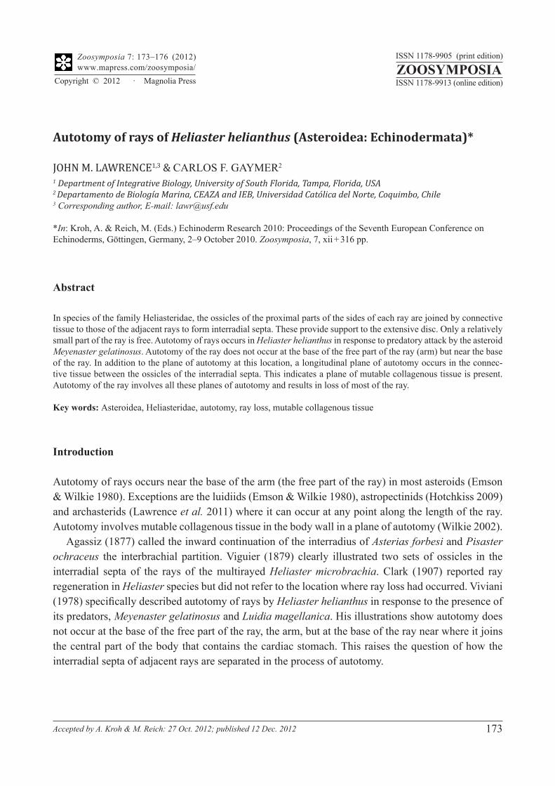
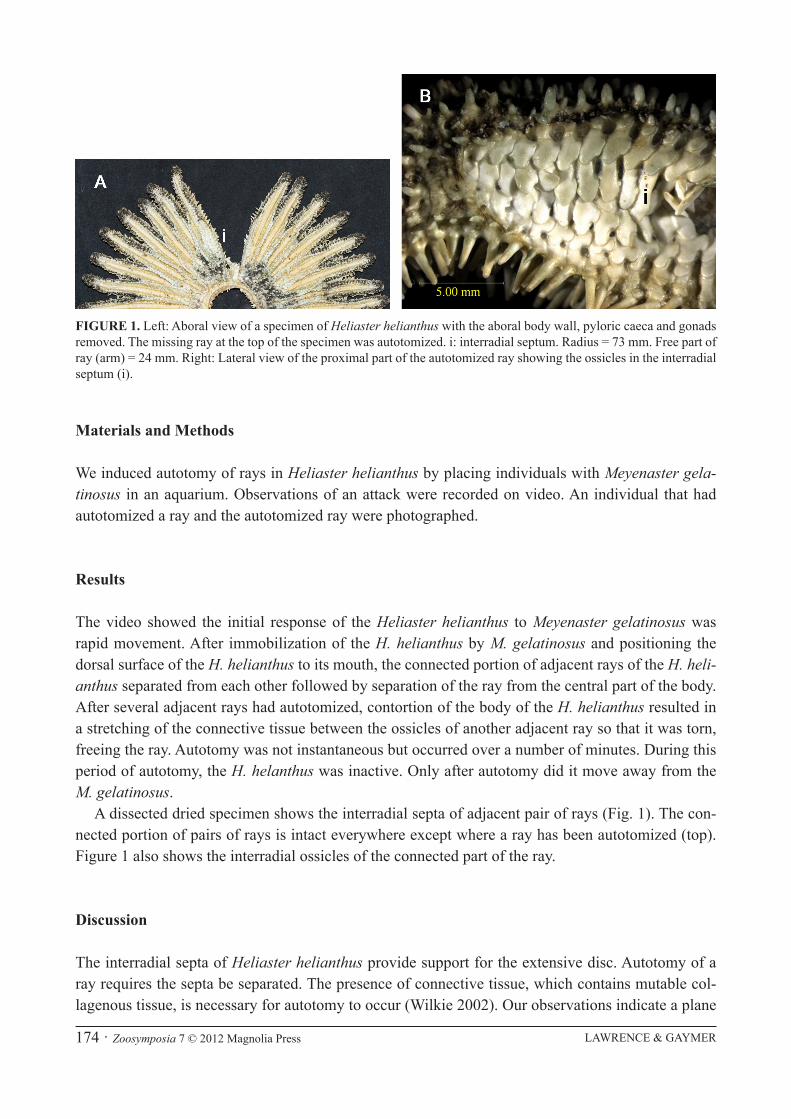
A dissected dried specimen shows the interradial septa of adjacent pair of rays (Fig. 1). The con-nected portion of pairs of rays is intact everywhere except where a ray has been autotomized (top). Figure 1 also shows the interradial ossicles of the connected part of the ray.
Discussion
The interradial septa of Heliaster helianthus provide support for the extensive disc. Autotomy of a ray requires the septa be separated. The presence of connective tissue, which contains mutable col-lagenous tissue, is necessary for autotomy to occur (Wilkie 2002). Our observations indicate a plane
FIGURE 1. Left: Aboral view of a specimen of Heliaster helianthus with the aboral body wall, pyloric caeca and gonads removed. The missing ray at the top of the specimen was autotomized. i: interradial septum. Radius = 73 mm. Free part of ray (arm) = 24 mm. Right: Lateral view of the proximal part of the autotomized ray showing the ossicles in the interradial septum (i).
AUTOTOMY OF RAYS OF HELIASTER HELIANTHUS Zoosymposia 7 © 2012 Magnolia Press · 175
of autotomy is present in the connective tissue joining the ossicles of the interradial septa of Heliaster helianthus in addition to the transverse plane of autotomy across the proximal part of the ray.
Clark (1907) stated the five-rayed Asterias ochracea (= Pisaster ochraceus) has well developed interbrachial walls that extend approximately 70 % of the ray length. Wilkie (2010, pers. comm.) found Asterias rubens (which is closely related to Pisaster) “does not have double septum linked by connective tissue, as in Heliaster” and “The lateral body wall of adjacent arms ruse basally to form a single ‘vertical’ partition that joins the mouth.”
Autotomy in large Asterias rubens and Asterias forbesi is at the base of the free ray, the arm, at the distal end of the interradial septa (Wilkie et al. 1990; Hotchkiss, pers. comm., respectively). This difference in the position of autotomy of Heliaster helianthus and Asterias rubens is interesting. What could be the basis of the difference between them? The proximate answer could be that H. helianthus has a double interradial septa and A. rubens does not. Wilkie (2010, pers. comm.) hypothesized “the autotomy plane cannot pass through it and the whole ray cannot be detached.”
Regarding the ultimate answer, it seems intuitive that as little of the ray as possible be lost. The answer may be due to the presence of a discobrachial septum in Heliaster species that is found in no other asteroid family (Clark 1907). Clark (1907) said the interradial septa end at the inner (proximal) end of the rays and unite laterally, more or less extensively, to form a discobrachial wall. As a result, the cavity of the disc is almost completely separated from the cavities of the rays so that the pyloric caeca and the cardiac stomach are connected only by a small tube. Autotomy at this position would result in a clean break that could facilitate healing and regeneration. This would not be the case with autotomy at a more distal position of the ray that would result in rupture of the pyloric caeca. Thus there may be a trade-off between the cost of the amount of ray loss and facilitation of healing and regeneration.
Lawrence (2013) gave this explanation for autotomy at the base of arms of asteroids, making the unwarranted assumption that this was the location where the pyloric caeca and gonads end in tubes. This is not true for Asterias rubens because these organs continue proximally to the true disc where they end in tubes. To say the arm is autotomized and also that the arm extends to the disc in A. rubens is an oxymoron except for small individuals. This is the case in small A. rubens (Cuénot 1948) but not in large ones because their interradius is extended. In the latter case, the arm is only the free part of the ray. In his memoir on the genus Heliaster, Clark (1907) referred to the distal free part of the ray.
This indicates correctness in terminology is necessary. The terms “ray” and “arm” are often used interchangeably. Yet they have different meanings. “Ray” has an anatomical meaning, the length of the radial water canal and its surrounding structures. Rays are present in all echinoderm classes, including echinoids and holothuroids. “Arm” has a meaning regarding form, an appendage to the central body. The two terms can be used interchangeably in asteroids when the ray/arm is attached to a distinct disc that contains the cardiac stomach. This distinction is important to avoid misinterpreta-tions and misunderstandings.
Acknowledgments
We thank Ian Wilkie for information about arm autotomy and the interradial septum in Asterias rubens, F.H.C. Hotchkiss for information about arm autotomy in Asterias forbesi, and F.H.C. Hotch-kiss and C.M. Pomory for comments on the manuscript.
LAWRENCE & GAYMER176 · Zoosymposia 7 © 2012 Magnolia Press
References
Agassiz, A. (1877) North American Starfishes. Memoirs of the Museum of Comparative Zoology, Harvard College, 5(1), 1–136.
Clark, H.L. (1907) The starfishes of the genus Heliaster. Bulletin of the Museum of Comparative Zoology, Harvard Col-lege, 51, 25–76.
Cuénot, L. (1948) Anatomie, éthologie et systématique des échinodermes. In: Grassé, P.-P. (Ed.), Traité de Zoologie, XI. Masson et Cie, Paris, pp. 3–272.
Emson, R.H. & Wilkie, I.C. (1980) Fission and autotomy in echinoderms. Oceanography and Marine Biology: An Annual Review, 8, 155–250.
Hotchkiss, F.H.C. (2009) Arm stumps and regeneration models in Asteroidea. Proceedings of the Biological Society of Washington, 122, 342–354.
Lawrence, J.M. (2013) Arm loss and regeneration in stellate echinoderms: an organismal view. In: Johnson, C. (Ed.), Echinoderms in a Changing World. Proceedings of the Thirteenth International Echinoderm Conference, Hobart, Tasmania, Australia, 5–9 January 2009. Taylor & Francis Group, London, pp. 53–66.
Lawrence, J.M., Keesing, J.K. & Irvine, T.R. (2011) Population characteristics and biology of two populations of Archaster angulatus (Echinodermata: Asteroidea) in different habitats off the central-western Australian coast. Journal of the Marine Biological Association of the United Kingdom, 91, 1577–1585.
Viguier, C. (1879) Anatomie comparée du squelette des Stellérides. Archives de Zoologie Experimentale, 7, 33–250.Viviani, C.A. (1978) Predación interespecífica, canibalismo y autotomía como mecanismo de escape en las especies de
Asteroídes (Echinodermata) en el litoral del desierto del Norte Grande de Chile. Universidad del Norte, Iquique, 116 pp.Wilkie, I.C. (2002) Is muscle involved in the mechanical adaptability of echinoderm mutable collagenous tissue? The
Journal of Experimental Biology, 205, 159–165.Wilkie, I.C., Griffiths, G.V.R. & Glennie, S.F. (1990) Morphological and physiological aspects of the autotomy plane
in the aboral integument of Asterias rubens L. In: De Ridder, C., Dubois, P., LaHaye, M.C. & Jangoux, M. (Eds.), Echinoderm Research: Proceedings of the Second European Conference on Echinoderms. Brussels, Belgium, 18–21 September 1989. A.A. Balkema, Rotterdam, pp. 301–313.