a simplified method for the reconstruction of fully competent mouse zygotes from adult somatic donor...
TRANSCRIPT
CLONINGVolume 2, Number 1, 2000Mary Ann Liebert, Inc.
A Simplified Method for the Reconstruction of Fully Competent Mouse Zygotes from
Adult Somatic Donor Nuclei
QI ZHOU,1,2 LAURENT BOULANGER,1 and JEAN-PAUL RENARD1
ABSTRACT
Until now, full-term development of mouse embryos reconstructed from somatic nuclei hasbeen convincingly achieved only when a piezoimpact pipette drive unit is used for the injec-tion of a donor nucleus into an enucleated recipient oocyte. Here we describe a simplifiedmethod for mouse cloning that requires neither electrofusion nor a piezo device. Efficient ratesof enucleation can be achieved without staining the chromosomes of the recipient oocyte andhigh survival rates are obtained after direct injection of the donor nucleus. Although a lowproportion of reconstructed embryos could implant after their transfer into the oviducts of fos-ter mothers (less than 5%), we show that at least some of them can develop into normal young.
35
INTRODUCTION
ANIMAL CLONING from somatic donor cells re-quires the introduction of a foreign nucleus
into a recipient enucleated oocyte or egg (cyto-plast). This can be obtained either indirectlythrough fusion of the donor cell with the recipi-ent cytoplast (virus-mediated fusion [McGrathand Solter, 1983]; electrofusion [Willadsen, 1986])or directly through intracytoplasmic injection(Bromhall, 1975; Illmensee and Hoppe, 1981; Col-las and Barnes, 1994). The former approach hasallowed the demonstration that somatic nucleifrom adult donor animals of different species canbe fully reprogrammed (Wilmut et al., 1997).However, it failed when applied to the mousespecies except when associated with an elegantbut time-consuming procedure that involves se-rial nuclear (of embryonic source) transfer fromthe same reconstructed embryo (Kwon and Kono,1996). Intracytoplasmic injection has undergone
extensive experimentation in the mouse speciesbecause of a first claim of success (Illmensee andHoppe, 1981). But it is only recently that this ap-proach has lead to the indubitable demonstrationthat full reprogramming from freshly isolatedadult somatic donor cells could also be obtainedin that species (Wakayama et al., 1998). This tech-nical breakthrough has now been extended todonor cells obtained from short-term culturedcumulus cells (Wakayama et al., 1999a) or estab-lished (Wakayama et al., 1999b) and geneticallymanipulated (Rideout et al., 2000) ES cell lines. Itinvolves the microsurgical isolation of a nucleusfollowed by its piezoelectrically actuated mi-croinjection into a metaphase II (MII)-derived cy-toplast. Taken together, these results, which un-til now have been obtained by or in closecollaboration with the same author (TeruhikoWakayama), suggest that some aspect of the enu-cleation procedure or some decisive advantageconferred by the piezocontrolled injection ap-
1Unité de Biologie du Développement et Biotechnologie, Institut National de la Recherche Agronomique (INRA),78352, Jouy en Josas, France.
2Institute of Developmental Biology, Chinese Academy of Sciences (CAS), Beijing, 100080, China.
proach, or both, are required for the success ofmouse somatic cloning. Elucidating the nature ofthese technical improvements could bring someclue to the routine use of the mouse model in ba-sic studies aimed at understanding the biology ofnuclear reprogramming.
We first adopted the same experimental condi-tions in term of strain of mice, type of donor cells,culture media, timing of manipulation, and use ofthe piezo device (Model PMAS-CT16, Prime-TechLtd., Ibaraki, Japan) as reported in Wakayama etal. (1998). Unfortunately, and despite numerousattempts, we only obtained a low rate of recon-structed embryos. Although the enucleation ratewas high (above 90%), we obtained a much lowerrate of survival after nucleus injection (only 12 outof 202; 5.9%) than the one reported by Wakayamaet al. (1998) and none of such reconstructed em-bryos developed into blastocysts. To overcomethis first disappointment, we decided to concen-trate essentially on the manipulations requiredboth for oocyte enucleation and nucleus injectionwithout the help of a piezo device and decided tokeep more closely to the routine culture proce-dures (in terms of strain of mice, media, timing ofegg recoveries) that we use in the laboratory forhandling mouse embryos. We tried to minimizetrauma to oocytes by improving an enucleationmethod that would reduce the amount of cyto-plasm removed together with the MII chromo-some-spindle complex (Kono et al., 1993). We alsodesigned a simple injection method for donor nu-clei that we adapted from the one initially pro-posed for rabbit embryos (Zhou et al., 1997).
We provide here evidence that the use of piezodevice is not a requirement for full-term repro-gramming of mouse somatic nuclei. Although theoverall efficiency of our procedure is slightlylower than the one described by Wakayama et al.(1998) with the same type of donor nuclei, it con-sistently provides in vitro development to theblastocyst stage thereby offering a means to ana-lyze early reprogramming events.
MATERIALS AND METHODS
Media
Oocytes before or after enucleation and alsonuclear transferred embryos were cultured intoM16 medium. The medium used during enucle-ation and injection of cumulus cells was M2medium (Brown and Whittingham, 1992).
Preparation of oocytes and cumulus cells
Eight-week-old F1 C57/CBA (C57B1/6 3 CBA,providing oocytes and donor nuclei in series A andB) or F1 B6D2 (C57BL6/DBA2, providing oocytesin series C) females were superovulated with preg-nant mare serum gonadoptropin (PMSG, 10 IU)and human chorionic gonadotropin (hCG, 5 IU).These donor females were either hybrid F1 pro-duced in our animal facilities after the mating ofC57B1/6 females with CBA males, or C57/CBAtransgenic females (thus of the same genetic back-ground as the F1 C57/CBA females) but harbor-ing two copies of a reporter construct that allowsdetection of luciferase from cultured cells (Thomp-son et al., 1995). Oocytes were collected fromoviducts 13 hours after hCG injection. Cumuluscells were removed with hyaluronidase (300IU/mL), and oocytes and cumulus cells werewashed with F M2 for several times. They werethen cultured in M16 medium at 37.5°C, under anatmosphere of 8% CO2 in air. In a part of the ex-periments (series C), cumulus cells were obtainedfrom a transgenic line harboring a luciferase re-porter construct produced in our animal facilities.
Preparation of oocyte holding, enucleating, andinjecting pipettes
We used thin-wall Clark capillaries (Phymep,Paris, France; GC100T-10, 1.0 mm outer diameter[OD] 0.78 mm inner diameter [ID]) without inter-nal filament, for the preparation of all the mi-croinstruments needed for manipulations. Hold-ing pipettes were first drawn by hand, then scoredat suitable region by diamond pen and their endsfire-polished under a microforge (Alcatel, France)for an outer and inner diameters of 80–100 mmand 15 mm, respectively. Enucleation and injec-tion pipettes were drawn with a pipette puller(Moving-coil microelectrode Puller-753; Camp-don Instruments Ltd., London, UK). The tip of theenucleation pipettes (inner diameter approxi-mately 8 to 10 mm) was first beveled at 40-degreeangle with a homemade grinder and then sharp-ened under microforge as described initially byMcGrath and Solter (1983). Injection pipette werebroken by gently tapping against the filament ofthe microforge in order to obtain a flat tip (bluntend) with an internal diameter of about 5–12 mm.
Enucleation of MII oocytes
Manipulations (Nikon-Narishige Micromanip-ulators MO-188, Tokyo, Japan) were performed
ZHOU ET AL.36
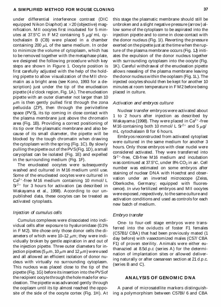
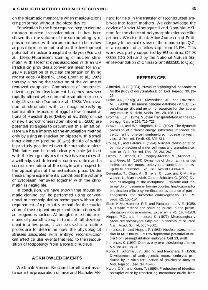
under differential interference contrast (DICequipped Nikon Diaphot) at 320 (objective) mag-nification. MII oocytes first incubated for 5 min-utes at 37.5°C in F M2 containing 5 mg/mL cy-tochalasin B (CB) were placed in a chambercontaining 200 mL of the same medium. In orderto minimize the volume of cytoplasm, which hasto be removed together with the metaphase plate,we designed the following procedure which keysteps are shown in Figure 1. Oocyte position isfirst carefully adjusted with the help of the hold-ing pipette to allow visualization of the MII chro-matin as a bright area (see Kono, 1993 for a de-scription) just under the tip of the enucleationpipette (4 o’clock region, Fig. 1A). The enucleationpipette with an outer diameter not larger than 10mm is then gently pulled first through the zonapellucida (ZP), then through the perivitellinespace (PVS), its tip coming in close contact withthe plasma membrane just above the chromatinarea (Fig. 1B). Providing a correct positioning ofits tip over the plasmatic membrane and also be-cause of its small diameter, the pipette will beblocked by the tough chromatin when drawingthe cytoplasm with the spring (Fig. 1C). By slowlypulling the pipette out of the PVS (Fig. 1D), a smallcaryoplast can be isolated (Fig. 1E) and expelledin the surrounding medium (Fig. 1F).
The enucleated oocytes were subsequentlywashed and cultured in M16 medium until use.Some of the enucleated oocytes were cultured inCa21-free M16 medium containing 10 mmol/LSr21 for 3 hours for activation (as described inWakayama et al., 1998). According to our un-published data, these oocytes can be treated asactivated cytoplasts.
Injection of cumulus cells
Cumulus complexes were dissociated into indi-vidual cells after exposure to hyaluronidase (0.1%in F M2). We chose only those donor cells the di-ameters of which were 10–12 mm; they were indi-vidually broken by gentle aspiration in and out ofthe injection pipette. Three outer diameters for in-jection pipettes (5 mm, 10 mm and 12 mm) were usedand all allowed an efficient isolation of donor nu-cleus with virtually no surrounding cytoplasm.This nucleus was placed close to the tip of thepipette (Fig. 1G) before its insertion into the PVS ofthe recipient oocyte through the hole made for enu-cleation. The pipette was advanced gently throughthe ooplasm until its tip almost reached the oppo-site of the side of the oocyte cortex (Fig. 1H). At
this stage the plasmatic membrane should still beunbroken and a slight negative pressure (arrow) al-low some of the cytoplasm to be aspirated into theinjection pipette and to come in close contact withthe donor nucleus (Fig. 1I). Reverting the pressureexerted on the pipette just at the time when the rup-ture of the plasma membrane occurs (Fig. 1J) initi-ates the expulsion of the donor nucleus togetherwith surrounding cytoplasm into the oocyte (Fig.1K). Careful withdrawal of the enucleation pipetteallows resealing of the plasma membrane leavingthe donor nucleus within the ooplasm (Fig. 1L). Theinjected oocytes should then be kept for another 10minutes at room temperature in F M2 before beingplaced in culture.
Activation and embryos culture
Nuclear transfer embryos were activated about1 to 2 hours after injection as described byWakayama (1998). They were placed in Ca21-freeM16 containing both 10 mmol/L Sr21 and 5 mg/mL cytochalasin B for 6 hours.
Embryos reconstructed from activated cytoplastwere cultured in the same medium for another 3hours. Only those embryos with clear nuclei wereconsidered activated. They were transferred intoSr21-free, CB-free M16 medium and incubationwas continued at 37.5°C, under 8% CO2 in air. Cellnumber was estimated on some embryos afterstaining of nuclear DNA with Hoechst and obser-vation under an inverted microscope (Zeiss,Oberkoche, Germany; equipped with fluores-cence). In vivo fertilized embryos and MII oocyteswere exposed, respectively, to the same culture andactivation conditions and used as controls for eachnew batch of medium.
Embryo transfer
One- to four-cell stage embryos were trans-ferred into the oviducts of foster F1 females(C57B1/CBA) that had been previously mated (1day before) with vasectomized males (C57/CBAF1) of proven sterility. Animals were either eu-thanazied at 8.5d.p.c (series A) for the determi-nation of implantation sites or allowed deliver-ing naturally or after caesarean section at 21 d.p.c.(series B and C).
ANALYSIS OF GENOMIC DNA
A panel of microsatellite markers distinguish-ing a polymorphism between C57Bl/6 and CBA
A SIMPLIFIED METHOD FOR MOUSE CLONING 37

FIG. 1. Drawings representing the different steps of the technique used to reconstruct one cell stage embryos fromsomatic nucleus without the help of a Piezo device. (A–F) enucleation of mouse MII oocyte. (G–L) injection of mousecumulus cell into enucleated oocyte (see text for details).
homozygous strains was used to confirm thatpups obtained from nuclear F1 hybrids C57/CBAdonors were truly F1 and not F2 hybrids as wouldhave been the case if foster mothers had been for-tuitously mated by F1 hybrids males. This panelof markers was determined from the MIT data-base established for recombinant congenic strainsderived from C57Bl/6J and NOD/Shi (Blake etal., 2000) and the data provided by Reifsnyder etal. (1999) for recombinant congenic strains de-rived from NOD/Shi and CBA/J genomes. It in-cluded the following microsatellites: D2Mit77;D6Mit1; D7Mit12; D7Mit46; D12Mit5; D13Mit21.Polymerase chain reaction (PCR) amplification ofDNA extracted from tail-tips was performed byusing primer pairs obtained from Research Ge-netics except for microsatellites D12Mit5 andD13Mit21 all purchased from Genset (Paris,France). Amplification products were rho-damine-labeled by R6G dCTP (Perkin ElmerBiosystems, Branchburg, NJ) incorporation dur-ing PCR cycle and separated by acrylimide gelmigration on a ABI 373 Sequencer (Perkin ElmerBiosystems). Because the above microsatellitesare polymorphic in length between the two ho-mozygous strains, F1 heterozygous animals willshow a biallelic pattern for each of the mi-crosatellites PCR products. In contrast, an F2 an-imal resulting from the mating of an heterozy-gous F1 female with an F1 C57/CBA male shouldshow some homozygous PCR patterns products.
The transgenic nuclei used in this study wereobtained from the transgenic C57/CBA femalesof the same genetic background but harboringtwo copies of a reporter luciferase gene per hap-loid genome (linearized Bluescript plasmidHSP70.1luc, [Thompson et al., 1995]). Constitu-tive luciferase expression was determined fromtail-tip biopsies or tissue samples as described inMenck et al. (1998).
RESULTS
Survival rate of oocytes after manipulation
We first found that using an enucleationpipette with an internal diameter of only about 8to 10 mm allowed an accurate removal of the MIIchromosomes. For that, MII oocytes (n 5 425)were first exposed to Hoechst dye but their chro-matin was observed only after micromanipula-tion. No condensed chromatin could be visual-
ized from the 422 oocytes (99.3%) that survivedmanipulation, whereas it was the case for each ofthe expelled karyoplasts. When injected with acumulus nucleus, 46.2% of the recipient cyto-plasts (1150/2491) did not lyse (Table 1). We usu-ally injected enucleated oocytes in groups of 6 to10 and found that their ability for survival aftermicroinjection greatly varied between groupseven within the same day and using the samepipette. Overall, 63.4% of the reconstituted eggsactivate after strontium treatment. In some in-stances all the injected oocytes from several con-secutive groups could activate.
The diameter of the injection pipettes provedto be an important factor for survival (Table 2).There was no significant difference between 5–7mm and 8–10 mm pipettes but the surviving rateof injected cytoplasts (lysis) rapidly decreasedwhen using a larger diameter. Changing the tem-perature of the microscope stage (10–30°C) didnot affect survival rates, neither did changes inthe osmolarity of manipulating medium follow-ing addition of 0.25%, 0.50%, or even 5% sucrose(respective values: 290 to 500 mOsm). Carefulcleaning of pipettes (with fluorydric acid) to-gether with their siliconisation appeared to be im-portant parameters for successful injection. Wealso found that reducing variations in the me-chanical tensions exerted on the plasmatic mem-brane during the injection of nucleus improvedthe survival of the reconstructed embryos. Thiswas obtained by careful control over the pro-gression of the pipette inside the recipient cyto-plast and by reducing the cytoplasmic volume ab-sorbed into the pipette before nucleus delivery.
In vitro development of reconstituted embryos
From a total of 239 nuclear transplant oocytesthat were submitted to the strontium treatment,145 (60.7%) formed pseudo-pronuclei and werethus considered activated. When these embryoswere subsequently cultured in M16 medium,about two-thirds (99/145, 68.3%) started to cleaveapparently normally but only 22.1% (n 5 32)could reached the morula/blastocyst stage and8.3% (n 5 12) formed enlarged blastocyst after 4days of culture (Table 3). This low in vitro devel-opmental rate to the blastocyst stage contrastedwith the one obtained with fertilized zygotes (108blastocysts obtained from 129 cultured one-cellstage, 83.7%) or activated oocytes (581 blastocystsobtained from 668 activated oocytes, 87.0%) ex-
A SIMPLIFIED METHOD FOR MOUSE CLONING 39
cluding any specific detrimental effect of our cul-ture and activation procedures. Number of cellscounted on 2 of the 32 morula/blastocysts ob-tained were 59 and 65, respectively. Nucleartransfer embryos with evidence of developmen-tal arrest at or before the four-cell stage (77.9%,113/145) occasionally formed a fluid-filled likecavity (blastocoel-like), with clearly no evidenceof inner cell mass cells F.
When preactivated cytoplasts were used, only8.5% of those reconstructed embryos formed apseudo-pronucleus (5/59, Table 3) and none coulddevelop to the morula/blastocyst stage in vitro.
In vitro development of reconstituted embryos
In order to assess the in vivo developmental po-tential of reconstructed embryos we finally trans-ferred them into foster recipients. Rate of implan-tation was first recorded at day 8.5 dpc from a seriesof experiments involving 11 recipients (Table 4, se-ries A). From 129 embryos that were transferred atthe one- or two-cell stage only 6 (4.6%) implanted.Size of these six implantation sites was smaller thanthe one observed at the same age of gestation aftereuthanasia of mated females. This suggests that de-velopment had already been compromised at day8.5 dpc. In a second experiment 246 embryos weretransferred at the one-, or two-, to four-cell stagesinto foster recipients (n5 15) that were allowed togo to term (Table 4, series B). One delivered a sin-gle morphologically normal pup. Genetic analysis(see Materials and Methods) confirmed that thisoffspring was of the same F1 (CBA and C57Bl/6)genotype as the donor nucleus (Fig. 2, panel F1 andMA) and excluded the possibility it could be an F2because would have been the case if the foster
mother had been mated fortuitously by a fertile F1male (only F1 males are available in our animal fa-cilities).
In order to confirm that our cloning method al-lows full reprogramming from cumulus nuclei,we decided to perform a third experiment usingcumulus cells of the same genetic background(C57/CBA) as a source of nuclei but bearing atransgene marker (see material and method).From 155 embryos transferred at the one- or two-cell stage on eight recipient females (Table 4, se-ries C) two live pups were obtained (from twodifferent recipient females). The first was ob-tained by caesarean section at 21 days of preg-nancy but was unfortunately eaten during thenight after its adoption by a lactating mother. Theother was born naturally at 20 days of gestation.Although normal in size and without phenotypicabnormalities, it died 1 day later. A sample of tis-sue obtained after autopsy proved to be positivefor luciferase thereby confirming that this pupwas derived from the injected nucleus. Other his-tological examinations performed from fixatedtissues remained inconclusive. There was clearevidence that the pup had apparently been fednormally by the lactating mother; this latter wasalso autopsied and we could confirm that she hada normally functioning mammary gland.
Although the rate of development to term ob-tained from series B and C is low (3 pups bornalive from 401 embryos transferred, 0.7%) theseresults provide evidence that a piezocontrolledinjection procedure is not a requirement for thesuccessful reconstitution of mouse embryos fromsomatic nuclei and that cloning in the mouse canbe achieved by classical manipulations of an in-jection pipette. Because of this, the first cloned
ZHOU ET AL.40
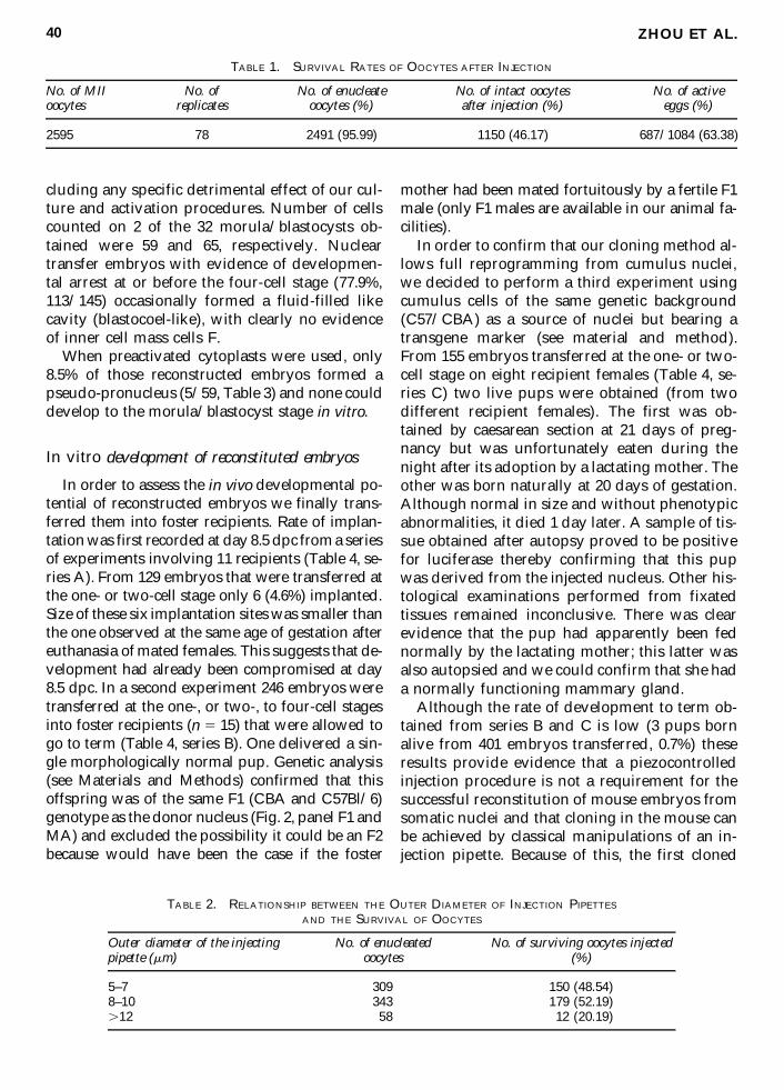
TABLE 1. SURVIVAL RATES OF OOCYTES AFTER INJECTION
No. of MII No. of No. of enucleate No. of intact oocytes No. of activeoocytes replicates oocytes (%) after injection (%) eggs (%)
2595 78 2491 (95.99) 1150 (46.17) 687/1084 (63.38)
TABLE 2. RELATIONSHIP BETWEEN THE OUTER DIAMETER OF INJECTION PIPETTES
AND THE SURVIVAL OF OOCYTES
Outer diameter of the injecting No. of enucleated No. of surviving oocytes injectedpipette (mm) oocytes (%)
5–7 309 150 (48.54)8–10 343 179 (52.19).12 58 12 (20.19)
mouse obtained in this study, which has nowproven to be fertile, with nine normal pups ob-tained after mating with a C57/CBA male, hasbeen called “Manuela.”
DISCUSSION
Since the publication of Wakayama et al. (1998)demonstrating that cloning in the mouse speciescould lead to a high implantation rate and de-velopment to term, several groups, includingours, have extensively tried to repeat the experi-ment but without marked success. We initiallyadopted the experimental conditions designed inDr. Yanagimashi’s laboratory by using the sameculture media during the manipulations (CZBwith or without HEPES), same hybrid mice (F1,B6/D2), same timing between nucleus injectionand activation, and also a piezo device (providedto us by the same company). Despite these effortswe could not achieve in vitro development to theblastocyst stage, and less than 10% of the embryosthat survived the reconstruction procedure couldcleave beyond the two-cell stage. Because wecould not make the piezo device work properlyand consistently from one experiment to anotherwe decided to use conventional micromanipula-tion methods and tried to minimize trauma in-duced to both recipient oocytes and nuclei dur-ing the micromanipulations. We also keep closerto the experimental conditions used in our labo-ratory in terms of culture media (M16 [Brown andWhittingham, 1992]) and strain of mice (F1,C57/CBA) because these conditions routinely
lead to a high rate of in vitro development fromin vivo produced one-cell stage embryos (morethan 70% of hatched blastocysts; unpublisheddata). We found that the proportion of success-fully reconstructed embryos that could developbeyond the two-cell stage in vitro was increasedby a factor of 3 times more (36% instead of 10%developed at and beyond the four-cell stage (seeTable 3). Because in the mouse, transcriptional ac-tivity of the zygotic genome becomes required atthe two-cell stage to allow further cleavage, thisobservation may reflect the fact that optimizationof procedures for the handling of manipulatedembryos greatly facilitates the remodeling of for-eign nuclei into a fully active zygotic genome.
A relatively high rate of embryos (nearly 50%)could survive nuclear injection without a piezodevice and we provide evidence that such recon-structed embryos obtained from an adult somaticnucleus can develop into normal fertile mice. Assuch, out technique appears to be similar to theone initially proposed by Illmensee for success-ful enucleation (Hoppe and Illmensee, 1977) andinjection (Illmensee and Hoppe, 1981) of mouseembryos with embryonic nuclei. For instance wealso found that a pipette with an internal diame-ter of 10 mm could be used not only for enucle-ation but also for injection as reported by Ill-mensee and Hoppe (1981) whereas othersgenerally use smaller sized pipette for injection(7 to 8 mm, see Wakayama et al., 1998). We fa-vored blunt-end pipettes for injection but wecould also obtain in vitro development when thesame beveled and tip-sharpened pipette wasused both for enucleation and injection of the
A SIMPLIFIED METHOD FOR MOUSE CLONING 41
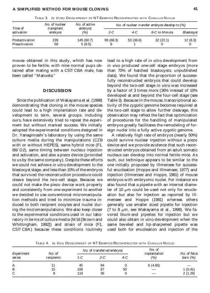
TABLE 4. IN VIVO DEVELOPMENT OF NT EMBRYOS RECONSTRUCTED WITH CUMULUS NUCLEI
No. ofExperiment No. of implantation No. of fetusseries recipients 1-C 2-C 4-C (%) born (%)
A 11 45 84 0 6 (4.65) —B 15 109 87 50 — 1 (0.41)C 8 119 36 0 — 2 (1.29)
No. of transferred embryos
TABLE 3. IN VITRO DEVELOPMENT OF NT EMBRYOS RECONSTRUCTED WITH CUMULUS NUCLEI
No. of nuclear No. of activeTime of transplant embryosactivation embryos (%) 2-C 4-C 8-C to Morula Blastocyst
Postactivation 239 145 (60.7) 99 (68.3) 53 (36.6) 32 (22.1) 12 (8.3)Preactivation 59 5 (8.5) 8 1 0 0
No. of nuclear transfer embryos develop to (%)
donor nucleus (Q.Z., unpublished observations).We were, however, unable to obtain evidence thatembryos could develop in vitro when recon-structed from cytoplasts already exposed to anactivation treatment in contrast to Illmensee andHoppe (1981). This result brings additional evi-dence to the experimental facts showing that theinjection method does not allow fertilized (acti-vated) zygotes to be used as recipients for somaticcell nuclei (Wakayama et al., 2000, but see alsoSolter, 1999). Thus, despite large technical simi-larities between our method of nuclear transferand the one described in Illmensee and Hoppe(1981) our data do not support the view that “time
for the correct evaluation of these earlier resultshas come” (Illmensee, 1999).
In several replicate experiments we could ob-tain a high rate of cleavage (up to 100%) but fur-ther development into blastocysts was very low.This does not occur in the work of Wakayama etal. (1998, 1999a) and could indicate that the verydelicate way in which the nucleus is injected be-cause of the piezo technique may benefit some bi-ological events, playing an important althoughdispensable role in early reprogramming. Suchevents could be linked, for instance, to the dis-turbance of some signaling pathway because ofthe higher mechanical effort apparently exerted
ZHOU ET AL.42
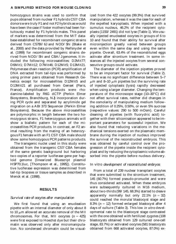
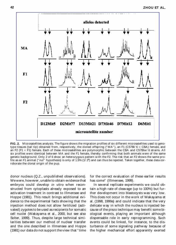
FIG. 2. Microsatellites analysis. The figure shows the migration profiles of six different microsatellites used to geno-type tissues (tail tip) obtained from, respectively, the cloned offspring (“MA”), an F1 (C57Bl/6 3 CBA) female, andan F2 (F1 3 F1) female. Each of these microsatellites are polymorphic between the CBA and C57Blw/6 strains. Allsix profiles were identical between MA and the F1 female, thereby confirming that both animals were of the samegenetic background. Only 2 of 6 show an heterozygous pattern with the F2. The risk that an F2 shows the same pro-file as an F1 animal (“nul” hypothesis) is only of 1.5% (1/26) and can thus be rejected. Taken together, these data cor-roborate the clonal origin of the pup.
on the plasmatic membrane when manipulationsare performed without the piezo device.
Enucleation is the first required step to cloningthrough nuclear transplantation. It has beenshown that the volume of the surrounding cyto-plasm removed with chromatin has to be as lowas possible in order not to affect the developmentpotential of nuclear transplant embryos (Peura etal., 1998). Fluorescent staining of nuclear chro-matin with Hoechst dyes associated with an UVirradiation provides a convenient mean for an insitu visualization of nuclear chromatin on livingrodent eggs (Albertini, 1984; Ebert et al., 1985)thereby allowing the reduction of the volume ofremoved cytoplasm. Competence of mouse fer-tilized eggs for development becomes howeverrapidly altered when time of irradiation exceedsonly 45 seconds (Tsunoda et al., 1988). Visualiza-tion of chromatin with an image-intensifyingcamera after exposure of eggs to low concentra-tions of Hoechst dyes (Debey et al., 1989) or useof new fluorochromes (Dominko et al., 2000) arepotential strategies to circumvent this limitation.Here we have improved the enucleation methodonly by using an enucleation pipette with a smallinner diameter (around 10 mm) the tip of whichis precisely positioned over the metaphase plate.This latter can be made clearly visible (at leastwith the two genotypes that we have used) witha well-adjusted differential contrast optics and acorrect orientation of the oocyte with respect tothe optical plan of the metaphase plate. Underthese simple experimental conditions the volumeof cytoplasm removed together with the chro-matin is negligible.
In conclusion, we have shown that mouse so-matic cloning can be performed using conven-tional micromanipulation techniques without therequirement of a piezo device both for the enucle-ation of the recipient oocyte and its injection withan exogenous nucleus. Although our technique re-mains of poor efficiency in terms of full develop-ment into live pups, it can be used as a routineprocedure to determine how the physiologicalstresses associated with embryo reconstructioncan affect cellular events that lead to the reacqui-sition of totipotency from a somatic nucleus.
ACKNOWLEDGMENTS
We thank Vincent Brochard for efficient assis-tance in the preparation of mice and Nathalie Mé-
nard for help in the transfer of reconstructed em-bryos into foster mothers. We acknowledge theadvice of Xavier Montagutelli and Dominique Si-mon for the choice of polymorphic microsatelliteprimers. We also thank Alice Jouneau and EdithLegouy for critical review of this manuscript. Q.Z.is a recipient of a fellowship from INRA. Thiswork was partly supported by EU contract CT 9800032 (DG XII) and by the National Natural Sci-ence Foundation of China (Grant 9802801 to Q.Z.).
REFERENCES
Albertini, D.F. (1984). Novel morphological approachesfor the study of oocyte maturation. Biol. Reprod. 30, 13–28.
Blake, J.A., Eppig, J.T., Richardson, J.E., and Davisson,M.T. (2000). The mouse genome database (MGD): Ex-panding genetic and genomic resources for the labora-tory mouse. Nucleic. Acids. Res. 28, 108–111.
Bromhall, J.D. (1975). Nuclear transplantation in the rab-bit egg. Nature 258, 719–722.
Brown, J.J., and Whittingham, D.G. (1992). The dynamicprovision of different energy substrates improves de-velopment of one-cell random-bred mouse embryos invitro. J. Reprod. Fertil. 95, 503–511.
Collas, P., and Barens, F. (1994). Nuclear transplantationby microinjection of inner cell mass and granulosa cellnuclear. Biol. Reprod. Dev. 38, 264–267.
Debey, P., Renard, J.P., Coppey-Moisan, M., Monnot, I.,and Geze, M. (1989). Dynamics of chromatin changesin live one-cell mouse embryos: a continuous follow-up by fluorescence. Exp. Cell. Res.183, 413–433.
Dominko, T., Chan, A., Simerly, C., Luetjens, C.M., He-witson, L., Martinovich, C., and Schatten, G. (2000). Dy-namics imaging of the metaphase II spindle and ma-ternal chromosomes in bovine oocytes: Implications forenucleation efficiency verification, avoidance of parth-enogenesis, and successful embryogenesis. Biol. Re-prod. 62, 150–154.
Ebert, K.M., Hammer, R.E., and Papaioannou, V.E. (1985).A simple method for counting nuclei in the preim-plantation mouse embryo. Experientia 41, 1207–1209.
Hoppe, P.C., and Illmensee, K. (1977). Microsurgicallyproduced homozygous-diploid uniparental mice. Proc.Natl. Acad. Sci. 74, 5657–5661.
Illmensee, K., and Hoppe, P. (1981). Nuclear transplanta-tion in Mus musculus: Developmental potential of nu-clei from preimplantation embryos. Cell 23, 9–18.
Illmensee, K. (1999). Controversy over the cloning of mice.Nature 398, 19–20.
Kono, T., Sotomaru, Y., Sato, Y., and Nakahara, T. (1993).Development of androgenetic mouse embryos pro-duced by in vitro fertilization of enucleated oocytes.Mol. Reprod. Dev. 34, 43–46.
Kwon, O.Y., and Kono, T. (1996). Production of identicalsextuplet mice by transferring metaphase nuclei from
A SIMPLIFIED METHOD FOR MOUSE CLONING 43
four-cell embryos. Proc. Natl. Acad. Sci. 93, 13010–13013.
McGrath, J., and Solter, D. (1983). Nuclear transplantationin mouse embryos. J. Exp. Zool. 228, 355–362.
Menck, M., Mercier, Y., Campion, E., Lobo, R.B., Heyman,Y., Renard, J.P., and Thompson, E.M. (1998). Predictionof transgene integration by noninvasive bioluminescentscreening of microinjected embryos. Transgenic Res. 7,331–341.
Peura, T.T., Lewis, I.M., and Trounson, A.O. (1998). Theeffect of recipient oocyte volume on nuclear transfer incattle. Mol. Reprod. Dev. 50, 185–191.
Reifsnyder, P.C., Flynn, J.C., Gavin, A.L., Simone, E.A.,Sharp, J.J., Herberg, L., and Leiter, E.H. (1999). Geno-typic and phenotypic characterization of six new re-combinant congenic strains derived from NOD/Shiand CBA/J genomes. Mamm. Genome 10, 161–167.
Rideout, W.M., Wakayama, T., Wutz, A., Eggan, K., Jack-son-Grusby, L., Dausman, J., Yanagimachi, R., andJaenisch, R. (2000). Generation of mice from wild-typeand targeted ES cells by nuclear cloning. Nat. Genet.24, 109–110.
Solter, D. (1999). Cloning claims challenged. Nature 399,13.
Thompson, E.M., Legouy, E., Christians, E., and Renard,J.P. (1995). Progressive maturation of chromatin struc-ture regulates HSP70.1 gene expression in the preim-plantation mouse embryo. Development 121, 3425–3437.
Tsunoda, Y., Shioda, Y., Onodera, M., Nakamura, K., andUchida, T. (1988). Differential sensitivity of mousepronuclei and zygote cytoplasm to Hoechst stainingand ultraviolet irradiation. J. Reprod. Fertil. 82, 173–178.
Wakayama, T., Perry, A.C., Zuccotti, M., Johnson, K.R.,
and Yanagimachi, R. (1998). Full-term development ofmice from enucleated oocytes injected with cumuluscell nuclei. Nature 394, 369–374.
Wakayama, T., and Yanagimachi, R. (1999a). Cloning ofmale mice form adult tail-tip cells. Nat. Genet. 22, 127–128.
Wakayama, T., Rodriguez, I., Perry, A.C., Yanagimachi,R., and Mombaerts, P. (1999b). Mice cloned from em-bryonic stem cells. Proc. Natl. Acad. Sci. USA 96, 14984–14989.
Wakayama, T., Tateno, H., Mombaerts, P., and Yanagi-machi, R. (2000). Nuclear transfer into mouse zygotes.Nat. Genet. 24, 108–109.
Willadsen, S.M. (1986). Nuclear transplantation in sheepembryos. Nature 320, 63–65.
Wilmut, I., Schnieke, A.E., McWhir, J., Kind, A.J., andCampbell, K.H. (1997). Viable offspring derived fromfetal and adult mammalian cells. Nature 385, 810–813.
Zhou, Q., Li, Z.Y., and Liu, Z.H. (1997). Nuclear trans-plantation by microinjection in rabbit. Theriogenology41, 231.
Address reprint requests to:Jean-Paul Renard
Unité de Biologie du Développement etBiotechnologie
INRA, 78352, Jouy en Josas, France
E-mail: [email protected]
Received for publication May 17, 2000; acceptedafter revision July 5, 2000.
ZHOU ET AL.44

This article has been cited by:
1. Z.-J. Gong, Y.-Y. Zhou, M. Xu, Q. Cai, H. Li, J.-B. Yan, J. Wang, H.-J. Zhang, S.-Y. Fan, Q. Yuan, S.-Z. Huang, F. Zeng. 2012. Aberrant expression of imprinted genes and their regulatory network in clonedcattle. Theriogenology 78:4, 858-866. [CrossRef]
2. Karlla Ribeiro-Mason, Claire Boulesteix, Vincent Brochard, Tiphaine Aguirre-Lavin, Juliette Salvaing,Renaud Fleurot, Pierre Adenot, Walid E. Maalouf, Nathalie Beaujean. 2012. Nuclear Dynamics of HistoneH3 Trimethylated on Lysine 9 and/or Phosphorylated on Serine 10 in Mouse Cloned Embryos As NewMarkers of Reprogramming?. Cellular Reprogramming 14:4, 283-294. [Abstract] [Full Text HTML] [FullText PDF] [Full Text PDF with Links]
3. Q.F. Lyu, L. Deng, S.G. Xue, S.F. Cao, X.Y. Liu, W. Jin, L.Q. Wu, Y.P. Kuang. 2010. New techniquefor mouse oocyte injection via a modified holding pipette. Reproductive BioMedicine Online 21:5, 663-666.[CrossRef]
4. Eunsong Lee, Jose Estrada, Jorge A. Piedrahita. 2008. A Comparative Study on the Efficiency of TwoEnucleation Methods in Pig Somatic Cell Nuclear Transfer: Effects of the Squeezing and the AspirationMethods. Animal Biotechnology 19:2, 71-79. [CrossRef]
5. C KEEFER. 2008. Lessons learned from nuclear transfer (cloning). Theriogenology 69:1, 48-54. [CrossRef]6. ErYao Wang, Yang Yu, XueMei Li, LiHong Jiao, Liu Wang. 2007. Effects of different nuclear transfer and
activation methods on the development of mouse somatic cell cloned embryos. Chinese Science Bulletin 52:2,209-214. [CrossRef]
7. Man-Xi Jiang, Zi-Li Lei, Ying-Chun Ouyang, Zi-Yu Zhu, Yue-Liang Zheng, Qing-Yuan Sun, Da-YuanChen. 2005. Full-term development of mouse eggs transplanted with male pronuclei derived from roundspermatids: The effect of synchronization between male and female pronucleus on embryonic development.Molecular Reproduction and Development 71:4, 439-443. [CrossRef]
8. Dr. R. Ribas, B. Oback, W. Ritchie, T. Chebotareva, P. Ferrier, C. Clarke, J. Taylor, E.J. Gallagher, A.C.Maurício, M. Sousa, I. Wilmut. 2005. Development of a Zona-Free Method of Nuclear Transfer in theMouse. Cloning and Stem Cells 7:2, 126-138. [Abstract] [Full Text PDF] [Full Text PDF with Links]
9. E.S. Park, W.S. Hwang, G. Jang, J.K. Cho, S.K. Kang, B.C. Lee, J.Y. Han, J.M. Lim. 2004. Incidence ofapoptosis in clone embryos and improved development by the treatment of donor somatic cells with putativeapoptosis inhibitors. Molecular Reproduction and Development 68:1, 65-71. [CrossRef]
10. Shee-Uan Chen, Kuang-Han Chao, Chia-Yi Chang, Fon-Jou Hsieh, Hong-Nerng Ho, Yu-Shih Yang. 2004.Technical aspects of the piezo, laser-assisted, and conventional methods for nuclear transfer of mouse oocytesand their efficiency and efficacy: Piezo minimizes damage of the ooplasmic membrane at injection. Journalof Experimental Zoology 301A:4, 344-351. [CrossRef]
11. Zhi-Ming Han, Da-Yuan Chen, Jin-Song Li, Qing-Yuan Sun, Qiu-Hong Wan, Zhao-Hui Kou, Gang Rao,Lei Lei, Zhong-Hua Liu, Sheng-Guo Fang. 2004. Mitochondrial DNA heteroplasmy in calves cloned byusing adult somatic cell. Molecular Reproduction and Development 67:2, 207-214. [CrossRef]
12. Cesare Galli, Irina Lagutina, Giovanna Lazzari. 2003. Introduction to Cloning by Nuclear Transplantation.Cloning and Stem Cells 5:4, 223-232. [Abstract] [Full Text PDF] [Full Text PDF with Links]
13. Sayaka Wakayama, Jose B. Cibelli, Teruhiko Wakayama. 2003. Effect of Timing of the Removal of OocyteChromosomes Before or After Injection of Somatic Nucleus on Development of NT Embryos. Cloning andStem Cells 5:3, 181-189. [Abstract] [Full Text PDF] [Full Text PDF with Links]
14. B. Oback, A.T. Wiersema, P. Gaynor, G. Laible, F.C. Tucker, J.E. Oliver, A.L. Miller, H.E. Troskie,K.L. Wilson, J.T. Forsyth, M.C. Berg, K. Cockrem, V. McMillan, H.R. Tervit, D.N. Wells. 2003. ClonedCattle Derived from a Novel Zona-Free Embryo Reconstruction System. Cloning and Stem Cells 5:1, 3-12.[Abstract] [Full Text PDF] [Full Text PDF with Links]
15. Y. Heyman, Qi Zhou, D. Lebourhis, P. Chavatte-Palmer, J.P. Renard, X. Vignon. 2002. Novel Approachesand Hurdles to Somatic Cloning in Cattle. Cloning and Stem Cells 4:1, 47-55. [Abstract] [Full Text PDF][Full Text PDF with Links]
16. András Dinnyés, Paul De Sousa, Tim King, Ian Wilmut. 2002. Somatic Cell Nuclear Transfer: RecentProgress and Challenges. Cloning and Stem Cells 4:1, 81-90. [Abstract] [Full Text PDF] [Full Text PDFwith Links]
17. Guang-Peng Li, Da-Yuan Chen, Li Lian, Zhi-Ming Han, Zi-Yu Zhu, George E. Seidel. 2002. Rabbitcloning: Improved fusion rates using cytochalasin B in the fusion buffer. Molecular Reproduction andDevelopment 61:2, 187-191. [CrossRef]
18. J. R. Hill. 2002. Abnormal in utero development of cloned animals: implications for human cloning.Differentiation 69:4-5, 174-178. [CrossRef]
19. Teruhiko Wakayama, Anthony C.F. PerryCloning of Mice 301-341. [CrossRef]20. Gábor Vajta, Ian M. Lewis, Poul Hyttel, George A. Thouas, Alan O. Trounson. 2001. Somatic Cell Cloning
without Micromanipulators. Cloning 3:2, 89-95. [Abstract] [Full Text PDF] [Full Text PDF with Links]21. Xiangang Zou, Yong Chen, Yuge Wang, Jinping Luo, Qingbo Zhang, Xuchen Zhang, Yaofei Yang, Huiming
Ju, Yu Shen, Weide Lao, Shaofu Xu, Miao Du. 2001. Production of Cloned Goats from Enucleated OocytesInjected with Cumulus Cell Nuclei or Fused with Cumulus Cells. Cloning 3:1, 31-37. [Abstract] [Full TextPDF] [Full Text PDF with Links]