a secreted complement-control-related protein ensures acetylcholine receptor clustering
TRANSCRIPT

LETTERS
A secreted complement-control-related proteinensures acetylcholine receptor clusteringMarie Gendrel1,2, Georgia Rapti1,2, Janet E. Richmond3 & Jean-Louis Bessereau1,2
Efficient neurotransmission at chemical synapses relies on spatialcongruence between the presynaptic active zone, where synapticvesicles fuse, and the postsynaptic differentiation, where neuro-transmitter receptors concentrate. Diverse molecular systemshave evolved to localize receptors at synapses, but in most cases,they rely on scaffolding proteins localized below the plasmamembrane1–3. A few systems have been suggested to control thesynaptic localization of neurotransmitter receptors through extra-cellular interactions, such as the pentraxins that bind AMPAreceptors and trigger their aggregation4. However, it is not yetclear whether these systems have a central role in the organizationof postsynaptic domains in vivo or rather provide modulatoryfunctions5. Here we describe an extracellular scaffold that isnecessary to cluster acetylcholine receptors at neuromuscularjunctions in the nematode Caenorhabditis elegans. It involvesthe ectodomain of the previously identified transmembraneprotein LEV-10 (ref. 6) and a novel extracellular protein, LEV-9.LEV-9 is secreted by the muscle cells and localizes at cholinergicneuromuscular junctions. Acetylcholine receptors, LEV-9 andLEV-10 are interdependent for proper synaptic localization andphysically interact based on biochemical evidence. Notably, thefunction of LEV-9 relies on eight complement control protein(CCP) domains. These domains, also called ‘sushi domains’, areusually found in proteins regulating complement activity in thevertebrate immune system7. Because the complement systemdoes not exist in protostomes, our results suggest that some ofthe numerous uncharacterized CCP proteins expressed in themammalian brain might be directly involved in the organizationof the synapse, independently from immune functions.
Ionotropic acetylcholine receptors (AChRs) are pentameric ligand-gated ion channels. In C. elegans, both heteromeric and homo-meric ionotropic AChRs are present at neuromuscular junctions.Heteropentameric AChRs are activated by the drug levamisole—anematode-specific cholinergic agonist which causes muscle hypercon-traction and death of wild-type animals at high concentrations8,9—and are inhibited by nicotine10. A second type of receptor, activatedby nicotine and partially blocked by levamisole, is most likelycomposed of homomers of the ACR-16 subunit10–12. Notably, distinctmachineries have evolved to localize levamisole-sensitive hetero-meric AChRs (L-AChRs) and nicotine-sensitive homomeric AChRs(N-AChRs) at the synapse. LEV-10, a type 1 transmembrane proteinwith a large extracellular region comprising five CUB (complement,urchin EGF, BMP) domains and one LDLa (low-density lipoproteinreceptor class A) domain, is required for the clustering of L-AChRsbut not N-AChRs at the neuromuscular junction6. LEV-10 isexpressed in muscle cells and co-localizes with L-AChRs at cholinergicneuromuscular junctions. When LEV-10 is absent, L-AChRs areexpressed at levels similar to the wild type, correctly transported to
the plasma membrane, but remain diffusely distributed at the surfaceof muscle cells. Interestingly, the synaptic localization of LEV-10 itselfrequires the expression of L-AChRs. Because the sole ectodomain ofLEV-10 was demonstrated to be necessary and sufficient for theclustering activity of this protein, these results suggested a novelmechanism for AChR clustering relying on extracellular protein–protein interactions.
To identify additional components of this clustering machinery, wesearched for mutants with a phenotype similar to lev-10(0) animals.lev-9 mutants were originally identified in a screen for resistance tolevamisole after chemical mutagenesis of the wild-type N2 strain8.Additional lev-9 alleles were generated by insertional mutagenesisusing the Drosophila transposon Mos1 (refs 13, 14). Similar tolev-10(0) animals, all strains containing a mutation of the lev-9 locusbecame paralysed on 1 mM levamisole but were more resistant thanthe wild type on lower drug concentrations (Supplementary Fig. 1).lev-9 was identified as T07H6.5 based on genetic mapping, identifica-tion of seven independent mutations in this locus, and rescue experi-ments (Fig. 1a, b). The lev-9 messenger RNA is trans-spliced to thesplice leader SL1, 210 bp upstream of the start codon (Fig. 1a; seeMethods). It encodes a predicted 622-amino-acid secreted proteincontaining a signal peptide followed by a WAP (whey acidic protein)domain and eight CCP modules (Fig. 1b and Supplementary Fig. 2).WAP domains are present in various known proteins implicated incell migration and cell adhesion as well as in secreted proteins withantimicrobial activity15. They seem to act mainly as proteinase inhibi-tors. However, animals with a deletion destroying the WAP domainare wild type, indicating that the WAP domain is not required forLEV-9 function (data not shown). CCP domains (also referred toas the ‘sushi domain’ and the ‘short consensus repeat’ SCR) aremodules frequently found in regulators of complement activation inmammals7. The three-dimensional structure of numerous CCPs hasbeen solved, showing that about 60 residues form a hydrophobic corewrapped byb-sheets held together by two strictly conserved disulphidebonds16. As in LEV-9, CCPs are frequently arranged in a chain, joinedby linking sequences of three to eight amino acids, and provide inter-acting surfaces for proteins or glycans. Proteins containing one amino-terminal WAP domain followed by CCP repeats could be identified inother nematodes, as well as insects and fishes (Supplementary Fig. 3),but none has a characterized function.
To characterize the lev-9 expression pattern, a green fluorescentprotein (GFP) coding sequence preceded by a SL2 splice leaderacceptor site was fused to the lev-9 genomic fragment. This constructbehaves as an artificial operon and leads to the expression of lev-9 andgfp mRNAs in identical cells using lev-9 regulatory sequences.Transgenic lev-9 mutants expressed GFP solely in body wall musclecells (Fig. 1c) and were rescued for levamisole sensitivity (Fig. 1d). Toconfirm that lev-9 expression in muscle was sufficient, we expressed a
1ENS, Biology Department, Paris, F-75005 France. 2INSERM, U789, Biologie cellulaire de la synapse, Paris F-75005, France. 3Department of Biology, University of Illinois, Chicago,Illinois 60607, USA.
Vol 461 | 15 October 2009 | doi:10.1038/nature08430
992 Macmillan Publishers Limited. All rights reserved©2009

lev-9 cDNA under the control of the muscle-specific promoter Pmyo-3and demonstrated the rescue of levamisole sensitivity of a lev-9 mutant(Fig. 1d).
The primary sequence of LEV-9 predicts it to be a secreted protein.To test this prediction, we expressed a GFP–LEV-9 translationalfusion in muscle. Fluorescence was detected in the pseudocoelomiccavity and in coelomocytes, six scavenger cells that filter the pseudo-coelomic fluid17 (Supplementary Fig. 5). Therefore, we concludedthat LEV-9 is effectively secreted from body-wall muscle cells. Toanalyse the subcellular localization of LEV-9, we took advantage ofthe recently developed MosTIC technique18 to knock-in a T7 epitope
tag sequence in the chromosomal lev-9 locus, just after the signalpeptide (Supplementary Fig. 6). lev-9::T7 animals display a similarsensitivity to levamisole as wild-type animals. Immunofluorescencestaining using anti-T7 antibodies specifically detected T7–LEV-9 aspuncta distributed along the ventral and dorsal nerve cords and in thenerve ring where head muscles are innervated (Fig. 2a, b). Thesepuncta were juxtaposed to cholinergic varicosities (Fig. 2c–e) andco-localized with L-AChR clusters (Fig. 2f–h and SupplementaryFig. 7). Together these results indicated that LEV-9 is a proteinsecreted by muscle cells, which localizes at cholinergic neuromuscu-lar junctions, most probably in the synaptic cleft.
SP
0
10
20
30
40
50
60
70
80
90
100
a
b
c d
ox177: Mos1
ox177: STOP x66: R→STOP x62: W→STOP x16: W→STOPkr104: IGPP...→IGTYAFFLSKSTOP
kr108kr107: STOP
kr107: Mos1
x66: CGA→TGA x62: TGG→TGA x16: TGG→TGA
kr104: TGgta→TGga
kr108: deletion of 439 bp
SL11 kb
100 aa
CCP 1WAP CCP 2 CCP 3 CCP 4 CCP 5 CCP 6 CCP 7 CCP 8
A P
WT
lev-9
(×66
)
lev-9
(ox1
77)
lev-9
(×66
)
Ex(fra
gmen
t)
lev-9
(ox1
77)
Ex(cDNA in
mus
cles)
lev-9
(ox1
77)
Ex(gen
omic-
SL2–G
FP)
Par
alys
ed w
orm
s (%
)
n = 9
n = 16n = 9
n = 26
n = 14
n = 8
*
Figure 1 | lev-9 encodes a muscle-expressedprotein containing a WAP domain and eight CCPdomains. a, Structure of the lev-9 locus. Blackboxes, coding regions; grey box, 59 untranslatedregion; SL1, SL1 trans-spliced leader; blacktriangle, Mos1 insertion; vertical black lines, pointmutations. The starting codon is 141 bp upstreamof the predicted ATG in Wormbase (WS200).b, LEV-9 is predicted to be a secreted protein.Horizontal black line, signal peptide; WAP, wheyacidic protein domain; CCP, complement controlprotein domain; black triangle, Mos1 insertioncreating a premature STOP codon; vertical blackline, point mutation resulting in the presence of apremature STOP codon; black box, deletion. c, lev-9 is expressed in body wall muscle cells. Anartificial operon containing a lev-9 genomicfragment and a gfp drives GFP expression in bodywall muscle cells (white arrows). Nonspecificfluorescence in posterior gut cells (white asterisk)is thought to be an indirect consequence of theunc-54 39 untranslated region (UTR)29. A–P,anterior–posterior. Scale bar, 100mm. d, A lev-9genomic fragment or a lev-9 cDNA expressed inbody wall muscle rescues the mutant phenotype.Bar graphs represent the percentage of deadanimals after overnight exposure to 1 mMlevamisole. (mean 6 s.e.m., n 5 number ofindependent experiments, total number of testedanimals is from 177 to 320). Transgenic lev-9animals have a wild-type (WT) phenotype whenthey contain the lev-9 genomic fragment, the samelev-9 genomic fragment in an artificial operonwith gfp, or the lev-9 cDNA under the control ofthe muscle promoter Pmyo-3.
Ant
i-T7
L-A
ChR
T7–
LEV-
9VA
ChT
Mer
ge
Mer
ge
T7–
LEV-
9
c
d
e
f
g
h
a b
nr
dc
vc
WT lev-9::T7
lev-9::T7lev-9::T7
Figure 2 | LEV-9 localizes at cholinergic neuromuscular junctions.a, b, Immunofluorescence staining using anti-T7 antibodies specificallydetects the T7–LEV-9 fusion protein in a lev-9 knock-in animal at the nervering (nr), the dorsal (dc) and the ventral (vc) nerve cord. Scale bar, 10mm.
c–e, T7–LEV-9 clusters (d) are juxtaposed to cholinergic boutons visualized byimmunostaining of the vesicular ACh transporter (VAChT) UNC-17 (c). Scalebar, 10mm. f–h, T7–LEV-9 clusters (g) co-localize with L-AChR clustersvisualized by immunostaining of the UNC-38 subunit (h). Scale bar, 10mm.
NATURE | Vol 461 | 15 October 2009 LETTERS
993 Macmillan Publishers Limited. All rights reserved©2009

The reduced sensitivity of the lev-9 mutants to levamisole suggestedthat disrupting lev-9 impairs L-AChR function. Consistently, immu-nofluorescence staining of L-AChRs in lev-9 mutants was no longerdetectable along the ventral and the dorsal nerve cords (Fig. 3a, b) andonly weak staining was observed in the nerve ring (data not shown).To characterize the expression of N-AChRs, we raised antibodiesagainst the ACR-16 subunit. L- and N-AChRs co-localized in the wildtype (Supplementary Fig. 8c–h). In contrast to L-AChRs, the distri-bution of the N-AChRs was unaltered in lev-9 mutants (Fig. 3c, d),suggesting that cholinergic synapses still differentiate. In agreementwith this hypothesis, the staining pattern of cholinergic boutons inlev-9 mutants was indistinguishable from wild-type animals (Fig. 3e,f). Because muscle cells also receive GABAergic innervation, weanalysed the distribution of the muscle GABAA receptor UNC-49by immunofluorescence. In lev-9 mutants UNC-49 was properlyclustered as in the wild type (Fig. 3g, h). Hence, the loss of lev-9function causes a specific loss of L-AChRs at the synapse. Westernblot analysis of fractionated worm extracts indicated that L-AChRswere expressed at similar levels in the wild type and lev-9 mutants(Fig. 3i). To test if L-AChRs were properly inserted in the plasmamembrane and functional, we performed electrophysiological ana-lysis. Pressure ejection of levamisole on to voltage-clamped musclecells elicited similar currents in the wild type and lev-9 mutants(Fig. 3j). These data indicated that the overall number of functionalL-AChRs in the plasma membrane of lev-9 mutants was the same as inthe wild type. To probe the synaptic population of L-AChRs, westimulated motor neurons in the ventral nerve cord and recordedevoked currents in individual muscle cells. This analysis wasperformed in an unc-49;acr-16 double mutant background toeliminate currents due to GABA receptor and N-AChR activation.In unc-49;acr-16;lev-9 triple mutants, the size of the evoked responsewas decreased by 84% compared with that of the double mutantunc-49;acr-16 (P 5 0.0002; Fig. 3k). In addition, the time to peakand decay time of the evoked currents were increased in unc-49;acr-16;lev-9 as compared to unc-49;acr-16 mutants, in agreement with adiffuse distribution of L-AChRs. In contrast, N-AChR-dependentevoked currents recorded in unc-49;lev-9 and in unc-49 mutants didnot reveal any significant difference (2,654 6 386 pA (n 5 5) versus2,140 6 168 pA (n 5 9), respectively; P 5 0.18). In combination withimmunostaining data, these results indicate that LEV-9 is specificallyrequired for the clustering of L-AChRs at cholinergic synapses.
Disrupting either lev-9 or lev-10 genes causes extremely similarphenotypes. Because LEV-9 is a secreted protein and LEV-10 func-tions through its ectodomain, both proteins might be part of thesame extracellular clustering machinery. To test this hypothesis, weanalysed the respective distribution of the two proteins by immuno-fluorescence. Double labelling of the lev-9::T7 knock-in strain usinganti-T7 and anti-LEV-10 antibodies demonstrated that both proteinsco-localized in discrete clusters (Fig. 4a–c). In lev-9(ox177) animalsthe LEV-10 synaptic clusters disappeared (Fig. 4d–g). However,western blot analysis of fractionated worm extracts indicated thatLEV-10 was still expressed at a wild-type level in lev-9 animals(Fig. 4h). These results were similar to those obtained in mutantslacking L-AChRs where LEV-10 was expressed but no longer detectedat the synapse by immunofluorescence6. Hence, the clustering ofLEV-10 requires LEV-9 and L-AChRs at the neuromuscular junctions.
We next analysed the distribution of LEV-9 in lev-10(kr26) and inunc-29(x29) null mutants. The T7–LEV-9 protein was no longerdetectable in animals that did not express LEV-10 or L-AChRs(Fig. 4i–n). To quantify the overall amount of protein present inthe different mutant backgrounds, we had to immunoprecipitatethe T7–LEV-9 protein because it could not be detected simply bywestern blot experiments using worm extracts (data not shown). Anapproximately 68-kDa protein was specifically identified in lev-9::T7worm extracts using anti-T7 antibodies (Fig. 4o), in agreement withthe size predicted from the LEV-9 primary sequence. By contrast, T7–LEV-9 was undetectable in lev-10(kr26) and unc-29(x29) worm
WT lev-9(ox177)a
c
e
g
b
d
f
h
GA
BA
RVA
ChT
N-A
ChR
L-A
ChR
50 kDa
100 kDa
WTlev-9
(ox177)lev-9(x62)
unc-29(x29)
50 pA1 s
100 pA10 ms
300
600500400300200100
0
200
100
0
Leva
mis
ole
amp
litud
e (p
A)
Evo
ked
amp
litud
e (p
A)
Levamisole responsesWild type
Evoked responseslev-9(+) lev-9(ox177)
lev-9(ox177)
Wild type lev-9(ox177)
lev-9(ox177)lev-9(+)
i
j
k
UNC-29(L-AChR)
VHA-5
Figure 3 | LEV-9 is specifically required to localize L-AChRs at theneuromuscular junction. a–h, The L-AChR UNC-29 subunit cannot bedetected at the dorsal cord by immunostaining in the lev-9(ox177) mutant(b) as compared to wild type (a). The N-AChR distribution is not affected inlev-9(ox177) mutants based on ACR-16 immunostaining (c, d). Presynapticcholinergic varicosities have a similar distribution in wild-type (e) and lev-9(ox177) mutants (f). Immunostaining of the GABA receptor atneuromuscular junctions using anti-UNC-49 antibody reveals a similardistribution in wild-type animals (g) and in lev-9(ox177) mutants (h). Scalebar, 10 mm. i, L-AChRs are expressed at similar levels in wild-type and lev-9mutants based on western blot analysis. UNC-29 levels were normalized toVHA-5, the V0 ‘a’ subunit of the vacuolar proton-translocating ATPase30
(percentage of wild-type levels in lev-9(ox177) and lev-9(x62) was124 6 32% (n 5 2) and 116 6 45% (n 5 2), respectively; mean 6 absolutedeviation). j. L-AChRs are functional in lev-9 mutants. Currents recordedfrom voltage-clamped body-wall muscles in response to pressure ejection oflevamisole are similar in wild type and lev-9(ox177) mutants. Black arrowsmark the 100-ms application onset for 5 3 1024 M levamisole. Error barsindicate mean of levamisole-elicited current amplitude plus s.e.m.; N2,n 5 11; lev-9(ox177), n 5 5. k, L-AChRs are specifically reduced atcholinergic synapses. Evoked currents recorded in a body-wall muscle aftereliciting neurotransmitter release by ventral nerve cord depolarization arestrongly reduced in lev-9(ox177) mutants compared to wild-type animals.Experiments were performed in an unc-49(e407);acr-16(ok789) backgroundto eliminate currents due to GABAR and N-AChR activation, respectively.Error bars indicate mean of evoked response amplitude plus s.e.m.; lev-9(1),n 5 11; lev-9(ox177), n 5 6. Time to peak and decay time were increased inunc-49;acr-16;lev-9 as compared to unc-49;acr-16 (6.45 6 0.6 ms (n 5 6)versus 4.2 6 0.3 ms (n 5 7), P 5 0.0035, and 22.4 6 2.6 ms (n 5 6) versus4.3 6 0.6 ms (n 5 7), P 5 0.0001, respectively), in agreement with a diffusedistribution of L-AChRs.
LETTERS NATURE | Vol 461 | 15 October 2009
994 Macmillan Publishers Limited. All rights reserved©2009
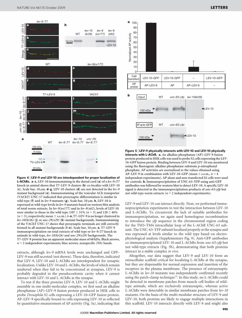
extracts, although lev-9 mRNA levels were unchanged and GFP–LEV-9 was still secreted (not shown). These data, therefore, indicatedthat LEV-9, LEV-10 and L-AChRs are interdependent for synapticlocalization. Unlike LEV-10 and L-AChRs, the levels of which remainunaltered when they fail to be concentrated at synapses, LEV-9 isprobably degraded in the pseudocoelomic cavity when it cannotinteract with LEV-10 and L-AChRs at the synapse.
To test if the three proteins LEV-9, LEV-10 and L-AChRs mightassemble in one multi-molecular complex, we first used an alkalinephosphatase (AP)–LEV-9 fusion protein produced in HEK cells toprobe Drosophila S2 cells expressing a LEV-10–GFP fusion protein.AP–LEV-9 specifically bound to cells expressing LEV-10 as reflectedby quantitative measurement of AP activity (Fig. 5a), indicating that
LEV-9 and LEV-10 can interact directly. Next, we performed immu-noprecipitation experiments to test the interaction between LEV-10and L-AChRs. To circumvent the lack of suitable antibodies forimmunoprecipitation, we again used homologous recombinationto introduce the yfp sequence in the chromosomal region codingfor the TM3–TM4 intracellular loop of the L-AChR UNC-63 sub-unit. The UNC-63–YFP subunit localized properly at the synapse andwas expressed at levels similar to the wild type based on electro-physiological analysis (Supplementary Fig. 9). Anti-GFP antibodiesco-immunoprecipitated LEV-10 and L-AChRs from unc-63::yfp butnot wild-type extracts (Fig. 5b), demonstrating that both proteinsinteract in a stable complex in vivo.
Altogether, our data suggest that LEV-9 and LEV-10 form anextracellular scaffold critical for localizing L-AChRs at the synapse,but they are dispensable for normal expression and insertion of thereceptors in the plasma membrane. The presence of extrasynapticL-AChRs in lev-10 mutants was independently confirmed recentlyusing the patch-clamp technique19. In this study, no L-AChRs couldbe detected in membrane patches from muscle cell bodies of wild-type animals, which are exclusively extrasynaptic, whereas activeL-AChRs were detectable in similar membrane patches from lev-10mutants. On the basis of the multi-modular structure of LEV-9 andLEV-10, both proteins are likely to engage multiple interactions inthis scaffold. LEV-10 interacts directly with LEV-9 and might also
Mer
geT7
–LE
V-9
LEV-
10VA
ChT
LEV-
10lev-9::T7
100 kDa
LEV-10
WTlev-10(kr26)
lev-9(ox177)
lev-9(x62)
WT lev-9(ox177)
T7–LEV-9 VAChT
a h
b
d
i
j
k
o
l
m
n
f
e
g
c
unc-
29;
lev-
9::T
7
unc-29;lev-9::T7
lev-
10;
lev-
9::T
7
lev-10;lev-9::T7lev-9::T7WT
lev-
9::T
7
100 kDa
75 kDa
50 kDa
TV–LEV-9NS
Figure 4 | LEV-9 and LEV-10 are interdependent for proper localization ofL-AChRs. a–c, LEV-10 immunostaining in the dorsal cord (a) of a lev-9::T7knock-in animal shows that T7–LEV-9 clusters (b) co-localize with LEV-10(c). Scale bar, 10 mm. d–g, LEV-10 clusters (d) are not detected in the lev-9mutant background (e). Immunostaining of the vesicular ACh transporter(VAChT) UNC-17 indicated that presynaptic differentiation is similar inwild type (f) and in lev-9 mutants (g). Scale bar, 10 mm. h, LEV-10 isexpressed at wild-type levels in lev-9 mutants based on western blot analysisof total worm extracts. In lev-9(ox177) and in lev-9(x62), levels of LEV-10were similar to those in the wild type (107 6 31% (n 5 3) and 120 6 46%(n 5 3), respectively; mean 6 s.e.m.). i–n, T7–LEV-9 is no longer clustered inlev-10(kr26) (j) or unc-29(x29) (k) mutant backgrounds. Immunostainingof the VAChT UNC-17 shows that presynaptic terminals are still correctlyformed in all mutant backgrounds (l–n). Scale bar, 10 mm. o, T7–LEV-9immunoprecipitation on total extracts of wild type or lev-9::T7 knock-inanimals in wild-type, lev-10(kr26) and unc-29(x29) backgrounds. TheT7–LEV-9 protein has an apparent molecular mass of 68 kDa. Black arrows,n 5 2 independent experiments; blue arrows, nonspecific (NS) bands.
0
10
20
30
40
50
60
70
80
90
100
Nor
mal
ized
AP
act
ivity
Celltransfect.
APprobe
LEV-10–GFP LEV-10–GFP
LEV-10
LEV-10
– LEV-10–GFP
–AP–LEV-9 AP AP–LEV-9
a
b Input WT
WT
unc-63::yfp
unc-63::yfp
lev-10(kr26)
IP:anti-GFP
100 kDa
100 kDa
Figure 5 | LEV-9 physically interacts with LEV-10 and LEV-10 physicallyinteracts with L-AChR. a, An alkaline phosphatase (AP)–LEV-9 fusionprotein produced in HEK cells was used to probe S2 cells expressing the LEV-10–GFP fusion protein. Binding between LEV-9 and LEV-10 was monitoredusing the fluorogenic alkaline phosphatase substrate p-nitrophenylphosphate. AP activities are normalized to the values obtained usingAP–LEV-9 in combination with LEV-10–GFP (mean 6 s.e.m., n 5 4independent experiments). AP alone and non-transfected S2 cells were usedfor controls. b, Immunoprecipitation of UNC-63–YFP using anti-GFPantibodies was followed by western blot to detect LEV-10. A specific LEV-10signal is detected in the immunoprecipitation products of unc-63::yfp butnot wild-type worm extracts (n 5 2 independent experiments).
NATURE | Vol 461 | 15 October 2009 LETTERS
995 Macmillan Publishers Limited. All rights reserved©2009

interact with L-AChRs as LEV-10 and L-AChRs can be immunopre-cipitated in vivo. Such a direct interaction could resemble the directassociation of CUB-domain-containing proteins similar to LEV-10with ionotropic receptors, such as the C. elegans SOL-1 with GLR-1(ref. 20) and the mouse NETO1 and NETO2 with NMDA (N-methyl-D-aspartate) and kainate receptors, respectively21,22.
The function of LEV-9 relies on a series of CCP domains, whichhave been mainly described in proteins of the complement system,either in regulators of complement activity or in components of theproteolytic cascade7,23. However, a growing number of CCP proteinsdo have a documented neuronal function. For example, Hikaru genki(Hig) is a secreted Drosophila protein containing four CCP domainswhich was detected in the synaptic cleft in the neuropile and pro-posed to modulate synapse physiology24,25. More recently, mutationsin the human gene SRPX2, which encodes a secreted protein withthree CCP domains and an immunoglobulin-related domain, weredemonstrated to cause rolandic seizures associated with oral andspeech dyspraxia and mental retardation26. Altogether, out of 52genes encoding predicted CCP-domain-containing proteins in themouse genome, 24 are expressed in the central nervous system basedon the Brain Allen database. Because the complement system doesnot exist in protostomes27, our results suggest that neuronal func-tions remain to be identified for these proteins in the mammalianbrain apart from regulation of the immune system.
METHODS SUMMARYMosTIC experiments. Generation of the KI strains was done as previously
described18,28. Germline expression of the Mos transposase in animals containing
the Mos1 transposon triggers its excision and causes a DNA double-strand break
at the excision site. The break can then be repaired by homologous recombina-
tion using a repair template containing sequence homologous to the broken
locus provided on an extrachromosomal transgenic array. PCR screening iden-
tifies animals that inserted the sequence present in the repair template into the
locus (Supplementary Fig. 6).
For generation of the lev-9::T7 KI strain, lev-9(ox177::Mos1) animals were
injected with a mixture of pJL44 (hsp-16.48::MosTase), pMG22 (repair template)
and pPD118.33 (Pmyo-2::gfp). After transgenic lines were established, transgenic
animals were heat-shocked and MosTIC events were screened by PCR as
described18,28 using primers 59-GCTAGCATGGCTTCTATGAC-39 and 59-TTT
CTGGGTATTTTGAGTGG-39 for the first PCR (1.5 mM MgCl2; annealing time,
30 s; annealing temperature, 62 uC) and primers 59-GAGGACAGCAGA
TGGGAGTC-39 and 59-TGTGGATATATTGCGGTTGC-39 for the nestedPCR (2.5 mM MgCl2; annealing time, 1 min; annealing temperature, 62 uC).
For the unc-63::yfp KI strain, transgenic unc-63(kr19::Mos1) animals containing
the unc-63 repair template and pJL44 were generated as described above. After
heat-shock, animals were allowed to recover overnight at 20 uC. Pools of 10 P0
worms were transferred to fresh plates every 12–24 h for 2 days to synchronize
their progeny. unc-63(kr19::Mos1) animals show mild uncoordination. To isolate
MosTIC events, we screened for individuals with wild-type locomotion and wild-
type sensitivity to levamisole. Individual candidates were cloned and wild-type
phenotypes were confirmed.
Full Methods and any associated references are available in the online version ofthe paper at www.nature.com/nature.
Received 5 July; accepted 14 August 2009.Published online 30 September 2009.
1. Elias, G. M. & Nicoll, R. A. Synaptic trafficking of glutamate receptors by MAGUKscaffolding proteins. Trends Cell Biol. 17, 343–352 (2007).
2. Kneussel, M. & Loebrich, S. Trafficking and synaptic anchoring of ionotropicinhibitory neurotransmitter receptors. Biol. Cell 99, 297–309 (2007).
3. Sanes, J. R. & Lichtman, J. W. Induction, assembly, maturation and maintenance ofa postsynaptic apparatus. Nature Rev. Neurosci. 2, 791–805 (2001).
4. O’Brien, R. J. et al. Synaptic clustering of AMPA receptors by the extracellularimmediate-early gene product Narp. Neuron 23, 309–323 (1999).
5. Bjartmar, L. et al. Neuronal pentraxins mediate synaptic refinement in thedeveloping visual system. J. Neurosci. 26, 6269–6281 (2006).
6. Gally, C., Eimer, S., Richmond, J. E. & Bessereau, J. L. A transmembrane proteinrequired for acetylcholine receptor clustering in Caenorhabditis elegans. Nature431, 578–582 (2004).
7. Kirkitadze, M. D. & Barlow, P. N. Structure and flexibility of the multiple domainproteins that regulate complement activation. Immunol. Rev. 180, 146–161 (2001).
8. Lewis, J. A., Wu, C. H., Berg, H. & Levine, J. H. The genetics of levamisoleresistance in the nematode Caenorhabditis elegans. Genetics 95, 905–928 (1980).
9. Fleming, J. T. et al. Caenorhabditis elegans levamisole resistance genes lev-1, unc-29, and unc-38 encode functional nicotinic acetylcholine receptor subunits.J. Neurosci. 17, 5843–5857 (1997).
10. Boulin, T. et al. Eight genes are required for functional reconstitution of theCaenorhabditis elegans levamisole-sensitive acetylcholine receptor. Proc. NatlAcad. Sci. USA 105, 18590–18595 (2008).
11. Touroutine, D. et al. acr-16 encodes an essential subunit of the levamisole-resistant nicotinic receptor at the Caenorhabditis elegans neuromuscular junction.J. Biol. Chem. 280, 27013–27021 (2005).
12. Francis, M. M. et al. The Ror receptor tyrosine kinase CAM-1 is required for ACR-16-mediated synaptic transmission at the C. elegans neuromuscular junction.Neuron 46, 581–594 (2005).
13. Bessereau, J. L. et al. Mobilization of a Drosophila transposon in the Caenorhabditiselegans germ line. Nature 413, 70–74 (2001).
14. Williams, D. C., Boulin, T., Ruaud, A. F., Jorgensen, E. M. & Bessereau, J. L.Characterization of Mos1-mediated mutagenesis in Caenorhabditis elegans: a methodfor the rapid identification of mutated genes. Genetics 169, 1779–1785 (2005).
15. Bingle, C. D. & Vyakarnam, A. Novel innate immune functions of the whey acidicprotein family. Trends Immunol. 29, 444–453 (2008).
16. Soares, D. C. et al. Large-scale modelling as a route to multiple surfacecomparisons of the CCP module family. Protein Eng. Des. Sel. 18, 379–388 (2005).
17. Fares, H. & Greenwald, I. Genetic analysis of endocytosis in Caenorhabditiselegans: coelomocyte uptake defective mutants. Genetics 159, 133–145 (2001).
18. Robert, V. & Bessereau, J. L. Targeted engineering of the Caenorhabditis elegansgenome following Mos1-triggeredchromosomal breaks. EMBO J. 26, 170–183 (2007).
19. Qian, H., Robertson, A. P., Powell-Coffman, J. A. & Martin, R. J. Levamisoleresistance resolved at the single-channel level in Caenorhabditis elegans. FASEB J.22, 3247–3254 (2008).
20. Zheng, Y., Mellem, J. E., Brockie, P. J., Madsen, D. M. & Maricq, A. V. SOL-1 is aCUB-domain protein required for GLR-1 glutamate receptor function in C. elegans.Nature 427, 451–457 (2004).
21. Ng, D. et al. Neto1 is a novel CUB-domain NMDA receptor-interacting proteinrequired for synaptic plasticity and learning. PLoS Biol. 7, e41 (2009).
22. Zhang, W. et al. A transmembrane accessory subunit that modulates kainate-type glutamate receptors. Neuron 61, 385–396 (2009).
23. Arlaud, G. J., Barlow, P. N., Gaboriaud, C., Gros, P. & Narayana, S. V. Decipheringcomplement mechanisms: the contributions of structural biology. Mol. Immunol.44, 3809–3822 (2007).
24. Hoshino, M., Suzuki, E., Nabeshima, Y. & Hama, C. Hikaru genki protein issecreted into synaptic clefts from an early stage of synapse formation inDrosophila. Development 122, 589–597 (1996).
25. Hoshino, M. et al. Neural expression of hikaru genki protein during embryonic andlarval development of Drosophila melanogaster. Dev. Genes Evol. 209, 1–9 (1999).
26. Roll, P. et al. SRPX2 mutations in disorders of language cortex and cognition. Hum.Mol. Genet. 15, 1195–1207 (2006).
27. Nonaka, M. & Yoshizaki, F. Primitive complement system of invertebrates.Immunol. Rev. 198, 203–215 (2004).
28. Robert, V. J., Katic, I. & Bessereau, J. L. Mos1 transposition as a tool to engineer theCaenorhabditis elegans genome by homologous recombination. Methodsdoi:10.1016/j.ymeth.2009.02.013 (in the press).
29. Boulin, T., Etchberger, J. F. & Hobert, O. Reporter gene fusions. WormBook doi/10.1895/wormbook.1.106.1, 1–23 (2006).
30. Liegeois, S., Benedetto, A., Garnier, J. M., Schwab, Y. & Labouesse, M. The V0-ATPase mediates apical secretion of exosomes containing Hedgehog-relatedproteins in Caenorhabditis elegans. J. Cell Biol. 173, 949–961 (2006).
Supplementary Information is linked to the online version of the paper atwww.nature.com/nature.
Acknowledgements We thank E. M. Jorgensen and D. Williams for thelev-9(ox171::Mos1) strain, M. Labouesse for the anti-VHA-5 antibodies, J. Rand forthe anti-UNC-17 antibodies, A. Fire for the GFP vectors, the Caenorhabditis GeneticCenter and W. R. Schafer for strains, I. Katic, M. Zhen and S. Marty for criticalreading of the manuscript, and H. Gendrot and B. Mathieu for technical help. M.G.was supported by a fellowship from the Ministere de la Recherche and by theAssociation Francaise contre les Myopathies. G.R. is a Ministere de la Recherchefellow. This work was funded by an INSERM Avenir grant, the Agence Nationale dela Recherche (ANR-07-NEURO-032-01) and the Association Francaise contre lesMyopathies. J.E.R. was supported by NIH RO1 MH073156.
Author Contributions M.G. performed most of the experiments. J.E.R. performedall the electrophysiology experiments (Fig. 3j, k and Supplementary Fig. 9d). G.R.generated and characterized the unc-63::YFP knock-in strain. M.G. and J.-L.B. wrotethe manuscript. J.-L.B. supervised the project.
Author Information The EMBL database accession number for lev-9 cDNA isFN433774. Reprints and permissions information is available at www.nature.com/reprints. Correspondence and requests for materials should be addressed to J.-L.B.([email protected]).
LETTERS NATURE | Vol 461 | 15 October 2009
996 Macmillan Publishers Limited. All rights reserved©2009

METHODSGeneral methods and strains. Strains were maintained at 20 uC on NG agar plates
under standard conditions31. OP50 Escherichia coli was used for feeding, except for
strains used for biochemistry, which were maintained on enriched peptone plates
with HB101 E. coli for feeding. The wild-type reference strain was N2 Bristol. The
following mutant alleles were used in this study: lev-9(ox177::Mos1), lev-9(x66),
lev-9(x62), lev-9(x16), lev-9(kr107::Mos1), lev-9(kr104), lev-9(kr108),
lev-9(kr184::T7), lev-10(kr26::Mos1), unc-29(x29), unc-38(x20), acr-16(ok789),
lin-15(n765ts), unc-49(e407), unc-49(e407);acr-16(ok789), unc-49(e407);acr-
16(ok789);lev-9(ox177), unc-29(x29);lev-9(kr184::T7), lev-10(kr26::Mos1);lev-
9(kr184::T7), lev-9(ox177::Mos1) lin-15(n765ts), unc-63(kr19::Mos1 sd), unc-
63(kr98::yfp).
The following transgenic lines were generated for this study: (1) in lev-9(x66):
krEx231 [Plev-9(big)::lev-9;Pmyo-3::rfp;Prab-3::gfp]; krEx232 and krEx233
[Plev-9(small)::lev-9;Pmyo-3::gfp;Prab-3::gfp]. (2) In lev-9(ox177): krEx245 and
krEx246 [Pmyo-3::lev-9(cDNA)-gfp;Pmyo-2::gfp]; krEx371, krEx372 and krEx373
[lev-9 MosTIC fragment with T7 insertion;Pmyo-2::gfp;Phsp::MosTransposase].
(3) In lev-9(ox177) lin-15(n765ts): krEx247 and krEx248 [Pmyo-3::lev-9(cDNA)-
gfp;lin-15(1)]; kr289 and kr290 [Pmyo-3::gfp-lev-9(cDNA);pEKL15 lin-15(1)];
krEx402, krEx403 and krEx404 [Plev-9(small)::lev-9-SL2-gfp, pEKL15 lin-15(1)].
(4) In unc-63(kr19): krEx227 [pGR06 (MosTIC template unc-63.rep.venus);Pmyo-
2::gfp;Phsp::MosTransposase].
Plasmid construction and PCR amplification. For Plev-9(big)::lev-9, a 6-kb
genomic fragment corresponding to C. elegans T07H6.5 was PCR-amplified
from N2 genomic DNA using Taq polymerase (Invitrogen) (primers 59- TCTG
CAAATCACCTGAACACA-39 and 59-GGGGAAACAGTTCTGAAATAGC-39).
For Plev-9(small)::lev-9, a 5.7-kb genomic fragment corresponding to C. ele-
gans T07H6.5 was PCR-amplified from N2 genomic DNA using Taq polymerase
(Invitrogen) (primers 59-TGAAAGTAAATGAAAAATCTTGCTG-39 and 59-GG
GGAAACAGTTCTGAAATAGC-39) .
For pMG11, Pmyo-3::lev-9(cDNA)-gfp, lev-9 cDNA was cloned by RT–PCR
using (1) primers 59-CGCACGTGACCGGTATGCGATTTCTACTACTACTC
G-39 and 59-TCTTCTCTGACTGGCACCAA-39 and (2) primers 59-CGCACGT
GTCGGCCCTATCTTGTCCGGA-39 and 59-CGTCTAGAAAGGTACCGAGCT
CACAGACCGAGACTCCATTGTC-39. Phusion polymerase (Promega) was
used. These PCR fragments were cloned in pPD115.62 using restriction sites
AgeI, SpeI and KpnI.
For pMG12, Pmyo-3::lev-9SP-NheI-SalI-lev-9, the lev-9 SP (signal peptide)
was PCR-amplified from pMG11 using 59-CCACTAGATCCATCTAGAGG-39
and 59-CGGTCGACGCTAGCTAGTAGGGCCGACGCGTAGGTAATGG-39
(fragment 1). lev-9 cDNA without SP was PCR-amplified from pMG11 using
59-CGGTCGACTCTTGTCCGGAAGTTACTCT-39 and 59-CGGAATTCGAGC
TCTCAACAGACCGAGACTCCATTGTC-39 (fragment 2). Fragment 1 was
digested by XbaI and SalI, and fragment 2 was digested by SalI and EcoRI.
These two digested fragments were ligated together into pPD115.62 digested
by XbaI and EcoRI. PCRs were done using Phusion polymerase (Promega).
For pMG13, Pmyo-3::lev-9SP-gfp-lev-9, GFP was PCR-amplified from
pPD115.62 using 59-GCGCTAGCAAAGGAGAAGAACTTTTCACTG-39 and
59-GCGTCGACTTTGTATAGTTCATCCATGCCA-39. The PCR fragment was
cloned in pMG12 using restriction sites NheI and SalI. PCR was done using
Phusion polymerase (Promega).
For pMG22, lev-9::T7 (MosTIC repair template for lev-9 KI), the two primers
59-CTAGCATGGCTTCTATGACCGGAGGACAGCAGATGGGAG-39 and 59-TC
GACTCCCATCTGCTGTCCTCCGGTCATAGAAGCCATG-39 were hybridized
in a 50 mM NaCl 5 mM MgCl2 buffer. The resulting fragment was subcloned in
pMG12 using restriction sites NheI and SalI to generate pMG20. A 3.6-kb genomic
fragment around the Mos1 insertion site of ox177 was PCR-amplified from N2
genomic DNA using Phusion polymerase (Promega) (primers 59-GCGAAGCT
TCACCAGCGAACGAAACTGACT-39 and 59-CGCGGTATACGCGTCTAACAA
AAGGGGACA-39). The PCR fragment was cloned in pATHScCRE using the
restriction sites HindIII and Bst1107I to generate pMG21. pMG22 was obtained
by subcloning a EagI–MluI fragment from pMG20 into pMG21. The sequence
coding for the T7 tag (MASMTGGQQMG) was inserted at a position correspond-
ing to residue 17 in LEV-9.
For pGR06, unc-63::venus (MosTIC repair template for unc-63 KI), the unc-63
39 UTR was PCR-amplified from N2 genomic DNA using Phusion polymerase
(Promega) (primers 59-ATCGATCCCCAACAACACAT-39 and 59-AGCG
GCCGCCGTAAGCTACCGGATTTCCA-39) and inserted in pAF6732 using the
restriction sites PstI and NotI to generate pGR01. Two unique restriction sites,
BglII and NheI, were inserted in the unc-63 gene coding sequence to generate
pGR02. The venus sequence optimized for expression in C. elegans was PCR-
amplified from a venus vector (gift from D. Y. M. Coudreuse)33 using Phusion
polymerase (Promega) (primers 59-ATAAGATCTAAAGGAGAAGAACTTTT
CACTGG-39 and 59- AGCTAGCTTTGTATAGTTCATCCATGCCAAG-39).
The venus sequence was inserted in-frame with unc-63 using the BglII–NheI
restriction sites in pGR02, at a position corresponding to residue 383 in the
TM3–TM4 loop (pGR04). The repair template pGR06 was generated by internal
deletion of pGR04 by Acc651 and Bsp120I digestion and self-religation.
For pMG23, Plev-9(small)::lev-9-SL2-gfp, a 5.7-kb genomic fragment corres-
ponding to C. elegans T07H6.5 was PCR-amplified from N2 genomic DNA using
Phusion polymerase (Promega) (primers 59-CGGCATGCGGGGAAACA
GTTCTGAAATAGC-39 and 59-CGGGTACCTCAACAGACCGAGACTCCAT
TG-39). This PCR fragment was cloned in pEXPR gcy-32-egl-2(gf) (gift from
M. de Bono) at the restriction sites SphI and KpnI.
For pMG26, PMT-lev-10-egfp, lev-10 cDNA was PCR-amplified using
Phusion polymerase (Promega) using 59-CGCGATATCATGCATTTGATC
TACCTATTC-39 and 59-CGCGCTCGAGAGCATACATATCACGCGATG-39
from pCG0346 (fragment 1). eGFP was PCR-amplified using Phusion polymer-
ase (Promega) from pIRES2-eGFP (59-GCGCTCGAGATGGTGAGCAAGGGC
GAGGAG-39 and 59-CGCGGGTACCTTACTTGTACAGCTCGTCC-39) (frag-
ment 2). Fragment 1 was digested by EcoRV and XhoI, and fragment 2 was
digested by XhoI and KpnI. The digested fragments were then ligated together
into pMT-GAL4 digested by EcoRV and KpnI (MT stands for metallothionein
promoter).
For pMG27, pCMV::T7-AP-lev-9(cDNA)-myc-his, lev-9 cDNA was PCR-
amplified using Phusion polymerase from pMG11 (59-GGCTCGAGCTATCTT
GTCCGGAAGTTAC-39 and 59-GCTCTAGAGAACAGACCGAGACTCCAT
TG-39) (fragment 1). Hybridization of the two primers 59-AGCTTTAATGG
CTTCTATGACCGGAGGACAGCAGATGGGAA-39 and 59-GATCTTCCCA
TCTGCTGTCCTCCGGTCATAGAAGCCATTAA-39 was done in a 50 mM
NaCl, 5 mM MgCl2 buffer (fragment 2). Fragment 1 was digested by XhoI and
XbaI and cloned into APtag-5 (ref. 34) to generate pMG25. Fragment 2 was
digested by HindIII and BglII and subcloned into pMG25.
Germline transformation. Transformation was performed by microinjection of
plasmid DNA into the gonad35.
For lev-9(x66) rescue, animals were injected with a DNA mixture containing
the large lev-9 genomic fragment (5 ng ml21), pAF75 (Pmyo-3::rfp; 10 ngml21),
pHU4 (Prab-3::gfp; 20 ng ml21) and 1 kb1 DNA ladder (Invitrogen; 65 ngml21),
or a small lev-9 genomic fragment (1 ngml21), pPD115.62 (Pmyo-3::gfp;
5 ngml21), pHU4 (Prab-3::gfp; 20 ngml21) and 1 kb1 DNA ladder (Invitrogen;
74 ngml21).
For the expression pattern, pMG23 (Plev-9(small)::lev-9-SL2-gfp) was injected
either in lev-9(ox177) at 10 ng ml21 with pAF75 (Pmyo-3::rfp; 10 ngml21) as co-
injection marker and 1 kb1 DNA ladder (Invitrogen; 80 ngml21) or in lev-
9(ox177) lin-15(n765ts) at 1 ngml21 with pEKL15 (lin-15(1); 99 ng ml21).
For tissue-specific rescue, pMG11 (Pmyo-3::lev-9(cDNA)-gfp) was injected
either in lev-9(ox177) at 10 ngml21 with pPD118.33 (Pmyo-2::gfp; 10 ng ml21)
as co-injection marker and 1 kb1 DNA ladder (Invitrogen; 80 ng ml21) or in lev-
9(ox177) lin-15(n765ts) at 1 ng ml21 with pEKL15 (lin-15(1); 80 ng ml21) as co-
injection marker. pMG13 (Pmyo-3::lev-9SP-gfp-lev-9(cDNA without SP)) was
injected in lev-9(ox177) lin-15(n765ts) at 10 ng ml21 with pEKL15 (lin-15(1);
90 ng ml21).
For MosTIC experiments, pMG22 (lev-9 repair template) was injected in lev-
9(ox177) either at 50 ng ml21 or at 25 ngml21 with pPD118.33 (Pmyo-2::GFP;
5 ngml21) as co-injection marker and pJL44 (hsp-16.48::transposase; 50 ngml21).
pGR06 (unc-63 repair template) was injected at 10 ngml21 with pPD118.33
(Pmyo-2::gfp; 5 ngml21) as co-injection marker and pJL44 (hsp-16.48::transpo-
sase; 50 ngml21).
Levamisole resistance assay. (2)-Tetramisole (Sigma) was dissolved in water
and added to 55 uC-equilibrated NG agar at a concentration of 0.2 mM, 0.4 mM,
0.6 mM, 0.8 mM or 1 mM just before plates were poured. For the 1 mM levami-
sole overnight exposure assay, young adult worms were placed overnight at 20 uCon 1 mM levamisole plates seeded with OP50 E. coli. Surviving animals were then
scored. For the levamisole dose response curve, young adult worms were scored
blind for paralysis after a 2 h exposure to different levamisole concentrations.
The plates were tapped ten times on the bench before scoring animals moving the
distance of at least one body length.
MosTIC experiments. Generation of the KI strains was done as previously
described18,28.
For generation of the lev-9::T7 KI strain, lev-9(ox177::Mos1) animals were
injected with a mixture of pJL44 (hsp-16.48::MosTase), pMG22 (repair template)
and pPD118.33 (Pmyo-2::gfp). After transgenic lines were established, transgenic
animals were heat-shocked and MosTIC events were screened by PCR as
described18,28 using primers 59-GCTAGCATGGCTTCTATGAC-39 and 59-TTT
CTGGGTATTTTGAGTGG-39 for the first PCR (1.5 mM MgCl2; annealing time,
30 s; annealing temperature, 62 uC) and primers 59-GAGGACAGCAGATG
doi:10.1038/nature08430
Macmillan Publishers Limited. All rights reserved©2009

GGAGTC-39 and 59-TGTGGATATATTGCGGTTGC-39 for the nested PCR
(2.5 mM MgCl2; annealing time, 1 min; annealing temperature, 62 uC).
For the unc-63::yfp KI strain, transgenic unc-63(kr19::Mos1) animals contain-
ing the unc-63 repair template and pJL44 were generated as described above.
After heat-shock, animals were allowed to recover overnight at 20 uC. Pools of 10
P0 worms were transferred to fresh plates every 12–24 h for 2 days to synchronize
their progeny. unc-63(kr19::Mos1) animals show mild uncoordination. To isol-
ate MosTIC events, we screened for individuals with wild-type locomotion and
wild-type sensitivity to levamisole. Individual candidates were cloned and wild-
type phenotypes were confirmed.
Antibody production. For anti-UNC-38 antibodies, a DNA fragment encoding
UNC-38 amino acids 375–418 was inserted into pGEX-3X. The glutathione
S-transferase (GST)–UNC-38 fusion protein was expressed in E. coli and puri-
fied. Rabbits were injected with 100mg of fusion protein and boosted three times
with 100mg each. Antibodies were then purified as previously described36 using
fusion protein GST–UNC-38 blotted on nitrocellulose.
For anti-ACR-16 antibodies, a DNA fragment encoding ACR-16 amino acids
348–459 was inserted into pATHSc. The histidine (His)–ACR-16 fusion protein
was expressed using BL21(DE3)pLysS bacteria and purified. Guinea-pigs were
injected with 50 mg of fusion protein and boosted three times with 50 mg each.
Antibodies were then purified as previously described36 using fusion protein
His–ACR-16 blotted on nitrocellulose.
Antibodies against UNC-49, UNC-29 and LEV-10 were produced as prev-
iously described6,37.
Immunocytochemical staining. Worms were prepared using the freeze-crack
method described previously38 using SuperFrost slides (Menzel-Glaser) and then
fixed either in 220 uC methanol for 5 min and in 220 uC acetone for 5 min or in
PFA 4% (paraformaldehyde) for 15 min when using anti-T7 antibodies. Worms
were then collected in PBS 13 and immunofluorescence staining was performedon fixed nematodes in suspension. Samples were blocked for 30 min at room
temperature with 0.2% gelatine from fish (Sigma). Anti-UNC-49 antibodies
were used at a 1:800 dilution. Anti-UNC-38 antibodies were used at a 1:800
dilution. Anti-ACR-16 antibodies were used at a 1:100 dilution. Anti-GFP
monoclonal antibodies (Roche) were used at a 1:500 dilution. Incubations were
done overnight at 4 uC.
For double-labelling experiments, procedures were optimized for the combina-
tion of proteins to detect. Anti-UNC-17 monoclonal antibodies39 were used at a1:3,000 dilution and incubated for 1 h at room temperature, and after a 1 h wash,
either anti-UNC-29 (1:800) or anti-LEV-10 antibodies were incubated overnight
at 4 uC. Anti-UNC-38 antibodies or rabbit anti-GFP (Molecular Probes; 1:500)
were incubated overnight at 4 uC, and after a 1 h wash, anti-UNC-17 antibodies
were incubated 1 h at room temperature. Anti-UNC-38 or anti-UNC-29 were
incubated overnight at 4 uC, then after 1 h of washing anti-GFP monoclonal
antibodies were incubated for 1 h at room temperature. Anti-ACR-16 and anti-
UNC-38 antibodies or anti-ACR-16 and anti-UNC-49 antibodies were incubated
together overnight at 4 uC. Anti-T7 monoclonal antibodies (Novagen) were used
at a 1:500 dilution and were incubated overnight at 4 uC. For double-labelling
experiments, mouse anti-T7 and anti-UNC-38 antibodies, mouse anti-T7 and
anti-LEV-10 antibodies, and rabbit anti-T7 (Genetex; 1:1,000) and anti-UNC-17
antibodies were incubated together overnight at 4 uC.
Secondary antibodies included Cy3-labelled goat anti-rabbit IgG (H1L)
(Jackson ImmunoResearch Laboratories) used at a 1:1,000 dilution, Cy3-labelled
goat anti-guinea-pig IgG (H1L) (Jackson ImmunoResearch Laboratories) 1:1,000,
Alexa-488-labelled goat anti-mouse (Molecular Probes) 1:500, Alexa-488-labelled
goat anti-rabbit IgG (H1L) (Molecular Probes) 1:2,000, and Cy3-labelled goat
anti-mouse IgG (H1L) (Jackson ImmunoResearch Laboratories) 1:1,000.
Electrophysiological studies. Electrophysiological methods were as previously
described40,41. Animals were immobilized with cyanoacrylic glue and a lateral
cuticle incision was made exposing the ventral medial body wall muscles. Muscle
whole-cell voltage-clamp recordings were obtained at a holding potential of
–60 mV using an EPC-10 patch-clamp amplifier and digitized at 1 kHz. The
extracellular solution consisted of (in mM): NaCl 150, KCl 5, CaCl2 5, MgCl24, glucose 10, sucrose 5, HEPES 15 (pH 7.3, ,340 mOsm). The patch pipette was
filled with (in mM): KCl 120, KOH 20, MgCl2 4, N-tris[hydroxymethyl] methyl-
2-aminoethane-sulphonic acid 5, CaCl2 0.25, Na2ATP 4, sucrose 36, EGTA 5
(pH 7.2, ,315 mOsm). Data were acquired using Pulse software (HEKA) run on
a Dell computer. Subsequent analysis and graphing were performed using
Pulsefit (HEKA), Mini analysis (Synaptosoft Inc.) and Igor Pro (Wavemetrics).
Protein extraction and western blotting. A mixed stage population of worms
(2 ml) was frozen at 280 uC until use. For extraction, worm pellets were ground
under liquid nitrogen and thawed in an equal volume of ice-cold homogenization
buffer (50 mM HEPES pH 7.7, 50 mM KCl, 2 mM MgCl2, 250 mM sucrose, 1 mM
EDTA pH 8, 2 mM PMSF and two tablets of complete Protease inhibitor cocktail
(Roche) in 50 ml). The suspension was further homogenized by sonication and
centrifuged at 5,800g for 12 min at 4 uC to remove worm debris. The low speed
supernatant (LSS) was either kept to detect the presence of LEV-10 protein or
centrifuged at 100,000g for 1 h at 4 uC to produce a high speed pellet (HSP). The
HSP was then dissolved in 100–500mL of resuspension buffer (10 mM Tris pH 8,
0.1% Triton X-100, 1 mM EDTA pH 8). UNC-29 protein was detected within the
HSP fraction. Western blot experiments were performed as described6.
Immunoprecipitation and western blotting. For GFP immunoprecipitation,
the LSS, obtained as described above, was brought up to 15 ml with homogen-
ization buffer supplemented with 100 mM NaCl and 1% Triton X-100, to which
50 ml of agarose-conjugated anti-GFP beads (MBL) was added. After overnight
incubation at 4 uC, beads were sedimented and washed once in 50 mM HEPES
pH 7.7, 100 mM NaCl, 50 mM KCl, 2 mM MgCl2, 250 mM sucrose, 1 mM EDTA,
1% Triton X-100, and once in 50 mM HEPES pH 7.7, 50 mM NaCl. Proteins
were eluted in Laemmli buffer and analysed by western blot as above. LEV-10 was
detected using anti-LEV-10 antibodies (dilution 1:250).
For T7 immunoprecipitation, a mixed stage population of worms (2 ml)
was cleaned using sucrose floatation and frozen at 280 uC before use. Worm
lysate was generated as above except the homogenization buffer contained
50 mM Tris pH 8 (instead of HEPES), 150 mM NaCl and 5 mM EDTA. The
lysate volume was brought up to 15 ml with homogenization buffer supple-
mented with 1% Triton X-100, 1% SDS and 1% deoxycholate and mixed for
6 h at 4 uC. Afterwards, the suspension was centrifuged at 5,800g for 12 min at
4 uC to remove worm debris. A total of 5ml of rabbit anti-T7 antibodies
(MBL) were incubated with the supernatant overnight at 4 uC. Protein G
sepharose beads (Sigma) were then added and mixed for 1 h at 4 uC. Beads
were sedimented and washed three times with the homogenization buffer
containing the detergents and three times with 50 mM HEPES pH 7.7,
50 mM NaCl. Proteins were eluted in Laemmli buffer and analysed by western
blot as above. T7–LEV-9 was detected using anti-T7 monoclonal antibodies
(Novagen; dilution 1:10,000).
Binding assays. S2 cells were maintained in Schneider’s Drosophila medium
(Invitrogen) with 10% FBS. pMG26 (PMT-lev-10-gfp) was transfected into S2
cells with Lipofectamine (Invitrogen). Twenty-four hours after transfection, the
metallothionein (MT) promoter was activated by adding CuSO4 at a 700mM
final concentration.
For AP–LEV-9 production, pAPtag-5 (pCMV::AP-myc-his) and pMG27
(pCMV::T7-AP-lev-9-myc-his) were transfected into HEK293T cells with
Lipofectamine (Invitrogen). Three days after transfection, supernatants of cul-
tured cells were collected and concentrated using iCON concentrator 9K (Pierce)
devices. The relative amount of AP fusion protein contained in the supernatant
was evaluated using phosphatase substrate (p-nitrophenyl phosphate; Sigma)
turning yellow in the presence of AP and that can be read spectrophotometrically
at 405 nm.
Quantitative assays were performed as described34. In brief, concentrated HEK
supernatants were applied to S2 cells for 90 min. The presence of AP on S2 cells was
detected by scraping cells and measuring AP activity using p-nitrophenyl phosphate.
Microscopy. Animals were anaesthetized with M9 buffer containing 3.8 mM tri-
caine, 0.42 mM tetramisole and 20 mM sodium azide, mounted on 13 M9, 2%
agarose pads, and examined using either a Leica SP2 confocal microscope or a Leica
5000B microscope and spinning disk CSU10 (Yokogawa). Immunofluorescence
staining was examined under the Leica 5000B microscope. Image reconstruction
and merges were obtained with Image J.
31. Brenner, S. The genetics of Caenorhabditis elegans. Genetics 77, 71–94 (1974).32. Ruaud, A. F. & Bessereau, J. L. Activation of nicotinic receptors uncouples a
developmental timer from the molting timer in C. elegans. Development 133,2211–2222 (2006).
33. Coudreuse, D. Y., Roel, G., Betist, M. C., Destree, O. & Korswagen, H. C. Wntgradient formation requires retromer function in Wnt-producing cells. Science312, 921–924 (2006).
34. Flanagan, J. G. et al. Alkaline phosphatase fusions of ligands or receptors as in situprobesforstainingofcells,tissues,andembryos.MethodsEnzymol.327,19–35(2000).
35. Mello, C. C., Kramer, J. M., Stinchcomb, D. & Ambros, V. Efficient gene transfer inC. elegans: extrachromosomal maintenance and integration of transformingsequences. EMBO J. 10, 3959–3970 (1991).
36. Miller, K. G., Emerson, M. D., McManus, J. R. & Rand, J. B. RIC-8 (Synembryn): anovel conserved protein that is required for Gqa signaling in the C. elegans nervoussystem. Neuron 27, 289–299 (2000).
37. Gally, C. & Bessereau, J. L. GABA is dispensable for the formation of junctional GABAreceptor clusters in Caenorhabditis elegans. J. Neurosci. 23, 2591–2599 (2003).
38. Duerr, J. S. et al. The cat-1 gene of Caenorhabditis elegans encodes a vesicularmonoamine transporter required for specific monoamine-dependent behaviors.J. Neurosci. 19, 72–84 (1999).
39. Duerr, J. S., Gaskin, J. & Rand, J. B. Identified neurons in C. elegans coexpressvesicular transporters for acetylcholine and monoamines. Am. J. Physiol. CellPhysiol. 280, C1616–C1622 (2001).
doi:10.1038/nature08430
Macmillan Publishers Limited. All rights reserved©2009

40. Richmond, J. E., Davis, W. S. & Jorgensen, E. M. UNC-13 is required for synapticvesicle fusion in C. elegans. Nature Neurosci. 2, 959–964 (1999).
41. Richmond, J. E. Electrophysiological recordings from the neuromuscular junctionof C. elegans. WormBook doi/10.1895/wormbook.1.112.1, 1–8 (2006).
doi:10.1038/nature08430
Macmillan Publishers Limited. All rights reserved©2009