a review of australian conescharellinidae (bryozoa ... · introduction the bryozoa sorted from...
TRANSCRIPT

Introduction
The Bryozoa sorted from dredge samples offshore from south-eastern and south-western Australia in the past 25 years haverevealed a wide diversity of species, with many apparentlyundescribed. The present study is of the family Cones-charellinidae. The principal collection programs were the BassStrait Survey by the Victorian Institute of Marine Science andthe National Museum of Victoria (now Museum Victoria) (stations with BSS prefix), Museum Victoria’s South-easternAustralian Slope Survey (SLOPE prefix), and the RV Franklin1995 shelf survey of the Great Australian Bight including areasto the west by Dr Y. Bone (University of Adelaide) (GAB pre-fix). Further collections were made by Gary C.B. Poore on anexpedition with the Western Australian Museum to theDampier Archipelago, north-western Australia in 1999 (DA-02prefix). All these surveys used epibenthic sleds and grabs tocollect sediments. Sampling of sandy sea-floor sediments, fol-lowed by careful sorting, yields examples from a wide range of
groups adapted to loose sediments (Hayward and Cook 1979,Bock and Cook, in press). In view of the unexpected diversityfrom the scattered survey stations, it is to be expected that yetmore species remain undiscovered.
In addition, an interesting series of partially sorted speci-mens, labelled in C.M. Maplestone’s hand, from the NMV col-lection, includes some boxes labelled “S.A.” (i.e. SouthAustralia) and others with no locality. These last are labelledwith the names of Maplestone’s species from New SouthWales, described by him in 1909 and include specimens ofspecies that have not been reported again. They do not occur inany other collections except as “types” in the AustralianMuseum, and as “cotypes” that are held in the Natural HistoryMuseum (London) (BMNH), that were originally sent toLondon by Maplestone and were registered in 1909. Amongothers, these include examples of five species ofConescharellinidae, labelled as Bipora biarmata, B. multiar-mata, B. magniarmata (all now referred to Conescharellina),Bipora (=Trochosodon) ampulla and Zeuglopora lanceolata.
Memoirs of Museum Victoria 61(2): 135–182 (2004)
ISSN 1447-2546 (Print) 1447-2554 (On-line)
http://www.museum.vic.gov.au/memoirs/index.asp
A review of Australian Conescharellinidae (Bryozoa: Cheilostomata)
PHILIP E. BOCK1, 2 AND PATRICIA L. COOK2
1 School of Ecology and Environment, Deakin University, Melbourne Campus, Burwood Highway, Burwood, Vic. 3125.([email protected])2 Honorary Associate, Marine Biology Section, Museum Victoria, GPO Box 666E, Melbourne, Vic. 3001, Australia
Abstract Bock, P.E. and Cook, P.L. 2004. A review of Australian Conescharellinidae (Bryozoa: Cheilostomata). Memoirs ofMuseum Victoria 61(2): 135–182.
The family Conescharellinidae Levinsen is defined and is regarded as comprising seven cheilostome genera(Conescharellina, Bipora, Trochosodon, Flabellopora, Zeuglopora, Crucescharellina and Ptoboroa). The astogeny ofcolonies, that consists of frontally budded zooids with “reversed” orientation, is briefly described and compared betweengenera. The morphology of zooids and heterozooids is defined and keys to genera and Australian species are provided.Taxa that were first described from Australia or from reliable subsequent records are redescribed and illustrated wherepossible. Australian specimens that have been identified as non-Australian species, have generally been found to be dis-tinct and are here redescribed as new species. Some non-Australian records of specimens previously assigned toAustralian species have also been re-examined. These are described and sometimes referred to other taxa. Altogether,eight previously described species that have not been found in the present material are discussed and 27 taxa aredescribed from collections, principally from the eastern and southern coasts of Australia and from the Tertiary of Victoria.Eighteen of these are considered to be new species. Where possible, type or at least topotype material of previouslydescribed species has been examined. Colonies from the collections of Museum Victoria (NMV) and the Natural HistoryMuseum, London (BMNH), have been examined. New species from Australia described here are: Conescharellina cog-nata, C. ecstasis, C. diffusa, C. obscura, C. stellata, C. plana, C. perculta, C. pustulosa, C. ocellata, C. macgillivrayi, C.humerus; Trochosodon fecundus, T. asymmetricus, T. diommatus, T. aster, T. anomalus, T. praecox and Crucescharellinaaustralis. In addition, the New Zealand bryozoan Trochosodon multiarmatus (Gordon, 1989) (not Bipora multiarmataMaplestone, 1909) is described as Trochosodon gordoni sp. nov.
Keywords Bryozoa, bryozoans, Cheilostomata, Conescharellinidae, fossil, Recent, Australia, new taxa

The Appendix includes full data on station locations andspecies occurrences.
A further collection from the Natural History Museum(London) was originally one of the sediment samples collectedby H.M.S. Challenger. These were stored in the MineralogyDepartment and remained uninvestigated until the 1970s. Onesample, from Challenger stn 185 (11°25'35"S, 144°2'0"E,249–286 m. near Raine Island, on the outer rim of the GreatBarrier Reef, Cape York, Queensland), was first examined in1972–73. This sample included foraminiferans and minute bryozoan colonies, some of which were figured by Cook andLagaaij (1976). Cook (1981) later emphasised and illustratedthe striking similarities in size and general appearance of thesetwo different components of the sample. Further examinationof the numerous bryozoan colonies has revealed that threespecies of Trochosodon and one of Crucescharellina are present. Busk (1884) did not include stn 185 in his Report as itsbryozoan component was undiscovered. Similarly, a specimenof Crucescharellina sp. from Challenger stn 169 (37°34'0"S,179°22'0"E, 1295 m, off New Zealand) also remained unre-ported although a preparation of the single colony is preservedin the BMNH collection.
Colonies of fossils Conescharellina from the Miocene ofVictoria are also included in this study (see Appendix).
Colonial development. The group of conescharellinids dis-cussed below construct small colonies that are anchored intothe soft-sediment substratum by one or several cuticular roots.The colony may develop and grow below the water-sedimentinterface or live slightly above the sea-floor. Colonies are conical or lenticular except in the genus Crucescharellinawhich branches into several horizontal arms.
Notes on astogeny of colonies. The astogeny of“conescharellinids”, like that of “batoporids” (Batoporidae),has been the subject of a considerable amount of theoreticaldiscussion that was reviewed by Waters (1919) and Harmer(1957: 722). Full explanation had to await the description ofconcepts of frontal budding (Banta, 1972) and reversed frontalbudding (Cook and Lagaaij, 1976). The type of astogeny gen-erally known as “reversed frontal budding” occurs in all generaof Conescharellinidae and Batoporidae but is not unique tothese families. A closely similar form of budding occurs in theorbicular, flattened colonies of Orbituliporidae. In addition therounded and lenticular colonies of the numerous species of thegenus Sphaeropora have a similar type of budding. This genus,however, is closely related to Celleporaria; both genera arereferable to the family Lepraliellidae.
Frontal budding was first described by Banta (1972) inencrusting colonies of Schizoporella; different sequences werealso illustrated by Cook (1985). Essentially, a frontal bud isformed by enlargement of an existing hypostegal coelom,bounded frontally by an intussusceptive expansion of frontalcuticle. The nutrients necessary to support the growth of thebud are derived from the pre-existing zooid or zooids, via thefrontal septular pores in the calcified frontal shield. Frontalbuds often have an orientation closely similar to that of the“parent” zooid but in some mammilliform growths where budsare derived from more than one “parent” zooid, the orientation
may be random, the orifices occurring with no reference to theposition or direction of the originating zooids.
These forms of frontal budding occur frequently inascophoran cheilostomes, particularly among “schizoporellid”and “celleporid” genera. However, different types of frontalbudding may occur among “anascan” and “cribrimorph” genera. For example, new branches in the erect “anascan”Rhabdozoum develop from an elongated frontal bud that arisesfrom extended calcification surrounding the opesia of a singlezooid (Cook and Bock, 1994). In the cribrimorph Anaskopora,interzooidal frontal buds arise from uncalcified “windows” inthe chambered pores surrounding each zooid and the resultantcolonies may resemble those of conescharellinids in organisa-tion (Arnold and Cook, 1997). In Corbulipora, buds arise fromthe uncalcified pelmatidia in the spines of the frontal shield(Bock and Cook, 2001). Encrusting colonies of Trematooeciaand Fatkullina exhibit a reversal of polarity of orifice withinzooids but new buds arise from vertical interior walls(Grischenko et al, 1998 (1999)). In the Conescharellinidae allzooid orifices are reversed with respect to the direction ofgrowth and all zooids are interzooidal frontal buds.
In “reversed frontal budding” the buds arise regularlybetween or among the series of frontal septular pores of two ormore neighbouring zooids. The orientation of the primary orifice is with the “distal” border directed towards the ancestrular or adapical region. In nearly all the colonies con-sidered here, most of the frontal shield of a zooid is over-grown and concealed by the next generation of zooids at thegrowing edge (see Cook and Lagaaij, 1976; Pizzaferri andBraga, 2000). The remaining frontal regions surrounding the orifices (exposed frontal shields) form the exterior sur-face of the colony except for the proliferal region. An analo-gous arrangement occurs in the leaf-like colonies ofFlabellopora and Zeuglopora where the zooids of either surface interdigitate, forming a superficially “bilaminate” erectcolony (see p. 175).
Mode of life. All living colonies of Conescharellinidae areknown or inferred to be anchored to a substratum by one ormore cuticular roots or extrazooidal rhizoid systems. Generally,the majority of roots, or the greater part of rhizoid systems, islocated at or near the adapical region of earliest astogeny. Thereis evidence from living specimens that metamorphosis of thelarva produces a binary complex consisting of a pair of ances-trular and root elements (Cook and Chimonides, 1985). Rootswere first described in living colonies of Conescharellina byWhitelegge (1887); they have also been illustrated by Silén(1947), Harmer (1957) and Cook (1979, 1981). The mode oflife of small, conescharelliniform and flabelloporiformcolonies, especially early in astogeny, appears to be interstitial,almost without exception. The minute colonies exist within theupper centimetres of the sediment surrounded by sand grainsand shell fragments. The colonies are anchored randomly tominute particles with no particular orientation with regard togravity. Colonies are robust and are preserved in the sedimentsamples after death. These samples include associations of sev-eral species, with each species showing colonies at differentgrowth stages. The function of the roots seems to be purely one
136 P. E. Bock and P. L. Cook

of anchorage in most genera, not of support, in contrast to theturgid, extrazooidal rhizoid systems of Sphaeropora andParmularia (Cook and Chimonides, 1981, 1985; Brown et al.,2002). In some species of Flabellopora, however, the morenumerous and larger roots may have a supportive function.Roots may extend up to 10 mm or more from the colony surface (Silén, 1947). They are usually thin and delicate, asillustrated by Cook (1981, pl. A fig. 1) in Trochosodon optatusHarmer, 1957. They arise from special pores that are formed inthe outer walls of frontally budded, interzooidal kenozooids.These are quite small and are in communication with the sur-rounding zooids and kenozooids through small septular pores,that were described by Levinsen (1909), Livingstone (1925)and Cook and Lagaaij (1976). In the Conescharellinidae, manyof the root pores that have been reported have a lunate shape,although others are circular. Both types have been reported tooccur in a single colony; it has been suggested that the circularpores may be an early ontogenetic stage of the lunate pores(Harmer, 1957). No colony has been observed here to developboth kinds of root pore. The lunate shape has given rise to a ter-minology that has included “lunooecia”, “semilunar pores” and“semilunar slits”. Root pores are frequent in the earlier stagesof astogeny, occurring amongst both the autozooid orifices andthe avicularian series. Lunate pores often possess a pair of lateral avicularia, whereas circular pores may be surrounded bya circlet of avicularia.
The association of a solitary coral, Dunocyathus parasiticusT. Woods, with colonies of Conescharellina was documentedby Maplestone (1910) in specimens from New South Wales andSouth Australia. He considered that the position of the coral,that usually occupies the entire antapical region of the bryozoancolony, was evidence of the orientation in life ofConescharellina, because “the delicate tentacles of the coralwould be crushed” if they rested on the substratum. Harmer(1957: 724, text-fig. 69) examined a specimen fromMaplestone in the collections of Cambridge Museum. He con-cluded that Maplestone’s theoretical orientation was probablycorrect, as the adapical region of the bryozoan colony was usually without feeding zooids but was the origin of roots. Ofcourse, as the actual, interstitial mode of life does not involve ahard substratum, and as the anchoring, not supportive, nature ofroots, together with the minute size of colonies, is unaffectedby gravity, these theories are of historical interest only. Itappears possible that the coral component of the association did not live interstitially. A total of 22 bryozoan-coral associ-ations has been found among the specimens examined here.Two of these involve Conescharellina multiarmata, seven C. magniarmata, ten C. cognata, and three C. species (Figs 1D,2F). Although the majority of coral specimens grow from theantapical surface of the bryozoan colony, three are asymmetri-cally developed and one occurs at the adapical end of a smallcolony. The adjustment of the growth of both organisms seemsto be mutually advantageous. There is no evidence of the bryozoan occluding the coral, although calcification has devel-oped laterally, that appears to originate from the bryozoan (Figs1D). The large avicularian mandibles of C. magniarmataprobably discouraged settlement on any other but the antapicalregion but C. multiarmata has only very small avicularia. One
significant correlation may be that all the colonies showing the association have a “high” conical shape and few or noantapical cancelli.
Abundance and diversity. The very strong correlation betweenthe occurrence of minute colonies and fine-grained sedimentswas noted by Harmer (1957) and was also emphasised by Cook(1981). The paucity of earlier records and of numbers of spec-imens from each sample is almost certainly an effect of collec-tion bias. Strikingly different observations have resulted wheresamples of the sediments themselves have been examined(Hayward and Cook, 1979; Cook, 1981). The Australian speci-mens described by Maplestone (1909), from a single dredgehaul in 146 m off New South Wales, also illustrate this differ-ence, as no fewer than 145 specimens were found, thatbelonged to eight nominal species, now known to be referableto four genera. A total of 79 specimens of Conescharellinidaewere reported by Harmer (1957) from 16 Siboga stations fromthe East Indies. These were described as belonging to 18 nom-inal species and five genera. Silén (1947) also listed 79 speci-mens, that he referred to nine species and three genera, fromeight stations that overlapped both the Siboga area and the“Philippines” region reported by Canu and Bassler (1929).Canu and Bassler included 25 stations with conescharellinids,identifying 32 nominal species belonging to four genera.Analysis of sediments from south-eastern Africa revealed 31specimens belonging to two genera from six stations (Haywardand Cook 1979). In contrast, Gordon (1985) listed only eightcolonies, belonging to two species, from five stations in theKermadec region. Unfortunately, other reports on collectionshave not always included consistently the total number of spec-imens of species from each locality. Gordon (1989) describedsix species from 41 stations from southern New Zealand;Gordon and d’Hondt (1997) also reported six species from 18New Caledonian stations but gave no estimate of abundance.Lu (1991) described 24 species referred to Conescharellinidaefrom the South China Sea and tabulated estimates of colonyabundance from each of 27 stations. As noted above, Harmer(1957) was the first to remark on the correlation of sedimenttype with the presence of minute, rooted colony forms. Apartfrom Gordon and d’Hondt (1997), all the above-mentionedauthors give some indication of sediment type at each collect-ing station. With hardly any exception, these are of sand, mud,or ooze, depending on the depths at which they occurred. Cone-scharelliniform colonies belonging to the Conescharellinidaeare often associated with slope (200 to 1000 m), or even abyssaldepths. Several records given by Harmer (1957), Gordon(1989) and Gordon and d’Hondt (1997) are from depths inexcess of 1000 m or even 4000 m.
Morphology of structures with characters used in specificdetermination
Colony shape and structure. The genera of Conescharellinidaeare characterised to a large extent by shape, that reflects thearrangement and proportion of autozooids, kenozooids andavicularia. The principal axis of most colonies extends from theancestrular or adapical region to the proliferal or antapicalregion. In Conescharellina, autozooids are arranged with their
Australian Conescharellinidae (Bryozoa) 137
orifices in apparent radial or in quincuncial series and alternatefrequently with series of avicularia. They often surround a coreof small kenozooids (cancelli). These are budded centrally fromthe frontal septular pores on the inner edge of the autozooidwalls and occupy a variable area on the antapical surface. Thesuccessive whorls of autozooids, in fact, always alternate radially in the antapical direction (quincuncial). The distancebetween whorls varies, so that the orifices may appear to formalmost continuous radial chains in colonies with a “high” con-ical shape, but are obviously quincuncially arranged in colonieswith a “low” cone. In Trochosodon, the conical autozooidarrangement is very similar but there is little or no central keno-zooidal core resulting in a more obvious quincuncial arrange-ment. In Ptoboroa, that does not occur from Australia, thecolonies are stellate with a prominent central root kenozooid. In Bipora, the radial axes occurring in Conescharellina aregreatly reduced in one dimension; the kenozooidal core is flat-tened producing an intervening layer of cancelli and a fan-shaped colony. In Flabellopora and Zeuglopora, the reductionof all but two of the radial axes is complete. The autozooids arebudded in alternating and interdigitating series with no inter-vening kenozooids. Colonies are elongated and leaf-like oroccasionally trilobate. In Crucescharellina, it is the adapical toantapical axis that is completely reduced and the radial axeselongated, discrete and often branched. This produces a cruci-form colony with only one series of zooid orifices on one faceand an antapical, “non-zooidal” series on the other (see alsoSilén 1947). The colonies of Crucescharellina and trilobateFlabellopora have the potential to grow far larger than those ofthe more conical genera such as Conescharellina, Trochosodonand Bipora. In Conescharellina, the shape of the cone appearsto be decided early in astogeny and is often apparently species-specific. For example, the cones of C. biarmata,C. multiarmata and C. diffusa are usually higher than wide,whereas those of C. eburnea and C. obscura are wider thanhigh. The angle of the frontal surface to the vertical axis alsoaffects the extent and nature of the kenozooidal core. Thisforms an interior cone, or cylinder, completely filling theantapical surface, or lines a shallow concavity (see C. cognata,Figs 3F, G). Most colonies of the conical genera have a maturegrowth stage antapically in that there is no further budding ofautozooids but in that the “cancellated” kenozooidal core isitself covered by a smooth extrazooidal lamina with small,intervening avicularia (C. eburnea, Fig. 1G; C. plana, Fig.10D). These are often derived from the frontal septular pores ofthe exposed shields of the most proliferal of the antapicalwhorls. Later development of cancelli may include alternatingseries of kenozooids and small avicularia.
For some species, examination of large samples has shownthat they may exhibit a wide range of colony shape and of avicularian size, although in other species variation appearsminor. Particularly in early astogenetic stages, orifices tend tobe quincuncial and the small colonies dome-shaped. In laterastogeny, the orifices may appear radially arranged and thecolonies conical (see C. ecstasis, Figs 5A, B). Ontogeneticchanges affect both the adapical and antapical regions, with thedevelopment of secondary calcification that obscures zooidalcharacteristics. In all colonies, zooid orifice and avicularian
dimensions increase with astogenetic age and there is no distinct zone of astogenetic repetition. Usually, root pores andother kenozooids remain almost constant in size, although theymay become surrounded by extrazooidal calcification or bygroups of secondary avicularia, forming specific patterns.Variation in colony shape and in the astogenetic timing of“mature” characteristics often reduce the value of past taxo-nomic descriptions, such as those of Canu and Bassler (1929).
The earliest astogenetic stages have not been observed inany genus but may be inferred from analogous structures inother “sand fauna” colonies and from study of minute stagesthat infrequently occur in samples. It is inferred that the ances-trula is anchored to a sand grain or similar object within theupper layers of sediment, as has been observed inConescharellina, Sphaeropora and Parmularia (Cook andChimonides, 1981, 1985). The position and orientation of thefirst zooidal buds relative to the ancestrula indicate the eventual mode of growth and structure of the subsequentcolony. For example, Harmer (1957) illustrated very youngcolonies of Trochosodon linearis and T. optatus and analysedtheir budding patterns. The ancestrula and paired primary budsformed a radially directed triad, followed by “cycles” (whorls)of alternating zooids, increasing in size and number.Kenozooids and small avicularia were budded on the adapicalsurface. Almost exactly the same series of astogenetic changesmay be traced in very young colonies of Conescharellina. Cook(1981: pl. A fig. 6) illustrated a young colony of Cruces-charellina (as Agalmatozoum sp.) showing a central adapicalarea of rhizoid pores (probably overlying the ancestrularregion), with four autozooids forming the earliest stages of acruciform colony. Gordon and d’Hondt (1997) illustrated aslightly older colony with five arms and a central, adapical areaof rhizoid pores and avicularia, very similar in appearance.
Primary orifice. The primary orifice is invariably sinuate, thesinus defined by a pair of condyles, that may be prominent orminute. The dimensions of all primary orifices increase withastogeny but the proportions appear to remain virtually thesame within species. Although the differences among speciesare minute and are usually only observable in scanning electronmicrographs, they are constant and correlated and thereforetaxonomically valid. The shape of the sinus varies from round-ed to subtriangular and is species-specific but it may varyslightly among populations.
Secondary orifice. Secondary orifices are variable, usuallybeing confined to raised lappets of lateral peristome. In appar-ently radial series, these produce an appearance described as“costulate” (Canu and Bassler, 1929). Sometimes peristomesare tubular and very prominent marginally (e.g. in Trochosodonand Ptoboroa) but may be elongated without being prominentat the colony surface (e.g. Conescharellina plana).
Ovicell. Ovicells are known in two of the genera describedhere, Conescharellina and Trochosodon. They are globular,hyperstomial and often extremely delicately calcified, with anexposed, frontal entooecium. Ovicells apparently originatefrom the small, adapical pore placed close to the border of thematernal zooid orifice. Gordon (1985) clearly illustrated the
138 P. E. Bock and P. L. Cook
early stages of ovicell ontogeny in Conescharellina, showingthe ectooecial and entooecial calcified layers developing fromthe adapical and antapical sides of this pore respectively.Colonies of C. diffusa and T. fecundus also show traces of bothlayers, associated with the adapical pore (Figs 6B, 17C).Harmer (1957) described ovicells as peristomial but, althoughthey are closely associated with the adapical edge of the peris-tome, they are not derived from it nor do they normally includeany part of it (see Trochosodon praecox). Ovicells have alsobeen illustrated by Maplestone (1910) and by Livingstone(1925b). Silén (1947) illustrated asymmetrically placed ovi-cells; these may be inferred to occur in T. asymmetricus, fromthe asymmetric position of the adapical pore, although ovicellshave not been found (Fig. 19A). Harmer (1957) also describedasymmetrically placed ovicells, and noted their fragility insome species. Lu (1991, pl. 17 fig. 5C) illustrated part of anovicell, in a species he called “Conescharellina radicata” fromthe South China Sea. No ovicells were mentioned in thedescription, that apparently refers to C. radiata Canu andBassler (1929: 493, pl. 67 figs 1–3). Although the majority ofrecorded ovicells is from the later astogenetic stages of growth,ovicells have been found very early in astogeny inConescharellina africana (see Cook 1966, 1981; Hayward andCook, 1979) and in Trochosodon praecox sp. nov. (asTrochosodon sp. in Cook and Lagaaij, 1976; Cook, 1981).Colonies of Conescharellina with embryos in their ovicellswere found within a rhizoid and sediment mass belonging toParmularia from off Townsville, Queensland, in 1982. Thesetoo, were very fragile, and often became detached whencolonies were moved. The mature colonies occurred togetherwith very young specimens that were anchored by means of aminute, turgid ancestrular rhizoid element. Embryos werereleased from the mature colonies, that apparently spent theirentire life interstitially (Cook and Chimonides, 1985). Theroots of adult colonies were not turgid or supportive butanchored the colonies with random orientations with regard togravity, within the rhizoid mass of Parmularia. Ovicells, some-times with embryos in situ, have been found in the present collection in colonies of Conescharellina plana (stn BSS-167),C. diffusa (Dampier, N.W. Australia), C. stellata (stn GAB-019), C. obscura (stn GAB-048), Trochosodon fecundus(Dampier, N.W. Australia), and T. praecox (Cape York,Queensland). Study of these examples strongly suggests that inmany cases the basal wall of the ovicell, that is formed byectooecium developing from the adapical side of the pore, iscovered by cuticle that is in contact with the frontal shield onlyat the point of origin. This explains the ease of detachment ofovicells in many specimens (see Figs 9H–I). The entooeciummay be ridged and occasionally is porous. The ridges appear toform pores marginally where the entooecial layer meets theectooecium. Ovicells are distinctive as there is no contributionto their structure by any zooid other than the maternal zooid.This is a result of the reversed nature of the frontal astogeny.
Avicularia. Avicularia vary considerably in size, distributionand orientation but may provide some distinguishing characterstates among species. The great majority has small, roundedrostra, that may be minute (Conescharellina multiarmata).
Some species have large avicularia; in Crucescharellina andZeuglopora these may be spathulate. Those near the orificesappear to be adventitious in most cases, derived from frontalseptular pores of zooids at the proliferal region, becomingslightly immersed as the next whorl of zooids is budded. Largeravicularia appear to be budded interzooidally (Fig. 3D).Avicularia on the antapical surface are often derived from can-celli and may alternate with them. Cancelli are kenozooidsoriginating from septular pores in the antapical part of thefrontal shield in the proliferal region. In some colonies ofConescharellina that have only just reached a mature asto-genetic stage, the avicularia follow the radial rows of frontalseptular pores of the last-budded, proliferal whorl, and may notbe accompanied by any cancelli (see Fig. 3F). Avicularia havea bar that often bears one or more calcareous spinous pro-cesses (ligulae) on the palatal side. In one species, C. diffusa,small spinous processes are present on the non-palatal side ofthe bar (Figs 6A, B); in another, C. stellata, the non-palatal areais sometimes occupied by a thin lamina that may be perforate(Figs 9B, E). Generally, the palates are without any expandedcryptocystal margin but this occurs in C. magniarmata (see Fig.3B). Large, acute avicularia also occur in C. ecstasis (Fig. 4)and were seen in the living specimens of Conescharellina sp.from Queensland mentioned above. These last were veryactive, snapping shut and holding any surrounding objects inthe sediment. Whether their function is one of stabilization or defence, or perhaps both, is unknown, as is that of smallavicularia.
Superfamily Conescharellinoidea Levinsen, 1909
D’Hondt (1985: 11) stated that the diagnosis was “confondueavec celle des Conescharellinidae publiée par Ryland (1982)”.He included only the family Conescharellinidae Levinsen,1909.
Family Conescharellinidae Levinsen, 1909
Type genus. Conescharellina d’Orbigny, 1852.
Description. Free-living Cheilostomata attached to small par-ticles by cuticular roots originating from kenozooidal pores. Allautozooids with reversed frontal budding; ancestrular regionadapical, with root pores and avicularia. Zooids elongated;frontal wall composed of two parts; “exposed”, surrounding theprimary orifice, and “concealed”, only visible completely in theantapical region of the colony. Primary orifice usually sinuate,often with paired condyles, almost terminal, in the centre of theexposed frontal wall. Avicularia adventitious or interzooidal,often in patterns among autozooids. Antapical regions oftenoccupied by kenozooids (cancelli), originally budded from theseptular pores of the frontal walls of zooids of the proliferalregion. Extrazooidal calcification and/or secondary kenozooidsand avicularia often budded from the primary cancelli. Otheravicularia budded directly from the proliferal zooids. Ovicellshyperstomial, originating from an adapical pore, globular, notclosed by the operculum, entooecium and ectooecium oftendelicate and fragile; usually distinct from the peristome butoccasionally associated with it through a foramen.
Australian Conescharellinidae (Bryozoa) 139

Remarks. The family includes closely related groups of specieswith distinctive colony forms that define genera. The typicalgrowth pattern of each genus restricts variation of the colonyform; minor differences in astogenetic pattern and zooid mor-phology may be important in distinguishing species. All knownspecies have in common: small size (usually less than 10 mmin maximum dimension), anchorage to sediment particles byroots arising from special kenozooids, and a strong associationwith fine-particle sediments, often from continental slope andlower slope depths. All species have sinuate primary orifices,often with condyles. Most species have interzooidal or adven-titious, frontally budded avicularia, that form patterns amongthe autozooid orifices. Special pores, derived from kenozooids,are the origin of roots. These may be generally distributed orconfined to the regions of earliest astogeny. The family resembles the Batoporidae and the Orbituliporidae in itsreversed frontal budding pattern but differs in the structure of the primary orifice and the few known ovicells. It alsoresembles the Lekythoporidae, another group including severalclosely related genera that have erect branching colonies with atype of reversed frontal budding (Bock and Cook, 2000). Theselast genera, however, have zooidal and ovicellular charactersthat show stronger links with the family Celleporidae. Gordon(1989) has suggested that the Conescharellinidae andOrbituliporidae should be included with the Lekythoporidae ina single superfamily This view was not accepted by Bock andCook (2000) who noted that Sphaeropora Haswell, 1881, thatalso has globular to lenticular colonies formed by reversedfrontal budding, anchored by supportive, turgid, extrazooidalrhizoids, is closely related to Celleporaria, and is thereforeassignable to the family Lepraliellidae. Reversed frontal budding itself may not therefore reflect any close systematicrelationships.
Six of the seven genera of Conescharellinidae are repre-sented in Australia, often by several species. However, the typespecies of Conescharellina, C. angustata d’Orbigny, 1852, wasdescribed from the Philippines, and that of Flabellopora,F. elegans d’Orbigny, 1852, from the China Sea. The typespecies of Trochosodon, T. linearis Canu and Bassler, 1927,occurred from Borneo, and the type species ofCrucescharellina, C. japonica Silén, 1947, from Japan. Two ofthe remaining genera, Bipora and Zeuglopora, have Australiantype species: Bipora flabellaris Levinsen, 1909, andZeuglopora lanceolata Maplestone, 1909 respectively. Thetype species of Ptoboroa, Trochosodon pulchrior Gordon,1989, occurs from New Zealand.
No attempt has been made here to review or revise thenumerous species of Conescharellina, Trochosodon andFlabellopora introduced and described by Canu and Bassler(1929) from the Philippine region. Similarly, the synonymies ofthese species, and of further new taxa introduced from the sameregion by Silén (1947), from the East Indies by Harmer (1957),and the South China Sea by Lu (1991), cannot be assessedwithout examination of all relevant material. It is possible thatsome of the species from eastern and south-eastern Australiadescribed by Tenison Woods (1880), Whitelegge (1887) andMaplestone (1909), may be synonymous with some, or part, ofthe nominal species described by later authors from the west
Pacific region. Similarly, it is possible that some taxa, intro-duced here as new, may have been described earlier by theseauthors, or even later by Gordon (1989), or Gordon andd’Hondt (1997) from the New Zealand and New Caledonianregions.
There is no unequivocal record of a member of theConescharellinidae, as defined here, from the EuropeanTertiary. Conescharellinopsis Labracherie, 1975, describedfrom the Lower Eocene of Aquitaine, has the type species C.vigneauxi Labracherie, 1975 (p. 151, pl. 4 figs 4–11). Thisspecies appears to be similar to species of Atactoporidra, asdescribed by Cook and Lagaaij (1976), with which it was associated and is not referred to the Conescharellinidae here.
Conescharellina perfecta Accordi (1947), from the UpperEocene of northern Italy, has been demonstrated to belong tothe genus Lacrimula (Batoporidae) by Cook and Lagaaij (1976)and more recently by Zágors̆ek and Kázmér (2001) who gave afull synonymy. Lacrimula perfecta also appears to be con-generic with another north Italian Eocene species,Conescharellina eocoena Neviani, 1895. Cook and Lagaaij(1976) suggested that it seems possible that all fossil records ofConescharellina from western Europe may “prove to belong toone species complex, attributable to Lacrimula.” TheConescharellinidae therefore seems to have an Indo-west-Pacific and Australasian distribution only, perhapsextending from the Eocene (Labracherie and Sigal, 1975), tothe present day.
Notes on the use of the name “Biporidae”. Zágors̆ek (2001:558) and Zágors̆ek and Kázmér (2001: 73) introduced a super-family “Biporidae Gregory, 1893” but no mention was made ofthe genus Bipora Whitelegge, 1887. The superfamily wasdescribed to include the family “Batoporoidea” (sic) Neviani,1901 and the genera Lacrimula Cook, 1966 and OrbituliporaStoliczka, 1862. Neviani (1901) had, however, included in hisfamily “Batoporideae” [sic] only the genera Batopora Reuss(for B. rosula) and Conescharellina d’Orbigny (for C. conica, amanuscript name, almost certainly referable to Lacrimula perfecta; see Cook and Lagaaij, 1976 for discussion).
Gregory (1893: 223) suggested a classification ofCheilostomata that included five Suborders. Two of theseincluded the “ascophorine” forms and consisted of theSuborders Schizothyriata and Holothyriata. Gregory’s treat-ment of families and subfamilies was not consistent but amongthe Schizothyriata the family Schizoporellidae and subfamilySchizoporellinae were provided (p. 239) with an informal des-ignation of Schizoporella as type genus, and a reference to itsdiagnosis by Hincks (1880). In a similar manner, the type genusSchizoretepora was designated in a footnote for the familySchizoreteporinae. The type genus Schismoporina was alsodesignated for the subfamily Schismoporineae in another foot-note. The treatment of the subfamily Biporineae was com-pletely different. No generic names were included but thedescription given was “Schizoporellidae with a patelliformunilaminate zoarium, with vibracularia systematicallyarranged”. This is a parallel of the description of a subfamily ofMicroporidae included in Gregory’s suborder Athyriata, calledthe Selenarinae, similarly described as “Microporidae with
140 P. E. Bock and P. L. Cook

patelliform zoaria and vibracularia systematically arranged”.The Biporineae may even have been introduced to provide aform of symmetrical concept between the Athyriata andSchizothyriata. Presumably, Gregory had in mind some lunu-litiform ascophoran genus or genera that would be included inhis Biporineae but he did not mention the subfamily again, ordescribe Bipora, or any other genus as belonging to it. In addition, although Whitelegge’s (1887) paper and its reprint(1888) were both listed in Gregory’s bibliography (on p. 274),no mention of either was made anywhere in his text. BothWhitelegge and Jelly (1889), whose Synonymic Catalogue wasalso listed by Gregory in his bibliography, gaveConescharellina in the synonymies of several species assignedto Bipora. In fact, Jelly (1889: 20) referred to Whitelegge’spaper under her entry for B. umbonata (Haswell), and again (onp. 64) under Conescharellina cancellata and C. elegans, whereBipora was given in synonymy.
Gregory must therefore have been aware that other, earlierauthors had described a relationship between the two genera.Any Conescharellinidae were, however, tacitly excluded fromthe subfamily Biporineae by Gregory (1893: 225, 251), as thegenus Conescharellina was listed as belonging to the familyCelleporidae, a member of his suborder Holothyriata. Gregoryregarded Conescharellina as a senior synonym of Batopora anddescribed one species from the British Eocene, Cones-charellina clithridiata, that is, in fact, referable to theBatoporidae. This species was illustrated as Batopora by Cookand Lagaaij (1976, pl. 2 fig. 1, pl. 5 fig. 5) and by Cook (1981,pl. B fig. 4). One other species, B. glandiformis, was erroneously referred to the cyclostome genus Heteropora byGregory (1893) but was briefly discussed and assigned toBatopora by Cheetham (1966) and subsequently was assignedto Atactoporidra by Cook and Lagaaij (1976). Waters (1904:96) made the illuminating remark, with reference to Gregory’s“undoubted abilities” that “sometimes angel visits stir up allthat has been done without establishing order” and “classifi-cation has been left in a much more hopeless condition than itwas before . . . made by those who have swooped down on theBryozoa for a short visit”.
It seems that Biporineae is not a synonym ofConescharellinoidea and there is no necessity to use anyemended suprafamilial name such as “Biporidae Gregory” toinclude the “Batoporoidea” as used by Zágors̆ek and Kázmér(2001), or the Conescharellinidae as used by Levinsen (1909).“Biporinae” Maplestone (1910) was an informal usage of aname and is a junior “synonym” of Levinsen’s (1909) nameConescharellinidae. As Conescharellinidae has been in common usage, the rule of priority can be ignored, as in ICZNRule 35.5.
Key to genera of Conescharellinidae
1. Colonies conical with circular cross section, or stellate 2— Colonies not as above . . . . . . . . . . . . . . . . . . . . . . . . . 42. Autozooids and avicularia frequently in antapically direct-
ed, alternating series. Autozooids not very prominent mar-ginally; kenozooids forming a central core or as antapicallayers . . . . . . . . . . . . . . . . . . . . . . . . . . Conescharellina
— Colonies stellate, without central core of kenozooids, auto-zooid orifices often arranged quincuncially, marginalzooids prominent; avicularia often absent . . . . . . . . . . 3
3. Colonies with elongated peripheral peristomes; antapicalavicularia and cancelli rare . . . . . . . . . . . . . Trochosodon
— Colonies with prominent central root kenozooid Ptoboroa4. Colonies compressed laterally in one plane . . . . . . . . . 5— Colonies compressed antapically in one plane, often
branching . . . . . . . . . . . . . . . . . . . . . . Crucescharellina5. Colonies with a laterally compressed cone, becoming fan-
shaped, zooids on each face separated by a narrow band ofkenozooids.. . . . . . . . . . . . . . . . . . . . . . . . . . . . . Bipora
— Colonies leaf-like, with no intervening kenozooidsbetween two interdigitating, frontally budded series ofzooids . . . . . . . . . . . . . . . . . . . . . . . . . . . . . . . . . . . . . 6
6. Lateral margins of colonies serrated, often with groups ofprominent zooids or enlarged avicularia . . . . .Zeuglopora
— Lateral margins not serrated, colonies sometimes trilobate . . . . . . . . . . . . . . . . . . . . . . . . . . . . . . . . . . Flabellopora
Key to Australian species of Conescharellina
1. Avicularian rostra acute, longer than orifice . . . . . . . . . 2— Avicularian rostra rounded, smaller than orifice . . . . . . 62. Colonies domed, height and/ or width > 3mm . . . . . . . 3— Colonies small, distinctly conical, higher than wide.
Avicularia paired, lateral oral, orientated laterally . . . . . . . . . . . . . . . . . . . . . . . . . . . . . . . . . . . . . . . . .C. biarmata
3. Avicularia lateral oral, single . . . . . . . . . . . . . . . . . . . . 4— Avicularia lateral oral paired, orientated laterally and
adapically . . . . . . . . . . . . . . . . . . . . . . . . . . . C. ecstasis4. Avicularia randomly orientated . . . . . . . . C. angulopora— Avicularia orientated laterally and adapically . . . . . . . . 55. Colonies domed, width and height subequal. Solid antapic-
ally. Avicularia with lateral cryptocyst lamina, and 3 largeligulae . . . . . . . . . . . . . . . . . . . . . . . . . C. magniarmata
— Colonies becoming very large, wider than high, diameter10 mm. Antapical surface hollow, covered by cancelli . . . . . . . . . . . . . . . . . . . . . . . . . . . . . . . . . . . . . . C. cognata
6. Colonies large, height and / or width > 4mm . . . . . . . . 7— Colonies small, height and / or width < 3mm . . . . . . . 107. Root pores circular, surrounded by avicularia . . . . . . . 8— Root pores lunate, with paired avicularia . . . . . . . . . . . 98. Colonies with patent orifices; avicularia lateral and ada-
pical, visible on antapical surface of marginal zooids . . . . . . . . . . . . . . . . . . . . . . . . . . . . . . . . . . . . . . . .C. eburnea
— Orifices at the base of a long peristome, that is not raisedat the colony surface; avicularia minute, scattered andpaired, adapical . . . . . . . . . . . . . . . . . . . . . . . . C. plana
9. Colonies conical, higher than wide, height up to 5 mm;avicularia and root pores in series alternating with orifices;root pores without small avicularia; rostra with non-palatalspinules . . . . . . . . . . . . . . . . . . . . . . . . . . . . . . C. diffusa
— Colonies flat, up to 13 mm diameter, or small and discoidal; avicularia scattered or single, antapical and peristomial; root pores with paired avicularia . . . . . . . . . . . . . . . . . . . . . . . . . . . . . . . . . . . . . . . . . . . . . .C. obscura
10. Colonies with lunate root pores with paired avicularia 11
Australian Conescharellinidae (Bryozoa) 141

— Colonies with circular root pores surrounded by avicularia . . . . . . . . . . . . . . . . . . . . . . . . . . . . . . . . . . . . . . . . . . 12
11. Colonies domed, orifices in radial series alternating withavicularia,root pores adapical (fossil) . . . . . C. aff. diffusa
— Colonies conical, higher than wide, orifices with minutelateral and antapical avicularia . . . . . . . . C. multiarmata
— Colonies stellate, marginal peristomes bilabiate or spout-like; avicularia with non-palatal lamina . . . . . . C. stellata
— Colonies with pustular calcification adapically and anta-pically; avicularia minute, one peristomial and antapical,others scattered . . . . . . . . . . . . . . . . . . . . . . C. pustulosa
12. Colonies with prominent spout-like marginal peristomesand numerous pairs of avicularia, some visible on theantapical surface marginally . . . . . . . . . . . . . C. perculta
— Colonies not as above . . . . . . . . . . . . . . . . . . . . . . . . 1313. Colonies slightly domed , or raised centrally; orifices with
a long subtriangular sinus, peristomes raised laterally;avicularia paired lateral-oral, visible on the antapical sur-face of marginal zooids; cancelli absent . . . . C. ocellata
— Colonies fairly flat, with bilabiate marginal peristomes;orifice with a small rounded sinus; avicularia rare, anta-pical surface with large cancelli . . . . . . C. macgillivrayi
— Colonies slightly raised centrally; orifices with a roundedsinus and laterally raised peristome, with paired lateralavicularia that form prominent “shoulders” on marginalzooids . . . . . . . . . . . . . . . . . . . . . . . . . . . . . C. humerus
Key to Australian species of Trochosodon
1. Colonies large, diameter 3–4.7 mm . . . . . . . . . . . . . . . 2— Colonies smaller . . . . . . . . . . . . . . . . . . . . . . . . . . . . . 32. Colonies fairly flat, domed centrally, with numerous
tubular marginal peristomes . . . . . . . . . . . . . T. ampulla— Colonies lenticular, with prominent radial rows of peris-
tomes with paired avicularia on the antapical surface; rootpores lunate . . . . . . . . . . . . . . . . . . . . . . . T. diommatus
3. Colony diameter 2–3 mm . . . . . . . . . . . . . . . . . . . . . . 4— Colony diameter <2 mm . . . . . . . . . . . . . . . . . . . . . . . 54. Colonies with bilabiate marginal peristomes, orifices quin-
cuncial with wide, shallow sinus; ovicells symmetrical,root pores lunate . . . . . . . . . . . . . . . . . . . . . T. fecundus
— Colonies with short, tubular peristomes, orifices radial,sinus rounded; adapical pores asymmetric, root pores circular . . . . . . . . . . . . . . . . . . . . . . . . . T. asymmetricus
5. Colonies conical, higher than wide; zooid peristomesprominent and curved; root pores rare, lunate T. anomalus
— Colonies as wide as high, or wider . . . . . . . . . . . . . . . . 66. Colonies very small, fairly flat, stellate; peristomes tubular,
with paired lateral avicularia; root pores lunate . T. aster— Colonies minute, with an antapical dome of mamillate
calcification; peristomes tubular, with paired lateral avic-ularia; ovicells small, robust, symmetrical; root poresrounded . . . . . . . . . . . . . . . . . . . . . . . . . . . . . T. praecox
Conescharellina d’Orbigny 1852
Conescarellina [sic] d’Orbigny 1852: 447.Conescharellina.—Canu and Bassler, 1917.—Waters, 1919: 93.—
Canu and Bassler, 1929: 480.—Silén, 1947: 33.—Harmer, 1957:726.—Gordon, 1989: 81.
Type species. Conescharellina angustata d’Orbigny, 1852, sub-sequent designation by Waters, 1919: 93. [Canu and Bassler(1917) had earlier incorrectly indicated C. cancellata (Busk,1884), see Harmer (1957: 726)]. The mis-spelling of the nameas Conescarellina occurs only in the genus heading ofd’Orbigny (1852: 447): all other spellings of the name are asConescharellina. Conescharellina angustata was included inBatopora by Reuss (1867: 224).
Description. Colony conical, with autozooids appearing to bein radial series, either placed in rows alternating with avicular-ia, or in quincunx with intervening avicularia. Cuticular rootsarise from circular or crescentic skeletal pores, concentrated inthe adapical region in some species. Orifices with an antapicalsinus, often with raised lateral peristomes. Avicularia adventi-tious and interzooidal, usually budded in distinct patterns, withacute or rounded mandibles, slung on a bar, that often has oneor more palatal ligulae. Ovicells hyperstomial, prominent,derived from an adapical pore, with thinly calcified ectooeciumand entooecium. Central part of colony cone occupied by a core of small kenozooids (cancelli), often accompanied byavicularia, that may cover the antapical surface late in astogeny.
Remarks. C. angustata was described by d’Orbigny (1852: 447,pl. 714 figs 14–16) from the Philippine island of Basilan(approx. 6°50'N, 122°E, in the Celebes Sea). The figuredcolony (fig. 15) was an elongated cone with 8–9 apparentlyradial series of zooids forming costules. The orifices are raised,circular-to-oval, each with an asymmetrically arranged pair ofpores adapically, and a single series of “special” pores alternat-ing with the zooid orifices in a radial depression. D’Orbignynoted that the orifices were in quincunx, and figured the anta-pical surface (fig. 16) showing five alternating series of prolif-eral and subproliferal zooids, with no central cancelli.D’Orbigny noted this particularly, comparing it with the antapi-cal side of C. dilatata (see below). In view of the relativelylarge size and possible maturity of the type colony (heightapproximately 2.5 mm), it is unusual in Conescharellina forcancelli to be absent. In fact, this is characteristic ofTrochosodon.
A scanning electron micrograph of the putative type speci-men, from the Muséum Nationale d’Histoire Naturelle, Paris,has been provided by Drs D.P. Gordon and P.D. Taylor. Itresembles d’Orbigny’s figure 15 in its elongated conical shapeand radial series of zooid orifices. The adapical region is lessregular than the figure, and there are fewer zooid series but thismay be the result of damage.The rounded secondary orifices,almost all of which have an adapical pore, are similar to thosefigured but the additional pores shown near the orifices are notpresent. D’Orbigny figured a radial series of pores in thedepression between zooid series, that were lateral to the ada-pical edge of the adjacent orifices. Avicularia occupy a similarposition in the micrograph of the specimen but are far largerand twice as frequent. These avicularia are small and roundedwith a delicate, simple bar. The orifices of fig. 15 are second-ary and show a slightly raised peristomial rim; a few of those inthe micrograph also show a sunken primary orifice with arounded sinus. The specimen of C. angustata resembles some
142 P. E. Bock and P. L. Cook

of the more elongated colonies of C. diffusa. These differ intheir proportionally larger avicularia and the presence ofnumerous lunate root pores, that are absent from C. angustata.Waters (1905: 9, pl. 1 fig. 7) examined the type material of C. angustata and gave a figure of the specimen from Basilan.This does not show the entire colony but only a formalised rep-resentation of four oval orifices and a single antapical avicular-ium. Later, Waters (1919: 93) indicated C. angustata as typespecies of Conescharellina, without comment. He also (1921:419, pl. 30 fig. 18) figured but did not describe a speci-men “from China, sent to me thus named by Jullien” as C. angustata. This colony was also conical and very elongated,with raised “costules” of radial rows of oval secondary orificesseparated by adapically placed pores. One elongated sinuate,perhaps primary, orifice was figured, and small scattered poresamong the orifices may have represented avicularia. Unlike thetype specimen, the adapical region was occupied by extra-zooidal or kenozooidal calcification. The figure is otherwisesimilar to that of d’Orbigny’s C. angustata, with “costules” ofsecondary orifices that are more elongated and with “pores”less regularly spaced. Harmer (1957) was doubtful that thethree Siboga collection specimens from East Java, that he nevertheless assigned to C. angustata, were identical withd’Orbigny’s species. These colonies were not elongated; theprimary orifices were patent, with little or no peristome, andwere relatively wide with a rounded sinus. These specimens donot appear to be conspecific with the type specimen.D’Orbigny (1852: 447) also introduced but did not figureConescharellina dilatata from “Manille et détroit de Malacca”[sic]. It differed from C. angustata in its greater width (“ensem-ble plus large”) and in the presence of “un espace poreux” (pre-sumably of cancelli) on the antapical surface. Waters (1905: 9,pl. 1 fig. 6) gave a semidiagrammatical figure of two zooid ori-fices from a specimen of C. dilatata from d’Orbigny’s collec-tion from Manila. There were “two species in the tube” but hedid not indicate which of these he regarded as C. dilatata. Asbefore, only examination of the type material can elucidatefully the characters and relationships of this species. However,it is obvious that d’Orbigny’s C. angustata is closely similar to,and congeneric with, many of the other taxa subsequentlyreferred to Conescharellina but description of its specific characters must await examination of the type specimen.
Species recorded from Australia but not recognised in thematerial examined here
Conescharellina philippinensis (Busk, 18540 and C. can-cellata (Busk, 1854)
Lunulites philippinensis Busk, 1854 and L. cancellata Busk,1854 were described and figured by Busk (1854: 101, pl. 113figs 1–3 and 4–7 respectively) from the Philippines. They areobviously species of Conescharellina but the charactersdescribed and figured are not sufficiently clear to allow theirrecognition and identification with other material with any cer-tainty. It has been possible to examine specimens from the“type suites” of L. philippinensis and L. cancellata but it mustbe emphasised that until all Busk’s specimen suites have been
revised, little may be concluded as to the nature and the identity of specimens later reported under these names.According to Waters (1921: 419), Busk’s specimens in theBritish Museum collection confused both species and includedat least two additional species. Harmer (1957: 742) did not,however, agree with all Waters’ conclusions. The “type” slideof L. philippinensis (BMNH 1854.11.15. 150) originallyincluded five colonies. Two of these have been lost in the past;one was remounted as an additional slide and labelled inKirkpatrick’s hand. This very worn, separated colony may bethe original of Busk’s figure (1854: pl. 113 fig. 2). The otherspecimens do not appear to have been figured, although allthree seem to be conspecific. The specimens are all worn andshow little detail. Two are flat and are less than 2 mm in diameter. They include approximately five quincuncial genera-tions of zooids and each whorl has nine to ten zooid orifices.The marginal peristomes are slightly prominent and tubular; theprimary orifices cannot be seen. Small rounded pores, inferredto have been avicularia, are interspersed randomly among thezooid orifices and the antapical surface has a central cancellatearea. In both the larger colonies, the centre of the adapical sur-face has two prominent rounded “bosses”, that are illustrated inBusk’s pl. 113, fig. 2. It is not possible to recognise this species,either among those described from the Philippines by Canu andBassler (1929) or in the Australian material examined here. The“type” slide of L. cancellata (BMNH 1854.11.15.151) includesfour specimens that are in a better state of preservation. Theoriginals of Busk’s pl. 113, figs 4–7 are recognisable; an addi-tional large, worn, unfigured colony, that does not seem to beconspecific, is present (Brown, 1958: 82). The figured coloniesare distinctly domed; the largest, that is less than 2 mm in diameter, includes approximately seven quincuncial zooid gen-erations and nine to ten zooids per whorl. The peristomes areonly slightly raised and circular; the primary orifices are visibleand are rounded with a short, wide, almost semicircular sinus.Traces of an adapical pore are present in a few zooids. Smalloval avicularia, with a delicate, simple bar, occur some-what irregularly among the zooid orifices. No root pores arevisible; the antapical surface has a central cancellate area. Thisspecies was apparently not among the other Philippine formsdescribed by Canu and Bassler (1929) and has certainly not been recognised among the Australian specimens examined here.
Waters’ (1921) account of L. cancellata is not at all clear. Heremarked “specimens from Busk’s own collection so named areC. angustata d’Orb.” Harmer (1957: 742), when discussing C. crassa, seems to have mistaken Waters’ (1921) reference toC. angustata, as describing part of the type material of L. cancellata. A specimen in Busk’s collection from the Sea ofJapan (BMNH 1899.7.1.1276 labelled Lunularia cancellata) isnarrowly conical, with seven to eight radial series of orificesand five to six zooid whorls. Zig-zag series of small oval avic-ularia alternate with the orifice series; these also occur on theantapical surface. The colony somewhat resembles d’Orbigny’sC. angustata and may be the one mentioned by Waters. Waters(1921) also stated that the specimens he described “from NewSouth Wales” (i.e. in 1887) “then called cancellata by me areseen to be philippinensis.” Both names have been used for
Australian Conescharellinidae (Bryozoa) 143

several Australian records; references to Recent materialassigned to these species are discussed below under C. diffusaand C. obscura.
It is unfortunate that little of the previously describedTertiary material is extant. Various combinations and spellingsof C. cancellata having been quoted, particularly for specimensfrom the Tertiary, by Waters (1881; 1882a; 1882b) and byMacGillivray (1895). Maplestone (1904) tabulated severaladditional fossil localities, including Campbells Point, MitchellRiver and Lake Gnotuk, together with his own observations ofmaterial from Mornington. Unfortunately, Maplestone’s specimens are not extant, and therefore his concept of fossil B. cancellata and B. philippinensis must remain unknown. Healso listed B. elegans Waters (=Bipora flabellaris), fromJimmys Point, that has otherwise not been reported as a fossil,and therefore seems unlikely to be this species. MacGillivray(1895: 89, pl. 12 fig.2) reported “Bipora philippinensis” fromthe Tertiary of Schnapper Point and Muddy Creek, Victoria. Hisspecimen from Muddy Creek is extant (NMV P27728). It is afairly flat colony, with quincuncial zooid orifices with a smallsinus and scattered avicularia. The antapical surface has a largecancellated area. This specimen appears to be referable to thefossil species described here as Conescharellina macgillivrayisp. nov. Waters (1881) mentioned Recent specimens of B. cancellata from Torres Strait but no fossil examples.However, he appears to have believed that he had specimensfrom the “Curdies Creek” locality, as he mentioned them(Waters, 1882a) in connection with the “better preserved”material he had from Bairnsdale, Victoria (Waters, 1882b: 512,pl. 22 figs 10, 11, as Lunulites cancellatus), that he figuredshowing the orifice and surrounding avicularia. These illustra-tions suggest that the species may also have beenConescharellina macgillivrayi. Whitelegge (1887: 341) listedC. cancellata, remarking that he had several fossil examplesfrom Muddy Creek, Victoria, that might be identical with thespecies recorded by Waters (1882b) but that in C. cancellataand C. philippinensis “the identity can only be definitely settledby comparison with the types”. Bipora cancellata was recorded by MacGillivray (1895: 89, pl. 12 fig. 1) fromBairnsdale; he noted that it was often difficult to distinguish itfrom B. philippinensis. His specimen (NMV P22727) is a conical colony with orifices arranged in radial series. The pri-mary orifice has a fairly wide, rounded sinus and is flankedantapically by a pair of small, rounded avicularia. The anta-pical surface has very few cancelli. His specimen resemblesothers from Bairnsdale, and is discussed here underMaplestone’s Recent colonies of C. diffusa. Colonies from theMiocene of Victoria and South Australia are numerous anddiverse; four species, C. ocellata, C. macgillivrayi , C. humerusand C. aff. diffusa are described below.
The ovicells of C. cancellata were mentioned, in passing, byLevinsen (1909: 310, pl. 23 figs 8a, b), who illustrated small,globular ovicells with marginal pores and an oval zooid orificewith an adapical pore. Three small rounded avicularia sur-rounded the ovicelled zooid orifice. Levinsen did not give anydetails of the provenance of the specimens illustrated and theinformation given is insufficient for identification of thespecies.
Conescharellina angulopora (Tenison Woods, 1880)
Lunulites angulopora Tenison Woods, 1880: 7, pl. 1 figs 3a–c.?Lunulites conica Haswell, 1881: 42, pl. 3 figs 7, 8.?Conescharellina incisa Hincks, 1881: 127 (sep. p. 68), pl. 4 figs
1–3.?Bipora angulopora.—Whitelegge, 1887 (1888): 18.not ?Lunulites angulopora.—MacGillivray, 1895: 46, pl. 8 fig. 1
(= Selenariopsis macgillivrayi Bock and Cook, 1996).?Conescharellina angulopora.—Levinsen, 1909: 311, pl. 23 figs
7a–f.not Conescharellina angulopora.—Gordon, 1985: 173, figs 20–23;
Gordon, 1989: 81, pl. 48B (see C. cognata).
Remarks. Search for type material of Tenison Woods has beenunsuccessful; consequently the characters of this speciesremain somewhat doubtful. The colony was figured as a distinct cone and the autozooids and avicularia occurred inapparent alternating radial series. However, the description ofthe orifice as “divided into two portions; one half triangular constricted in the middle; the other semicircular”, taken together with the illustration, indicates that Tenison Woods hadconfused the avicularia with the secondary orifices. The illus-tration shows at least one triangular avicularium accompaniedby a typical lunate root pore that he did not recognise as distinctstructures. His later remark “the cells are obliquely placed;sometimes in contrary directions alternately”, also appears torefer to avicularia, that have been described in other materialassigned to this species as having alternating orientations. Thedescription of “the vibracular pores” as “long and narrow, andin a depressed area” and the illustration, showing irregularlyovoid openings, apparently refers, in fact, to the secondaryautozooid orifices. Waters (1887: 199), describing specimenshe assigned to C. incisa (Hincks), remarked “This may beLunulites angulopora T. Woods, but apparently the aviculariawere mistaken for zooecial cells, and the zooecia for vibracula”. Tenison Woods had only two specimens from Port Stephens, New South Wales, that he noted were “worn”;his type material has not been found. It seems unlikely that hisspecies is recognisable. Livingstone (1924) regarded C. conicaHaswell (1881), Lunulites incisa Hincks (1881, 1892), Biporabiarmata, and B. magniarmata Maplestone (1909), all as junior synonyms of L. angulopora Tenison Woods (1880). BothLivingstone (1928) and Hageman et al. (1996) reported C. angulopora from South Australia, and specimens labelledBipora angulopora occur in Maplestone’s collection from thisarea. These specimens belong to at least two other taxa (see C. cognata and C. diffusa) but specimens in Maplestone’s collection (NMV), inferred to have been from New SouthWales, are described below as C. species (C. anguloporasensu Maplestone not T. Woods). Haswell (1881) gave an illustration of his C. conica showing the orifices “upsidedown”, so that the apparent antapical primary sinuses are infact, adapical parts of the peristome. He did not label his types or conserve entire specimens (Livingstone, 1924).Hincks’ (1881) type specimens of L. incisa are not avail-able, so that the identity of these species and their pos-sible synonyms remains in doubt, in spite of the superficial similarity of his figure of L. incisa with that of C. conica (seebelow).
144 P. E. Bock and P. L. Cook

Conescharellina crassa (Tenison Woods, 1880)
Lunulites (Cupularia) crassa Tenison Woods, 1880: 5, pl. 1 figs1a–c.
Bipora crassa.—Whitelegge, 1887: 343 [reprinted 1888: 18].Conescharellina crassa.—Livingstone, 1924: 212.—Livingstone,
1925: 301, pl. 46 figs 1–5, text-fig. 1.
Description. (modified in part from Livingstone’s 1925account). Colony a large, shallow cone, maximum diameter 10mm, height 5 mm. Zooids arranged in quincunx. Primary ori-fice elongated, with a fairly narrow but rounded sinus; lateralperistomes raised, marginal peristomes prominent. Adapicalpore (“special pore”) large, on the edge of the peristome, form-ing a tube. Root pores rounded, not lunate. Avicularia small androunded; with a bar and one ligula; one (possibly the “vibracu-lar pore”) placed adapically to the orifice; others minute, some-times paired, antapical and lateral, or irregularly scatteredamong orifices, rounded. Antapical surface “spongy”, (inferredto have consisted of cancelli), and “solid”.
Remarks. Tenison Woods (1880) mentioned “about a dozenspecimens” from Cape Three Points and Port Stephens, NewSouth Wales. They were collected from depths of approxi-mately 130–150 metres. Whitelegge (1887, 1888) examinedthese, the type specimens of C. crassa, that were then in theMacleay Museum, Sydney. He remarked on the raised lateralperistomes, the primary orifice, the subcircular avicularianmandibles and the large, antapically placed pore (inferred byHarmer (1957) to have been an avicularium) but did not mention the antapical surface or the form of root pore.Whitelegge (1887) noted that Tenison Woods’ figure was “thefirst published figure which exhibits the form of the true operculum-bearing aperture”. This was narrow and elongated,with a rounded sinus. Livingstone (1925) also examined thetype specimens, and other colonies from New South Wales. Heredescribed C. crassa, noting that some of the “vibracularpores” were “filament pores”, i.e. root pores. These wererounded, not lunate. The raised lateral peristomes obscured theorifice, with its fairly elongated, narrow sinus. The adapicalpore (“special pore”) was figured on the edge of the peristome,forming a tube, very similar to the pore illustrated here inC. multiarmata (Fig. 2D). Livingstone (1925) was the first tosuggest that “lunoecia” and “filament pores” had the samefunction.
Harmer (1957: 740, pl. 48 figs 1–6, text-figs 70, I, 73)described specimens from West Timor, the Arafura Sea andHolothuria Bank (north-west Australia) as C. crassa. Thecolonies resembled those reported from eastern Australia insize and shape, having a concave antapical surface lined bycancelli, and bordered by prominent zooids; they were, how-ever, not solid antapically. The orifices had an elongated sinusbut were arranged in quincunx, not in apparently radial series.Harmer noted that both the “vibracular pore” of Tenison Woods(1880) and the “filament pore” of Livingstone (1925) mighthave been avicularia. A small adapical pore (“proximal pore”)was sometimes present in his material but the circular rootpores were found scattered among the orifices, not directlyassociated with the peristomes. Three of Harmer’s preparationshave been examined (BMNH Siboga stn 59, West Timor, 390
m, 1964.3.2.8 part, and from Murray Island, Torres Strait, fromHaddon, 1890.3.24.17). The latter was mentioned byKirkpatrick (1890) who described the operculum as “broadlypyriform”. They are large colonies, ranging from 10 to12 mmin diameter but are all very worn. Only one primary orifice isclearly visible: it is wide, with a rounded sinus, unlike TenisonWoods’ figure. Otherwise, Harmer’s C. crassa resembles theoriginal description but only examination of Tenison Woods’type material, and comparison with that seen by Haswell fromQueensland, can decide if any of them are conspecific.
Livingstone (1925: fig. 1) described ovicells in the “small-est specimen” of a group of colonies of C. crassa from north-east of Port Jackson, at 137–146 m. These were “bean-shaped”,wider than long, flattened frontally. They appear to have had anectooecial rim bordered by “a row of elongated pores”. The figure of the ovicells depicts these pores as minute and cer-tainly not elongated. Curiously, Livingstone (1925: 303) notedthe absence of “special pores” in the smallest colony that borethe ovicells. His illustration (pl. 46 fig. 3) leaves no doubt thatthe adapical pore is depicted. As its presence is a necessary partof ovicell development, his observation requires explanation.The illustration of the ovicells in C. crassa given byLivingstone (1925: Fig. 1) is remarkably similar to that of theovicells of “Batopora pulchrior” Gordon (1989: 81, pls 47F, G,48A) from very deep water (914–3347 m) off New Zealand.The ovicells of B. pulchrior lack marginal pores. B. pulchrioris the type species of Ptoboroa Gordon and d’Hondt (1997), agenus that appears to have closer links with Trochosodon thanwith Batopora (see below).
Although specimens of C. crassa should be recognisablefrom the descriptions of authors mentioned above, no colony inthe collections examined here appears to be assignable to thisspecies. Two species described here with large, relatively flattened colonies are C. cognata and C. obscura.
Conescharellina depressa Haswell, 1881
Conescharellina? depressa Haswell, 1881: 41, pl. 3 fig. 4.Conescharellina depressa.—Livingstone, 1924: 212.
Description. Colony forming a low cone, concave antapically,with prominent marginal zooids. Orifices with raised peris-tomes, arranged in apparent radial series, alternating with largeavicularia with elongated, rounded, or semicircular mandibles.Lunate root pores occur among the avicularia. Antapical cancelli lining the concave surface (based on Livingstone,1924).
Remarks. C. depressa was originally described from PortDenison, Queensland. Whitelegge (1887) mentioned “5 or 6specimens” but these do not seem to have been part of the typematerial, although he mentioned no locality other thanHaswell’s. Livingstone (1924: 205) noted that Whitelegge hadinformed him that Haswell did not label his type specimens,and that he himself had seen only small fragments of eachspecies. The orifice was described with a sinus “about half thediameter of the mouth; or ovate with a sub-triangular denticleon each side near the base”. Harmer (1957: 743) regarded C. depressa it as “nearly allied” to his C. crassa but the wide
Australian Conescharellinidae (Bryozoa) 145
primary orifice and lunate root pores suggest that it may be dis-tinct. Until type material can be examined, the characters of thisspecies must remain uncertain; no colony resembling it hasbeen found in the present material.
Conescharellina conica Haswell, 1881
Conescharellina conica Haswell, 1881: 43, pl. 3 figs 7, 8.?Lunulites incisa Hincks, 1881: 68 (sep. p. 127), pl. 4 figs 1–3.
Description. Colony conical, distinctly higher than wide. Zooidorifices apparently in radial rows, peristome raised laterally.Primary orifice elongated, with a minute sinus. Avicularialarge, in raised rows, mandibles acute, orientated laterodistally,in both directions; bar with well developed ligula.
Remarks. Haswell’s material was from Holborn Island,Queensland, from 37 m, and Hincks’ specimens were fromBass Strait (from less than 73 m) but their illustrations of theprimary orifices and avicularia appear to be closely similar.Haswell’s figure, however, does not illustrate the primary ori-fice, as the antapical side is uppermost. Hincks’ figure has areversed orientation and the apparent similarity in orifice shapeis accidental. Hincks (1892: 331, sep. p. 194), however, regard-ed his species as synonymous with C. conica Whitelegge(1887). Livingstone (1924) placed both species in the syn-onymy of Conescharellina angulopora, see above. Withoutexamination of type material, the relationships of these threenominal taxa remains uncertain.
Descriptions of species present in the material examined
Conescharellina sp.
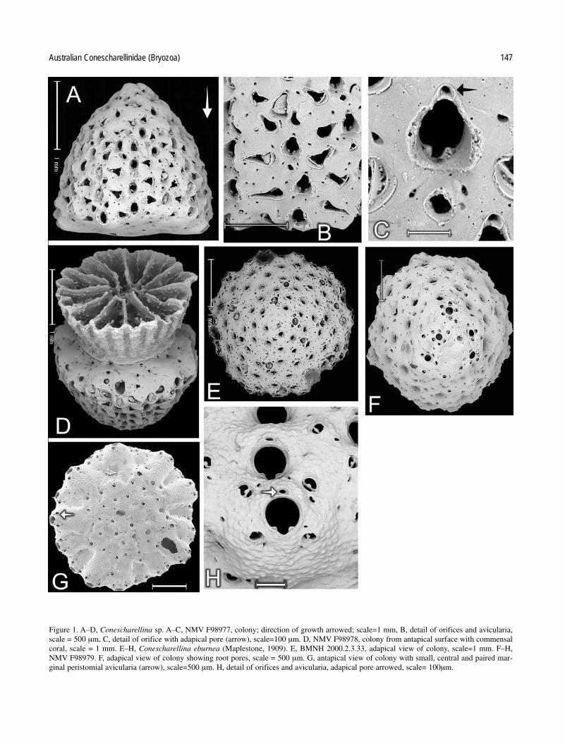
Figures 1A–D.
Bipora angulopora.—Maplestone 1909: 268 (not Conescharellinaangulopora Tenison Woods).
Specimens examined. NMV F98977, F98978, F101878. 13 specimens,all somewhat worn, labelled “Bipora angulopora” are present in theMaplestone material, inferred to be from New South Wales.
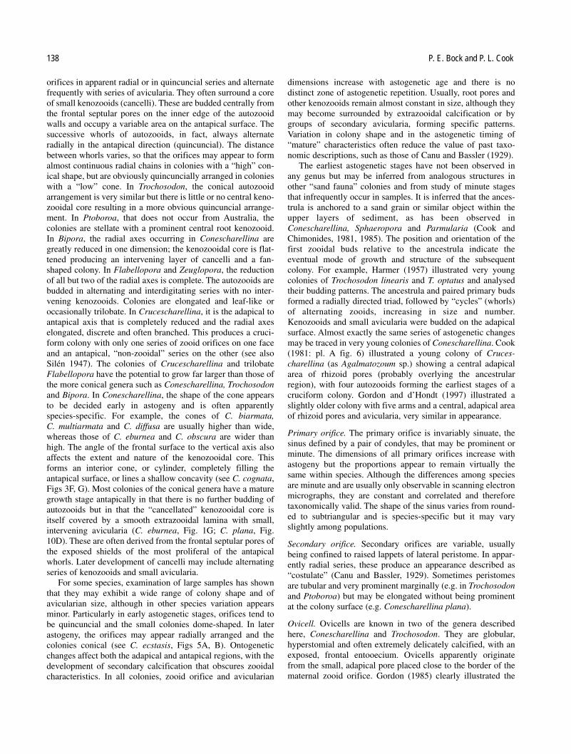
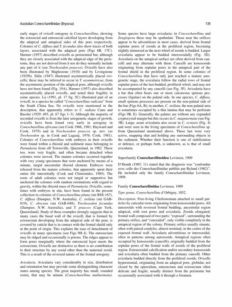
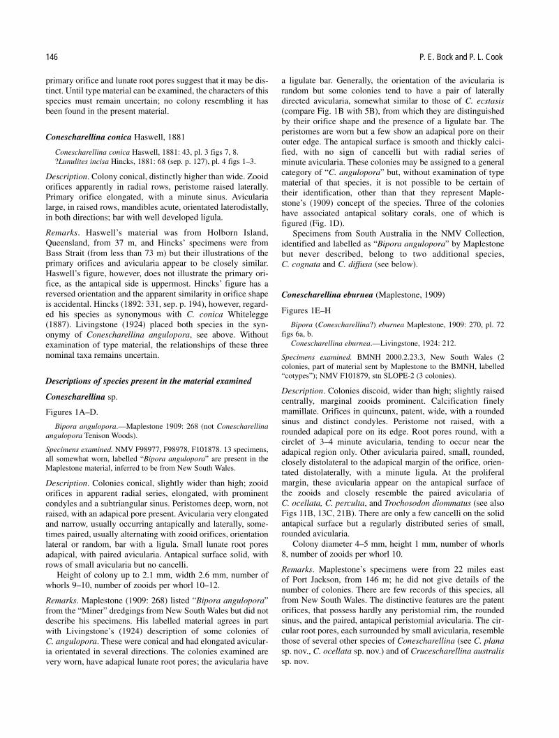
Description. Colonies conical, slightly wider than high; zooidorifices in apparent radial series, elongated, with prominentcondyles and a subtriangular sinus. Peristomes deep, worn, notraised, with an adapical pore present. Avicularia very elongatedand narrow, usually occurring antapically and laterally, some-times paired, usually alternating with zooid orifices, orientationlateral or random, bar with a ligula. Small lunate root poresadapical, with paired avicularia. Antapical surface solid, withrows of small avicularia but no cancelli.
Height of colony up to 2.1 mm, width 2.6 mm, number ofwhorls 9–10, number of zooids per whorl 10–12.
Remarks. Maplestone (1909: 268) listed “Bipora angulopora”from the “Miner” dredgings from New South Wales but did notdescribe his specimens. His labelled material agrees in partwith Livingstone’s (1924) description of some colonies of C. angulopora. These were conical and had elongated avicular-ia orientated in several directions. The colonies examined arevery worn, have adapical lunate root pores; the avicularia have
a ligulate bar. Generally, the orientation of the avicularia is random but some colonies tend to have a pair of laterallydirected avicularia, somewhat similar to those of C. ecstasis(compare Fig. 1B with 5B), from which they are distinguishedby their orifice shape and the presence of a ligulate bar. The peristomes are worn but a few show an adapical pore on theirouter edge. The antapical surface is smooth and thickly calci-fied, with no sign of cancelli but with radial series of minute avicularia. These colonies may be assigned to a generalcategory of “C. angulopora” but, without examination of typematerial of that species, it is not possible to be certain of their identification, other than that they represent Maple-stone’s (1909) concept of the species. Three of the colonieshave associated antapical solitary corals, one of which is figured (Fig. 1D).
Specimens from South Australia in the NMV Collection,identified and labelled as “Bipora angulopora” by Maplestonebut never described, belong to two additional species, C. cognata and C. diffusa (see below).
Conescharellina eburnea (Maplestone, 1909)
Figures 1E–H
Bipora (Conescharellina?) eburnea Maplestone, 1909: 270, pl. 72figs 6a, b.
Conescharellina eburnea.—Livingstone, 1924: 212.
Specimens examined. BMNH 2000.2.23.3, New South Wales (2colonies, part of material sent by Maplestone to the BMNH, labelled“cotypes”); NMV F101879, stn SLOPE-2 (3 colonies).
Description. Colonies discoid, wider than high; slightly raisedcentrally, marginal zooids prominent. Calcification finelymamillate. Orifices in quincunx, patent, wide, with a roundedsinus and distinct condyles. Peristome not raised, with a rounded adapical pore on its edge. Root pores round, with a circlet of 3–4 minute avicularia, tending to occur near theadapical region only. Other avicularia paired, small, rounded,closely distolateral to the adapical margin of the orifice, orien-tated distolaterally, with a minute ligula. At the proliferal margin, these avicularia appear on the antapical surface of the zooids and closely resemble the paired avicularia of C. ocellata, C. perculta, and Trochosodon diommatus (see alsoFigs 11B, 13C, 21B). There are only a few cancelli on the solidantapical surface but a regularly distributed series of small,rounded avicularia.
Colony diameter 4–5 mm, height 1 mm, number of whorls8, number of zooids per whorl 10.
Remarks. Maplestone’s specimens were from 22 miles east of Port Jackson, from 146 m; he did not give details of the number of colonies. There are few records of this species, allfrom New South Wales. The distinctive features are the patentorifices, that possess hardly any peristomial rim, the roundedsinus, and the paired, antapical peristomial avicularia. The cir-cular root pores, each surrounded by small avicularia, resemblethose of several other species of Conescharellina (see C. planasp. nov., C. ocellata sp. nov.) and of Crucescharellina australissp. nov.
146 P. E. Bock and P. L. Cook
Australian Conescharellinidae (Bryozoa) 147
Figure 1. A–D, Conescharellina sp. A–C, NMV F98977, colony; direction of growth arrowed; scale=1 mm, B, detail of orifices and avicularia,scale = 500 µm, C, detail of orifice with adapical pore (arrow), scale=100 µm. D, NMV F98978, colony from antapical surface with commensalcoral, scale = 1 mm. E–H, Conescharellina eburnea (Maplestone, 1909). E, BMNH 2000.2.3.33, adapical view of colony, scale=1 mm. F–H,NMV F98979. F, adapical view of colony showing root pores, scale = 500 µm. G, antapical view of colony with small, central and paired mar-ginal peristomial avicularia (arrow), scale=500 µm. H, detail of orifices and avicularia, adapical pore arrowed, scale= 100µm.

Conescharellina biarmata (Maplestone, 1909).
Figures 2A, B
Bipora biarmata Maplestone, 1909: 268, pl. 75 figs 1a, b.Conescharellina biarmata.—Harmer, 1957: 729.
Specimens examined. BMNH 2000.2.23.2, (4 colonies, part of material sent by Maplestone to BMNH, labelled “cotypes”); NMVF98980, no locality (89 colonies, labelled by Maplestone, probablypart of type material); NMV F101880, South Australia, (9 colonies,from Maplestone, with “S.A.” on box); NMV F101881, stn SLOPE-19(2 colonies); NMV F101882, stn BSS-170 (1 colony); NMV F101883,no locality, slide labelled E3195, suspected material from “Endeavour”(New South Wales) but no other information (2 colonies).
Description. Colonies very small, conical, distinctly higherthan wide. Calcification smooth. Orifices in radial series,patent, with little raised peristome, elongated oval, with a verysmall sinus formed by paired condyles. Frontal septular poresslit-like. Adapical pore placed just outside the edge of the peri-stome. Avicularia regularly paired, proximolateral to the orifi-cial sinus, small, elongated, narrow, triangular, with a ligulatebar, orientated laterally and slightly antapically. Lunate rootpores adapical, uncommon. Antapical surface with small avicularia in mature specimens, adapical surface solid withkenozooidal calcification and scattered avicularia.
Specimens 2.0–2.3 mm high, 1.4–1.6 mm wide, and com-prise approximately 10–13 astogenetic generations arranged inradial rows. The number of zooids in each whorl, 6–8.
Remarks. Livingstone (1924) placed C. biarmata in the synonymy of C. angulopora. Maplestone’s material differsfrom Livingstone’s concept of this species in the consistentlysmall colonies, and the characters of orifices and avicularianorientation. Harmer (1957) did not describe C. biarmata buttreated it as a distinct species. Maplestone’s specimens arenumerous and very consistent in characters that are shared bythe specimens from stn SLOPE-19 and the slide E3195. Otherspecimens with elongated, triangular, paired avicularia fromthe SLOPE stations differ in several respects and are heredescribed as Conescharellina ecstasis (see below).
Conescharellina multiarmata (Maplestone, 1909)
Figures 2C–F
Bipora multiarmata Maplestone, 1909: 268, pl. 75 figs 2a, b.Conescharellina multiarmata.—Livingstone, 1924: 212.not Trochosodon multiarmatus.—Gordon, 1989: 83, pl. 49 D–F (=
Trochosodon gordoni sp. nov., see below).
Specimens examined. BMNH 2000.2.23.4; (3 colonies, part of material sent by Maplestone to the BMNH, labelled “cotypes”); NMVF98981, probably NSW (31 small colonies from two boxes labelled byMaplestone, probably part of type material); NMV F101884, stn BSS-169 (1 colony); NMV F101885, stn BSS-170 (1 colony); NMVF98982, F98983, stn SLOPE-2 (20 colonies, 4 with roots, 2 with coralson antapical side); NMV F101886, stn SLOPE-7 (1 colony); NMVF101887 stn SLOPE-39 (2 colonies); NMV F101888, stn SLOPE-40(47 colonies); NMV F101889. stn SLOPE-48 (1 colony); NMVF101890, stn GAB-030 (1 colony).
Description. Colony conical, higher than wide. Calcificationsmooth to finely mamillate. Orifices in radial series, small,
elongated, with a small sinus flanked by distinct condyles, anda raised lateral peristome. Adapical pore obviously tubular,opening on the inside of the peristome edge. Avicularia verysmall, rounded, 4 placed laterally and 1 proximolaterally nearthe orifice sinus; bar with a minute ligula. Frontal pores minute,forming a pattern among the orifices. Antapical surface solidwith pores and small avicularia at maturity; marginal peristomewith small paired avicularia. Lunate root pores in radial serieswith the zooid orifices, present towards the adapical region ofthe colony.
Colonies 1.9–2.2 mm in height and 1.8–2.2 mms wide. Theycomprise approximately 4–12 or more astogenetic generations,arranged radially, and include 8–10 zooids per whorl. Thenumerous colonies from the SLOPE stations are wider thanthose from Maplestone’s collection, inferred to be from NewSouth Wales. Mature colonies may show a small central area ofcancelli on the antapical surface.
Remarks. C. multiarmata differs from C. biarmata, that alsohas very small colonies, in the details of the primary orifice andcharacter and distribution of the minute avicularia. These alter-nate with minute frontal pores and have a rounded rostrum; thebar has a single small ligula. Paired avicularia are visible on the antapical surface of marginal zooids but are not as promi-nent as those of C. eburnea. In some zooids, the adapical poreis prominent and a tubular extension of its calcification can beseen to descend into the peristome, like that of Trochosodonasymmetricus (Fig. 2D). The colonies from the SLOPE stationsare generally larger than those from the Maplestone collection.A colony from stn SLOPE-2 has an incorporated antapical solitary coral present (Fig. 2F).
Gordon (1989) identified specimens from New Zealand asMaplestone’s species and assigned them all to Trochosodon.The New Zealand material appears to be referable toTrochosodon but certainly not to Conescharellina multiarmata:it is here renamed Trochosodon gordoni sp. nov.
Conescharellina magniarmata (Maplestone, 1909)
Figures 3A, B
Bipora magniarmata Maplestone, 1909: 269, pl. 75 figs 3a, b.Conescharellina magniarmata.—Harmer, 1957: 729.
Specimens examined. BMNH 2000.2.23.5 (2 colonies, part of materialsent by Maplestone to the BMNH, labelled “cotypes”); NMV F98984(12 colonies, labelled in Maplestone’s hand, inferred to be from NSW);NMV F101891, South Australia (1 colony, Maplestone’s specimens,no other information); NMV F101892, stn BSS-167 (4 colonies withroots and antapical corals); NMV F101893, stn BSS-170 (2 colonieswith antapical corals); NMV F101894, stn BSS-171 (1 colony); NMVF101895, stn GAB-015 (1 colony); NMV F101896, stn GAB-020 (1colony); NMV F101897, stn GAB-056 (1 colony).
Description. Colony conical, becoming large, broader than pre-vious species, with adapical extrazooidal, kenozooidal andavicularian growth forming a prominent “lump” quite early inastogeny. Orifices in 6–8 radial series, slightly elongate, withan extended sinus; secondary orifices with laterally raised peristomes, forming “costules”. Adapical pore outside the peristome. Avicularia very large, originating beside the orifice,
148 P. E. Bock and P. L. Cook

acute, orientated distolaterally, with a wide, curved, palatalflange surrounding an opesia, and a bar with at least 3 largeligulae. Lunate root pores present, each with a pair of small lateral avicularia. Mature colonies with solid antapical extrazooidal calcification and avicularia.
Height of colonies 3.0 mm, diameter 3.0 mm, number ofzooid whorls 8–9, number of zooids per whorl 8.
Remarks. Livingstone (1924) regarded C. magniarmata as asynonym of his “C. angulopora” but Harmer (1957) was
doubtful of the identity of the two forms. The avicularia cer-tainly distinguish C. magniarmata. Few of the coloniesassigned here to C. cognata show any intermediate characters,although the two forms are obviously very closely related. Allthe specimens from eastern Australia have elongated conicalcolonies but one from south-western Australia (stn GAB-015)is very small and domed. However, it has typical “magniar-mata”- type avicularia, with a wide palatal flange and is there-fore referred to this species.
Australian Conescharellinidae (Bryozoa) 149
Figure 2. A–B, Conescharellina biarmata (Maplestone, 1909). NMV F98980. A, lateral view of colony, growth direction arrowed, scale=1 mm.B, detail of orifices with adapical pore, avicularia and slit-like frontal septular pores, scale= 100 µm. C–D, Conescharellina multiarmataMaplestone, NMV F98981. C, Lateral view of colony, growth direction arrowed, scale=500 µm, D, detail of orifices from slightly antapical view,showing tubular adapical pores, avicularia and minute frontal septular pores, scale = 200 µm. E–F, Conescharellina multiarmata (Maplestone,1909). E, NMV F98983, orifices and avicularia; note pattern of septular pores, scale= 200 µm. F, NMV F98982, Antapical view of colony show-ing small solitary coral. Note paired, marginal peristomial avicularia, scale= 500 µm.

150 P. E. Bock and P. L. Cook
Fig.3. A–B, C. magniarmata Maplestone, NMV F98984. A, Lateral view of colony, growth direction arrowed, note adapical secondary calcifica-tion, scale= 1 mm. B, detail of orifice with adapical pore and avicularium, scale= 100 µm. C–G, Conescharellina cognata sp. nov. C–E. NMVF98985, holotype. C, lateral view of large colony, growth direction arrowed, scale = 2 mm. D, margin of proliferal region in antapical view, show-ing zooid orifices with adapical pores and “concealed” frontal shields with marginal septular pores. Chambers of alternating developing inter-zooidal avicularia arrowed, scale = 500 µm. E, detail of orifices with adapical pores, one with entooecial and ectooecial laminae of developingovicell (arrow); large, interzooidal avicularia and lunate root pore with small paired avicularia, scale = 200 µm. F, NMV F98986, antapical viewof small colony, showing central area of cancelli and alternating “concealed” zooids with marginal septular pores, scale = 500 µm. G. NMVF98985, holotype, detail of antapical surface of large colony, showing alternating series of cancelli, minute avicularia and extensions of septularpores, scale = 200 µm.

Conescharellina cognata sp. nov.
Figures 3C–G
Holotype. NMV F98985, Maplestone collection, Kangaroo Island,South Australia, labelled “Bipora angulopora”.
Paratypes. NMV F101898, locality as above (23 colonies).Other specimens. NMV F101899, Maplestone collection, South
Australia (no details) (23 colonies); NMV F101900, stn BSS-55 (1 colony); NMV F101901, stn BSS-65 (1 colony); NMV F101902, stnBSS-117 (4 colonies with roots); NMV F101903, stn BSS-127 (1 colony); NMV F101904, stn BSS-130 (4 colonies); NMV F101905,stn BSS-132 (1 colony); NMV F101906, stn BSS-138 (1 colony);NMV F98986, stn BSS-155 (34 colonies); NMV F101907, stn BSS-158 (12 colonies); NMV F101908, stn BSS-159 (12 colonies); NMVF101909, stn BSS-161 (11 colonies); NMV F101910, stn BSS-162 (3 colonies); NMV F101911, stn BSS-170 (2 colonies); NMVF101912, stn BSS-171 (8 colonies); NMV F101913, stn BSS-176 (6 colonies); NMV F101914, stn BSS-194 (3 colonies); NMVF101915, stn GAB-019 (1 colony); NMV F101916, stn GAB-020 (5 colonies); NMV F101917, stn GAB-030 (3 colonies); NMVF101918, stn GAB-045 (2 colonies, one with root); NMV F101919, stnGAB-049 (1 colony with root and antapical coral); NMV F101920, stn GAB-067 (1 colony); NMV F101921, stn GAB-098 (2 colonies);NMV F101922, stn GAB-101 (1 colony).
Etymology. cognatus (L.) – related, referring to the similaritiesof the species with some descriptions of “C. angulopora”.
Diagnosis. Conescharellina with large, often flattenedcolonies; antapical surface flat or hollow, with large cancelli.Zooid orifices with a narrow sinus and large condyles.Avicularia large, lateral, interzooidal, with subtriangular ros-trum orientated adapically and laterally. Root pores frequent,lunate.
Description. Colonies very large, usually flattened, occasion-ally conical, particularly early in astogeny. Calcificationsmooth. Zooids in irregular quincuncial series, tending toappear radial in very large colonies. Primary orifice an elon-gated oval with a narrow sinus and prominent condyles, peristome raised laterally and antapically but only a little adapi-cally; adapical pore just outside, or on edge of the peristome.Avicularia interzooidal, lateral and adapical, large, rostrum sub-triangular, directed adapically and laterally (usually in the samedirection); bar with 1–3 ligulae. Lunate root pores frequentamong orifices, apparently replacing avicularia, each with apair of small lateral avicularia with a ligulate bar. Antapical sur-face with large, central cancelli, or hollow, covered by series ofcancelli and minute avicularia. Proliferal region growing edgewith prominent frontal shields visible at all stages of growth.
Colony diameter up to 10 mm, height 3 mm, number ofwhorls (radial) 18, number of zooids per whorl, at least 10.
Remarks. C. cognata differs from the accepted character ofMaplestone’s concept of C. angulopora (see above) that itresembles in several features: its colony shape and the variablebut generally slightly wider primary orifice. There is a variationin orifice shape between colonies from Bass Strait, where theyare very narrow, and those from Southern Australia. The largeavicularia can be seen at the growing edge to be interzooidal(Fig. 3D) and are usually consistently orientated in one direc-tion within a single colony, although they vary within samples.
The pair associated with each root pore is orientated laterally.Root pores are abundant, apparently replacing avicularia. In thelarger colonies, the hollow antapical surface is completely covered by series of cancelli, interspersed with minute, rounded avicularia, totally unlike the solid surface of the conical colonies of C. angulopora (sensu Maplestone).
C. cognata is common among the samples from GAB andBSS stations and in the Maplestone collection from SouthAustralia. Gordon (1985, 1989) described and figured speci-mens from the Kermadec region and from New Zealand, that heassigned to C. angulopora. The colonies were not conical butflat. The antapical surface had no avicularia and a small centralcancellate area. Gordon used an identical description for bothsets of specimens and figured developing ovicells in one of thecolonies from the Kermadec region (1985: fig. 23). Althoughclosely similar to the colonies from South Australia, these spec-imens differ in the more elongated shape of the primary orificesand the characters of the antapical surface.
Conescharellina ecstasis sp. nov.Figures 4, 5A–D.
Holotype. NMV F98987, stn SLOPE-6.Paratypes. NMV F101923, stn SLOPE-6 (45 colonies, many with
roots and opercula and mandibles).Other specimens. NMV F101924, stn SLOPE-2 (7 colonies, 4 with
roots); NMV F98988, stn SLOPE-7 (10 colonies, 2 with roots); NMV
Australian Conescharellinidae (Bryozoa) 151
Figure 4. Conescharellina ecstasis sp. nov. NMV 98987, holotype.Radial series of zooid orifices with adapical pores and large avicular-ia, scale = 200µm.

F101925, stn SLOPE-39 (1 colony with root); NMV F101926, stnSLOPE-40 (2 colonies); NMV F101927, stn SLOPE-45 (1 colony);NMV F101928, stn SLOPE-48 (1 colony); NMV F101929, stnSLOPE-53 (3 colonies).
Etymology. ecstasis (L.) – joy, with reference to the appearanceof the orifice and paired lateral avicularia.
Diagnosis. Conescharellina with large, conical colonies, solidantapically. Zooid orifices in radial series, primary orifice witha rounded sinus. Avicularia very large, paired, elongated, lateralto orifice, orientated laterally and adapically. Root pores lunate.
Description. Colonies large, conical, wider than high, domedand stellate in early astogeny. Calcification finely mamillate.Zooids in apparently radial series, peristomes prominent mar-ginally, particularly in young, stellate colonies. Primary orificewith a distinct, rounded sinus and paired condyles, adapicalpore on the edge of the peristome. Avicularia paired, lat-eral, very large, with acutely triangular rostra, nearly alwaysdirected laterally and adapically; bar without a ligula. Adapicalregion with large avicularia and lunate root pores, each with a
pair of small lateral avicularia. Antapical region solid, withradiating series of small avicularia and a few cancelli.
Colony diameter up to 4.7 mm, height 2.3 mm, number ofwhorls 6, number of zooids per whorl 8.
Remarks. The colonies of C. ecstasis sp. nov. are recognisableimmediately, even to the naked eye, by the pairs of large avic-ularia, with mandibles of dark brown cuticle. The orientation ofthe rostra varies a little; those of the one specimen from stnSLOPE-45 being almost horizontal, like the rostra of C. biarmata. In contrast, one of the two colonies from stnSLOPE-7 has rostra directed almost adapically. Young coloniesare stellate, with prominent peristomes, especially marginally,giving the colony a “Trochosodon-like” appearance (Fig. 5A).C. ecstasis differs completely from C. biarmata in colonyshape and size, the characters of the primary orifice, and lackof avicularian ligulae. Except for stn SLOPE-6, only a fewspecimens of C. ecstasis were present at each of the eightSLOPE stations. The two colonies of C. biarmata sensu strictofrom stn SLOPE-19 were easily distinguished by their much
152 P. E. Bock and P. L. Cook
Figure 5. A–C, Conescharellina ecstasis sp. nov. A, NMV 98988. Adapical view, scale = 500 µm. B–C, NMV 98987, holotype. B, lateral view ofcolony, growth direction arrowed, scale = 1 mm. C, detail of adapical surface showing secondary calcification, avicularia and lunate root porewith three small avicularia, scale = 200 µm. D–F, Conescharellina diffusa sp. nov. D, NMV F98990. Adapical view of colony showing lunate root pores and broken ovicell (arrow), scale = 500 µm. E, NMV F98989, holotype. Lateral view of large colony, growth direction arrowed, scale = 1 mm. F. Conescharellina aff. diffusa sp. nov. NMV P311803, Bairnsdale, Victoria, Miocene. Lateral view of colony, scale = 500 µm.

smaller dimensions and orientation of avicularia. All records ofC. ecstasis are from deep water. The SLOPE stations rangefrom south-eastern New South Wales, to eastern Victoria andTasmania. The bathymetric range of records is from 400 m to1096 m.
Conescharellina diffusa sp. nov.
Figures 5D–F, 6A–B
Holotype. NMV F98989, South Australia (no other details), from boxlabelled “Bipora philippinensis” in Maplestone’s hand.
Paratypes. NMV F101930, South Australia, as above (3 colonies).Other specimens. NMV F101931, South Australia, Maplestone
Collection (55 colonies); NMV F101932, Kangaroo Island, SouthAustralia, Maplestone Collection (1 colony); NMV, F101933, probablyNSW, Maplestone Collection (3 colonies); NMV F101934, stn BSS-065 (8 colonies, 5 with roots); NMV F101935, stn BSS-171 (1 colony);NMV F101936, stn SLOPE-49, (2 colonies); NMV F101937, stnGAB-020 (1 colony); NMV F101938, stn GAB-067 (1 colony); NMVF101939, stn GAB-069 (1 colony); NMV F101940, stn GAB-118 (3 colonies); NMV F101941, stn GAB-129 (2 colonies); NMVF98990, Dampier DA-2-37-01, North-western Australia (2 colonies,one with 8 ovicells, both with roots); NMV F101942, DmitriMendeleev collection, Tasmania (1 colony).
C. aff. diffusa: Specimens from the Tertiary of Victoria withvery similar but not identical characters: Bairnsdale, NMVP311803 (Fig. 5F), P311804, plus 20 additional colonies;Muddy Creek (three colonies).
Etymology. diffusus (L.) – extended, dispersed, with referenceto the wide distribution of this species.
Diagnosis. Conescharellina with large, often elongated, conicalcolonies, zooid orifices radial, surrounded by a rim of peris-tome. Root pores frequent, lunate, without avicularia.Avicularia in series alternating with orifices, small, rounded tosubtriangular, bar without a ligula; non-palatal area with spinous processes. Ovicells fragile, with a wide ectooecial rim.
Description. Colony often large, conical, very narrow ordomed, higher than wide. Zooid orifices apparently radial,alternating with radial series of rounded avicularia.Calcification smooth. Primary orifice with a fairly deep, round-ed sinus and paired condyles, surrounded by a peristome rim;adapical pore outside peristome. Ovicells fragile, with a wideectooecial rim and a semitransparent entooecial frontal area.Avicularia paired, lateral and slightly adapical, or in seriesalternating with orifices. Orientation lateral and adapical; ros-trum rounded to subtriangular, bar without ligula, but with 3 ormore very fine, spinous processes on the non-palatal side.Antapical surface solid, with a small, central cancellate area.Lunate pores frequent, occurring in series with the aviculariabut without any accompanying small avicularia.
Recent colonies with up to 28 zooid whorls and more than14 zooids per whorl, height up to 5.0 mm, diameter 3.5 mm.Fossil colonies rounded, height 2.5 mm, diameter 3 mm.
Remarks. The Recent colonies are among the largest and mostwidely distributed of the Australian species examined. Theavicularia appear to be unique in possessing small calcareousspine-like structures on the non-palatal side of the bar. Some
Australian Conescharellinidae (Bryozoa) 153
Figure 6. Conescharellina diffusa sp. nov. NMV F98989, holotype. A,detail of zooid orifices with adapical pores, lunate root pores and avic-ularia with non-palatal spinules (arrow), scale = 200 µm. B, detail oforifices with adapical pores; orifice at left shows broken laminae ofectooecium and overlying entooecium of developing ovicell; orifice atright shows adapical pore surrounded by ectooecial lamina of ovicell(arrow). Note non-palatal spinules of avicularia, scale = 200 µm.

zooids show evidence of the development of an ectooecial lamina surrounding the adapical pore; others appear to havealso developed an entooecial layer above the pore (Fig. 6B).Usually, colonies are distinctly higher than wide but five ofeight from Bass Strait (stn 64) are shorter and more rounded inoutline. All five have long roots, in one case, anchored ter-minally to a fragment of a “scrupocellariid” bryozoan. The twocolonies from north-western Australia are also conical butsomewhat rounded; their avicularia show non-palatal spinousprocesses. One colony has eight fragile and only partially com-plete ovicells (Fig. 5D), showing that these have a very thinlycalcified entooecium and a wider ectooecium than those ofTrochosodon fecundus sp. nov. and C. stellata sp. nov.
Records of C. diffusa are widely separated. It ranges fromnorth-west Australia to New South Wales, the west and centralAustralian Bight, South Australia, Bass Strait, and Tasmania,from 15 m. (north-west Australia) to 200 m. (Tasmania).
The fossil colonies are much smaller and domed; their avic-ularia do not possess any non-palatal projections and it appearsprobable that, although closely related, they are not referable toC. diffusa sensu stricto (Fig. 5F). The specimen of “Bipora cancellata” from Bairnsdale described by MacGillivray (1895:89, pl. 12 fig. 1; NMV P22727) appears to be conspecific withthose listed here from Bairnsdale and Muddy Creek.
Conescharellina obscura sp. nov.
Figures 7, 8A–C
Bipora philippinensis.—Maplestone, 1910: 6, pl. 1 figs 2, 2a (notBusk, 1854).
Holotype NMV F98991, stn BSS-155.Paratype NMV F98992, stn BSS-155.Other specimens. BMNH as C. philippinensis, 1909.11.12.12 and
13, Green Point, Port Jackson, NSW (Maplestone Collection, possiblyfrom Whitelegge’s material, 2 colonies with ovicells); BMNH1899.5.1.1148, Port Jackson (Hincks Collection, 5 colonies); BMNH,
as L. cancellatus, 1934.10.20.88, Port Stephens, NSW (VineCollection, 2 colonies); NMV F101943, stn GAB-048; (1 colony withovicells); NMV F101944, stn GAB-074; (3 colonies); NMV F101945,stn GAB-108 (1 colony); NMV F101946, stn GAB-113 (2 colonies);NMV F101947, stn GAB-118 (2 colonies); NMV F101948, stn GAB-131 (1 colony); NMV F101949, Dampier DA-2-09-02 (1 colony withroot); NMV F101950, Dampier DA-2-73-01 (2 colonies with roots).
Etymology. obscura (L.) – hidden, referring to the confusion ofrecords with those of C. cognata, C. stellata and C. philip-pinensis.
Diagnosis. Conescharellina with flat, often large colonies.Zooid orifices oval with a short sinus, peristomes not promi-nent. Avicularia rounded, lateral and antapical, near the orifice,bar with 1–3 ligulae. Root pores lunate. Ovicells globular, veryfragile, with an extensive area of entooecium frontally.
Description. Colony fairly flat, even lenticular, distinctly widerthan high. Zooid orifices quincuncial, sunken in surroundingperistome, that is not prominent. Orifice oval, with a short,
154 P. E. Bock and P. L. Cook
Figure 7. Conescharellina obscura sp. nov. A, NMV F98991, holo-type. Lateral and adapical view of large colony, scale= 500 µm.
Figure 8. Conescharellina obscura sp. nov. A–B, NMV P98992. A, adapical view of small colony, showing lunate root pores, scale = 500 _m. B,detail of zooid orifices, avicularia, and one lunate root pore, scale = 200 µm. C, BMNH 1899.5.1.1148, Hincks Collection, Port Jackson, NewSouth Wales. Detail of zooid orifices with adapical pores, avicularia and one lunate root pore. Note the wide orifice sinuses, scale = 200 µm.

rounded sinus and small condyles. Adapical pore within peris-tome calcification. Avicularia rounded, lateral and antapicalnear the orifice, bar with 1–3 ligulae. Lunate root pores fre-quent adapically, with 1, occasionally 2, lateral avicularia.Antapical surface covered with small cancelli.
Colonies up to 14 mm in diameter but usually much smaller, maximum number of zooid whorls estimated as 15,and zooids per whorl 20.
Remarks. The close but superficial similarity in colony formmeans that records of this species were originally confusedwith those of C. cognata, that also has large, flat colonies withnumerous antapical cancelli. The colonies from the GAB sta-tions are very large, ranging from 9 to 12 mm in diameter andare hollow antapically; those from stns GAB-074 and GAB-118 are more domed and nearly solid antapically. In thesmaller colonies from stn BSS-155, the lunate root pores arepresent but they are very rare or absent from the large colonies.Again, this is in contrast to C. cognata, where they are commonthroughout colony growth. Although the large, flat coloniesresemble those described in “C. crassa” by Tenison Woods(1880), they differ in orifice shape and the types of avic-ularia and root pores present. Antapical oral avicularia are also found only in C. pustulosa, from which C. obscura differs in colony shape and size, orifice shape and form of the antapical cancelli. Whitelegge (1887) described speci-mens from Port Jackson as Bipora philippinensis, with adepressed conical shape and orifices with a wide sinus.Avicularia with subcircular mandibles occurred in pairs andsometimes on the antapical side of each orifice. The anta-pical surface had cancelli and avicularia. Ovicells were presentand were “globose and smooth, with a faint fimbriated stigmain front.”
Colonies referred to this species were observed alive byWhitelegge (1887: 347) for three days. He noted pairs of “tubular filaments” attached to annelid tubes and to fragmentsof shell. He thought that these roots originated from aviculariaand did not recognise the function of the lunate root pore thathe also reported. Maplestone (1910: 6, pl. 1, fig. 2) illustratedovicells in specimens of Whitelegge’s material that he alsoreferred to C. philippinensis but noted that he could see nofrontal stigma. Maplestone’s specimens from Port Jackson, inthe BMNH collection, are probably part of Whitelegge’s mater-ial and are here referred to C. obscura sp. nov., whereas thosein the NMV collection from South Australia, also labelledBipora philippinensis, are here assigned to another newspecies, C. diffusa. The two slides from Green Point(1909.11.12.12 and 13) each contain a single, fairly flat colony,less than 2.5 mm in diameter, with most of the opercula andmandibles intact. They each include two fragile ovicells and upto eight partially developed ovicells. These are globular, likethose figured by Maplestone (1910). They appear to have anextensive area of frontal entooecial calcification and a series ofminute pores close to the ovicell base, that may mark the limitof the ectooecium. There is no sign of any striations or a “stigma”. The other, smaller specimens from the BMNH col-lections are obviously conspecific but have no ovicells. Theytoo, have most of the opercula and mandibles present; only one
preparation shows the primary orifice clearly. One of the fivespecimens from Port Jackson (Fig. 8C, Hincks collection,BMNH 1899.5.1.1148) is similar in characters to specimensfrom Bass Strait (Fig. 8B, stn BSS-155) except for the greaterwidth of the orifice sinus. C. obscura occurs from north-westAustralia and across the Great Australian Bight to New SouthWales, from a depth range of 12 to 125 metres.
Conescharellina stellata sp. nov.
Figures 9A–I
Holotype. NMV F98993, stn GAB-019.Paratypes. NMV F98994, stn GAB-019 (8 colonies).Other specimens. NMV F98995, stn GAB-128 (1 colony).
Etymology. stellata (L.) – starry, referring to the appearance ofthe colonies from the adapical side.
Diagnosis. Conescharellina with small, domed colonies.Orifices with rounded sinus and distinct condyles, surroundedby a raised peristome laterally and sometimes antapically.Avicularia lateral, rounded; bar without a ligula, non-palatalarea sometimes filled by a lamina. Ovicells fragile, with adepressed, marginally striated entooecium.
Description. Colony small, domed, wider than high; zooid orifices quincuncial. Calcification smooth and slightly tuber-culate, adapical region sometimes with small, secondarilythickened mamillae. Peristomes raised laterally, forming aprominent, stellate pattern, especially at the colony margin;sometimes extended adapically and very prominent. Primaryorifices rounded with a fairly wide sinus and small to distinctcondyles. Adapical pore large, on the edge or outer face of theperistome, surrounded by a rim of calcification, sometimesslightly asymmetrically placed. Avicularia rounded, lateral andpaired, widely separated from the peristomes; bar without aligula, non-palatal side sometimes with a thin lamina, occa-sionally pierced by a pore. Lunate root pores tending to occuradapically, each with a pair of closely apposed, rounded lateralavicularia. Ovicells present on subperipheral zooids, very fragile, with a raised, smooth, transparent ectooecium and adepressed entooecium, striated marginally, forming pores at the ectooecial junction. Antapical surface with a small centralcancellate area.
Colony diameter 1–1.5 mm, height 1 mm, number of alter-nating whorls 6, number of zooids per whorl 6–8.
Remarks. The colonies have a very regular, stellate appearancefrom the adapical side. The peristomes are usually well devel-oped laterally but, in one colony (from stn GAB-128), they arealso extensive antapically, forming a funnel. The ovicells areextremely fragile and were detached soon after initial scanningelectron microscopy. The lower face of the ectooecium showsthat it was almost certainly covered by cuticle and apposed butnot attached to the surface of the zooid adapically (Figs 9H, I).Although closely similar to the smaller colonies of C. obscurain several features, C. stellata differs in the form and distribu-tion of the avicularia, that do not include a solitary one on theantapical side of the peristome. The avicularia also differ in thelack of ligulae and the presence of a lamina filling the non-
Australian Conescharellinidae (Bryozoa) 155

156 P. E. Bock and P. L. Cook
Figure 9. Conescharellina stellata sp. nov. A–C NMV F98993, holotype. A, adapical view of colony showing prominent bilabiate marginal peri-stomes and lunate root pores, scale = 500 µm. B, detail of marginal zooid orifice with adapical pore. Avicularium with porous non-palatal lami-na (arrow), compare Figure 9E, scale = 200 µm. C, lateral view of colony showing lunate root pore, avicularia, scale = 200 µm. D–E, NMVF98995. D, adapical view of colony with prominent, rounded, funnel-shaped marginal peristomes, scale = 500 µm. E, marginal view showingextended peristomes, orifices and avicularia with non-palatal laminae, one with pore (arrow), compare Figure 9B, scale = 200 µm. F–I, NMVP98994. F, Colony with cuticle in situ and two ovicells near margin, scale = 100 µm, G, detail of ovicells, scale = 100µm. H, I, frontal and basalsurfaces of detached ovicell. H, showing ridges in margin of entooecium, forming pores at junction with ectooecium, with basal smooth ectooe-cium. I, with fracture at original margin of adapical pore, scales = 50 µm.

palatal area. This is not always developed in the specimensfrom stn GAB-019 but is constantly present in the distinctlylarger, more prominent avicularia of the colony from stn GAB-128. The lamina may be pierced by a pore in both populations.C. stellata has been found from the western end of the GreatAustralian Bight at a depth of 59 m and from the central regionat a depth of 304 m. Although these two populations show differences in detail, the number of colonies does not allow anyestimate of its significance.
Conescharellina plana sp. nov.
Figures 10A–D
Holotype. NMV F98996, stn SLOPE-2.Paratypes. NMV F98997, stn SLOPE-2 (26 colonies).Other specimens. NMV F101951, stn BSS-167 (4 colonies, 1
with root and ovicells); NMV F101952, stn BSS-169; 3 colonies, 1 with root); NMV F101953, stn SLOPE-6 (5 colonies); NMVF101954, stn SLOPE-7 (1 colony); NMV F101955, stn SLOPE-40 (57colonies, 2 with roots); NMV F101956, stn SLOPE-56 (13 colonies);NMV F101957, stn GAB-020 (6 colonies); NMV F101958, stn GAB-030 (2 colonies, 1 with root); NMV F101959, stn GAB-044 (1 colonywith root); NMV F101960, stn GAB-049 (1 colony).
Etymology. planus (L.) – smooth, with reference to the lack ofraised peristomes above the colony surface.
Diagnosis. Conescharellina with large, slightly flattenedcolonies, solid antapically. Zooid orifices in radial rows, deeplysunken within a circular peristome, that is not raised above thecolony surface. Avicularia paired, small, with a minute ligula.Root pores numerous, circular, surrounded by avicularia.Ovicells fragile with a fairly wide ectooecial rim.
Australian Conescharellinidae (Bryozoa) 157
Figure 10. Conescharellina plana sp. nov. A–C, NMV F98996, holotype. A, adapical view of colony, scale = 1 mm. B, detail of root pore withsurrounding avicularia, scale = 200 µm. C, detail of colony margin showing orifices, peristomes, and avicularia, scale = 200 µm. D, NMV F98997,paratype, antapical surface of colony, scale = 500 µm.

Description. Colonies large, slightly flattened, wider than high.Zooid orifices in marked, apparently radial series, peristomestubular, deep but not prominent at the surface. Calcificationsmooth to finely tuberculate. Primary orifice with a small,rounded sinus, deeply hidden at the base of the circular peris-tome, adapical pore just outside the edge of the peristome.Ovicells fragile, with a fairly wide ectooecial rim and a semi-transparent entooecial frontal area. Avicularia paired, close tothe edge of the peristome, adapical and antapical, very small,rounded, with a minute ligula. Root pores numerous, large, cir-cular, surrounded by a circlet of 3–4 small avicularia. Antapicalsurface solid and flat, with small, scattered avicularia.
Colony diameter 4.5 mm, height 2 mm. Number of whorls5, number of zooids per whorl 8.
Remarks. The large, circular root pores of C. plana are similarin appearance to those of Conescharellina eburnea, C. perculta,C. humerus and Crucescharellina australis that are also sur-rounded by a circlet of small, rounded avicularia. The peris-tomes of C. plana are unusual in being elongated but not promi-nent and the colony surface is smooth. Two colonies exhibit asingle, marginal zooid each, with a prominent peristomial avic-ularium (Fig. 10C). Only one of the two ovicells present in thecolony from stn BSS-167 is complete; a deeply pigmentedembryo is visible through the thin frontal calcification.Colonies of C. plana are widely distributed off the southern andeastern coasts of Australia occurring from the westernAustralian Bight to the eastern border of Victoria, through BassStrait, from depths ranging from 80 to 1096 m.
Conescharellina perculta sp. nov.
Figures 11A–D
Holotype. NMV F98998, slide labelled E3195 (Locality unknown,probably off New South Wales).
Paratypes. NMV F98999, locality as above.
Etymology. percultus (L.) – highly adorned, with reference to the patterning of the numerous avicularia and colony calcification.
Diagnosis and description. Colonies small, discoid, distinctlywider than high, with a mamillate centre and prominent mar-ginal peristomes, calcification delicate and finely tuberculate.Orifices quincuncial, becoming radial. Primary orifice with arounded sinus. Peristomes elongated and tubular, raised anta-pically and prolonged into a spout, prominent at the colonymargin. Adapical pore present on outer face of the peristomes.Root pores circular, surrounded by up to 5 small avicularia. Allavicularia small, rostrum rounded, bar with a minute ligula.Each orifice with 1 adapical, 1 lateral and 1 antapical pair ofavicularia. Further pairs of lateral and antapically placed avicularia, that are visible from the antapical surface, areaccompanied by pairs of pores.
Largest colony about 2.3 mm wide and 0.5 mm high, with 6astogenetic generations and probably up to 11–12 zooids perwhorl at margin.
Remarks. The locality from that the three small colonies ofwere collected also provided two well preserved colonies of
C. biarmata and therefore is inferred to have been collectedfrom New South Wales. C. perculta is distinguished by its delicate, semitransparent, finely tuberculate calcification, withnumerous avicularia surrounding the spout-shaped peristomialorifices. As in C. eburnea, C. ocellata and T. diommatus, themarginal peristomes can be recognised from the antapical surface by the pattern or outline of the associated paired avicularia. In many other respects, such as the depth of the peristome, the distribution of circum-oral avicularia, and type of root pore, C. perculta greatly resembles C. plana, fromwhich it differs principally in colony size, the patterning andshape of the orifices, and nature of the antapical surface,including the peristomes. The circular root pores, with their surrounding avicularia, resemble those of C. eburnea,C. plana and C. humerus, as well as those of Crucescharellinaaustralis.
Conescharellina pustulosa sp. nov.
Figures 12A–D
Holotype. NMV F99000, stn SLOPE-2.Paratypes. NMV F99001, stn SLOPE-2 (9 colonies).Other specimens. NMV F99002, unlabelled Maplestone specimens,
probably from NSW (5 colonies); NMV F101961, stn BSS-158 (1colony); NMV F101962, stn BSS-169 (1 colony); NMV F101963, stnSLOPE-40 (14 colonies); NMV F101964, stn SLOPE-45 (1 colony);NMV F101965, stn GAB-049 (1 colony).
Etymology. pustula (L) - a bubble or blister, with reference tothe calcification of the zooid surfaces.
Diagnosis. Conescharellina with small, domed colonies.Zooids orifices with a subtriangular sinus; surrounded byraised, pustular secondary calcification, that also covers theantapical surface. Avicularia antapical, peristomial, raised, barwithout a ligula.
Description. Colonies small, domed. Surface irregular, formedby raised, pustular secondary calcification. Zooids in quin-cuncial series, peristomes raised laterally, prominent only atcolony margins. Primary orifice with a wide subtriangular sinusand minute condyles. Adapical pore outside the peristome rim.One small peristomial avicularium lateral and antapical, nearlyvertical to the rim of the peristome, rostrum rounded, bar with-out a ligula; other occasional avicularia scattered among thezooids. Adapical region with a few, lunate root pores; antapicalregion with more pustular secondary calcification, cancelli andavicularia.
Colony diameter 2.2 mm, height 1.2 mm, number of whorls6, number of zooids per whorl 8.
Remarks. The pustular calcification occurs among the zooidorifices and is a prominent feature of the antapical surface ofthe small colonies. The only other species in the samples examined that possesses an antapical peristomial avicularium isC. obscura, but this is not placed on the edge of the peristomeas in C. pustulosa. C. pustulosa bears a close but superficialsimilarity to C. papulifera Harmer (1957: 734, pl. 47 figs 7–9,text-fig. 70C). Harmer did not describe or figure the primaryorifice, that he was not certain was visible. He mentioned
158 P. E. Bock and P. L. Cook

paired avicularia and radially costulate zooid orifices but didnot figure them. Specimens of C. papulifera (BMNH,1964.3.2.3, paratype?, Java Sea and 1964.3.2.2, Siboga stn 77,Borneo Bank, 59 m) have been examined. They are minute;their dimensions being less than half of those of C. pustulosa atthe same astogenetic stage. The peristomes are raised and tubular, arranged in radial series; the primary orifices are not visible. Minute avicularia alternate with the zooid orifices andnone are antapical and peristomial.
C. pustulosa is distributed from the coasts of New SouthWales to Bass Strait and the Great Australian Bight, from awide depth range of 36 to 800 m. There is, however, little variation in the characters of the colonies from the differentpopulations.
Conescharellina ocellata sp. nov.
Figures 13A–D
Holotype. NMV P311805; Miocene, Balcombe Bay, Victoria (seeappendix).
Paratype. NMV P311806; Balcombe Bay, Victoria.Other specimens. Victoria, Balcombe Bay (55 colonies); Batesford
Quarry (see appendix) (45 colonies).
Etymology. ocellata (L.) – having little eyes, with reference tothe appearance of the antapical peristomial avicularia.
Diagnosis. Conescharellina with minute, slightly domedcolonies. Zooid orifices radial. Avicularia small, paired, lateral,with a minute ligula. Paired avicularia visible on the antapicalside of the marginal peristomes.
Australian Conescharellinidae (Bryozoa) 159
Figure 11. Conescharellina perculta sp. nov. A, NMV F98998, holotype. A, adapical view of colony, scale = 200 µm. B, NMV P98999, antapi-cal view of marginal peristomes, scale = 200 µm. C–D, NMV F98998, holotype. C, detail of orifices, peristomes, and avicularia, scale = 200 µm.D, detail of root pore, scale = 200 µm.

Description. Colonies very small, domed, orifices appearing tobe in radial series. Primary orifice with a short, narrow sinusand distinct paired condyles, with a laterally and antapicallyraised peristome. Adapical pore on the edge of the peristome.Avicularia small, rounded, paired and antapical, lateral to thesinus, bar with a minute ligula, orientated laterally and ada-pically. Adapical region with a few rounded pores, inferred tobe root pores, with 2–3 small adjacent avicularia. Antapicalregion with a few avicularia and central cancelli; and withprominent, paired avicularia present on the antapical side of theperistome of marginal zooids.
Diameter of colony 2.4 mm, height 1.6 mm, up to 6 radialwhorls, 5–7 zooids per whorl.
Remarks. C. ocellata is easily recognisable in unworn coloniesby the paired antapical avicularia on the marginal peristomes.C. ocellata resembles C. eburnea and Trochosodon diammotos
in this character but differs in all its dimensions and in theshape of the primary orifice. It differs from C. macgillivrayi inits domed colonies, radially arranged orifices, shape of the primary orifice, and rarity of antapical cancelli. It differes fromC. humerus in the position and shape of the primary orifice andposition of the lateral avicularia, especially in antapical view.
Conescharellina macgillivrayi sp. nov.
Figs 13E-F, 14A
Bipora philippinensis.—MacGillivray, 1895: 89, pl. 12 fig. 2.
Holotype. NMV P311810, Miocene, Balcombe Bay, Victoria.Paratypes. NMV P311811, Balcombe Bay.Other specimens, Miocene, Balcombe Bay (approximately 209
colonies); Miocene, Bairnsdale (12 colonies); Miocene, BatesfordQuarry (22 colonies); Miocene, Heywood Bore (approximately 63worn colonies); Miocene, Muddy Creek (13 colonies), Miocene,
160 P. E. Bock and P. L. Cook
Figure 12. Conescharellina pustulosa sp. nov. A–B, NMV F99000, holotype. A, adapical view of colony, scale = 500 µm. B, detail of two mar-ginal orifices, showing thick secondary calcification and peristomial avicularia, scale = 200 µm. C, NMV F99001, paratype, group of orifices,adapical pores with developing ectooecial lamina arrowed, scale = 200 µm. D, NMV F99002, colony lateral view, growth direction (antapical)arrowed, scale = 500 µm.

Puebla Clay, Torquay (16 colonies); Miocene, Mount Schanck, SouthAustralia (approximately 100 colonies).
Etymology. Named for P.H. MacGillivray.
Diagnosis. Conescharellina with fairly flat, small colonies.Zooid orifices arranged quincuncially, marginal peristomesprominent. Avicularia rare. Root pores circular. Antapical surface with cancelli.
Description. Colonies fairly flat, distinctly wider than high,peristomes prominent marginally. Calcification finely tuber-culate. Zooid orifices obviously quincuncial, sinus short androunded, with paired condyles. Peristomes raised laterally andantapically; adapical pore outside the peristome. Root poresround, small, adapical. Avicularia rare, lateral or antapical,
rounded, bar with a minute ligula. Antapical surface with largecancelli and minute avicularia.
Colony diameter 1.9 mm, height 1.25 mm, number ofwhorls 6, zooids per whorl 8.
Remarks. Colonies of C. macgillivrayi are the most numerousof the fossils found in the Victorian and South AustralianTertiary samples, although many colonies are worn and theiridentity has had to be inferred from their proportions and orifice pattern. Comparison of the colonies with MacGillivray’s(1895) specimen of “Bipora philippinensis” (NMV P 27728)indicates that they are conspecific and it is also possible thatthis species is the “Lunulites cancellatus” of Waters (1882b:512, pl. 22 figs 10, 11) from Bairnsdale, although this was fig-ured with more numerous avicularia. None of his specimenshas been examined.
Australian Conescharellinidae (Bryozoa) 161
Figure 13. A–D, Conescharellina ocellata sp. nov. A–B, NMV P311805. A, adapical view of colony, scale = 500 µm. B, detail of orifices, avic-ularia and septular pores, scale = 200 µm. C–D, NMV P311806. C, antapical view, scale = 500 µm. D, marginal view showing peristomes andpaired avicularia (arrow), scale = 200 µm. E–F, Conescharellina macgillivrayi sp. nov. NMV P311810. E, adapical view of colony, scale = 500µm. F, detail of orifices with adapical pores, peristomes and avicularia, scale = 200 µm.

Conescharellina humerus sp. nov.
Figures 14B–F
Holotype. NMV P311812, Miocene, Balcombe Bay, Victoria .Paratypes. NMV P311813, P311814, Miocene, Balcombe Bay.Other specimens. Miocene, Balcombe Bay (43 colonies); Miocene,
Batesford Quarry (170 colonies); Miocene, Muddy Creek (7 colonies);Miocene, Paraatte Bore (8 colonies); Miocene, Puebla Clay, Torquay(16 colonies); Miocene, Mount Schanck, South Australia (approxi-mately 125 colonies).
Etymology. humerus (L.) – a shoulder, with reference to theoutline of the lateral avicularia and peristome from antapicalview.
Diagnosis. Conescharellina with slightly domed colonies.Zooid orifices radial. Avicularia small, lateral, forming a
“shoulder” visible on marginal peristomes. Round root porenear the centre adapically, surrounded by small avicularia.
Description. Colonies small, slightly domed, distinctly widerthan high. Orifices radially arranged towards the margin of thecolony. Primary orifices with a distinct, deep, rounded sinusand paired condyles, peristome raised laterally, adapical poreoutside peristome. Avicularia small, paired, rounded, lateraland antapical, directed inwardly, bar with ligula, subrostralchamber prominent, visible as a lateral “shoulder” in marginalzooids. A fairly large, rounded root pore near the centre of theadapical region, surrounded by a circlet of six avicularia.Antapical surface cancellate centrally, otherwise smooth, withsmall avicularia.
Colony diameter 3.3 mm, height 1.5 mm, number of whorls4, number of zooids per whorl 7.
162 P. E. Bock and P. L. Cook
Figure 14. Conescharellina macgillivrayi sp. nov. and C. humerus sp. nov. A, C. macgillivrayi NMV P311811, antapical view of colony, showingcancelli, scale = 500 _m. B–F. C. humerus, B, NMV P311814, adapical view of colony, showing root pore, scale = 500 µm. C–D. NMV P311812.C, Adapical view of colony, scale = 500 mm. D, detail of orifices and avicularia, scale = 200 µm. E, NMV P311814, detail of root pore (see Figure14B), scale = 100 µm. F, NMV P311813, detail of marginal zooids, showing profiles of peristomes and avicularia, scale = 100 µm.

Remarks. The colonies of C. humerus are widely distributed inthe Victorian Tertiary but are not as numerous as those of C. macgillivrayi. C. humerus is immediately recognisable bythe profile of the marginal peristomes formed by the prominentlateral avicularian rostra. The rounded root pore with circlet of avicularia is reminiscent of those found in C. eburnea,C. plana, C. perculta and in Crucescharellina australis.
Bipora Whitelegge, 1887
Bipora Whitelegge, 1887: 340 (part).—Levinsen, 1909: 312.—Harmer, 1957: 754.
Type species. Flabellipora [sic] flabellaris Levinsen, 1909(subsequent designation by Levinsen, 1909).
Description. Colony fan-shaped, laterally flattened, zooidsarranged in 2 apposing, frontally budded expanses, separatedby a series of cancelli, visible antapically. Orifices sinuate, withpaired condyles, surrounded by a peristome that is not promi-nent. Avicularia small and rounded, with a bar but no ligula.Root pores lunate, paired, adapical. Ovicells not known, butadapical pore present.
Remarks. Whitelegge (1887) described seven species that heassigned to Bipora but did not indicate a type species. Heassigned specimens from Port Jackson to “Bipora (?) elegans”of d’Orbigny (1852) somewhat doubtfully, remarking “if thisspecies proves to be different (as I think it will) from the fossilform described by d’Orbigny as Flabellopora elegans, it canremain as B. elegans Waters”. D’Orbigny’s species was not afossil: Whitelegge’s reference was to a remark by Waters(1887a: 71) who mentioned receiving a specimen of“Flabellopora elegans” from New South Wales that grew in an“irregular subcrescentic form with two layers of zooecia separated by a cellular structure formed of avicularian cells”.This specimen was apparently from Brazier, as Waters (1887:200) listed specimens from Port Stephens (from approximately13–15 m depth), collected by him, some of which had “betweenthe layers a cancellous structure”. Waters’ figures (pl. 5 figs13–17) leave no doubt that they represent “Bipora flabellaris”,even though Waters (1889) remarked that Whitelegge had“favoured me with further specimens of Flabellopora elegans,d’Orb., and I feel no doubt as to the correctness of my identifi-cation”. However, Waters later (1905, 1921) amended this viewand stated that he had adopted Levinsen’s name. Levinsen(1909) had somewhat informally and irregularly designatedFlabellipora [sic] elegans Waters (1887) not d’Orbigny (1852),that he then renamed Flabellipora flabellaris, as the typespecies of Bipora. Harmer (1957: 755) remarked that “Biporais a genus of uncertain validity” but that B. flabellaris was theonly species mentioned by Whitelegge (1887: 346), as Bipora(?) elegans, that would be available as type species, as all theother species had subsequently been referred either toConescharellina or Flabellopora. Presumably, the type speci-mens of B. flabellaris are among those figured by Waters(1887). Harmer (1957: 755) incorrectly listed the registrationnumbers of some specimens in the collections of the NaturalHistory Museum. The numbers should read “99.5.1.1147” indi-cating Hincks’ material and “97.5.1.807” indicating
Bracebridge Wilson material. Harmer concluded that thereseemed to be “sufficient reason for regarding Bipora, with thisgenotype” (i.e. B. flabellaris) “as a distinct genus ofConescharellinidae”. Lu (1991) described three species ofZeuglopora from the South China Sea as Bipora.
Maplestone (1904: 209) listed specimens of “Bipora ele-gans” among his own collection of fossils from Jimmy’s Point,Victoria. No specimens of Maplestone’s material are extant andit cannot be established whether or not this is the only fossilrecord of Bipora.
Bipora flabellaris Levinsen, 1909
Figures 15A–E
Bipora (?) elegans.—Whitelegge, 1887 (not d’Orbigny, 1852).Flabellopora elegans.—Waters, 1887: 200.Flabellipora flabellaris Levinsen, 1909.—Livingstone, 1924: 211.
Specimens examined. NMV F99003, stn GAB-020 (2 colonies); NMVF101966, stn GAB-030 (2 colonies); NMV F99004, stn GAB-116 (1colony); NMV F101967, stn GAB-118 (1 colony).
Description. Colony fan-shaped, composed of 2 apposedzooidal faces, separated by an intervening cancellated and avic-ularian layer. Adapical region often extrazooidally thickened,with rhizoids arising from small lunate pores. Zooid primaryorifice with a subtriangular sinus and paired condyles. Adapicalpore present outside peristome. Peristome raised laterally.Avicularia paired, lateral and antapical, rostrum rounded,directed adapically, bar without ligula.
Colonies up to 10 mm wide, 8 mm deep.
Remarks. Many of the specimens examined here are worn.Only one, from stn GAB-020, has three long roots (width 0.25mm, length 2.0 mm), that arise from the adapical region of alarge colony from 155 m depth.
B. flabellaris is obviously very closely related to species of Conescharellina. The early growth stages are hardly distinguishable, except for the slight flattening of the colony.Later stages, however, emphasise the cancellated region, thatcurves round the antapical edge and protrudes beyond the orifices of the zooidal series of each face, producing the typical fan-shaped colony. All the specimens examined hereappear to belong to one species but it is possible that otherforms of Bipora may eventually be found from the Australianregion.
Trochosodon Canu and Bassler, 1927
Trochosodon Canu and Bassler, 1927: 11.—Canu and Bassler, 1929:493.—Harmer, 1957: 744.
Type species. Trochosodon linearis Canu and Bassler, 1927(original designation).
Description. Colonies forming a low cone, orifices both radial-ly and quincuncially arranged, antapical marginal series ofzooids tubular, projecting, often prominent; frequently withoutavicularia. Adapical pores and kenozooids present, among largerounded root pores; lunate pores also reported to be present.Avicularia and ovicells present. Antapical cancelli usually rareor absent.
Australian Conescharellinidae (Bryozoa) 163

Remarks. Canu and Bassler (1927, 1929) considered thatTrochosodon was characterised by the absence of avicularia.The type species, T. linearis (Canu and Bassler, 1927: 11, 42,pl. 1 fig. 12; 1929: 493, pl. 1 figs 11–13), was from Sibuko Bay,Borneo (Albatross stn 5586), from a depth of 247 fathoms (625m). The unique figured colony was 2.5 mm in diameter, withapproximately 6–8 zooids per whorl and a strong tendency forthe orifices to be arranged radially. The marginal peristomeswere prominent and the antapical surface was convex, with little structure except some scattered pores, that may have beenminute avicularia. No avicularia appear to have occurred on theadapical surface near the peristomes. Canu and Bassler (1929:494, pl. 70 figs 7–10) also described T. quincuncialis from thesame station. It was distinguished by its quincuncially arrangedorifices but pl. 70, fig. 10 clearly shows some series to be
radially arranged; it is probable that the two species are syn-onymous. Canu and Bassler (1927, 1929) gave no details of theprimary orifices except that they were sinuate. Their figureswere all retouched but pl. 70 fig. 12 perhaps shows a few ada-pical pores. Harmer (1957: 744) noted difficulties in definingTrochosodon, remarking “it is not easy to establish a clear dis-tinction between this genus and Conescharellina”, maintainingthat all the abyssal species, including T. linearis that heassigned to the genus, possessed avicularia. However, he couldnot have examined the unique type specimen of T. linearissensu stricto and it seems possible that his Siboga specimensbelonged to a distinct species (see below). Generally, the dis-tinguishing features of Trochosodon include prominent, tubularmarginal peristomes and virtual or complete absence of anta-pical cancelli. Gordon (1989) introduced several abyssal
164 P. E. Bock and P. L. Cook
Figure 15. Bipora flabellaris (Levinsen, 1909). A–D, NMV F99003. A, young colony, direction of growth arrowed, scale =100 µm. B, antapicalview of colony, showing narrow kenozooidal region (arrowed), scale = 1 mm. C, detail of orifices with adapical pores, and avicularia, scale = 200 µm. D, adapical region, showing lunate root pores, scale = 200 µm. E, NMV F99004, large worn colony at late growth stage, showing flabelliform shape and everted antapical region, direction of growth arrowed, scale = 2 mm.
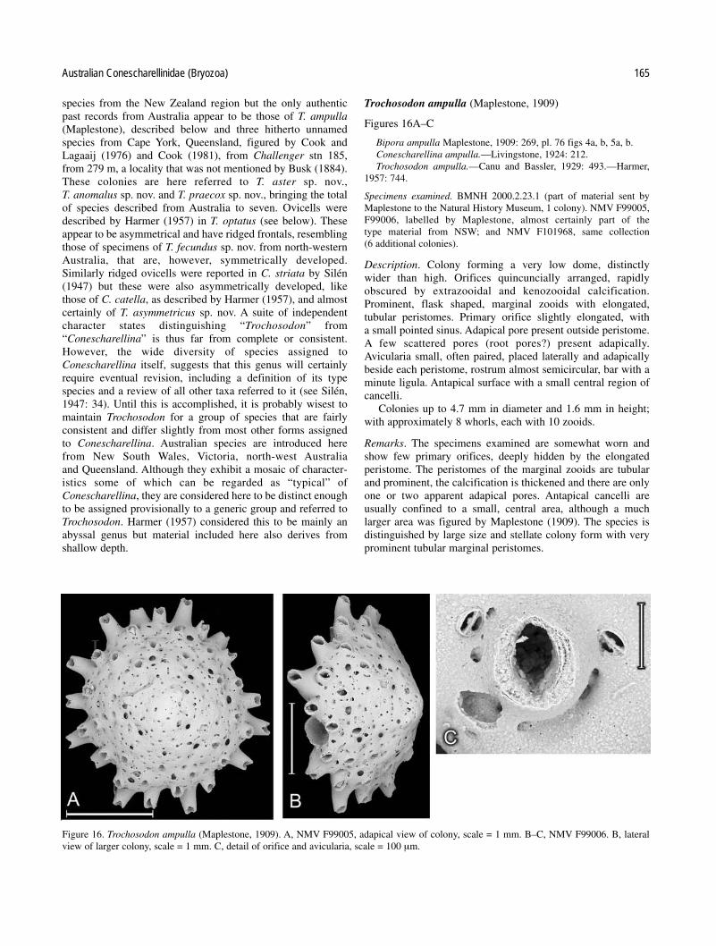
species from the New Zealand region but the only authenticpast records from Australia appear to be those of T. ampulla(Maplestone), described below and three hitherto unnamedspecies from Cape York, Queensland, figured by Cook andLagaaij (1976) and Cook (1981), from Challenger stn 185,from 279 m, a locality that was not mentioned by Busk (1884).These colonies are here referred to T. aster sp. nov., T. anomalus sp. nov. and T. praecox sp. nov., bringing the totalof species described from Australia to seven. Ovicells weredescribed by Harmer (1957) in T. optatus (see below). Theseappear to be asymmetrical and have ridged frontals, resemblingthose of specimens of T. fecundus sp. nov. from north-westernAustralia, that are, however, symmetrically developed.Similarly ridged ovicells were reported in C. striata by Silén(1947) but these were also asymmetrically developed, likethose of C. catella, as described by Harmer (1957), and almostcertainly of T. asymmetricus sp. nov. A suite of independentcharacter states distinguishing “Trochosodon” from“Conescharellina” is thus far from complete or consistent.However, the wide diversity of species assigned toConescharellina itself, suggests that this genus will certainlyrequire eventual revision, including a definition of its typespecies and a review of all other taxa referred to it (see Silén,1947: 34). Until this is accomplished, it is probably wisest tomaintain Trochosodon for a group of species that are fairly consistent and differ slightly from most other forms assigned to Conescharellina. Australian species are introduced here from New South Wales, Victoria, north-west Australia and Queensland. Although they exhibit a mosaic of character-istics some of which can be regarded as “typical” ofConescharellina, they are considered here to be distinct enoughto be assigned provisionally to a generic group and referred toTrochosodon. Harmer (1957) considered this to be mainly anabyssal genus but material included here also derives fromshallow depth.
Trochosodon ampulla (Maplestone, 1909)
Figures 16A–C
Bipora ampulla Maplestone, 1909: 269, pl. 76 figs 4a, b, 5a, b.Conescharellina ampulla.—Livingstone, 1924: 212.Trochosodon ampulla.—Canu and Bassler, 1929: 493.—Harmer,
1957: 744.
Specimens examined. BMNH 2000.2.23.1 (part of material sent byMaplestone to the Natural History Museum, 1 colony). NMV F99005,F99006, labelled by Maplestone, almost certainly part of the type material from NSW; and NMV F101968, same collection (6 additional colonies).
Description. Colony forming a very low dome, distinctly wider than high. Orifices quincuncially arranged, rapidlyobscured by extrazooidal and kenozooidal calcification.Prominent, flask shaped, marginal zooids with elongated, tubular peristomes. Primary orifice slightly elongated, with a small pointed sinus. Adapical pore present outside peristome.A few scattered pores (root pores?) present adapically.Avicularia small, often paired, placed laterally and adapicallybeside each peristome, rostrum almost semicircular, bar with aminute ligula. Antapical surface with a small central region ofcancelli.
Colonies up to 4.7 mm in diameter and 1.6 mm in height;with approximately 8 whorls, each with 10 zooids.
Remarks. The specimens examined are somewhat worn andshow few primary orifices, deeply hidden by the elongatedperistome. The peristomes of the marginal zooids are tubularand prominent, the calcification is thickened and there are onlyone or two apparent adapical pores. Antapical cancelli are usually confined to a small, central area, although a much larger area was figured by Maplestone (1909). The species isdistinguished by large size and stellate colony form with veryprominent tubular marginal peristomes.
Australian Conescharellinidae (Bryozoa) 165
Figure 16. Trochosodon ampulla (Maplestone, 1909). A, NMV F99005, adapical view of colony, scale = 1 mm. B–C, NMV F99006. B, lateralview of larger colony, scale = 1 mm. C, detail of orifice and avicularia, scale = 100 µm.

Specimens additional to the types suites of T. ampullahave not been reported since its first description. This, takentogether with Maplestone’s labelling of the NMV specimensand the occurrence in this collection of other, apparently uniquerecords from New South Wales, of Zeuglopora lanceolata etc.,strongly suggests that these specimens are part of the originaltype suite.
Trochosodon fecundus sp. nov.
Figures 17A–F
Holotype. NMV F99007, Dampier Archipelago, stn DA-2-75-02.Paratypes. NMV F99008, F99009, Dampier Archipelago, stn
DA-2-75-02.
Etymology. fecundus (L.) – fertile, prolific, with reference tothe numerous ovicells present in the specimens.
Diagnosis. Trochosodon with peristomes raised laterally andarranged quincuncially. Zooid orifices concealed, with a verywide, shallow sinus. Avicularia rounded. Ovicells prominent,symmetrical, with a thin marginal ectooecium. Root poreslunate.
Description. Colony forming a low cone, wider than high, withprominent peristomes, particularly at the margin. Calcificationsmooth to finely mamillate. Orifices in irregular, quincuncialseries, oval, with a pair of minute condyles that delineate abroad, very shallow sinus. Peristomes raised laterally andantapically, forming a partial, shallow tube. One aviculariumnear and lateral to each orifice, rostrum semicircular, often orientated adapically, with a bar but no ligula. Adapical poresymmetrically placed. Ovicells fragile, symmetrical, promi-nent, with an ectooecial layer visible marginally, that extendslaterally to form paired leaflike lobes above the orifice and thelateral part of the peristomes. Entooecium flat and smoothfrontally, with raised marginal striations forming a series ofpores where it meets the edge of the ectooecium. Small lunateroot pores present. Antapical surface with large cancelli.
Colonies with maximum diameter 2.25 mm and height 0.75mm, number of whorls 6–7, number of zooids per whorl 10–12.
Remarks. The extreme fragility of the ovicell calcificationmakes it impossible to treat specimens with bleach before electron microscopy. The striated ovicells are similar to those
166 P. E. Bock and P. L. Cook
Figure 17. T. fecundus sp. nov. A, NMV F99009, paratype. Detail of orifice and adapical pore, scale = 50 µm. B–D, NMV F99007, holotype. B,Adapical view of colony with ovicells and lunate root pores, scale = 500 µm. C. detail of orifices and avicularia, note one adapical pore withdeveloping ectooecial lamina (arrowed), scale = 200 µm. D, ovicells, orifices and lunate root pore, scale = 200 µm. E, NMV F99009, paratype.Adapical view of small colony with associated “anascan” ancestrula (arrowed), scale = 500 µm. F, NMV 99008, paratype, detail of ovicell, showing marginal ectooecium with lateral lappets and ridged entooecium, scale =100 µm.

figured in Trochosodon optatus by Harmer (1957: 747, pl. 48figs 16–18, text-figs 77, 78). As the locality from that thesecolonies were collected is off the north-west coast of Australia,it is therefore not very remote from the type locality of T. optatus, from the coast of Java (Siboga stn 318, KangeangIsland, 88 m). However, examination of two of the colonies
from this station (BMNH 1964.3.2.12 part), shows that theydiffer in having raised, radial series of zooid orifices, minuteavicularia, and only rare, adapical, lunate root pores. The principal differences occur in the relationships and position ofthe ovicells, that are not exactly as described by Harmer (1957).They are, in fact, asymmetrically developed, like the ovicells ofC. striata Silén (1947) but, unlike that and other similarspecies, have no obvious orifice. Instead, the ovicell opens intothe base of the peristome through a laterally placed foramen.The peristome is long and tubular and completely obscures theovicell opening. The frontal entooecium is striated, as figuredby Harmer, and resembles that of C. striata and T. fecundus.The ectooecial wall of the ovicell of T. optatus is closelyapposed to the walls of both the neighbouring peristomes; theovicells are wedged in between them and difficult to observe.The only other species observed in that the ovicell orifice opens into the peristome is T. praecox (see below), and that hassymmetrical ovicells.
The occurrence of an ancestrula with seven marginal spines(Fig. 17E) on the adapical centre of two colonies is unique.They are not referable to Trochosodon; this suggests that theyare extraneous and belong to another, possibly “anascan” species.
T. fecundus is known only from north-western Australiafrom 20 m.
Trochosodon asymmetricus sp. nov.
Figures 18, 19A–C
Holotype. NMV F99010, stn SLOPE-6 (colony with root).Paratypes. NMV F99011 (figured), stn SLOPE-6 (1 worn colony).Other specimens. NMV F101969, stn SLOPE-7 (1 colony).
Etymology. asymmetros (Gr.) – without symmetry, referring tothe position of the adapical pore.
Australian Conescharellinidae (Bryozoa) 167
Figure 18. Trochosodon asymmetricus sp.nov. NMV F99011, paratype.Adapical view of colony, scale = 1 mm.
Figure 19. Trochosodon asymmetricus sp.nov. NMV F99011, paratype. A, detail of orifices and asymmetrically placed, tubular adapical pores,scale =100 µm. B, adapical region showing rounded root pores and avicularia, scale = 200 µm. C, detail of marginal peristomes, orifices, adapi-cal pores, and avicularia, scale = 200 µm.

Diagnosis. Trochosodon with radial series of peristomes, alter-nating with minute, rounded avicularia. Zooid orifices deeplyconcealed. Adapical pore asymmetrically placed. Root porescircular.
Description. Colony domed, very small, wider than high; orifices apparently arranged radially; calcification granular.Peristomes raised, tubular, with intervening radial series ofminute, rounded avicularia; occasional, asymmetrically placedadapical avicularia; bar without ligula. Primary orifice oval,deeply concealed, with a short, rounded sinus. Adapical poretubular, present in peripheral and subperipheral zooids, asymmetrically placed inside the margin of the peristome.Ovicells inferred to be asymmetrical. Root pores adapical, circular, with a rim and 1 adjacent avicularium. Antapical sur-face with occasional short radial series of isolated cancelli,derived from the frontal septular pores of the antapical surfaceof the zooids.
Colony diameter 2.5 mm, height 1.5 mm, 5 whorls of 8–9zooids per whorl.
Remarks. T. asymmetricus is the only species among thoseexamined (except T. optatus, see above, and a few zooids of C. stellata), that exhibits an asymmetrically placed adapicalpore. No ovicells have been found but it may be inferred thatthese, too, would be in an asymmetrical position between therows of zooid orifices, as are the ovicells of T. optatus Harmer(1957), together with those of C. striata, C. brevirostris and C. longirostris of Silén (1947), as well as the specimensdescribed by Harmer (1957) assigned to C. catella Canu andBassler (1929). The tubular appearance of the adapical poreresembles that figured by Livingstone (1925) in “C. crassa”.There are more zooids per whorl than in Trochosodon anomalus but there are several closely similar characters sharedby these two species. Both have finely tuberculate calcificationand similar radial series of avicularia alternating with the orifices. The primary orifice is also almost identical in appear-ance (compare Figs 19A, 24F). However, the adapical pores arecompletely different in position, so it is inferred that the typesof ovicells would be an important distinction between the twotaxa. T. asymmetricus occurs from two adjacent stations fromthe New South Wales slope, from 770 to 1096 m.
Trochosodon diommatus sp. nov.
Figures 20, 21A–C
Holotype. NMV F99012, figured specimen, stn SLOPE-7.Paratype. NMV F99013, F99014, figured specimens, stn SLOPE-7.Other specimens. NMV F101970, stn SLOPE-6 (4 colonies, 3 very
young); NMV F101971, stn SLOPE-7 (22 colonies, 10 with roots);NMV F101972, stn SLOPE-45 (1 colony with root).
Etymology. di – two and ommatos – an eye (Gr.), referring tothe paired antapical peristomial avicularia.
Diagnosis. Trochosodon with stellate, radial peristomes, calci-fication smooth to finely tuberculate. Zooid orifices deeplyconcealed, with a narrow sinus. Frontal avicularia minute; a prominent pair on the antapical surface of the marginal peristomes.
Description. Colony stellate, fairly flat, distinctly wider thanhigh, with prominent marginal peristomes. Orifices quincuncialat first, becoming radial. Primary orifice at the base of the long,tubular but not prominent peristome, with an elongate, fairlynarrow sinus and large, paired condyles. An adapical pore present on the edge of the peristome of some peripheral zooids.Avicularia single, lateral and antapical between the peristomes,rostrum semicircular, with a bar but no ligula; other small avic-ularia scattered. Lunate root pores frequent in the adapicalregion, each with a pair of avicularia laterally. Antapical sur-face with marginal pores and avicularia; a pair of avicularia onthe antapical surface of each peristome (cf. C. ocellata and C. eburnea).
Colony with up to 4 whorls and 4–5 zooids per whorl.Diameter up to 4.7 mm, height up to 1.5 mm.
Remarks. Ovicells have not been seen in T. diommatus but thecentral position of the adapical pore suggests that they wouldbe symmetrical, like those of T. fecundus, rather than asymmet-rical, as in T. asymmetricus. Several colonies from stn SLOPE-7 have roots present; these are 0.5–1.0 mm long. T. diommatusis easily distinguished by the presence of the pair of minuteavicularia on the antapical side of the marginal zooid peris-tomes. It resembles two other species in the presence of antapi-cal peristomial avicularia. It differs completely from fossil C. ocellata in dimensions and arrangement of the zooid ori-fices, that have a longer, more acutely subtriangular sinus. Itdiffers from C. eburnea in its long peristomes and narrow ori-ficial sinus, as well as the form of its root pores. T. diommatus
168 P. E. Bock and P. L. Cook
Figure 20. Trochosodon diommatus sp. nov. NMV F99012, holotype.adapical view of colony showing lunate root pores, scale = 500 µm.

has a similar distribution to T. asymmetricus, with the additionof a record from 800 m depth off Tasmania.
Trochosodon aster sp. nov.
Figures 22A–C, 23
Trochosodon sp. 1.—Cook and Lagaaij, 1976, pl. 1 figs 3, 4.
Holotype. BMNH 1976.1.6.2 part, Challenger stn 185, Cape York,Queensland, 279 m.
Paratypes. BMNH 1976.1.6.2. part (20 colonies) and BMNH1969.1.2.2 (7 colonies). NMV F99015, F99016, F99017, and F101973,same locality (7 colonies).
Etymology. aster (L.) – a star, referring to the budding pattern.
Diagnosis and description. Colonies very small, stellate, bud-ded in alternating zooid triads early in astogeny, orificesbecoming radial later, fairly flat, but mamillate and raised cen-trally. Primary orifice almost circular, with a wide sinus, usual-ly obscured by the elongated peristome, that has a pair of small,rounded, lateral avicularia. Adapical pores present, root poreslunate, rare. Calcification mamillate, on adapical and antapicalsurfaces.
Colony diameter up to 2 mm, height 0.3 mm, number ofwhorls up to 4 and 3–4 zooids per whorl.
Remarks. The colonies from Cape York are heavily calcifiedand often somewhat worn. They range in size from 0.25 mm tonearly 2 mm in diameter and have long marginal peristomesthat bear small avicularia laterally. One colony, figured byCook and Lagaaij (1976), shows lunate root pores among theadapical mamillae. T. aster resembles T. pacificum Lu (1991:74, pl. 21 fig. 4) from the South China Sea but differs in thepresence of lunate root pores and minute lateral peristomialavicularia. T. aster also has some characteristics similar tothose described for T. linearis from the East Indies by Harmer(1957). Two of his specimens have been examined (BMNH1964.3.2.10, Strait of Makassar, Siboga stn 88, 1301 m, and1964.3.2.11 , the Banda Sea, stn 227, 2081 m). These are pro-portionally larger than T. aster, with bilabiate peristomes. Onecolony was figured by Harmer (1964.3.2.11, p1. 48 fig. 14,text-fig.75), who gave a very detailed description of the earlyastogeny. The colony has a central, rounded root pore. Canuand Bassler’s (1929) unique type specimen of T. linearis wasdredged from 635 m depth, from Borneo. The description is notadequate to decide its synonymy with Harmer’s specimens, thathe could not have compared with the type.
Trochosodon anomalus sp. nov.
Figures 24A–F
Holotype. NMV F99018 stn SLOPE-7.Paratypes. NMV F99019, stn SLOPE-7 (2 colonies).Other specimens. BMNH 1976.1.6.2, Challenger stn 185, Cape
York, Australia, 279 m (26 colonies); NMV F101974, Challenger stn185 (10 colonies).
Etymology. From anomalos (Gr.) – irregular, inconsistent, deviat-ing, with reference to the combination of character states foundin several genera, that are uniquely possessed by this species.
Australian Conescharellinidae (Bryozoa) 169
Figure 21. Trochosodon diommatus sp. nov. A. NMV F99012, holo-type. Detail of orifices and adapical pores, scale = 200 µm. B, NMVF99013, paratype, antapical view of marginal peristomes, showingpaired avicularia, scale =200 µm. C. NMV F99014, paratype, detail ofperistome, orifice, and lunate root pores, scale = 200 µm.
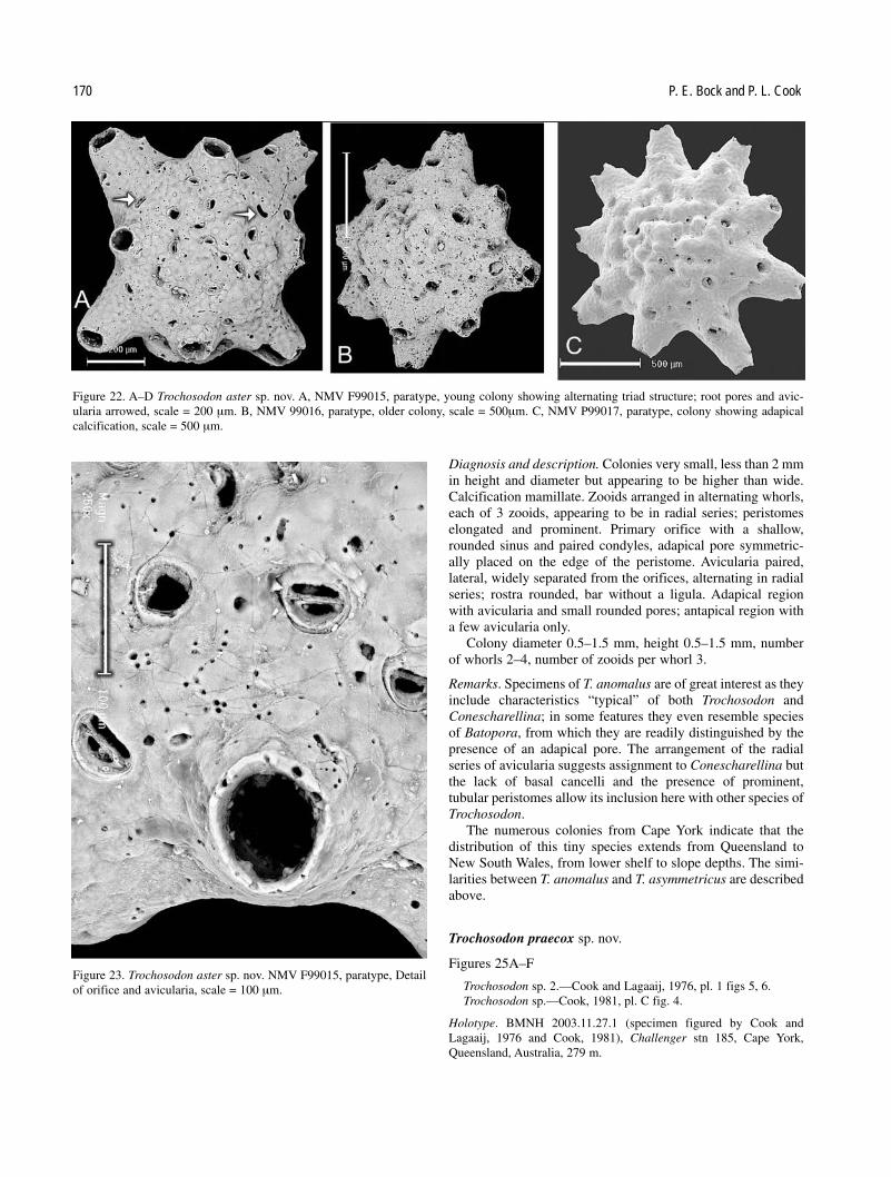
Diagnosis and description. Colonies very small, less than 2 mmin height and diameter but appearing to be higher than wide.Calcification mamillate. Zooids arranged in alternating whorls,each of 3 zooids, appearing to be in radial series; peristomeselongated and prominent. Primary orifice with a shallow,rounded sinus and paired condyles, adapical pore symmetric-ally placed on the edge of the peristome. Avicularia paired, lateral, widely separated from the orifices, alternating in radialseries; rostra rounded, bar without a ligula. Adapical regionwith avicularia and small rounded pores; antapical region witha few avicularia only.
Colony diameter 0.5–1.5 mm, height 0.5–1.5 mm, numberof whorls 2–4, number of zooids per whorl 3.
Remarks. Specimens of T. anomalus are of great interest as theyinclude characteristics “typical” of both Trochosodon andConescharellina; in some features they even resemble speciesof Batopora, from which they are readily distinguished by thepresence of an adapical pore. The arrangement of the radialseries of avicularia suggests assignment to Conescharellina butthe lack of basal cancelli and the presence of prominent, tubular peristomes allow its inclusion here with other species ofTrochosodon.
The numerous colonies from Cape York indicate that thedistribution of this tiny species extends from Queensland toNew South Wales, from lower shelf to slope depths. The simi-larities between T. anomalus and T. asymmetricus are describedabove.
Trochosodon praecox sp. nov.
Figures 25A–F
Trochosodon sp. 2.—Cook and Lagaaij, 1976, pl. 1 figs 5, 6.Trochosodon sp.—Cook, 1981, pl. C fig. 4.
Holotype. BMNH 2003.11.27.1 (specimen figured by Cook andLagaaij, 1976 and Cook, 1981), Challenger stn 185, Cape York,Queensland, Australia, 279 m.
170 P. E. Bock and P. L. Cook
Figure 22. A–D Trochosodon aster sp. nov. A, NMV F99015, paratype, young colony showing alternating triad structure; root pores and avic-ularia arrowed, scale = 200 µm. B, NMV 99016, paratype, older colony, scale = 500µm. C, NMV P99017, paratype, colony showing adapical calcification, scale = 500 µm.
Figure 23. Trochosodon aster sp. nov. NMV F99015, paratype, Detailof orifice and avicularia, scale = 100 µm.
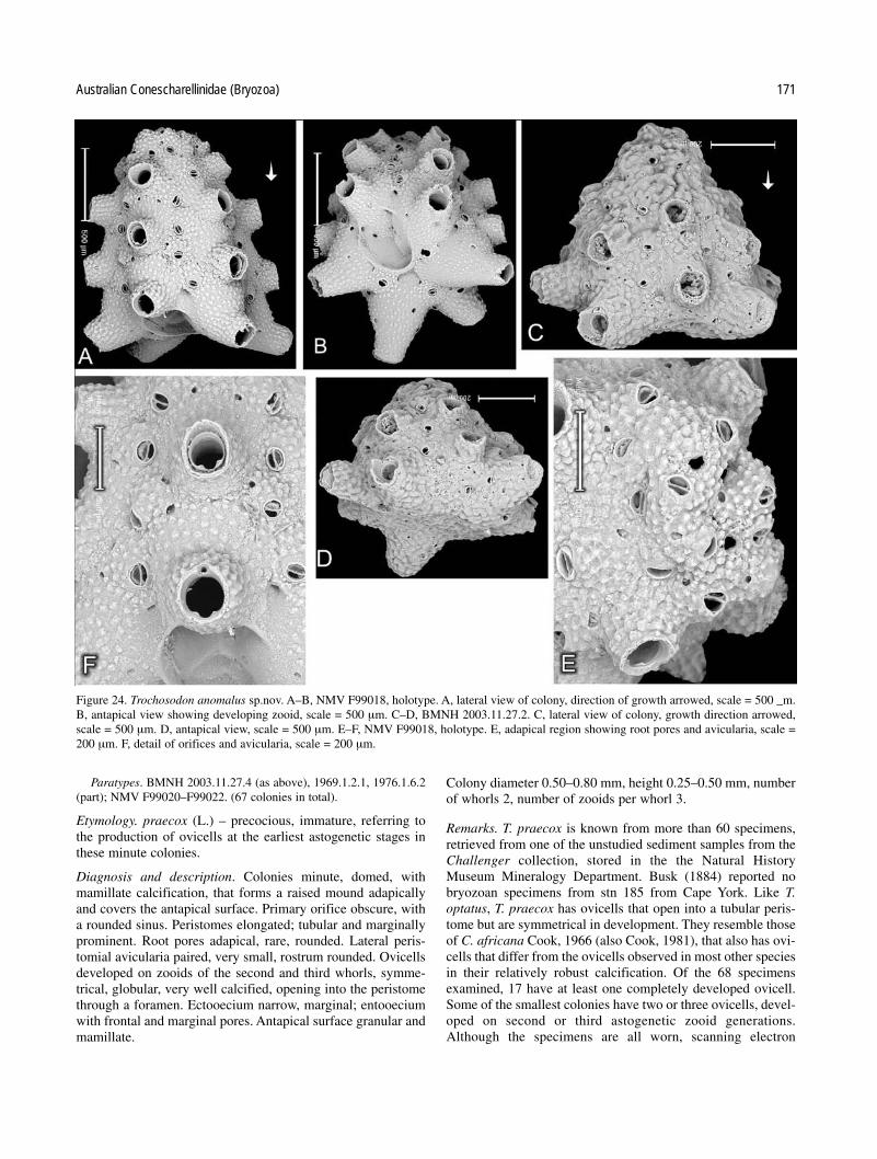
Paratypes. BMNH 2003.11.27.4 (as above), 1969.1.2.1, 1976.1.6.2(part); NMV F99020–F99022. (67 colonies in total).
Etymology. praecox (L.) – precocious, immature, referring tothe production of ovicells at the earliest astogenetic stages inthese minute colonies.
Diagnosis and description. Colonies minute, domed, withmamillate calcification, that forms a raised mound adapicallyand covers the antapical surface. Primary orifice obscure, witha rounded sinus. Peristomes elongated; tubular and marginallyprominent. Root pores adapical, rare, rounded. Lateral peris-tomial avicularia paired, very small, rostrum rounded. Ovicellsdeveloped on zooids of the second and third whorls, symme-trical, globular, very well calcified, opening into the peristomethrough a foramen. Ectooecium narrow, marginal; entooeciumwith frontal and marginal pores. Antapical surface granular andmamillate.
Colony diameter 0.50–0.80 mm, height 0.25–0.50 mm, numberof whorls 2, number of zooids per whorl 3.
Remarks. T. praecox is known from more than 60 specimens,retrieved from one of the unstudied sediment samples from theChallenger collection, stored in the the Natural HistoryMuseum Mineralogy Department. Busk (1884) reported nobryozoan specimens from stn 185 from Cape York. Like T.optatus, T. praecox has ovicells that open into a tubular peris-tome but are symmetrical in development. They resemble thoseof C. africana Cook, 1966 (also Cook, 1981), that also has ovi-cells that differ from the ovicells observed in most other speciesin their relatively robust calcification. Of the 68 specimensexamined, 17 have at least one completely developed ovicell.Some of the smallest colonies have two or three ovicells, devel-oped on second or third astogenetic zooid generations.Although the specimens are all worn, scanning electron
Australian Conescharellinidae (Bryozoa) 171
Figure 24. Trochosodon anomalus sp.nov. A–B, NMV F99018, holotype. A, lateral view of colony, direction of growth arrowed, scale = 500 _m.B, antapical view showing developing zooid, scale = 500 µm. C–D, BMNH 2003.11.27.2. C, lateral view of colony, growth direction arrowed,scale = 500 µm. D, antapical view, scale = 500 µm. E–F, NMV F99018, holotype. E, adapical region showing root pores and avicularia, scale =200 µm. F, detail of orifices and avicularia, scale = 200 µm.

microscopy has revealed details of ovicell development. Zooidsapparently develop an elongated peristome, with a foramen onits adapical surface. This is in contact with an adapical pore(Fig. 25E) at its edge. An ectooecial and an entooecial laminathen grow together, one “below” the other, from the adapicalpore in the adapical direction (Fig. 25C).The two laminae thencurve in an antapical direction, forming a capsule. The fusionof the laminae with the lateral and antapical edges of the fora-men finally closes it, forming the complete ovicell (Fig. 25B).It should be noted that this does not produce a peristomial ovi-cell, the walls of which are expansions of the frontal shield. The
ovicells of T. praecox comprise a separate development ofectooecium and entooecium. Other characteristics that are distinct enough for colonies to be recognised from additional,better preserved material, should it ever become available, arealso revealed by scanning electron microscopy. The minute sizeof the colonies of T. praecox is comparable to those of accom-panying foraminiferans; Cook (1981) noted the close similarityin appearance among them.
The occurrence of reproductive precocity in interstitial bryozoans with very small colonies has been described and discussed by Winston and Håkansson (1986: 43).
172 P. E. Bock and P. L. Cook
Figure 25. T. praecox sp. nov., Cape York. A, NMV F99020, paratype, detail of orifices and avicularia. with two complete ovicells and one peristomial foramen (arrowed), scale = 200 µm. B–C, NMV F99022, paratype, B, marginal zooids of colony with complete ovicell, avic-ularia arrowed, scale = 200 µm. C, detail of ovicells, showing ectooecial margin and central foramina of entooecium (arrowed); note avicularia,scale = 100 µm. D–E, NMV F99021, paratype. D, young colony with one peristomial foramen, root pores arrowed, scale = 200 µm. E, detail offoramen and adapical pore (arrowed), scale = 100 µm. F, NMV P99022, paratype. Colony with one complete and two developing ovicells(arrowed), scale = 200 µm.

Trochosodon gordoni sp. nov.
Trochosodon multiarmatus.—Gordon, 1989: 83, p1. 49 figs D–F(not Bipora multiarmata Maplestone, 1909: 268).
Etymology. Named for Dr Dennis P. Gordon.
Description. Colony domed, wider than high, calcification fine-ly mamillate. Zooid orifices in quincunx, sinus rounded, peris-tomes raised slightly marginally. Rounded avicularia and largefrontal septular pores scattered among orifices. Circular rootpores adapical. Antapical surface with a few avicularia.
Remarks. Gordon (1989) identified specimens from NewZealand as Maplestone’s species and assigned them all toTrochosodon. He appears to have confused the dimensions ofhis colonies with those of Conescharellina multiarmata, thatare always “higher than wide”, not “typically wider than high”,as he described. The broad orifice sinus of the New Zealandspecies is also quite unlike that of C. multiarmata. Gordon’sspecies lacks any basal cancelli and has one or two large, cir-cular, central rhizoid pores adapically. It appears to be referableto Trochosodon but certainly not to Conescharellina multiar-mata. Gordon figured a specimen from Station P927(40°50.1'S, 168°14.8'E, 1005–1009 m, western South Island,New Zealand) and reported it from numerous other localitiesfrom southern New Zealand, from a range of 540 to 1676 mdepth.
Crucescharellina Silén, 1947
Crucescharellina Silén, 1947: 44.Agalmatozoum Harmer, 1957: 757.
Type species. Crucescharellina japonica Silén, 1947 (originaldesignation).
Description. Colonies are cruciform or star-shaped and mayhave branches that bifurcate terminally. The adapical zooid orifices are sinuate and interspersed with lunate or rounded rootpores. The antapical growing edges are positioned at the limitsof the branches but an antapical surface, that is the equivalentof the exposed frontal wall of conescharelliniform colonies, isalso continuous and present on the “lower, non-orificial side”of colonies. It is inferred that the colonies live, in fact, with thisantapical surface upward with the orifices directed downward,because the rhizoids that occur among them are inferred toanchor the colonies above or into the surface of the bottom sediments. Rounded or acute avicularia occur, that are occa-sionally large and spathulate. The orifices possess an adapicalpore but ovicells have not been seen. Roots were figured in C. japonica by Silén (1947: pl. 1 fig. 11) and the position of theroot pores suggests that the mode of life is similar to thatinferred for the genus Euginoma (Hayward 1978), that alsooccurs from abyssal depths (d’Hondt and Schopf ,1984).
Remarks. Crucescharellina was introduced by Silén (1947) forC. japonica from near the Goto Islands, Japan, from a depth of175 m. Only one colony was found; it was stellate but eachbranch originated from a narrow neck, one or two zooids inwidth. The branches rapidly expanded and then bifurcated,each subbranch starting with one or two zooids. The
subbranches also expanded rapidly, so that within two astogenetic generations, the segments were 4 zooids wide. Lunateroot pores were present but these were not associated withbranch bifurcations and no large, spathulate avicularia weredescribed. Gordon and d’Hondt (1997: 73, figs 221–223)described “C. japonica” from the Philippines from 640–668 m.They too, had only one colony. It differed in having much lessexpanded branches, regular lunate root pores, and rare largeaxillary avicularia. The primary orifice had a shallow sinus andpaired condyles. Silén (1947: 44) stated that he referredTrochosodon decussis Canu and Bassler (1929: 495, pl. 71 figs7–10, from 456 m, east of Mindanao in the Philippines) to hisgenus Crucescharellina. Harmer (1957) was unaware of Silén’swork, that was not available to him during the war of1939–1945, and introduced Agalmatozoum for Trochosodondecussis Canu and Bassler (1929). Colonies of this specieswere cruciform, with triserial branches, and were describedwith lunate root pores and an elliptical secondary orifice.Avicularia or small pores were present antapically but no largeavicularia were mentioned in the original description. Harmer(1957) listed more than ten colonies of A. decussis from sevenlocalities in the Sulu, Banda, and Celebes Seas. The depthswere nearly all abyssal, ranging from 535 to 3112 m. Thebranches of the colonies were mostly biserial and the root poreswere circular, placed regularly at bifurcations, and surroundedby a ring of small avicularia. In addition, large, axillary spathu-late avicularia sometimes occurred on the lateral sides ofbranches. The species from the Siboga area described byHarmer (1957) as A. decussis strongly resemblesCrucescharellina australis from Australia described below, notthe original form from the Philippines described by Canu andBassler (1929). Gordon and d’Hondt (1997: 74, figs 224–227)introduced another very similar stellate species, C. aster, withbiserial branches, from several New Caledonian and NewZealand localities at a depth range of 760 to 1573 m. The rootpores were central and rounded but no large avicularia werepresent. Their material included numerous colonies, that theynoted resembled “clusters of snowflakes”. A single preparationof a colony in the Natural History Museum collection (BMNH1963.8.18.18) closely resembles the description of C. aster buthas slightly more extended, spiny peristomes. The specimen isfrom Challenger stn 169, off New Zealand (37°34'S, 179°22'E,1295 m), a station that was not mentioned by Busk (1884).Gordon (1989: 84, pl. 1E figs 50B–E) described another biser-ial species, C. jugalis, from northern New Zealand, from adepth range of 1217–1357 m. The colonies were irregularlybranched but had circular root pores very similar to those of theAustralian C. australis and A. decussis sensu Harmer (1957).
Although there is no doubt of the synonymy of the two genera Crucescharellina and Agalmatozoum, there are uncer-tainties as to the identity of the various taxa referred to them inthese previous descriptions. Among other records, Cook (1981)figured one of two very young, cruciform colonies from CapeYork, from 279 m (BMNH 1976.1.6.2, part), as Agalmatozoumspecies. These, with the specimens of C. australis describedhere from Point Hicks, Victoria and from eastern Tasmania,remain the only records of Crucescharellina from Australianwaters to date.
Australian Conescharellinidae (Bryozoa) 173
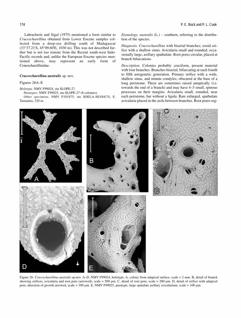
Labracherie and Sigal (1975) mentioned a form similar toCrucescharellina obtained from Lower Eocene samples col-lected from a deep-sea drilling south of Madagascar(33°37.21'S, 45°09.60'E, 1030 m). This was not described fur-ther but is not too remote from the Recent south-west Indo-Pacific records and, unlike the European Eocene species men-tioned above, may represent an early form ofConescharellinidae.
Crucescharellina australis sp. nov.
Figures 26A–E
Holotype. NMV F99024, stn SLOPE-27.Paratypes. NMV F99025, stn SLOPE-27 (8 colonies).Other specimens. NMV F101975, stn SOELA-S03/84/74, E.
Tasmania, 320 m.
Etymology. australis (L.) – southern, referring to the distribu-tion of the species.
Diagnosis. Crucescharellina with biserial branches; zooid ori-fice with a shallow sinus. Avicularia small and rounded; occa-sionally large, axillary spathulate. Root pores circular, placed atbranch bifurcations.
Description. Colonies probably cruciform, present materialwith four branches. Branches biserial, bifurcating at each fourthto fifth astogenetic generation. Primary orifice with a wide,shallow sinus, and minute condyles; obscured at the base of along peristome. These are sometimes raised antapically (i.e.towards the end of a branch) and may have 4–5 small, spinousprocesses on their margins. Avicularia small, rounded, neareach peristome, bar without a ligula. Rare enlarged, spathulateavicularia placed in the axils between branches. Root pores reg-
174 P. E. Bock and P. L. Cook
Figure 26. Crucescharellina australis sp.nov. A–D, NMV F99024, holotype. A, colony from adapical surface, scale = 2 mm. B, detail of branchshowing orifices, avicularia and root pore (arrowed), scale = 500 µm. C, detail of root pore, scale = 200 µm. D, detail of orifice with adapicalpore, direction of growth arrowed, scale = 100 µm. E, NMV F99025, paratype, large spatulate axillary avicularium, scale = 100 µm.

ularly placed at bifurcation of branches; circular, with a distinct rim, surrounded by 3–4 small avicularia. Adapical porewithin the calcification of the edge of the peristome, present inmany zooids, but no ovicells seen. Antapical surface finelygranular, with approximately one small rounded aviculariumper zooid.
Branches 0.6 mm width, length 6 mm.
Remarks. The specimens from stn SLOPE-27 comprise ninecolony fragments. The largest show evidence of having oncebeen cruciform but only four complete branches are now present. Two colonies have enlarged, spathulate avicularia inthe axil between two branches, very similar to that shown in his“A. decussis” by Harmer (1957, pl. 49 fig. 13). The colonystructure, type and distribution of root pores, and the large avic-ularia of C. australis make it virtually certain that it is con-specific with some specimens of A. decussis sensu Harmer butit is distinct from T. decussis Canu and Bassler (1929) and theother species mentioned above. Some fragmentary preparationsof Harmers’ Siboga material, labelled A. decussis, from theBMNH collection, have been examined, The specimens are allslightly worn, and none possesses large avicularia. A cruciformcolony and fragments from Siboga stn 211 (BMNH1964.3.2.21, south of Celebes, 1158 m.), most closely resem-bles C australis. Other fragments from Siboga stn 102 (BMNH1964.3.2.23, Sulu Archipelago, 535 m.) differ in having triser-ial branches and distinctly elongated avicularia near the zooidorifices. Some partially decalcified fragments from Siboga stn221 (BMNH 1964.3.2.25, Banda Sea, 2798–3112 m.) alsoresemble C. australis but have raised peristomes on the anta-pical side of the orifices. They possess three long roots (over 20mm), that emanate from the root pores. It is possible thatHarmer’s material, identified as A. decussis, may belong tomore than one species. Circular root pores surrounded by smallavicularia also occur in Conescharellina eburnea, C. plana,C. perculta and C. humerus.
Zeuglopora Maplestone, 1909
Zeuglopora Maplestone, 1909: 272.—Canu and Bassler, 1929:510.—Harmer, 1957: 755.
Type species. Zeuglopora lanceolata Maplestone, 1909 (original designation).
Description. Colony similar to Flabellopora, ligulate, appar-ently composed of 2 laminae but in fact consisting of a pair ofalternating and interdigitating expanses of frontally buddedzooid series. Single or small groups of marginal zooidsenlarged and prominent, forming a serrated edge, occasionallywith enlarged avicularia. Orifices oval, with paired condylesforming a subtriangular antapical sinus; peristome tubular. Arounded adapical pore is present but ovicells are unknown.Avicularia usually small, rounded, with a bar but no ligula.Colony anchored by 1 or 2 roots, arising from lunate pores inthe adapical region.
Remarks. Both Canu and Bassler (1929) and Harmer (1957)analysed the colony structure of Zeuglopora and maintained itsdistinction from Flabellopora.
Zeuglopora lanceolata Maplestone, 1909
Figures 27, 28A–C
Zeuglopora lanceolata Maplestone, 1909: 272, pl. 78, fig. 11.—Harmer, 1957: 757.
Bipora lanceolata.—Livingstone, 1924: 211.
Specimens examined. NMV F99026, 1 colony labelled byMaplestone, probably part of the type material from NSW.
Diagnosis and description. As for the genus, serrated edges ofcolony formed by prominent zooids, that occur in alternatingunequal pairs. Primary orifice with a subtriangular sinus andwell developed paired condyles; obscured at the base of a tubular peristome, that is most prominent adapically. Arounded adapical pore, at a little distance from the edge of theperistome, is present in some central and antapical zooid orifices. Surface of zooids mammilate, interspersed with minute, rounded avicularia, with a bar but no ligula. Root poreslunate, adapical, paired, large and surrounded by extrazooidalcalcification.
Colony length 7 mm, breadth 2.25 mm. Number of astoge-netic generations 12–13, number of zooids per generation 10.
Remarks. The single colony from New South Wales resemblesMaplestone’s (1909) description. The orifices of the enlargedmarginal zooids are surrounded by up to five small aviculariaand resemble the root pores of C. eburnea and C. plana.However, the adapical end of the colony shows that the actualroot pores resemble those of Flabellopora and consist of largelunate pores surrounded by massive, secondary extrazooidalcalcification. The adapical pore is large and a little offset inposition towards the colony margin. It seems probable that anyovicell would be slightly asymmetrically placed. Harmer(1957: 737) examined a colony that was part of Maplestone’s(1909) type material in the BMNH collection (BMNH1909.11.12.3). It has a large adapical foramen that was filledwith detritus. It seems almost certain that this colony actuallypossessed paired, lunate adapical root pores like other speci-mens (Fig. 28C). There are no additional records of Z. lanceo-lata from Australia but Canu and Bassler (1929: 511, pl. 75, fig.6) described a very similar colony from deep water (630 m)north-east of Borneo as Z. lanceolata, an identification accepted by Harmer (1957). The locality was remote fromAustralia; the figured colony is slightly narrower than thosefrom Australia, with more prominent marginal zooids than thetype specimens. The character of the primary orifices, rootpores and avicularia are uncertain. Harmer (1957) alsodescribed a new species, Z. arctata, from two minute coloniesfrom 82 m off Java, where the bottom sediment consisted offine grey mud. Each colony had large marginal avicularia, anda long single root emanating from the adapical region. Cadée(1987: 52) noted the occurrence of several hundred colonies ofZ. arctata together with an undescribed species from soft-bottom sediments in the Banda Sea but gave no detaileddescriptions. Lu (1991) described several species of Zeuglo-pora (as Bipora) from the South China Sea. His B. pagoda(p.70, pl. 18 fig. 2) and B. trinodata (p. 71, pl. 18 fig. 3) bothresemble the unnamed species mentioned by Cadée (1987).
Australian Conescharellinidae (Bryozoa) 175
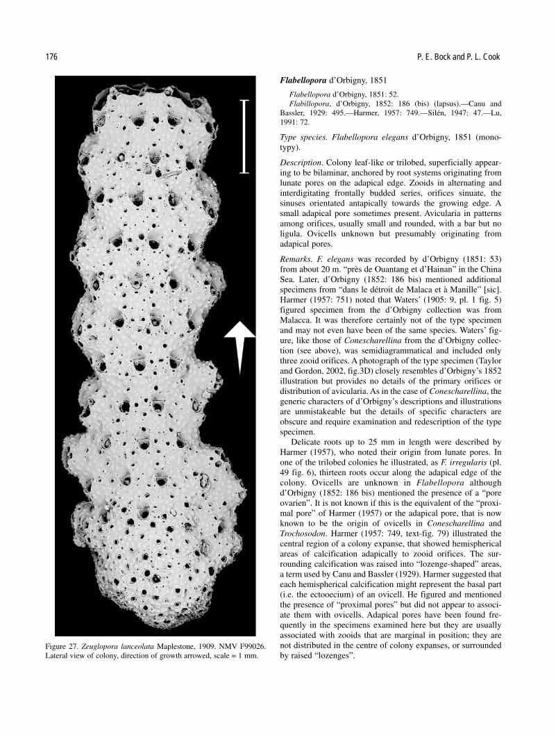
Flabellopora d’Orbigny, 1851
Flabellopora d’Orbigny, 1851: 52.Flabillopora, d’Orbigny, 1852: 186 (bis) (lapsus).—Canu and
Bassler, 1929: 495.—Harmer, 1957: 749.—Silén, 1947: 47.—Lu,1991: 72.
Type species. Flabellopora elegans d’Orbigny, 1851 (mono-typy).
Description. Colony leaf-like or trilobed, superficially appear-ing to be bilaminar, anchored by root systems originating fromlunate pores on the adapical edge. Zooids in alternating andinterdigitating frontally budded series, orifices sinuate, thesinuses orientated antapically towards the growing edge. Asmall adapical pore sometimes present. Avicularia in patternsamong orifices, usually small and rounded, with a bar but noligula. Ovicells unknown but presumably originating fromadapical pores.
Remarks. F. elegans was recorded by d’Orbigny (1851: 53)from about 20 m. “près de Ouantang et d’Hainan” in the ChinaSea. Later, d’Orbigny (1852: 186 bis) mentioned additionalspecimens from “dans le détroit de Malaca et à Manille” [sic].Harmer (1957: 751) noted that Waters’ (1905: 9, pl. 1 fig. 5)figured specimen from the d’Orbigny collection was fromMalacca. It was therefore certainly not of the type specimenand may not even have been of the same species. Waters’ fig-ure, like those of Conescharellina from the d’Orbigny collec-tion (see above), was semidiagrammatical and included onlythree zooid orifices. A photograph of the type specimen (Taylorand Gordon, 2002, fig.3D) closely resembles d’Orbigny’s 1852illustration but provides no details of the primary orifices ordistribution of avicularia. As in the case of Conescharellina, thegeneric characters of d’Orbigny’s descriptions and illustrationsare unmistakeable but the details of specific characters areobscure and require examination and redescription of the typespecimen.
Delicate roots up to 25 mm in length were described byHarmer (1957), who noted their origin from lunate pores. Inone of the trilobed colonies he illustrated, as F. irregularis (pl.49 fig. 6), thirteen roots occur along the adapical edge of thecolony. Ovicells are unknown in Flabellopora althoughd’Orbigny (1852: 186 bis) mentioned the presence of a “poreovarien”. It is not known if this is the equivalent of the “proxi-mal pore” of Harmer (1957) or the adapical pore, that is nowknown to be the origin of ovicells in Conescharellina andTrochosodon. Harmer (1957: 749, text-fig. 79) illustrated thecentral region of a colony expanse, that showed hemisphericalareas of calcification adapically to zooid orifices. The sur-rounding calcification was raised into “lozenge-shaped” areas,a term used by Canu and Bassler (1929). Harmer suggested thateach hemispherical calcification might represent the basal part(i.e. the ectooecium) of an ovicell. He figured and mentionedthe presence of “proximal pores” but did not appear to associ-ate them with ovicells. Adapical pores have been found fre-quently in the specimens examined here but they are usuallyassociated with zooids that are marginal in position; they arenot distributed in the centre of colony expanses, or surroundedby raised “lozenges”.
176 P. E. Bock and P. L. Cook
Figure 27. Zeuglopora lanceolata Maplestone, 1909. NMV F99026.Lateral view of colony, direction of growth arrowed, scale = 1 mm.

Flabellopora umbonata (Haswell, 1881)
Figures 29A–C
Eschara umbonata Haswell, 1881: 41, pl. 2 figs 5, 6.Bipora umbonata.—Whitelegge, 1887: 345.—Livingstone, 1924:
209.—Livingstone, 1926: 98, pl. 5 figs 4, 5.Bipora mamillata Maplestone, 1909: 270, pl. 77 fig. 7.Conescharellina mamillata.—Bretnall, 1922: 191.
Specimens examined. NMV F99395, stn SLOPE-40 (1 colony); NMV F101976, stn GAB-020 (2 young and 1 trilobed colony withroots, plus fragments); NMV F101977, stn GAB-030 (2 young and 2 trilobed colonies with roots, plus fragments); NMV F101978, stnGAB-045 (1 trilobed colony with roots, plus fragments); NMVF101979, stn GAB-056 (1 colony with fragments); NMV F101980, stnGAB-067 (1 trilobed colony); NMV F101981, stn GAB-074 (5 fragments); NMV F101982, stn GAB-084 (2 trilobed colonies plusfragments); NMV F101983, stn GAB-088 (1 colony plus fragments); NMV F101984, stn GAB-093 (1 large colony); NMVF101985, stn GAB-112 (1 colony); NMV F101986, stn GAB-117 (1 trilobed colony plus fragments); NMV F101987, stn GAB-119 (2 young and 3 trilobed colonies); NMV F101988, stn GAB-128 (1 trilobed colony); NMV F101989, stn SOELA-S03/84/74, E.Tasmania, 320 m (1 colony).
Description. Colony leaf-shaped, sometimes trilobed. Zooidfrontal shield continuous, without zooid borders; calcificationsmooth with umbonate mamillae occurring among the orificesand the avicularia. Orifices almost circular, patent, with arounded sinus, peristomial rim raised, narrow; adapical pores present. Avicularia small, rostra subtriangular or rounded,bar without a ligula. Septular pores rare, scattered. Root pores lunate, on the adapical edge, surrounded by thickenedcalcification.
Remarks. Livingstone (1924, 1926) and Harmer (1957) exam-ined specimens from Queensland that were reported to be fromHaswell’s original material, although only fragments of thiswere preserved. Maplestone’s (1909) type specimen of Biporamamillata was unique but Livingstone (1924) mentioned“types” that may have had a different provenance. BothLivingstone and Harmer were convinced that F. umbonata wasidentical with B. mamillata, Maplestone (1909) however hadnoted some differences, both within Haswell’s suite of speci-mens and between them and his colonies from New South Wales.
The numerous specimens examined here are often slightlyworn and very few possess roots. There are small differencesamong specimens but these seem to be the result of astogenet-ic position, ontogenetic thickening and wear. Some specimenshave larger umbonate mamillae among orifices than others andsome have larger avicularia but none of these differences is cor-related with locality. The specimens from stations GAB-020,GAB-030 and GAB-119 include several very young colonies.These are lanceolate and consist principally of parallel series ofantapically directed zooids with few laterally inclined series.Later growth illustrated the development of paired lateral lam-inae, giving the typically trilobed shape. One regeneratedcolony from station GAB-093 has a diameter of 28 mm and hasdeveloped nine thickened rays from an irregular central area. Itis possible that other species of Flabellopora occur inAustralian waters but have not yet been recognised.
Ptoboroa Gordon and d’Hondt, 1997
Ptoboroa Gordon and d’Hondt, 1997: 70, pl. 47F, G, 48A.
Type species. Trochosodon pulchrior Gordon, 1989: 81 (original designation).
Australian Conescharellinidae (Bryozoa) 177
Figure 28. Zeuglopora lanceolata Maplestone, 1909. NMV F99026. A, detail of orifice with adapical pore, scale = 100 µm. B, detail of large,marginal zooid orifice with surrounding avicularia, direction of growth arrowed, scale = 200 µm. C. adapical region showing lunate root pores,scale =200 µm.
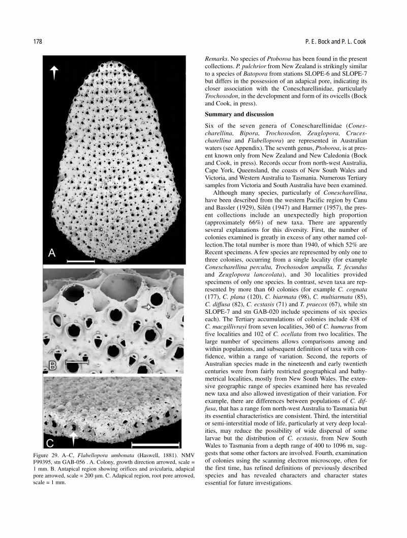
Remarks. No species of Ptoboroa has been found in the presentcollections. P. pulchrior from New Zealand is strikingly similarto a species of Batopora from stations SLOPE-6 and SLOPE-7but differs in the possession of an adapical pore, indicating itscloser association with the Conescharellinidae, particularlyTrochosodon, in the development and form of its ovicells (Bockand Cook, in press).
Summary and discussion
Six of the seven genera of Conescharellinidae (Cones-charellina, Bipora, Trochosodon, Zeuglopora, Cruces-charellina and Flabellopora) are represented in Australianwaters (see Appendix). The seventh genus, Ptoboroa, is at pres-ent known only from New Zealand and New Caledonia (Bockand Cook, in press). Records occur from north-west Australia,Cape York, Queensland, the coasts of New South Wales andVictoria, and Western Australia to Tasmania. Numerous Tertiarysamples from Victoria and South Australia have been examined.
Although many species, particularly of Conescharellina,have been described from the western Pacific region by Canuand Bassler (1929), Silén (1947) and Harmer (1957), the pres-ent collections include an unexpectedly high proportion(approximately 66%) of new taxa. There are apparently several explanations for this diversity. First, the number ofcolonies examined is greatly in excess of any other named col-lection.The total number is more than 1940, of which 52% areRecent specimens. A few species are represented by only one tothree colonies, occurring from a single locality (for exampleConescharellina perculta, Trochosodon ampulla, T. fecundusand Zeuglopora lanceolata), and 30 localities provided specimens of only one species. In contrast, seven taxa are rep-resented by more than 60 colonies (for example C. cognata(177), C. plana (120), C. biarmata (98), C. multiarmata (85),C. diffusa (82), C. ecstasis (71) and T. praecox (67), while stnSLOPE-7 and stn GAB-020 include specimens of six specieseach). The Tertiary accumulations of colonies include 438 of C. macgillivrayi from seven localities, 360 of C. humerus fromfive localities and 102 of C. ocellata from two localities. Thelarge number of specimens allows comparisons among andwithin populations, and subsequent definition of taxa with con-fidence, within a range of variation. Second, the reports ofAustralian species made in the nineteenth and early twentiethcenturies were from fairly restricted geographical and bathy-metrical localities, mostly from New South Wales. The exten-sive geographic range of species examined here has revealednew taxa and also allowed investigation of their variation. Forexample, there are differences between populations of C. dif-fusa, that has a range fom north-west Australia to Tasmania butits essential characteristics are consistent. Third, the interstitialor semi-interstitial mode of life, particularly at very deep local-ities, may reduce the possibility of wide dispersal of some larvae but the distribution of C. ecstasis, from New SouthWales to Tasmania from a depth range of 400 to 1096 m, sug-gests that some other factors are involved. Fourth, examinationof colonies using the scanning electron microscope, often forthe first time, has refined definitions of previously describedspecies and has revealed characters and character states essential for future investigations.
178 P. E. Bock and P. L. Cook
Figure 29. A–C, Flabellopora umbonata (Haswell, 1881). NMVF99395, stn GAB-056 . A. Colony, growth direction arrowed, scale =1 mm. B. Antapical region showing orifices and avicularia, adapicalpore arrowed, scale = 200 µm. C. Adapical region, root pore arrowed,scale = 1 mm.

The Australian fauna of Conescharellinidae appears to bequite distinct from the Indo-West-Pacific fauna to the north andthe New Zealand fauna to the east. Further investigations mayshow similarities among the deeper water faunas of NewCaledonia and the eastern Australian coast, for example, amongspecies of Crucescharellina. There are also some tenuous linkswith New Zealand illustrated by populations of C. cognata.Little is known of fossil populations of Conescharellinidae;only Conescharellina has an established fossil record. Thethree most abundant fossil species are not only very similar toone another in characters, they appear to have little in commonwith any of the Recent forms. All have very small colonies thatare apparently astogenetically mature; all have rounded rootpores with a circlet of avicularia. C. ocellata resembles C.eburnea in possessing a pair of antapical avicularia on margin-al peristomes but there is no evidence of any descendantsequence among the specimens examined.
Acknowledgements
Dr K. J. Tilbrook provided the initial impetus for this study byfinding specimens of Maplestone species in the collections ofthe Natural History Museum, that were kindly lent by MarySpencer Jones. Chris Rowley, Museum Victoria, has been help-ful in facilitating access to that collection. Samples fromDampier were from collections made by Dr Gary Poore.Samples from the GAB series are provided through the activities of Dr Yvonne Bone (University of Adelaide) and theMaster and crew of RV Franklin. The contribution of CSIRO isgratefully acknowledged.
References
Accordi, B. 1947. Nuove forme di Briozoi eocenici. Studi Trentini diScienze Naturali Acta Geologica 25: 103–110.
Arnold, P., and Cook, P.L. 1997. Some Recent species of the genusAnaskopora Wass, 1975 (Bryozoa: Cribriomorpha) fromQueensland. Memoirs of the Queensland Museum 42: 1–11.
Banta, W.C. 1972. The body wall of cheilostome Bryozoa, V. Frontalbudding in Schizoporella unicornis floridana. Marine Biology 14:63–71.
Bock, P.E., and Cook, P.L. 1996. The genus Selenariopsis Maplestone,1913 (Bryozoa, Ascophorina). Records of the South AustralianMuseum 29: 23–31.
Bock, P.E., and Cook, P.L. 2000. Lekythoporidae (Bryozoa,Cheilostomata) from the Tertiary and Recent of southeasternAustralia. Memorie di Scienze Geologiche 52: 167–174.
Bock, P.E., and Cook, P.L. 2001. Revision of the multiphased genusCorbulipora MacGillivray (Bryozoa: Cribriomorpha). Memoirs ofMuseum Victoria 58: 191–213.
Bock, P.E., and Cook, P.L, in press. New species of the bryozoan genera Batopora and Lacrimula (Batoporidae) from Australia.Proceedings of the Royal Society of Victoria.
Bretnall, R.W. 1922. Studies on Bryozoa 2. 1. On a collection ofBryozoa from 26–38 fathoms off Norah Head. Records of theAustralian Museum 13: 189–192.
Brown, D.A. 1958. Fossil cheilostomatous Polyzoa from south-westVictoria. Memoirs of the Geological Survey of Victoria 10: 1–90.
Brown, K.M., Schmidt, R., and Bone, Y. 2002. Observations on eco-logical adaptations of Lanceopora smeatoni (MacGillivray), fromWest Island, South Australia. Pp. 61–65 in: Wyse Jackson, P.N.,
Buttler, C.J., and Spencer-Jones, M. (eds), Bryozoan Studies 2001.A.A. Balkema Publishers: Lisse, Abingdon, Exton, Tokyo.
Busk, G. 1854. Catalogue of marine Polyzoa in the collection of theBritish Museum, II. Cheilostomata (part). Trustees of the BritishMuseum (Natural History): London. pp. viii, 55–120.
Busk, G. 1884. Report on the Polyzoa collected by H.M.S. Challengerduring the years 1873–1876. Part 1. The Cheilostomata. Report onthe Scientific Results of the Voyage of the H.M.S. “Challenger”,Zoology 10: 1–216.
Cadée, G.C. 1987. The shallow soft-bottom faunas of the Java Sea andBanda Sea. Pp. 49–56 in: Ross, J.R.P. (ed.), Bryozoa: Present andPast. Western Washington University: Bellingham.
Canu, F., and Bassler, R.S. 1917. A synopsis of American EarlyTertiary Cheilostome Bryozoa. United States National MuseumBulletin 96: 1–87.
Canu, F., and Bassler, R.S. 1927. Classification of the cheilostomatousBryozoa. Proceedings of the United States National Museum 69:1–42.
Canu, F., and Bassler, R.S. 1929. Bryozoa of the Philippine region.United States National Museum Bulletin 100: 1–685.
Cheetham, A.H. 1966. Cheilostomatous Polyzoa from the UpperBracklesham Beds (Eocene) of Sussex. Bulletin of the BritishMuseum (Natural History) (Geology) 13: 1–115.
Cook, P.L. 1966. Some “sand fauna” Polyzoa (Bryozoa) from EasternAfrica and the northern Indian Ocean. Cahiers de Biologie Marine7: 207–223.
Cook, P.L. 1979. Mode of life of small, rooted “sand fauna” colonies of Bryozoa. Pp. 269–281 in: Larwood, G.P., and Abbott, M.B. (eds), Advances in Bryozoology. Academic Press:London.
Cook, P.L. 1981. The potential of minute bryozoan colonies in theanalysis of deep sea sediments. Cahiers de Biologie Marine 22:89–106.
Cook, P.L. 1985. Bryozoa from Ghana. A preliminary survey. AnnalesMusee Royale de l'Afrique Centrale, Sciences Zoologiques,Tervuren 238: 1–315.
Cook, P.L., and Bock, P.E. 1994. The astogeny and morphology ofRhabdozoum wilsoni Hincks (Anasca, Buguloidea). Pp. 47–50 in:Hayward, P. J., Ryland, J.S., and Taylor, P.D., (eds), Biology andPalaeobiology of Bryozoans. Olsen and Olsen: Fredensborg.
Cook, P.L., and Chimonides, P.J. 1981. Morphology and systematics ofsome rooted cheilostome Bryozoa. Journal of Natural History 15:97–134.
Cook, P.L., and Chimonides, P.J. 1985. Larval settlement and earlyastogeny of Parmularia (Cheilostomata). Pp. 71–78 in: Nielsen, C.,and Larwood, G.P. (eds), Bryozoa: Ordovician to Recent. Olsen andOlsen: Fredensborg.
Cook, P.L., and Chimonides, P.J. 1987. Recent and fossil Lunu-litidae (Bryozoa, Cheilostomata), 7. Selenaria maculata (Busk) andallied species from Australasia. Journal of Natural History 21:933–966.
Cook, P.L., and Lagaaij, R. 1976. Some Tertiary and Recentconescharelliniform Bryozoa. Bulletin of the British Museum(Natural History), Zoology 29: 317–376.
Gordon, D.P. 1984. The marine fauna of New Zealand: Bryozoa:Gymnolaemata from the Kermadec Ridge. New ZealandOceanographic Institute Memoir 91: 1–198.
Gordon, D.P. 1985. Additional species and records of GymnolaemateBryozoa from the Kermadec region. Records of the New ZealandOceanographic Institute 4: 160–183.
Gordon, D.P. 1989. The marine fauna of New Zealand: Bryozoa:Gymnolaemata (Cheilostomida Ascophorina) from the westernsouth Island continental shelf and slope. New ZealandOceanographic Institute Memoir 97: 1–158.
Australian Conescharellinidae (Bryozoa) 179

Gordon, D.P., and d’Hondt, J.-L. 1997. Bryozoa: Lepraliomorpha andother Ascophorina from New Caledonian waters. Mémoires duMuséum National d’Histoire Naturelle, Paris 176: 9–124.
Gregory, J.W. 1893. On the British Palaeogene Bryozoa. Transactionsof the Zoological Society of London 13: 219–279.
Grischenko, A.V., Gordon, D.P., and Taylor, P.D. 1998 (1999). Aunique new genus of cheilostomate bryozoan with reversed-polari-ty zooidal budding. Asian Marine Biology 15: 105–117.
Hageman, S.J., Bone, Y., McGowran, B., and James, N.P. 1996.Bryozoan species distributions on the cool-water Lacepede Shelf,southern Australia. Pp. 109–116 in: Gordon, D.P., Smith, A.M., andGrant-Mackie, J.A. (eds), Bryozoans in Space and Time. NIWA:Wellington.
Harmer, S.F. 1957. The Polyzoa of the Siboga Expedition, Part 4.Cheilostomata Ascophora II. Siboga Expedition Reports 28d:641–1147.
Haswell, W.A. 1881. On some Polyzoa from the Queensland Coast.Proceedings of the Linnean Society of New South Wales 5: 33–44.
Hayward, P.J. 1978. The morphology of Euginoma vermiformis Jullien(Bryozoa, Cheilostomata). Journal of Natural History 12: 97–106.
Hayward, P.J., and Cook, P.L. 1979. The South African Museum’sMeiring Naude Cruises. Part 9, Bryozoa. Annals of the SouthAfrican Museum 79: 43–130.
Hincks, T. 1880. A history of the British Marine Polyzoa. Van Voorst:London. cxli + 601 pp.
Hincks, T. 1881. Contributions towards a general history of the marinePolyzoa. (Part VI. Polyzoa from Bass’s Straits continued, no title).Annals and Magazine of Natural History (5) 8: 122–129 (separatepp. 63–70).
Hincks, T. 1892. Contributions towards a general history of the marinePolyzoa. Appendix. Annals and Magazine of Natural History (6) 9:327–334 (separate pp. 190–197).
d’Hondt, J.-L. 1985. Contribution à la systématique des BryozoairesEurystomes. Apports récents et nouvelles propositions. Annales desSciences Naturelles, Zoologie and Biologie Animale (13) 7: 1–12.
d’Hondt, J.-L., and Schopf, T.J.M. 1984. Bryozoaires des grandes pro-fondeurs recueillis lors des campagnes océanographiques de laWoods Hole Oceanographic Institution de 1961 à 1968. Bulletin duMuséum National d’Histoire Naturelle, Paris (4) 6A (4): 907–973.
Jelly, E.C. 1889. A synonymic catalogue of the Recent marine Bryozoa.Dulau and Company: London. 322 pp.
Kirkpatrick, R. 1890. Reports on the zoological collections made inTorres Straits by Professor A.C.Haddon, 1888–1889. Hydroida andPolyzoa. Scientific Proceedings of the Royal Dublin Society, newseries 6: 603–626.
Labracherie, M. 1975. Sur quelques bryozoaires de l'Eocene inferieurnord-aquitain. Revista Española de Micropaleontología 7:127–164.
Labracherie, M., and Sigal, J. 1975. Les Bryozoaires cheilostomes desformations Eocene Inferieur du Site 246 (crosière 25, Deep DeaDrilling Project). Pp. 449–466 in: Pouyet, S. (ed.), Bryozoa 1974(Documents de Laboratoires de Géologie Faculté de sciences deLyon, HS 3) Université Claude Bernard: Lyon.
Levinsen, G.M.R. 1909. Morphological and systematic studies on thecheilostomatous Bryozoa. Nationale Forfatterers Forlag:Copenhagen. 431 pp.
Livingstone, A.A. 1924. Studies on Australian Bryozoa. No. 1.Records of the Australian Museum 14: 189–212.
Livingstone, A.A. 1925. Studies on Australian Bryozoa. No. 2.Records of the Australian Museum 14: 301–305.
Livingstone, A.A. 1926. Studies on Australian Bryozoa. No. 3.Records of the Australian Museum 15: 79–99.
Livingstone, A.A. 1928. Bryozoa from South Australia. Records of theSouth Australian Museum 4: 111–124.
Lu Linhuang 1991. Holocene bryozoans from the Nansha sea area. Pp. 11–81, 473–486 in: Multidisciplinary OceanographicExpedition Team of Academia Sinica to the Nansha Islands (eds),Quaternary Biological Groups of the Nansha Islands and theNeighbouring Waters. Zhongshan University Publishing House:Guangzhou.
MacGillivray, P.H. 1895. A monograph of the Tertiary Polyzoa ofVictoria. Transactions of the Royal Society of Victoria 4: 1–166.
Maplestone, C.M. 1904. Tabulated list of the fossil cheilostomatousPolyzoa in the Victorian Tertiary deposits. Proceedings of the RoyalSociety of Victoria (new series) 17: 182–219.
Maplestone, C.M. 1909. The results of deep-sea investigations in theTasman Sea. The expedition of the H.M.C.S “Miner”, 5. ThePolyzoa. Records of the Australian Museum 7: 267–273.
Maplestone, C.M. 1910. On the growth and habits of the Biporae.Proceedings of the Royal Society of Victoria (new series) 23: 1–7.
Neviani, A. 1895. Briozoi Eocenici del calcare nummulitico diMosciano presso Firenze. Bolletino della Società GeologicaItaliana 14: 119–129.
Neviani, A. 1901. Briozoi neogenici delle Calabrie. PalaeontographiaItalica 6 (1900), 115–266.
d’Orbigny, A. 1851–1854. Paléontologie française, Terrains Crétacés,V, Bryozoaires. Victor Masson: Paris (1851: 1–188; 1852, 185bis–472; 1853, 473–984; 1854, 985–1192).
Pizzaferri, C., and Braga, G. 2000. Nuove osservazioni sullo sviluppoastogenetico di Batopora rosula (Reuss), Bryozoa Cheilostomatidadel Miocene del Pedeappennino Parmense. Annali dei MuseoCivico – Rovereto 14 (1998): 55–88.
Reuss, A.E. 1867. Über einige Bryozoen aus dem deutschenUnteroligozän. Sitzungsberichte der Akademie der Wissenschaftenin Wien (Abt. 1) 55: 216–234.
Ryland, J.S. 1982. Bryozoa. Pp. 743–769 in: Parker, S.P. (ed.),Synopsis and classification of living organisms. McGraw-Hill: NewYork.
Silén, L. 1947. Conescharellinidae (Bryozoa Gymnolaemata) collectedby Prof. Dr Sixten Bock’s expedition to Japan and the Bonin Islands1914. Arkiv för Zoologi 39A: 1–59.
Stoliczka, F. 1862. Oligocäne Bryozoen van Latdorf in Bernburg.Sitzungberichte der Kaiserlichen Akademie Wissenschaften Wien45, Abt. 1: 71–94.
Taylor, P.D., and Gordon, D.P. 2002. Alcide d’Orbigny’s work onRecent and fossil bryozoans. Comptes Rendus Palevol 1 (7):533–547.
Tenison Woods, J.E. 1880. On some Recent and fossil species ofAustralian Selenariidae (Polyzoa). Transactions of the RoyalSociety of South Australia 3: 1–12.
Waters, A.W. 1881. On fossil chilostomatous Bryozoa from south-westVictoria, Australia. Quarterly journal of the geological society(London) 37: 309–347.
Waters, A.W. 1882a. On fossil Chilostomatous Bryozoa from MountGambier, South Australia. Quarterly Journal of the GeologicalSociety (London) 38: 257–276.
Waters, A.W. 1882b. On fossil chilostomatous Bryozoa fromBairnsdale, (Gippsland). Quarterly Journal of the GeologicalSociety (London) 38: 502–513.
Waters, A.W. 1887. Bryozoa from New South Wales, North Australia,etc. Part II. Annals and Magazine of Natural History (5) 20:181–203.
Waters, A.W. 1889. Bryozoa from New South Wales, North Australia,etc. Part IV. Annals and Magazine of Natural History (6) 4: 1–24.
Waters, A.W. 1904. Bryozoa. Résultats du Voyage du S.V. “Belgica”,Zoologie. Expedition Antarctique Belge 4: 1–114.
Waters, A.W. 1905. Notes on some Recent Bryozoa in d’Orbigny’s collection. Annals and Magazine of Natural History (7) 15: 1–16.
180 P. E. Bock and P. L. Cook
Waters, A.W. 1919. On Batopora and its allies. Annals and Magazineof Natural History (9) 3: 79–94.
Waters, A.W. 1921. Observations on the relationships of the (Bryozoa)Selenariidae, Conescharellinidae, etc., fossil and Recent. Journal ofthe Linnean Society (Zoology) London 34: 399–427.
Whitelegge, T. 1887. Notes on some Australian Polyzoa. Proceedingsof the Linnean Society of New South Wales 2: 337–347.
Whitelegge, T. 1888. Notes on some Australian Polyzoa. Annals andMagazine of Natural History (6) 1: 13–22.
Winston, J.E., and Håkansson, E. 1986. The interstitial fauna of theCapron Shoals, Florida. American Museum Novitates 2865: 1–98.
Zágors̆ek, K. 2001. Upper Eocene Bryozoa from the Alpine ForelandBasin in Salzburg, Austria (Borehole Helmberg-1). ÖsterreichischeAkademie der Wissenschaften, Schriftenreihe der Erdwissenschäft-lichen Kommissionen Band 14: 509–609.
Zágors̆ek, K., and Kázmér, M. 2001. Eocene Bryozoa from Hungary.Courier Forschungsinstitut Senckenberg 231: 1–159.
Australian Conescharellinidae (Bryozoa) 181
Appendix. Station numbers, details of localities, latitude, longitude, depth, with distribution of species
Maplestone specimens
South Australia, C. biarmata, C. magniarmata, C. cognata, C. diffusaKangaroo I., South Australia, C. cognata.Locality assumed to be NSW and includes part of type material described by Maplestone (1909). “C. angulopora” sensu
Maplestone, C. biarmata, C. magniarmata, C. cognata, C. diffusa, C. pustulosa, Trochosodon ampulla, Zeuglopora lanceolataBMNH 1976.1.6.2, Cape York, Queensland, Challenger stn 185, 279 m, Trochosodon fecundus, T. anomalus, T. praecox
Museum Victoria Bass Strait Survey
BSS-055, 39°9'S, 143°26', 85 m, C. cognata.BSS-065, 39°5'S, 142°33' 207 m, C. cognata.BSS-117, 40°38'S, 145°23'E 36 m, C. pustulosa.BSS-130, 39°38'S, 145°5.01'E 66 m, C. cognata.BSS-155, 38°24'S, 144°54.03'E, 70 m, C. cognata.BSS-158, 38°34'S, 144°54.03'E, 82 m, C. cognata, C. pustulosa.BSS-159, 39°46'S, 146°18'E, 80 m, C. cognata.BSS-161, 39°47'S, 147°19.3'E, 60 m, C. cognata.BSS-162, 40°9.4'S, 147°32'E, 51 m, C. cognata.BSS-167, 39°44.8'S, 148°40.6'E, 124 m, C. magniarmata; C. plana.BSS-169, 39°2.4'S, 148°30.6'E, 120 m, C. multiarmata; C. plana, C. pustulosa.BSS-170, 38°52.6'S, 148°25.2'E, 140 m, C. biarmata, C. multiarmata, C. magniarmata, C. cognata.BSS-171, 38°53.7'S, 147°55.2'E, 71 m, C. magniarmata, C. cognata, C. diffusa.BSS-176, 38°54.3'S, 147°13.4'E, 58 m, C. cognata.
Museum Victoria eastern Australian continental slope, RV Franklin, 1986
SLOPE-2, off Nowra, NSW, 34°57.90'S, 151°8'E, 503 m, C. eburnea, C. multiarmata, C. ecstasis, C. plana, C. pustulosaSLOPE-6, off Nowra ,NSW, 34°51.90'S, 151°12.60'E, 770 m, C. ecstasis, C. plana, T. asymmetricus, T, diommatusSLOPE-7, off Nowra, NSW, 34°52.29'S, 151°15.02'E, 1096 m, C. multiarmata, C. ecstasis, C. plana, T. asymmetricus, T. diom-
matus, T. anomalusSLOPE-19, off Eden, NSW, 37°07.3'S, 15°20.2'E, 520 m, C. biarmataSLOPE-27, S of Point Hicks, Vic., 38°25'S, 149°E, 1500 m, Crucescharellina australisSLOPE-39, S of Point Hicks, Vic., 38°19.1'S, 149°14.3'E, 600 m, C. multiarmata, C. ecstasis, C. pustulosaSLOPE-40, S of Point Hicks, Vic., 38°17.7'S, 149°11.3'E, 400 m, C. multiarmata, C. ecstasis, C. plana, C. pustulosa, Flabellopora
umbonataSLOPE-45, off Freycinet Peninsula, Tas., 42°02.2'S, 148°38.7'E, 800 m C. ecstasis, C. pustulosa, T. diommatusSLOPE-48, off Freycinet Peninsula, Tas., 41°57 5'S, 148°37.9'E, 400 m, C. multiarmata, C. ecstasisSLOPE-49, off Freycinet Peninsula, Tas., 41°56.5'S, 148°37.9'E, 200 m, C. diffusaSLOPE-53, 54 km ESE, of Nowra, NSW, from 34°52.77S, 151°15.04'E, 996 m to 34°54.03' 151°19.05'E, 990 m, C. ecstasis.SLOPE-56, 44 km E, of Nowra, NSW, from 34°55.79'S, 151°08.06'E, 429 m to 34°56.06'S, 151°07.86'E, 466 m, C. plana.
Great Australian Bight, Y. Bone collection, RV Franklin, 1995
GAB-015, 33°20'S, 130°00'E, 203 m, C. magniarmataGAB-019, 33°22'S, 129°19'E, 301 m, C. cognata, C. stellataGAB-020, 33°20'S, 129°18'E, 157 m, C. magniarmata, C. cognata, C. diffusa, C. plana, Bipora flabellaris, Flabellopora
umbonataGAB-030, 33°13'S, 128°29'E, 137 m, C. multiarmata, C. cognata, C. plana, Bipora flabellaris, Flabellopora umbonataGAB-044, 33°25'S, 125°58'E, 163 m, C. plana

182 P. E. Bock and P. L. Cook
Appendix. Continued
GAB-045, 33°25'S, 125°58'E, 143.5 m, C. cognata, Flabellopora umbonataGAB-048, 33°53'S, 125°22'E, 182 m, C. obscuraGAB-049, 33°53'S, 125°22'E, 156 m, C. cognata, C. plana, C. pustulosaGAB-056, 33°19'S, 125°43'E, 72.5 m, C. magniarmata, Flabellopora umbonataGAB-067, 33°22'S, 124°23'E, 50 m, C. cognata, C. diffusa, Flabellopora umbonataGAB-069, 33°43'S, 124°23'E, 65.5 m, C. diffusaGAB-074, 34°15'S, 124°24'E, 117–125 m, C. obscura, Flabellopora umbonataGAB-084, 34°20'S, 124°08'E, 96 m, Flabellopora umbonataGAB-088, 34°35'S, 123°38'E, 98 m, Flabellopora umbonataGAB-093, 34°32'S, 122°58'E, 95 m, Flabellopora umbonataGAB-098, 34°39'S, 122°26'E, 156 m, C. cognataGAB-101, 34°33S, 121°33'E, 236 m, C. cognataGAB-108, 34°29'S, 121°32'E, 101 m, C. obscuraGAB-112, 34°20'S, 119°55'E, 65 m, Flabellopora umbonataGAB-113, 34°36'S, 119°55'E, 106 m, C. obscuraGAB-116, 34°37'S, 119°21'E, 66 m, Bipora flabellarisGAB-117, 34°35'S, 119°00'E, 65.5 m, Flabellopora umbonataGAB-118, 34°59'S, 119°00'E, 87 m, C. diffusa, C. obscura, Bipora flabellarisGAB-119, 35°00'S, 119°00'E, 149 m, Flabellopora umbonataGAB-128, 35°07'S, 116°52'E, 59 m, C. stellata, Flabellopora umbonataGAB-129, 35°07'S, 116°20'E, 70 m, C. diffusaGAB-131, 35°07'S, 115°51'E, 160 m, C. obscura
Dampier Archipelago, north-western WA, G.C. B. Poore collection, 1999
DA-2-09-02, 20°20.5'S, 117°05.4'E, 33 m, off Delambre I., C. obscuraDA-2-75-02, 20°32.17'S, 116°33.63'E, 20.5 m, off Goodwyn I., T. fecundusDA-2-73-01, 20°40.0'S, 116°27.7'E, 12.5 m, off Eaglehawk I., C. obscuraDA-2-37-01, 20°36.5'S, 116°35.0'E, 15 m, off Enderby I., C. diffusa
Other Museum Victoria collections
Off Tasmania?, RV Dmitri Mendeelev, C. diffusaS03/84/74, off eastern Tas., RV Soela, 42°41'S, 148°25.0'E, 320 m, Flabellopora umbonata, Crucescharellina australis
Fossil localities from Victoria and South Australia
Bairnsdale (Skinner’s): Mitchell River bank, about 12 km W of Bairnsdale, Vic., 37°47.9'S, 147°29.5'E. C. macgillivrayi, C. aff.diffusa
Balcombe Bay: also known as Fossil Beach, Mornington, Mount Martha and possibly “Schnapper Point” (MacGillivray); on coastof Port Phillip Bay, about 3 km S of Mornington, Vic., 38°14.5'S, 145°01.7'E. Fyansford Clay. Age: Balcombian; MiddleMiocene, (Langhian). C. ocellata, C. macgillivrayi, C. humerus
Batesford Quarry: upper levels of Batesford Limestone Quarry, 7 km W of Geelong, Vic., 38°06.5'S, 144°17.3'E. Fyansford Clay.Age: Balcombian; Middle Miocene, (Langhian). C. ocellata, C. macgillivrayi, C. humerus
Heywood No. 10 Bore, Mines Department of Victoria, 38°07.9'S, 141°37.6'E. Age: Miocene. C. macgillivrayi.Mount Schanck: limestone quarry about 1 km W of Mount Schanck, about 15 km S of Mount Gambier, SA, 37°57'S, 140°43.2'E.
Gambier Limestone. Age: Early Miocene, (Longfordian). C. macgillivrayi, C. humerusMuddy Creek: Clifton Bank, Muddy Creek, 8 km W of Hamilton, Vic., 37°44.6'S, 141°56.4'E. Muddy Creek Marl (= Gellibrand
Marl). Age: Balcombian. C. macgillivrayi, C. humerus, C. aff. diffusaPaaratte No.1 Bore. Mines Department bore in the Parish of Paaratte, located in the village of Port Campbell, Vic., 38°36.8'S,
143°00.0'E. Age: Middle Miocene. C. humerusPuebla: coastal section, about 3 km W of Torquay, Vic., 38°21.4'S, 144°17.8'E. Jan Juc Formation. Age: Longfordian; Early
Miocene, (Aquitanian). C. macgillivrayi, C. humerus