a reverse binding motif that contributes to ... - mpecmpec.ucsf.edu/pdf/pmc3268006.pdfa reverse...
TRANSCRIPT

doi:10.1016/j.jmb.2011.11.036 J. Mol. Biol. (2012) 415, 699–715
Contents lists available at www.sciencedirect.com
Journal of Molecular Biologyj ourna l homepage: ht tp : / /ees .e lsev ie r.com. jmb
A Reverse Binding Motif That Contributes to SpecificProtease Inhibition by Antibodies
Eric L. Schneider 1†, Melody S. Lee 2†, Aida Baharuddin 1,David H. Goetz 1, Christopher J. Farady 3, Mick Ward 4,Cheng-I Wang 1 and Charles S. Craik 1⁎1Department of Pharmaceutical Chemistry, University of California, San Francisco, Genentech Hall, 600 16th Street,San Francisco, CA 94143-2280, USA2Graduate Group in Chemistry and Chemical Biology, University of California, San Francisco, Genentech Hall,600 16th Street, San Francisco, CA 94143-2240, USA3Graduate Group in Biophysics, University of California, San Francisco, Genentech Hall, 600 16th Street,San Francisco, CA 94143-2240, USA4Genencor, Inc. 925 Page Mill Road, Palo Alto, CA 94304, USA
Received 23 September 2011;received in revised form12 November 2011;accepted 18 November 2011Available online27 November 2011
Edited by I. Wilson
Keywords:antibody;specificity;matriptase;structure;protease inhibitor
*Corresponding author. E-mail add† E.L.S. and M.S.L. contributed eqPresent address: C. I. Wang, SingAbbreviations used: scFv, single-c
antigen binding; tPA, tissue plasminMes, 4-morpholineethanesulfonic ac
0022-2836/$ - see front matter © 2011 E
The type II transmembrane serine protease family consists of 18 closelyrelated serine proteases that are implicated in multiple functions. Toidentify selective, inhibitory antibodies against one particular type IItransmembrane serine protease, matriptase [MT-SP1 (membrane-typeserine protease 1)], a phage display library was created with a naturalrepertoire of Fabs [fragment antigen binding (Fab)] from human naïve Bcells. Fab A11 was identified with a 720 pM inhibition constant and highspecificity for matriptase over other trypsin-fold serine proteases. ATrichoderma reesei system expressed A11 with a yield of ∼200 mg/L. Thecrystal structure of A11 in complex with matriptase has been determinedand compared to the crystal structure of another antibody inhibitor (S4) incomplex with matriptase. Previously discovered from a synthetic single-chain variable fragment library, S4 is also a highly selective and potentmatriptase inhibitor. The crystal structures of the A11/matriptase andS4/matriptase complexes were solved to 2.1 Å and 1.5 Å, respectively.Although these antibodies, discovered from separate libraries, interactdifferently with the protease surface loops for their specificity, the structuresreveal a similar novel mechanism of protease inhibition. Through theinsertion of the H3 variable loop in a reverse orientation at the substrate-binding pocket, these antibodies bury a large surface area for potentinhibition and avoid proteolytic inactivation. This discovery highlights thecritical role that the antibody scaffold plays in positioning loops to bind andinhibit protease function in a highly selective manner. Additionally, FabA11 is a fully human antibody that specifically inhibits matriptase over
ress: [email protected] to this work.
apore Immunology Network, BMSI, A-STAR, Singapore 138648, Singapore.hain variable fragment; HGFA, hepatocyte growth factor activator; Fab, fragmentogen activator; BPTI, bovine pancreatic trypsin inhibitor; PDB, Protein Data Bank;id; PEG, polyethylene glycol; DMSO, dimethyl sulfoxide.
lsevier Ltd. All rights reserved.

700 Specific Protease Inhibition by Antibodies
other closely related proteases, suggesting that this approach could beuseful for clinical applications.
© 2011 Elsevier Ltd. All rights reserved.
Introduction
The use of antibodies as therapeutics has increasedsignificantly over the past decade. 1 The highspecificity and tight binding characteristics com-monly attributed to antibodies give them enormouspotential as therapeutics. This specificity allows forprecise targeting of protein functions, which helpsminimize side effects resulting from off-targetbinding. Currently, therapeutic antibodies in clinicaluse function through three modes of action: (1) asinducers of immune system cytotoxicity, (2) ascarriers of a specific cytotoxic agent, and (3) asinhibitors of the target protein function.2 To date, themajority of therapeutic antibodies have fallen intothis last group, acting as antagonists of proteins indisease-related signaling pathways such as Avastinfor vascular endothelial growth factor, Erbitux forepidermal growth factor receptor, and Humira fortumor necrosis factor.1 With the combination ofselectivity and a large binding footprint, antibodieshave proven to be ideal for creating sufficient sterichindrance by direct or indirect means to blockligand/receptor interactions, thereby inhibitingdownstream signaling cascades and functions in-volved in disease progression.Many diseases are dependent upon dysregulated
enzyme function for their pathogenesis. Proteases,in particular, have been implicated in a number offunctions essential for cancer progression fromextracellular matrix remodeling and release ofcytokines to loss of apoptotic response.3–5 Forexample, certain members of the type II transmem-brane serine protease family are thought to play arole in cancer progression.5 The localization of theseproteases at the extracellular surface of the plasmamembrane suggests their involvement in the regu-lation of cellular signaling events. One protein fromthis family that is of specific interest is MT-SP1(membrane-type serine protease 1) or matriptase.6
Matriptase is over-expressed on the surface ofepithelial cells involved in breast, colon, andprostate cancers. This protease is involved in theactivation of other proteases, growth factors, andreceptors, all of which result in extracellular matrixremodeling, angiogenesis, and invasive growth.7–10
Protease activity is normally regulated post-translationally by zymogen activation, cofactor bind-ing, or expression of cognate inhibitors. These cognateinhibitors often compete with substrates for bindingto the protease substrate-binding pocket. Since thesebinding pockets are generally conserved amongmembers of a family of proteases, using the macro-
molecular scaffold of cognate inhibitors to achieveselective inhibition of a single protease is difficult.Therefore, recent studies have instead investigatedthe use of antibodies as selective inhibitors of proteasefunction in the clotting cascade and cancer.11–16 Theseantibody inhibitors block substrate binding throughsteric hindrance or cause conformational changesafter binding at allosteric sites.11,13,14,17 Additionally,the molecular basis of inhibition for some of theseantibodies has been determined from the crystalstructures of the antibody/protease complexes formatriptase and hepatocyte growth factor activator(HGFA).16–18 These structures show that the interac-tions with protease surface loops are necessary forantibody specificity, while the insertion of a heavy-chain variable loop into the substrate-binding pocketinhibits enzyme machinery in a way reminiscent ofsmall-molecule inhibitors. Two of these antibodies, E2and Ab58, inhibit matriptase and HGFA proteaseactivity, respectively, through direct interaction withthe substrate-binding pocket.16,18
Here, we describe two new structures of antibodyinhibitors in complex with the target proteasematriptase. The antibodies are highly potent, selec-tive inhibitors of protease activity: one (A11) newlydiscovered from a fully humanized fragment anti-gen binding (Fab) phage display library and thesecond (S4) previously discovered from a syntheticsingle-chain variable fragment (scFv) phage displaylibrary.15,19 The structures of A11 and S4 in complexwith matriptase confirm biochemical studies thatthe antibodies use different interactions with theprotease surface loops to selectively inhibit theprotease. However, these antibodies directly inhibitthe substrate-binding pocket with a similar, newlyidentified mechanism. These studies demonstratethe utility of the antibody scaffold to identifydifferent approaches for protease inhibition andreveal the significance of a novel reverse orientationinhibitory motif that was identified from bothnatural and synthetic antibody fragment phagedisplay libraries. Furthermore, the discovery ofA11 from a fully humanized Fab library provides aselective agent with potential clinical utility forspecific protease inhibition in human cancer.
Results
Identification of a potent matriptase inhibitorfrom a human Fab library
A Fab phage display library created from humannaïve B cells was used to identify inhibitory

Fig. 1. Inhibition of matriptase by identified Fabs. Therelative inhibition of matriptase by eight Fabs identifiedby phage display is shown. The most potent inhibitor A11was further investigated and fully characterized.
701Specific Protease Inhibition by Antibodies
antibodies against the protease domain of humanmatriptase. Seven unique antibodies were identifiedthat exhibited inhibitory activity against matriptasein preliminary activity assays with purified protein(Fig. 1). Of these, Fab A11 demonstrated the mostpotent inhibition of matriptase. Analysis of theamino acid sequence shows that A11 has a VH3heavy-chain template and a Vκ3 light chain tem-plate. The sequences of the hypervariable regions ofA11 are shown in Fig. 2. A11 has a 16-residue heavy-chain complementarity-determining region 3 (H3)loop, which is longer than the average 12-14residues H3 loop found in most humanantibodies.20,21 The unusual length of the H3 loopsuggested that it may play a key role in binding tomatriptase.
Expression and purification of anti-matriptaseFabs
S4 was raised from a fully synthetic combinatorialscFv phage display library and biochemicallycharacterized previously.15 The scFv constructproved unsuitable for structural studies; thus, thevariable fragment of S4 was transferred to a Fabscaffold by ligating the variable region to a human
Fab constant region.22 The conversion from a scFv toFab scaffold had minimal effect on the inhibitorypotency of the antibody (data not shown). Bothrecombinant A11 and S4 Fabs were periplasmicallyexpressed in Escherichia coli BL21 (DE3) cellsutilizing the original phagemid vector.22 Purifica-tion of the periplasmic fraction over a Ni2+ columnfollowed by a size-exclusion column yielded ap-proximately 3 mg of protein per liter of growthmedia. The purified protein was determined to beN98% pure by SDS-PAGE analysis. To boost theproduction levels of the A11 Fab for subsequentstructural studies, a Trichoderma reesei system wasused. This expression system significantly increasedthe yield of A11 compared to the E. coli system by60-fold, resulting in a final yield of ∼200 mg/L ofculture from the growth media that was N98% pureby SDS-PAGE analysis. The expression levelachieved is higher than the majority of expressionlevels reported for Fabs and is at the upper end ofFab expression in T. reesei, affirming that T. reeseioffers a relatively simple, low-cost system for highexpression of Fab antibodies.23,24
Steady-state kinetics show that A11 is a potentand specific protease inhibitor
Steady-state kinetics experiments were performedto investigate the inhibition of matriptase by A11.A11 binds tightly to matriptase and competitivelyinhibits turnover of a synthetic peptide substrate(Spectrozyme® tPA) with a KI of 720 pM (Table 1).To determine the specificity of A11 against a panelof related proteases, assays were performed with theserine proteases factor Xa, thrombin, plasmin, tissueplasminogen activator (tPA), urokinase plasmino-gen activator, HGFA, hepsin, and prostasin. A11showed no inhibition of these proteases at aconcentration of 1 μM. These results confirm thepotency and selectivity of A11.The mouse homologue of matriptase, epithin,
shares 93% sequence similarity to matriptase in theprotease domain; however, the KI of A11 measured
Fig. 2. Comparison of the aminoacid sequences (Kabat numbering)of the A11, S4, and E2 heavy-chain(H1, H2, and H3) and light-chain(L1, L2, and L3) variable loops.

Table 1. KI values for A11 and S4 point mutants againstmatriptase
KI (nM) Fold difference
A11 0.72A11 H R100aA 1.5 2A11 H R100bA 180 250A11 H R100bK 180 250S4 1S4 H R100bA 93 93S4 H R100bK 110 110
Fig. 3. Matriptase alanine point mutants and their effecton protease inhibition by A11 and S4. (a) The sixmatriptase surface loops surround the protease activesite, which consists of a binding cleft and the catalytic triad(sticks, in yellow). (b) Matriptase surface map of KIchanges with A11. (c) Matriptase surface map of KIchanges with S4. Point mutants that had minimal effect onprotease inhibition are shaded in gray, mutations that hada 3- to 10-fold increase in inhibitor KI are shaded in pink,and point mutants that increased inhibitor KI by morethan 10-fold are shaded in red. Point mutants thatdecreased inhibitor KI are shaded in green. The pointmutant/inhibitor KI values are given in Table 2. Structuralfigures were generated using PyMOL (Schrödinger, LLC).
702 Specific Protease Inhibition by Antibodies
against epithin was 87 nM, 120-fold lower thanthat of matriptase. The surface of matriptasecontains six surface loops that differentiate itfrom other type II transmembrane serine proteasesin sequence and structure (Fig. 3a). Epithin con-tains important sequence differences in the 60s,140s, and 220s protease surface loops, where A11binds matriptase (see below) that may contribute toreduced A11 potency. The sequence of the 60s loop(QDDKNFKYSDYTM) in epithin contains the mostdifferences in comparison with matriptase (IDDRG-FRYSDPTQ). In the 140s loop, the presence of K145and E146 charged residues in epithin (Q145 andY146 in matriptase) may affect the potency of A11due to its proximity to the S1 pocket and the H3loop. In the 220s loop, instead of the D217 residuefound in matriptase, epithin contains an E217residue that has been shown previously to be animportant interaction point for improving the cross-reactivity of the E2 matriptase antibody to epithin.25
Mutational analysis of matriptase revealssurface loop binding by A11
In order to map the surface interactions of A11with A11 inhibition of a library of matriptase alaninemutants was tested. The differences in the proteo-lytic activity of most of these matriptase mutantsversus wild type were less than 2-fold.19 The T98Amutant was 6-fold less efficient thanwild type, whilethe D217A mutant had a 3-fold decrease in proteo-lytic activity.19 The results indicate that side chainsfrom four of the six matriptase surface loops areimportant for binding (Table 2 and Fig. 3b).Mutations at Asp96 and Phe97 had the largest effecton apparent KI, increasing it to N1 μM in both cases,which is more than 104-fold over wild type. Thispartially explains the high selectivity of A11 formatriptase over other related serine proteases. Of thenine additional proteases tested, HGFA has a Pheresidue at position 97, and tPA has an Asp residue atposition 96. The mouse homologue of matriptase,epithin, is the only protease tested that contains bothAsp96 and Phe97. Additionally, mutation of Asp217showed a large increase in apparent KI to 99 nM, anapproximately 250-fold increase. Other point mu-tants revealed that residues Ile60, Asp60a, Asp60b,

Table 2. Apparent KI values for A11 and S4 withmatriptase surface mutants
A11 S419
KI,app
Folddifference
KI,app(pM)
Folddifference
Wild-typematriptase
392 pM 70.4
Q38A 383 pM 0.98 73.6 1I41A 989 pM 2.5 208 3I60A 2055 pM 5.2 40.2 0.57D60aA 2176 pM 5.6 125 1.8D60bA 1369 pM 3.5 427 6.1R60cA 172 pM 0.44 11.7 0.17F60eA 1053 pM 2.7 102 1.4R60fA 615 pM 1.6 88.9 1.3Y60gA 942 pM 2.4 151 2.1R87A 592 pM 1.5 54.5 0.77F94A 280 pM 0.71 1036 15N95A 1590 pM 4.1 108 1.5D96A N1 μM 10,000 897 13F97A N1 μM 10,000 154 2T98A 226 pM 0.58 239 3.4H143A 369 pM 0.94 1671 24Q145A 336 pM 0.86 116 1.6Y146A 3119 pM 8 1405 20T150A 206 pM 0.53 94.6 1.3L153A 563 pM 1.4 116 1.6E169A 1376 pM 3.5 199 2.8Q174A 614 pM 1.6 63.7 0.9Q175A 711 pM 1.8 246 3.5D217A 98,658 pM 252 838 12Q221aA 289 pM 0.74 65.7 0.93R222A 744 pM 1.9 61.1 0.87K224A 2052 pM 5.2 59.1 0.84
KI is calculated from the IC50 value; all errors b6%.Bolded values are ≥ 3.5 fold over wild type and are noted in text.
703Specific Protease Inhibition by Antibodies
Asn95, Tyr146, Glu169, and Lys224 are important forantibody recognition of matriptase; however, theseinteractions play amuch smaller role in A11/matrip-tase binding, resulting in a change in apparent KI ofbetween 3- and 6-fold.The apparent KI values for S4 inhibition against
the same library of matriptase alanine mutants weresignificantly different from those from A11 inhibi-tion (Table 2). The results show that the S4/matrip-tase interaction involves a number of importantresidues on the six protease loops surrounding theactive site.19 However, unlike A11, no surface loopresidue contributes critical binding energy. Whenthe fold apparent KI values are mapped onto thesurface of matriptase, different binding modes andareas of key importance are observed for A11 and S4(Fig. 3b and c). While these point mutants indicatethat binding to the surface loops of matriptase iscritical for the antibody inhibitor, this is not the casefor the canonical serine protease inhibitor bovinepancreatic trypsin inhibitor (BPTI).19 Mutations tothe matriptase surface loops minimally affect theinhibitory activity of BPTI, allowing for the broadspecificity of these Kunitz-type inhibitors againstserine proteases. The importance of the protease
surface loops for A11 binding offers an explanationfor its highly specific interaction with matriptase.
Point mutations of the H3 loops of A11 and S4suggest a noncanonical binding motif
The longer H3 loop length and the presence of anArg–Arg sequence motif in the H3 loop of A11 aresimilar to those of the E2 Fab, another antibodyinhibitor of matriptase (Fig. 2). This suggests thatArgH100a and ArgH100b (Kabat numbering) ofA11 may play a significant role in the binding tomatriptase and the direct interaction with theprotease S1 substrate-binding pocket in a mannerreminiscent of E2/matriptase interactions.18 Muta-tions were made to the Arg–Arg motif to investigatethe possible role of these residues in matriptaseactive-site binding. The mutations ArgH100aAla,ArgH100bAla, and ArgH100bLys were made, andthe KI values were measured (Table 1). ForArgH100aAla, the KI was determined to be1.5 nM, about 2-fold greater than that of the wild-type inhibitor, indicating the limited matriptasebinding energy contributed by this residue. How-ever, both ArgH100bAla and ArgH100bLys pointmutants had KI values of 180 nM against matriptase.These KI values are 250-fold higher than those of thewild type A11, indicating that ArgH100b plays animportant role in matriptase binding.In contrast, the H3 loop of S4 has only one Arg
residue ArgH100b that can interact with the S1pocket. Mutation of ArgH100b to Ala and Lysresidues also exhibited significant effects on prote-ase inhibition with KI values 93- and 110-fold higher,respectively. The change in KI resulting from themore extreme Arg-to-Ala mutation seen with bothS4 and A11 demonstrates that ArgH100b is the keyresidue that interacts with the S1 pocket. However,the more conservative Arg-to-Lys mutation for S4and A11 also increased the KI values 110- and 250-fold, respectively. This loss of binding can be relatedto the fact that the original ArgH100b residue is notfully inserted and instead makes a water-mediatedhydrogen bond with the Asp residue at the bottomof the S1 pocket (see below). Furthermore, the samerigidity in the H3 loop that positions the ArgH100bresidue in this partially inserted position preventsthe shorter Lys residue from interacting with theAsp residue.
Matriptase does not cleave A11 under digestiveconditions
To further test whether the H3 loop binds in asubstrate-like fashion, substrate-like fashion, A11was incubated with matriptase at both pH 8.0 andpH 6.0. Previous experiments have shown that theincubation of standard mechanism inhibitors withthe target protease at low pH can result in

Fig. 4. Close-up view of the H3 loop in the (a) A11/matriptase and (b) S4/matriptase complexes. The Fab (heavy chain,light pink; light chain, light orange) caps matriptase surface loops (space-filling models) with the 60 s loop (red), 37 s loop(purple), 140 s loop (orange), 220 s loop (dark pink), 170 s loop (light blue), and 90 s loop (green) shaded as indicated. TheH3 loops (dark blue) of A11 and S4 are inserted directly into the active site of matriptase (catalytic triad indicated inyellow), while the remaining variable loops (L1, L2, and L3, red; H1 and H2, sky blue) interact with the protease surfaceloops. Structural figures were generated using PyMOL. (c) Buried surface area as determined by PISA28 shows thecontribution of the heavy and light chains to the antibody/matriptase interactions. The number in parentheses representsthe percentage of the total buried surface area.
704 Specific Protease Inhibition by Antibodies
cleavage.19,26,27 For A11, matriptase was unable tocleave the Fab at both the standard reaction pH (8.0)and the more acidic pH (6.0), indicating that eitherA11 does not insert a loop into the active site ofmatriptase or it is not bound in a substrate-likemanner. Conditions lower than pH 6.0 were nottested because matriptase does not tolerate moreacidic pH levels. These results alongwith the findingsfrom the A11 H3 loop mutations suggest that the H3loopmay bind at the active site in a non-substrate-likefashion, thereby avoiding cleavage. S4 was alsopreviously shown to be not cleaved by matriptaseunder these conditions,19 further indicating that theymay be utilizing similar binding mechanisms.
Three-dimensional structures of theFab / matriptase complexes confirm surfaceloop binding and identify specificity interactions
To understand the molecular basis for the potencyand specificity of these antibodies, the crystalstructures of A11 and S4 in complex with matriptasewere determined. Both A11/matriptase andS4/matriptase crystallized with one copy of thecomplex in the asymmetric unit. The structure wasdetermined by molecular replacement to 2.1 Å and1.5 Å for A11/matriptase and S4/matriptase,
respectively. As suggested by the mutational anal-ysis above, the inhibitors bind to matriptase and capthe active site through numerous interactions withthe protease surface loops (Fig. 4a and b; Table 2).Furthermore, binding of the antibodies to matrip-tase did not result in remote conformational changesat either the oxyanion hole or the activating Val16–Asp194 salt bridge.Both the light-chain and the heavy-chain loops of
A11 make significant contacts with the proteasesurface. In total, A11 buries 1216 Å2 of surface area(Fig. 4c). The light chain buries 538 Å2 of surfacearea, while the heavy chain is responsible forburying a similar 678 Å2 of surface area. For theheavy chain, the long H3 loop is responsible for themajority of contacts made with matriptase, burying466 Å2 of surface area (69% of heavy chain total). S4on the other hand has a total buried surface area of1080 Å2, and the heavy chain accounts for 71% of thetotal buried surface area (767 Å2) with the H3 loopcontributing 89% of this total. Although the overallsurface area buried for S4 is less than that for A11, S4is more potent than A11 (KI for S4=70.4 pM
19 and KIfor A11=720 pM). The potency of S4 could be dueto the increased binding of the H3 loop surfacearea (682 Å2) to the protease surface and key inter-actions between the light chain and the 140s loop ofthe matriptase.

705Specific Protease Inhibition by Antibodies
The heavy-chain loops of A11 make contacts withthe 60s and 90s loops of matriptase while insertingthe H3 loop into the substrate-binding pocket (Fig.4a). The heavy-chain complementarity-determiningregion 1 (H1) loop interacts mainly with the 60sprotease loop with the Oγ of SerH30 and SerH31forming hydrogen bonds with the Asp60b sidechain. The side chain of AlaH33 also in the H1 loopis involved in a hydrophobic interaction with Phe97,contributing to the importance of this residue withinthe 90s loop. The heavy-chain complementarity-determining region 2 (H2) loop also makes hydro-phobic interactions with Phe97 through AlaH50 andTyrH58. Additional hydrogen bonds are made fromthe Oγ of SerH52, SerH53, and SerH56 and the sidechain of TyrH58 in the H2 loop to the side chains ofAsn95 and Asp96 in the 90s loop. On the other halfof A11, the Vκ3 architecture of the A11 light chainallows for an extended light-chain complementarity-determining region 3 (L3) loop, which binds in agroove between the 90s and 170s loop on theprotease. TrpL94, in particular, makes significantinteractions with the protease 170s loop. The indoleside chain of TrpL94 interacts with Thr177 and thering of Pro178, while the backbone amide makes astrong hydrogen bond with the backbone carbonylof Gln174 (2.9 Å). In addition, TyrL96 makes ahydrophobic interaction with Phe97. The light-chaincomplementarity-determining region 1 (L1) loopalso makes several contacts with the protease 170sand 220s loops. The Oγ of SerL31 hydrogen bondsto Asp217 Oδ2 (2.6 Å) and makes a water-mediatedhydrogen bond to Lys224 on the protease 220s loop,and the backbone carbonyl of SerL27a forms a 3.0 Åhydrogen bond with Nɛ of the protease Gln174. Thelight-chain complementarity-determining region 2(L2) loop is positioned so that it makes no in-teractions with matriptase.Unlike A11, the heavy chain of S4 interacts
significantly with the 90s, 140s, and 220s proteasesurface loops, while the light chain interacts mainlywith the 140s loop and, to a small extent, the 60sloop (Fig. 4b). No extensive interactions wereobserved between the S4 heavy or light chain andthe 170s loop or the 90s loop, although a hydrogenbond (2.7 Å) is formed between SerH31 Oγ of the H1loop and Asp96 Oδ 2, and weak hydrophobicinteractions are found between IleH53 and PheH54of the H2 loop and Phe97. The L1 loop binds Tyr146in the 140s loop via a pi-stacking interaction withTyrL32 and a water-mediated hydrogen bond withthe AsnL31 Oδ1, while the H3 loop contributes awater-mediated hydrogen bond with the backbonecarbonyl of ArgH100b to Tyr146. The L1 loop alsocontributes to 140s loop binding via a stronghydrogen bond (2.6 Å) between the TyrL32 OγandHis143 Nδ1.The A11 structure elucidates why Phe97 and
Asp96 are hotspots for inhibitor binding, which
was suggested by the analysis of the matriptasealanine mutants. The L3 and H2 loops combine tograb the 90s protease loop containing the criticalPhe97 side chain in matriptase. Phe97 binds into ahydrophobic cavity formed by TyrH58 of H2 andProL95a of L3 with an additional cation-pi interac-tion made between the ArgL91 of L3 and the phenylring of Phe97. The A11/matriptase structure revealsthat Asp96 is rotated 180° from its position in theapo matriptase structure where it forms the bottomof the S4 pocket. The H1 loop has two serineresidues, SerH52 and SerH53, that make hydrogenbonds with Asp96 in the 90s loop. The structuralbasis for the Asp217 hotspot revealed by thematriptase mutational analysis is not as clear.Although the residue forms a hydrogen bond withthe SerL31 Oγ of L1, this single interaction alonecannot explain the 250-fold change in the apparentKI; however, the 3-fold decrease in proteolyticactivity of this mutant19 may help account for thislarge change. Structural changes to the entire 220sloop from the Ala mutation may explain theinterruption of additional interactions with A11.Unlike the interaction between A11 and matrip-
tase residue Phe97 with the antibody loops forminga clamp around this residue, the S4/matriptasecomplex does not show a similar binding hotspot.The Phe97Ala mutation had little effect on inhibitionby S419 since the hydrophobic interactions of the H1and H2 loops of S4 were fairly weak. In contrast,matriptase residues Phe146 and His143 in the 140sloop appear to be anchor points that interact withresidues from both S4 light and heavy chains, whichis in agreement with the positions exhibiting thelargest changes in the mutational studies.19 Theinteraction of the light chain with the 140s loop alsoexplains why the H143A and Y146A mutationsexhibit 24- and 20-fold increased apparent KI valuescompared to the wild type protease. These changeswere not observed for A11 and E2. The moderateincrease in the apparent KI (13-fold increase) forD96A is due to the loss of a strong hydrogen bondbetween SerH31 Oγ of H1 loop and Asp96 Oδ2. Themutation to D217A also caused a moderate increasein apparent KI (12-fold) due to the loss of a hydrogenbond with ArgH100b carbonyl mediated by twowater molecules.
A11 and S4 inhibit matriptase with novelpresentation of the H3 loops at the proteasesubstrate-binding pocket
The H3 loops of both A11 and S4 adopt similarconformations in the protease active site (Fig. 5a andb). The H3 loop of A11 forms a β-hairpin turn thatreaches into the protease active site but makes fewcontacts with the protease. AlaH99, AlaH100, andValH100d combine to bury 174 Å2 of surface area inhydrophobic interactions with the protease as the β-

Fig. 5. Interaction of the H3 variable loops of A11 and S4 with the matriptase substrate-binding pocket. (a) The H3 loopof A11 (blue) accounts for the majority of the buried surface area contributed by the heavy-chain variable loops. The loopinserts ArgH100b into the active site at the S1 pocket while making very few additional contacts. A11 is superimposedwith chloromethylketone inhibitor D-FPR-cmk (purple; PDB code 2FIR). (b) The H3 loop of S4 (green) inserts into thematriptase substrate-binding pocket and makes contacts with a number of the matriptase residues. S4 is superimposedwith D-FPR-cmk (purple). (c) ArgH100b makes a water-mediated hydrogen bond with Asp189. Benzamidine (red, balland stick; PDB code 1EAX) bound to matriptase adapts the expected binding orientation for an arginine containingsubstrate, forming a salt bridge with Asp189 at the bottom of the S1 pocket of matriptase (distance of 3.1 Å). H3 loop ofA11 in blue andH3 loop of S4 in green. (d) Residue ArgH99 of S4 is a critical binding residue for matriptase inhibition, andArgH100b of S4 makes the same water-mediated hydrogen bond with Asp189 as A11.
706 Specific Protease Inhibition by Antibodies
hairpin strand is extended into the active site. At theapex of the turn, A11 has two arginine residues. TheC-terminal arginine (ArgH100b) binds sub-optimal-ly, relative to a substrate, in the S1 pocket, while theN-terminal arginine (ArgH100a) extends toward theprime side of the protease active site. This confor-mation results in the binding of the putative scissilebond in a reverse orientation in the active site, thusexplaining the lack of cleavage at this position by theprotease (Fig. 6a and b). In the S1 pocket, ArgH100bmakes a 2.8 Å water-mediated hydrogen bond to
Asp189 of matriptase (Fig. 5c). This positioning is incontrast to the typical salt bridge formed by P1arginine substrates and mimics such asbenzamidine,29 and this is instead similar to thebinding of a shorter P1 Lys residue observed inBPTI. The water-mediated interaction explains whythe ArgH100bLys mutation is so deleterious to A11inhibition (Table 1). The lysine side chain is onecarbon shorter than the arginine side chain; there-fore, ArgH100bLys cannot make a similar water-mediated hydrogen bond to the Asp residue at the
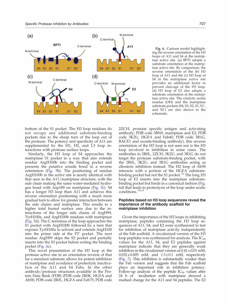
Fig. 6. Cartoon model highlight-ing the reverse orientation of the H3loops of A11 and S4 at the matrip-tase active site. (a) BPTI adopts asubstrate orientation at the matrip-tase active site. By comparison, thereverse orientation of the (b) H3loop of A11 and the (c) H3 loop ofS4 in the matriptase active siteprovides an additional factor toprevent cleavage of the H3 loop.(d) H3 loop of E2 also adopts asubstrate orientation at the matrip-tase active site. The catalytic serineresidue (OH) and the matriptasesubstrate pockets (S4, S3, S2, S1, S1′,and S2′) are also shown in theschematic.
707Specific Protease Inhibition by Antibodies
bottom of the S1 pocket. The H3 loop residues donot occupy any additional substrate-bindingpockets due to the sharp turn of the loop out ofthe protease. The potency and specificity of A11 aresupplemented by the H1, H2, and L3 loop in-teractions with protease surface loops.Similarly, the H3 loop of S4 approaches the
matriptase S1 pocket in a way that also extendsresidue ArgH100b into the binding pocket andpresents the putative scissile bond in a reverseorientation (Fig. 5b). The positioning of residueArgH100b in the active site is nearly identical withthat seen in the A11/matriptase structure, with theside chain making the same water-mediated hydro-gen bond with Asp189 on matriptase (Fig. 5c). S4has a longer H3 loop than A11 and achieves thisreverse orientation positioning with a much moregradual turn to allow for greater interaction betweenthe side chains and matriptase. This results in ahigher total buried surface area due to the in-teractions of the longer side chains of ArgH99,TyrH100a, and ArgH100b residues with matriptase(Fig. 5d). The C-terminus of the loop approaches theS1 pocket with ArgH100b followed by a turn thatexposes TyrH100a to solvent and extends ArgH100into the prime side at the P2′ pocket. The nextresidue ArgH99 skips the S2 pocket and partiallyinserts into the S3 pocket before exiting the bindingpocket (Fig. 6c).This novel presentation of the H3 loop at the
protease active site in an orientation reverse of thatfor a standard substrate allows for potent inhibitionof matriptase and avoidance of proteolytic inactiva-tion of the A11 and S4 Fabs. In the otherantibody/protease structures available in the Pro-tein Data Bank (PDB) (PDB code 2R0K, HGFA andAb58; PDB code 2R0L, HGFA and Fab75; PDB code
2ZCH, prostate specific antigen and activatingantibody; PDB code 3BN9, matriptase and E2; PDBcode 3K2U, HGFA and Fab40; PDB code 3R1G,BACE1 and exosite-binding antibody), this reverseorientation of the H3 loop is not seen nor is the H3loop involved in inhibition in some cases. Theantibodies in 2R0L, 2ZCH, 3K2U, and 3R1G do nottarget the protease substrate-binding pocket, withthe 2R0L, 3K2U, and 3R1G antibodies acting asallosteric inhibitors instead. The H2 loop of Ab58interacts with a portion of the HGFA substrate-binding pocket but not the S1 pocket.16 The long H3loop of E2 inserts into the matriptase substrate-binding pocket but binds in a canonical fashion (Fig.6d) that leads to proteolysis of the loop under acidicconditions.18,19
Peptides based on H3 loop sequences reveal theimportance of the antibody scaffold formatriptase inhibition
Given the importance of the H3 loops in inhibitingmatriptase, peptides containing the H3 loop se-quences of A11, S4, and E2 were synthesized to testfor inhibition of matriptase activity independentlyof the Fab scaffold. A circularized version of the H3loop peptides was synthesized for analysis. The IC50values for the A11, S4, and E2 peptides againstmatriptase indicate that they are generally weakinhibitors in the circularized version at 0.41±0.01mM,0.032±0.005 mM, and 1.3±0.1 mM, respectively(Fig. 7). This inhibition is substantially weaker thanthe Fab version and suggests that the Fab scaffoldplays an important role in inhibition potency.Follow-up analysis of the peptide IC50 values after24 h of incubation with matriptase showed amarked change for the A11 and S4 peptides. The E2

Fig. 7. IC50 plots for the A11, S4,and E2 circularized H3 peptidesagainst matriptase. (a) IC50 of cir-cularized A11 H3 peptide at time0 (red) and 24 h (blue). The IC50increases over time due to digestionof the peptide. (b) IC50 of circular-ized S4 H3 peptide at time 0 (red)and 24 h (blue). The IC50 improvesmodestly over time. (c) IC50 ofcircularized E2 H3 peptide at time0 (red) and 24 h (blue). The IC50remains the same.
708 Specific Protease Inhibition by Antibodies
peptides showed little change in IC50 values after24 h. Mass spectrometric analysis of the A11peptides confirmed that the change in IC50 was theresult of near complete cleavage of the A11 peptideby matriptase, with the cleavage event C-terminal toArgH100a. This result indicates that the A11 peptidemay not bind in a reverse orientation as seen in theA11 Fab. The S4 peptide showed modest improve-ment in inhibition after 24 h, with a decreased IC50value of 0.010±0.002 mM. However, partial diges-tion of the S4 peptide was observed, with the majorcleavage event C-terminal to ArgH100b. The slightincrease in inhibition over time may be a result ofproduct inhibition from the cleaved peptide. The E2peptide showed no cleavage after 24 h, supportingthe unchanged IC50.Since the H3 loop of A11 makes limited contacts
with the protease outside the S1 pocket, it isunlikely that the A11 peptide would adopt thereverse binding orientation without the scaffoldand therefore unsurprising that the peptide iscleaved. Furthermore, the H3 loop of A11 buries asmall percentage (38%) of surface area relative tothe total buried surface area in the matriptasesubstrate-binding pocket when compared to S4(63%) and E2 (68%). The H3 loops of S4 and E2 are
assisted by more extensive interactions with thesubstrate-binding pocket,18 and these contactsmake it more likely that the circularized peptidewould act as an inhibitor. The weak inhibitionexhibited by the E2 peptide in the absence of anycleavage indicates that the sequence may bind inan orientation similar to that of the H3 loop of theFab, allowing it to avoid cleavage, presumably dueto the rigidity provided by the proline residues inthe sequence. Since the S4 peptide was the mostpotent inhibitor out of the three, this suggests thatits interactions with the substrate-binding pocketmay contribute more significantly to the inhibitionof matriptase independent of the entire scaffold,though whether it does so in a reverse orientationis difficult to determine. The cleavage of some ofthe S4 peptide by matriptase over time suggeststhat it may not adopt this novel conformationconsistently or at all without the aid of thescaffold. While the H3 loop alone contributes toa certain degree to protease inhibition, theseresults demonstrate that the antibody scaffoldplays a significant role in the correct orientationof the H3 loop for inhibition and that additionalinteractions provided by the remaining Fab loopsare necessary to achieve potent inhibition.

709Specific Protease Inhibition by Antibodies
Discussion
As antibodies continue to demonstrate theirusefulness in many therapeutic areas, one particularfield of recent interest is protease inhibition fortreatment of cancer progression. Since proteaseshave historically been difficult to target selectivelywith small molecules and macromolecular cognateinhibitors, the high affinity and specificity ofantibodies are ideal for this purpose.30,31 Anemphasis of the current study was to identifyselective antibodies that competitively inhibit pro-teolytic activity. As a result, three different inhibi-tory antibodies of matriptase that all utilize a longH3 loop with a P1 Arg residue to partially fill the S1pocket were isolated and characterized. The inter-action between the Arg and the Asp residues at thebottom of the binding pocket is mediated by a watermolecule, resulting in a binding mode that preventsproteolysis of the Fab. These antibodies are differentfrom the HGFA antibodies that are mostly allostericinhibitors containing shorter H3 loops.16,17 Here, thestructural characteristics of specific protease inhibi-tion using A11 and S4 Fabs are revealed. As withmany enzyme-inhibiting antibodies, A11 and S4inhibit substrate binding by capping the active sitethrough interactions with the surrounding surfaceloops (Fig. 4).14,16,18 These loops are areas of lowhomology among trypsin-fold serine proteases andcan be targeted with high selectivity by antibodies.Since these antibodies use the surface loop in-teractions for specificity, this differentiates themfrom naturally occurring macromolecular proteaseinhibitors that bind a broader array of proteases byinteracting with the highly conserved proteaseactive site.32
The crystal structures of the A11/matriptase andS4/matriptase complexes reveal a number of simi-larities with other antibody protease inhibitors forwhich structures have recently been reported, inparticular, the HGFA inhibitor Ab5816 and thematriptase inhibitor E2.18 All of these antibodiesdepend on the protease surface loops for recognitionand binding to their target protease. For A11, E2,and Ab58, the 90s loop of the protease and the Phe97residue, in particular, acts as a key anchor point forthese antibodies. However, upon protease capping,the buried surface area of the Ab58/HGFA interac-tion is considerably less (890 Å2)16 when comparedto A11/matriptase (1216 Å2) and E2/matriptase(1241 Å2) (Fig. 4c), which are both significantlygreater than a typical antibody/antigen interaction(b900 Å2).28,33 Ab58 only partially occludes thesubstrate-binding pocket at the S2 and S3 pockets.16
Conversely, the matriptase inhibitors fully occludethe substrate-binding pocket and interact with theS1 pocket by inserting a long H3 loop, resulting inthe enhanced interactions.
Focusing on the antibody inhibitors of matriptase,a closer similarity in antibody/protease interactionsis observed with A11 and E2. A11 and E2 haveidentical H1 and H2 loops even though A11 wasisolated from a naïve human B cell phage displaylibrary and E2 was isolated from a fully syntheticphage display library.15 These loops are critical foranchoring the antibody between the 60s and 90sloops on the surface of matriptase (Fig. 4a), creatingvery similar binding footprints for A11 and E2. Mostof the interactions in the A11/matriptase andE2/matriptase complexes are made by nonpolarresidues. In contrast, S4 binds matriptase in adifferent mode and does not utilize the interactionwith the 90s loop (Fig. 4b). S4/matriptase interac-tions are instead formed by polar residues of the H3and L1 loops interacting with the 140s loop via astrong network of hydrogen bonds and pi-stackinginteractions. S4 is the most potent matriptaseinhibitor out of the three, and this may be the resultof the increased buried surface area localized to theH3 loop (682 Å2) found in the substrate-bindingpocket (Fig. 4c). Instead of relying on the S1 pocketfor critical binding energy and inhibition as withA11 and E2, S4 adopts a unique mechanism wherethe ArgH99 residue forms a strong salt bridge withGln175 in the S3 pocket to position the H3 loop inthe most energetically stable orientation (Fig. 5b andd). On the other hand, the H3 loop of E2 is thelongest of our matriptase inhibitors and adopts amore extended conformation in the matriptaseactive site when compared to the A11 H3 loop. Asa result, the H3 loop of A11 buries less surface area(466 Å2) compared to the H3 loop of E2 (847 Å2)(Fig. 4c). To compensate for this decrease in buriedsurface area, A11 utilizes a different light chain. TheVκ3 light chain of A11 buries 538 Å2 of surface area,while the light chain of E2 buries just 175 Å2 (Fig.4c). Attempts to make a chimera composed of theA11 light chain and the E2 heavy chain that shouldbury a larger total surface area resulted in reducedaffinity (data not shown), indicating that the loopsare precisely positioned for each antibody and notinterchangeable despite the similarities in the twoantibodies.The discovery and characterization of antibodies
that inhibit through H3 loop interactions with theprotease active site represent a new approach tospecific inhibition of protease activity. The novel,reverse orientation of the H3 loop in the proteaseactive site demonstrates that the antibody scaffold iscapable of revealing completely new inhibitionmechanisms, suggesting that there are still undis-covered approaches to inhibiting these enzymes. Inthe case of A11 and S4, interactions with theprotease surface loops orient the antibody so thatthe long H3 loop can be inserted into the proteaseactive site. Interaction of the H3 loop with the activesite occurs in a unique fashion, with the H3 loop

710 Specific Protease Inhibition by Antibodies
binding in a reverse orientation while partiallyinserting an Arg residue into the S1 pocket (Fig. 6).The reverse orientation combined with the shallowarginine binding at the S1 pocket prevents hydro-lysis of the H3 loop while, at the same time,providing critical binding energy. A few cases ofreverse binding of peptides near the active site havebeen recorded, most notably, inhibition of cathepsinpro-peptide and BIR2 domain inhibition of caspase 3and caspase 7.34 Although they share some similar-ities with A11 and S4, the mechanism utilized by ourantibodies remains unique, relying upon a directinteraction with the primary substrate-bindingpocket of the protease. Even so, the resemblance ofthis reverse orientation in the mechanism used byBIR2 inhibition34 of the cysteine proteases caspase 3and caspase 7 indicates that this approach could alsobe used in identifying selective active-site inhibitorsof protease classes beyond the serine proteases. Forexample, this approach could be extremely benefi-cial for the study of metalloproteases and aspartylproteases, two classes that have been historicallydifficult to design specific inhibitors against.Selective protease inhibition is particularly useful
in the clinic to target misregulated protease activityresponsible for disease pathophysiology. Forexample, the recombinant polypeptide ecallantide(Kalbitor) approved by the Food and DrugAdministration is used to specifically inhibit theserine protease plasma kallikrein that causes acutehereditary angioedema attacks in patients.35
Moreover, selective protease inhibition may beparticularly important in treating and monitoringcancer progression. Matriptase is over-expressedin breast, colon, and prostate cancers. It is normallyregulated by its cognate inhibitor HGFA inhibitor-1;however, dysregulation of matriptase activitypromotes cancer progression.36 With the use ofmatriptase-specific antibodies, protease activitycan be monitored in disease to develop a novelapproach to follow early-staged malignancies. Inthe recent work by Darragh et al., the A11antibody was used as a noninvasive imagingprobe to characterize protease activity in vivo inorder to define matriptase as an early biomarkerto visualize epithelial cancers in preclinical mousemodels.37 Furthermore, the recent discovery of therole of matriptase in squamous cell carcinoma38
highlights the need for agents that can selectivelyinhibit protease activity to pharmacologicallyprobe the pathophysiological role of the enzymeand to provide potential therapeutic applications.Here, we have shown that antibodies can providenovel solutions for the selective inhibition ofproteases. Our discovery highlights the importanceof the antibody scaffold to uncover unique andunpredictable positioning of the inhibitory loops tobind and inhibit protease function in a highly selectivemanner. The identification of a fully human, inhibitory
recombinant antibody A11 validates this approachand reaffirms the use of antibodies for selectiveinhibition of protease targets in cancer.
Materials and Methods
Identification of inhibitory Fabs from a human phagedisplay library
A Fab library created from naïve B cells was used toidentify inhibitory antibodies against the human matrip-tase protease domain (hMT-SP1).39 Active matriptase wasimmobilized in wells of a 96-well ELISA plate. Thepanning was accomplished in three rounds with increas-ing stringency against hMT-SP1 adsorbed to wells.ELISAs were performed to verify binding of the identifiedFabs to hMT-SP1. ELISA-positive clones were expressed,purified, and tested for inhibition of matriptase. Individ-ual clones were sequenced to verify their uniqueness.
Protein expression and purification from E. coli
Matriptase andmatriptase mutants were expressed in E.coli and purified as previously described.6,19 S4 wascloned into the Fab scaffold following a procedure similarto that described in Farady et al.18 A11 and S4 Fabs wereexpressed in E. coli BL21 (DE3) cells. Cultures were grownin 1 L of 2× YT containing 100 μg/mL ampicillin and 0.1%glucose at 37 °C and 250 rpm to an OD600 of 0.6–0.8. Thetemperature was then reduced to 25 °C, and the cultureswere induced with the addition of 0.5 mM IPTG. After18 h of growth, the bacteria were harvested and pelletedby centrifugation. The cells were resuspended in 25 mL ofbuffer containing 0.2 M Tris (pH 8.0), 0.5 mM ethylene-diaminetetraacetic acid, and 0.5 M sucrose. The resus-pended solution was left on ice for 1 h. The solution wasthen pelleted, and the periplasmic fraction was run over aNi2+ column prewashed with wash buffer [50 mM Tris(pH 8.0) and 250 mM NaCl]. The Ni2+column was thenwashed with 10 column volumes of the wash buffer, andthe Fab was eluted with 250 mM imidazole in 50 mM Tris(pH 8.0) and 100 mM NaCl. Size-exclusion chromatogra-phy was carried out on the eluted A11 using a SuperdexS75 26/60 with a 50 mM Tris (pH 8.0), 100 mM KCl, and5% glycerol buffer.
Site-directed mutagenesis of A11and S4 antibodies
A11 mutants ArgH100aAla, ArgH100bAla, andArgH115bLys and S4 mutants ArgH100bAla andArgH100bLys were all created using the QuikChange™kit from Stratagene. Sequences were verified by DNAsequencing. Expression and purification of A11 and S4mutants were carried out as described above.
T. reesei expression vector construction
Two independent expression vectors were constructed:one for expression of the Fab heavy chain (pCBHIxFabA11H1) and one for the light chain (pCBHIxFabA11 L1). In

711Specific Protease Inhibition by Antibodies
each case, the Fab chains were produced as fusion proteinswith the T. reeseiCBHI (cellobiohydrolase I; cel7a) catalyticcore and linker region. A Kex2 cleavage site (Val Ala ValTyr Lys Arg) was positioned between the CBHI and theFab chain to allow cleavage of the fusion protein after theArg residue and release of the Fab chain during secretion.The following segments of DNA were assembled in the
construction of pCBHIxFabA11H1 and pCBHIxFabA11 L1.The T. reesei cbh1 promoter and coding region start at anaturally occurring XbaI site approximately 1500 bp up-streamof the coding region. The synthetic, codon-optimizedcoding region for each Fab chainwas fused to the end of theCBHI linker region at a created SpeI restriction site (seebelow). Immediately after, the Fab stop codon was an AscIrestriction site followed by the T. reesei cbh1 terminatorregion (356 bp). This was followed by a 2.75-kb fragment ofAspergillus nidulans genomic DNA, including the promoter,coding region, and terminator of the amdS (acetamidase)gene. The above DNA fragments were inserted in pNEB193(New England Biolabs, Inc., USA) between the XbaI andKpnI sites of the multiple cloning site.The following changes were made within the cbh1 open
reading frame. The codon for amino acid 212 of the matureCBHI protein was changed from GAG (glutamic acid) toCAG (glutamine), resulting in production of an inactiveform of CBHI. Within the coding region for the CBHIlinker, a change was made to create a SpeI restriction site.This altered the DNA sequence from ACCCAG toACTAGT, changing the amino acid sequence at the endof the CBHI linker region from Thr Gln to Thr Ser. The Glnin this sequence represents the first amino acid of thecellulose binding domain of CBHI.
T. reesei transformation
T. reesei GICC20000150 was derived from strain RL-P3740 by sequential deletion of the genes encoding the fourmajor secreted cellulases (cel7a, cel6a, cel7b, and cel5a).Transformation was performed using a Bio-Rad Labora-tories, Inc. (Hercules, CA) model PDS-1000/He biolisticparticle delivery system according the manufacturer'sinstructions. Transformants were selected on solid medi-um containing acetamide as the sole nitrogen source. Forantibody production, transformants were cultured in aliquid minimal medium containing lactose as carbonsource as described previously,41 except that 100 mMpiperazine-N,N′-bis(3-propanesulfonicacid) (Calbiochem)was included tomaintain the pH at 5.5. In order to produceFab, it was necessary for transformants to have taken upboth the heavy-chain and the light-chain expressionvectors. However, both expression vectors had the sameamdS selectable marker; thus, it was not immediatelypossible to recognize co-transformants. Culture superna-tants were analyzed by SDS-PAGE under reducingconditions, and those that contained the highest level ofa 25-kDa band (representing heavy and/or light chain)and an apparent 60-kDa band (representing the CBHI coreand linker) were selected for further analysis.
Purification of A11 from T. reesei expression
Media from the Trichoderma expression was adjusted topH 5.5. For an initial crude purification, media were run
over an SP Sepharose column equilibrated with WashBuffer 1 [100 mM 4-morpholineethanesulfonic acid (Mes)(pH 5.5) and 50 mM NaCl]. The column was then washedwith 5 column volumes of Wash Buffer 1, followed by 5column volumes of Wash Buffer 2 [50 mM N-[2-hydroxy-1,1-bis(hydroxymethyl)ethyl]glycine (pH 8.0)]. A11 waseluted with 3 column volumes of 50 mM N-[2-hydroxy-1,1-bis(hydroxymethyl)ethyl]glycine (pH 8.0) and 500mMNaCl. The elution was buffer exchanged into 100 mMMes(pH 5.5) and 50 mM NaCl and was loaded onto a MonoSHR 5/5 column. The column was washed with WashBuffer 1 followed by Wash Buffer 2. Elution was thencarried out in a 0–100% gradient of Wash Buffer 2 to WashBuffer 2 containing 500 mM NaCl. Further purificationwas carried out on a Superdex 75 26/60 size-exclusioncolumn with a 50 mM Tris (pH 8.0), 100 mM KCl, and 5%glycerol buffer.
Steady-state kinetics
Kinetics were carried out as previously described.19
Briefly, all reactions were carried out in 50 mM Tris(pH 8.8), 50 mM NaCl, and 0.01% Tween-20 in a 96-well,medium binding, flat-bottomed plate (Corning) wherecleavage of substrate Spectrozyme® tPA (hexahydrotyr-osyl-Gly-Arg-pNA; American Diagnostica, Greenwich, CT)was monitored in a UVmax Microplate Reader (MolecularDevices Corporation, Palo Alto, CA). KI values weremeasured using the tight binding inhibition equations ofWilliams and Morrison.42 When measuring the effectmutations to matriptase had on the strength of theinteraction between the protease and inhibitor, IC50 valueswere used instead of KI values. Reactions to determine theIC50 values were carried out by incubating 0.2 nM enzymewith inhibitor forN5 h to assure steady-state behavior of thesystem. Apparent KI values were then calculated from IC50values as shown previously in order to normalize the IC50with respect to the strength of the protease/substrateinteraction.43 Inhibitory activity against related proteaseswas measured using a similar assay monitoring thecleavage of a p-nitroanilide substrate. Thrombin, factor Xaand plasmin (Haematologic Technologies, Inc., Essex Junc-tion, VT) at 10 nM concentration were incubated with 1 μMFab, and the reaction was monitored using 1 mM substrateT1637 (Sigma, St. Louis, MO). The enzymes, tPA and uPA(American Diagnostica) at 10 nM concentration wereincubated with 1 μM Fab, and the reaction was monitoredusing 1mMSpectrozyme® tPAand 400mMSpectrozyme®UK (American Diagnostica), respectively. Hepsin andprostasin (R&D Systems, Minneapolis, MN) at 1 nM and5 nM concentration respectively were incubated with 1 μMFab, and the reaction was monitored using 0.5 mMSpectrozyme® tPA. KaleidaGraph 3.6 was used to fit allgraphs and equations (Synergy Software, Reading, PA).
A11 inhibitor digestion
The digestion of A11 by matriptase was carried out aspreviously described.19 A11 was incubated at 2 μM with0.1 nM matriptase in either 100 mM Mes (pH 6.0) and100 mM NaCl buffer or 50 mM Tris (pH 8.0) and 100 mMNaCl. After 120 h, the samples were run on a 4–20% Trisglycine SDS-PAGE gel (Invitrogen) to visualize.

Table 3. Summary of crystallographic information, 3NPS and 3SO3
S4/matriptase 3NPS A11/matriptase 3SO3
Data collection and processingNumber of crystals used 1 1Wavelength (Å) 1.000 1.000Space group P21 P64Unit cell parameters
a, b, c (Å) 39.2, 84.0, 101.4 130.6, 130.6, 96.9α, β, γ (°) 90.0, 91.5, 90.0 90.0, 90.0, 120.0
Matthews coefficient (A3/Da) 2.27 3.23Solvent content (%) 45.9 61.9Molecules per asymmetric unit 3 3Beamline ALS 8.3.1 ALS 8.3.1
Diffraction dataResolution range (Å) 43.4–1.4 (1.46–1.40) 113.0–2.1 (2.21–2.10)Unique reflections 104,800 (2150) 54,872 (7998)R(I)sym (%)a 5.8 (17.6) 9.4 (93.2)Completeness (%) 84.5 (80.0) 100.0 (100.0)Redundancy 3.7 (3.1) 7.3 (6.9)I/σ(I) 16.0 (8.0) 11.7 (2.2)
RefinementResolution range (Å) 28.6–1.5 19.9–2.1Reflections used in refinement (work/free) 88,531/4447 51,923/2774Final R values for all reflections (workb/freec) (%) 18.8/22.8 16.1/19.4Number of atomsProteins 5157 5109Ligands/iond 46 35Water 676 462
RMSD valuesBonds (Å) 0.014 0.017Angles (°) 1.61 1.64
Ramachandran parameters (%)e
Residues in most favored regions 89.3 91.6Residues in additional allowed regions 10.1 8.3Residues in generously allowed regions 0.2 0.0Residues in disallowed regions 0.4 0.2
Mean B-factor (Å2)Protein 19.8 59.6Ligand/ion 48.3 57.7Water 31.4 63.4
Numbers in parentheses represents the highest-resolution shell.ALS, Advanced Light Source
a Rsym=(∑|(I − ⟨I⟩)|/∑I), where ⟨I⟩ is the average intensity of multiple measurements.b Rwork=(∑|(Fobs −Fcalc)|/∑|Fobs|).c Rfree=Rwork based on ∼1000 (at least 5%) of reflections excluded from refinement.d Ligands/ion are ethylene glycol, Na, and Cl in S4/matriptase and glycerol and sucrose in A11/matriptase.e Calculated using PROCHECK.44
712 Specific Protease Inhibition by Antibodies
Crystallization and data collection
A11 and S4 was incubated with matriptase at 1:1 molarratio, and the complex was purified by gel filtration usinga Superdex S75 26/60 column in a buffer containing50 mM Tris (pH 8.0), 100 mM NaCl, and 5% glycerol. Thepurified complex (A11/matriptase and S4/matriptase)was then concentrated to ∼15 mg/mL. Initial crystalliza-tion conditions were discovered using a Mosquitonanoliter-scale robot (TTP Labtech). The A11/matriptasecomplex was crystallized in 16% polyethylene glycol(PEG) 3350, 0.23 M MgSO4, 0.4% isopropanol, 3%glycerol, and 0.12 M AmSO4 in hanging drop by vapordiffusion. Crystals belonging to the hexagonal space
group P64 (a=b=130.6 Å and c=96.9 Å; β=90.0°) grewin 3 days and were cryoprotected in the mother liquorsupplemented with 30% sucrose. Needle-like crystals ofthe S4/matriptase complex were grown from 0.2 M NaCl,25% PEG 3355, and 1% ethyl acetate as additive at 25 °Cusing the hanging drop vapor diffusion technique.These crystals belong to the monoclinic space groupP21 (a=39.2 Å, b=84.0 Å, c=101.4 Å; β=91.5°), diffractX-rays to beyond 1.5 Å resolution, and have onemolecule in the asymmetric unit. The S4/matriptasecrystals were transferred to 0.2 M NaCl, 25% PEG 3355,1.0% ethyl acetate, and 25% ethylene glycol. Diffractiondata were collected at beamline 8.3.1 at the AdvancedLight Source at Lawrence Berkeley National Laboratory

713Specific Protease Inhibition by Antibodies
(see Table 3). Diffraction images for A11/matriptase andS4/matriptase were reduced and integrated usingDENZO, and the intensities were merged and scaledusing SCALEPACK from the HKL-2000 suite.45
Structure determination and refinement
The structures of A11/matriptase and S4/matriptasewere solved by molecular replacement using Phaser46 inCCP4,47 first searching for matriptase (using 1EAX.pdb assearch model), then searching for the Fab fragment withits H3 loop truncated (using 2HFF.pdb as search model).After molecular replacement, automatic building inARP/wARP48 and manual building followed by re-strained refinement cycles using REFMAC549 yieldedthe final structures. TLS refinement was applied at the laststage of the refinement for A11/matriptase. In the finalmodel building/refinement cycles, water molecules wereinserted at stereochemically reasonable sites that hadfeatures in electron density, and individual restrained B-values were refined. Five percent of all reflections wereomitted from the refinement to calculate Rfree; the final Rand Rfree are 18.8 and 22.8%, respectively, for S4/matrip-tase. For A11/matriptase, the final R and Rfree are 16.0 and19.4%, respectively. The final model of both complexesconverged rapidly, yielding a model with excellentparameters (see Table 3). In the final structure ofA11/matriptase, there was no density for the heavy-chain residues 129–133 and 213–215 or protease Ala204.There was no side-chain density for A11 light-chainresidues Glu1, Glu143, Lys188, and Glu213 or heavy-chain residues Glu1, Lys201, and Lys210; thus, the sidechains were not modeled. These regions are oftendisordered in Fab structures. The whole main chain ofthe S4/matriptase catalytic domain is in appropriateelectron density. In S4/matriptase, the side-chain occu-pancy was set to 0.5 since the side chains exhibit twoconformations. The quality of the final structures wasassessed using Molprobity.50 Buried surface area calcula-tions were performed using PISA.28
Synthesis of H3 loop peptides
The H3 loop sequences of A11 (circularized:GLGIAARRFVSGEG), S4 (circularized: GFHIRRYRS-GYYEG), and E2 (circularized: GLTYPQRRGPQNVSEG)were synthesized by standard solid-phase Fmoc chemistryon a Protein Technologies Symphony Quartet automatedpeptide synthesizer using Wang resin preloaded with theC-terminal amino acid (Novabiochem). An ODmab-pro-tected glutamic acid residue was inserted near the C-terminus (Novabiochem). To circularize the peptide, theODmab protecting group was selectively removed by 2%hydrazine in dimethylformamide followed by Fmoc de-protection of the N-terminal residue and a standardcoupling reaction with HOBt/HBTU/DIPEA. The finishedpeptideswere deprotected and cleaved from the resin using95% trifluoroacetic acid and purified by HPLC on a C-18column. The peptide sequences were confirmed by matrix-assisted laser desorption/ionization mass spectrometry.
Peptide IC50 determination
The IC50 of each peptide were carried out underconditions similar to those for the steady-state kinetics
described earlier. Assays containing 200 μM substrate(Spectrozyme® tPA), 0.2 nM matriptase, and varyingpeptide concentrations were run. A 50 mM stock solutionof each peptide in dimethyl sulfoxide (DMSO) was usedfor all assays. Additional DMSO was added to all assaysso that the DMSO concentration was equal throughout.Assays were run for 30 min and monitored on afluorescent plate reader at 405 nm. Data were plottedusing KaleidaGraph.
Accession numbers
Coordinates and structure factors have been depositedin the PDBwith accession numbers 3SO3 (A11/matriptasecomplex) and 3NPS (S4/matriptase complex).
Acknowledgements
This work was funded in part by grants from theNational Institutes of Health CA128765 andGM082250 (C.S.C.) and CA108462 (E.L.S.). We alsothank Professors Robert Fletterick and MatthewJacobson for helpful discussions and Dr. GregoryLee for careful reading of the manuscript.
References
1. Carter, P. J. (2006). Potent antibody therapeutics bydesign. Nat. Rev., Immunol. 6, 343–357.
2. Brekke, O. H. & Sandlie, I. (2003). Therapeuticantibodies for human diseases at the dawn of thetwenty-first century. Nat. Rev., Drug Discov. 2, 52–62.
3. Chambers, A. F. & Matrisian, L. M. (1997). Changingviews of the role of matrix metalloproteinases inmetastasis. J. Natl. Cancer Inst. 89, 1260–1270.
4. Coussens, L. M. & Werb, Z. (1996). Matrix metallo-proteinases and the development of cancer. Chem. Biol.3, 895–904.
5. Netzel-Arnett, S., Hooper, J. D., Szabo, R., Madison,E. L., Quigley, J. P., Bugge, T. H. & Antalis, T. M.(2003). Membrane anchored serine proteases: arapidly expanding group of cell surface proteolyticenzymes with potential roles in cancer. CancerMetastasis Rev. 22, 237–258.
6. Takeuchi, T., Shuman, M. A. & Craik, C. S. (1999).Reverse biochemistry: use of macromolecular proteaseinhibitors to dissect complex biological processes andidentify a membrane-type serine protease in epithelialcancer and normal tissue. Proc. Natl Acad. Sci. USA, 96,11054–11061.
7. Bhatt, A. S., Welm, A., Farady, C. J., Vasquez, M.,Wilson, K. & Craik, C. S. (2007). Coordinate expres-sion and functional profiling identify an extracellularproteolytic signaling pathway. Proc. Natl Acad. Sci.USA, 104, 5771–5776; [Epub 2007 Mar 27].
8. Kilpatrick, L. M., Harris, R. L., Owen, K. A., Bass, R.,Ghorayeb, C., Bar-Or, A. & Ellis, V. (2006). Initiation ofplasminogen activation on the surface of monocytesexpressing the type II transmembrane serine proteasematriptase. Blood, 108, 2616–2623; [Epub 2006 Jun 22].

714 Specific Protease Inhibition by Antibodies
9. Suzuki, M., Kobayashi, H., Kanayama, N., Saga, Y.,Lin, C. Y., Dickson, R. B. & Terao, T. (2004). Inhibitionof tumor invasion by genomic down-regulation ofmatriptase through suppression of activation ofreceptor-bound pro-urokinase. J. Biol. Chem. 279,14899–14908; [Epub 2004 Jan 27].
10. Takeuchi, T., Harris, J. L., Huang, W., Yan, K. W.,Coughlin, S. R. & Craik, C. S. (2000). Cellularlocalization of membrane-type serine protease 1 andidentification of protease-activated receptor-2 andsingle-chain urokinase-type plasminogen activator assubstrates. J. Biol. Chem. 275, 26333–26342.
11. Colwell, N. S., Blinder, M. A., Tsiang, M., Gibbs, C. S.,Bock, P. E. & Tollefsen, D. M. (1998). Allosteric effectsof a monoclonal antibody against thrombin exosite II.Biochemistry, 37, 15057–15065.
12. Devy, L., Huang, L., Naa, L., Yanamandra, N., Pieters,H., Frans, N. et al. (2009). Selective inhibition of matrixmetalloproteinase-14 blocks tumor growth, invasion,and angiogenesis. Cancer Res. 69, 1517–1526; [Epub2009 Feb 10].
13. Dickinson, C. D., Shobe, J. & Ruf, W. (1998). Influenceof cofactor binding and active site occupancy on theconformation of the macromolecular substrate exositeof factor VIIa. J. Mol. Biol. 277, 959–971.
14. Petersen, H. H., Hansen, M., Schousboe, S. L. &Andreasen, P. A. (2001). Localization of epitopes formonoclonal antibodies to urokinase-type plasminogenactivator: relationship between epitope localizationand effects of antibodies on molecular interactions ofthe enzyme. Eur. J. Biochem. 268, 4430–4439.
15. Sun, J., Pons, J. & Craik, C. S. (2003). Potent and selectiveinhibition ofmembrane-type serine protease 1 by humansingle-chain antibodies. Biochemistry, 42, 892–900.
16. Wu, Y., Eigenbrot, C., Liang, W. C., Stawicki, S.,Shia, S., Fan, B. et al. (2007). Structural insight intodistinct mechanisms of protease inhibition byantibodies. Proc. Natl Acad. Sci. USA, 104,19784–19789; [Epub 2007 Dec 5].
17. Ganesan, R., Eigenbrot, C., Wu, Y., Liang, W. C., Shia,S., Lipari, M. T. & Kirchhofer, D. (2009). Unravelingthe allosteric mechanism of serine protease inhibitionby an antibody. Structure, 17, 1614–1624.
18. Farady, C. J., Egea, P. F., Schneider, E. L., Darragh,M. R. & Craik, C. S. (2008). Structure of an Fab–protease complex reveals a highly specific non-canonical mechanism of inhibition. J. Mol. Biol. 380,351–360; [Epub 2008 May 11].
19. Farady, C. J., Sun, J., Darragh, M. R., Miller, S. M. &Craik, C. S. (2007). The mechanism of inhibition ofantibody-based inhibitors of membrane-type serineprotease 1 (MT-SP1). J. Mol. Biol. 369, 1041–1051;[Epub 2007 Apr 4].
20. Wu, T. T., Johnson, G. & Kabat, E. A. (1993). Lengthdistribution of CDRH3 in antibodies. Proteins, 16, 1–7.
21. Zemlin, M., Klinger, M., Link, J., Zemlin, C., Bauer, K.,Engler, J. A. et al. (2003). Expressed murine andhuman CDR-H3 intervals of equal length exhibitdistinct repertoires that differ in their amino acidcomposition and predicted range of structures. J. Mol.Biol. 334, 733–749.
22. de Haard, H. J., van Neer, N., Reurs, A., Hufton, S. E.,Roovers, R. C., Henderikx, P. et al. (1999). A large non-immunized human Fab fragment phage library that
permits rapid isolation and kinetic analysis of highaffinity antibodies. J. Biol. Chem. 274, 18218–18230.
23. Arbabi-Ghahroudi, M., Tanha, J. & MacKenzie, R.(2005). Prokaryotic expression of antibodies. CancerMetastasis Rev. 24, 501–519.
24. Keranen, S. & Penttila, M. (1995). Production ofrecombinant proteins in the filamentous fungusTrichoderma reesei. Curr. Opin. Biotechnol. 6, 534–537.
25. Farady, C. J., Sellers, B. D., Jacobson, M. P. &Craik, C. S. (2009). Improving the species cross-reactivity of an antibody using computationaldesign. Bioorg. Med. Chem. Lett. 19, 3744–3747;[Epub 2009 May 7].
26. McGrath, M. E., Hines, W. M., Sakanari, J. A.,Fletterick, R. J. & Craik, C. S. (1991). The sequenceand reactive site of ecotin. A general inhibitor ofpancreatic serine proteases from Escherichia coli. J. Biol.Chem. 266, 6620–6625.
27. Ozawa, K. & Laskowski, M., Jr (1966). The reactive siteof trypsin inhibitors. J. Biol. Chem. 241, 3955–3961.
28. Krissinel, E. & Henrick, K. (2007). Inference ofmacromolecular assemblies from crystalline state. J.Mol. Biol. 372, 774–797; [Epub 2007 May 13].
29. Friedrich, R., Fuentes-Prior, P., Ong, E., Coombs, G.,Hunter, M., Oehler, R. et al. (2002). Catalytic domainstructures of MT-SP1/matriptase, a matrix-degradingtransmembrane serine proteinase. J. Biol. Chem. 277,2160–2168; [Epub 2001 Nov 5].
30. Coussens, L. M., Fingleton, B. & Matrisian, L. M.(2002). Matrix metalloproteinase inhibitors and can-cer: trials and tribulations. Science, 295, 2387–2392.
31. Lee, G. M. & Craik, C. S. (2009). Trapping movingtargets with small molecules. Science, 324, 213–215.
32. Laskowski, M. & Kato, I. (1980). Protein inhibitors ofproteinases. Annu. Rev. Biochem. 49, 593–626.
33. Huang, M., Syed, R., Stura, E. A., Stone, M. J.,Stefanko, R. S., Ruf, W. et al. (1998). The mechanismof an inhibitory antibody on TF-initiated bloodcoagulation revealed by the crystal structures ofhuman tissue factor, Fab 5G9 and TF·G9 complex. J.Mol. Biol. 275, 873–894.
34. Riedl, S. J., Renatus, M., Schwarzenbacher, R., Zhou,Q., Sun, C., Fesik, S. W. et al. (2001). Structural basisfor the inhibition of caspase-3 by XIAP. Cell, 104,791–800.
35. Cicardi, M., Levy, R. J., McNeil, D. L., Li, H. H.,Sheffer, A. L., Campion, M. et al. (2010). Ecallantide forthe treatment of acute attacks in hereditary angioe-dema. N. Engl. J. Med. 363, 523–531.
36. Oberst, M. D., Johnson, M. D., Dickson, R. B., Lin,C. Y., Singh, B., Stewart, M. et al. (2002). Expressionof the serine protease matriptase and its inhibitorHAI-1 in epithelial ovarian cancer: correlation withclinical outcome and tumor clinicopathologicalparameters. Clin. Cancer Res. 8, 1101–1107.
37. Darragh, M. R., Schneider, E. L., Lou, J., Phojanakong,P. J., Farady, C. J., Marks, J. D. et al. (2010). Tumordetection by imaging proteolytic activity. Cancer Res.70, 1505–1512; [Epub 2010 Feb 9].
38. Szabo, R., Rasmussen, A. L., Moyer, A. B., Kosa, P.,Schafer, J. M., Molinolo, A. A. et al. (2011). c-Met-induced epithelial carcinogenesis is initiated by theserine protease matriptase. Oncogene, 30, 2003–2016;[Epub 2011 Jan 10].

715Specific Protease Inhibition by Antibodies
39. Duriseti, S., Goetz, D. H., Hostetter, D. R., LeBeau,A. M., Wei, Y. & Craik, C. S. (2010). Antagonisticanti-urokinase plasminogen activator receptor(uPAR) antibodies significantly inhibit uPAR-medi-ated cellular signaling and migration. J. Biol. Chem.285, 26878–26888; [Epub 2010 May 25].
40. Shier-Neiss, G. & Montenecourt, B. S. (1984).Characterization of the secreted cellulases of Tricho-derma reesei wild type and mutants during con-trolled fermentations. Appl. Microbiol. Biotechnol. 20,46–53.
41. Ilmen, M., Saloheimo, A., Onnela, M. L. & Penttila,M. E. (1997). Regulation of cellulase gene expressionin the filamentous fungus Trichoderma reesei. Appl.Environ. Microbiol. 63, 1298–1306.
42. Williams, J. W. &Morrison, J. F. (1979). The kinetics ofreversible tight-binding inhibition. Methods Enzymol.63, 437–467.
43. Chou, T. (1974). Relationships between inhibitionconstants and fractional inhibition in enzyme-cata-lyzed reactions with different numbers of reactants,different reaction mechanisms, and different typesand mechanisms of inhibition. Mol. Pharmacol. 10,235–247.
44. Laskowski, R. A., MacArthur, M. W., Moss, D. S. &Thornton, J. M. (1993). PROCHECK: a program to
check the stereochemical quality of protein structures.J. Appl. Crystallogr. 26, 283–291.
45. Otwinowski, Z., Minor, W. & Carter, C. W. (1997).Processing of X-ray diffraction data collected inoscillation mode. Methods Enzymol. 276, 307–326.
46. Read, R. J. (2001). Pushing the boundaries ofmolecular replacement with maximum likelihood.Acta Crystallogr., Sect. D: Biol. Crystallogr. 57,1373–1382; [Epub 2001 Sep 21].
47. Collaborative Computational Project, Number 4.(1994). The CCP4 suite: programs for protein crystal-lography.Acta Crystallogr., Sect. D: Biol. Crystallogr. 50,760–763.
48. Evrard, G. X., Langer, G. G., Perrakis, A. & Lamzin,V. S. (2007). Assessment of automatic ligandbuilding in ARP/wARP. Acta Crystallogr., Sect. D:Biol. Crystallogr. 63, 108–117; [Epub 2006 Dec 13].
49. Murshudov, G. N., Vagin, A. A., Lebedev, A., Wilson,K. S. & Dodson, E. J. (1999). Efficient anisotropicrefinement of macromolecular structures using FFT.Acta Crystallogr., Sect. D: Biol. Crystallogr. 55, 247–255;[Epub 1999 Jan 1].
50. Lovell, S. C., Davis, I. W., Arendall, W. B., III, deBakker, P. I., Word, J. M., Prisant, M. G. et al. (2003).Structure validation by Cα geometry: ϕ,ψ and Cβdeviation. Proteins, 50, 437–450.