the mhc motif viewer: a visualizationtheory.bio.uu.nl/pdf/rapin.cpi10-0.pdfthe mhc motif viewer: a...
TRANSCRIPT

UNIT 18.17The MHC Motif Viewer: A VisualizationTool for MHC Binding Motifs
Nicolas Rapin,1 Ilka Hoof,2,3 Ole Lund,3 and Morten Nielsen3
1Department of Pharmaceutics and Analytical Chemistry, Faculty of PharmaceuticalSciences, University of Copenhagen, Copenhagen, Denmark2Department of Theoretical Biology/Bioinformatics, Utrecht University, Utrecht,The Netherlands3Center for Biological Sequence Analysis, Department of Systems Biology,Technical University of Denmark, Lyngby, Denmark
ABSTRACT
In vertebrates, the onset of cellular immune reactions is controlled by presentation of peptides incomplex with major histocompatibility complex (MHC) molecules to T cell receptors. In humans,MHCs are called human leukocyte antigens (HLAs). Different MHC molecules present differentsubsets of peptides, and knowledge of their binding specificities is important for understandingdifferences in the immune response between individuals. Algorithms predicting which peptidesbind a given MHC molecule have recently been developed with high prediction accuracy. Theutility of these algorithms is hampered by the lack of tools for browsing and comparing specificityof these molecules. We have developed a Web server, MHC Motif Viewer, which allows the displayof the binding motif for MHC class I proteins for human, chimpanzee, rhesus monkey, mouse,and swine, as well as HLA-DR protein sequences. The binding motif for each MHC moleculeis predicted using state-of-the-art, pan-specific peptide-MHC binding-prediction methods, and isvisualized as a sequence logo, in a format that allows for a comprehensive interpretation of bindingmotif anchor positions and amino acid preferences. Curr. Protoc. Immunol. 88:18.17.1-18.17.13.C© 2010 by John Wiley & Sons, Inc.
Keywords: MHC � HLA � T cell epitope � binding motif � binding specificity � viewer
INTRODUCTIONIn most higher vertebrates, the onset of cel-
lular immune reactions is controlled by thepresentation of peptides in complex with majorhistocompatibility complex (MHC) moleculesto T cell receptors (TCR) (Thompson, 1995).The most selective step in the pathway of pep-tide presentation to the TCR is the bindingof the peptide to the MHC molecule (Yewdelland Bennink, 1999). Every human carries 12classical HLA genes, two alleles of each of theclass I loci A, B, and C, and two alleles of eachof the class II loci DR, DQ, and DP. Each ofthese alleles expresses HLA molecules whichpotentially present a distinct set of antigenicpeptides to the immune system (Falk et al.,1991), making all MHC combinations imposea unique signature on the repertoire of pep-tides presented to the immune system. The hu-man MHC genomic region (called HLA, shortfor human leukocyte antigen) comprises sev-eral thousand allelic variants (Robinson et al.,2001). This immense polymorphism makes ra-tional epitope discovery a daunting task, and
has made it highly challenging to correlate im-mune responses to pathogen infection and hostMHC genetic background (Frahm et al., 2007).
For most MHC molecules the bindingspecificity is still uncharacterized. Of the morethan 2000 known HLA class I alleles, for ex-ample, the binding specificity has been experi-mentally characterized for less than 5% (Ram-mensee et al., 1999; Sette et al., 2005). Fornonhuman species, there is an even greaterlack of experimental validation. In the caseof nonhuman primates, less than 15 alleleshave been characterized experimentally (Setteet al., 2005). Characterizing the binding motifof a given MHC molecule requires a signifi-cant amount of experimental work. It has beenshown that on the order of 100 binding pep-tides are needed to train an accurate MHC classI binding prediction method. (Yu et al., 2002).
Many MHC molecules share a large frac-tion of their peptide-binding repertoire, and theuse of so-called supertypes has often been con-sidered a solution for dealing with the MHCpolymorphism. An MHC supertype is a set of
Current Protocols in Immunology 18.17.1-18.17.13, February 2010Published online February 2010 in Wiley Interscience (www.interscience.wiley.com).DOI: 10.1002/0471142735.im1817s88Copyright C© 2010 John Wiley & Sons, Inc.
Ligand-ReceptorInteractions inthe ImmuneSystem
18.17.1
Supplement 88
The MHC MotifViewer
18.17.2
Supplement 88 Current Protocols in Immunology
class I molecules that bind largely overlappingpeptide repertoires (Sette and Sidney, 1999;Lund et al., 2004; Sidney et al., 2008). Re-cent studies, however, seem to indicate thatthe supertype concept might give a highlyoversimplified picture of the diversity in MHCspecificity. Many MHC molecules show cross-supertype specificities (Frahm et al., 2007;Perez et al., 2008), and some alleles definedas belonging to the same supertype displayvery little overlap in peptide repertoire (Hillenet al., 2008).
Development of in silico methods aimedat predicting the binding motif for uncharac-terized MHC molecules is therefore impor-tant. Several groups have developed predictionmethods designed to provide a broad alleliccoverage of the MHC polymorphism (Jojicet al., 2006; Nielsen et al., 2007; Hoof et al.,2008; Jacob and Vert, 2008). In contrast to con-ventional allele-specific methods, these pan-specific methods take both the peptide andthe peptide-MHC interaction environment intoaccount, thus allowing for extrapolations toaccurately predict the binding specificity ofuncharacterized MHC molecules. In the orig-inal NetMHCpan publication, it was demon-strated that a pan-specific method trained onquantitative human data could predict nonhu-man primate binding motifs (Nielsen et al.,2007). Recently, this coverage was extended,and it was demonstrated that a pan-specificmethod trained on quantitative human, nonhu-man primate, and mouse data could accuratelypredict the binding motifs for HLA-C (Hoofet al., 2008). The NetMHCpan-2.0 methodthus provides quantitative peptide MHC bind-ing predictions for all HLA class I proteinsin humans (including HLA-C), as well aschimpanzee (Pan troglodytes), rhesus monkey(Macaca mulatta), and mouse (Mus musculus)MHCs. For MHC class II, Nielsen et al. (2008)have published a method providing peptide-binding predictions covering all HLA-DR al-leles with known protein sequence.
While these methods are important foranalyzing host-pathogen interactions and foridentifying potential T cell epitopes, their use-fulness for studying the diversity of the speci-ficity of the immune system within and be-tween species is limited. The authors of thisunit have therefore developed a Web inter-face, the MHC Motif Viewer (http://www.cbs.dtu.dk/biotools/MHCMotifViewer), which al-lows for easy visualization and comparison ofpredicted binding motifs for MHC class I andclass II molecules (Rapin et al., 2008).
Here, we present an updated version ofthis server with an improved user interfaceand a novel binding motif visualization formatthat allows for a comprehensive interpretationof binding motif anchor positions and corre-sponding amino acid preferences. Further, weexplain the use of the Web server for non-expert users and give examples on how theservice can be used to interpret complex im-munoassay data and understand peptide-MHCbinding promiscuity.
METHODS
Pan-Specific MHC Class I and IIPrediction Methods
The main engines powering the MHC Mo-tif Viewer are the pan-specific NetMHCpan(Nielsen et al., 2007; Hoof et al., 2008) andNetMHCIIpan (Nielsen et al., 2008) predic-tion methods. The NetMHCpan method al-lows for prediction of peptide binding to anyMHC class I molecule of known protein se-quence. The accuracy of the method has beendescribed in several benchmark studies (Linet al., 2008a; Zhang et al., 2009). The maindifference between the pan-specific NetMHC-pan method and conventional allele-specificmethods like NetMHC (Nielsen et al., 2003;Lundegaard et al., 2008), SMM (Peters andSette, 2005), and ARB (Bui et al., 2005)lies in the fact that the pan-specific methodscan leverage information from multiple MHCmolecules to extrapolate the binding speci-ficity for uncharacterized MHC molecules.The NetMHCpan method achieves this by in-cluding both the peptide amino acid sequenceand the amino acids of the MHC moleculedefining the binding environment (the so-called pseudo sequence) in the training ofthe binding-prediction algorithm. This addi-tional information on the binding environmentfor each peptide binding measurement allowsthe method to learn peptide-MHC amino acidbinding preferences and to extrapolate fromthese to uncharacterized MHC molecules.
The HLA-DR pan-specific MHC class IIbinding prediction method, NetMHCIIpan, al-lows for prediction of binding to any HLA-DRmolecule of known protein sequence (Nielsenet al., 2008). Like the NetMHCpan method, theHLA-DR pan-specific method achieves thisby including both peptide (including flank-ing amino acids) and MHC environment-determining amino acids in the training of thebinding-prediction algorithm. This combina-tion of interaction-environment, peptide-core,

Ligand-ReceptorInteractions inthe ImmuneSystem
18.17.3
Current Protocols in Immunology Supplement 88
and peptide-flanking amino acids allows theNetMHCIIpan method to achieve a predic-tive performance comparable to the state ofthe art for already characterized MHC classII molecules (Lin et al., 2008b), while simul-taneously making it possible to extrapolateand predict the specificity of uncharacterizedMHC class II molecules.
Position-Specific Scoring MatrixConstruction
To determine the binding motif for eachMHC molecule, the binding affinity for a setof 1,000,000 random natural 9-mer peptides(15-mers for the MHC class II binding motifs)was predicted using the NetMHCpan method,and the 1% best-binding peptides (1,000) wereselected for the position-specific scoring ma-trix (PSSM) construction. The PSSM was con-structed as described by Nielsen et al. (2004)including pseudo count correction for lowcounts. In short, the PSSM value for aminoacid a at position i in the binding motif is cal-culated as a log-odds score using the relation:
Sp
qia
ia
a
= log
Equation 18.17.1
where pia is the foreground frequency of theamino acid a at position i, and qa is the back-ground frequency of amino acid a. The fore-ground frequency pia is calculated from theobserved amino acid frequency at position i,fia, combined with the pseudo frequency gia
(Altschul et al., 1997):
ia ia
ia
f gp
α ⋅ + β ⋅=
α + β
Equation 18.17.2
where α = N – 1 (N is the number of peptides)and β is the so-called “weight on prior.” Toderive the PSSMs for the MHC Motif Viewer,β was set equal to 200 (Nielsen et al., 2004).The pseudo frequency gia is calculated fromthe amino acid frequencies fib at position i us-ing the relation:
( )ia ib
b
a|bg f q= ⋅∑Equation 18.17.3
where the sum is over the 20 amino acids,and q(a|b) is the Blosum conditional mutationprobability of matching amino acid a to aminoacid b (Henikoff and Henikoff, 1992). The
background frequencies for the 20 amino acidsare obtained from UniProt (UniProt, 2008).
According to the relation used to derivethe PSSM values, amino acid a at position iwill contribute positively to the binding if itsforeground frequency is greater than its back-ground frequency, and, likewise, it will con-tribute negatively to the binding at position iif its foreground frequency is smaller than itsbackground frequency.
Sequence Logos and How to InterpretThem
Sequence logos (as seen in the left part ofFig. 18.17.1) are a graphical representationof aligned multiple amino or nucleic acid se-quences. Sequence logos were originally de-veloped by Tom Schneider and Mike Stephens(Schneider and Stephens, 1990). For each po-sition, the frequency of all 20 amino acids (or4 bases in the case of nucleic acids) is dis-played as a stack of letters. The total heightof the stack represents the sequence conserva-tion, while the individual height of the symbolsrelates to the relative frequency of that partic-ular symbol at that position. This representa-tion is more precise than a simple consensussequence. The total height is expressed in bits.The higher the stack at a given position, themore conserved the position across all the se-quences, and the higher the information con-tent for this position. The total height of thestack, i.e., the information content (R) in bits,is calculated using Claude Shannon’s measure(Shannon, 1948) of uncertainty (H) at a givenposition i:
H f fi b,i
b
b,i= − ⋅∑ log
2
Equation 18.17.4
summing over the twenty amino acids, andwhere i is the ith position in the protein se-quence alignment, and fb,i the frequency ofamino acid b at position i. Hi is expressed inbits. The information content Ri at position iis expressed as:
R H
f f
i i
b,i
b
b,i
= − =
+ ⋅
( )
( ) ∑log
log log
2
2 2
20
20
Equation 18.17.5
The relative height of every letter repre-senting a particular amino acid b at position iis proportional to its frequency fb,i.

The MHC MotifViewer
18.17.4
Supplement 88 Current Protocols in Immunology
Y7
F9
A24
M45
Y59
G62
E63
K66
V67
A69
H70
T73
H74
V76
D77
T80
L81
Y84
V95
R97
Y99
H114
Y116
Y118
T149
W147
A150
V152
L156
A158
Y159
T163
W167
Y171
HLA A0201 pseudo sequence
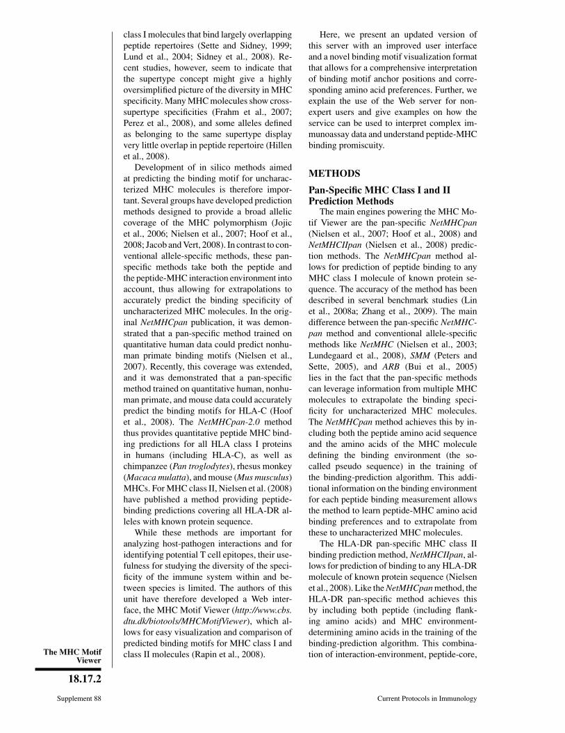
Figure 18.17.1 Left: Kullback-Leibler (KL) sequence logo for the HLA-A*0201 allele. The KL information content isplotted along the 9-mer peptide sequence (solid blue line). Amino acids with positive influence on the binding are plottedon the positive y axis, and amino acids with a negative influence on binding are plotted on the negative y axis. Therelative height of each amino acid is given by Equation 18.17.7, in the text. Right: The contact map for the HLA-A*0201allele visualizes which residues of the MHC pseudosequence are in contact with which positions in the 9-mer peptide.For color figure go to http://www.currentprotocols.com/protocol/im1817.
In the logo plots used in the MHC MotifViewer Web site, the amino acids are coloredaccording to their physicochemical properties:
Acidic [DE]: redBasic [HKR]: blueHydrophobic [ACFILMPVW]: blackNeutral [GNQSTY]: green.
Modified Kullback-Leibler LogoRepresentation
In the Shannon information equation, it isassumed that the different symbols (aminoacids or nucleic acids) have equal backgrounddistribution. In nature, amino acids are foundwith different frequencies. The Kullback-Leibler (KL) logos, as opposed to the logosdescribed earlier, explicitly take this differencein background amino acid distribution into ac-count when estimating the information contentat each position (Kullback and Leibler, 1951).The KL information content is calculated as:
b,i
i b,i 2
b b
logf
R fq
= ⋅∑Equation 18.17.6
where fb,i is the observed frequency of aminoacid i at position b, and qb is the corre-sponding background probability. Again, the
background probability is derived from largesequence databases like UniProt (UniProt,2008). Note that for a uniform background fre-quency distribution, qb is equal to 1/20 for alltwenty amino acids, and the KL informationcontent reduces to the Shannon informationcontent.
For the KL logos of the MHC Motif Viewer,the relative height of amino acid b at positioni is proportional to the corresponding term inthe relation for the KL information content:
b,i
b,i 2
b
b,i
c,i
c,i 2
c c
log
log
ff
qR
ff
q
⋅=
⋅∑Equation 18.17.7
In the KL logos, amino acids with a neg-ative log odds value are depicted as upside-down characters on the negative y axis, andamino acids with a positive log odds value asupright characters above the positive y axis.This way the logo directly reflects the matrixused to generate it and allows for a direct in-terpretation of which amino acids will have apositive or negative influence on the bindingaffinity, respectively. A blue histogram is used

Ligand-ReceptorInteractions inthe ImmuneSystem
18.17.5
Current Protocols in Immunology Supplement 88
to denote the total information content at eachposition (see left part of Fig. 18.17.1).
Contact MapsAn essential feature of the MHC Motif
Viewer interface is the visualization of contactmaps. The contact map displays the poten-tial interactions between a peptide sequenceand the MHC binding environment (the MHCpseudo sequence). The contact residues in theMHC molecule are defined as being within4.0
◦A of the peptide in any of a representa-
tive set of HLA-A and -B protein structureswith a bound nonamer peptide. Only residuespolymorphic across the HLA-A, B, and C al-leles are included giving rise to a pseudo se-quence consisting of 34 amino acid residues.Note that due to multiple possible conforma-
tions, the central peptide residues could chooseto interact with different subsets of residues inthe binding groove (Nielsen et al., 2007). Anexample of such a contact map for the HLA-A*0201 allele is shown in Figure 18.17.1.
Predictive PerformanceThe predictive performance of the
NetMHCpan method is to a very high degreedetermined by the density of characterizedMHC class I molecules in the immediateneighborhood of an uncharacterized MHCmolecule (Nielsen et al., 2007). This findingwas employed in the work by Hoof et al.(2008) to estimate the prediction accuracy ofNetMHCpan in terms of Pearson’s correlationcoefficient for any given MHC molecule. Inshort, the reliability index is estimated from
0
1
2
3
bit
s |AIHMRSK
TWCYDNPQFVEGL
VSITML
W
CHYNPDRFKQGEA
A
WLNMFY
H
CPDQESRTGI
KV
WKTFRYNSA
HMDCPEGQIVL
MKFHRWYS
Q P
V
L
TC
NIDAEG
QW
YLPMSTF
NCR
HAIVKGDE
TWHIYPMFSL
CNAQDVREKG
RNSLPFY
K
EW
THCQMADIGV
YK
WCMFHIV
NPQDTGLRASE
|Figure 18.17.2 Home screen for the MHC Motif Viewer Web site. The different pictures areclickable and take the user to different sections of the Web site for the six animal species (human,pig, mouse, gorilla, chimpanzee, and macaque) for which MHC binding motifs were computed.On top of the screen, a quick menu bar allows the user to navigate to the Help and MHC Fightsections.
18.17.6
Supplement 88 Current Protocols in Immunology
the pseudo sequence distance to the nearestneighbor for which the binding specificity iswell characterized. This measure of predictionaccuracy is included in the logo representationof the binding motif as a pie chart indicatingthe estimated predictive performance (seeFig. 18.17.1). A value of 1 suggests perfectaccuracy and a value of 0.0 suggests randompredictions.
The MHC Motif Viewer Web SiteThe MHC Motif Viewer Web site allows
for easy browsing of MHC binding motifsfor a large set of alleles. Included HLA al-leles cover the class I loci A, B, C, and Gand the DR class II families. In addition,the viewer offers binding motifs for mouse,gorilla, macaque, chimpanzee, chicken, andswine MHC class I alleles. The binding mo-tifs are represented by a modified version ofthe Kullback-Leibler logo, which allows for aneasy interpretation of which amino acids aredecisive (over-represented) or less important(under-represented) for binding.
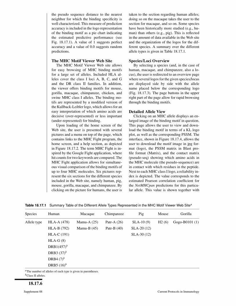
Upon loading of the home screen of theWeb site, the user is presented with severalpictures and a menu on top of the page, whichcontains links to the MHC Fight program, thehome screen, and a help section, as depictedin Figure 18.17.2. The term MHC Fight is in-spired by the Google Fight application, wherehit counts for two keywords are compared. TheMHC Fight application allows for simultane-ous visual comparison of the binding motifs ofup to four MHC molecules. Six pictures rep-resent the six sections for the different speciesincluded in the Web site, namely human, pig,mouse, gorilla, macaque, and chimpanzee. Byclicking on the picture for humans, the user is
taken to the section regarding human alleles;doing so on the macaque takes the user to thesection for macaque, and so on. Some specieshave been historically more studied (e.g., hu-man) than others (e.g., pig). This is reflectedin the amount of data available in the Web siteand the organization of the logos for the dif-ferent species. A summary over the differentallele types is given in Table 18.17.1.
Species/Loci OverviewBy selecting a species (and, in the case of
human, macaque, and chimpanzee, also a lo-cus), the user is redirected to an overview pagewhere several logos for the given species/locusare displayed side by side with the allelename placed below the corresponding logo(Fig. 18.17.3). The page buttons in the upperright part of the page allow for rapid browsingthrough the binding motifs.
Detailed Allele ViewClicking on an MHC allele displays an en-
larged image of the binding motif in question.This page allows the user to view and down-load the binding motif in terms of a KL logoplot, as well as the corresponding PSSM. Theinterface, shown in Figure 18.17.4, allows theuser to download the motif image in jpg for-mat (logo), the PSSM matrix in Blast pro-file format (Matrix), and the contact matrix(pseudo-seq) showing which amino acids inthe MHC molecule (the pseudo-sequence) arein contact with which residues in the peptide.Next to each MHC class I logo, a reliability in-dex is depicted. The value corresponds to theestimated Pearson correlation coefficient forthe NetMHCpan predictions for this particu-lar allele. This value is shown together with
Table 18.17.1 Summary Table of the Different Allele Types Represented in the MHC Motif Viewer Web Sitea
Species Human Macaque Chimpanzee Pig Mouse Gorilla
Allele type HLA-A (478) Mamu-A (25) Patr-A (26) SLA-10 (9) H2 (6) Gogo-B0101 (1)
HLA-B (792) Mamu-B (45) Patr-B (40) SLA-20 (12)
HLA-C (191) SLA-30 (12)
HLA-G (8)
DRB1(457)b
DRB3 (37)b
DRB4 (7)b
DRB5 (16)b
aThe number of alleles of each type is given in parentheses.bClass II alleles.

Ligand-ReceptorInteractions inthe ImmuneSystem
18.17.7
Current Protocols in Immunology Supplement 88
0
1
2
3
bit
s
MHSKR
W
C
A
FYPNDQETIV
GL
MIASVT
C
W
H
QPYRDFKNEGL
HKR
W
C
MYPFD
GQTVI
NESAL
H
P
N
TYKRA
Q M
W
V
C
F
S
E
L
D
IG
WKY
P
AF
THSR
N
V
M
C
QIG
DE
L
IT
K
W
H
YNFSR
PQ
M
C
L
VD
AGE
INH
W
T
M
YAS
LFP
R
C
V
QDK
EG
L
A
YPFS
W
H
N
Q
C
I
M
V
T
R
E
DKG
AK
W
CM
VHY
PF
DN
QITRELGS
0
1
2
3
bit
s
MHSKR
W
C
Y
F
PNDAQEITVGL
M
AVST
I
C
W
H
YPQRD
FEKNGL
HKR
F
W
C
M
YPD
QGTVNIESAL
HQN
Y
P
T
K
RAVF
W
M
C
S
LDIEG
VY
P
F
A
HTRS
W N
K
M
C
Q
I
GDE
L
LTKPWHY
NFSR
I
C
Q
M
VA
D
EG
IH
T
W
N
M
Y
SALFP
C
RQ
DVKEG
L
A
PYSF
NW
H
C
VM
Q
I
T
E
R
D
K
G
MAK
V
W
C
H
Y
PF
DQNLIRTEGS
0
1
2
3
bit
s
MHSKR
W
C
A
FYPNDQETIV
GL
MIASVT
C
W
H
QPYRDFKNEGL
HKR
W
C
MYPFD
GQTVI
NESAL
H
P
N
TYKRA
QM
W
V
C
F
S
E
L
D
IG
WKY
P
AF
THSR
N
V
M
C
QIG
DE
L
IT
K
W
H
YNFSR
PQ
M
C
L
VD
AGE
INH
W
T
M
YAS
LFP
R
C
V
QDK
EG
L
A
YPFS
W
H
N
Q
C
I
M
V
T
R
E
DKG
AK
W
CM
VHY
PF
DN
QITRELGS
0
1
2
3
bit
s
MHSKR
W
C
A
FYPNDQETIV
GL
MIASVT
C
W
H
QPYRDFKNEGL
HKR
W
C
MYPFD
GQTVI
NESAL
H
P
N
TYKRA
Q M
W
V
C
F
S
E
L
D
IG
WKY
P
AF
THSR
N
V
M
C
QIG
DE
L
IT
K
W
H
YNFSR
PQ
M
C
L
VD
AGE
INH
W
T
M
YAS
LFP
R
C
V
QDK
EG
L
A
YPFS
W
H
N
Q
C
I
M
V
T
R
E
DKG
AK
W
CM
VHY
PF
DN
QITRELGS
0
1
2
3
bit
s
THMSKR
IWCPY
L
DA
FQNEVG
VMAST
Y
W
CH
FPRDQIK
NEGL
Y
HMRFK
NCWSI
DQAPETGVL
W
T
N
R
FYS
A
HP
M
D
C
V
K
Q
G
I
L
E
M
TW
R
H
IYSFV
N
C
L
Q
K
PDE
AG
W
I
YSHNRF
V
M
T
C
P
L
Q
K
DE
GA
TWMHASYF
PC
N
V
QKIRDG
EL
HPLYF
W
C
E
S
M
NQADRTI
GKV
RK
W
C
H
FMYPINDQVTGESLA
0
1
2
3
bit
s
S
MHKR
W
C
Y
F
PNDQEVAITGL
MAVST
I
WC
H
YPRD
QFKN
EGL
HKR
W
C
FMYPDQ
GT
VI NESAL
QVSH
NY
P
T
KRA
M
W
F
C
DIE
LG
WKPY
F
ATHSR
N
V
C
M
Q
GI DEL
LTW
HYNFSR
I
MP
C
K
V
Q
ADGE
IN
W
H
T
M
Y
S
ALFP
C
Q
RVDE
KG
L
A
PYSF
HW
NCM
I R
QT
V
ED
K
G
M
L
VAK
W
C
I H
PYDFNQ
RETGS
0
1
2
3
bit
s
MHSKR
W
C
A
FYPNDQETIV
GL
MIASVT
C
W
H
QPYRDFKNEGL
HKR
W
C
MYPFD
GQTVI
NESAL
H
P
N
TYKRA
QM
W
V
C
F
S
E
L
D
IG
WKY
P
AF
THSR
N
V
M
C
QIG
DE
L
IT
K
W
H
YNFSR
PQ
M
C
L
V
D
AGE
INH
W
T
M
YAS
LFP
R
C
V
QDKEG
L
A
YPFS
W
H
N
Q
C
I
M
V
T
R
E
DKG
AK
W
CM
VHY
PF
DN
QITRELGS
0
1
2
3
bit
s
MHSKR
W
C
A
FYPNDQETIV
GL
MIASVT
C
W
H
QPYRDFKNEGL
HKR
W
C
MYPFD
GQTVI
NESAL
H
P
N
TYKRA
QM
W
V
C
F
S
E
L
D
IG
WKY
P
AF
THSR
N
V
M
C
QIG
DE
L
IT
K
W
H
YNFSR
PQ
M
C
L
V
D
AGE
INH
W
T
M
YAS
LFP
R
C
V
QDK
EG
L
A
YPFS
W
H
N
Q
C
I
M
V
T
R
E
DKG
AK
W
CM
VHY
PF
DN
QITRELGS
0
1
2
3
bit
s
SHMKR
W
C
PYD
FN
IQ
TEAVGL
AVQMST
C
W
H
PYI D
RNF
KE
GL
H
LMYRFK
W
NC
I
PDQTE
SGAV
PR
H
W
K
FYSNA
M
C
E
T
D
Q
VI
GL
LVTWHFSRY
N
M
C
QI K
P
G
D
EA
M
Y
I
H
WNT
RSF
C
Q
K
L
PV
DEAG
L
T
MWPAHSYF
C
NKQDI
RVEG
A
HSPYF
W
C
N
MLQ
ETDRGKIV
RYK
C
W
MH
P
NDI
QGFTVESAL0
1
2
3
bit
s
Y
IA
HSMRK
W
C
P
F
LDQN
EVTG
A
VMST
I
W
C
H
L
Y
QPN
DRFKEG
I
H
MNFKARSY
W
C
P
Q
DTEVGL
TDWH
Y
PKANS
G
C
F
M
R
E
Q
I
VL
RW
S
Y
Q
GFL
H
I
T
M
N
CV
D
K
P
E
A
Q
I
P
WM
F
NST
RY
H
C
L
GDA
VKE
W
S
HGPAYFL
M
C
NQDTRKIEV
Y
A
PSLF
REWHCM
T
G
Q
N
KI D
V
YC
W
M
HDP
NQ
GRTKEI FVSAL
0
1
2
3
bit
s
MHSKR
W
C
A
FYPNDQETIV
GL
MIASVT
C
W
H
QPYRDFKNEGL
HKR
W
C
MYPFD
GQTVI
NESAL
H
P
N
TYKRA
Q M
W
V
C
F
S
E
L
D
IG
WKY
P
AF
THSR
N
V
M
C
QIG
DE
L
IT
K
W
H
YNFSR
PQ
M
C
L
V
D
AGE
INH
W
T
M
YAS
LFP
R
C
V
QDK
EG
L
A
YPFS
W
H
N
Q
C
I
M
V
T
R
E
DKG
AK
W
CM
VHY
PF
DN
QITRELGS0
1
2
3
bit
s
FLAIHYSMRK
W
C
PDV
QNETG
LAVMST
W
I
C
H
Y
PDRNFQKGE
I
MRNKFASY
WH
C
PQ
DTEV
GL
TWD
H
P
YKANS
F
G
R
C
M
E
Q
IV
L
RSQWYGLHFIN
C
M
T
K
V
D
E
P
A
ILPWMFNSTRQY
H
C
DG
AK
EV
W
PHG
AYFL
MS
C
NQDKRTI
EV
A
P
YSFL
E
W
R
C
H
M
Q
T
IN
GK
DV
YC
W
M
HDP
NQ
GRTKEI FVSAL
0
1
2
3
bit
s
I
YHSMRK
W
C
F
PDANQ
LETVG
AVMST
L
YC
Q
W
F
H
I
PN
DRKE
G
H
K
I
WM
NASFY
C
RLQ
PDTGEV
H
D
W
K
G
YPANS
TF
E
C
M
QRIVL
N
I
Y
G
H
W
FL
Q
M
C
SR
V
D
TAP
K
E
LPI
MY
W
N
FTS
H
R
Q
C
D
A
GV
KE
P
HAWYFL
M
CQDNKRIVTGES
YPSFL
EW
A
C
H
M
T
G
RQ
N
I K
D
V
Y
C
WFM
HD
PNQGRTEI KVSAL
0
1
2
3
bit
s
MHSKR
WC
A
FYNDQPVEIT
GL
M
IASVT
W
C
H
YFNP
RDQKEGL
M
YHFKR
C
W
PDQGTNVIESAL
K
T
N
P
YRAH
S
F
W
M
C
DQ
V
E
GI
L
W
YN
F
A
TP
HRS
CV
MQ
G
K
I D
EL
W
T
HI
L
N
FRSYV
MC
P
Q
A
K
G
DE
H
YI
A
SMFLP
W
T
C
V
NQ
D
RK
EG
L
YASFP
W
H
N
C
M
TQI
RV
E
DGK
M
VAK
I
W
CH
F
Y
PLD
NQRGTES0
1
2
3
bit
s
I
H
SYMRK
F
W
C
A
PD
N
Q
LTE
VG
AFVLSTM
Q
W
Y
C
H
PN
DI RKEG
NWIMSAFY
H
K
C
R
P
LQ
DT
EVG
YH
W
G
PKANS
T
D
FQ
C
M
ERIVL
Q I
NYFWHLG
C
S
D
M
R
E
V
T
P
K
A
L
N
Y
P
W
MFTS
I
HC
Q
R
A
D
GK
EV
MA
HPWYFL
CS
GQ
NKRDEITV
AY
P
SFL
E
W
TH
C
M
Q
G
R
K
N
I
D
V
YC
WF
M
HD
PNQGRTKEIVSAL
0
1
2
3
bit
s
Y
IA
HSMRK
W
C
P
F
LDQN
EVTG
A
VMST
I
W
C
H
L
Y
QPN
DRFKEG
I
H
MNFKARSY
W
C
P
Q
DTEVGL
TDWH
Y
PKANS
G
C
F
M
R
E
Q
I
VL
RW
S
Y
Q
GFL
H
I
T
M
N
CV
D
K
P
E
A
Q
I
P
WM
F
NST
RY
H
C
L
GDA
VKE
W
S
HGPAYFL
M
C
NQDTRKIEV
Y
A
PSLF
REWHCM
TG
Q
N
KI D
V
YC
W
M
HDP
NQ
GRTKEI FVSAL
0
1
2
3
bit
s
WH
FYMSRK
C
PD
I
QAN
TE
VLG
VMST
IWCHA
QP
YNDRF
KGEL
HWMNKISYAF
R
C
PQDTEVGL
H
D
K
G
W
F
PYNAS
C
M
T
Q
E
R
IV
L
GN
Y
H
W
LFI
V
T
C
M
Q
S
D
R
P
A
KE
Y
P
W
L
I
MFNST
HQC
R
V
DGKA
E
P
W
GHLAFY
S
M
C
NVQR
TKD
IE
G
P
AYSLF
EW
HR
C
M
TQNDI
KV
F
Y
C
W
MHDPNQGRTEIVSAKL
0
1
2
3
bit
s
WH
FYMSRK
C
PD
I
QAN
TE
VLG
VMST
IWCHA
QP
YNDRF
KGEL
HWMNKISYAF
R
C
PQDTEVGL
H
D
K
G
W
F
PYNAS
C
M
T
Q
E
R
IV
L
GN
Y
H
W
LFI
V
T
C
M
Q
S
D
R
P
A
KE
Y
P
W
L
I
MFNST
HQC
R
V
DGKA
E
P
W
GHLAFY
S
M
C
NVQR
TKD
IE
G
P
AYSLF
EW
HR
C
M
TQNDI
KV
F
Y
C
W
MHDPNQGRTEIVSAKL
0
1
2
3
bit
s
Y
IA
HSMRK
W
C
P
F
LDQN
EVTG
A
VMST
I
W
C
H
L
Y
QPN
DRFKEG
I
H
MNFKARSY
W
C
P
Q
DTEVGL
TDWH
Y
PKANS
G
C
F
M
R
E
Q
I
VL
RW
S
Y
Q
GFL
H
I
T
M
N
CV
D
K
P
E
A
Q
I
P
WM
F
NST
RY
H
C
L
GDA
VKE
W
S
HGPAYFL
M
C
NQDTRKIEV
Y
A
PSLF
REWHCM
TG
Q
N
KI D
V
YC
W
M
HDP
NQ
GRTKEI FVSAL
0
1
2
3
bit
s
Y
IA
HSMRK
W
C
P
F
LDQN
EVTG
A
VMST
I
W
C
H
L
Y
QPN
DRFKEG
I
H
MNFKARSY
W
C
P
Q
DTEVGL
TDWH
Y
PKANS
G
C
F
M
R
E
Q
I
VL
RW
S
Y
Q
GFL
H
I
T
M
N
CV
D
K
P
E
A
Q
I
P
WM
F
NST
RY
H
C
L
GDA
VKE
W
S
HGPAYFL
M
C
NQDTRKIEV
Y
A
PSLF
REWHCM
TG
Q
N
KI D
V
YC
W
M
HDP
NQ
GRTKEI FVSAL
Figure 18.17.3 Species/loci overview. The alleles are arranged on a grid and are clickable.Comparison is made easy because the user has a simultaneous overview of many alleles.
the closest well characterized MHC neighborand the distance to this neighboring allele.Note that accurate reliability estimations arenot available for MHC class II alleles (Nielsenet al., 2008).
APPLICATIONSThe MHC Motif Viewer offers easy brows-
ing of the MHC binding specificity space.Employing the different viewing features, themotif viewer may be used to unravel unex-pected similarities between HLA alleles ofdifferent serotype as well as unexpected dis-similarities between HLA alleles of the sameserotype. In the following, several exampleswill be presented that demonstrate the valueof the MHC Motif Viewer.
Discovering Unexpected Differences inSpecificity
The first two digits of each HLA allelename describe its allele family, which is of-ten determined serologically. The full-lengthprotein sequences of the alleles HLA-A*3001and A*3002 differ only at four positions, cor-responding to a sequence identity of 98.9%.However, comparing the binding-motif logosof these two alleles reveals that the bind-ing specificity differs, most notably at theC-terminal anchor position. A*3001 showsa preference for basic amino acids (Lys) atP9, whereas A*3002 prefers the polar aminoacid tyrosine (see Fig. 18.17.5A). A lookat the contact matrix reveals the reason forthis dramatic difference. All four substitutions

The MHC MotifViewer
18.17.8
Supplement 88 Current Protocols in Immunology
0
1
2
bit
s |
1
PDQWEN
CRHGKATVMSYIFL
2
HCPWNRDKE
GQYASIFVMTL
3
PR
KHCW
E
QDTGVMNSLIFYA
4
CMW
HQI
RVY
EFDKLTPNGAS
5
PC
M
KR
Q
DTH
EWA
NYSVG
IFL
6
CQH
K
WEPY
DGMN
RV
AI
SFTL
7
E
CKR
DPQTNHMSGWIVAYFL
8
HWC
MDQRNIGPTVKEYASFL
9
C
DMPQ
NTG
RWESKI
AVHLFY| 0
1
2
bit
s |
1
PDQWEN
CRHGKATVMSYIFL
2
HCPWNRDKE
GQYASIFVMTL
3
PR
KHCW
E
QDTGVMNSLIFYA
4
CMW
HQI
RVY
EFDKLTPNGAS
5
PC
M
KR
Q
DTH
EWA
NYSVG
IFL
6
CQH
K
WEPY
DGMN
RV
AISFTL
7
E
CKR
DPQTNHMSGWIVAYFL
8
HWC
MDQRNIGPTVKEYASFL
9
C
DMPQ
NTG
RWESKI
AVHLFY| 0
1
2
bit
s |
1
PWD
EC
NQ
TFGHYMVSALIRK
2
CHWP
RD
EYKNF
GQAILMVST
3
PCWDET
H
VQ
GMLI
KNASFRY
4
CMWQ
ILHVRY
FEDTPKGNAS
5
CMD
WPKNE
T
H
RIAY
QVSG
FL
6
CWH
M
E
P
KYD
GVA
QINFRLST
7
K
D
CW
E
T
IM
QP
VRNHYFSAGL
8
CWM
HQ
D
NVKIPTR
EYAGSFL
9
C
DPMQ
NWTREGKSAI
HVLFY|
0
1
2
bit
s |
1
WCFP
YNDQ
EVTI
GMLHSARK
2
CP
HWRD
E
KNYQMGFLI
ASVT
3
WCPD
TGVISQNAEYMFHLKR
4
WCMG
HQFD
ES
YI
PTVLNKRA
5
CWDEMQY
GK
H
NPI
TLVFRSA
6
DCEWMKGH
Q
P
Y
NT
AVRIS
FL
7
CD
WE
K
QHT
GMNYRVISFAPL
8
CWMHG
QDEKNR
PITVAYSFL
9
WCPHYD
QNFETGSRMIVLAK| 0
1
2
bit
s |
1
WC
PF
DYNEQVTGLIHMSARK
2
WHC
YPRDFEKNQGMLI
ASVT
3
C
P
WD
TEQGVINSAMHYLFKR
4
WCM
H
Q
EG
IF
SY
T
PDNVRLK
A
5
CDWME
Q
H
KNPYI
GLTRVFAS
6
CDWM
E
Y
H
QKG
PATVNRIFL
S
7
CDEWK
QG
HN
TRM
YSVI
APFL
8
CWM
DHQGN
KRET
IVPYASLF
9
WC
H
PYFDNQTEGMSRIVLAK|0
1
2
bit
s |
1
PWD
EC
NQ
TFGHYMVSALI
RK
2
CHWP
RD
EYKNF
GQAILMVST
3
PCWDET
H
VQ
GMLI
KNASFRY
4
CMWQ
ILHVRY
FEDTPKGNAS
5
CMD
WPKNE
T
H
RIAY
QVSG
FL
6
CWH
M
E
P
KYD
GVA
QINFRLST
7
K
D
CW
E
T
IM
QP
VRNHYFSAGL
8
CWM
HQ
D
NVKIPTR
EYAGSFL
9
C
DPMQ
NWTREGKSAI
HVLFY| 0
1
2
bit
s |
1
PD
C
ENW
Q
HFGTMAYSVLIRK
2
CHWPD
RENK
G
FYAI
QSLVMT
3
PCD
E
TWQH
VGMNSI
KLARFY
4
C
MI
W
VL
QRH
FTE
YDPKNGAS
5
CMP
D
KWT
EN
QRHIV
AYSGFL
6
CWHKE
MPYV
DQGN
IAR
FSLT
7
KC
DE
TIW
Q
RMVNPHYSFGAL
8
WCMH
Q
D
NV
KIR
EP
YGTASFL
9
C
DPM
QN
WTERGSKAVIHLFY|
0
1
2
bit
s |
1
WPDEQN
C
TGHFYMSAVILRK
2
CHWPRDE
KNGYFQI
ALMVST
3
PC
DEH
W
V
T
QMG
INKLSRAFY
4
CMWQV
RIHLT
DYEFPKNGAS
5
CM
PD
KTWEQNH
A
SGRYIVFL
6
CW
ME
HKG
PQYVIDAN
SRLTF
7
K
DE
C
TI
RPMNQWVHGSYAFL
8
CWM
HDQVGNKIRPETAYSFL
9
C
DPMQ
NWTREG
SAKI
HVLFY|0
1
2
bit
s |
1
PDWECQN
GHT
FAYVMKRI
SL
2
WCHP
RDENKY
FGQILMAVST
3
CPWDEQ
GHVT
LMNI
RKYFSA
4
CWQ
M
RHV
I
E
D
LTYPKFN
GAS
5
CMDPW
KQ
ETGANYH
RSIVFL
6
WC
H
E
GK
M
P
YQ
DI
VAN
LFRTS
7
K
P
E
CD
WITQMRVNHYFGASL
8
WCM
HQDNK
PVGRITYEASFL
9
C
DPMQNWTREGSAKI
HVLFY| 0
1
2
bit
s |
1
PDWECQN
GHT
FAYVMKRISL
2
WCHP
RDENKY
FGQILMAVST
3
CPWDEQ
GHVT
LMNI
RKYFSA
4
CWQ
M
RHV
I
E
D
LTYPKFN
GAS
5
CMDPW
KQ
ETGANYH
RSIVFL
6
WC
H
E
GK
M
P
YQ
DI
VAN
LFRTS
7
K
P
E
CD
WITQMRVNHYFGASL
8
WCM
HQDNK
PVGRITYEASFL
9
C
DPMQNWTREGSAKIHVLFY|
0
1
2
3
bit
s
KSMWYF
C
AHPDRQENTIVGL
QMLW
CHNYDPGRFKEISATV
DSWIYLAFM
CHQRNKPEVTG
NWSPED
AH
CG
YQMIFTRKVL
MI
VFHAWGYSTC
DLNEQPRK
M
YTFIL
VWAHCSNRQPKDEG
QTAHMWPYF
CSI
NVRDEKGL
NQWGSEAFYP
HTCMLDRI KV
ILV
W
HCANYQMDPRGKEFST
Figure 18.17.4 Detailed view of the human HLA-A*0201 motif logo. The Logo link allows theuser to download the motif image in jpg format (logo), the Matrix link allows the user to downloadthe PSSM matrix in Blast profile format, and the Pseudo seq link directs the user to a graphicalplot of the contact-matrix (see Fig. 18.17.1). The pie-chart above the logo shows the estimatedPearson correlation coefficient for the NetMHCpan predictions for the given allele. This value isshown together with the closest neighbor and the distance to this neighboring allele.
are located in the binding groove of themolecules and are part of the pseudo sequence:Q70H (in contact with peptide positions 2 and3, Q denotes the amino acid at position 70in A*3001, H the amino acid in A*3002),V76E (peptide position 8), D77N (peptide po-sitions 8 and 9), and W152R (peptide posi-tion 7). Position 77 is a key residue in deter-mining the specificity of the F-pocket (Sidneyet al., 2008), and the substitution of the neg-atively charged Asp (D) by the polar Asn(N) may explain the change of binding speci-ficity from positively charged (Lys) to polar(Tyr).
Another, similar example is given by theHLA-A*6801 and A*6901 alleles. These two
molecules differ at five positions on proteinsequence level (i.e., 98.6% sequence iden-tity), three of which are part of the pseudose-quence (M97R, R114H, D116Y) and take partin the formation of the F-pocket. These sub-stitutions change the C-terminal binding pref-erence from basic (A*6801) to hydrophobic(A*6901); see Figure 18.17.5B.
These two examples illustrate that high sim-ilarity on sequence level does not necessarilyentail similar binding specificity. A small num-ber of substitutions at key positions of the HLAmolecule, namely in the binding pockets thatdefine the specificity of the anchor positions,are sufficient to change the binding specificitydramatically.

Ligand-ReceptorInteractions inthe ImmuneSystem
18.17.9
Current Protocols in Immunology Supplement 88
A
B
Figure 18.17.5 Motif logos of (A) HLA-A*3001 and HLA-A*3002 and (B) HLA-A*6801 and HLA-A*6901. Both panels show examples of allele pairs that show a high similarity on a protein-sequence level while revealing clear differences in the C-terminal amino acid preference. Logoswere displayed using the MHC Fight Viewer.
Discovering Unexpected SimilaritiesThe HLA-alleles A*0265 and A*0280 have
been reported by Sidney et al. (2008) to be ofA3-supertype specificity at P9. While our pre-dictions agree with the A*0265 assignmentto A3, for A*0280 the predictions suggestthat this allele indeed shares the A2-supertypespecificity, i.e., hydrophobic preference at P2and P9 (see Fig. 18.17.6).
Comparing Binding Motifs AcrossSpecies
Chimpanzees and humans have been sug-gested to share peptide-binding motifs (Sidneyet al., 2006). Figure 18.17.7 illustratestwo such examples: Patr-A*0701 and HLA-
A*2402, showing A24 supertype specificity,and Patr-B*1301 and HLA-B*0702, whichshow B7 supertype specificity.
Finding shared binding specificity for hu-man and chimpanzee MHC molecules may notbe very surprising given that chimpanzees areour closest relatives. We can, however, also de-tect similarities between the binding motifs ofhuman and pig MHC alleles. Figure 18.17.8illustrates an example of such a pair, SLA-2*0601 and HLA-B*4001, which both showa preference for acidic amino acids at P2 andhydrophobic C-terminal binding preference.
These examples illustrate the possible ap-plication of the MHC Motif Viewer in com-paring binding motifs across species borders.

The MHC MotifViewer
18.17.10
Supplement 88 Current Protocols in Immunology
Figure 18.17.6 Motif logos of HLA-A*0201, A*0301, A*0265, and A*0280. The logos reveal the similarity in bindingspecificity between A*0301 and A*0265, which is unexpected given the serotype of these molecules. A*0280 sharesthe A2 supertype binding preference, exemplified by A*0201. Logos were displayed using the MHC Fight Viewer.
Figure 18.17.7 Motif logos of the Patr-A*0701, HLA-A*2402, Patr-B*1301, and HLA-B*0702. The motif logosillustrate the shared binding specificity that can be observed for some chimpanzee and human MHC class I molecules.Logos were displayed using the MHC Fight Viewer.
Explaining Unexpected CTLResponses
Elispot assays are a common way of screen-ing a patient for epitope-specific CTL re-sponses. Perez et al. (2008) have tested anHIV-1 infected patient cohort for CTL re-sponses against a panel of 9-mer peptides.Some of the observed responses in this studycould not be explained based on the patients’HLA genotype, meaning that the peptide thatraised the immune response did not fit any ofthe binding motifs of the responding patient’sHLA alleles. Figure 18.17.9 illustrates onesuch example. A patient, typed to possess thealleles HLA-A*A1101, A*0201, B*4001, andB*3501, showed a high CTL response againstthe peptide QVPLRPMTY. Surprisingly, noneof the expressed HLA molecules has a prefer-
ence for hydrophobic residues at P2 and polarresidues at P9, and the strongest binding affin-ity predicted using the NetMHCpan method(Hoof et al., 2008) is 3000 nM. A closer lookat the binding motifs and the peptide, however,reveals that the 9-mer peptide itself containsa nested 8-mer (VPLRPMTY), which fits thebinding preference of B*3501 with a predictedbinding affinity of 68 nM.
SUMMARYWe have presented the MHC Motif Viewer,
which can be used to get a quick impressionand overview of the (predicted) binding speci-ficities for a large number of human and non-human MHC molecules.
The motifs are presented as sequence lo-gos in a way that allows for comprehensive

Ligand-ReceptorInteractions inthe ImmuneSystem
18.17.11
Current Protocols in Immunology Supplement 88
Figure 18.17.8 Motif logos of the pig MHC class I allele SLA-2*0601 and the human allele HLA-B*4001. The logos illustrate their shared binding specificity. Logos were displayed using the MHCFight Viewer.
Allele:HLA-A*0201
Allele:HLA-A*1101
Allele:HLA-B*3501
Allele:HLA-B*4001
Logo: Logo: Logo: Logo:
0
1
2
3
bit
s
KSMWYF
C
AHPDRQENTIVGL
QML
W
CHNYDPGRFKEISATV
SWIYLAFM
CHQRNKPEVTG
NWSPED
A
HCGYQMIFTRKVL
IVFHAWGY
C
DLNEQPRK
M
YTFIL
AHCSNRQPKDEG
Q
TAHMWPYF
CSI
NVRDEKGL
WGSEAFYP
H
TCMLDRI KV
ILV
W
HCANYQMDPRGKEFST
0
1
2
3
bit
s
RHATMKS
W
CI
GYQFDPNVEL
MIVST
W
CHAYFPRNDQKGEL
WNI
ALYMF
SCHRQPDKEVTG
W
DFYTNAS
C
MHKPQIEVL
QVTYWFS
I L
CREDAKG
W
MNYSTLF
HC
VRIAKDEG
T
HWISYMLF
V
CPNAQKRDEG
NPLFY
A
WH
CQMTRKSDI
GV
KW
CMHFYIVNRQTPGDLASE
0
1
2
3
bit
s
NITWLYFM
CHPQKRGDVAS
AP
W
CHYMFQNRDI
KEGLTVS
TWFMISYNVA
Q
LCHEDRPKG
Q
DSWEAP
CNVKRIL
LT
VHWGSFA
I
PEY
QNRK
PDGAST
QC
MNYREI
KL
YNHPITFAL
VQDKRG
EVTSPA
W
CMHYFQI
NRDKGL
MFY
WCPHNDQRGKETSVI
AL
0
1
2
3
bit
s
YSWGKAR
FMCHDPQNI
ETVL
EW
CMHFYIPVNGTQRLDAKS
S
WLAFYQI
M
V
HCNERTKPDG
SN
PQEGA
YTRILVK
LSIVAG
WY
DCNEQRKP
W
GNHLSFA
MP
RVTIEDK
Q
ASWTPLVYF
CENDRGK
TESPA
W
VQC
HMYFI
GNRDKL
MIL
WHCNDQYPGEKRFSTAV
Figure 18.17.9 Motif logos representing a patient’s HLA genotype.
interpretation of peptide binding anchor po-sitions and identification of amino acids thatpromote binding, as well as those that have anegative effect on binding.
For a more detailed comparison, the MHCFight Viewer enables the user to compare upto four binding motifs side-by-side. This canbe used to compare the specificity of differ-ent alleles. We have shown how this featurecan be used to study unexpected functionaldifferences in specificity between alleles thatmay be serologically identical and geneticallysimilar. We demonstrated how such compar-
isons might also be useful for the detectionof unexpected similarities between MHC al-leles, and how such similarity might explainpeptide cross-reactivity to alleles belonging todifferent supertypes. The viewer may even beused to study similarities across species bor-ders. We illustrate further how the server canbe used to correlate complex immune responsedata to host MHC genotypes.
The motif viewer is not intended forprediction of MHC-peptide binding. Forthis, the interested user is referred to thebinding-prediction methods NetMHCpan and

The MHC MotifViewer
18.17.12
Supplement 88 Current Protocols in Immunology
NetMHCIIpan, which were used to generatethe predictions that formed the basis for thebinding motifs presented by the MHC MotifViewer. Both prediction methods are availableas online Web servers.
ACKNOWLEDGEMENTSThis work was supported by NIH con-
tracts HHSN266200400083C, HHSN266200400025C, and HHSN266200400006C.
LITERATURE CITEDAltschul, S.F., Madden, T.L., Schaffer, A.A., Zhang,
J., Zhang, Z., Miller, W., and Lipman, D.J. 1997.Gapped BLAST and PSI-BLAST: A new gen-eration of protein database search programs.Nucleic Acids Res. 25:3389-3402.
Bui, H.H., Sidney, J., Peters, B., Sathiamurthy,M., Sinichi, A., Purton, K.A., Mothe, B.R.,Chisari, F.V., Watkins, D.I., and Sette, A.2005. Automated generation and evaluation ofspecific MHC binding predictive tools: ARBmatrix applications. Immunogenetics 57:304-314.
Falk, K., Rotzschke, O., Stevanovic, S., Jung, G.,and Rammensee, H.G. 1991. Allele-specific mo-tifs revealed by sequencing of self-peptideseluted from MHC molecules. Nature 351:290-296.
Frahm, N., Yusim, K., Suscovich, T.J., Adams, S.,Sidney, J., Hraber, P., Hewitt, H.S., Linde, C.H.,Kavanagh, D.G., Woodberry, T., Henry, L.M.,Faircloth, K., Listgarten, J., Kadie, C., Jojic,N., Sango, K., Brown, N.V., Pae, E., Zaman,M.T., Bihl, F., Khatri, A., John, M., Mallal,S., Marincola, F.M., Walker, B.D., Sette, A.,Heckerman, D., Korber, B.T., and Brander, C.2007. Extensive HLA class I allele promiscu-ity among viral CTL epitopes. Eur. J. Immunol.37:2419-2433.
Henikoff, S. and Henikoff, J.G. 1992. Amino acidsubstitution matrices from protein blocks. Proc.Natl. Acad. Sci. U.S.A. 89:10915-10919.
Hillen, N., Mester, G., Lemmel, C., Weinzierl,A.O., Muller, M., Wernet, D., Hennenlotter, J.,Stenzl, A., Rammensee, H.G., and Stevanovic,S. 2008. Essential differences in ligand presenta-tion and T cell epitope recognition among HLAmolecules of the HLA-B44 supertype. Eur. J.Immunol. 38:2993-3003.
Hoof, I., Peters, B., Sidney, J., Pedersen, L.E., Sette,A., Lund, O., Buus, S., and Nielsen, M. 2008.NetMHCpan, a method for MHC class I bind-ing prediction beyond humans. Immunogenet-ics. 61:1-13.
Jacob, L. and Vert, J.P. 2008. Efficient peptide-MHC-I binding prediction for alleles with fewknown binders. Bioinformatics 24:358-366.
Jojic, N., Reyes-Gomez, M., Heckerman, D., Kadie,C., and Schueler-Furman, O. 2006. Learn-ing MHC I–peptide binding. Bioinformatics22:E227-E235.
Kullback, S. and Leibler, R.A. 1951. On informa-tion and sufficiency. Ann. Inst. Stat. Math. 22:76-86.
Lin, H.H., Ray, S., Tongchusak, S., Reinherz, E.L.,and Brusic, V. 2008a. Evaluation of MHC class Ipeptide binding prediction servers: Applicationsfor vaccine research. BMC Immunol. 9:8.
Lin, H.H., Zhang, G.L., Tongchusak, S., Reinherz,E.L., and Brusic, V. 2008b. Evaluation of MHC-II peptide binding prediction servers: Applica-tions for vaccine research. BMC Bioinformatics9:S22.
Lund, O., Nielsen, M., Kesmir, C., Petersen, A.G.,Lundegaard, C., Worning, P., Sylvester-Hvid,C., Lamberth, K., Roder, G., Justesen, S., Buus,S., and Brunak, S. 2004. Definition of supertypesfor HLA molecules using clustering of speci-ficity matrices. Immunogenetics 55:797-810.
Lundegaard, C., Lamberth, K., Harndahl, M., Buus,S., Lund, O., and Nielsen, M. 2008. NetMHC-3.0: Accurate web accessible predictions of hu-man, mouse and monkey MHC class I affinitiesfor peptides of length 8-11. Nucleic Acids Res.1:36
Nielsen, M., Lundegaard, C., Worning, P.,Lauemoller, S.L., Lamberth, K., Buus, S.,Brunak, S., and Lund, O. 2003. Reliable pre-diction of T-cell epitopes using neural networkswith novel sequence representations. ProteinSci. 12:1007-1017.
Nielsen, M., Lundegaard, C., Worning, P., Hvid,C.S., Lamberth, K., Buus, S., Brunak, S., andLund, O. 2004. Improved prediction of MHCclass I and class II epitopes using a novel Gibbssampling approach. Bioinformatics 20:1388-1397.
Nielsen, M., Lundegaard, C., Blicher, T., Lamberth,K., Harndahl, M., Justesen, S., Roder, G., Peters,B., Sette, A., Lund, O., and Buus, S. 2007.NetMHCpan, a method for quantitative predic-tions of peptide binding to any HLA-A and -Blocus protein of known sequence. PLoS ONE2:E796.
Nielsen, M., Lundegaard, C., Blicher, T., Peters,B., Sette, A., Justesen, S., Buus, S., and Lund,O. 2008. Quantitative predictions of peptidebinding to any HLA-DR molecule of knownsequence: NetMHCIIpan. PLoS Comput. Biol.4:E1000107.
Perez, C.L., Larsen, M.V., Gustafsson, R.,Norstrom, M.M., Atlas, A., Nixon, D.F.,Nielsen, M., Lund, O., and Karlsson, A.C. 2008.Broadly immunogenic HLA class I supertype-restricted elite CTL epitopes recognized in a di-verse population infected with different HIV-1subtypes. J. Immunol. 180:5092-5100.
Peters, B. and Sette, A. 2005. Generating quantita-tive models describing the sequence specificityof biological processes with the stabilized ma-trix method. BMC Bioinformatics 6:132.
Rammensee, H., Bachmann, J., Emmerich, N.P.,Bachor, O.A., and Stevanovic, S. 1999. SYF-PEITHI: Database for MHC ligands and peptidemotifs. Immunogenetics 50:213-219.

Ligand-ReceptorInteractions inthe ImmuneSystem
18.17.13
Current Protocols in Immunology Supplement 88
Rapin, N., Hoof, I., Lund, O., and Nielsen, M. 2008.MHC motif viewer. Immunogenetics 60:759-765.
Robinson, J., Waller, M.J., Parham, P., Bodmer,J.G., and Marsh, S.G.E. 2001. IMGT/HLADatabase: A sequence database for the hu-man major histocompatibility complex. NucleicAcids Res. 29:210-213.
Schneider, T.D. and Stephens, R.M. 1990. Se-quence logos: A new way to display consen-sus sequences. Nucleic Acids Res. 18:6097-6100.
Sette, A. and Sidney, J. 1999. Nine major HLAclass I supertypes account for the vast pre-ponderance of HLA-A and –B polymorphism.Immunogenetics 50:201-212.
Sette, A., Fleri, W., Peters, B., Sathiamurthy, M.,Bui, H.H., and Wilson, S. 2005. A roadmapfor the immunomics of category A-C pathogens.Immunity 22:155-161.
Shannon, C.E. 1948. A mathematical theory ofcommunication. Bell Labs Tech. J. 27:379-423;623-656.
Sidney, J., Asabe, S., Peters, B., Purton, K.A.,Chung, J., Pencille, T.J., Purcell, R., Walker,
C.M., Chisari, F.V. and Sette, A. 2006. Detailedcharacterization of the peptide binding speci-ficity of five common Patr class I MHCmolecules. Immunogenetics 58:559-570.
Sidney, J., Peters, B., Frahm, N., Brander, C., andSette, A. 2008. HLA class I supertypes: A re-vised and updated classification. BMC Immunol.9:1.
Thompson, C.B. 1995. New insights into V(D)Jrecombination and its role in the evolution ofthe immune system. Immunity 3:531-539.
UniProt. 2008. The universal protein resource(UniProt). Nucleic Acids Res. 36:D190-D195.
Yewdell, J.W. and Bennink, J.R. 1999. Immun-odominance in major histocompatibility com-plex class I-restricted T lymphocyte responses.Annu. Rev. Immunol. 17:51-88.
Yu, K., Petrovsky, N., Schonbach, C., Koh, J.Y., andBrusic, V. 2002. Methods for prediction of pep-tide binding to MHC molecules: A comparativestudy. Mol. Med. 8:137-148.
Zhang, H., Lundegaard, C., and Nielsen, M. 2009.Pan-specific MHC class I predictors: A bench-mark of HLA class I pan-specific predictionmethods. Bioinformatics 25:83-89.