a practical approach to anemia
TRANSCRIPT

A Practical Approach to Anemia Dr Ali Maleki
PhD in Laboratory Hematology & Transfusion Sciences Kermanshah University of Medical Sciences
1

Diseases affecting RBCs are among the most common illnesses worldwide. RBC disorders are the most common human genetic diseases, Acquired anemias affect up to 25% of the world’s population.
Red blood cell disorders 2
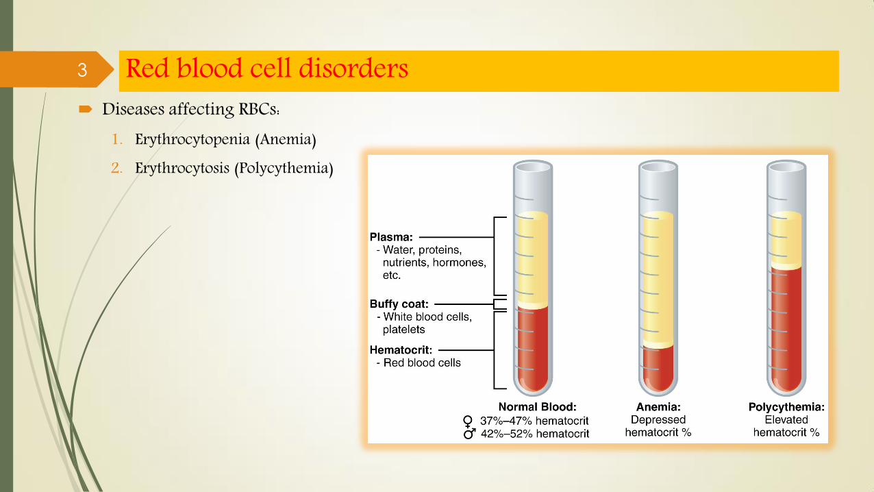
Diseases affecting RBCs: 1. Erythrocytopenia (Anemia) 2. Erythrocytosis (Polycythemia)
Red blood cell disorders 3

A Practical Approach to Anemias
4
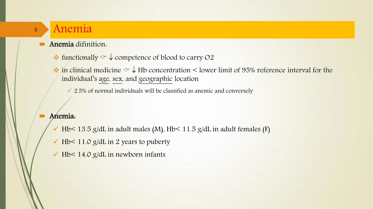
Anemia difinition: functionally competence of blood to carry O2 in clinical medicine Hb concentration < lower limit of 95% reference interval for the
individual’s age, sex, and geographic location 2.5% of normal individuals will be classified as anemic and conversely
Anemia:
Hb< 13.5 g/dL in adult males (M), Hb< 11.5 g/dL in adult females (F) Hb< 11.0 g/dL in 2 years to puberty Hb< 14.0 g/dL in newborn infants
5 Anemia

WHO defines anemia in adults as: Hb< 13 g/dL in M or Hb< 12 g/dL in F On this basis, anemia was estimated ~ 33% of global population (in 2010)
The main causes of anemia: iron deficiency (hookworm, schistosomiasis) sickle cell diseases thalassemia malaria anemia of chronic disorders (ACD)
Prevalence: F > M at all ages most frequent in children <5 years old most frequent in South Asia, and Central, West and East of Africa
Anemia 6

Anemia can develop if: 1. BM erythrocyte production is impaired
or 2. RBC loss or destruction exceeds the
maximal capacity of BM production
Anemia
BM can compensate RBC survival with production to a level 5–8 times normal (maximal functional capacity of BM). when RBC life span to ~18 days BM compensation is inadequate and anemia develops
7

is usually made from the CBC results generally relies on Hb/Hct: In general, Hct / Hb both move and ↓ together By contrast, changes in RBC count do not always parallel changes in Hct/Hb
In a patient with Thal trait: ↓ Hct or ↓ Hb + N or RBC count
Screening for anemia
o Sometimes, Hb/Hct can be misleading as changes in Hct /Hb can due to altered plasma volume also
8
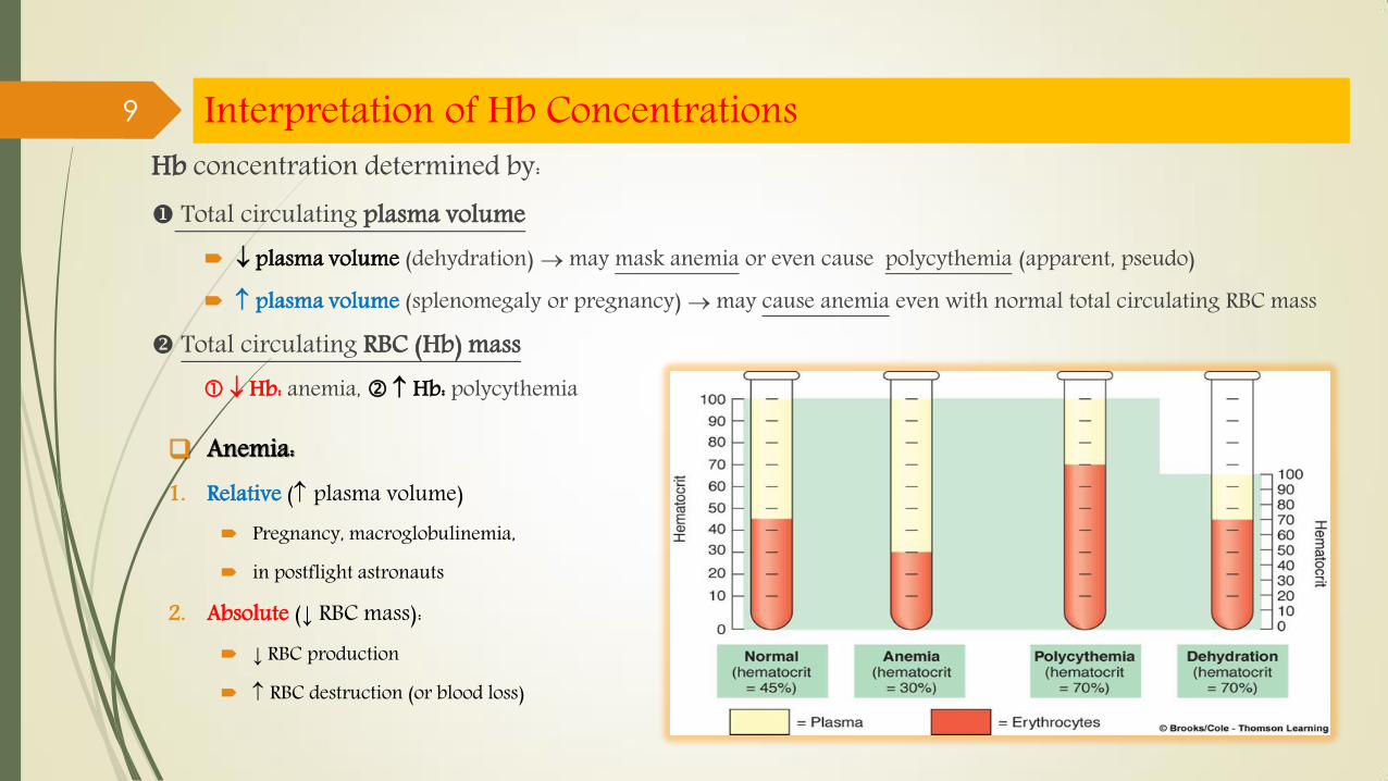
Hb concentration determined by: Total circulating plasma volume
plasma volume (dehydration) may mask anemia or even cause polycythemia (apparent, pseudo) plasma volume (splenomegaly or pregnancy) may cause anemia even with normal total circulating RBC mass
Total circulating RBC (Hb) mass Hb: anemia, Hb: polycythemia
Interpretation of Hb Concentrations
Anemia: 1. Relative ( plasma volume)
Pregnancy, macroglobulinemia, in postflight astronauts
2. Absolute (↓ RBC mass): ↓ RBC production
RBC destruction (or blood loss)
9
10 Diagnosis of Anemia- Lab investigation

11
The initial screening test is the CBC Depending on CBC results additional tests can be suggested:
Retic count, bilirubin, and PBS exam (for abnormal cell morphology) Urine (UA) and stool (SE) for the presence of blood
Diagnosis of Anemia- Lab investigation
12
RBC, Hb / Hct to screen for presence of anemia: in 1 of these parameters followed by other Lab tests
The CDC recommended cutoff values for diagnosis of anemia according to age and sex.
CDC: Centers for Disease Control and Prevention
Lab investigation- RBC, Hb, Hct

13
Hb/ Hct Cutoffs for diagnosis of anemia in Children, Males and nonpregnant Females.
Lab investigation- RBC, Hb, Hct

14
Upward adjustments for Hb/Hct cutoff values should be utilized for individuals living at high altitudes.
Lab investigation- RBC, Hb, Hct
15
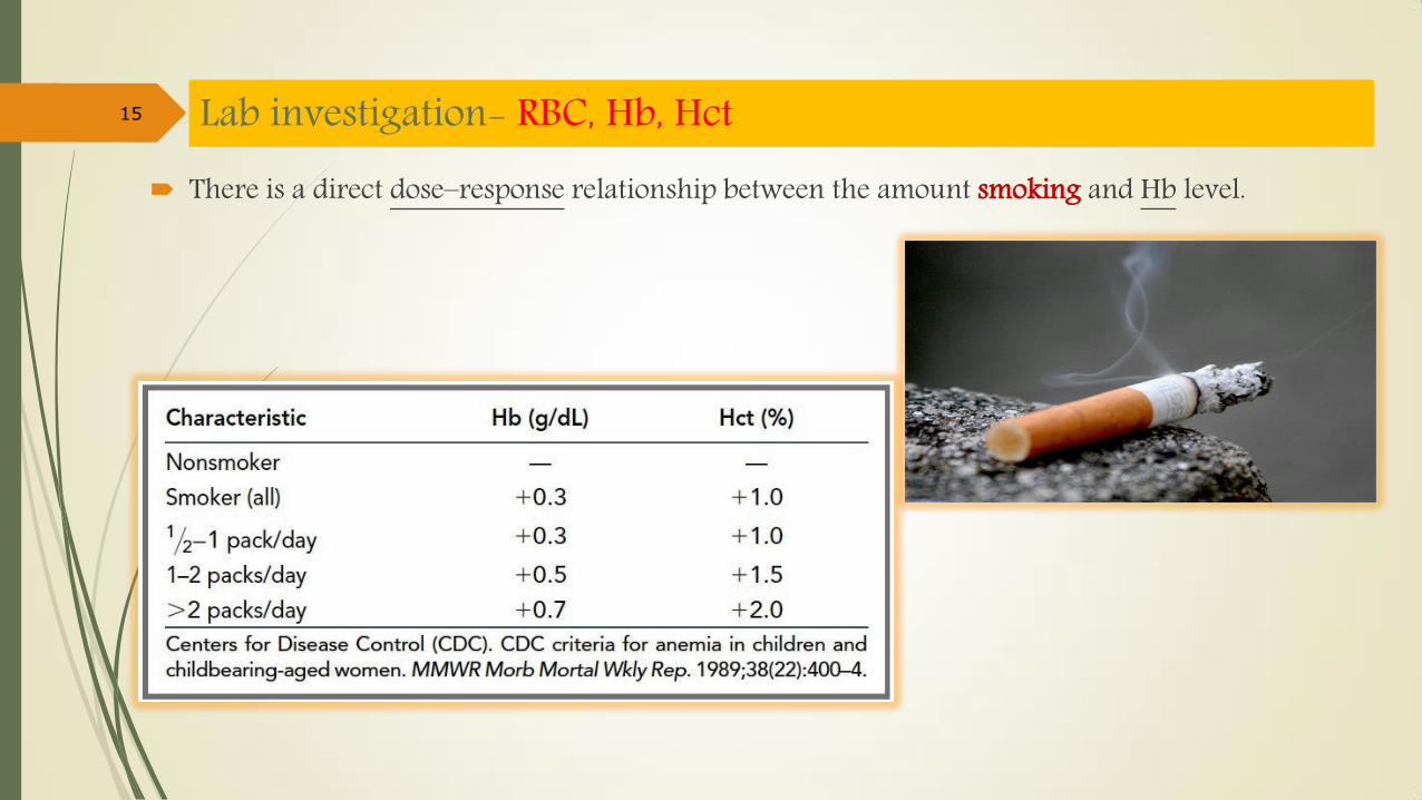
There is a direct dose–response relationship between the amount smoking and Hb level.
Lab investigation- RBC, Hb, Hct
16
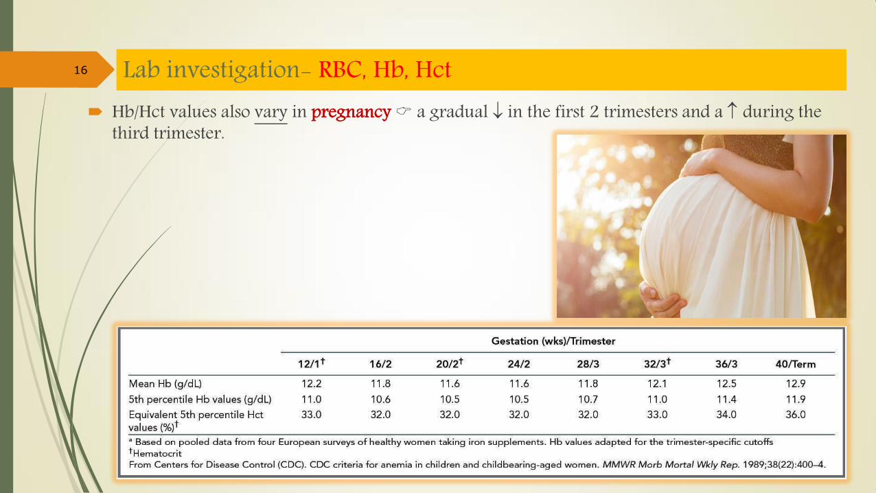
Hb/Hct values also vary in pregnancy a gradual in the first 2 trimesters and a during the third trimester.
Lab investigation- RBC, Hb, Hct

17
Anemia is prevalent in elderly persons but it not a normal part of aging. After age 65 prevalence of anemia (11% in M; 10.2% in F)
prevalence for those in nursing homes is higher. The highest prevalence in ages > 85 years (26% of M and 20% of F). In this group:
1. 1/3 was due to blood loss or nutritional deficiency, 2. 1/3 was due to ACD, inflammation, or chronic renal failure, 3. 1/3 was unexplained can be due to multiple causes.
ACD: anemia of chronic disease
Lab investigation- RBC, Hb, Hct

18
Variations in Hb due to blood-drawing techniques. in upright position compared with supine Hb values are
~0.7 g/dL higher
Lab investigation- RBC, Hb, Hct
Prolonged vasoconstriction by tourniquet cause hemoconcentration of sample and elevate Hb value.

19
RBC indices (MCV, MCH, MCHC, RDW) give important clues to the pathophysiology of anemia help to direct reflex testing Microcytic hypochromic cells highly suggestive of IDA Macrocytic normochromic cells associated with B12 or folate deficiency
IDA: iron-deficiency anemia
Lab investigation- RBC Indices
20
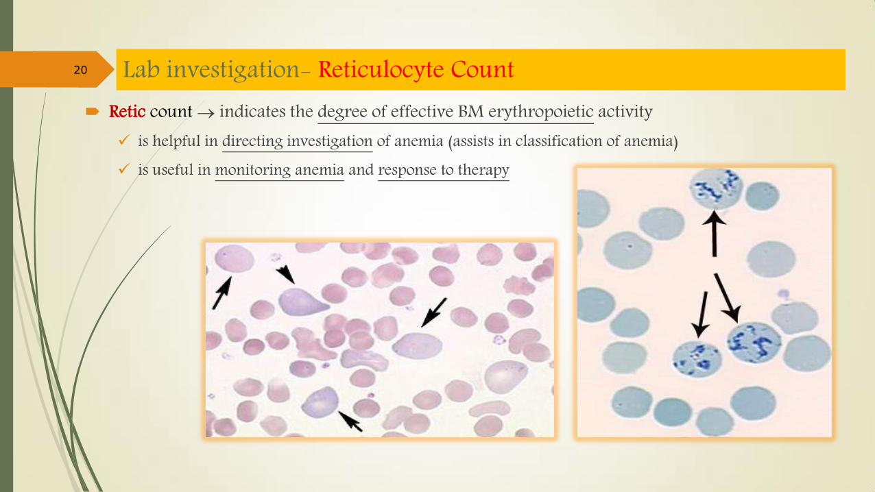
Retic count indicates the degree of effective BM erythropoietic activity is helpful in directing investigation of anemia (assists in classification of anemia) is useful in monitoring anemia and response to therapy
Lab investigation- Reticulocyte Count

21
Various pathological conditions (intrinsic or extrinsic) can alter RBC’s morphology careful examination of PBS assists in diagnosing the type of anemia in ~25% of cases.
PBS: peripheral blood smear
Lab investigation- Blood Smear Examination

22
WBC & PLT counts helps to distinguish ‘pure’ anemia from ‘pancytopenia’
Pancytopenia suggests: a general BM defect (e.g. Hypoplasia, Infiltration) a general destruction of cells (e.g. hypersplenism)
in anemias caused by hemolysis or hemorrhage Neut & PLT counts in Infections and Leukemia leucocyte count + abnormal leucocytes precursors
Lab investigation- WBC and PLT Abnormalities

23
BM examination usually is not necessary to determine the cause of an anemia. it can provide supplemental diagnostic information, when other Lab tests are not conclusive.
BM evaluation in hypoproliferative anemias can reveal myelodysplasia or infiltration with malignant cells or granulomas.
Erythroid hyperplasia (with fat & consequently M:E) is more pronounced in Hemolytic anemia than in non-hemolytic anemias
Lab investigation- BM examination
24
Purpose: to assist physician in identifying the cause by using Lab results in addition to other clinical data also is useful to Lab professionals when they correlate various test results for accuracy and make
suggestions for additional reflex testing
Anemias can be classified by: 1. Morphology 2. Pathophysiology
Classification of Anemias

Anemia Classification Morphologic & Pathophysiologic classifications of Anemias
25

The first step in approaching anemia is to classify based on RBC volume (MCV): 1. Microcytic (MCV, <80 fL), 2. Normocytic (MCV, 80-100 fL), 3. Macrocytic (MCV, >100 fL)
This exercise markedly narrows the differential diagnosis that needs to be considered in each patient.
26 Anemia- Morphologic Classification
It strongly recommend obtaining a PBS during the initial evaluation of anemia (regardless of subtype)

Approach to Microcytic Anemia The most common anemias in clinical practice are the microcytic anemias. a simple acronym summarizes the causes of microcytic anemia: TAILS:
1. T (thalassemia and the thalassemic hemoglobinopathies) 2. A (anemia of chronic disease; ACD) 3. I (iron deficiency; IDA) 4. L (lead poisoning) 5. S (congenital sideroblastic anemia; SA)
Expressed in order of frequency:
IDA: the most common microcytic anemia, (depending on one’s patient population) ACD or thalassemia, Lead poisoning (a normocytic anemia) → classically found in association with IDA usually listed with the microcytic anemias Congenital sideroblastic anemia.
27

The 3 major diagnostic possibilities for microcytic anemia are: 1. Iron deficiency anemia (IDA) 2. Thalassemia 3. Anemia of chronic disease (ACD) Lead poisoning and SA are not prevalent enough for routine consideration
28 Microcytic anemia
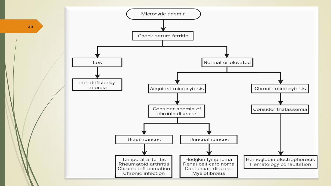
Step 1. Rule Out Iron Deficiency Anemia Since the most common of the microcytic anemias is IDA it recommend determination of
serum ferritin level as initial step for all patients with microcytic anemia: Low ferritin level is diagnostic of IDA
contrary to current dogma regarding acute phase reaction a diagnosis of IDA is unlikely in the presence of a persistently N or serum ferritin level.
in general, it not recommend either other serum iron studies (serum iron, TIBC, transferrin saturation) or BM biopsy for evaluation of IDA. o Instead, a limited treatment trial with iron supplementation is both a cost-effective & definitive way of
addressing the issue in equivocal cases.
29 Microcytic anemia
IDA: Iron deficiency anemia / TIBC: total iron-binding capacity
Important notes: microcytic anemia associated with RDW favors a diagnosis of IDA over that of ACD microcytic anemia associated with RBC count is characteristic of thalassemia trait Microcytosis without anemia could occur in thalassemia trait in polycythemia associated with iron deficiency. PBS in IDA usually shows anisocytosis and poikilocytosis in severe cases, cigarshaped RBCs and elliptocytes are characteristically present. Polychromasia, basophilic stippling, and target cells are characteristic features in thalassemia. IDA may be associated with reactive thrombocytosis.
30 Microcytic anemia
Step 2. Evaluation of Microcytic Anemia with Normal Serum Ferritin Normal ferritin level the next step is to determine whether the microcytosis is new or previously recognized?
in patients with chronic microcytosis a diagnosis of thalassemia should be considered Hb electrophoresis should be ordered as the initial test.
If the microcytosis is new a nonthalassemic condition associated with acquired microcytosis is a possibility.
31 Microcytic anemia
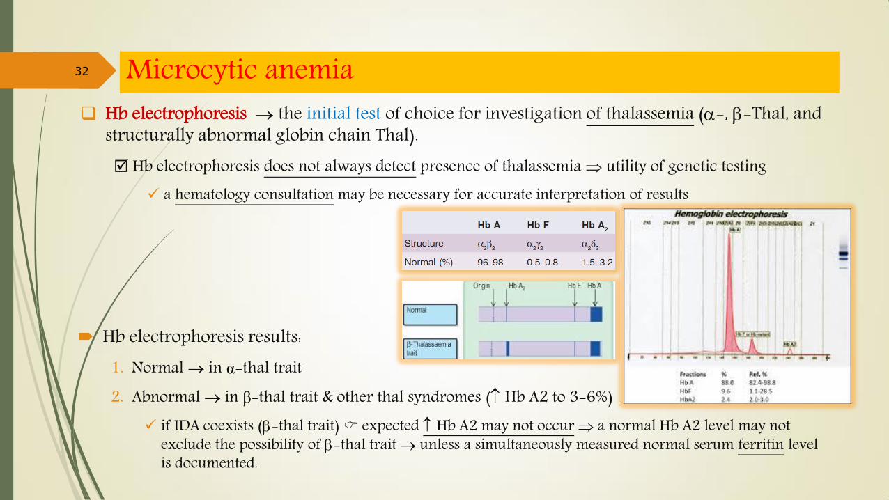
Hb electrophoresis the initial test of choice for investigation of thalassemia (-, -Thal, and structurally abnormal globin chain Thal). Hb electrophoresis does not always detect presence of thalassemia utility of genetic testing
a hematology consultation may be necessary for accurate interpretation of results
32 Microcytic anemia
Hb electrophoresis results: 1. Normal in α-thal trait 2. Abnormal in β-thal trait & other thal syndromes ( Hb A2 to 3-6%)
if IDA coexists (-thal trait) expected Hb A2 may not occur a normal Hb A2 level may not exclude the possibility of -thal trait unless a simultaneously measured normal serum ferritin level is documented.

33 Microcytic anemia -Thal Genetic testing (PCR–based DNA tests and Southern blot analysis) can reveal the molecular defect.
However, a genetic counseling can be initiated on the basis of family history and ethnic origin and without resorting to DNA testing.
-Thal a slight or moderate in Hb F may also be seen in -thal trait in general, Hb electrophoresis is often adequate for evaluating -thal, and genetic testing may be unnecessary.
Structurally Abnormal Globin Chain Thalassemia. Some structural Hb-pathies can produce a thalassemic (microcytic) phenotype as a result of globin
synthesis (Hb E, Hb Lepore, Hb CS) These thalassemic syndromes usually are identified by routine Hb electrophoresis genetic testing
may not be required.

34 Microcytic anemia Nonthalassemic (Acquried), non-IDA microcytic anemia
The differential diagnosis includes ACD and Sideroblastic anemia (SA). SA: a rare disorder characterized by RDW, dimorphic RBCs, and BM ring sideroblasts.
Acquired, non-IDA microcytic anemia is labeled as microcytic ACD is indicative of an underlying systemic disease (usual & unusual)
Anemia in ACD is usually normocytic but in some systemic diseases can be microcytic anemia. o Further clinical and Lab investigation in this instance is dictated by:
1. patient history 2. findings from physical examination 3. examination of PBS
ACD: Anemia of chronic disease
35

36 Normocytic anemia
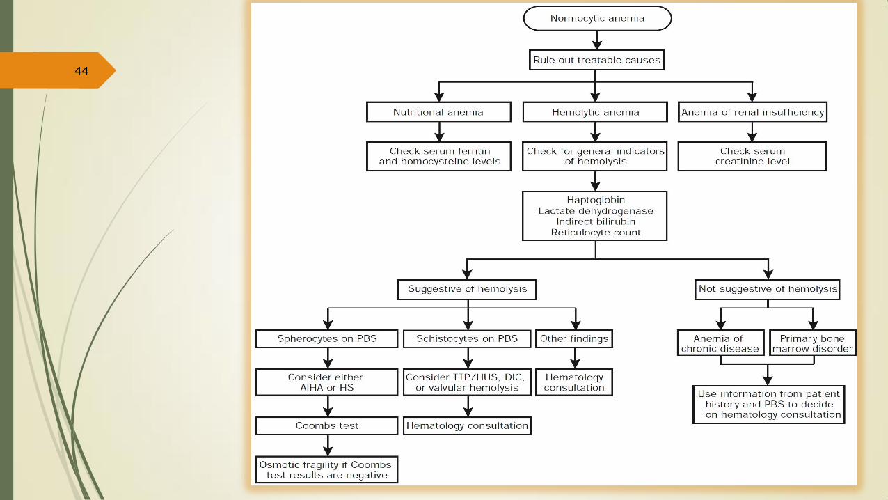
Step 1. Rule Out Readily Treatable Causes The first step in approaching normocytic anemia to exclude potentially treatable causes from
others including: 1. Anemia due to bleeding, 2. Nutritional anemia, 3. Anemia of renal insufficiency, 4. Hemolytic anemia
Patient history is key in implicating bleeding as a cause of anemia o if indicated fecal occult blood (OB) test can be ordered
37 Normocytic anemia
Nutritional anemia both iron and B12/folate deficiencies are possible causes of “normocytic” anemia (despite their usual
association with micro- & macrocytic anemia, respectively) Therefore, the initial investigation of normocytic anemia should include determination of both
serum ferritin and serum B12/ folate levels
Anemia of renal insufficiency is addressed easily by checking serum creatinine level. Anemia is associated with: an unremarkable PBS and an inappropriately normal serum EPO level.
in advanced kidney disease (serum creatinine, >3 mg/dL) anemia is severe and symptomatic
If initial tests are unrevealing the possibility of hemolyytic anemia (HA) should be considered.
38 Normocytic anemia
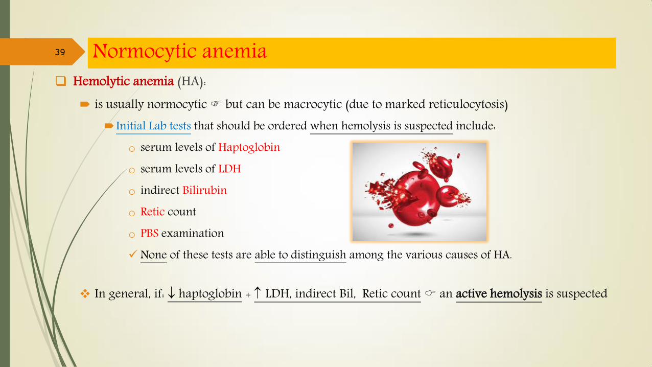
Hemolytic anemia (HA): is usually normocytic but can be macrocytic (due to marked reticulocytosis)
Initial Lab tests that should be ordered when hemolysis is suspected include: o serum levels of Haptoglobin o serum levels of LDH o indirect Bilirubin o Retic count o PBS examination None of these tests are able to distinguish among the various causes of HA.
In general, if: haptoglobin + LDH, indirect Bil, Retic count an active hemolysis is suspected
39 Normocytic anemia

Hemolytic anemia (HA) can be classified in many ways: 1. One classification separates causes: that are intrinsic or extrinsic to RBC. 2. in clinical practice it may be preferable to first distinguish EV-HA from IV-HA using urinary
hemosiderin test In general:
RBC–intrinsic and immune-mediated HA are extravascular MAHA, infection-associated, and chemical-induced HAs are intravascular
a drug-induced mechanism always should be considered in any hemolytic process.
40 Normocytic anemia
IV-HA: Intravascular hemolytic anemia / EV-HA: Extravascular hemolytic anemia / MAHA: microangiopathic HA
Hemolytic anemias Test EV-HA IV-HA Retic count LDH or N Indirect Bilirubin Haptoglobin - + Urinary Hemosiderin
41 Normocytic anemia

Step 2. Normocytic Anemia Not Associated With Bleeding, Nutritional Deficiency, Renal Insufficiency, or Hemolysis The primary consideration is:
1. a normocytic ACD 2. a primary BM disorder
Differentiating between the two is not always easy Patient history and PBS results provide helpful informations
42 Normocytic anemia
Anemia Due to Primary BM Disorder PBS is most helpful in providing clues for a primary BM disease:
In MDS RDW often is , PBS may show: oval macrocytes, Pseudo-Pelger- Huët anomaly, or monocytosis. In BM infiltration (PMF, metastatic cancer) NRBCs and IG are noted. In MM RBC rouleaux formation may be seen. Severe anemia + exremely Retic count suggests PRCA or AA. primary BM disease often is associated with disorders of WBCs & PLTs.
deciding to obtain a BM biopsy or no depend on: likelihood of discovering a primary BM disease therapeutic & prognostic value of the derived information. For example: BM biopsy in an elderly patient with mild anemia is unnecessary even if PBS suggests a primary
hematologic disease (because the results may not affect overall management decisions). In contrast, a younger patient with a history of chemotherapy or abnormal PBS should undergo BM biopsy
43 Normocytic anemia
PRCA: pure red cell aplasia / AA: aplastic anemia / MM: multiple myeloma
44
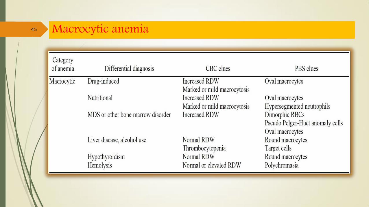
45 Macrocytic anemia
Step 1. Rule out the Presence of Drugs that Cause Macrocytosis The first considerations during evaluation of macrocytic anemia:
to exclude certain drugs (eg, hydroxyurea, Methotrexate, Trimethoprim, zidovudine, …) and alcohol consumption among them: Hydroxyurea is the most notorious induces the largest in MCV (oval macrocytosis >110 fL). a lesser degree of macrocytosis (100-110 fL) may result from use of:
o Zidovudine & chemotherapy (oval macrocytosis), or o Alcohol (round macrocytosis).
46 Macrocytic anemia
Step 2. Rule Out Nutritional Causes of Macrocytic Anemia The next step is to rule out nutritional causes (B12 or folate deficiency)
it prefer to use serum homocysteine for initial screening (because of its higher sensitivity) a normal homocysteine folate deficiency extremely unlikely
it advocate concomitant determination of serum B12 to safeguard against Lab error in view of the dire clinical consequences associated with Vit-B12 deficiency
In B12 deficiency, serum B12 B12 levels may be spuriously low during: pregnancy, in elderly patients, and in patients with low WBC counts. In these instances + in borderline-low B12 levels mesurement of methylmalonic acid level
If one of or tests has abnormal results serum methylmalonic acid level should be checked serum methylmalonic acid level:
an increased level strongly suggests B12 deficiency
47 Macrocytic anemia

Once vitamin B12 deficiency is confirmed the next step is to determine the cause: 1. The initial test: is to screen for the presence of IF-antibodies
if IF-Ab present diagnosis of PA additional testing may be unnecessary. 2. Otherwise, the Schilling test is performed to differentiate PA from primary malabsorptive
disorders (tropical sprue, celiac sprue, IBD, amyloidosis, and intestinal lymphoma)
48 Macrocytic anemia
IF intrinsic factor PA: pernicious anemia IBD: inflammatory bowel disease

Step 3. Evaluating Non–Drug Induced, Non-nutritional Macrocytic Anemia Further investigation of these macrocytic anemia is subcategorizing based on MCV: 1. Marked macrocytic anemia (MCV >110 fL)
almost always associated with a primary BM disease (eg, MDS, AA, PRCA, or LGL disorder) BM biopsy is indicated.
2. Mild macrocytic anemia (MCV, 100-110 fL) can be associated with MDS or more benign conditions (liver disease, alcohol consumption,
hypothyroidism, and marked reticulocytosis from hemolysis) it is important to obtain detailed information from PBS before proceeding to BM biopsy.
o polychromasia suggests hemolysis as the cause of macrocytosis, o round morphology of RBCs suggests liver disease (target cells are also evident) or hypothyroidism.
49 Macrocytic anemia
PRCA: pure red cell aplasia / AA: aplastic anemia / LGL: large granular lymphocyte

Macrocytic anemia 50

51