a paediatric spinal injury andreas crede emergency medicine registrar
TRANSCRIPT
A Paediatric Spinal InjuryA Paediatric Spinal Injury
Andreas CredeAndreas Crede
Emergency Medicine RegistrarEmergency Medicine Registrar

IntroductionIntroduction
• 5 Year old male5 Year old male
• Involved in MVA as restrained passenger Involved in MVA as restrained passenger near Beaufort Westnear Beaufort West
• Head on collision, no history about other Head on collision, no history about other passengerspassengers
• Referred because of abdominal pain and Referred because of abdominal pain and distension - ?blunt abdominal organ injurydistension - ?blunt abdominal organ injury
IntroductionIntroduction
• Arrived via AMSArrived via AMS
• Immobilised on spine boardImmobilised on spine board
• No significant past medical historyNo significant past medical history

IntroductionIntroduction
Clinical examination: Clinical examination:
• ABC’s stableABC’s stable
• Chest, CVS: NADChest, CVS: NAD
• Abdo: soft, suprapubic distension and Abdo: soft, suprapubic distension and discomfort. Urinary catheter drained discomfort. Urinary catheter drained 900ml clear urine 900ml clear urine

IntroductionIntroduction
• CNS: CNS: GCS 15/15. GCS 15/15. No signs of head injuryNo signs of head injury T12 Sensory level on rightT12 Sensory level on right L1 Sensory level on leftL1 Sensory level on left Lower limbs: complete motor deficit, bilateral Lower limbs: complete motor deficit, bilateral
unresponsive plantar reflexesunresponsive plantar reflexes Absent anal toneAbsent anal tone Right upper limb: C4-T2 sensory deficit, no Right upper limb: C4-T2 sensory deficit, no
motor deficitmotor deficit

InvestigationsInvestigations
• Bloods normalBloods normal
• Lodox normal, incl c-spine viewsLodox normal, incl c-spine views
• Thoraco lumbar spine x-rays:Thoraco lumbar spine x-rays:


MRI findingsMRI findings
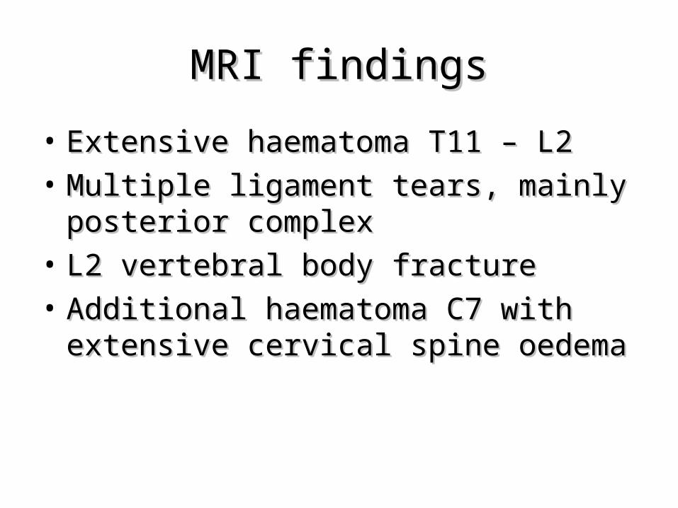
• Extensive haematoma T11 – L2 Extensive haematoma T11 – L2
• Multiple ligament tears, mainly posterior Multiple ligament tears, mainly posterior complexcomplex
• L2 vertebral body fractureL2 vertebral body fracture
• Additional haematoma C7 with extensive Additional haematoma C7 with extensive cervical spine oedemacervical spine oedema

Mechanism of InjuryMechanism of Injury

3 Column Model of Denis3 Column Model of Denis

Column ModelColumn Model
• 3 columns required to maintain spinal 3 columns required to maintain spinal stabilitystability
Wedge fracture = stableWedge fracture = stableWedge fracture with ligamentous rupture = Wedge fracture with ligamentous rupture =
unstableunstablePredictors of soft tissue injury: Angulation Predictors of soft tissue injury: Angulation
>20° or translation >3,5mm>20° or translation >3,5mm

Adult ClassificationAdult Classification
• A: Classic Chance FractureA: Classic Chance Fracture
• B: Fulcrum FractureB: Fulcrum Fracture
• C: Soft tissue flexion-distraction injuryC: Soft tissue flexion-distraction injury

Paediatric ClassificationPaediatric Classification
• Different to adultsDifferent to adults
• Presence of growth platePresence of growth plate
• Different characteristics of intervertebral Different characteristics of intervertebral disc allowing greater deformity: disc allowing greater deformity:
1.1. more water content of nucleus pulposusmore water content of nucleus pulposus
2.2. more elastic contentmore elastic content

Paediatric ClassificationPaediatric Classification• Type IType I: physeal injury of the superior growth plate associated with : physeal injury of the superior growth plate associated with
posterior lesion above the pedicle (soft tissue injury or superior facet posterior lesion above the pedicle (soft tissue injury or superior facet fracture). fracture).
• Type IIType II: osseous type. Fracture through the vertebral body, pedicle, : osseous type. Fracture through the vertebral body, pedicle, lamina and spinous process.lamina and spinous process.
• Type IIIType III: physeal injury of the inferior growth plate associated with : physeal injury of the inferior growth plate associated with posterior lesion below the pedicle (soft tissue injury or inferior facet posterior lesion below the pedicle (soft tissue injury or inferior facet fracture). fracture).

Type IType I
• Physeal injury of the superior growth plate associated with posterior Physeal injury of the superior growth plate associated with posterior lesion above the pedicle (soft tissue injury or superior facet lesion above the pedicle (soft tissue injury or superior facet fracture).fracture).

Type IIType II
• Osseous type. Fracture through the vertebral body, Osseous type. Fracture through the vertebral body, pedicle, lamina and spinous process.pedicle, lamina and spinous process.

Type IIIType III
• Physeal injury of the inferior growth plate Physeal injury of the inferior growth plate associated with posterior lesion below the associated with posterior lesion below the pedicle (soft tissue injury or inferior facet pedicle (soft tissue injury or inferior facet fracture). fracture).

Alternate ClassificationAlternate Classification
• Rumball and Jarvis A-D (X-ray classification)Rumball and Jarvis A-D (X-ray classification)• A: Disruption of Posterior Column extending into A: Disruption of Posterior Column extending into
middle columnmiddle column• B: Avulsion of Posterior elements with facet joint B: Avulsion of Posterior elements with facet joint
disruptiondisruption• C: Posterior ligament disruption with fracture line C: Posterior ligament disruption with fracture line
extending into vertebraextending into vertebra• D: Posterior ligament rupture with fracture line D: Posterior ligament rupture with fracture line
through lamina extending into physis of adjacent through lamina extending into physis of adjacent vertebral bodyvertebral body

ImagingImaging
• Standard X-rays view boney components/ Standard X-rays view boney components/ alignmentalignment
• X-rays cannot view soft tissuesX-rays cannot view soft tissues• MRI can identify ligamentous/ soft tissue MRI can identify ligamentous/ soft tissue
and growth plate injuriesand growth plate injuries• Absent epiphysis in human spines Absent epiphysis in human spines • CT scan not indicated unless MRI not CT scan not indicated unless MRI not
available or intra-abdominal injury available or intra-abdominal injury suspectedsuspected

Chance FracturesChance Fractures
• Unique to thoracolumbar spine (T10 – L2)Unique to thoracolumbar spine (T10 – L2)• Variant of flexion-distraction injuryVariant of flexion-distraction injury• Due to lap belt injury without shoulder belt Due to lap belt injury without shoulder belt
restraintrestraint• Fulcrum of flexion lies anterior to vertebral Fulcrum of flexion lies anterior to vertebral
column allowing no compression of vertebral column allowing no compression of vertebral bodybody
• Flexion results in either ligamentous tear or Flexion results in either ligamentous tear or combination of ligament, bone and disc injuriescombination of ligament, bone and disc injuries
Chance FracturesChance Fractures
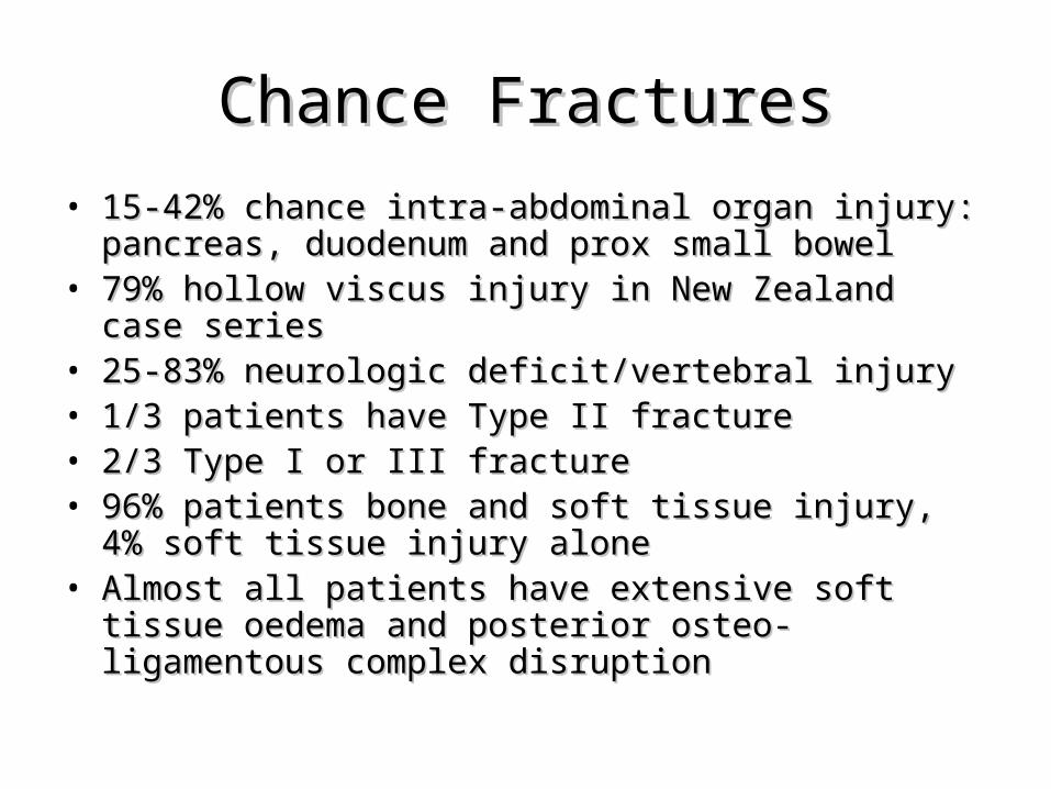
• 15-42% chance intra-abdominal organ injury: pancreas, 15-42% chance intra-abdominal organ injury: pancreas, duodenum and prox small bowelduodenum and prox small bowel
• 79% hollow viscus injury in New Zealand case series 79% hollow viscus injury in New Zealand case series • 25-83% neurologic deficit/vertebral injury25-83% neurologic deficit/vertebral injury• 1/3 patients have Type II fracture 1/3 patients have Type II fracture • 2/3 Type I or III fracture2/3 Type I or III fracture• 96% patients bone and soft tissue injury, 4% soft tissue 96% patients bone and soft tissue injury, 4% soft tissue
injury aloneinjury alone• Almost all patients have extensive soft tissue oedema Almost all patients have extensive soft tissue oedema
and posterior osteo-ligamentous complex disruptionand posterior osteo-ligamentous complex disruption

ManagementManagement
• ABC’sABC’s
• Prevent secondary injuryPrevent secondary injury
• High index of suspicion in patients High index of suspicion in patients restrained by lap seat beltsrestrained by lap seat belts
• Regular reassessment for abdo injuriesRegular reassessment for abdo injuries
• Unstable fracture: requires immobilisation/ Unstable fracture: requires immobilisation/ stabilisationstabilisation

ManagementManagement
• Conservative: reduction of dislocation + Conservative: reduction of dislocation + application of TLSO 2-3 monthsapplication of TLSO 2-3 months
• Surgical: large body habitus, polytrauma, Surgical: large body habitus, polytrauma, failure to stabilise conservatively.failure to stabilise conservatively.
ReferencesReferences• www.radiologyassistant.nl• www.imaging.consult.com• www.emedicine.medscape.comwww.emedicine.medscape.com• Ceroni, Mousny, Lironi, Kaelin. Paediatric seat belt injuries: Unusual Ceroni, Mousny, Lironi, Kaelin. Paediatric seat belt injuries: Unusual
Chance’s fracture associated with intra-abdominal lesions in a child. Eur Chance’s fracture associated with intra-abdominal lesions in a child. Eur Spine J 2004; 13:167-171Spine J 2004; 13:167-171
• De Gauzy et al. Classification of Chance Fracture in Children Using De Gauzy et al. Classification of Chance Fracture in Children Using Magnetic Resonance Imaging. Spine 2007; 32: E89-E92Magnetic Resonance Imaging. Spine 2007; 32: E89-E92
• Sheperd, Hamill, Segedin. Paediatric lap-belt injury: A 7 year experience. Sheperd, Hamill, Segedin. Paediatric lap-belt injury: A 7 year experience. Emergency Medicine Australasia 2006; 18: 57-63Emergency Medicine Australasia 2006; 18: 57-63
• Leonard M, Sproule J, McCormack D. Paediatric Spinal Trauma and Leonard M, Sproule J, McCormack D. Paediatric Spinal Trauma and Associated Injuries. Injury 2007; 38: 188-193Associated Injuries. Injury 2007; 38: 188-193
• Groves CJ et al. Chance type flexion-extension injuries in the thoracolumbar Groves CJ et al. Chance type flexion-extension injuries in the thoracolumbar spine: MR imaging characteristics. Radiology 2005; 236: 601-8spine: MR imaging characteristics. Radiology 2005; 236: 601-8

Also Check…Also Check…
• ‘‘Seatbelt syndromes’Seatbelt syndromes’– Google/pubmed etc itGoogle/pubmed etc it
• Soft tissue injuries associated with ‘seat Soft tissue injuries associated with ‘seat belt sign’belt sign’