a novel gene of the phosducin-like (phlp) family, expressed … · 2 summary we identified a new...
TRANSCRIPT

A novel gene of the phosducin-like (PhLP) family, expressed in the germ
line-a meiotic function conserved from yeast to mice*
Pascal Lopez, Ruken Yaman, Luis A. Lopez-Fernandez§, Frédérique Vidal, Daniel Puel,
Philippe Clertant, François Cuzin¶ and Minoo Rassoulzadegan
From the University of Nice, INSERM U470, Nice, France
Foot-notes
* This work was supported by grants from Association pour la Recherche sur le Cancer
(France).
§ Present address: Department of Immunology and Oncology, Centro Nacional de
Biotecnologıa, Campus Universidad Autónoma, Madrid, Spain.
¶ To whom correspondence should be addressed: INSERM U470, Faculté des Sciences,
06108 Nice cedex 2, France. Tel.: +33 492 07 64 11; Fax: +33 492 07 64 02; E-mail:
Copyright 2002 by The American Society for Biochemistry and Molecular Biology, Inc.
JBC Papers in Press. Published on November 6, 2002 as Manuscript M207434200 by guest on June 1, 2018
http://ww
w.jbc.org/
Dow
nloaded from

2
SUMMARY
We identified a new member of the phosducin-like (PhLP) protein family, predominantly, if
not exclusively expressed in male and female germ cells. In situ analysis on testis sections
and analysis of purified spermatogenic cell fractions evidenced a stage-specific expression,
with high levels of RNA and protein in pachytene spermatocytes and round spermatids.
Three mRNA species were detected, which correspond to different polyadenylation sites and
vary in abundance during germ cell maturation. Only low levels of RNA were detected in
whole ovary extracts, but expression of the protein became detectable within hours after
hormonal induction of superovulation. The gene (Mgcphlp) is located on mouse chromosome
5 in the immediate vicinity of the Clock locus. The predicted aminoacid sequence shows
extensive similarities not only with the known mammalian PhLP proteins, but also with the
yeast phosducin-like protein Plp2, required for the production and growth of haploid cells.
Expression of the murine protein was found to complement the defect of a yeast plp2D
mutant. We propose that the MgcPhLP/Plp2 proteins exert in germ cell maturation a function
conserved from yeast to mammals.
by guest on June 1, 2018http://w
ww
.jbc.org/D
ownloaded from

3
INTRODUCTION
Phosducin, a protein highly expressed in retina and pineal gland, has been considered as
playing a role in retinal phototransduction by interacting with the bg subunits of G proteins
and thereby modulating their signaling functions (1). A partially similar widely expressed
phosducin-like protein (PhLP)1 has been identified, which also inhibits Gbg function (2,3).
Two related genes were identified in the yeast Saccharomyces cerevisiae, designated PLP1
and PLP2 (4). The Plp1 protein was shown to bind efficiently the Gbg subunits. Binding of
Plp2 was also evidenced, but with a lesser affinity. On the other hand, genetic analysis
evidenced a role of Plp2 in the generation of viable haploid cells.
The mammalian phosducin and phosducin-like proteins are ubiquitously expressed (5).
Whether tissue specific homologues exist remains an open question. In the course of
screening a mouse testis cDNA library, we have identified a novel germ cell specific
phosducin-like protein, designated MgcPhLP for “mouse germ cell specific phosducin-like
protein”, which exhibits significant similarities with both the mouse phosducin and phosducin-
like proteins. Expression is strictly restricted to the male and female germ cells in a regulated
manner depending on the stage of germ cell maturation. The murine gene complemented the
defect of a yeast plp2D mutant, suggesting an evolutionary conserved function in meiotic
and/or post-meiotic cells.
1 The abbreviations used are : PhLP, phosducin-like protein; PCR, polymerase chain reaction; GST:
glutathione S-transferase; bp, base pairs; kbp, kilobase pairs; kDa, kilodaltons.
by guest on June 1, 2018http://w
ww
.jbc.org/D
ownloaded from
4
EXPERIMENTAL PROCEDURES
Mice—Mice used in all experiments were C57BL/6 x DBA/2 F1. Eggs were collected from
either naturally ovulated females or after hormonal induction of superovulation according to
standard procedures (6).
Cell cultures, preparation of germ cells–Cultures of the 15P-1 established Sertoli cell line
and primary Sertoli cell cultures, preparation of total germ cells and fractionation by elutriation
centrifugation were performed as previously described (7).
RNA analysis–Total RNAs were extracted and analyzed by Northern blot hybridization as
previously described (7). Dot blot hybridization was performed on Mouse RNA MasterblotTM
(Clontech).
DNA sequencing–Purified PCR products were first subcloned in pGEM-T Easy vector
System I, ref: A1360 Promega. The purified plasmids were then sequenced with DNA
Sequencing System kit, Big Dye Terminator ref: 4303149, according to manufacturer’s
instructions. Sequences were analyzed with the PE Biosystems ABI PRISM 310 genetic
analyzer.
Reverse transcription and PCR amplification–Quantitation of RNA amounts in cell extracts
was done by comparison with that of Gapdh reverse transcripts amplified as an internal
standard in the same reaction mixture. Total RNA (1µg) prepared using the total RNA
Isolation Kit (Roche Mannheim) was reverse transcribed using the MuLV reverse
transcriptase according to the supplier instructions. PCR amplification was performed with
the Taq DNA polymerase (Roche Mannheim). The following pairs of oligonucleotide primers
were used (RACE and polymerization conditions according to the supplier’s instruction
Roche Manheim). 5’ end analysis: RT, Rev0: 5’-gccagtgtcatcttctcatatgg-3’, first round, Rev-1:
5’-ggcctcctgctgtaagcg-3’ plus AAP from kit, and second round of PCR, Rev-2: 5’-
by guest on June 1, 2018http://w
ww
.jbc.org/D
ownloaded from

5
gcgcaacaccatctcttc-3’ plus AUAP from kit. 3’ end analysis: RT with oligos dT, first round, E11:
5’-gttggagcgatacagagtg-3’ plus AAP from kit, and second round of PCR, E12: 5’-
cattgcagatatgatggtg-3’ plus AUAP from kit. The 3’ Race products were then isolated after
hybridization with oligos: F01: 5’-gtgacactgaggctaagtag-3’. To routinely follow Mgcphlp
expression in testis and ovary, two pairs of oligonucleotide primers (common to the three
isoforms) were used, namely Phos-1: 5’-ctggaaatcagtatgtgaatg-3’ and Phos-2 5’-
ctgtatcgctccaacttctga-3’, and Mgc271 5’-cggttacaggaatggaaagc-3’ and Mgc483
5’-atgcagctattcacgatggc-3’. In yeast DNA, detection of the PLP2 wild type allele was
performed using primers Plp2-s: 5’-gataaagacttgtcggatttgg-3’, and Plp2-r: 5’-gtaatgcaattttctc-
tcttccc-3’, and detection of the URA3, plp2D mutated allele, using Plp2-s and Ura3-r 5’-tcatc-
tcttccacccatgtc-3’.
In situ hybridization–In situ hybridization was performed as previously described (8) using
single strand digoxigenine labeled RNA probes corresponding to sense- or antisense-
Mgcphlp messengers.
Purification of the MgcPhLP protein–Mgcphlp cDNA was inserted into the pGEX-5X vector
(Roche Co) by taking advantage of a BamHI site in the N-terminal region (a.a. 5), generating
a GST (glutathione S-transferase) fusion gene, whose expression was induced in
Escherichia coli cultures during overnight growth at 25°C in the presence of 0.1 mM IPTG.
Bacteria from 500 ml culture were washed and extracted in 50 ml of 100 mM Tris-HCl pH 8.0,
100 mM NaCl, 1 mM EDTA, 2 mM DTT, 1 mM PMSF, 50 µg/ml of both leupeptin and
aprotinin, by sonication (6 times 10 sec) at 4°C. Extract was clarified by high-speed
centrifugation (30 min, 30 kRPM, 4°C, in a Beckman 50Ti rotor), and submitted batchwise to
adsorption on Glutathione Sepharose beads (0.50 ml packed volume, pre-washed with
extraction buffer) with gentle agitation for 2 hours at 4°C. Beads were washed five-fold at 4°C
with 1 ml of 20 mM Hepes pH 7.5, 1.0 M NaCl, 1 mM EDTA, 2 mM DTT, 0.1% NP40 and 4
by guest on June 1, 2018http://w
ww
.jbc.org/D
ownloaded from
6
times with proteolysis buffer (20 mM Hepes pH 7.5, 100 mM NaCl, 2 mM CaCl2, 2 mM DTT,
10% glycerol, and 20 µg/ml bovine serum albumin). Biotinylated factor Xa (Roche
Boehringer) was added (10 µg in 1 ml final volume) and reaction was let to proceed for 2 hrs
at 4°C under gentle agitation. PMSF was then added (to a final concentration of 1 mM), and,
after having spun down the beads, the supernatant was cleared of protease by incubation for
1 hr at 4°C with streptavidin beads (50 µl packed volume, Roche Boehringer). The latter were
removed by centrifugation and the final supernatant was dialyzed against PBS (400 ml, 2
changes, 4 h, 4°C). The final preparation was analyzed by SDS-PAGE (see Fig. 6), and
stored frozen at –70°C. In parallel, control buffer was prepared in the same way by using
bacteria expressing GST.
Immunological techniques–GST-MgcPhLP fusion protein, prepared bound to glutathione-
Sepharose beads as described above, was eluted by PBS containing 20 mM reduced
glutathione, and used to raise antiserum in rabbits. The resulting antiserum recognized both
GST and MgcPhLP, in Western blot analysis (data not shown). Specific antibodies were
prepared by depleting the rabbit serum with Sepharose beads carrying the GST protein, and
used as primary antibodies in immunofluorescence and Western immunotransfer analyses.
Western transfers were obtained from mouse organs minced and boiled in Laemmli's
buffer by standard procedure (9); mouse oocytes were collected and treated in the same
way. Secondary antibody was a mouse monoclonal anti-rabbit IgG Fc fragment antibody
coupled to horse-radish peroxydase (Sigma). Staining was performed by the ECL technique
(Amersham).
Immunoprecipitation of 14-3-3 and associated proteins was performed using 14-3-3ß(H-8):
sc-1657 mouse monoclonal antibody according to the supplier’s instructions (Santa Cruz
Biotechnology Inc.). This antibody reacts with all the known proteins of the family.
by guest on June 1, 2018http://w
ww
.jbc.org/D
ownloaded from
7
Yeast strain, media, plasmid–The mutant Saccharomyces cerevisiae strain
(plp2::URA3,plp2D) used in this study was obtained from Dr H.G.Dohlman (Yale University
School of Medicine, New Haven, CT). For expression in yeast, the full length Mgcphlp cDNA
was inserted in the expression vector pRS315 (10).
by guest on June 1, 2018http://w
ww
.jbc.org/D
ownloaded from

8
RESULTS
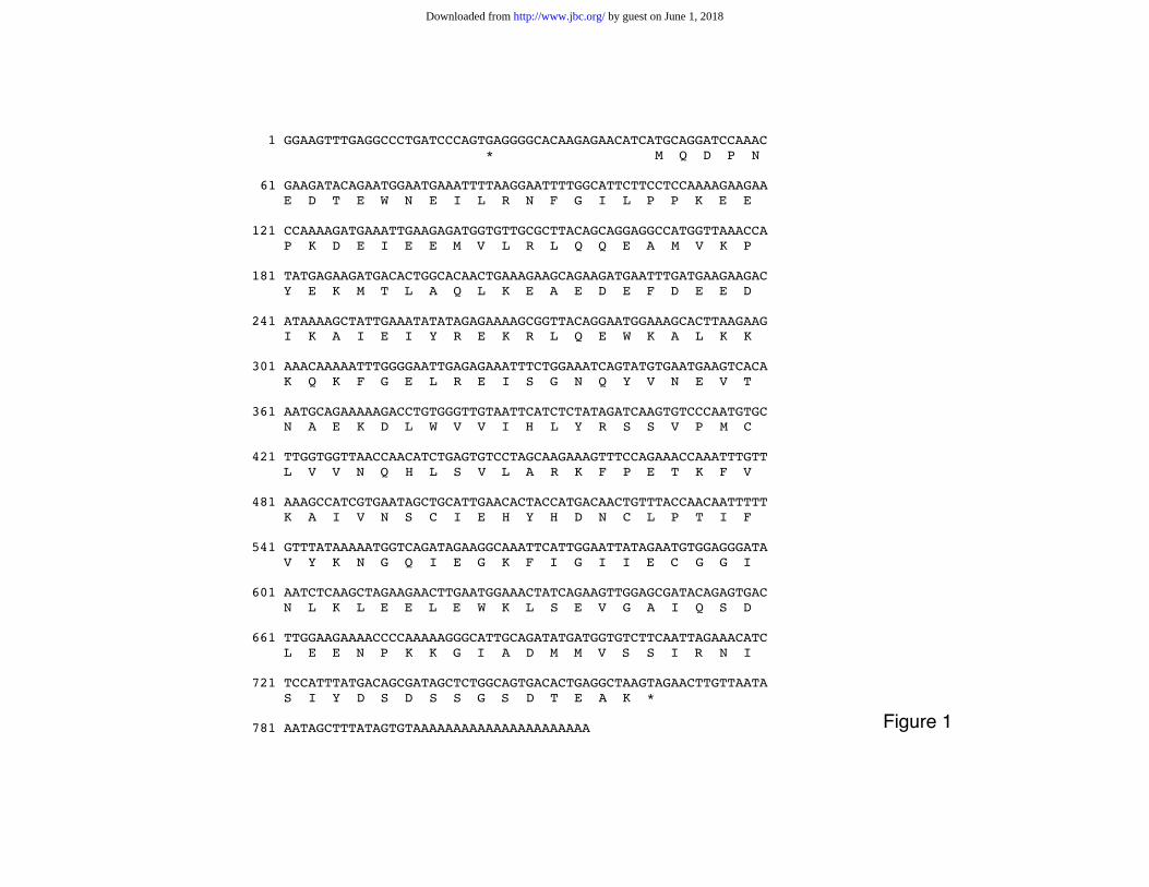
A new phosducin-like mRNA–During the course of an unrelated expression-based screen
of a mouse testis library, we have identified a cDNA uniquely expressed in germ cells. Figure
1 shows 818 base pairs of cDNA sequence with an open reading frame of 240 amino acids,
encoding a putative protein of 27.824 kDa, designated MgcPhLP, which exhibits 30-36%
amino acid identity to the mouse and bovine phosducins and to the rat phosducin-like protein
in a 143 amino acid core region (Fig. 2). Important amino acid residues conserved between
the phosducin and phosducin like proteins of various species (human (11), rat (12), mouse
(13)) are also conserved in MgcPhLP protein. As documented in more detail in a subsequent
section, significant similarities were also detected with two phosducin-related proteins of the
yeast Saccharomyces cerevisiae.
Computer search indicated that, in the mouse genome, the Mgcphlp gene is located within
a 204 Kb segment of chromosome 5 (accession number AF146795) which also includes the
Clock locus and has been designated PdcI2 in the MGI database (MG: 1890655). The
existence of two potential proteins, named protein B and PDCL2 (corresponding to a.a 3-204
and 3-195 of MgcPhLP, respectively) had been predicted from the genomic DNA sequence
(accession number AAD30564). The complete nucleotide sequence derived from the initial
cDNA clone and the 5’ and 3’ RACE products now allows us to determine the correct position
of the initiating methionine and thus, the complete aminoacid sequence.
Analysis of mouse EST libraries showing identities with Mgcphlp sequences (Unigene
cluster Mm.143764 plus additional clones) strongly suggested that the gene was at least
preferentially expressed in the testis. All identities were found in testis derived library
sources, and none among clones derived from other tissues, with the best match found in a
purified spermatocyte library (GI12596242).
by guest on June 1, 2018http://w
ww
.jbc.org/D
ownloaded from
9
Expression of Mgcphlp in the testis
Northern blot analysis on a limited series of tissues detected expression only in the testis
(Fig. 3A). A larger screen performed by dot blot analysis on a membrane spotted with polyA+
RNA from 22 mouse organs (Fig. 3B) showed again expression only in the testis. Mgcphlp
RNA was not detected in any of the somatic organs tested. The same conclusion was
reached using the most sensitive RT-PCR assay, which could not detect Mgcphlp RNA
sequences in two somatic tissues (Fig. 3C). The RNA was present in purified germ cells (see
below Fig. 5), but neither in Sertoli cells, whose primary cultures were negative (Fig. 3D), nor
cells of the Sertoli line 15P-1 (not shown). Although expression of the gene cannot be
excluded either in yet another tissue, or in a minor fraction of cells, possibly under specific
physiological conditions, it would appear to occur mostly in testicular germ cells. In fact, as
shown in a subsequent section, Mgcphlp is also expressed in female germ cells during a
limited period of meiotic maturation.
In whole testis extracts, three mRNA species were detected. Analysis of cDNA ends by 5’
and 3’ RACE revealed that these RNAs share the same 5’ extremity but differ by their 3’
ends. Three major polyadenylation sites were identified, which correspond to three canonical
polyadenylation signals in the genomic sequence (Fig. 4). As shown in Figure 3D, distribution
of the three isoforms significantly differed between the pachytene spermatocyte, the round
spermatid and the elongated spermatid fractions prepared by elutriation centrifugation.
During post-natal development, RNA was first detected at d.p.p. 20 and the maximum level of
expression was reached at day 30. The middle size mRNA was the only one identified at day
20, while the two other isoforms appeared only at the later time points.
Stage specific expression in the testis was confirmed by in situ hybridization. The probe
consisted of the entire coding sequence of Mgcphlp and thus detected all three mRNA
by guest on June 1, 2018http://w
ww
.jbc.org/D
ownloaded from

10
isoforms. As shown in Figure 5, expression was detected in pachytene to round spermatids
at all spermatogenesis stages.
Expression of MgcPhLP was examined at the protein level by using a rabbit antiserum
raised against a GST fusion protein (see Methods). Immunofluorescence staining of purified
fractions of male germ cells was clearly positive from the meiotic to late haploid stages of
spermatogenesis, and the protein was also present in the mature spermatozoa of epididymal
sperm. It was not detectable by Western blotting at 10 d.p.p., but clearly present in the testis
at day 18 (Fig. 5B-F).
Expression in ovary–In spite of the facts that no EST sequences corresponding to Mgcphlp
were present in ovarian libraries from various species, and that RNA was not detected by dot
blot hybridization (Fig. 3B), low levels of expression were evidenced by RT-PCR analysis in
total extracts from adult ovary (Fig. 6A). Western blot analysis (Fig. 6B) failed to detect
expression of the protein in extracts from either ovary or unfertilized eggs. It was, however
detected in fertilized eggs (Fig. 6C). Taking into account the smaller proportion of germ cells
in the female gonad, these results did not exclude the possibility of stage-specific expression
during meiotic maturation. This was shown to be the case by Western blot analysis following
injection of human chorionic gonadotropin (hCG) as part of a super-ovulation regime (6)
(Fig. 6D). The protein was detected as early as 3 hrs after hormone injection, a time
corresponding to the nuclear breakdown step of pre-ovulatory meiotic maturation.
Association with 14-3-3 protein(s)-The 14-3-3 family includes a series of closely related
proteins which bind phosphorylated components of signal-transduction pathways and
modulate their interactions (reviewed in ref. 14). Several of them, prominently the 14-3-3
q protein, are expressed in a stage-dependent manner in the spermatogenic differentiation
pathway (15). As shown in Fig. 7, a complex of the MgcPhLP protein with 14-3-3 protein(s)
was evidenced by immunoprecipitation of testicular protein extracts with polyvalent anti-14-3-
by guest on June 1, 2018http://w
ww
.jbc.org/D
ownloaded from
11
3 antibodies followed by Western blot analysis of the precipitated complexes with anti-
MgcPhLP antiserum. Further experiments are in progress to identify more precisely the
protein(s) present in these complexes.
Evolutionary conservation–The Mgcphlp coding sequence appears to have been relatively
well conserved throughout evolution. Coding regions of the murine gene are 87-92% identical
at the nucleotide level to human EST sequences contained in Unigene cluster Hs.223712,
derived exclusively from germ cells or testes. The cluster sequences are located in human
chromosome 4q11 region, again in close proximity to the human homologue of the mouse
Clock gene. In the Saccharomyces cerevisiae genomic sequence, two genes, PLP1 and
PLP2, encode proteins with a clear similarity to the mammalian phosducins and phosducin-
like sequences (4). Alignments of phosducin-like sequences (Fig. 2) show that the murine
MgcPhLP protein is more closely related to Plp2. One may note that, in the amino-terminal
part of the sequences, an 11 aminoacid-region implicated in Gßg binding (16), is completely
conserved between the phosducin proteins and Plp1, but present neither in Plp2 nor in
MgcPhLP. That would distinguish two groups of phosducin-like proteins: on one hand, the
mammalian phosducin and PHLP1, with the region that interacts with Gbg proteins, and on
the other, the MgcPhLP and PLP2 proteins, with a distinct pattern of conserved amino acids.
Other discrete patches of pairwise similarities could also be observed in the central and
carboxyterminal parts of the sequences.
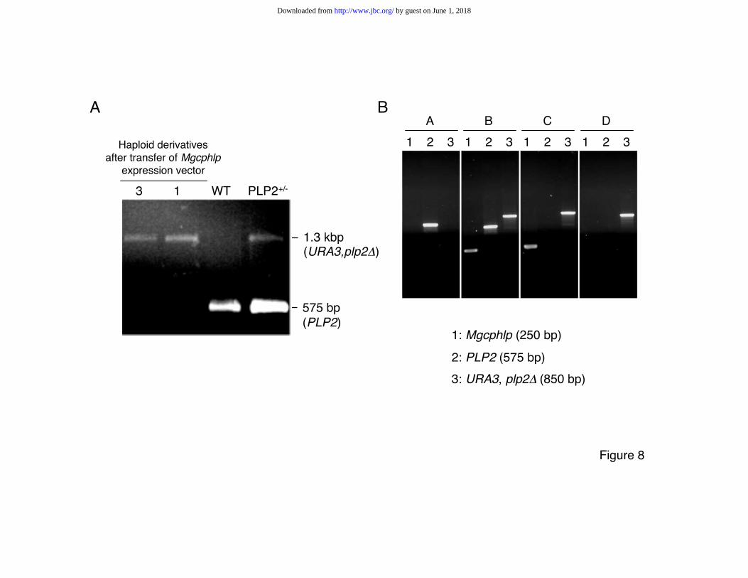
Complementation of a yeast plp2 mutant by the murine Mgcphlp gene–A yeast strain
bearing mutations in the TRP1 and URA3 nutritional markers, and in which one PLP2 allele
has been replaced by URA3 (plp2::URA3, plp2D) was previously established (4). The
deletion of PLP2 prevented the recovery of mutated haploid clones upon induction of
sporulation in diploid heterozygotes. The mutated strain was transformed with an expression
vector for Mgcphlp and TRP1 (see Materials and Methods), and the resulting colonies were
by guest on June 1, 2018http://w
ww
.jbc.org/D
ownloaded from
12
induced to sporulate. Isolated spores were tested in duplicate in media selective for either
URA3 (plp2D) or TRP1 (Mgcphlp). Haploid colonies were then revealed by replica plating
with yeast strains of either the a or a mating types carrying a mutation in the nutritional HIS
gene and selection in histidine-free medium. Haploid derivatives identified as carrying both
the URA3 (plp2D) allele and TRP1 (Mgcphlp) were grown. Analysis by PCR amplification
confirmed the expected absence of the wild type PLP2 and the presence of the Mgcphlp
allele (Fig. 8A). These complemented haploid clones were successfully grown for successive
generations. As expected from the published data, transfer of the empty vector did not result
in the production of viable haploid cells.
A meiotic function conserved from yeast to mammals?–It was initially reported (4) that
haploid derivatives could not be grown from the mixture of meiotic products generated by the
diploid heterozygous genotype (plp2::URA3, plp2D). One could not on this basis distinguish
between a function of the gene during meiosis (or at an early stage of spore formation) and a
general requirement for cellular growth. In view of the results shown in Fig. 8B, we may now
conclude that the gene is not required for growth. Genotyping haploid clones which had been
maintained in culture for 30-40 generations after their isolation from the complemented
parent strain showed that the mouse gene was eventually lost. Growth properties of the
clones in the absence of a functional PLP2 gene remained, however, unaffected. It is
therefore most likely that the protein, not necessary for growth, was required for the
establishment of the haploid state, a conclusion which is consistent with the expression of the
mouse gene being restricted to the meiotic and early post-meiotic stages.
by guest on June 1, 2018http://w
ww
.jbc.org/D
ownloaded from
13
DISCUSSION
Phosducins are regulators of G-protein activity in the retina, and the phosducin-like
proteins are considered to be potential ubiquitous regulators of Gßg signaling. We describe in
this report a novel phosducin-like mRNA specific of the meiotic and post-meiotic germ cells,
which shows significant amino acid sequence similarities to the phosducin and phosducin-like
proteins of various species. Searching existing EST clones, both from the mouse and human,
suggested germ cell specificity, since related sequences were found in EST libraries from
testes, but not from other tissues. In the mouse, expression of Mgcphlp was at least
predominantly observed in the male and female germ cells, in both sexes at the meiotic and
post-meiotic stages. It must be, however, taken into account that a detailed in situ analysis
could not be performed on every possible tissue, so that we cannot exclude that the gene
might be expressed in a minor cell population and/or only during a limited physiological or
developmental period (as it is in fact the case in the ovary). In the male gonad, three Mgcphlp
RNA isoforms were identified at different stages of differentiation. These three specific
mRNA’s correspond to different polyadenylation sites in the locus. It is interesting to note the
presence in the vicinity of the polyadenylation sites in genomic DNA of AU rich sequences
similar to the sequences which were described as cytoplasmic polyadenylation elements
mediating polyadenylation and translation of messages during the ovocyte release from the
meiotic block at ovulation and prior to the activation of zygotic genome at the two cell stage
(17,18). The presence of stage-specific isoforms of the Mgcphlp message may thus reflect a
translational control during meiotic and post-meiotic maturation. The conclusions of RNA
analysis were confirmed by direct determination of the protein by polyclonal antibodies
specific for the mouse protein. This was especially informative in the ovary, in which
by guest on June 1, 2018http://w
ww
.jbc.org/D
ownloaded from

14
expression is normally limited to the small number of ovocytes undergoing meiosis, but could
be readily evidenced after hormonal stimulation leading to superovulation.
As it is the case for the other phosducin-related proteins, the function of the protein at the
molecular level remains largely to be established. A possibly significant feature in this respect
is its association with at least one of the proteins of the 14-3-3 family. Binding may be
mediated by the RSSVP motif (a.a. 119-123, Fig. 1), which resembles the sites of interaction
identified in other 14-3-3-binding proteins (14). 14-3-3 binds phosphorylated serine residues
in a number of proteins active in signal transduction. In retinal photoreceptors, it is
considered as regulating binding of phosducin to Gbg by sequestering the phosphorylated
phosducin molecules and blocking their binding. It was also recently shown to interact in the
brain with a phosducin-like protein (19). The specificity of 14-3-3 binding, its relationship with
the phosphorylation of serine residues in MgcPhLP are currently being studied.
Two phosducin-related genes were recently described in yeast, PLP1 and PLP2, and .
MgcPhLP displays a greater amino acid similarity with PLP2 than with PLP1. The inability of
a plp2D mutant to generate viable haploid products was successfully complemented by
transfer of the mouse gene. Regarding the function of the yeast gene, published data (4)
have left two possibilities open: the Plp2 protein could be either necessary for growth in
general or specifically required for the generation of haploid products, either during or after
meiosis. The observation that haploid clones bearing the plp2D mutation could not be grown
from a sporulating culture is compatible with both interpretations and made impossible to
evaluate the phenotype of the diploid homozygous mutant. Our data in fact rather favor the
hypothesis of a meiotic function. We observed, upon long term growth of several of the
complemented haploid strains, a loss of the murine gene that did not impair their growth
ability. A meiotic function of the yeast gene is consistent with the restricted meiotic and post-
meiotic expression of Mgcphlp in the mouse.
by guest on June 1, 2018http://w
ww
.jbc.org/D
ownloaded from
15
Mgcphlp is included in the 204 Kb DNA fragment of the Clock locus (accession number
AF146795). Taking into account that phosducin, which is expressed abundantly in the retina,
has been considered as being involved in signal phototransduction cascades, one might
speculate that Mgcphlp expression could be part of the same type of signaling cascade
initiated by dark/light stimuli. In the mouse, the ovulation cycle is known to be dependent on
light periodicity and circadian periods have been reported for the ovarian melatonin and
rhythm of cAMP accumulation. Increase in Mgcphlp expression within hours after induction of
superovulation by hCG injection clearly points to hormonal regulation. It is clear, however,
that beyond such speculations, the function of the mouse protein in germ cell differentiation
will require the use of site-directed and/or temporally controlled mutagenesis technologies.
Acknowledgments–We thank Rob Arkowitz for help in the design of yeast experiments,
and Yan Fantei and Mireille Cutajar for skilled technical help.
by guest on June 1, 2018http://w
ww
.jbc.org/D
ownloaded from

16
REFERENCES
1. Lee, R. H., Whelan, J. P., Lolley, R. N., and McGinnis, J. F. (1988) Exp Eye Res 46(6),
829-840.
2. Schroder, S., and Lohse, M. J. (1996) Proc Natl Acad Sci U S A 93, 2100-2104.
3. Thibault, C., Sganga, M. W., and Miles, M. F. (1997) J Biol Chem 272, 12253-12256.
4. Flanary, P. L., DiBello, P. R., Estrada, P., and Dohlman, H. G. (2000) J Biol Chem 275,
18462-18469.
5. Danner, S., and Lohse, M. J. (1996) Proc Natl Acad Sci U S A 93, 10145-10150.
6. Hogan, B., Costantini, F., and Lacy, L. (1994) Manipulating the mouse embryo - a
laboratory manual, Second edition, Cold Spring Harbor Laboratory, Cold Spring Harbor, NY
7. Lopez, P., Vidal, F., Martin, L., Lopez-Fernandez, L. A., Rual, J. F., Rosen, B. S., Cuzin,
F., and Rassoulzadegan, M. (2002) Mol Cell Biol 22, 3488-3496.
8. de Luis, O., Lopez-Fernandez, L. A., and del Mazo, J. (1999) Exp Cell Res 249, 320-326.
9. Harlow, E., and Lane, D. (1988) Antibodies - a laboratory manual, Cold Spring Harbor
Laboratory, Cold Spring Harbor, NY
10. Sikorski, R. S., and Hieter, P. (1989) Genetics 122, 19-27
11. Abe, T., Kikuchi, T., and Shinohara, T. (1994) Genomics 19, 369-372.
12. Craft, C. M., Lolley, R. N., Seldin, M. F., and Lee, R. H. (1991) Genomics 10, 400-409.
13. Abe, T., Kikuchi, T., Chang, T., and Shinohara, T. (1993) Gene 133, 179-186.
14. Aitken, A. (1996) Trends Cell Biol 6, 341-347
15. Perego, L., and Berruti, G. (1997) Mol Reprod Dev 47, 370-379.
16. Craft, C. M., Xu, J., Slepak, V. Z., Zhan-Poe, X., Zhu, X., Brown, B., and Lolley, R. N.
(1998) Biochemistry 37, 15758-15772
by guest on June 1, 2018http://w
ww
.jbc.org/D
ownloaded from

17
17. Oh, B., Hwang, S., McLaughlin, J., Solter, D., and Knowles, B. B. (2000) Development
127, 3795-3803.
18. Stutz, A., Conne, B., Huarte, J., Gubler, P., Volkel, V., Flandin, P., and Vassalli, J. D.
(1998) Genes Dev 12, 2535-2548.
19. Garzon, J., Rodriguez-Diaz, M., Lopez-Fando, A., Garcia-Espana, A., and Sanchez-
Blazquez, P. (2002) Neuropharmacology 42, 813-828.
20. Nakano, K., Chen, J., Tarr, G. E., Yoshida, T., Flynn, J. M., and Bitensky, M. W. (2001)
Proc Natl Acad Sci U S A 98, 4693-4698.
by guest on June 1, 2018http://w
ww
.jbc.org/D
ownloaded from

18
FIGURE LEGENDS
FIG. 1: Nucleotide sequence of Mgcphlp cDNA and open reading frame of the
corresponding protein sequence.
FIG. 2. Aminoacid sequence alignments of phosducin and phosducin related proteins.
A. Alignments were generated by standard algorithms, first by pairwise comparison, and then
by grouping all alignments for optimizing conserved position. Black boxes indicate similarities
between MgcPhLP (NM023508), mouse phosducin (L08075), rat phosducin-like
(NM022247), yeast Plp1 (YDR183w) and Plp2 (YOR281c) proteins. Shown in grey are
similarities with the Plp2 aminoacid sequence. B. Identical residues (per cent) among the
phosducin and phosducin-like proteins.
FIG. 3. Mgcphlp RNA in somatic and germinal tissues.
A. Northern blot analysis of total RNAs prepared from brain (“br”), heart (“he”), liver (“li”),
intestine (“in”), lung (“lu”), spleen (“sp”), and adult testis (“t”). B. RNA dot blot analysis of
polyA+ RNA from 22 different mouse tissues and seven different control RNAs and DNAs
indicated in the diagram (Mouse RNA MasterblotTM, Clontech). Hybridization was performed
with a complete Mgcphlp cDNA probe according to the manufacturer’s instructions. C. Total
RNA (1µg) from testis (“t”), liver (“li”) and bran (“br”) was reverse transcribed using oligo-dT
primers and amplified by PCR with primers Mgc271 and Mgc483, with controls performed
with cloned complete Mgcphlp cDNA (C) or without reverse transcriptase (RT-). “M” :
molecular weight markers. D. Testis RNA at days 10, 20, 25, 30, and 2 months (“Ad”) after
birth; “Sc”: freshly isolated Sertoli cells (primary culture); “eS”, “rS”, “Pac”: 80-90% pure
elutriated preparations of, respectively, elongated spermatids, round spermatids and
pachytene spermatocytes. Hybridization was performed in succession with a complete
Mgcphlp cDNA probe and with a cDNA probe for the ubiquitous ribosomal S26 protein.
by guest on June 1, 2018http://w
ww
.jbc.org/D
ownloaded from
19
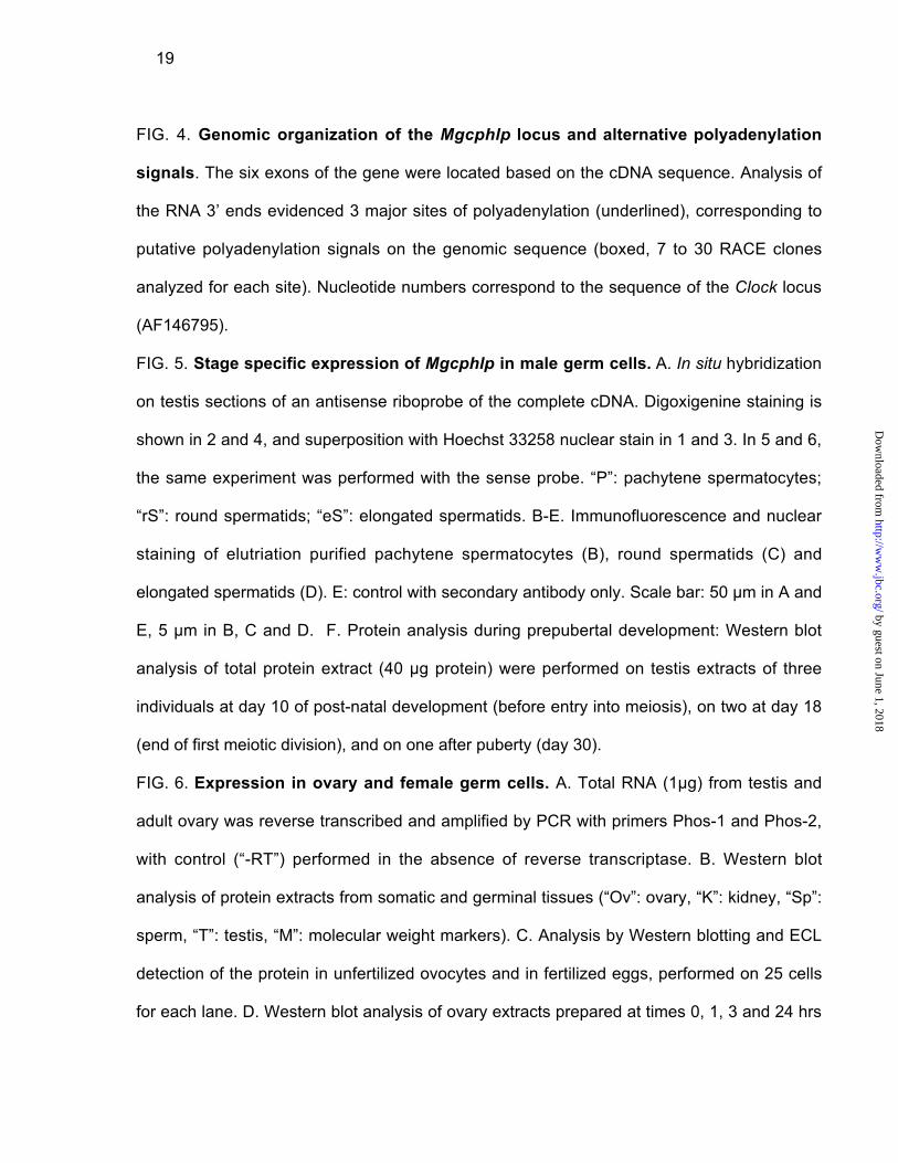
FIG. 4. Genomic organization of the Mgcphlp locus and alternative polyadenylation
signals. The six exons of the gene were located based on the cDNA sequence. Analysis of
the RNA 3’ ends evidenced 3 major sites of polyadenylation (underlined), corresponding to
putative polyadenylation signals on the genomic sequence (boxed, 7 to 30 RACE clones
analyzed for each site). Nucleotide numbers correspond to the sequence of the Clock locus
(AF146795).
FIG. 5. Stage specific expression of Mgcphlp in male germ cells. A. In situ hybridization
on testis sections of an antisense riboprobe of the complete cDNA. Digoxigenine staining is
shown in 2 and 4, and superposition with Hoechst 33258 nuclear stain in 1 and 3. In 5 and 6,
the same experiment was performed with the sense probe. “P”: pachytene spermatocytes;
“rS”: round spermatids; “eS”: elongated spermatids. B-E. Immunofluorescence and nuclear
staining of elutriation purified pachytene spermatocytes (B), round spermatids (C) and
elongated spermatids (D). E: control with secondary antibody only. Scale bar: 50 µm in A and
E, 5 µm in B, C and D. F. Protein analysis during prepubertal development: Western blot
analysis of total protein extract (40 µg protein) were performed on testis extracts of three
individuals at day 10 of post-natal development (before entry into meiosis), on two at day 18
(end of first meiotic division), and on one after puberty (day 30).
FIG. 6. Expression in ovary and female germ cells. A. Total RNA (1µg) from testis and
adult ovary was reverse transcribed and amplified by PCR with primers Phos-1 and Phos-2,
with control (“-RT”) performed in the absence of reverse transcriptase. B. Western blot
analysis of protein extracts from somatic and germinal tissues (“Ov”: ovary, “K”: kidney, “Sp”:
sperm, “T”: testis, “M”: molecular weight markers). C. Analysis by Western blotting and ECL
detection of the protein in unfertilized ovocytes and in fertilized eggs, performed on 25 cells
for each lane. D. Western blot analysis of ovary extracts prepared at times 0, 1, 3 and 24 hrs
by guest on June 1, 2018http://w
ww
.jbc.org/D
ownloaded from
20
after induction of super-ovulation by human chorionic gonadotropin (hCG). Testis extract and
the purified GST-MgcPhLP protein are shown for comparison.
FIG. 7. 14-3-3 binding to MgcPhLP. Immunoprecipitates prepared using the monoclonal
antibody 14-3-3ß(H-8): sc-1657 reacting with all the 14-3-3 family members were analyzed by
Western blot analysis for the presence of MgcPhLP as shown in Fig. 6. Lane 1: testis extract,
lane 2: liver extract, lane 3: molecular weight markers. Arrow indicates the expected
migration of MgcPhLP (27.8 kDa).
FIG. 8. Complementation of the S. cerevisiae plp2D mutation by the murine Mgcphlp
gene. A. Genotypes of two representative yeast clones (#1 and #3) derived from spores
generated by a plp2::URA3, plp2D yeast clone after transfer of the Mgcphlp expression
vector. These clones were checked to be haploid on the basis of their ability to conjugate with
reference a and a yeast strains. PCR analysis was performed with primers Plp2-s and Plp2-r,
generating a 525 bp product diagnostic of the wild type allele, and a 1.3 kbp fragment from
the deleted gene with the URA3 insert. “WT”: wild type. B. Loss of the mouse gene upon long
term growth of the complemented haploid clones. PCR analysis performed with primers
Mgc271 and Mgc483 (lanes 1: Mgcphlp), primers Plp2-s and Plp2-r (lanes 2: PLP2) and
primers Plp2-s and Ura3-r (lanes 3: URA3, plp2D). Panel A: wild type diploid; B: diploid
Mgcphlp, plp2::URA3, plp2D; C: haploid clone #1 early after isolation (same as in A); D: clone
#1 after 30 generations in culture.
by guest on June 1, 2018http://w
ww
.jbc.org/D
ownloaded from
1 GGAAGTTTGAGGCCCTGATCCCAGTGAGGGGCACAAGAGAACATCATGCAGGATCCAAAC
* M Q D P N
61 GAAGATACAGAATGGAATGAAATTTTAAGGAATTTTGGCATTCTTCCTCCAAAAGAAGAA
E D T E W N E I L R N F G I L P P K E E
121 CCAAAAGATGAAATTGAAGAGATGGTGTTGCGCTTACAGCAGGAGGCCATGGTTAAACCA
P K D E I E E M V L R L Q Q E A M V K P
181 TATGAGAAGATGACACTGGCACAACTGAAAGAAGCAGAAGATGAATTTGATGAAGAAGAC
Y E K M T L A Q L K E A E D E F D E E D
241 ATAAAAGCTATTGAAATATATAGAGAAAAGCGGTTACAGGAATGGAAAGCACTTAAGAAG
I K A I E I Y R E K R L Q E W K A L K K
301 AAACAAAAATTTGGGGAATTGAGAGAAATTTCTGGAAATCAGTATGTGAATGAAGTCACA
K Q K F G E L R E I S G N Q Y V N E V T
361 AATGCAGAAAAAGACCTGTGGGTTGTAATTCATCTCTATAGATCAAGTGTCCCAATGTGC
N A E K D L W V V I H L Y R S S V P M C
421 TTGGTGGTTAACCAACATCTGAGTGTCCTAGCAAGAAAGTTTCCAGAAACCAAATTTGTT
L V V N Q H L S V L A R K F P E T K F V
481 AAAGCCATCGTGAATAGCTGCATTGAACACTACCATGACAACTGTTTACCAACAATTTTT
K A I V N S C I E H Y H D N C L P T I F
541 GTTTATAAAAATGGTCAGATAGAAGGCAAATTCATTGGAATTATAGAATGTGGAGGGATA
V Y K N G Q I E G K F I G I I E C G G I
601 AATCTCAAGCTAGAAGAACTTGAATGGAAACTATCAGAAGTTGGAGCGATACAGAGTGAC
N L K L E E L E W K L S E V G A I Q S D
661 TTGGAAGAAAACCCCAAAAAGGGCATTGCAGATATGATGGTGTCTTCAATTAGAAACATC
L E E N P K K G I A D M M V S S I R N I
721 TCCATTTATGACAGCGATAGCTCTGGCAGTGACACTGAGGCTAAGTAGAACTTGTTAATA
S I Y D S D S S G S D T E A K *
781 AATAGCTTTATAGTGTAAAAAAAAAAAAAAAAAAAAAA Figure 1
by guest on June 1, 2018http://www.jbc.org/Downloaded from

mouse Ph mekaksqslEedfEGqathTGPKGVINDWRkFK..L.ESEDgdsippskkEILRqMsspqSR...............DDKdSkErmSrKMs
rat.PhLP1 .pqtsspaeaElagEGivSnTGPKGVINDWRrFK.QL.EtEqkE.......EqcReMerLikKlsmScrshldEeeEqqkQkdlqEiSgKMT
MgcPhLP.... .. MQD.........pnEDtE.....WNEILRnfGiLPpKeEPKDe...eEmVlrlqQEAmvKpyEKMT
yeast.Plp2..... ................. MQnepmFqvQvdESEDsE.....WNdILRakGviPeRA.PSp....takVEealeEAiaKqhErle
yeast Plp1........... ........... MeD......................kldRyytnvlSnAE.KDk...httVdsDDKsSgEE......
mouse Ph. ..iQeyEL..ihqDkEDEgcLrkYRrQcMQdMhQKLsfGPryGfVyEletGEQFL.EtIeKEQ.................KvTtIVVnIYEDG
rat PhLP1. ....LKEcgmmdknlDDEEFLqqYRkQRMdEMRQqLhKGPqFkqVlEIpSGEgFL.dmIDKEQ.................KqTlImVHIYEDG
MgcPhLP. .LaQLKEaE.DefDeEDikaiEiYReKRLQEwk.aLKKkqKFGElrEI.SGnQYvNEVTnAE..................KdlwVVIHLYrsS
yeast Plp2 NkDdLsdLE.elEDDEDEdFLEAYkiKRLmEIR.KLqersKFGEVfhInkpE.YnkEVTlAsQgkkyegaqtndngeeddggvyVfVHLsLqS
yeast Plp1 NLDeLln.ElDrElDEDhEFLsAYRseRLQqIsdhLKqvkKnvEddgygrlqcidNEa.DAiQi..............ctKtTmVVIHfeLet
mouse Ph VrGCdALNSsLeCLAA.EYPmVKFCKIRaSntGAgdRFs.Sdv..LPTLLVYKgGELIsNFIsVaEQfaE...DFFaaDvEsFLnEyGLL...
rat PhLP1. VPGteAMNgcmiCLAA.EYPTVKFCrVRsSViGAssRFt.rNa..LPaLLVYKaGELIGNFvrVtdQLGE...DFFavDLEaFLqEfGLL...
MgcPhLP VPmClvvNqHLsvLA.rKFPETKFVKai..VNsCIEhYhd.NC..LPTifVYKNGqLEGkFIgLiE.cGG..iNlKlEeLEwkLsEVGAIqsD
yeast Plp2. klqsriL.SHLfqsAAcKFrEiKFVeIp..aNrAIEnYpeSNC...PTLiVYyrGEvIKNmItLlE.LGG..NNsKmEDfEdFmvkVGAv...
yeast Plp1. fgkCqyMNekLenLA.krYlTTrFiKVn..VqtCpflvnklNikvLPfvvgYKNG.LEKvryvgfskLGndpNgFdirrLEqsLahsGvI.eD
mouse Ph . ....PErEIhDL.............gqtntmeDEDme.
rat PhLP1. ....PEKEvlvL..SSVRN.....satchSESDlEiD.
MgcPhLP. lEEnPKKgIaDmmvSSiRN..isIydsDsSgSDtEaK.
yeast Plp2 aEgdnr................lImNrDdeESrEErKlhygekksirsgirgkfnvgiggnddgninddddgfdd.
yeast Plp1 tfEirKh.......SSVnterfastNhDrSESDsDlDi.
A
BPhosducin Phosducin-like Plp1 Plp2
MgcPhLP 17.2 18.0 17.2 27.2
Phosducin 49.6 12.9 16.8
Phosducin-like 15.1 14.1
Plp1 12.5Figure 2
by guest on June 1, 2018http://www.jbc.org/Downloaded from

BA
Figure 3
Hss26
Mgcphlp
d.p.p.
10 20 25 30 Ad Sc eS rS Pac
C
RT + + +- - -C t li br
M+
Hss26
Mgcphlp
br he li in lu sp t
D
by guest on June 1, 2018http://www.jbc.org/Downloaded from

26390 GAACTTGAAT GGAAACTATC AGAAGTTGGA GCGATACAGA GTGACTTGGA
26440 AGAAACCCCC AAAAAGGGCA TTGCAGATAT GATGGTGTCT TCAATTAGAA
26490 ACATCTCCAT TTATGACAGC GATAGCTCTG GCAGTGACAC TGAGGCTAAG
26540 TAGAACTTGT TAATAAATAG CTTTATAGTG TATCCATCGA ATGCCTTATT
26590 TCCTGCTGAC TTTAGCTACA AATGAATTGA CCTCATTTTC ATTCTTATGT
26640 AAGAAGTATA ACCTACATGT CAATTGAAAA AATTTCAGGT ATTTAAGAAT
26690 ATTGTCTCTT GCAAATATGA ATATTTACTC CTTAATATTT TGTGACTTAT
26740 TTTTTCTGAC TCCTGTTGGA CAAAACCCAA CAAATACTTT AAGTCCTCTG
26790 TGTTGCCTAA TCACTTCCAA AAAGACACAT GGAAGGAAAT AAATATGGCT
26840 TCATAAATAA AATTAAGACA TTTTGTATGT GTGCACCATG TGTGTATAGG
26890 TGTGCATGCC TGTGAATGTG CATGTGATGG TCAGAGATCA GTTTTGGGTG
26940 TCTTCTTTAA CTGCACTCTA CCTTAATTTC TGAGAGAGAG CCCCTCACAG
26990 AGAAGCTGGA C
Figure 4
7,000 10,000 15,000 20,000 25,000 27,000
1 51 52 172 173 263 264 407 408 616 617 796
946
1072
by guest on June 1, 2018http://www.jbc.org/Downloaded from

Figure 5
1 2
3 4
5 6
BA
F
C
D
E
P
eS
rS
P
eS
rS
by guest on June 1, 2018http://www.jbc.org/Downloaded from

A
Figure 6
+RT -RT +RT -RT
324 bp
Testes Ovary
B
175
83
62
47
32
25
kDa
D
32 kDa
C ovocyte fertilized
testis ovary (hrs after hCG) GST
1 3 24 0 Mgcphlp M
Female Male
Ov K Sp K T M
by guest on June 1, 2018http://www.jbc.org/Downloaded from
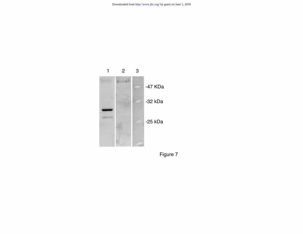
Figure 7
1 2 3
-25 kDa
-32 kDa
-47 KDa
by guest on June 1, 2018http://www.jbc.org/Downloaded from
575 bp (PLP2)
1.3 kbp (URA3,plp2D)
3 1 WT PLP2+/-
Haploid derivatives
after transfer of Mgcphlpexpression vector
Figure 8
1 2 3 1 2 3 1 2 3 1 2 3
A B C D
1: Mgcphlp (250 bp)
2: PLP2 (575 bp)
3: URA3, plp2D (850 bp)
A B
by guest on June 1, 2018http://www.jbc.org/Downloaded from
Philippe Clertant, François Cuzin and Minoo RassoulzadeganPascal Lopez, Ruken Yaman, Luis A. Lopez-Fernandez, Frédérique Vidal, Daniel Puel,
function conserved from yeast to miceA novel germ line specific gene of the phosducin-like (PhLP) family: A meiotic
published online November 6, 2002J. Biol. Chem.
10.1074/jbc.M207434200Access the most updated version of this article at doi:
Alerts:
When a correction for this article is posted•
When this article is cited•
to choose from all of JBC's e-mail alertsClick here
by guest on June 1, 2018http://w
ww
.jbc.org/D
ownloaded from