a novel approach for the isolation and long-term expansion
TRANSCRIPT

METHODOLOGY Open Access
A novel approach for the isolation andlong-term expansion of pure satellite cellsbased on ice-cold treatmentAnna Benedetti1, Gianluca Cera2,3, Daniele De Meo2,3, Ciro Villani2,3, Marina Bouche1† andBiliana Lozanoska-Ochser1*†
Abstract
Satellite cells (SCs) are muscle stem cells capable of regenerating injured muscle. The study of their functionalpotential depends on the availability of methods for the isolation and expansion of pure SCs with preservedmyogenic properties after serial passages in vitro. Here, we describe the ice-cold treatment (ICT) method, which is asimple, economical, and efficient method for the isolation and in vitro expansion of highly pure mouse and humanSCs. It involves a brief (15–30 min) incubation on ice (0 °C) of a dish containing a heterogeneous mix of adherentmuscle mononuclear cells, which leads to the detachment of only the SCs, and gives rise to cultures of superiorpurity compared to other commonly used isolation methods. The ICT method doubles up as a gentle passagingtechnique, allowing SC expansion over extended periods of time without compromising their proliferation anddifferentiation potential. Moreover, SCs isolated and expanded using the ICT method are capable of regeneratinginjured muscle in vivo. The ICT method involves minimal cell manipulation, does not require any expertise orexpensive reagents, it is fast, and highly reproducible, and greatly reduces the number of animals or humanbiopsies required in order to obtain sufficient number of SCs. The cost-effectiveness, accessibility, and technicalsimplicity of this method, as well as its remarkable efficiency, will no doubt accelerate SC basic and translationalresearch bringing their therapeutic use closer to the clinic.
Keywords: Satellite cell isolation, Satellite cells in vitro expansion, Skeletal muscle regeneration
BackgroundThe muscle is endowed with an exceptional regenerativeability primarily due to a resident population of stemcells called satellite cells (SCs) [1–3]. Ensconced betweenthe basal lamina and the plasma membrane of muscle fi-bers, SCs respond to injury or various stress stimuli bybecoming activated, and undergoing proliferation, self-renewal, and differentiation to form new myofibers [4,
5]. As SCs become activated, a proportion of them iden-tified as Pax7+MyoD- replenish the stem cell pool, whileothers acquire the expression of MyoD (Pax7+MyoD+),differentiate into myoblasts and enter the myogenic pro-gram. After several rounds of division myoblasts giverise to Myogenin+ myocytes, which fuse together to formnew myofibers [1, 4, 6–9].A major stumbling block in the study of the functional
potential of SCs has been the lack of isolation methodsinvolving minimal cell manipulation and allowing theisolation and expansion of highly pure SCs with pre-served myogenic properties after serial passages in vitro[10–12]. Moreover, the success of SC transplantationtherapy depends on having an efficient method to isolate
© The Author(s). 2021 Open Access This article is licensed under a Creative Commons Attribution 4.0 International License,which permits use, sharing, adaptation, distribution and reproduction in any medium or format, as long as you giveappropriate credit to the original author(s) and the source, provide a link to the Creative Commons licence, and indicate ifchanges were made. The images or other third party material in this article are included in the article's Creative Commonslicence, unless indicated otherwise in a credit line to the material. If material is not included in the article's Creative Commonslicence and your intended use is not permitted by statutory regulation or exceeds the permitted use, you will need to obtainpermission directly from the copyright holder. To view a copy of this licence, visit http://creativecommons.org/licenses/by/4.0/.The Creative Commons Public Domain Dedication waiver (http://creativecommons.org/publicdomain/zero/1.0/) applies to thedata made available in this article, unless otherwise stated in a credit line to the data.
* Correspondence: [email protected]†Bouche Marina Lozanoska-Ochser Biliana are co-senior authors.1Department of Anatomical, Histological, Forensic and Orthopedic Sciences,Section of Histology and Embryology, Sapienza University of Rome, Rome,ItalyFull list of author information is available at the end of the article
Benedetti et al. Skeletal Muscle (2021) 11:7 https://doi.org/10.1186/s13395-021-00261-w

and expand these cells in vitro in undifferentiated stateand in sufficient numbers [13].Presently, there are three main methods commonly
used for the isolation of SCs: the pre-plating method,fluorescence activated cell sorting (FACS), and magneticbead isolation method.The pre-plating method is based on the differing
adhesive properties of muscle cells, with SCs beingthe least adherent. Following enzymatic digestion, aheterogeneous mix of skeletal muscle cells is platedonto uncoated culture dishes and after a 1–24-h incu-bation at 37 ○C, the non-adherent cells are collectedand plated onto new collagen coated dishes [10–12,14]. The resulting cell culture contains both SCs andfibroblasts in variable proportion. To improve SCpurity, the pre-plating step can be repeated every 24h over 6 days [10]. Although cheap and straightfor-ward to perform, this method’s main disadvantage isthat it is time consuming and gives rise to cultures ofvariable purity, with fibroblast contamination andovergrowth by day 7 of culture, leading to early sen-escence and detachment of myotubes [10]. A recentlydescribed version of the pre-plating method intro-duces a re-plating step whereby after a 2-day expan-sion, the adhered cells are detached with trypsin andreplated onto Matrigel coated dishes, giving rise toSC cultures of much improved purity [15].The FACS sorting method sorts muscle mononuclear
cells pre-labelled with SC specific antibodies. Followingdigestion of muscle with various enzymes the resultingmixture of cells is labelled with specific antibodies to fa-cilitate the identification of SCs, which are then sortedusing a FACS sorter instrument [16–21]. At present, theFACS sorting method represents the gold standard forthe isolation and study of SCs. Nevertheless, there areseveral disadvantages to this method including high costand the requirement for a FACS sorter instrument.Moreover, this method is time consuming, requires ex-pertise to perform and cell purity can be variable. Thecell labelling step followed by the sorting procedure canpotentially stress or damage the cells, decrease their via-bility, or alter their functional properties in vitro [12].The third method is based on magnetic cell separation(MACS) and uses magnetic columns and SC specificmagnetic bead kits [22]. It is based on negative selec-tion of SCs by magnetically labelling and removingother cell lineages. Because this method assumes thatall the other cell types are successfully removed fromthe muscle cell preparation, it is less precise than theFACS sorting method. This method is expensive toperform, time consuming, and stressful for the cells.As for the other two methods, cell purity is variableand often the SC cultures become overgrown by fi-broblasts by day 7 [10, 12].
There is therefore a need for new and improvedmethods for the isolation, expansion and culture of SCs.Here, we describe a simple, inexpensive, and efficientmethod for the isolation of highly pure mouse and hu-man SCs that can be serially expanded in vitro to obtainsufficient number of SCs with preserved proliferationpotential, capable of regenerating injured muscle in vivo.
MethodsMiceC57BL/10ScSn-Dmdmdx, C57BL/10ScSn, and C57BL/6Jmice were purchased from the Jackson laboratory (BarHarbor, ME, USA). Both male and female mice wereused. The mice were housed in the Histology Depart-ment–accredited animal facility at the University ofSapienza. All the procedures were approved by the Ital-ian Ministry for Health and were conducted accordingto the EU regulations and the Italian Law on AnimalResearch.
Human muscle sourcingMuscle biopsies (gluteus maximus) were obtained frompatients (8 males and 7 females, age range 50–90 years)undergoing surgery at the Department of Orthopaedicsand Traumatology, Umberto I Hospital in Rome, Italy.According to the Italian law, the authors are not re-quired to ask for approval from an institutional reviewboard or ethical committee for this type of study. In anycase, all patients gave their approval to undergo intraop-erative muscle biopsy and to publish the clinical and la-boratory data obtained.
Satellite cell isolation with the ice-cold treatment methodSCs were isolated from hind-limb muscles of 4–8-week-old mice or from human biopsies. Muscles were dis-sected with scissors and finely diced with a scalpel in adish containing DMEM (Sigma-Aldrich, St. Louis, MO,USA, D5671). This was followed by enzymatic digestionwith 10 ml/g of muscle of Collagenase type II (Sigma-Al-drich, SCR103) at a concentration of 0.4 mg/ml in PBS(Sigma-Aldrich), for 45 min in a water bath at 37 °Cwith agitation. Digestion was blocked with DMEM 10%FBS and after centrifuging the muscle preparation andremoving the supernatant, a second digestion was per-formed with 10 ml/g of muscle of Collagenase/Dispaseat a concentration of 1 mg/ml (Roche, Basel, CH,11097113001) in PBS Calcium-Magnesium free (Sigma-Aldrich), for 30 min at 37 °C in a water bath with agita-tion. The digested muscle was then passed first througha 70-μm cell strainer followed by 40-μm cell strainer toobtain single cell suspension. Next, the cells were centri-fuged, resuspended in DMEM 10% FBS (Sigma-Aldrich,F2442), counted and plated at 2 × 10⁶ cells/100 mm dish(uncoated) (Corning, NY, USA, 430167), and incubated
Benedetti et al. Skeletal Muscle (2021) 11:7 Page 2 of 12

at 37 °C for 1 h. Non-adherent cells were collected, cen-trifuged, and the cell pellet was resuspended in DMEM10% FBS, plated again, and incubated for another 1 h at37 °C. After the second pre-plating, non-adherent cellswere collected, centrifuged, counted, resuspended in SCGrowth Medium (GM) DMEM, 20% Horse Serum(Thermo Fisher Scientific, Waltham, MA, USA,26050088), 3% Chicken Embryo Extract (Seralab, CE-650-J), and plated into 100 mm dishes coated with 0.1%gelatin (Stem Cell Technologies, Vancouver, BC, CAN,07903), at 106 cells/dish. The next day, the dishes con-taining a heterogeneous mix of adhered muscle cellswere washed 3 times with PBS, and 10 ml of ice-coldPBS was added into each dish. The dishes were thenplaced on ice (0 °C) for 15–30 min with occasional gen-tle manual shaking (swirling motion). The detached cellswere collected, centrifuged, resuspended in GM, andplated into 0.1% gelatin-coated 35-mm dishes (Corning,353001) at a density of 103 cells/dish. To induce differ-entiation, proliferating cells (day 3 after adding GM)were cultured in differentiating medium (DM) contain-ing DMEM 5% Horse Serum, 1% Chicken EmbryoExtract.
Satellite cell isolation with magnetic bead labellingMouse SC isolation by magnetic bead labelling was per-formed by using a SC Isolation Kit (Miltenyi Biotech,Bergisch Gladbach, DE, REF: 130-104-268) as previouslydescribed [23]. Briefly, minced muscle was digested asdescribed above. The digested muscle was passedthrough 70 μm and 40 μm cell strainers, and the result-ing single cell suspension was centrifuged, resuspendedin 80 μl buffer (PBS pH 7.2, 0.5% FBS, 2 mM EDTA),and incubated with 20 μl of Satellite Cell Isolation Kitper gram of muscle, for 15 min at 4 °C. Next, the cellsuspension was passed through a LS column (MiltenyiBiotech, 130-042-401) placed in a magnetic field of aMACS Separator (Miltenyi Biotech). Unlabeled SCs werecollected in the flow-through, counted, washed, resus-pended in growth medium (GM), and plated into 35-mm dishes at a density of 103 cells/dish.Mouse and human SCs were cultured either in GM
containing DMEM, 20% Horse Serum (Sigma-Aldrich),3% Chicken Embryo Extract, or in DM containingDMEM 5% Horse Serum, 1% Chicken Embryo Extract.
SC detachment with trypsinSCs were rinsed once with PBS and then incubated withtrypsin-EDTA solution (Sigma-Aldrich, T3924) for 5min at 37 °C. SCs were then collected, centrifuged, re-suspended in GM, and plated at a density of 103 cells/dish.
SC transplantationAcute muscle injury was induced the day before SCtransplantation. To induce muscle injury tibialis muscleswere injected with 0.01 ml of Cardiotoxin from Naja Pal-lida (10 μM) (Latoxan ZA, Les Auréats, Fr), using a 30Gauge micro-syringe [23–26].For cell transplantation, 15,000 SCs were resuspended
in 20 μl of DMEM 2% FBS (Sigma-Aldrich) and injectedinto the TA muscle of one leg with a single injection byusing a 30 Gauge micro-syringe. Contralateral TAmuscle was injected with only PBS and used as control.
Immunofluorescence and microscopyFor immunofluorescence analysis cultured SCs werefixed in PFA 4% for 10 min RT, permeabilized in coldmethanol at – 20 °C for 6 min, blocked in 5% GoatSerum in PBS for 30 min RT, and incubated overnightat 4 °C in 4% BSA in PBS with the following primaryantibodies: mouse anti-Pax7-c (1:10 DSHB, Iowa City,IA, USA), rabbit anti-MyoD (1:50 Santa Cruz C20: sc-304, Dallas, TX, USA), mouse anti-myogenin (1:20,DSHB), mouse anti-Myosin Heavy Chain (1:20 DSHB),and mouse anti-desmin (1:20, DSHB). The next day, SCswere washed 3 times in PBS for 15 min, and then incu-bated with secondary antibodies goat anti-rabbit AlexaFluor 488 (1:1000, Abcam) and goat anti-mouse AlexaFluor 555 (1:1000, Thermo Fisher Scientific) diluted in1% BSA in PBS, for 1 h RT. Nuclei were counterstainedwith Hoechst.For immunofluorescence analysis of mdx TA muscle
transplanted with WT SCs, 8-μm-thick muscle cryosec-tions were fixed in 4% PFA for 10 min at roomtemperature (RT), and then permeabilized in coldmethanol for 6 min at – 20 °C. Sections were thenblocked in 5% Goat Serum (Sigma-Aldrich) in PBS for30 min at RT. Next, sections were incubated with pri-mary rabbit anti-dystrophin antibody (1:200, Abcam,Cambridge, UK) overnight at 4 °C. The next day, sec-tions were washed and incubated with a secondary anti-body goat anti rabbit Alexa Fluor 488 (1:1000 Abcam).Nuclei were counterstained with Hoechst.Samples were analyzed under an epifluorescence Zeiss
Axioskop 2 Plus microscope (Carl Zeiss, Oberkochen,DE).Bright field images were acquired with an inverted
phase-contrast microscope (Nikon Eclipse, TS100). Im-ages were acquired with a Nikon DS-Fi2 camera andNIS Elements version 4.0 Imaging System.
CFSE stainingIsolated SCs were stained with CFSE (ThermoFisher Sci-entific) at a concentration of 1 μM for 10 min at 37 °Cin the dark prior to culture. After 4 days of culture, thecells were detached with Accutase Solution (Sigma-
Benedetti et al. Skeletal Muscle (2021) 11:7 Page 3 of 12

Aldrich). Samples were acquired with a CyAn ADP(DAKO) flow cytometer and acquired data were ana-lyzed using FlowJo software version 10 (FlowJo LLC,Ashland, OR, USA).
RNA isolation and quantitative real-time PCRFor RNA preparation, cells were lyzed with TRI reagent(Sigma-Aldrich) and processed as previously described[23]. Reverse transcription was performed with Sensi-FAST™ cDNA Synthesis Kit (Bioline, Memphis, TN,USA). Quantitative real-time PCR assays were per-formed according to the MIQE criteria, using Sensi-FAST™ SYBR No-ROX Kit (Bioline) followingmanufacturer’s protocol. All reactions were performedin duplicate. Data were collected and analyzed using ABIPRISM 7500 Sequence Detection System (Life Technolo-gies, Carlsbad, CA, USA). Quantitative RT–PCR valueswere normalized to the expression of GAPDH mRNA.The relative gene expression level was calculated usingthe 2−ΔΔCT method and reported as mean fold changein gene expression.The following primers were used for amplification:
Pax7 (FW: 5′ GTCCCAGTCTTACTGCCCAC 3′, RV:5′ TGTGGACAGGCTCACGTTTT 3′), Myogenin (FW:5′ GCATGGAGTTCGGTCCCAA 3′, RV: 5′ TATCCTCCACCGTGATGCTG 3′), GAPDH (FW: 5′ ACCCAGAAGACTGTGGATGG 3′, RV: 5′ CACATTGGGGGTAGGAACAC 3′).
Clonal myogenicity assayFor the clonal myogenicity assay, SCs were plated into0.1% gelatin coated 96-well plates, (excluding the outerwells of the plate) at 1 cell per well, in growth medium.Colony formation and number of cells were assessed at24, 48, and 72 h of culture.
Statistical analysisAll statistical analyses were performed using Graph-Pad Prism software version 8 (La Jolla, CA, USA).Data are presented as mean ± SEM. Statistical signifi-cance was determined using unpaired 2-tailed Stu-dent’s t test with Welch’s correction for unequalvariances. A P value of ≤ 0.05 was considered statisti-cally significant.
ResultsIsolation and characterisation of muscle SCs using the ice-cold treatment methodPrevious studies have demonstrated that coldtemperature causes a reduction in cell adhesion, likelydue to the downregulation of adhesion receptors [27,28]. Compared to other cells, such as fibroblasts, whichtypically contaminate SC cultures, SC are considered tobe less adherent [11, 14]. Taking this into consideration,
as well as the notion that like all stem cells, SCs are sen-sitive to stress signals and are among the first muscleresident cells to respond to injury [4, 5], we hypothe-sized that subjecting a heterogeneous culture of musclecells to a mild stress stimulus such as ice-coldtemperature will lead to the detachment of only the SCs.To test this hypothesis, we obtained a mix of musclecells following enzymatic digestion, and after 2 h of pre-plating on uncoated dishes, cultured them overnight ongelatin-coated dishes. The next day, after washing andremoving the non-adhered cells and debris, we placedthe dishes of heterogeneous muscle cells on ice for 30’(Fig. 1a, b). This time point was chosen based on thepurity and number of SCs obtained following 15, 30, 45,and 60’ on ice. Although the number of SCs obtained in-creased with longer incubations, the purity of SCs de-creased from 100% at 15–30’ to 95 and 90% at 45 and60’ on ice, respectively (data not shown). The ice-coldtreatment (ICT) method can be used to harvest SCsfrom the original dish containing the heterogeneousmuscle cells for at least the first 3 days of culture. Pla-cing the heterogeneous muscle culture dish on ice for30’ resulted in the detachment of only the SCs givingrise to a highly pure culture of SCs that proliferated anddifferentiated into myotubes upon culture in differentiat-ing medium (Fig. 1b–d). Satellite cells isolated using theICT method were 100% pure as determined by the ex-pression of the SC markers Pax7 and MyoD (Fig. 1c). Atday 3 of proliferation, 100% of the cells were positive forthe satellite cell marker Pax7 and of these 97% were acti-vated and expressed MyoD (Pax7+MyoD+) (Fig. 1c). Toexamine the myogenic capacity of ICT-isolated SCs weperformed a clonal myogenicity assay, by plating a singlecell per well and analyzing the formation of myogeniccolonies. Satellite cells isolated with the ICT method dis-played a similar clonal myogenicity of 40 % and a doub-ling time of 17 h to SCs isolated using the magneticbeads method (Figure S1A and B). The myogenic iden-tity of the cultured cells was further confirmed by theexpression of myosin heavy chain (MHC) and the forma-tion of myotubes. After 3 days in differentiating mediumthe SCs differentiated into myoblasts that fused intoMHC expressing myotubes with a fusion index of 90%(Fig. 1d). The purity of the isolated SCs at the begin-ning of culture, as well as the fusion index and num-ber of myonuclei per myotubes were similar betweenSCs isolated with the ICT and magnetic cell separ-ation (MACS) method (Figure S1C–E). However, byday 5–7, the cultures obtained with the MACSmethod became overgrown by non-myogenic cellssuch as PDGFRα+ fibroblasts, causing premature myo-tube detachment, whereas cultures obtained with theICT method remained almost free of contaminatingcells (Fig. 1e, f).
Benedetti et al. Skeletal Muscle (2021) 11:7 Page 4 of 12
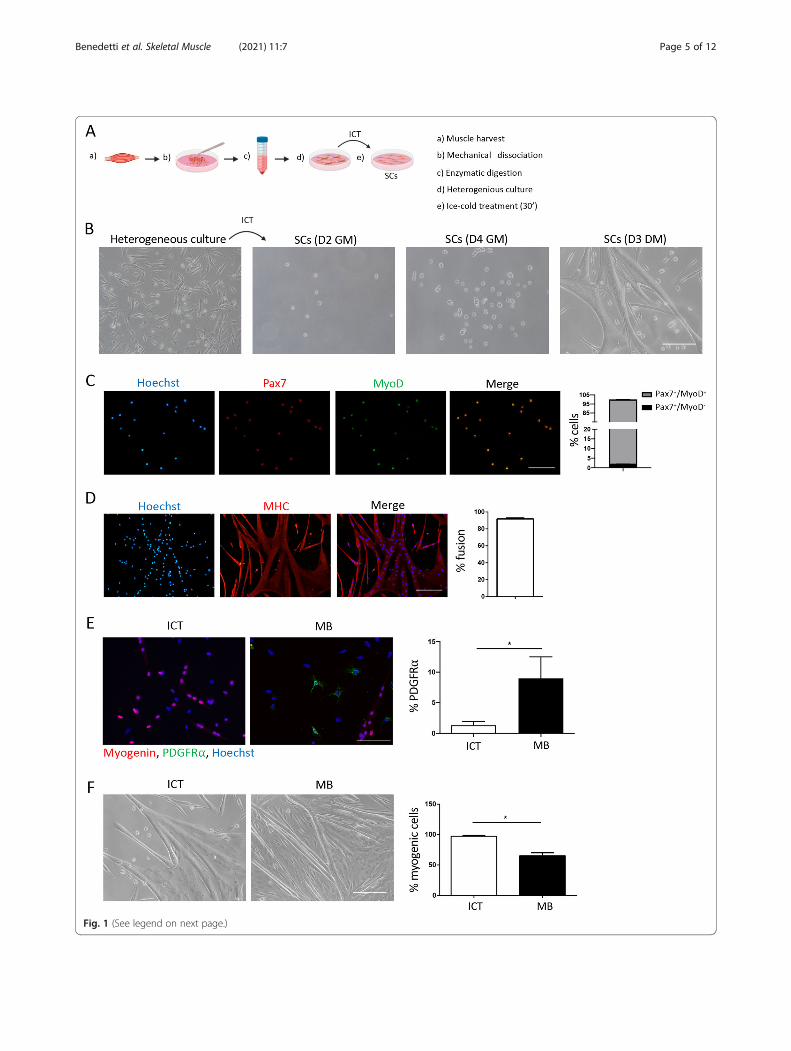
Fig. 1 (See legend on next page.)
Benedetti et al. Skeletal Muscle (2021) 11:7 Page 5 of 12

Overall, these results confirm our hypothesis that ICTleads to the preferential detachment of SCs and usingthis method allows the isolation of 99–100% pure SCs.
The ICT approach can also be used for serial passagingand long-term expansion of SCsA major obstacle in SC research has been that culturedSCs lose their proliferation potential after a couple ofpassages and begin to differentiate into myotubesthereby limiting the number of SCs that can be seriallyexpanded in vitro. Loss of differentiation potential hasalso been noted with increasing number of passages[10–13, 29]. We reasoned that, since the ICT leads toSC detachment, it can also be used to passage growingcultures of proliferating SCs. Indeed, placing the dishesof proliferating SCs on ice for 30 min led to the detach-ment of around 30% of the proliferating cells and wewere able to serially passage and expand the proliferatingSCs without compromising their proliferation and differ-entiation capacity (Fig. 2a). We used the ICT approachon proliferating SCs and then successively on eachestablished culture of proliferating SCs (for more than10 passages). This approach yielded on average 2.5 × 10⁶
cells/g of muscle (Fig. 2b). The SCs passaged using theICT method did not lose their proliferation and differen-tiation potential and displayed a minimally altered Pax7and myogenin gene expression (Fig. 2c, d). While ICT-passaged SCs retained their proliferation and differenti-ation capacity even after 10 passages, SCs passaged withthe most commonly used passaging reagent trypsin, lostthe ability to form new myogenic colonies after just 2passages and instead begun to differentiate into myo-tubes (Fig. 2e–g). Similar to SCs detached by trypsin,SCs detached using a gentler detachment solution suchas Accutase, exhibited accelerated differentiation aftermore than 3 passages (data not shown).Previous studies have demonstrated that based on
their rate of proliferation, SCs can be divided into twosubpopulations: fast dividing and slow dividing [30]. Theslow-dividing are a subset of SC that have been shownto retain stemness and long-term self-renewal ability
[31]. Unlike the fast-dividing SCs, which have a limitedability to form secondary myogenic colonies after pas-sage and instead undergo differentiation, the slow divid-ing SCs form secondary myogenic colonies whenpassaged [30, 31]. To examine the prevalence of fast andslow dividing SCs within our population, we labelled theICT-passaged SCs with CFSE prior to plating and ana-lyzed the rate of proliferation 3 days later. We comparedthe proliferation rate of ICT-isolated and passaged SCswith that of SCs isolated using magnetic beads and pas-saged with trypsin. In line with previous observations,we found that most of the activated SCs isolated usingthe commercial kit and passaged with trypsin were fast-dividing cells (CFSElo), with slow-dividing cells (CFSEhi)representing less than 10% of the total. Interestingly,while similarly heterogeneous, the SCs isolated and pas-saged using the ICT method were enriched in the slowdividing SC population (Fig. 2h, i).Next, we examined the longevity of differentiated ICT-
isolated SCs in culture. The highly pure cultures of SCsisolated using the ICT method gave rise to myotubesthat could be maintained in culture for up to 1 month,compared to just 7 days when isolated using the mag-netic beads (Figure S2A–C) or the pre-plating method(Figure S2D).Overall, these data show that the ICT method can be
used for the serial expansion of SCs with preserved pro-liferation and myogenic potential over an extendedperiod of time compared to other methods.To examine the potential of ICT-isolated and ex-
panded SCs to regenerate injured muscle, we injected15,000 SCs isolated from wild-type mice into the tibialisanterior of mdx mice (lacking dystrophin) CTX injured24 h previously. SCs were either injected immediatelyafter ICT isolation, or after 3-day in vitro expansion fol-lowing ICT. Transplantation of both SCs immediatelyafter ICT-isolation and after in vitro expansion contrib-uted to the regeneration process to a similar extent asevidenced by the appearance of newly formeddystrophin-positive fibers (Figure S3A and B). There wasno difference in the ability of transplanted ICT-isolated
(See figure on previous page.)Fig. 1 Isolation and characterization of muscle SCs using the ICT method. a Schematic representation of the ICT method. b Representative brightfield images of the heterogeneous muscle mononuclear cell culture from which SCs were isolated by ICT, and representative images of the ICT-isolated SCs at D2 and D4 in growth medium (GM) and at D3 after adding differentiation medium (DM) (n = 15 independent experiments). cRepresentative immunofluorescence images of ICT-isolated SCs stained for Pax7 (red), MyoD (green), and nuclei (blue) and a graph showing thepercentage of cells positive for Pax7 and/or MyoD at day 3 of culture in GM (n = 3 independent experiments). d Representativeimmunofluorescence images of ICT-isolated SCs stained for myosin heavy chain (MHC) (red) and nuclei (blue) and a graph showing percentfusion after differentiation (4 days in GM followed by 3 days in differentiating medium (DM)) (n = 3 independent experiments). e Representativeimmunofluorescence images of ICT-isolated SCs and MB-isolated SCs stained for Myogenin (red) PDGFR⍺ (green) and nuclei (blue) and a graphshowing percent PDGFR⍺+ cells at day 5 of culture in GM. f Representative bright field images of ICT- and magnetic beads (MB)-isolated SCs anda quantification graph showing percentage of myogenic cells in ICT- and MB-isolated SCs after differentiation at day 7 of culture (n = 3independent experiments). Non-myogenic cells were identified as Pax7-MyoD-nuclei outside the myotubes. Scale bar = 100 μm. Error barsrepresent mean ± sem. *p < 0.05 by Student’s t test.
Benedetti et al. Skeletal Muscle (2021) 11:7 Page 6 of 12
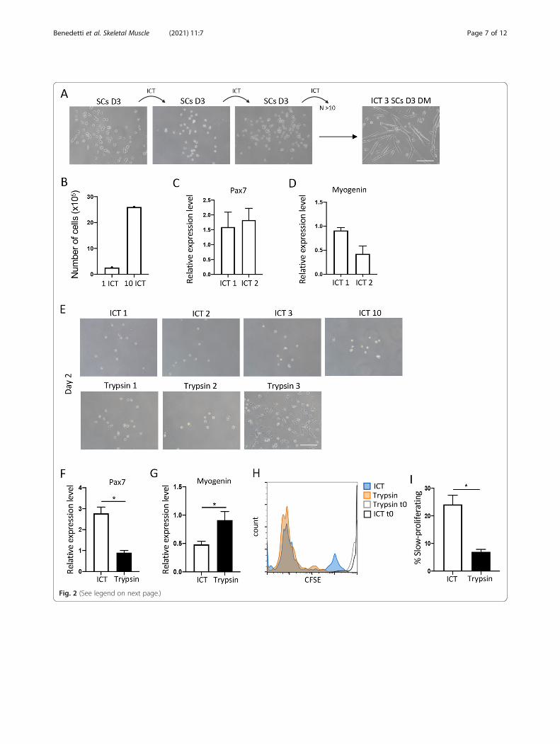
Fig. 2 (See legend on next page.)
Benedetti et al. Skeletal Muscle (2021) 11:7 Page 7 of 12

SCs and SCs isolated by magnetic beads to regenerateinjured muscle (Figure S3C and D). These experimentsconfirmed that SCs isolated with the ICT method suc-cessfully engraft after transplantation and do not losetheir potential to regenerate injured muscle after expan-sion in vitro.
Efficient isolation, serial expansion and long-term cultureof human satellite cells with the ICT methodThe study of human SCs (hSCs) has generally lagged be-hind that of mouse SCs due to the difficulties associatedwith obtaining muscle tissue, as well as the lack ofmethods for the isolation of pure hSCs that can be ex-panded in vitro without altering their myogenic potential[13]. Having demonstrated the remarkable efficiency ofthe ICT method in the isolation of pure mouse SCs, weset to reproduce these findings using human muscle bi-opsies. We obtained gluteus maximus specimens frompatients undergoing surgery, aged between 50 and 90years. Using the same approach as described above, weconsistently and reproducibly obtained a highly pure cul-ture of hSCs (Fig. 3a, d–g) from a heterogeneous popu-lation of human muscle cells (Figure S4), that could beserially expanded for more than 10 passages (Fig. 3a). Aspreviously reported [32], we found that hSCs proliferatedslower than mouse SCs, with a doubling time of 46 h(Fig. 3b), reaching a peak around day 10 post isolation,and slowing down thereafter. On average we isolatedaround 20 × 103 SCs/g of muscle. Using the ICTmethod, these hSCs could be expanded 300-fold over aperiod of 2 months to a final total of 6 × 10⁶/g of muscle(Fig. 3c). Previous studies have shown that hSCs rapidlydownregulate Pax7 expression in culture [33]. In agree-ment with others [33], we found that the expression ofPax7 was variable and ranged between 45 and 50% atday 2 of culture in GM (Fig. 3d). Almost 100% of thehSCs were myogenin positive at day 5 of culture in GM,suggesting that most of them have activated their myo-genic program (Fig. 3e). The myogenic purity of the hSCculture was further confirmed by desmin at day 5 (Fig.3f) and MHC expression at day 10 after shifting to dif-ferentiating medium (Fig. 3g).
These data show that the ICT method performsequally well when used for the isolation of human SCs.
DiscussionOver the past decade, a considerable progress has beenmade in the development of new methodologies for theisolation of SCs. Nevertheless, each of the availablemethods suffers from at least one disadvantage be it pur-ity, cost, expertise, or a combination of these [11]. In thisstudy, we describe a novel method for the isolation ofpure mouse and human SCs, that is inexpensive, simpleto perform, and reproducibly efficient. The ICT methodtakes advantage of the differing adhesive properties ofmuscle cells as well as the ability of SCs to rapidly re-spond to stress stimuli [5, 11, 14]. Thus, the combin-ation of a mild cold-stress stimulus and cold-inducedreduction in adhesion, leads to the detachment of onlythe SCs. Exposure of mammalian cells to cold stress canslow down the progression through the cell cycle and in-hibit protein synthesis. Moreover, depending on the in-tensity and duration, cold stress can activate theapoptotic program, or lead to necrosis [34]. Thus, it isconceivable that prolonged exposure to coldtemperature could interfere with the myogenic proper-ties of SCs. Nevertheless, we found that the brief periodof exposure to ice-cold temperature did not interferewith the ability of SCs to proliferate and differentiatein vitro or with their in vivo regeneration potential, sug-gesting that ICT does not alter SC function. Indeed, SCsisolated by ICT behaved similarly to those isolated usingthe pre-plating method or the magnetic bead isolationkit. Of note, both the magnetic bead and the FACS sort-ing isolation methods involve far lengthier incubationtimes on ice compared to our method without interfer-ing with SC behavior and function in vitro and in vivo[16, 17, 21, 22]. Interestingly, Marg A et al. recentlyfound that storing human muscle biopsies at 4 °C, inlow serum medium and no O2 for up to 35 days andsubsequent culture at 37 °C and 21% oxygen led to SCsexpansion outside the fiber fragments. Surprisingly, thepurity of the outgrowing colonies of SCs was 100% myo-genic cells, since non-myogenic cells such as fibroblasts
(See figure on previous page.)Fig. 2 Serial expansion and long-term proliferation potential of SCs using the ICT method. a Representative bright field images of SCs at day 3 ofculture in GM following ICT 1, ICT 2, and ICT 3, and differentiated cells at day 3 of culture in DM following ICT 3 (n = 10 independentexperiments). b Total number of SCs at day 3 of culture in GM, after 1 and 10 ICTs (n = 10 independent experiments). c Pax7 gene expression inSCs at day 2 of culture in GM after ICT 1 and ICT 2 analyzed by quantitative real time PCR (n = 3 independent experiments). d Myogeninexpression in SCs at day 5 of culture in GM after ICT1 and ICT 2 analyzed by quantitative real-time PCR (n = 3 independent experiments). eRepresentative images of SCs at day 2 of culture in GM, after 1–3 detachments with ICT (top panels) and trypsin (bottom panels). f Pax7 geneexpression in SCs detached with ICT or trypsin, at day 2 of culture in GM, analyzed by quantitative real-time PCR (n = 3 independentexperiments). g Myogenin gene expression in SCs detached with ICT or trypsin, at day 5 of culture in GM, analyzed by quantitative real time PCR(n = 3 independent experiments). h Representative overlay of histogram plots of CFSE labeled ICT and trypsin detached SCs at time 0 and day 4of culture in GM. i Graph showing percent CFSE-low or slow-proliferating SCs after ICT or trypsin detachment, at day 4 of culture in GM (n = 3independent experiments). Error bars represent mean ± sem, *p < 0.05, calculated by Student’s t test.
Benedetti et al. Skeletal Muscle (2021) 11:7 Page 8 of 12

Fig. 3 (See legend on next page.)
Benedetti et al. Skeletal Muscle (2021) 11:7 Page 9 of 12

did not survive prolonged storage at hypothermic condi-tions [33]. Therefore, it is likely that SCs tolerance forcold stress is high compared to other cell types, and thistrait can be exploited as we did with our ICT method, toimprove the purity of SCs grown in vitro.Recently, Yoshioka K et al. described an improved ver-
sion of the pre-plating method, reducing the isolationand purification procedure to 2.5 days in total, while in-creasing the cell yield, and significantly improving thepurity of the resulting SC culture by introducing a re-plating step [15]. The re-plating step performed at day2.5 of culture involves the detachment of all adheredcells including fibroblasts and SCs with trypsin, andreplating on matrigel coated dishes [15]. While the pur-ity of the resultant SC culture is comparable to ours, ourmethod involves fewer steps in total and only one over-night pre-plating. In addition, the ICT method doublesup as a very gentle passaging technique, allowing long-term serial expansion of SCs ex vivo, without alteringtheir proliferation and differentiation properties.Whereas the FACS sorting method is and will remainthe gold standard for the study of SCs immediately afterisolation, the ICT approach will likely become themethod of choice for the in vitro expansion of SCs. Aftereach ICT passage, the already expanded SCs can becryopreserved, and stored until needed. With the ICTmethod we were able to passage proliferating mouse andhuman SCs for at least 10 times, expanding their num-ber 150- and 300-fold, respectively. This represents aclear advantage over the most commonly used passagingreagent trypsin, which we and others have shown, typic-ally accelerates the differentiation of passaged SCs afteronly two passages [11]. Apart from being a relativelyharsh enzymatic passaging reagent, trypsin leads to thedetachment of all the cells in the dish including SCs thatare already committed to differentiate into myotubes, aswell as any contaminating cells, which might contributetowards the loss of SC proliferative potential and accel-erated differentiation. Indeed, even a gentler detachmentsolution like Accutase leads to loss of myogenic prolifer-ative properties and accelerates differentiation. By con-trast, the ICT approach favors the detachment of onlythe SCs that have not yet committed to differentiate,and in particular the slow dividing population which has
previously been shown to retain stemness and long-termself-renewal ability [31]. It is conceivable that being ‘true’stem cells, the slow dividing SCs detaching in responseto cold temperature are the so-called first responders tostress or injury in vivo [5], a hypothesis that will be thesubject of future investigation in our laboratory.In a recent study, Gregory WC et al. demonstrated
that hSCs differentiate and lose their proliferative poten-tial when maintained in high mitogen conditionsex vivo. They used inhibition of p38 signalling to preventthe differentiation of SCs and promote their expansion[35]. Using our method, we were able to achieve thesame but with minimal manipulation, maintaining theproliferative capacity of hSC ex vivo for an extendedperiod of time, to a similar degree using muscle biopsiestaken from a wide range of ages (between 50 and 90years old). This is an important technical advance forboth basic and clinical research since it will allow re-searchers to obtain sufficient number of cells for trans-plantation or intervention studies, while reducing thenumber of human biopsies required. Indeed, a majorobstacle to stem cell-based therapies has been the scar-city of human muscle tissue specimens and the limitednumber of cells that can be obtained for transplantation.Moreover, successful transplantation requires the use of
freshly isolated SCs because culturing and expandingthem in vitro greatly reduces their engraftment capacity[13]. Here, we show that SCs isolated and expanded usingthe ICT method do not lose their regenerative capacity.Another advantage of the ICT method is the im-
proved longevity of cultured myotubes. Generally, SCsdifferentiate into myotubes by day 7 of culture andshortly after, begin to detach [11, 12]. Notably, SCsisolated and passaged with the ICT method could bemaintained in culture for up to 2 months, even oncethey have differentiated into myotubes, likely due tothe lack of contaminating cells such as fibroblasts. Thepurity of the isolated SCs is of paramount importancefor in vitro studies since even 97% purity is insufficientto prevent overgrowth by non-myogenic cells, as wedemonstrated. While important for cell growth,growth factors produced by fibroblasts, have beenlinked to senescence induction in long term culturesof mesenchymal stem cells [36].
(See figure on previous page.)Fig. 3 Isolation and in vitro expansion of human satellite cells using the ICT method. A. Representative bright field images of ICT- isolated humanSCs following 1, 3, 5, and 10 ICTs, at day 3 and 10 of culture in GM, and at day 10 of culture in DM (n = 15 independent experiments). b Numberof human SCs at 24, 48, and 72 h following ICT 1. c Total number of human SCs at day 3 of culture in GM after 1 and 10 ICTs. d Representativeimmunofluorescence images of human SCs stained for Pax7 (red) and nuclei (blue). Graph shows percentage of cells positive for Pax7 at day 2 ofculture in GM. e Representative immunofluorescence images of human SCs stained for myogenin (red) and nuclei (blue). Graph showspercentage of cells positive for myogenin at day 5 of culture in GM. f Representative immunofluorescence images of human SCs stained fordesmin (red) and nuclei (blue) after differentiation (10 days in GM + 5 days in DM). g Representative immunofluorescence images of human SCsstained for myosin heavy chain (MHC) (red) and nuclei(blue). Graph shows percent fusion after differentiation (10 days GM + 10 days DM). (n = 3independent experiments, 10 images analyzed per experiment) Scale bar = 100 μm. Error bars represent mean ± sem
Benedetti et al. Skeletal Muscle (2021) 11:7 Page 10 of 12

ConclusionsIn the quest for new and improved SC isolationmethods, the ideal technique would permit the isolationof pure SCs with minimal manipulation, that can be ex-panded ex vivo without losing their stemness and regen-erative capacity. In terms of purity of the isolated cellpopulation, the ICT method outperforms others such asthe pre-plating method or the magnetic beads isolationmethod. Compared to other commonly used methods, itis fast and easy to perform, and apart from the time re-quired for enzymatic digestion (1.5 h), it involves min-imal manipulation of the cells. Finally, using the ICTapproach, SCs can be expanded for extended periods oftime without losing their proliferation and differentiationpotential. This in turn drastically reduces the number ofmice or muscle biopsies required to obtain sufficientnumber of cells.Overall, the cost-effectiveness, accessibility and tech-
nical simplicity of this method, as well as its remarkableefficiency, represent major improvements over existingmethods, and will no doubt accelerate SC basic andtranslational research bringing their therapeutic usecloser to the clinic. Finally, this is a proof of conceptstudy, and the ICT method can be further optimised,adapted, and improved for use in different experimentalsettings.
Supplementary InformationThe online version contains supplementary material available at https://doi.org/10.1186/s13395-021-00261-w.
Additional file 1: Figure S1. Myogenic properties of SCs isolated withthe ICT method. A. Percent of myogenic colony formation was calculatedas percent growing clones out of the total seeded single cells per well(60 per 96-well plate) among ICT- and MB-isolated SCs (n=3 independentexperiments). B. Number of cells per clone in single clone-derived ICT-and MB-isolated SCs at 48 and 72 h of culture in GM. ICT SCs, n= 37clones analyzed per experiment. MB SCs, n= 37 clones analyzed per ex-periment. C. Percent of ICT- and MB-isolated SCs positive for Pax7 at day2 of culture in GM. (n=3 independent experiments). D. Percent fusion ofsingle clone-derived ICT and MB-isolated SCs after differentiation (4 daysin GM followed by 3 days in DM). Fusion index: number of nuclei withinmyotubes divided by total number nuclei. E. Number of nuclei per myo-tube in single clone-derived ICT and MB-isolated SCs after differentiation(4 days in GM followed by 3 days in DM). (n=3 independent experi-ments). Error bars represent mean ± sem.
Additional file 2: Figure S2. Increased longevity in culture of ICT-isolated SCs. A. Representative bright field images of ICT-isolated SCs atday 3, 5, 11 and 17 of culture in DM. B. Representative bright field imageof MB isolated SCs at day 3 of culture in DM. C. Total number of days inculture of ICT- and MB-isolated SCs. D. Representative bright field imagesshowing the heterogeneous muscle cell culture after pre-plating, at 3and 5 days of culture in GM. Error bars represent mean ± sem. *P < 0.05by Student’s t-test.
Additional file 3: Figure S3. In vivo functional validation of SCsisolated using the ICT and MB method. A. Representativeimmunofluorescence images of dystrophin positive fibers (green) andnuclei (blue) in mdx tibialis muscle at 30 days following intra-muscular in-jection of 15 x 103 SCs immediately after ICT isolation or after 3 day-expansion in culture following ICT isolation. B. Quantification of the
number of dystrophin positive fibers per TA muscle section in mdx mice(ICT, n=5 mdx mice; ICT-expanded, n=4 mdx mice). Scale bar=100μm.Error bars represent mean ± sem. C. Representative immunofluorescenceimages of dystrophin positive fibers (green) and nuclei (blue) in mdx tibi-alis muscle at 30 days following intra-muscular injection of 15 x 103 ICT-isolated SCs (left), or MB-freshly isolated SCs (right). B. Quantification ofthe number of dystrophin positive fibers per TA section in mdx mice (ICT,n=5 mdx mice; MB, n=5 mdx mice). Scale bar=100μm. Error bars repre-sent mean ± sem.
Additional file 4: Figure S4. Characterisation of the human muscle-derived cells obtained after pre-plating and prior to ICT. A. Representativebright field images of human muscle-derived cells at day 3 and 10 of cul-ture in GM, and at day 10 of culture in DM. B. Representative immuno-fluorescence images of the heterogeneous culture of human muscle-derived cells stained for Pax7 (red) and nuclei (blue). Graph shows per-centage of cells positive for Pax7 at day 2 of culture in GM. C. Representa-tive immunofluorescence images of the heterogeneous human musclecell culture stained for myogenin (red) and nuclei (blue). Graph showspercent of cells positive for myogenin at day 5 of culture in GM. D. Repre-sentative immunofluorescence images of the heterogeneous culture ofhuman muscle-derived cells stained for MHC (red) and nuclei (blue).Graph shows percent cell fusion after differentiation (10 days in GMfollowed by 10 days in DM). E. Graph shows percent myogenic cells inthe human heterogenous muscle cells obtained after pre-plating or afterICT at day 5 of culture in GM, calculated by IF staining for myogenin (n=3 independent experiments, 10 images analysed per experiment). Scalebar=100μm. Error bars represent mean ± sem. ****P < 0.0001 by Stu-dent’s t-test.
AcknowledgementsWe thank Dr. Luca Madaro for critical reading of the manuscript and helpfuldiscussions.
Authors’ contributionsBLO conceived, developed, and standardized the method. BLO, AB, and MBdesigned the experiments. AB performed experiments. AB and BLO analyzeddata and prepared the figures. BLO and MB supervised the study. CG, DMD,and VC provided the human muscle biopsy specimens. BLO wrote themanuscript. All authors critically read, edited, and approved the finalmanuscript.
FundingThis work was supported by a grant from The Dutch Duchenne ParentProject NL (DPP NL) to BLO; research grants from Parent Project Italy (PP,Italy) to MB and from the University of Rome (# RP11715C7D238352,RM118164275C7EBE, and RM11916B7E20311C to MB; and #AR11715C7F9E158E, AR11816436905518, and AR11916B7E2A7B64 to AB).
Availability of data and materialsAll data generated or analyzed during this study are included in thispublished article [and its supplementary information files].The datasets used and/or analyzed during the current study are availablefrom the corresponding author on reasonable request.
Declarations
Ethics approval and consent to participateAll procedures involving mice were approved by the Italian Ministry forHealth and were conducted according to the EU regulations and the ItalianLaw on Animal Research.According to the Italian law, the authors are not required to ask for approvalfrom an institutional review board or ethical committee for the use ofhuman tissue removed during surgical procedures. All patients gave theirapproval to undergo intraoperative muscle biopsy and to publish the clinicaland laboratory data obtained.
Consent for publicationNot applicable.
Benedetti et al. Skeletal Muscle (2021) 11:7 Page 11 of 12

Competing interestsThe authors declare that they have no competing interests.
Author details1Department of Anatomical, Histological, Forensic and Orthopedic Sciences,Section of Histology and Embryology, Sapienza University of Rome, Rome,Italy. 2Department of Anatomical, Histological, Forensic and OrthopedicSciences, Section of Orthopedics, Sapienza University of Rome, Rome, Italy.3Department of Orthopaedics and Traumatology, Policlinico Umberto I,Rome, Italy.
Received: 21 December 2020 Accepted: 18 February 2021
References1. Chang NC, Rudnicki MA. Satellite Cells: The Architects of Skeletal Muscle.
Curr Top Dev Biol. 2014;107:161–81.2. Wang YX, Dumont NA, Rudnicki MA. Muscle stem cells at a glance. J Cell
Sci. 2014;127:4543–8.3. Mauro A. Satellite cell of skeletal muscle fibers. J Biophys Biochem Cytol.
1961;9:493–5.4. Wang YX, Rudnicki MA. Satellite cells, the engines of muscle repair. Nat Rev
Mol Cell Biol. 2012;13:127–33.5. Evano B, Tajbakhsh S. Skeletal muscle stem cells in comfort and stress. npj
Regen Med. 2018;3:24.6. Feige P, Rudnicki MA. Muscle stem cells. Curr Biol. 2018;28:581–98.7. Sambasivan R, Yao R, Kissenpfennig A, van Wittenberghe L, Paldi A,
Gayraud-Morel B, et al. Pax7-expressing satellite cells are indispensable foradult skeletal muscle regeneration. Development. 2011;138:3647–56.
8. Relaix F, Zammit PS. Satellite cells are essential for skeletal muscleregeneration: The cell on the edge returns centre stage. Development(Cambridge). 2012;139:2845–56.
9. Tedesco FS, Dellavalle A, Diaz-Manera J, Messina G, Cossu G. Repairingskeletal muscle: Regenerative potential of skeletal muscle stem cells. J ClinInvestig. 2010;120:11–9.
10. Keire P, Shearer A, Shefer G, Yablonka-Reuveni Z. Isolation and culture ofskeletal muscle myofibers as a means to analyze satellite cells. Methods MolBiol. 2013;946:431–68.
11. Danoviz ME, Yablonka-Reuveni Z. Skeletal muscle satellite cells: Backgroundand methods for isolation and analysis in a primary culture system.Methods Mol Biol. 2012;798:21–52.
12. Jonah D, Lee BCS, Lisa M, Larkin KWV. Isolation and Purification of SatelliteCells for Skeletal Muscle Tissue Engineering. J Regen Med. 2015;3:117.
13. Rinaldi F, Perlingeiro RCR. Stem cells for skeletal muscle regeneration:Therapeutic potential and roadblocks. Transl Res. 2014.
14. Gharaibeh B, Lu A, Tebbets J, Zheng B, Feduska J, Crisan M, et al. Isolationof a slowly adhering cell fraction containing stem cells from murine skeletalmuscle by the preplate technique. Nat Protoc. 2008;163:409–17.
15. Yoshioka K, Kitajima Y, Okazaki N, Chiba K, Yonekura A, Ono Y. A ModifiedPre-plating Method for High-Yield and High-Purity Muscle Stem CellIsolation From Human/Mouse Skeletal Muscle Tissues. Front Cell Dev Biol.2020;8:793.
16. Pasut A, Oleynik P, Rudnicki MA. Isolation of muscle stem cells byfluorescence activated cell sorting cytometry. Methods Mol Biol. 2012;798:53–64.
17. Liu L, Cheung TH, Charville GW, Rando TA. Isolation of skeletal muscle stemcells by fluorescence-activated cell sorting. Nat Protoc. 2015;10:1612–24.
18. Chapman MR, Balakrishnan KR, Li J, Conboy MJ, Huang H, Mohanty SK, et al.Sorting single satellite cells from individual myofibers reveals heterogeneityin cell-surface markers and myogenic capacity. Integr Biol (UnitedKingdom). 2013;5:692–702.
19. Fukada SI, Higuchi S, Segawa M, Koda KI, Yamamoto Y, Tsujikawa K, et al.Purification and cell-surface marker characterization of quiescent satellitecells from murine skeletal muscle by a novel monoclonal antibody. Exp CellRes. 2004;296:245–55.
20. Sherwood RI, Christensen JL, Conboy IM, Conboy MJ, Rando TA, WeissmanIL, et al. Isolation of adult mouse myogenic progenitors: Functionalheterogeneity of cells within and engrafting skeletal muscle. Cell. 2004;119:543–54.
21. Montarras D, Morgan J, Collins C. Direct Isolation of Satellite Cells forSkeletal Muscle Regeneration. Mol Cell Biol. 2005;309:2064–7.
22. Blanco-Bose WE, Yao CC, Kramer RH, Blau HM. Purification of mouse primarymyoblasts based on α7 integrin expression. Exp Cell Res. 2001;265:212–20.
23. Benedetti A, Fiore PF, Madaro L, Lozanoska-Ochser B, Bouché M. Targetingpkcθ promotes satellite cell self-renewal. Int J Mol Sci. 2020;21:2419.
24. Fiore PF, Benedetti A, Sandonà M, Madaro L, de Bardi M, Saccone V, et al.Lack of PKCθ promotes regenerative ability of muscle stem cells in chronicmuscle injury. Int J Mol Sci. 2020;21:932.
25. Rizzo G, di Maggio R, Benedetti A, Morroni J, Bouche M, Lozanoska-OchserB. Splenic Ly6Chi monocytes are critical players in dystrophic muscle injuryand repair. JCI Insight. 2020;5:e130807.
26. Lozanoska-Ochser B, Benedetti A, Rizzo G, Marrocco V, di Maggio R, Fiore P,et al. Targeting early PKCθ-dependent T-cell infiltration of dystrophic musclereduces disease severity in a mouse model of muscular dystrophy. J Pathol.2018;244:323–333.
27. Juliano RL, Gagalang E. The adhesion of Chinese hamster cells. I. Effects oftemperature, metabolic inhibitors and proteolytic dissection of cell surfacemacromolecules. J Cell Physiol. 1977;92:209–20.
28. Rico F, Chu C, Abdulreda MH, Qin Y, Moy VT. Temperature modulation ofintegrin-mediated cell adhesion. Biophys J. 2010;99:1387–96.
29. Machida S, Spangenburg EE, Booth FW. Primary rat muscle progenitor cellshave decreased proliferation and myotube formation during passages. CellProlif. 2004;37:267–77.
30. Tierney MT, Sacco A. Satellite Cell Heterogeneity in Skeletal MuscleHomeostasis. Trends Cell Biol. 2016;26:434–444.
31. Ono Y, Masuda S, Nam HS, Benezra R, Miyagoe-Suzuki Y, Takeda S. Slow-dividing satellite cells retain long-term self-renewal ability in adult muscle. JCell Sci. 2012;125:1309–17.
32. Charville GW, Cheung TH, Yoo B, Santos PJ, Lee GK, Shrager JB, et al. Ex vivoexpansion and in vivo self-renewal of human muscle stem cells. Stem CellReports. 2015;5:621–32.
33. Marg A, Escobar H, Gloy S, Kufeld M, Zacher J, Spuler A, et al. Humansatellite cells have regenerative capacity and are genetically manipulable. JClin Investig. 2014;124:4257–65.
34. Sonna L, Fujita J, Gaffin SL, Craig M. Molecular Biology of ThermoregulationInvited Review: Effects of heat and cold stress on mammalian geneexpression. J Appl Physiol. 2002;92:1725–42.
35. Judson RN, Quarta M, Oudhoff MJ, Soliman H, Yi L, Chang CK, et al.Inhibition of Methyltransferase Setd7 Allows the In Vitro Expansion ofMyogenic Stem Cells with Improved Therapeutic Potential. Cell Stem Cell.2018;22:177–90.
36. Ito T, Sawada R, Fujiwara Y, Seyama Y, Tsuchiya T. FGF-2 suppresses cellularsenescence of human mesenchymal stem cells by down-regulation of TGF-β2. Biochem Biophys Res Commun. 2007;359:108–14.
Publisher’s NoteSpringer Nature remains neutral with regard to jurisdictional claims inpublished maps and institutional affiliations.
Benedetti et al. Skeletal Muscle (2021) 11:7 Page 12 of 12