isolation and characterization of novel plant growth promoting
TRANSCRIPT

1
Author version: Plant Physiol. Biochem., vol.48(12); 2010; 987-992
Isolation and characterization of novel plant growth promoting
Micrococcus sp NII-0909 and its interaction with cowpea
Syed G Dastager1,2, C.K. Deepa1 and Ashok Pandey1
Present Address: 1Biological Oceanography Division, National Institute of Oceanography (CSIR), Dona Paula-403004, Goa, India
2National Institute for Interdisciplinary Science and Technology (CSIR)-
Trivandrum-695019, India. Running head: Plant growth promoting Micrococcus sp
† Both Authors contributed equally to the work
Author for correspondence: Dr.Syed G Dastager Tel: 832-2450446, 447; Fax: +91-832- 2450606; Email: [email protected]
Abstract
A phosphate-solubilizing bacterial strain NII-0909 isolated from the Western ghat forest soil in India
was identified as Micrococcus sp on the basis of phenotypic characteristics, carbon source utilization
pattern, fatty acid methyl esters analysis, and 16S rRNA gene sequence. The strain exhibited the plant
growth-promoting attributes of phosphate solubilization, auxin production, 1-aminocyclopropane-1-
carboxylate deaminase activity, and siderophore production. It was able to solubilize (122.4 µg of
Ca3PO4 ml–1), and produce IAA (109 µg ml–1) at 30°C. P-solubilizing activity of the strain NII-0909 was
associated with the release of organic acids and a drop in the pH of the NBRIP medium. HPLC analysis
detected two organic acids in the course of P-solubilization. A significant increase in the growth of cow
pea was recorded for inoculations under controlled conditions. Scanning electron microscopic study
revealed the root colonization of strain on cow pea seedlings. These results demonstrate that isolates
NII-0909 has the promising PGPR attributes to be develop as a biofertilizer to enhance soil fertility and
promote the plant growth.
Key words: Plant growth activity, 16S rRNA, Micrococcus sp, Western ghat forest

2
1. Introduction
Microorganisms play an important role in effecting the availability of soil phosphorous to plant roots,
and increasing P-mobilization in soil. The ability of soil microorganisms to convert insoluble forms of
phosphorus to a soluble form is an important trait in plant growth-promoting bacteria for increasing
plant yields [1]. The main advantage of using rhizobia as P-solubilizing microorganism will be their
dual beneficial nutritional effect resulting both from phosphorous mobilization, N2-fixation [2] and their
well-documented synergistic interactions with arbuscular mycorrhizal fungi [3]. Many P-solubilizing
bacteria belongs to the Pseudomonas, Bacillus, Enterobacter, Serratia, Pantoea, Rhizobium,
Flavobacterium and to the fungal genera Aspergillus and Penicillium [4-8].Current trends in agriculture
are focused on the reduction of the use of pesticides and inorganic fertilizers, forcing the search for
alternative ways to improve a more sustainable agriculture [9]. Beneficial free-living soil bacteria
isolated from the rhizosphere, which have been shown to improve plant health or increase yield, are
usually referred to as plant growth-promoting rhizobacteria (PGPR) [10]. The use of plant growth
rhizobacterial (PGPR) inoculants as biofertilizers and/or antagonists of phytopathogens provide a
promising alternative to chemical fertilizers and pesticides. However, the ability of introduced bacterial
strains to colonize roots and survive in soil is often limited, reducing the expected PGP effect [11]. As a
consequence, the selection and use of PGPR should be done taking into account the adaptation of the
inoculant to a particular plant and soil in the rhizosphere ecosystem, though the development of effective
microbial inoculants remains a major scientific challenge [1]. Furthermore, a good selection of a PGPR
strain requires understanding the dynamic and composition of the bacterial communities colonizing the
rhizosphere and the characterization of its plant growth promoting (PGP) related properties. To date,
only limited information exists on microbial diversity and dynamic of population in agricultural soil [9,
11, 12]. The properties more often related to the PGP character are: auxin production, nitrogen fixation,
phytopathogen antagonism, cyanogenesis, phosphate solubilization and ACC deaminase activity [13].
The exact mechanisms by which PGPR promote plant growth are not fully understood. Soil–plant–
microbe interactions are complex and there are many ways in which the outcome can influence the plant
health and productivity [14]. The interaction may be harmful, beneficial and neutral to the plants.
However, our focus should be to exploit the beneficial interaction of plants and microbes. While
considerable attention has been given to the immense potential of using fluorescent pseudomonads for
enhancing crop growth and yield in a sustainable manner. The use of microbial technologies in
agriculture is currently expanding quite rapidly with the identification of new bacterial strains, which are

3
more effective in promoting plant growth. In the present study we have isolated and identified a
potential isolate of Micrococcus sp isolated from Western ghat dense forest with their effective plant
growth promoting activity.
2. Materials and Methods
2.1. Isolation
The soil used for bacterial isolation was collected from a root-free soil of rhizosphere of Western ghat
forest in west coast of India, located at an altitude of 900 m above mean sea level. The processed soil
sample was serially diluted, spread plated on full strength nutrient agar and incubated at 28°C for 48 h.
A total of 180 different colonies were isolated on nutrient agar (NA) and were purified with repeated
culturing and maintained in 20% glycerol at –80°C. A potential isolates were screened and selected on
the basis of halo zone produced in Pikovskaya agar. Strains were assessed for morphology, physiology
and Gram reaction and other characterization.
2.2. Bacterial identification and characterization
Isolated strain was subsequently differentiated by Gram reaction, salt tolerance, biochemical
characterization and microscopic observation. The ability of the isolates to grow in diverse temperature
range was carried out by growing the isolate NII-090in nutrient broth and incubated at different
temperatures i.e. 5, 10, 15, 20, 30 and 40°C respectively. Growth was recorded every 6h at OD600 upto
48h. The ability of the isolates to grow in different salt concentrations was carried out by inoculating
bacterial culture on nutrient agar plates supplemented with 0-25% (w/v) NaCl and the plates were
incubated at 28 ± 2 °C for three days. The ability of the isolates to grow in alkaline or acidic media was
tested in nutrient agar plates in which the pH was adjusted from 4.0 to 12.0 (at a pH 1.0 unit interval),
using the different buffer system: pH 4.0–5.0: 0.1 M citric acid/0.1 M sodium citrate; pH 6.0–8.0: 0.1 M
KH2PO4/0.1 M NaOH; pH 9.0–12.0: 0.1 M NaHCO3/0.1 M Na2CO3 and incubated at 28 ± 2 °C for three
days. Phenotypic characterization of isolates was done based on their colony morphology, microscopic
observations, and biochemical tests using Hi-25 Kit (HiMedia, Mumbai). A cream whitish colored
bacterial colony showing above 20mm zone of P-solubilization, maximum of 7% NaCl tolerance and
growth in wide pH range was selected for further analysis. Strain NII-0909 was then screened for traits
that might be associated with ability to functions as PGPR, each test performed in triplicate.

4
2.3. Quantitative estimation of phosphate solubilization and IAA production
Initial qualitative estimation of the P-solubilizing activity of the isolate was carried out on Pikovskaya
agar [15]. Quantitative estimation of P solubilization was carried out as per standard methodology, by
inoculating 1 ml of bacterial suspension (3 × 10⁷ cells ml–1) in 50 ml of National Botanical Research
Institute Phosphate NBRIP broth [16], in Erlenmeyer flasks (150ml), and incubating the flasks for 7
days. At the end of the incubation period the cell suspension was centrifuged at 10,000 rpm min–1 for 10
min. and the P content in the supernatant was spectrophotometrically estimated by the ascorbic acid
method [17], pH of the medium was recorded with a pH meter equipped with glass electrode.
For the analysis of organic acids, bacterial cultures were filtrated through 0.2 mm filter (Millipore,
GTBP) and 20 µl of filtrates were injected to HPLC (Shimadzu Corporation, Kyoto, Japan) equipped
with Poto Diode Array detector. The organic acid separation was carried out on C-18 column (Bio-Rad
Laboratories, Inc.) with 10.8% acetonitrile in 0.0035 M H2SO4 as mobile phase. Retention time of each
signal was recorded at a wavelength of 210 nm and compared with the standard acids. Estimation of
indole acetic acid (IAA) was done by inoculation of 200 µl of bacterial suspension (3 × 10⁷ cells ml–1) in
10 ml Luria Bertani (LB) broth amended with L-tryptophan (100 µg ml–1) and incubating it at 28°C for
48 h. The IAA content in the culture suspension was estimated by the standard procedure [18]. All the
studies were repeated on three independent dates to confirm the results.
2.4. Extraction of IAA and ACC-deaminase activity
Single bacterial colonies of isolates were inoculated in 200 ml of nutrient broth amended with 1 or 5
mg/ml of tryptophan and incubated at 28 ± 2°C for 3-5 days on a shaker incubator. Bacterial cells were
separated from the supernatant by centrifugation at 10,000 rpm for 30 min. The supernatant was
acidified to pH 2.5 to 3.0 with 1 N HCl and extracted twice with ethyl acetate at double the volume of
the supernatant. Extracted ethyl acetate fraction was evaporated to dryness in a rotator evaporator at
40°C. The extract was dissolved in 300 ml of methanol and kept at -20°C. Methanol extract was
quantified by reverse phase HPLC (Shimadzu Corporation, Kyoto, Japan). The mobile phase was
methanol/water/acetic acid (36:64:1) at a flow rate of 1ml/min. Elutes were detected at 220nm and IAA
was quantified by integrating the areas under the peaks. Authentic IAA was used as a standard.
ACC-deaminase activity (1-aminocyclopropane-1-carboxylate deaminase) of the strain NII-0909
was detected on plates with DF minimal medium containing 1-aminocyclopropane-1-carboxylate (ACC)

5
as the sole source of nitrogen [19]. Germinating of seed bioassay for ethylene reduction due to ACC
deaminase activity was performed. Measurement of the root length of cow pea treated with 48-h-old
Micrococcus strain NII-0909 culture (OD600 = 1.0 ~ 1× 10⁹ colony forming unit [CFU]/ml) for 1 h in
nutrient broth was compared with uninoculated controls after 5 days of incubation at 30°C in petri plates
as described earlier [20].
2.5. Qualitative measurement of siderophore and hydrocyanic acid (HCN) production
Siderophore production was detected by the standard Chrome Azurol-S (CAS) assay [21] in 110 mm
Petri dishes, and the diameter of the clearing zone was measured. HCN production was inferred by the
qualitative method of Bakker and Schipper [22]. The change in the color of the filter paper previously
dipped in 2% sodium carbonate prepared in 0.05% picric acid, from yellow to dark brown was rated
visually depending on the intensity of the colour change.
2.6. DNA extraction, 16S rRNA Gene sequencing and phylogenetic analysis
Extraction and amplification of genomic DNA for 16S rRNA gene sequence analysis was carried out as
described by Cui et al. [23]. The 16S rRNA gene fragment was amplified by using universal primers
corresponding to positions 8–27 for the forward primer and 1492–1510 for the reverse primer
(Escherichia coli numbering system [24]). Based on 1469 bp long 16S rRNA gene sequences,
phylogenetically related bacteria were aligned by using a BLAST search [25] against the GenBank
database. Multiple alignments with sequences of related taxa of the genus Micrococcus were
implemented by using CLUSTAL_X [26]. The 16S rRNA gene sequence similarity values were
calculated by pairwise comparison [27]. A neighbour-joining phylogenetic tree was constructed [28]
from evolutionary distances calculated using the Jukes–Cantor coefficient [29]. The topology of the
phylogenetic tree was evaluated by the bootstrap resampling method of Felsenstein [30] with 1000
replicates. The GenBank/EMBL/DDBJ accession number for the isolate is FJ897464.
2.7. Bioassay-based plant growth promotion ability in Cow pea (Vigna unguiculata)
A bioassay-based determination of the plant growth promotion ability of the isolate was conducted using
cow pea seedlings in sterile soil under glasshouse conditions. The cowpea seeds were sterilized in 70%
ethanol for 2 min and in 2% sodium hypochlorite for 2 min and followed ten times washing in sterile tap
water. For this experiment, pure cultures were grown in nutrient broth at 28°C and diluted to a final

6
concentration of 108 colony-forming units (cfu) mL–1 in sterile saline water (0.85%). The surface sterile
seeds were inoculated by immersion in the appropriate PGPR suspension (ca. 108 cfumL–1) for 45min on
a rotary shaker (140 rev min–1), air-dried, and sown immediately. The cell densities in the suspension
were adjusted to a final density of approximately 108 cfu seed–1 [31]. Control seeds were treated with
sterile distilled water. Seeds were sown in plastic pots (15-cm diameter) containing 1 kg of sterile soil
(pH-7.2, organic carbon-2.6%, available P-537.5 kg ha–1, available K-448 kg ha–1, iron-40 mg kg–1) and
placed in a temperature controlled growth chamber at 26 ± 1°C. Thinning of seedlings was done 7 days
after sowing and two seedlings per pot were maintained throughout the experimental period. The soil
was moistened to 50% of its water-holding capacity. The whole experiment was conducted in three
independent trials. For each treatment, the plants of each pot were harvested 3 weeks after the
emergence of seedlings and washed; morphological characteristics of each plant were recorded: plant
height, root length, leaf area, dry shoot and root weights. The total root number per plant (TRN; the
number of seminal roots + the number of crown roots) was counted after washing away the soil from the
roots. At harvest, the root system was separated from shoots, and both were oven-dried for overnight at
65°C and dry weights were recorded against the control.
2.8. Statistical Analysis
Data were statistically analyzed by analysis of variance using the general linear model developed by the
SAS Institute (version 9.1; Cary, NC), and means were compared using the least significant difference
(LSD) method; P ≤ 0.05 was considered significant.
2.9. Electron microscopic studies
Cowpea seedlings of 20-d old were randomly selected from growth pots of each treatment for electron
microscopic examination. Tissue samples from inoculated and non-inoculated seedling roots of cowpea
were thoroughly washed in water to remove soil particles and were fixed in 2% glutaraldehyde (made up
in 0.1 M cacodylate buffer) in the refrigerator (8°C) for 1.5 hr. Samples were washed two times in the
same buffer for 10 min, post fixed in 1% OsO4 for 4 hrs, and dehydrated as follows: 30%, 50%, 70%,
85%, and 95% ethanol for 15 min; 100% ethanol, two times for 15 min each. For scanning electron
microscopy, sputter coating, and a JEOL–JSM 5600LV scanning electron microscope operating at 20 kv
were used. Root vascular systems and rhizobacterial colonization patterns were observed by SEM.
7
3. Results
3.1. Isolation and Characterization of the Bacterial Isolate
A bacterial strain producing about a 20-mm zone of P-solubilization after 48h incubation on Pikovskaya
agar and morphologically different from other colonies showed a resemblance to Micrococcus in major
phenotypic characteristics. The bacterial strain was Gram-positive, nonmotile coccus, with circular,
smooth, convex, entire and pale yellow in colour. The strain was positive for nitrate reduction,
degradation of tween 80 and 40, negative for H2S, casein utilization, gelatin hydrolysis, starch
utilization, phenol degradation, lipase and cellulose and utilizes number of carbon sources (Table I). It
was able to grow over a wide range of temperatures 5-40°C, with optimum at 28±2°C. It had a pH
tolerance over the range of 4-11, with optimum 7.0±0.5 and could tolerate 7% of NaCl concentration
(w/v). Strain NII-0909 was initially identified using the BIOLOG identification system and was
confirmed by 16S rRNA gene sequencing. The cell wall fatty acid composition showed that iso-C15:0,
anteiso-C15:0, anteiso-C17:0 and iso-C17:0 were the predominant forms of fatty acids. Small amounts of
iso-C16:0, C16:0, iso-C11:0 3-OH and iso-C14:0 also were detected as showed by gas chromatograph-fatty
acid methyl esters (GC-FAME) analysis (Figure 1).Molecular analysis based on 16S rRNA homology of
1464-bp partial sequence confirmed that strain NII-0909 belongs to Micrococcus genus. In the
phylogenetic tree, strain NII-0909 and other Micrococcus species were grouped together (Figure 2).
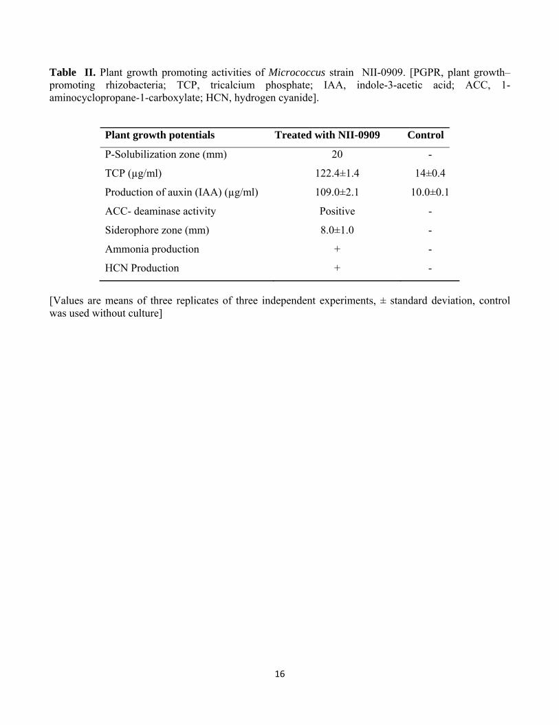
3.2. Plant growth attributes
The different plant growth promotion traits of the isolate were determined at different incubation
temperatures from 4-30°C. Strain NII-0909 was able to solubilize tri-calcium phosphate of about
23.5±0.9, 59.02±1.9 and 122.4±2.1µg ml-1 at 4, 20 and 30°C respectively after 10th day of incubation
(Table II). The production of indole acetic acid was about 109.0 µg ml-1 by NII-0909 when
supplemented with L-tryptophan, and 11.40 µg ml–1 of indole acetic acid production detected without L-
tryptophan which was confirmed by HPLC analysis. The IAA production reported in this study was
higher than the earlier reports of IAA production by any rhizosphere isolates of Enterobacteriaceae
family [32].
HPLC analysis also reveals that strain NII-0909 produces organic acids during P-solubilization.
Two different organic acids were detected, and were confirmed as malic acid and fumaric. The retention
times of these organic acids were 7.86 and 7.97 when compared with standard acids (data not shown)

8
The pH of the P-solubilization in broth was found to decline, in each case, due to bacterial activity;
lowering of pH coincided with increase in the efficiency of phosphate-solubilizing activity. The pH was
found to decline from 7.0 to 4.0-3.0. Qualitative detection of siderophore production and HCN were also
observed in all tested temperature. It was interesting to observe that the isolate was able to retain its
functional traits even at 4°C, which was the lower temperature extreme for its growth, while higher
values for all parameters were recorded at 28±2°C.
3.3. Plant growth promotion potential
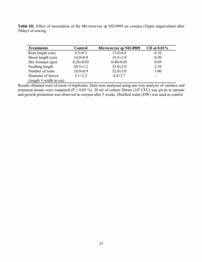
The plant growth promotion potential of Micrococcus sp NII-0909 was determined by a root
colonization bioassay in cowpea seeds. A significant influence on growth was resulted with treatment of
Micrococcus sp NII-0909 in cowpea seedling grown in pots under controlled conditions. It was
observed that the bacterized seedlings recorded 100 and 39.2% higher root and shoot lengths compared
to uninoculated control (Table III). Seed bacterization resulted in greater enhancement of the root
growth, as compared to the shoot growth. Increase in dry biomass, as well as number of roots were also
observed.
3.4. Scanning electron microscopic observations
Primary root sections of cowpea bacterized with NII-0909 which are showing potential plant growth
ability was examined by SEM. The results revealed that cells of isolates NII-0909 was consistently
distributed on the surface of roots. Surface furrows appeared to be located at epidermal cell junctions.
Root seedlings free of inoculant bacteria typically revealed a smooth, undamaged epidermal root
surface. Root surfaces from isolate NII-0909 inoculated seedlings were colonized with many clusters of
cells associated with fibrillar material, which contributed to the formation of microcolonies (Figure 3 A
& B).
4. Discussion
Bacterial plant growth promotion is a well-established and complex phenomenon, and is often
achieved by the activities of more than one plant growth-promoting traits exhibited by the associated
bacterium [33]. Phosphate is abundant in several soils and is one of the major nutrients limiting the plant
growth. The overall phosphate use efficiency following phosphate fertilizer application is low because
of the formation of insoluble complexes [34]. It is well established fact that improved phosphorous

9
nutrition influences overall plant growth and root development [35]. Hence, frequent application of
soluble forms of inorganic phosphate is necessary for crop production and which leaches to the ground
water and results in eutrophication of aquatic systems. In view of environmental concerns and current
developments in sustainability, research efforts are concentrated on elaboration of techniques that
involve the use of less expensive, though less bio-available sources of plant nutrients such as rock
phosphate and by application of phosphate solubilizing bacteria and the agronomic effectiveness can be
enhanced [36]. A potential Micrococcus strain NII-0909 isolated from Western ghat forest possessed
multiple plant growth traits, like P-solubilization, IAA and Siderophore production. Plant hormones are
central endogenous regulators of many aspects of plant growth and development. Auxin, one of the most
extensively studied hormones regulates cell division, cell elongation, cell differentiation and pattern
formation in plants [37]. Biosynthesis of IAA is not limited to higher plants. Organisms such as bacteria
are able to make physiologically active IAA that may have pronounced effects on plant growth and
development. About 80% of bacteria isolated from plant rhizospheres are able to produce indole-3-acetic
acid. Like plants, L-tryptophan is also considered as the IAA precursor in bacteria, because its addition
to IAA producing bacterial cultures promotes and increases IAA synthesis [38]. Root exudates are
natural source of L-tryptophan for rhizosphere microflora, which may enhance auxin biosynthesis in the
rhizosphere. Production of IAA in the presence of a suitable precursor such as tryptophan has been
reported for several PGPR belonging to the genera Azospirillum, Azotobacter, Bacillus, Burkholderia,
Enterobacter, Erwinia, Pantoea, Pseudomonas, and Serratia. The root exudates of various plants
contain rich supplies of tryptophan, which are used by the microorganisms for synthesis and release of
auxins as secondary metabolites in the rhizosphere [39]. Another important trait of the microorganisms
that influences plant growth is the production of siderophores, which suppress fungal pathogens in the
rhizosphere by chelating iron. In the current studies the bacterial strain also exhibited the production of
siderophores at 20 and 30°C. The results further suggested that the bacterium also could be indirectly
augmenting the availability of phosphorus because the siderophore production also is one of the
mechanisms involved in the solubilization of iron-bound phosphorus by the microorganisms. The strain
tested positive for the production of HCN as a secondary metabolite and positive for ACC-deaminase
activity. The bacterial strain exhibited growth in DF medium with ACC as the sole source of nitrogen
and also significantly enhanced root length in cowpea compared with the uninoculated control. The
bacteria producing ACC deaminase are known to promote root elongation and plant growth by lowering
the ethylene level, as also observed in the current studies [40] . The strain NII-0909 also exhibited strong
10
production of ammonia, which is taken up by plants as a source of nitrogen for their growth [40]. It was
interesting to observe that the isolate was able to retain its plant growth attributes even at lower
temperature, which was extreme for its growth, while higher values for all parameters were recorded at
30°C. Micro colony formation by deleterious rhizobacteria (DRB) on root surfaces frequently occurs
with effective colonization. Fibrillar materials are likely extracellular polymeric substances (EPS)
composed of proteins and nucleic acids as well as polysaccharides. When rhizobacteria are entrapped in
such matrices, production of high IAA concentrations is possible, shown previously for rhizobacteria
colonizing maize roots.
In this study, an increase in the plant growth by seed bacterization has been demonstrated. This
phenomenon can be attributed to the ability of the isolate to produce IAA, as IAA positively influences
root growth and development, thereby enhancing nutrient uptake [40]. It is a well-established fact that
improved phosphorous nutrition influences overall plant growth and root development [35]. Worldwide,
there is a profound need to explore varied agro-ecological niches for the presence of native beneficial
micro-organisms. Many studies have been undertaken to understand the nature and properties of these
unique microbes which harbor potential plant growth promoting traits. With increasing awareness about
the chemical-fertilizers-based agricultural practices, it is important to search for region-specific
microbial strains which can be used as a potential plant growth promoter to achieve desired product.
Strain NII-0909 stimulated the growth of cowpea seedlings under pot culture conditions. The increased
nutrient uptake parameters could be attributed to the enhancement of the root growth and development.
Although other parameters could have positively influenced the growth of cowpea seedlings, auxin
production by the isolates is proposed as a major means of attaining growth promotion. Future studies
are required to prove the nature of these isolate and to harness their potential as bio-inoculants in
agriculture.
Acknowledgement
The authors would like to thank CSIR Task force network programme on Exploration of India’s Rich
Microbial Diversity (NWP 0006) for providing the financial support.
11
References
[1] A.E. Richardson, Prospects for using soil microorganism to improve the acquisition of phosphate by plant. Aust. J. Plant. Physiol. 28 9(2001)897–906.
[2] Peix, A.A. Rivas-Boyero, P.F. Mateos, C. Rodriguez-Barrueco, E. Martinez-Molina, E.Velazquez, Growth promotion of chickpea and barley by a phosphate solubilizing strain of Mesorhizobium mediterraneum under growth chamber conditions. Soil. Biol. Biochem. 33(2001)103–110.
[3] J.M.Barea, R. Azcón, C. Azcón-Aguilar, Mycorhizosphere interactions to improve plant fitness and soil quality. Antonie van Leeuwenhoek. 81(2002) 343–351.
[4] M.A. Whitelaw, Growth promotion of plants inoculated with phosphate-solubilizing fungi. Adv. Agron. 69(2000)99–151.
[5] T. T. N. Son, C. N. Diep, T. T. M. Giang, Effect of bradyrhizobia and phosphate solubilizing bacteria application on Soybean in rotational system in the Mekong delta. Omonrice. 14(2006)48-57.
[6] Buch, G. Archana, K.G. Naresh, Metabolic channelling of glucose towards gluconate in phosphate solubilizing Pseudomonas aeruginosa P4 under phosphorus deficiency. Res. Microbiol. 159(2008) 635–642.
[7] A.Gulati, P. Rahi, P. Vyas, Characterization of phosphate-solubilizing Fluorescent Pseudomonads from the rhizosphere of seabuckthorn growing in the cold deserts of Himalayas. Curr. Microbiol. 56(2008) 73–79.
[8] M. Sulbarán, E. Pérez, M.M. Ball, A. Bahsas, L.A. Yarzábal, Characterization of the mineral phosphate-solubilizing activity of Pantoea agglomerans MMB051 isolated from an iron-rich soil in southeastern Venezuela (Bolívar State). Curr. Microbio. 58(4) (2009) 378–383.
[9] E. Smit, P. Leeflang, S. Gommans, J.Van den Broek, S.Van Mil, K. Wernars, Diversity and seasonal fluctuations of the dominant members of the bacterial soil community in a wheat field as determined by cultivation and molecular methods. Appl. Environ. Microbiol. 67(2001) 2284–2291.
[10] T.V. Suslow, M.N. Schroth, Rhizobacteria of sugar beets: effects of seed application and root colonization on yield. Phytopathology. 72 (1982) 199–206.
[11] B. Normander, J.I. Prosser, Bacterial origin and community composition in the barley phytosphere as a function of habitat and presowing conditions. Appl. Environ. Microbiol. 66(2000)4372– 4377.
[12] J. Dunbar, L. O.Ticknor, C. R. Kuske, Assessment of microbial diversity in four southwestern United States soils by 16S rRNA gene terminal restriction fragment analysis. Appl. Environ. Microbiol. 66(2000) 2943–2950.
[13] J. Cattelan, P. G. Hartel, J. J. Fuhrmann, Screening for plant growth-promoting rhizobacteria to promote early soybean growth. Soil. Sci. Soc. Am. J. 63(1999)1670–1680.

12
[14] A.C.Kennedy, The rhizosphere and spermosphere. In: Sylvia DM, Fuhrmann JJ, Hartel PG, Zuberer DA, eds. Principles and applications of soil microbiology. Upper Saddle River, New Jersey: Prentice Hall (1998) 389–407.
[15] R.I.Pikovskaya, Mobilization of phosphorus in soil in connection with the vital activity of some microbial species. Mikrobiologiya. 17(1948)362-370.
[16] S. Mehta, C.S.Nautiyal, An Efficient Method for Qualitative Screening of Phosphate-Solubilizing Bacteria. Curr. Microbiol. 43(2001)51–56.
[17] J.P. Murphy, J.P. Riley, A modified single solution method for the determination of the phosphate in natural waters. Anal. Chem. Acta. 27(1962)31–36.
[18] A.S. Gordon, R.P. Weber, Colorimetric estimation of indole acetic acid, Plant. Physiol. 26(1951) 192–195.
[19] B.C.Jacobson, J.J. Pasternak, B.R. Glick, Partial purification and characterization of 1-aminocyclopropane-1-carboxylate deaminase from the plant growth-promoting rhizobacterium Pseudomonas putida GR 12–2. Can. J. Microbiol. 40(1994)1019–1025.
[20] R. Dey, K.K. Pal, D.M. Bhatt, S.M. Chauhan, Growth promotion and yield enhancement of peanut (Arachis hypogaea L.) by application of plant growth-promoting rhizobacteria. Microbiol. Res. 159(2004)371–394.
[21] B.Schwyn, J.B. Neilands, Universal chemical assay for the detection and determination of siderophore. Anal. Biochem. 160(1987)47–56.
[22] A.W. Bakker, B. Schipper, Microbial cyanide production in the rhizosphere in relation to potato yield reduction and Pseudomonas spp. mediated plant growth stimulation. Soil. Biol. Biochem. 19(1987)451–457.
[23] X.L. Cui, P. H. Mao, M. Zeng, W.J. Li, L.P. Zhang, L.H. Xu, C.L. Jiang, Streptomonospora salina gen. nov., sp. nov., a new member of the family Nocardiopsaceae. Int. J. Syst. Evol. Microbiol. 51(2001) 357–363.
[24] W.G. Weisburg, S.M. Barns, D.A. Pelletier, D.J. Lane, 16S ribosomal DNA amplification for phylogenetic study. J. Bacteriol. 173(1991)697-703.
[25] S.F. Altschul, T.L. Madden, A.A. Schaffer, J. Zhang, Z. Zhang, W. Miller, D.J. Lipman, Gapped BLAST and PSI_BLAST: a new generation of protein database search programs. Nucleic Acids Res. 25(1997) 3389–3402.
[26] J.D. Thompson, T.J. Gibson, F. Plewniak, F. Jeanmougin, D.G. Higgins, The CLUSTAL_X windows interface: flexible strategies for multiple sequence alignment aided by quality analysis tools. Nucleic Acids Res. 25(1997)4876–4888.

13
[27] M. Kimura, A simple method for estimating evolutionary rates of base substitutions through comparative studies of nucleotide sequence. J. Mol. Evol. 16(1980)111–120.
[28] N. Saitou, M. Nei, The neighbor-joining method: a new method for reconstructing phylogenetic trees. Mol. Biol. Evol. 4(1987)406–425.
[29] T.H. Jukes, C.R. Cantor, Evolution of protein molecules. In Mammalian Protein 242 Metabolism. (1969) 21-132. Edited by H. N. Munro. New York: Academic Press.
[30] J. Felsenstein, Confidence limits on phylogenies: an approach using the bootstrap. Evolution. 39(1985)783–791.
[31] L.F. Elliott, J.M. Lynch, Pseudomonas as a factor in the growth of winter wheat (Triticum aestivum L.). Soil. Biol. Biochem. 16(1984)69–71.
[32] L. Halda-Alija, Incidence of antibiotic-resistant Klebsiella pneumoniae & Enterobacter species in freshwater wetlands. Lett. Appl. Microbiol. 39(2004)445–450.
[33] R. Lifshitz, J.W. Kloepper, M. Kozlowski, C. Simonson, J. Carlson, E.M. Tipping, I. Zaleska, Growth promotion of canola (rapeseed) seedlings by a strain of Pseudomonas putida under gnotobiotic conditions. Can. J. Microbiol. 8(1987) 102–106.
[34] N. Vassilev, M. Vassileva, Biotechnological solubilization of rock phosphate on media containing agro-industrial wastes. Appl. Microbiol. Biotechnol. 61(2003) 435–440.
[35] D.L. Jones, P.R. Darrah, Role of root derived organic acids in the mobilization of nutrients from the rhizosphere. Plant. Soil. 166(1994) 247–257.
[36] M.A. Whitelaw, Growth promotion of plants inoculated with phosphate solubilizing fungi. Adv. Agron. 69(2000) 99–151.
[37] T. Berleth, T.Sachs, Plant morphogenesis: long distance coordination and local patterning. Current Opinion in Plant Biology. 4(2001) 57–62.
[38] E.A. Tsavkelova, T.A. Cherdyntseva, S.G. Botina, A.I. Netrusov, Bacteria associated with orchid roots and microbial production of auxin. Microbiol. Res. 162(2007) 69–76.
[39] L.V. Kravchenko, T.S. Azarova, N.M. Makarova, I.A. Tikhonovich, The effect of tryptophan present in plant root exudates on the phytostimulating activity of rhizobacteria. Microbiology. 73(2004)156–158.
[40] F. Ahmad, I. Ahmad, M.S. Khan, Screening of free-living rhizospheric bacteria for their multiple plant growth-promoting activities. Microbiol. Res. 163(2008)173–181.

14
Figure 1. Gas-Chromatography profile showing peaks of fatty acid methyl esters (FAME) analysis for Micrococcus sp NII-0909
Figure 2. Neighbour-joining phylogenetic dendrogram based on 16S rRNA sequences showing relationships between strain NII-0909 and related taxa. Zhihengliuella alba YIM 90734T (EU847536) was used as an outgroup. The numbers represent the confidence levels from 1000 replicate bootstrap sampling. Only the bootstrap percentages higher than 50% are shown at branching points. Bar, 0.005 substitutions per nucleotide position.
Figure 3. Scanning electron micrograph of cowpea seedlings (A) Root surface of untreated seedlings free of bacteria; (B) Seedlings treated Micrococcus sp NII-0909. Formations of microcolonies or clustered cells are denoted by arrowheads.

15
Table I. Differences in phenotypic characteristics of strain NII-0909 and related Micrococcus species. 1. NII-0909 ; 2. M. lylae DSM 20315T; 3. M. antarcticus T2T; 4. M. flavus LW4T ; 5. M. endophyticus YIM 56238T; 6. M. yunnanensis YIM 65004T ; 7. M. luteus DSM 20030T. +, Positive or present; -, negative; w, weakly positive. Data were obtained during this study and compared with published data from Chen et al., [6] and Zhao et al., [32].
Characteristic 1 2 3 4 5 6 7
Colony Pigmentation Growth at
Pale yellow
Orange Creamy white to pale yellow
Yellow
Yellow
Yellow
Creamy yellow
4°C + - + - - + - 45°C + + - - - + - 7%NaCl + - + - + + + Nitrate reduction + - + - + - - Voges–Proskauer reaction
+ - + - - - -
Hydrolysis of: Tween 80 + - + - - + - Starch + - + + - - - Acid production from L-Arabinose + - + - + - - Inositol - - + - - - - Mannitol - + - - - - - Melibiose + - + - - + - Rhamnose w + + - - - - Sorbitol w + - - + - -
16
Table II. Plant growth promoting activities of Micrococcus strain NII-0909. [PGPR, plant growth–promoting rhizobacteria; TCP, tricalcium phosphate; IAA, indole-3-acetic acid; ACC, 1-aminocyclopropane-1-carboxylate; HCN, hydrogen cyanide].
Plant growth potentials Treated with NII-0909 Control
P-Solubilization zone (mm) 20 -
TCP (µg/ml) 122.4±1.4 14±0.4
Production of auxin (IAA) (µg/ml) 109.0±2.1 10.0±0.1
ACC- deaminase activity Positive -
Siderophore zone (mm) 8.0±1.0 -
Ammonia production + -
HCN Production + -
[Values are means of three replicates of three independent experiments, ± standard deviation, control was used without culture]
17
Table III. Effect of inoculation of the Micrococcus sp NII-0909 on cowpea (Vigna unguiculata) after 20days of sowing.
Treatments Control Micrococcus sp NII-0909 CD at 0.01% Root length (cm) 6.5±0.3 13.0±0.8 0.10 Shoot length (cm) 14.0±0.9 19.5±1.0 0.30 Dry biomass (gm) 0.26±0.05 0.40±0.05 0.05 Seedling length 20.5±1.2 33.0±2.0 2.10 Number of roots 18.0±0.9 32.0±3.0 1.00 Diameter of leaves (length × width in cm)
3.1×2.2 4.4×2.7 -
Results obtained were of mean of triplicates. Data were analysed using one-way analysis of variance and treatment means were compared (P ≤ 0.05 %). 20 ml of culture filtrate (108 CFU) was given to sprouts and growth promotion was observed in cowpea after 3 weeks. Distilled water (DW) was used as control

18
Fig.1.
min2.5 5 7.5 10 12.5 15 17.5 20
pA
16
18
20
22
24
26
28
30
32
FID1 A, (E09910.432\A0121353.D)
1.7
34 1
.851
5.1
44
7.0
89
8.6
10 8
.752
10.
260
10.
887
11.
991 1
2.15
4
Fig.2.
Micrococcus luteus DSM 20030T (AJ536198) Micrococcus yunnanensis YIM 65004 T (FJ214355)
Micrococcus endophyticus YIM 56238 T (EU005372) Micrococcus flavus LW4 T (DQ491453)
Micrococcus antarcticus T2 T (AJ005932) Micrococcus lylae DSM 20315 T (X80750)
Micrococcus sp NII-0909 (FJ897464) Arthrobacter cumminsii DMMZ 445T(X93354)
Citricoccus alkalitolerans YIM 70010T ( AY376164) Citricoccus muralis 4-0T (AJ344143)
Zhihengliuella alba YIM 90734T (EU847536)
100 65
87
93
99
0.005

19
Fig.3.