a new preprocessing approach to improve the performance of
TRANSCRIPT
https://doi.org/10.1007/s11517-021-02355-5
ORIGINAL ARTICLE
A new preprocessing approach to improve the performanceof CNN-based skin lesion classification
Hadi Zanddizari1 ·NamNguyen1 · Behnam Zeinali1 · J. Morris Chang1
Received: 9 October 2020 / Accepted: 19 March 2021© International Federation for Medical and Biological Engineering 2021
AbstractSkin lesion is one of the severe diseases which in many cases endanger the lives of patients on a worldwide extent. Earlydetection of disease in dermoscopy images can significantly increase the survival rate. However, the accurate detection ofdisease is highly challenging due to the following reasons: e.g., visual similarity between different classes of disease (e.g.,melanoma and non-melanoma lesions), low contrast between lesions and skin, background noise, and artifacts. Machinelearning models based on convolutional neural networks (CNN) have been widely used for automatic recognition of lesiondiseases with high accuracy in comparison to conventional machine learning methods. In this research, we proposed a newpreprocessing technique in order to extract the region of interest (RoI) of skin lesion dataset. We compare the performanceof the most state-of-the-art CNN classifiers with two datasets which contain (1) raw, and (2) RoI extracted images. Ourexperiment results show that training CNN models by RoI extracted dataset can improve the accuracy of the prediction (e.g.,InceptionResNetV2, 2.18% improvement). Moreover, it significantly decreases the evaluation (inference) and training timeof classifiers as well.
Keywords Region of interest · Segmentation · Convolutional neural network · Skin lesion
1 Introduction
As the growing advance of deep learning, a numerousnumber of tasks have been solved by artifact intelligence(A.I). Especially, the demand of A.I for medical imageshas become emerging in recent years, since with the earlydetection of the disease, we are now able to provide a bettertreatment plans. However, the main issue related to medicalimage classification is due to the lack of enough number ofsample images. Skin is the largest organ of the body that
� Hadi [email protected]
Behnam [email protected]
J. Morris [email protected]
1 Department of Electrical Engineering, University of SouthFlorida, Tampa, 33620, USA
contains lots of information about the individual’s healthcondition and also their identity [1]. Skin lesion is a seriousdisease that, if not diagnose in a proper time, may leadto detrimental consequences. There are many sources forskin image datasets, among them, the International SkinImaging Collaboration (ISIC) provides public datasets thatare mainly used for skin lesion classification [2–6]. Imagesegmentation is one of the most important computer visiontasks, which is used to partition a given image into multiplesegments. The main objective of segmentation is to locateobjects of interest and its boundary in order for enablingmore efficient and effective further analysis. Segmentationhas been widely investigated and implemented in manyworks [7–9].
There are many machine learning methods for objectsegmentation. One of the well-known methods for objectsemantic segmentation tasks is U-Net [10]. In this method,the network can be trained on both the original and aug-mented dataset. This characteristic is primarily appro-priate when the target datasets are from medical fields(mostly limited) since data augmentation enriches trainingsamples. Likewise, [11]’s residual multitasking networkachieved second place (among 28 teams) in ISBI 2016 SkinLesion Analysis Towards Melanoma Detection Challenge
/ Published online: 26 April 2021
Medical & Biological Engineering & Computing (2021) 59:1123–1131
segmentation task [3]. This model includes more than 50layers with residual layers, separated into two sub-architec-tures for classification and segmentation.
Another promising method, namely fully convolutionalnetwork (FCNs), is introduced by [12]. This deep neuralarchitect aims to localize the coarse approximation in theearly learning stage; then, the exact approximation will belearned later. Besides, the author also introduced a fusionframework to facilitate their model’s performance. The finalmodel achieved 90.66% in the PH2 dataset and 91.18% inISIC 2016.
In [13], an end-to-end training procedure has been pro-posed that utilizes the Jaccard Distance loss. The modelincludes 19 layers which were trained thoroughly by theirproposed loss function. Although the result is not outstand-ing for more challenging samples (involving hairs, badges,poor lightning condition, etc.), their approach outperformsthe [3] and [12] within the same datasets.
The first attempt towards multi-class segmentation onISBI 2016 was conducted by [14], enabling segmentationwith classes’ information. The sequential learning methodinvolved Faster-RCNN and U-Net in [15] also tacklesthe same segmentation task. In [16] a fully resolutionconvolutional network for learning visual representationfrom skin lesion images, reaching 77.11% Jaccard Index onISIC 2017 private test set has been proposed.
In this study, we investigated the effectiveness ofROI extraction that comes after segmentation step toimprove the performance of the classification task. We haveexperimented and evaluated recently developed methods ofsemantic segmentation so as to isolate and extract the RoI(lesion) of the images. It enables us to remove unwantedbackground image and artifacts such as hairs and badgesbefore training CNN models.
2Material andmethod
We started our study by a light-weight non-training-basedsegmentation method, then to have better result, we extendedour study by implementing a complex training-based seg-mentation method. One of the non-training-based segmen-tation algorithm that we investigated is Otsu’s thresholding[17] which clusters the background (skin) and the fore-ground (lesions) based on the optimal threshold from thehistogram of the pixel counts. Several previous works utiliz-ing Otsu’s thresholding segmentation due to its simplicity,for example H&E staining images [18], MRI and CT scanimages [19], and also melanoma lesions detection [20].However, the main assumption of this segmentation methodrelies on the histogram of pixel counts, which is assumed tobe bimodal distribution. Hence, the performance of Otsu’s
segmentation on noisy images that possess badges, hairs,and black borders is unsatisfactory.
The second approach for non-training-based segmenta-tion is K-means clustering based on the color spaces [21].The unsupervised cluster took three inputs: (1) two com-ponents of three color spaces: Hue which is related to thecolor’s position on a color wheel, Cr and Cb which are theblue-difference and red-difference chroma components ofan image, (2) pixel-based features, and (3) rough estimationof skin’s boundary gained from color-based classifier. Inboth segmentation algorithms, we have used Jaccard indexwhich is used for evaluating segmentation performance asfollows
J (A, B) = |A ∩ B||A ∪ B| (1)
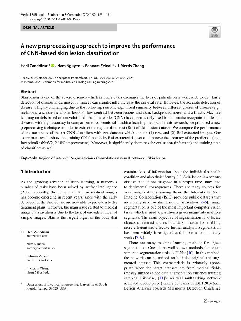
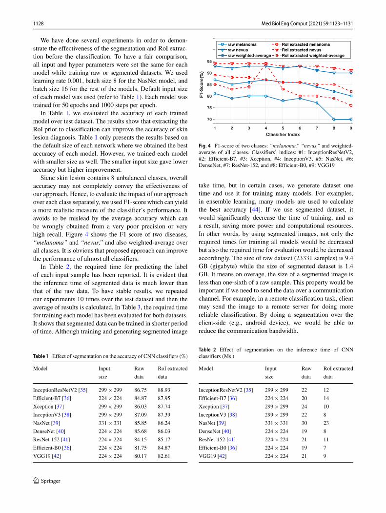
where A corresponds to the ground truth binary segmenta-tion mask, B represents the predicted binary segmentationmask, |A ∩ B| is area of overlap and |A ∪ B| is areaof union [22]. We used ISIC-2017 which contains 2000skin lesion samples with 2000 corresponding masks as aground truth to evaluate the performance of these two algo-rithms [23]. Although K-means clustering method was well-performed than Otsu’s thresholding segmentation by havingJaccard Index of 76.2% in compare to 71.7%, it is still notable to eliminate artifacts efficiently. However, our experi-ments over skin dataset showed that both non-training-basedapproaches cannot segment images very well, since arti-facts like badges are more often than not segmented as skinlesions. In addition, K-means approach does not considerthe border of the region. We observed that ground truthmask of skin lesion are mostly solid closed-contour shape,however the K-means approach does not give solid shape.In other words, there are some small dark region inside adetected lesion contour. This issue is due to the fact thatthe algorithm tries to separate pixels into multiple clustersbased on mean, and regardless of the position and value ofnear pixels. Figure 1 shows two samples of the ISIC-2017that have been segmented based on K-means approach. Thered circles inside lesion contour show the disability of thisapproach for detecting whole lesion part.
To address this issue, and regarding the availability of2000 masks of ISIC-2017, we followed our investigationby evaluating training-based approaches. One of the mostcommon training-based approaches for image segmentationis “U-net” convolutional neutral network [10]. U-net archi-tecture is an evolution of traditional convolutional neutralnetwork, which is so-called end-to-end fully convolutionalnetwork (FCN). The architecture includes two parts: (1)the contraction path (the encoder) and (2) the symmetricexpanding path (the decoder). The encoder is basically a con-ventional CNN, which is a sequence of matrix operations
1124 Med Biol Eng Comput (2021) 59:1123–1131
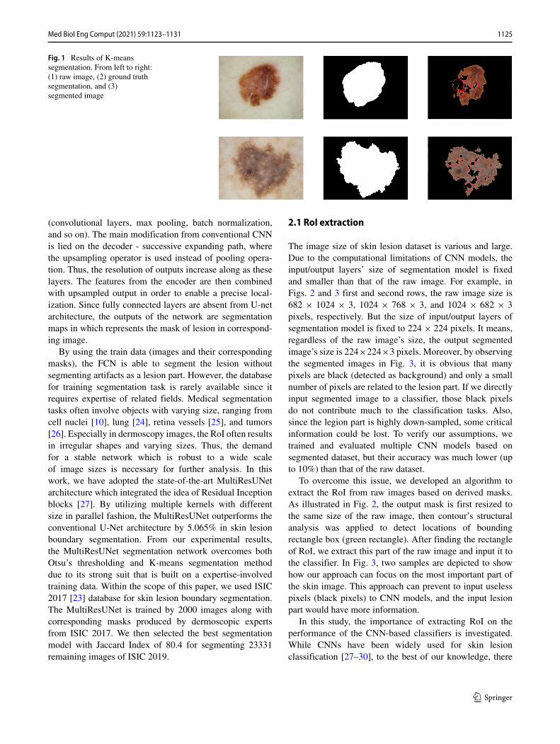
Fig. 1 Results of K-meanssegmentation. From left to right:(1) raw image, (2) ground truthsegmentation, and (3)segmented image
(convolutional layers, max pooling, batch normalization,and so on). The main modification from conventional CNNis lied on the decoder - successive expanding path, wherethe upsampling operator is used instead of pooling opera-tion. Thus, the resolution of outputs increase along as theselayers. The features from the encoder are then combinedwith upsampled output in order to enable a precise local-ization. Since fully connected layers are absent from U-netarchitecture, the outputs of the network are segmentationmaps in which represents the mask of lesion in correspond-ing image.
By using the train data (images and their correspondingmasks), the FCN is able to segment the lesion withoutsegmenting artifacts as a lesion part. However, the databasefor training segmentation task is rarely available since itrequires expertise of related fields. Medical segmentationtasks often involve objects with varying size, ranging fromcell nuclei [10], lung [24], retina vessels [25], and tumors[26]. Especially in dermoscopy images, the RoI often resultsin irregular shapes and varying sizes. Thus, the demandfor a stable network which is robust to a wide scaleof image sizes is necessary for further analysis. In thiswork, we have adopted the state-of-the-art MultiResUNetarchitecture which integrated the idea of Residual Inceptionblocks [27]. By utilizing multiple kernels with differentsize in parallel fashion, the MultiResUNet outperforms theconventional U-Net architecture by 5.065% in skin lesionboundary segmentation. From our experimental results,the MultiResUNet segmentation network overcomes bothOtsu’s thresholding and K-means segmentation methoddue to its strong suit that is built on a expertise-involvedtraining data. Within the scope of this paper, we used ISIC2017 [23] database for skin lesion boundary segmentation.The MultiResUNet is trained by 2000 images along withcorresponding masks produced by dermoscopic expertsfrom ISIC 2017. We then selected the best segmentationmodel with Jaccard Index of 80.4 for segmenting 23331remaining images of ISIC 2019.
2.1 RoI extraction
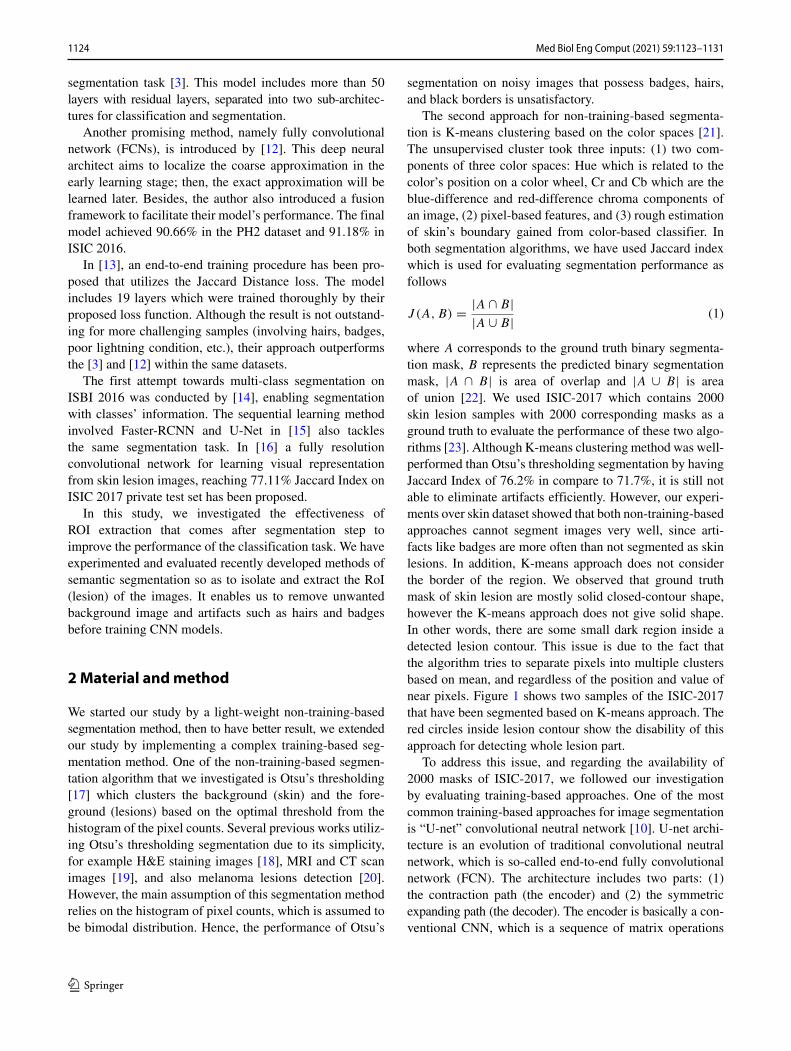
The image size of skin lesion dataset is various and large.Due to the computational limitations of CNN models, theinput/output layers’ size of segmentation model is fixedand smaller than that of the raw image. For example, inFigs. 2 and 3 first and second rows, the raw image size is682 × 1024 × 3, 1024 × 768 × 3, and 1024 × 682 × 3pixels, respectively. But the size of input/output layers ofsegmentation model is fixed to 224 × 224 pixels. It means,regardless of the raw image’s size, the output segmentedimage’s size is 224×224×3 pixels. Moreover, by observingthe segmented images in Fig. 3, it is obvious that manypixels are black (detected as background) and only a smallnumber of pixels are related to the lesion part. If we directlyinput segmented image to a classifier, those black pixelsdo not contribute much to the classification tasks. Also,since the legion part is highly down-sampled, some criticalinformation could be lost. To verify our assumptions, wetrained and evaluated multiple CNN models based onsegmented dataset, but their accuracy was much lower (upto 10%) than that of the raw dataset.
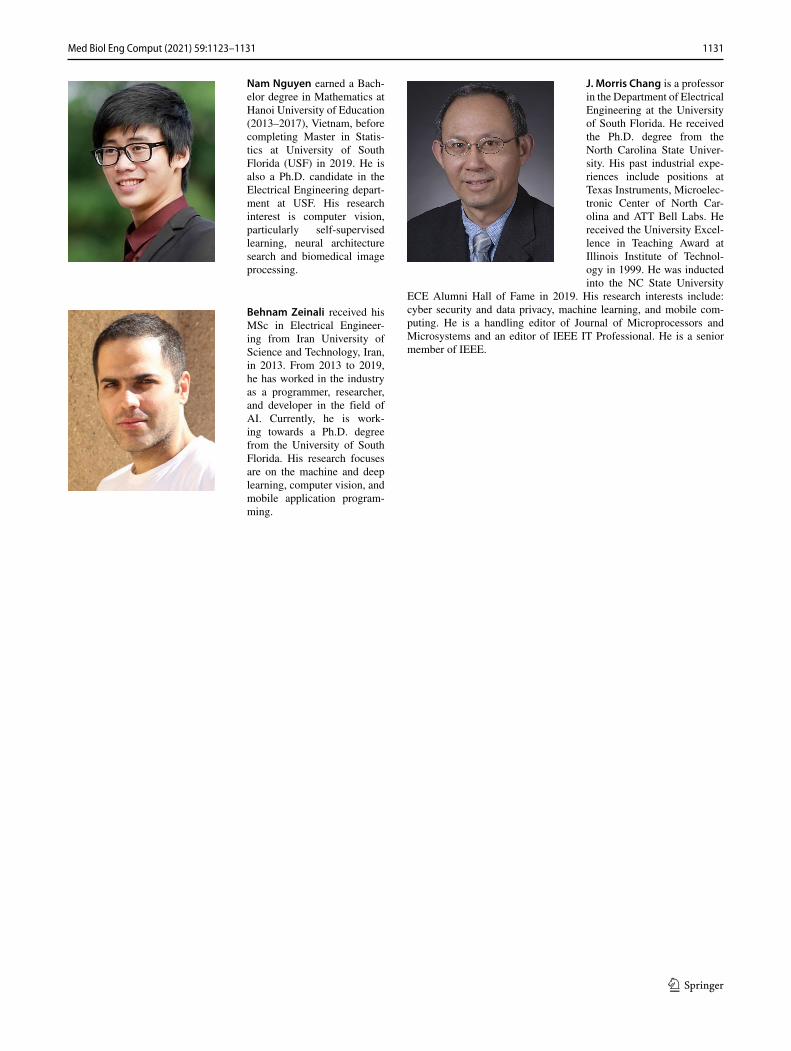
To overcome this issue, we developed an algorithm toextract the RoI from raw images based on derived masks.As illustrated in Fig. 2, the output mask is first resized tothe same size of the raw image, then contour’s structuralanalysis was applied to detect locations of boundingrectangle box (green rectangle). After finding the rectangleof RoI, we extract this part of the raw image and input it tothe classifier. In Fig. 3, two samples are depicted to showhow our approach can focus on the most important part ofthe skin image. This approach can prevent to input uselesspixels (black pixels) to CNN models, and the input lesionpart would have more information.
In this study, the importance of extracting RoI on theperformance of the CNN-based classifiers is investigated.While CNNs have been widely used for skin lesionclassification [27–30], to the best of our knowledge, there
1125Med Biol Eng Comput (2021) 59:1123–1131
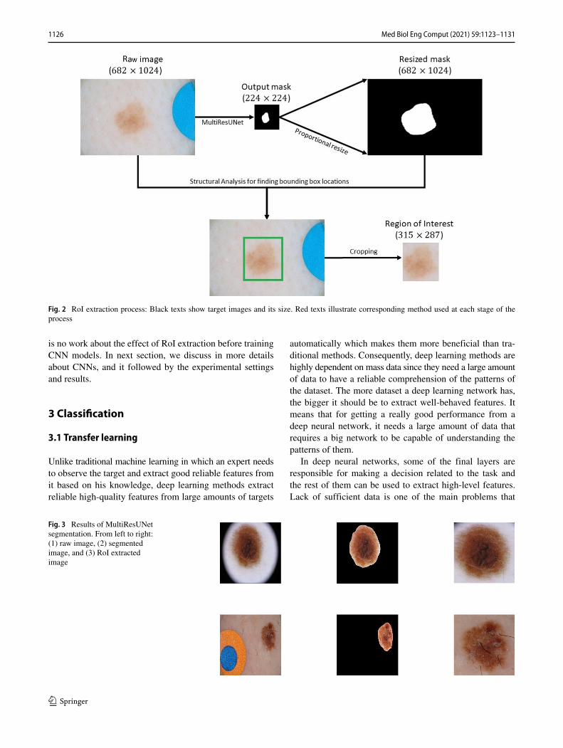
Fig. 2 RoI extraction process: Black texts show target images and its size. Red texts illustrate corresponding method used at each stage of theprocess
is no work about the effect of RoI extraction before trainingCNN models. In next section, we discuss in more detailsabout CNNs, and it followed by the experimental settingsand results.
3 Classification
3.1 Transfer learning
Unlike traditional machine learning in which an expert needsto observe the target and extract good reliable features fromit based on his knowledge, deep learning methods extractreliable high-quality features from large amounts of targets
automatically which makes them more beneficial than tra-ditional methods. Consequently, deep learning methods arehighly dependent on mass data since they need a large amountof data to have a reliable comprehension of the patterns ofthe dataset. The more dataset a deep learning network has,the bigger it should be to extract well-behaved features. Itmeans that for getting a really good performance from adeep neural network, it needs a large amount of data thatrequires a big network to be capable of understanding thepatterns of them.
In deep neural networks, some of the final layers areresponsible for making a decision related to the task andthe rest of them can be used to extract high-level features.Lack of sufficient data is one of the main problems that
Fig. 3 Results of MultiResUNetsegmentation. From left to right:(1) raw image, (2) segmentedimage, and (3) RoI extractedimage
1126 Med Biol Eng Comput (2021) 59:1123–1131
researchers usually face when they want to train a modelon specific data. In the biomedical image classificationtasks, the problem is more severe since it is much harderto find a large amount of a high-quality dataset. Transferlearning is addressing this problem and is a solution. Intraditional machine learning, we should consider the factthat training data must be independent and identicallydistributed with the test data. However, transfer learningassumes that training data and test data do not need to beindependent and identically distributed entries. It means thatfor a specific task, the network is not required to train fromscratch which has two specific benefits: first, it eliminatesthe requirement of accessing a big dataset and second, itreduces the time of training the network.
CNN pre-trained models usually trained on large imageclassification tasks. Convolutional layers are responsible forextracting high-level features from an image, while denselayers must decide on those features. There are two kinds oftransfer learning: feature extraction and fine-tuning. In theformer, the convolutional layer parameters are being frozenduring back-propagation and are used to extract features ona new dataset and the new dense layer is added to fit thenetwork for the new dataset. In the latter, after adding anew dense layer, relax back-propagation will be done on thewhole parameters for tuning them with the new dataset. Pre-trained models by ImageNet have been widely used for skinlesion classification[31–33]. We also tested this propertyand found that if we initialize the models with ImageNetpre-traiend wights, the training process converges in muchfewer number of epochs while maintaining the higheraccuracy. Also, we used data augmentation by randomlyrotating the training images up to 90 degrees, and flippingthem horizontally.
3.2 Deep learningmodels
Since 2012, when Alex Krizhevsky et al. introduced theAlexNet [34], CNN models which were not able to absorbattention came back to the play and in the next few years,many researchers and experts tried to come up with newdeep neural networks in the similar way to improve theaccuracy on different tasks [35–42]. We have used someof these pre-trained networks via transfer learning in ourwork to evaluate the effectiveness of our proposed method.We have used InceptionResNetV2 [35], Xception [37], Incep-tionV3 [38], DenseNet [40], ResNet-152 [41], and VGG19[42] which each of them has different architecture in thenumber of fully connected layers and convolutional layers.
All of these man-made models have tried to look atdifferent problems and are designed by some experts sinceit requires a suitable selection of architectures that needa high-quality knowledge in machine learning as well as
it is a tedious and time-consuming task. Moreover, fordifferent targets, different architectures should be designedto get a better result. However, some works named theneural architecture search(NAS) are introduced recently thataddress this problem and try to find a good architecturefor a certain target automatically which is logically suitableto use for different types of image classification. In ourwork, we also tried to use some of these networks whichare trained well on ImageNet dataset to evaluate our work.We have used two networks EfficientNet [36] and NasNet[39] which both are automatically designed by Google brainteam members.
4 Experiments and results
We used ISIC-2019 dataset that contains 25331 dermo-scopic images belong to 8 classes: melanoma,melanocyticnevus,basal cell carcinoma,actinic keratosis,benign kerato-sis, dermatofibroma, vascular lesion, and squa-mous cellcarcinoma. On the other hand, ISIC-2017 dataset contains2000 samples with masks for skin lesion segmentation andclassification. These 2000 samples of ISIC-2017 are exactlyavailable in ISIC-2019 as well. We used those 2000 sam-ples and their masks to train the MultiResUNet model, andwe did not involve them for the evaluation. We set theinput/output size of segmentation model to 224 × 224 pix-els. After training MultiResUNet model for 50 epochs, wegenerated 23,331 segmented images (2000 samples out of25,331 were excluded). The average required time for seg-menting each image was 11 ms (Ms). Then, we appliedour RoI extraction algorithm in order to focus on the maininformation of the image. After doing so, two sets of imagewere generated for our experiments: one contained 23,331raw images and the other had 23,331 corresponding RoIextracted images. We randomly split the 23,331 samplesinto three sub-sets, training (18,890 samples), validation(2101 samples), and test (2340 samples). We applied samedata augmentation from Keras framework over both trainingdatasets by randomly rotating the images up to 90 degreesand horizontally flipping them.
Python was used as the programming language. We usedKeras, a high-level neural networks API which is written inPython and is able to run on top of TensorFlow. All modelswere directly selected from Keras documanetation and thedefualt input size of first layer was chosen according tothe Keras documantation [43]. Keras framework providesmodels that can be converted to tensorflow lite (TFLite)format. TFLite models can be deployed over androidoperating system. We used a single GPU (Nvidia GeForceGTX 1080 Ti with 11 GB GDDR5X memory) for all of ourexperiments (training and evaluation).
1127Med Biol Eng Comput (2021) 59:1123–1131
We have done several experiments in order to demon-strate the effectiveness of the segmentation and RoI extrac-tion before the classification. To have a fair comparison,all input and hyper parameters were set the same for eachmodel while training raw or segmented datasets. We usedlearning rate 0.001, batch size 8 for the NasNet model, andbatch size 16 for the rest of the models. Default input sizeof each model was used (refer to Table 1). Each model wastrained for 50 epochs and 1000 steps per epoch.
In Table 1, we evaluated the accuracy of each trainedmodel over test dataset. The results show that extracting theRoI prior to classification can improve the accuracy of skinlesion diagnosis. Table 1 only presents the results based onthe default size of each network where we obtained the bestaccuracy of each model. However, we trained each modelwith smaller size as well. The smaller input size gave loweraccuracy but higher improvement.
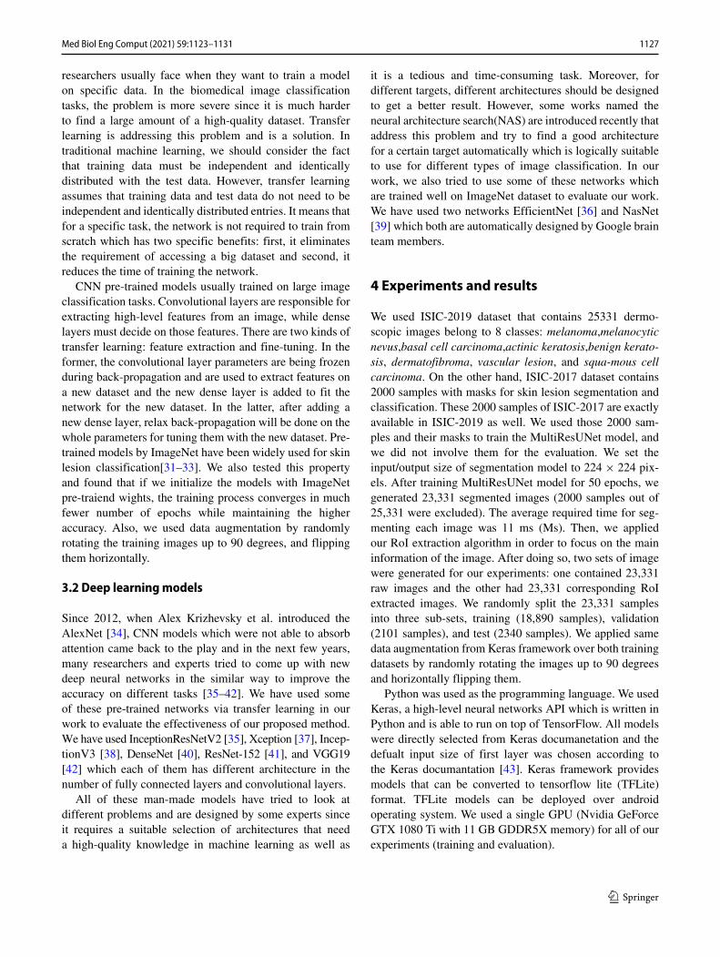
Sicne skin lesion contains 8 unbalanced classes, overallaccuracy may not completely convey the effectiveness ofour approach. Hence, to evaluate the impact of our approachover each class separately, we used F1-score which can yielda more realistic measure of the classifier’s performance. Itavoids to be mislead by the average accuracy which canbe wrongly obtained from a very poor precision or veryhigh recall. Figure 4 shows the F1-score of two diseases,“melanoma” and “nevus,” and also weighted-average overall classes. It is obvious that proposed approach can improvethe performance of almost all classifiers.
In Table 2, the required time for predicting the labelof each input sample has been reported. It is evident thatthe inference time of segmented data is much lower thanthat of the raw data. To have stable results, we repeatedour experiments 10 times over the test dataset and then theaverage of results is calculated. In Table 3, the required timefor training each model has been evaluated for both datasets.It shows that segmented data can be trained in shorter periodof time. Although training and generating segmented image
Table 1 Effect of segmentation on the accuracy of CNN classifiers (%)
Model Input Raw RoI extracted
size data data
InceptionResNetV2 [35] 299 × 299 86.75 88.93
Efficient-B7 [36] 224 × 224 84.87 87.95
Xception [37] 299 × 299 86.03 87.74
InceptionV3 [38] 299 × 299 87.09 87.39
NasNet [39] 331 × 331 85.85 86.24
DenseNet [40] 224 × 224 85.68 86.03
ResNet-152 [41] 224 × 224 84.15 85.17
Efficient-B0 [36] 224 × 224 81.75 84.87
VGG19 [42] 224 × 224 80.17 82.61
1 2 3 4 5 6 7 8 9
Classifier Index
70
75
80
85
90
95
F1-
Sco
re(%
)
raw melanoma RoI extracted melanomaraw nevus RoI extracted nevusraw weighted-average RoI extracted weighted-average
Fig. 4 F1-score of two classes: “melanoma,” “nevus,” and weighted-average of all classes. Classifiers’ indices: #1: InceptionResNetV2,#2: Efficient-B7, #3: Xception, #4: InceptionV3, #5: NasNet, #6:DenseNet, #7: ResNet-152, and #8: Efficient-B0, #9: VGG19
take time, but in certain cases, we generate dataset onetime and use it for training many models. For examples,in ensemble learning, many models are used to calculatethe best accuracy [44]. If we use segmented dataset, itwould significantly decrease the time of training, and asa result, saving more power and computational resources.In other words, by using segmented images, not only therequired times for training all models would be decreasedbut also the required time for evaluation would be decreasedaccordingly. The size of raw dataset (23331 samples) is 9.4GB (gigabyte) while the size of segmented dataset is 1.4GB. It means on overage, the size of a segmented image isless than one-sixth of a raw sample. This property would beimportant if we need to send the data over a communicationchannel. For example, in a remote classification task, clientmay send the image to a remote server for doing morereliable classification. By doing a segmentation over theclient-side (e.g., android device), we would be able toreduce the communication bandwidth.
Table 2 Effect of segmentation on the inference time of CNNclassifiers (Ms )
Model Input Raw RoI extracted
size data data
InceptionResNetV2 [35] 299 × 299 22 12
Efficient-B7 [36] 224 × 224 20 14
Xception [37] 299 × 299 24 10
InceptionV3 [38] 299 × 299 22 8
NasNet [39] 331 × 331 30 23
DenseNet [40] 224 × 224 19 8
ResNet-152 [41] 224 × 224 21 11
Efficient-B0 [36] 224 × 224 19 7
VGG19 [42] 224 × 224 21 9
1128 Med Biol Eng Comput (2021) 59:1123–1131
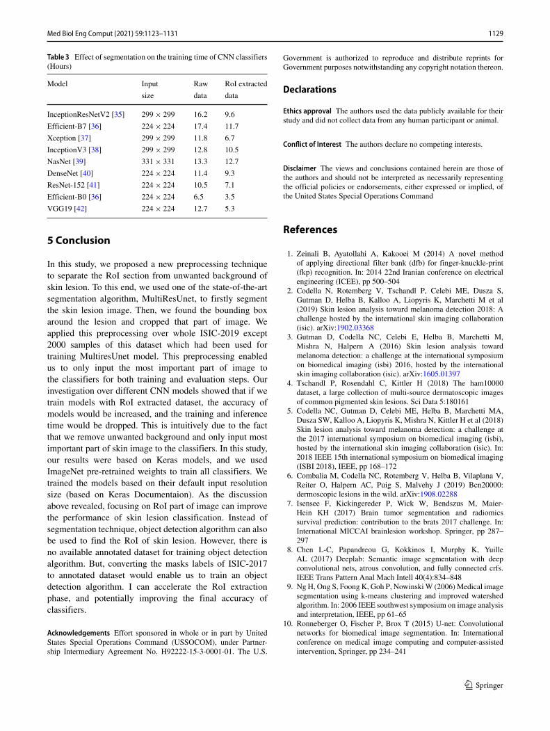
Table 3 Effect of segmentation on the training time of CNN classifiers(Hours)
Model Input Raw RoI extracted
size data data
InceptionResNetV2 [35] 299 × 299 16.2 9.6
Efficient-B7 [36] 224 × 224 17.4 11.7
Xception [37] 299 × 299 11.8 6.7
InceptionV3 [38] 299 × 299 12.8 10.5
NasNet [39] 331 × 331 13.3 12.7
DenseNet [40] 224 × 224 11.4 9.3
ResNet-152 [41] 224 × 224 10.5 7.1
Efficient-B0 [36] 224 × 224 6.5 3.5
VGG19 [42] 224 × 224 12.7 5.3
5 Conclusion
In this study, we proposed a new preprocessing techniqueto separate the RoI section from unwanted background ofskin lesion. To this end, we used one of the state-of-the-artsegmentation algorithm, MultiResUnet, to firstly segmentthe skin lesion image. Then, we found the bounding boxaround the lesion and cropped that part of image. Weapplied this preprocessing over whole ISIC-2019 except2000 samples of this dataset which had been used fortraining MultiresUnet model. This preprocessing enabledus to only input the most important part of image tothe classifiers for both training and evaluation steps. Ourinvestigation over different CNN models showed that if wetrain models with RoI extracted dataset, the accuracy ofmodels would be increased, and the training and inferencetime would be dropped. This is intuitively due to the factthat we remove unwanted background and only input mostimportant part of skin image to the classifiers. In this study,our results were based on Keras models, and we usedImageNet pre-retrained weights to train all classifiers. Wetrained the models based on their default input resolutionsize (based on Keras Documentaion). As the discussionabove revealed, focusing on RoI part of image can improvethe performance of skin lesion classification. Instead ofsegmentation technique, object detection algorithm can alsobe used to find the RoI of skin lesion. However, there isno available annotated dataset for training object detectionalgorithm. But, converting the masks labels of ISIC-2017to annotated dataset would enable us to train an objectdetection algorithm. I can accelerate the RoI extractionphase, and potentially improving the final accuracy ofclassifiers.
Acknowledgements Effort sponsored in whole or in part by UnitedStates Special Operations Command (USSOCOM), under Partner-ship Intermediary Agreement No. H92222-15-3-0001-01. The U.S.
Government is authorized to reproduce and distribute reprints forGovernment purposes notwithstanding any copyright notation thereon.
Declarations
Ethics approval The authors used the data publicly available for theirstudy and did not collect data from any human participant or animal.
Conflict of Interest The authors declare no competing interests.
Disclaimer The views and conclusions contained herein are those ofthe authors and should not be interpreted as necessarily representingthe official policies or endorsements, either expressed or implied, ofthe United States Special Operations Command
References
1. Zeinali B, Ayatollahi A, Kakooei M (2014) A novel methodof applying directional filter bank (dfb) for finger-knuckle-print(fkp) recognition. In: 2014 22nd Iranian conference on electricalengineering (ICEE), pp 500–504
2. Codella N, Rotemberg V, Tschandl P, Celebi ME, Dusza S,Gutman D, Helba B, Kalloo A, Liopyris K, Marchetti M et al(2019) Skin lesion analysis toward melanoma detection 2018: Achallenge hosted by the international skin imaging collaboration(isic). arXiv:1902.03368
3. Gutman D, Codella NC, Celebi E, Helba B, Marchetti M,Mishra N, Halpern A (2016) Skin lesion analysis towardmelanoma detection: a challenge at the international symposiumon biomedical imaging (isbi) 2016, hosted by the internationalskin imaging collaboration (isic). arXiv:1605.01397
4. Tschandl P, Rosendahl C, Kittler H (2018) The ham10000dataset, a large collection of multi-source dermatoscopic imagesof common pigmented skin lesions. Sci Data 5:180161
5. Codella NC, Gutman D, Celebi ME, Helba B, Marchetti MA,Dusza SW, Kalloo A, Liopyris K, Mishra N, Kittler H et al (2018)Skin lesion analysis toward melanoma detection: a challenge atthe 2017 international symposium on biomedical imaging (isbi),hosted by the international skin imaging collaboration (isic). In:2018 IEEE 15th international symposium on biomedical imaging(ISBI 2018), IEEE, pp 168–172
6. Combalia M, Codella NC, Rotemberg V, Helba B, Vilaplana V,Reiter O, Halpern AC, Puig S, Malvehy J (2019) Bcn20000:dermoscopic lesions in the wild. arXiv:1908.02288
7. Isensee F, Kickingereder P, Wick W, Bendszus M, Maier-Hein KH (2017) Brain tumor segmentation and radiomicssurvival prediction: contribution to the brats 2017 challenge. In:International MICCAI brainlesion workshop. Springer, pp 287–297
8. Chen L-C, Papandreou G, Kokkinos I, Murphy K, YuilleAL (2017) Deeplab: Semantic image segmentation with deepconvolutional nets, atrous convolution, and fully connected crfs.IEEE Trans Pattern Anal Mach Intell 40(4):834–848
9. Ng H, Ong S, Foong K, Goh P, NowinskiW (2006)Medical imagesegmentation using k-means clustering and improved watershedalgorithm. In: 2006 IEEE southwest symposium on image analysisand interpretation, IEEE, pp 61–65
10. Ronneberger O, Fischer P, Brox T (2015) U-net: Convolutionalnetworks for biomedical image segmentation. In: Internationalconference on medical image computing and computer-assistedintervention, Springer, pp 234–241
1129Med Biol Eng Comput (2021) 59:1123–1131

11. Yu L, Chen H, Dou Q, Qin J, Heng P.-A. (2016) Automatedmelanoma recognition in dermoscopy images via very deepresidual networks. IEEE Trans Med Imag 36(4):994–1004
12. Bi L, Kim J, Ahn E, Kumar A, Fulham M, Feng D (2017) Der-moscopic image segmentation via multistage fully convolutionalnetworks. IEEE Trans Biomed Eng 64(9):2065–2074
13. Yuan Y, Chao M, Lo Y-C (2017) Automatic skin lesionsegmentation using deep fully convolutional networks withjaccard distance. IEEE Trans Med Imaging 36(9):1876–1886
14. Goyal M, Yap MH, Hassanpour S (2017) Multi-class semanticsegmentation of skin lesions via fully convolutional networks.arXiv:1711.10449
15. Vesal S, Patil SM, Ravikumar N, Maier AK (2018) A multi-taskframework for skin lesion detection and segmentation. In: OR2.0 Context-aware operating theaters, computer assisted roboticendoscopy, clinical image-based procedures, and skin imageanalysis, Springer, pp 285–293
16. Soudani A, Barhoumi W (2019) An image-based segmentationrecommender using crowdsourcing and transfer learning for skinlesion extraction. Expert Syst Appl 118:400–410
17. Zhang J, Hu J (2008) Image segmentation based on 2d otsumethod with histogram analysis. In: 2008 International conferenceon computer science and software engineering, vol 6. IEEE,pp 105–108
18. Haggerty JM, Wang XN, Dickinson A, O’Malley CJ, Martin EB(2014) Segmentation of epidermal tissue with histopathologicaldamage in images of haematoxylin and eosin stained human skin.BMC Med Imaging 14(1):7
19. Bindu CH, Prasad KS (2012) An efficient medical imagesegmentation using conventional otsu method. Int J Adv SciTechnol 38(1):67–74
20. Premaladha J, Ravichandran K (2016) Novel approaches fordiagnosing melanoma skin lesions through supervised and deeplearning algorithms. J Med Syst 40(4):96
21. Buza E, Akagic A, Omanovic S (2017) Skin detection basedon image color segmentation with histogram and k-meansclustering. In: 2017 10th International conference on electrical andelectronics engineering (ELECO), pp 1181–1186
22. McGuinness K, O’Connor NE (2010) A comparative evaluation ofinteractive segmentation algorithms. Pattern Recogn 43(2):434–444. interactive Imaging and Vision. [Online]. Available: http://www.sciencedirect.com/science/article/pii/S0031320309000818
23. Berseth M (2017) Isic 2017-skin lesion analysis towardsmelanoma detection. arXiv:1703.00523
24. Zhao T, Gao D, Wang J, Tin Z (2018) Lung segmentation in ctimages using a fully convolutional neural network with multi-instance and conditional adversary loss. In: 2018 IEEE 15thInternational symposium on biomedical imaging (ISBI 2018),IEEE, pp 505–509
25. Xiao X, Lian S, Luo Z, Li S (2018) Weighted res-unet for high-quality retina vessel segmentation. In: 2018 9th Internationalconference on information technology in medicine and education(ITME), IEEE, pp 327–331
26. Li X, Chen H, Qi X, Dou Q, Fu C-W, Heng P-A (2018) H-denseunet: hybrid densely connected unet for liver and tumorsegmentation from ct volumes. IEEE Trans Med Imaging37(12):2663–2674
27. Ibtehaz N, Rahman MS (2020) Multiresunet: Rethinking the u-net architecture for multimodal biomedical image segmentation.Neural Netw 121:74–87
28. Jafari MH, Karimi N, Nasr-Esfahani E, Samavi S, SoroushmehrSMR, Ward K, Najarian K (2016) Skin lesion segmentation inclinical images using deep learning. In: 2016 23rd Internationalconference on pattern recognition (ICPR), IEEE, pp 337–342
29. Kawahara J, Hamarneh G (2016) Multi-resolution-tract cnn withhybrid pretrained and skin-lesion trained layers. In: Internationalworkshop on machine learning in medical imaging, Springer,pp 164–171
30. Saba T, Khan MA, Rehman A, Marie-Sainte SL (2019) Regionextraction and classification of skin cancer: a heterogeneousframework of deep cnn features fusion and reduction. J Med Syst43(9):289
31. Mahbod A, Schaefer G, Wang C, Ecker R, Dorffner G, Ellinger I(2020) Investigating and exploiting image resolution for transferlearning-based skin lesion classification
32. Hosny KM, Kassem MA, Foaud MM (2018) Skin cancerclassification using deep learning and transfer learning. In:2018 9th Cairo international biomedical engineering conference(CIBEC), pp 90–93
33. Adegun AA, Viriri S (2020) Deep learning-based system forautomatic melanoma detection. IEEE Access 8:7160–7172
34. Krizhevsky A, Sutskever I, Hinton GE (2012) Imagenet classifi-cation with deep convolutional neural networks. In: NIPS
35. Szegedy C, Ioffe S, Vanhoucke V, Alemi AA (2017) Inception-v4, inception-resnet and the impact of residual connectionson learning. In: Thirty-first AAAI conference on artificialintelligence
36. Tan M, Le QV (2019) Efficientnet: rethinking model scaling forconvolutional neural networks. arXiv:1905.11946
37. Chollet F (2016) Xception: Deep learning with depthwiseseparable convolutions. arXiv:1610.02357
38. Szegedy C, Vanhoucke V, Ioffe S, Shlens J, Wojna Z (2015)Rethinking the inception architecture for computer vision.arXiv:1512.00567
39. Zoph B, Vasudevan V, Shlens J, Le QV (2017) Learn-ing transferable architectures for scalable image recognition.arXiv:1707.07012
40. Huang G, Liu Z, van der Maaten L, Weinberger KQ (2016)Densely connected convolutional networks. arXiv:1608.06993
41. He K, Zhang X, Ren S, Sun J (2015) Deep residual learning forimage recognition. arXiv:1512.03385
42. Simonyan K, Zisserman A (2014) Very deep convolutionalnetworks for large-scale image recognition. arXiv:1409.1556
43. Chollet F et al (2015) Keras. https://keras.io, [accessed April 12020]
44. Liu Y, Yao X (1999) Ensemble learning via negative correlation.Neural Netw 12(10):1399–1404
Publisher’s note Springer Nature remains neutral with regard tojurisdictional claims in published maps and institutional affiliations.
Hadi Zanddizari receivedMaster’s in Electrical Engi-neering from University ofScience and Technology, Iran,in 2015. From 2015 to 2017,he worked at Pardis Technol-ogy Park as a researcher andprogrammer. Currently, heis working towards the PhDdegree from the University ofSouth Florida. His researchfocuses are on machinelearning, sparsity, cyberse-curity, and biomedical imageprocessing.
1130 Med Biol Eng Comput (2021) 59:1123–1131
Nam Nguyen earned a Bach-elor degree in Mathematics atHanoi University of Education(2013–2017), Vietnam, beforecompleting Master in Statis-tics at University of SouthFlorida (USF) in 2019. He isalso a Ph.D. candidate in theElectrical Engineering depart-ment at USF. His researchinterest is computer vision,particularly self-supervisedlearning, neural architecturesearch and biomedical imageprocessing.
Behnam Zeinali received hisMSc in Electrical Engineer-ing from Iran University ofScience and Technology, Iran,in 2013. From 2013 to 2019,he has worked in the industryas a programmer, researcher,and developer in the field ofAI. Currently, he is work-ing towards a Ph.D. degreefrom the University of SouthFlorida. His research focusesare on the machine and deeplearning, computer vision, andmobile application program-ming.
J. Morris Chang is a professorin the Department of ElectricalEngineering at the Universityof South Florida. He receivedthe Ph.D. degree from theNorth Carolina State Univer-sity. His past industrial expe-riences include positions atTexas Instruments, Microelec-tronic Center of North Car-olina and ATT Bell Labs. Hereceived the University Excel-lence in Teaching Award atIllinois Institute of Technol-ogy in 1999. He was inductedinto the NC State University
ECE Alumni Hall of Fame in 2019. His research interests include:cyber security and data privacy, machine learning, and mobile com-puting. He is a handling editor of Journal of Microprocessors andMicrosystems and an editor of IEEE IT Professional. He is a seniormember of IEEE.
1131Med Biol Eng Comput (2021) 59:1123–1131