a new insight into the treatment of renal anemia with hif
TRANSCRIPT
REVIEW Open Access
A new insight into the treatment of renalanemia with HIF stabilizerSatoru Kuriyama1,2*, Yukio Maruyama3 and Hirokazu Honda4
Abstract
The long-term clinical experiences with recombinant human erythropoietin (rHuEPO) and its analog derivativeshave clearly proven that correction of anemia with erythropoiesis stimulating agent (ESA) not only reduces bloodtransfusion and improves patients’ QOL but has multiple benefits for the concurrent complications of CKD such asCardio-Renal–Anemia (CRA) syndrome and/or malnutrition-inflammation-atherosclerosis (MIA) syndrome.Unlike ESA, the newly available agent, hypoxia-inducible factor (HIF) stabilizer, stimulates endogenouserythropoietin (EPO) by mimicking hypoxia with HIF prolyl hydroxylase domain enzyme (HIF-PHD) inhibition. Thephase 2 and 3 clinical studies have shown that HIF stabilizers are as efficacious as ESA in ameliorating renal anemia.Whether the same clinical benefits on CRA and MIA syndrome hold true in patients given HIF stabilizers is a matterfor future debate. Given that HIF stabilizers act on the multiple target genes, the use of this novel agent may leadto unwanted adverse events.Launching HIF stabilizers into the treatment of renal anemia provokes a concern about how this alternativetreatment will be taken up in the daily clinical practice. However, guideline-oriented strategies on how to use HIFstabilizer is not available at this limited point due to scant clinical information. Nevertheless, this opinion-basedreview provides a future insight into the management of renal anemia with HIF stabilizer by reference to the pastexperiences with ESA. HIF stabilizers can preferably be indicated for CRA syndrome at pre-dialysis stage, ESAresistant anemia at advanced CKD stage, and perhaps for dysregulated iron metabolism akin to MIA syndrome inpatients on dialysis.
Keywords: Hypoxia inducible factor (HIF) stabilizer, Hypoxia, Erythropoiesis stimulating agent (ESA), CRA syndrome,MIA syndrome, Iron metabolism
IntroductionThe epoch-making recombinant human erythropoietin(rHuEPO) came into clinical practice for renal anemia inchronic kidney disease (CKD) patients on dialysis in1990, and its indication was extended to those at pre-dialysis stage in 1994 in Japan. The pharmacologicalpotency of correction of anemia with rHuEPO and itspharmacological analogues was so efficacious that most
of the CKD patients benefited from the treatment. Oneof the remarkable effects of the erythropoiesis stimulatingagents (ESA) was the reduction in blood transfusion thatcontributed to the prevention of blood-transfusion-borneinfections such as viral hepatitis and other intractable life-threatening diseases before the Era of ESA. ESA has provento be cardio-renal protective, reducing cardio-renal-anemia(CRA) syndrome, a highly risky cardiovascular (CV) condi-tion associated with CKD. Indeed, a large body of evidencehas clearly proven that ESA regresses left ventricular hyper-trophy (LVH) and retards the progression of CKD[1, 2]. In addition, ESA also improves malnutrition-inflammation-atherosclerosis (MIA) syndrome, an abnormal
© The Author(s). 2020 Open Access This article is licensed under a Creative Commons Attribution 4.0 International License,which permits use, sharing, adaptation, distribution and reproduction in any medium or format, as long as you giveappropriate credit to the original author(s) and the source, provide a link to the Creative Commons licence, and indicate ifchanges were made. The images or other third party material in this article are included in the article's Creative Commonslicence, unless indicated otherwise in a credit line to the material. If material is not included in the article's Creative Commonslicence and your intended use is not permitted by statutory regulation or exceeds the permitted use, you will need to obtainpermission directly from the copyright holder. To view a copy of this licence, visit http://creativecommons.org/licenses/by/4.0/.The Creative Commons Public Domain Dedication waiver (http://creativecommons.org/publicdomain/zero/1.0/) applies to thedata made available in this article, unless otherwise stated in a credit line to the data.
* Correspondence: [email protected] University School of Medicine, 3-25-8, Nishi-shinbashi, Minato-ku, Tokyo105-8471, Japan2Nephrology & Hypertension Research Unit, Internal Medicine, Miho Clinic,Shin-Osaki-kangyo Bld 2F, Osaki, Shinagawa-ku, Tokyo 141-0032, JapanFull list of author information is available at the end of the article
Kuriyama et al. Renal Replacement Therapy (2020) 6:63 https://doi.org/10.1186/s41100-020-00311-x
iron metabolism characterized by the increased hepcidinand serum ferritin levels [3, 4].Lagging about 3 decades behind ESA in 2019, the hyp-
oxia inducible factor (HIF) stabilizer which stimulatesendogenous erythropoietin (EPO) by mimicking hypoxiawith HIF prolyl hydroxylase domain enzyme (HIF-PHD)inhibition was launched for the first time into theclinical practice of renal anemia in Japan. Its clinicalindication was limited only to patients on dialysis at thebeginning in 2019, and it was extended to those in pre-dialysis stage in August 2020. Although the pharmaco-logical mode of action of HIF stabilizer to stimulateerythropoiesis is totally different from ESA, the phase-2and phase-3 clinical studies have shown that HIF stabi-lizers are as efficacious as ESA in ameliorating renalanemia [5–19]. However, at this point whether HIFstabilizer exerts the same benefits in patients with CRAand MIA syndrome is a matter for future debate. Inaddition, upcoming problems to be solved are to whatextent the assumptive adverse effects originate from themultiple target genes that the HIF activates will be clin-ically problematic.Treatment of renal anemia with HIF stabilizers provokes
a clinical concern about how this alternative treatmentwill be taken up in our daily CKD practice. Clinical guide-lines on how to use HIF stabilizer are not yet available dueto insufficient clinical information. Therefore, by referenceto many clinical experiences with ESA in the past threedecades, this opinion-based review may provide anew afuture insight into how we clinicians should manage renalanemia with HIF stabilizer.
The differential diagnosis on renal anemiaThe total number of human cells is approximately 30 ×1012. The largest contributor to the overall number ofhuman cells is red blood cells (RBCs) with a total of 25× 1012, indicating that 84% of the total body cells areRBCs [20]. The regulatory system of RBCs is a dynamicand actively operating one, constantly producing 2.0 ×10⁶ (cells/second) and destroying the same number atthe same time. Iron is an essential element for erythro-poiesis and the regulatory system of iron is basically theclosed circuit in which iron in the RBCs is constantlyreused, as long as loss of RBCs such as gastrointestinal(GI) bleeding did not happen. Once RBCs are destroyedin the reticuloendothelial system, iron is reused efficientlyas heme iron. The total body iron storage is estimated ap-proximately 3 ~ 4 g. In a physiological condition in whichthere is no blood loss, only 1 ~ 2mg of iron is excretedfrom the decidual cells in the GI tracts which is routinelyreplenished with food.Causative conditions/diseases for anemia in CKD pa-
tients are multifactorial; these include CKD-associatedreduced EPO production, loss of blood, iron deficiency,
malabsorption of iron, dysregulation of iron metabolism,hemolysis, vitamin B12 and/or folic acid deficiency, useof RAS inhibitors, malnutrition, autoantibodies to EPOreceptor, and coincidental hematological disorders suchas MDS and aplastic anemia. The definition of renalanemia, in a narrow sense, is a normocytic normochro-mic anemia without the elevation of reticulocytes. Formaking a differential diagnosis of renal anemia, measure-ment of circulating EPO concentration is practically im-portant. In renal anemia in CKD, due to the reducedproduction of EPO in the kidney, the EPO concentrationis normally less than 50 mIU/mL. In contrast, in anemicpatients without CKD, the most of them have a circulatingEPO exceeding 50 mIU/mL. It is also known that patientswith acute GI bleeding sometimes have increased EPOconcentration above 200 ~ 300 mIU/mL. All in all, thedifferential diagnostic criteria for renal anemia include; (1)normocytic normochromic anemia without increase inreticulocytes, (2) EPO < 50 mIU/mL, and (3) able to ruleout other diseases that cause anemia. Regarding how tomake a differential diagnosis on renal anemia, the guide-lines published in the Japanese Society of Dialysis andTransplantation (JSDT) described it in detail [21].
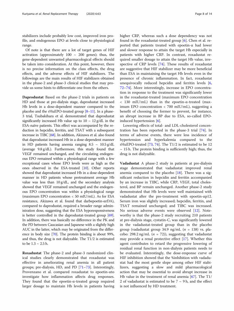
Cardio-renal effect of ESA therapyEffect of ESA in patients with CRA syndromeAnemia and/or anemia-induced hypoxia increasescardiac burden and accelerates LVH [22]. In general, themore the CKD progresses into the further advancedstage, the more the deterioration of LVH advances. Pa-tients with extremely advanced LVH could occasionallyfurther deteriorate into congestive heart failure (CHF),which jeopardizes patient’s life prognosis. In the pres-ence of renal anemia in CKD patients, the reduced car-diac function further accelerates the decline in renalfunction, creating a vicious circle so-called the cardio (orcardiovascular)-renal-anemia (CRA) syndrome. CKD in-creases inflammation and oxidative stress via hypoxiaand eventually activates renin-angiotensin-aldosteronesystem (RAS) via reduced renal perfusion, which also fa-cilitates cardiovascular disease (CVD). CKD also inducessodium and water retention which frequently leads tohypertension. These changes trigger to aggravate LVHand reduce cardia output, which in turn decreases perfu-sion of the kidney. Inflammation in CVD also becomes arisk for CKD progression. After all, CKD and CVD to-gether with renal anemia create an intractable viciouscircle. The concept of CRA syndrome has been widelyacknowledged and discussed elsewhere [23] (Fig. 1).Silverberg et al. reported that treatment of anemia withESA in patients with the CRA syndrome having congest-ive heart failure (HF) improved not only patients’ sub-jective symptoms, but also the ejection fraction (EF),NYHA classification, eGFR, and shortened the duration
Kuriyama et al. Renal Replacement Therapy (2020) 6:63 Page 2 of 14

of hospitalization, suggesting that ESA serves as cardio-renal protective [24]. The similar study was conductedby Ayus et al. who confirmed that ESA significantlyregressed the LVM index (LVMI) in CKD patients atpre-dialysis stage [25]. After collecting 15 related studies,Parfrey et al. confirmed in a meta-analysis that theregression of LVMI was unequivocally found in theanemia treatment with ESA [1]. Supporting this con-clusion is a multicenter study recruiting pre-dialysispatients in Japan that maintaining hemoglobin (Hb)levels above 12 g/dL with the use of darbepoetin-α(DA) significantly reduced LVMI [26].As for the effect of ESA on residual renal function in
pre-dialysis patients, early studies showed that the use ofESA retarded the progression of CKD [27, 28]. Recently,Covic et al. performed an extensive meta-analysis on therenal protective effect of ESA and found that there wasno benefit when evaluated using the hard-endpoint (EP)such as composite EP including mortality, commencement
of dialysis, and renal death. However, when using a doub-ling of Cr concentration, the relative risk was as low as 0.53(95%CI, 0.31 ~ 0.89), suggesting that early intervention withESA may serve as renal protective [2]. Focusing on renalprotection, Akizawa et al. and Tsubakihara et al. also con-firmed that the early intervention maintaining the Hb levelsabove 12 g/dL with ESA was advantageous in protectingthe kidney from failing [29, 30]. On the contrary, a random-ized multicenter study by Hayashi et al. in patients withadvanced CKD patients with an eGFR of 8 to 20mL/min/1.732 demonstrated that there was no difference in therenal survival between the high Hb-group and the low-Hbgroup [31].Taking all the above mentioned studies into consider-
ation, it can be safely concluded that ESA regressesLVH; thus, serving as cardio-protective, preventing HFand/other cardiac diseases, and eventually contributingto prolonging patients’ lives. With respect to renal pro-tection, ESA appears not to have benefits in advanced
Fig. 1 Cardio-renal-anemia (CRA) syndrome. CKD-induced anemia produces hypoxic condition which leads to an increase in oxidative stress. CKDalso facilitates chronic inflammation and hypoxia in renal tissue, activating systemic, and local RAS. These changes trigger to aggravate cardiachypertrophy and reduce cardiac output, which in turn decreases organ perfusion including the kidney. With such a mechanism, renal anemia inCKD creates a vicious circle in conjunction with CVD/HF, so-called the CRA syndrome, which may eventually result in poor patients’ prognosis.CVD cerebrovascular disease, LVH left ventricular hypertrophy, HF heart failure. Quoted from reference # 23,24
Kuriyama et al. Renal Replacement Therapy (2020) 6:63 Page 3 of 14

CKD patients, but early intervention with ESA is prob-ably advantageous in retarding the progression of CKD.All in all, the effect of ESA in improving the CRAsyndrome appears to be indisputable. Of note is thatwhether the same holds true with the use of HIF stabi-lizers remains undetermined, thus needs to be addressedin the future trials.
Factors to explain ESA hyporesponsivenessNormal hematocrit cardiac (NHC) trial was the firstlarge-scale RCT to evaluate the effect of ESA in patientson dialysis. Defining death and non-fatal myocardialinfarction as the primary EP, comparison was made be-tween the high-hematocrit (Ht) (Ht 42%) and the low-Ht (Ht 30%) group. The NHC trial was not completedbecause of a trend of unexpectedly increased number ofdeath and the excessive amount of iron-supplementationdosage [32]. This study widely provokes a concern that“normalization of Ht with ESA” may not always benecessary for the better outcomes, and thus, setting thetarget Ht level in the neighbor of “normal” was in gen-eral not recommended. Discussion on the normalizationof anemia with ESA was later years extended to patientsat pre-dialysis stage in the CREATE, CHOIR, and TREAT studies. The CREATE showed that therapy with ESAaiming at normal Hb level had no beneficial effect onthe prevention of CV diseases [33]. Second, the CHOIRshowed that there was an increasing rate of death, con-gestive HF, non-fatal myocardial infarction, and cerebralapoplexy [34]. Third, the TREAT study indicated thatthe incidence of cerebral apoplexy had risen significantlyin the high Hb group [35]. Following these trials, theRED-HF study aiming at the target Hb level above 13 g/dL in patients with chronic HF resulted in an increase inthe incidence of cerebral thrombosis [36]. The clinicalmessage of these clinical studies is that a targeted Hbwell over 13 g/dL or near normal may have an inherentCV risk that would lead to the poor prognosis of theCKD patients. Notably, post hoc analyses on the CREATE, CHOIR, and TREAT studies suggested that the truecause for the unexpected adverse events is not the ESAtreatment aiming at the high Hb level per se, but “theESA hyporesponsiveness” in which there is either highdose of ESA (as a result of weakened response to ESA)or large dose iron supplementation [37–39]. The under-lying abnormality to explain ESA-hyporesponsiveness ischronic inflammation and the subsequently accompany-ing iron dysregulation in CKD patients.Based on the abovementioned studies, the KDIGO
guideline is rather cautious about using ESA. The guide-line recommends that iron therapy is the first priorityand that ESA should not be the choice even if patient’Hb > 10 g/dL. Moreover, it recommends that the targetHb should be less than 11.5 g/dL [40]. However, unlike
the KDIGO guideline, the JSDT guideline paid a specialattention to set the stage-specific individual target Hbvalue, especially distinguishing patients on dialysis fromthose at pre-dialysis stages. Based on them, the ESAshould be started when the Hb value measured repeat-edly is less than 11 g/dL if patients are at pre-dialysisstage or on peritoneal dialysis (PD), and it is less than10 g/dL if patients are on hemodialysis (HD). Inaddition, the targeted Hb value should be set to 10 ~ 12g/dL if the patients were on HD and 11 ~ 13 g/dL if theywere in pre-dialysis stage or on PD [41].Now that the guideline for HIF stabilizer is not avail-
able at this limited point, these values obtained from thestudies of ESA should be applicable for the treatment ofrenal anemia with HIF stabilizers.As mentioned above, high-dose ESA treatment for
anemia in CKD patients might be associated with in-creased mortality in those patients. With respect to EPOconcentration, levels of EPO after administration of ESAis obviously increased compared with those with HIFstabilizers. HIF stabilizers could control renal anemiawhile keeping EPO within an almost normal physio-logical range. That could be the advantage in CKD pa-tients with anemia who would be required high dose ofESA for the management.
Dysregulated iron metabolism in CKD patientsCKD and MIA syndromeThe incidence of renal anemia increases linearly as theCKD progresses into its further advanced stage. Recruit-ing more than 1,000,000 individuals in the in USA inresearch, Go et al. clearly showed that the progression ofrenal function to end stage renal disease (ESRD) acceler-ates risks of death and CV events in an exponential fash-ion [42, 43]. What are the hidden mechanisms to createthis vicious cycle in CKD? One of the answers to thisquestion is the CRA syndrome, as was discussed previ-ously in this review. But there is another crucial mechan-ism to cause deterioration of CKD; that is, the abnormalregulation of iron in an association with chronic inflam-mation. Steinvinkel et al. proposed for the first time theconceptual disease entity of the MIA syndrome [3, 4].MIA syndrome can be found in most of advanced CKD.Via increase in circulating cytokines such as interleukin(IL)-6, chronic inflammation, and abnormal iron metabol-ism plays a crucial role in the ESA unresponsiveness andother CKD-accompanying complications in the MIA syn-drome. It is conceivable that its pathogenetic mechanismsare more or less overlapped with the CRA syndrome.
Iron sequestration inside of the cells in CKDUnder the influence of chronic inflammation, secondaryto increased inflammatory cytokines and oxidative stress,the iron metabolism is dysregulated in patients with
Kuriyama et al. Renal Replacement Therapy (2020) 6:63 Page 4 of 14
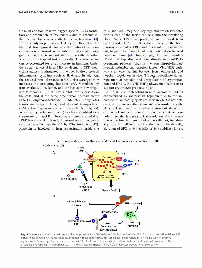
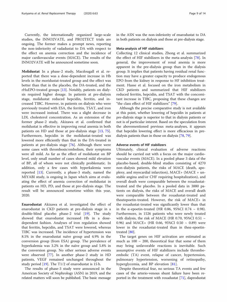
CKD. In addition, reactive oxygen species (ROS) forma-tion and production of free radicals due to chronic in-flammation also adversely affects iron metabolism [44].Utilizing polymorphonuclear leukocytes, Otaki et al. forthe first time proved clinically that intracellular ironcontent was increased in patients on dialysis [45], sug-gesting that iron is sequestrated in the cells. In otherwords, iron is trapped inside the cells. This mechanismcan be accounted for by an increase in hepcidin. Underthe circumstances akin to MIA syndrome in CKD, hep-cidin synthesis is stimulated in the liver by the increasedinflammatory cytokines such as IL-6, and in addition,the reduced renal clearance in CKD also synergisticallyincreases the circulating hepcidin level. Stimulated byiron overload, IL-6, leptin, and the hepcidin downregu-late ferroportin-1 (FPN-1) to inhibit iron release fromthe cells, and at the same time tumor necrosis factor(TNF)-NFolipopolysaccharide (LPS), etc., upregulatestransferrin receptor (TfR) and divalent transporter-1(DMT-1) to trap more iron into the cells [46] (Fig. 2a).Recently, erythroferrone (ERFE) has been identified as asuppressor of hepcidin. Honda et al. demonstrated thatERFE levels are significantly increased with a concomi-tant decrease in hepcidun-25 by ESA treatment [47].Hepcidin is involved in iron sequestration inside the
cells, and ERFE may be a key regulator which facilitatesiron release of the inside the cells into the circulatingblood. Since ERFE are produced and released fromerythroblasts, ESA or HIF stabilizer acts on the bonemarrow to stimulate ERFE and as a result inhibits hepci-din, helping the dysregulated iron mobilization to yieldbetter outcomes [48]. Interestingly, HIF could regulateFPN-1 and hepcidin production directly in non-ERFE-dependent pathway. That is, the von Hippel–Lindau/hypoxia-inducible transcription factor (VHL/HIF) path-way is an essential link between iron homeostasis andhepcidin regulation in vivo. Through coordinate down-regulation of hepcidin and upregulation of erythropoi-etin and FPN-1, the VHL-HIF pathway mobilizes iron tosupport erythrocyte production [49].All in all, iron metabolism in renal anemia of CKD is
characterized by increase in hepcidin due to the in-creased inflammatory cytokines. Iron in CKD is not defi-cient, and there is rather abundant iron inside the cells.Nevertheless, functionally deficient iron outside of thecells is not sufficient enough to elicit efficient erythro-poiesis. So, this is a paradoxical regulation of iron where“Excessive iron is present inside the cells but, function-ally iron is deficient outside the cells”. Incidentally,elevation of EPO by either ESA or HIF stabilizer lowers
Fig. 2 “Iron sequestration in the cells” (a) and “hematopoietic action of HIF stabilizers” (b). As a result of the HIF-PHD inhibition with HIF stabilizers, HIFinduces endogenous EPO and facilitates RBC production in the bone marrow. HIF also induces genes related to iron mobilization for effectiveerythropoiesis. Serum hepcidin levels are increased in CKD patients, and HIF inhibits hepcidin through the stimulation of erythroferrone (ERFE) toaccelerate erythropoiesis. FPN ferroportin, DMT-1 divalent metal transporter-1, TfR transferrin receptor. Quoted from reference # 46
Kuriyama et al. Renal Replacement Therapy (2020) 6:63 Page 5 of 14
hepcidin level and improves the utilization of iron moreeffectively through the increase in ERFE [46] (Fig. 2b).
Serum ferritin and patients’ mortalitySerum ferritin concentration is crucial in the determin-ation of a patient’s prognosis. It is known that the higherthe serum ferritin concentration is, the poorer thepatients’ mortality becomes [50]. Serum ferritin levelsare substantially different country by country. It is 83ng/mL in Japan, 405 ng/mL in Europe, and 718 ng/mLin the USA [51]. The similar trend has been found intwo other Japanese studies in which serum ferritin levelswere relatively low of 73 ng/mL in HD patients [52], andthe rate of patients with ferritin levels > 500 ng/mLaccounted for only 11.4% in a separate study population[53]. Of note is that Hamano et al. investigated thresh-olds of iron markers for iron deficiency erythropoiesisand suggested that the patient subgroup with TSAT <20% and ferritin > 100 ng/mL had significantly higherERIs compared with the subgroup with TSAT > 20%and ferritin < 100 ng/mL, implying that TSAT, ratherthan ferritin, should be a primary iron marker predictingESA response [53].Coincidentally, the mortality is the lowest in Japan,
followed by Europe and the USA in this order, suggest-ing a possibility that higher ferritin levels can be associ-ated with worse outcomes. Japan is a leading countrywhen it comes to the outcomes of dialysis treatment.Death risk in dialysis patients is 1 (a reference) in Japan,2.4 in Europe, and 2.8 in the USA, which is in goodaccordance with the abovementioned patients’ ferritinstatus [54]. Using the baseline data of 191,902 patientson dialysis, Maruyama et al. performed the nationwidesurvey on the relationship between serum ferritin andmortality. The study found that higher baseline serumferritin levels were associated with higher mortality rateamong patients undergoing HD [55]. Nevertheless, thereason why Japan leads Europe and the USA is notsimply because the iron management therapy in Japan issuperior to other countries. It may be influenced bymultiple factors and reasons, i.e., health insurancesystems, reimbursement system, differences in dialysismodality, quality of dialysis technique, the availability ofA-V fistula, quantity of blood flow, and patients’ adher-ence to the therapy.
Appropriate iron supplement therapyExcessive iron overload may have a deleterious effect onlife prognosis even in individuals without CKD. Notably,in Iowa women’s health study recruiting 38,772 womenin the USA, the hazard risk was higher in individualswho consumed iron supplement for a longer period anda larger amount [56]. Because iron metabolism is aclosed reuse circuit without efficient excretion pathway,
iatrogenic excessive iron overload will easily result inexcessive iron burden in humans. Rostoker et al. usingMRI demonstrated that HD patients whose serumferritin level exceeded 290 ng/mL frequently haveserious iron deposition in the liver [57]. Through theobservational study in patients on HD, Ogawa et al.recommended that an appropriate criterion in ironmanagement might be “serum ferritin < 90 ng/mL andTSAT < 20%” [58]. In addition, Anraku et al. foundthat acute and frequent administration of intravenousiron induced a substantial increase in oxidized albu-min and serum ferritin level, suggesting that the ironsupplement plan should be carefully managed [59].Based on these studies, the JSDT takes these studiesinto consideration and creates a guideline, recommendingthe moderate use of iron. The JSDT guideline suggests thatiron supplementation has priority when serum ferritin level< 50 ng/mL in ESA naïve patients. The guideline also sug-gests iron therapy for patients who are treated with ESAand cannot maintain target Hb levels if both the followingconditions are satisfied: absence of disease that decreasesiron utilization rate; and serum ferritin level < 100 ng/mLor TSAT < 20%. On the other hand, iron therapy is not rec-ommended when ferritin level ≥ n300ng/mL [60].Compared to the JSDT guideline which imposes rela-
tively strict restriction on the use of iron, overseas guide-lines are affirmative to allow a relatively high-dose ironsupplementation. In the PIVOTAL study in which pa-tients on maintenance dialysis were randomly assignedto the two dose of iron, Macdougall et al. demonstratedthat the high-dose intravenous iron administration (400mg/month) improved death rate, CV event, and reduc-tion in blood transfusion treatment [61]. Moreover,there was no difference in the prevalence of infectionbetween the high dose (400 mg/month) and the low dose(100 mg/month) [62]. These results may support anargument that iron supplementation might be requiredeven though patients’ ferritin levels exceed 200 ~ 300ng/mL. Similarly, Coyne and Fishbane insisted that aniron status of “serum ferritin around 200 ng/mL andTSAT < 20%” is regarded as a functional iron deficiencyand that iron supplementation should be recommended[63]. This concept of relatively high dose of iron therapyhas been supported by the other oversea researchers thateven if serum ferritin level exceeds 700 ng/mL under thecondition of ESA resistance, the iron supplementation isstill acceptable. An AIM-HD trial, performed in Taiwanrecruiting 42,230 HD patients during 2001 to 2008, dem-onstrated that patients with Hb levels lower than 10 g/dLhad a higher mortality [64]. In contrast, the AIM-HDfound that those having ferritin level of 300 ~ 800 ng/mLand TSAT of 30 ~ 50% had a lower mortality, and thisalso defends that the high-dose iron may be acceptable.Incidentally, according to the KDIGO guideline, iron
Kuriyama et al. Renal Replacement Therapy (2020) 6:63 Page 6 of 14
therapy is the first priority even if patients’ iron status isTSAT 30% and serum ferritin 500 ng/mL [40].Although at this point, there is no direct clinical evi-
dence that excessive iron load would lead to poor patients’prognosis, one must be alert to the possibility that carelessand aimless iron supplementation is far physiologicalconsidering that the regulatory system of iron is a closedcircuit. One must also be aware of a fact that even withoutESA or HIF stabilizers, iron replacement therapy, per se,at least in part, improves renal anemia. But that does notnecessarily imply that the treatment is scientifically justi-fied. Physicians must recognize that there is “A pitfall iniron therapy in CKD”; CKD is unequivocally associatedwith “Sequestration of iron in the cells with functionaliron deficiency outside the cells”. After all, iron therapyshould also be considered on the premise that the use ofeither ESA or HIF stabilizers improves the dysregulatediron metabolism via the hepcidin-ERFE axis.
Clinical significance of HIF stabilizersDevelopment of HIF stabilizersNow that we come to understand the pathophysiologyof renal anemia and its therapeutic interventionsthrough the experiences with ESA, the latter part of thisarticle will deal with how HIF stabilizer will be appliedfor CKD in our daily clinical practice.Given that high altitude training improves physical
performance of athletes, continuous exposure to hypoxiastimulates endogenous EPO. The similar effect is expectedeven in patients with impaired renal function. Focusing onthe relationship between land altitude and the death rateof inhabitants who were on dialysis, Winkelmayer et al.performed a large-scale observational study recruiting 80,000 HD patients for a period of 20 years. The cohortsuggests that those who live in high lands over 1800 m inaltitude had a lower relative death rate by 15% [65].It implies that even if kidney function is severelydamaged, it responds well to the altitude-dependenthypoxic condition.In CKD, glomerular sclerosis, interstitial fibrosis, and
vascular lesions accelerate hypoxia in the kidney. Underthese abnormal conditions, the activation of HIF is notsufficient to induce the secretion of endogenous EPOfrom the EPO-producing (REP) cells, resulting in renalanemia. HIF stabilizer is an inhibitor of HIF-PHD which
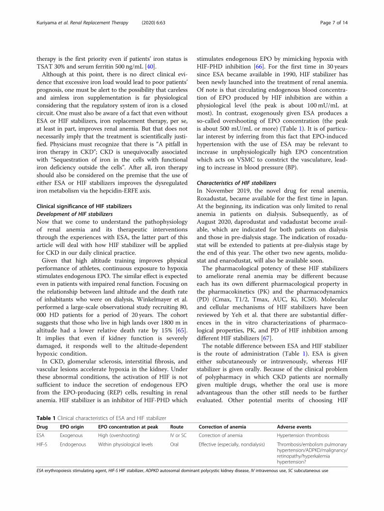
stimulates endogenous EPO by mimicking hypoxia withHIF-PHD inhibition [66]. For the first time in 30 yearssince ESA became available in 1990, HIF stabilizer hasbeen newly launched into the treatment of renal anemia.Of note is that circulating endogenous blood concentra-tion of EPO produced by HIF inhibition are within aphysiological level (the peak is about 100mU/mL atmost). In contrast, exogenously given ESA produces aso-called overshooting of EPO concentration (the peakis about 500 mU/mL or more) (Table 1). It is of particu-lar interest by inferring from this fact that EPO-inducedhypertension with the use of ESA may be relevant toincrease in unphysiologically high EPO concentrationwhich acts on VSMC to constrict the vasculature, lead-ing to increase in blood pressure (BP).
Characteristics of HIF stabilizersIn November 2019, the novel drug for renal anemia,Roxadustat, became available for the first time in Japan.At the beginning, its indication was only limited to renalanemia in patients on dialysis. Subsequently, as ofAugust 2020, daprodustat and vadadustat become avail-able, which are indicated for both patients on dialysisand those in pre-dialysis stage. The indication of roxadu-stat will be extended to patients at pre-dialysis stage bythe end of this year. The other two new agents, molidu-stat and enarodustat, will also be available soon.The pharmacological potency of these HIF stabilizers
to ameliorate renal anemia may be different becauseeach has its own different pharmacological property inthe pharmacokinetics (PK) and the pharmacodynamics(PD) (Cmax, T1/2, Tmax, AUC, Ki, IC50). Molecularand cellular mechanisms of HIF stabilizers have beenreviewed by Yeh et al. that there are substantial differ-ences in the in vitro characterizations of pharmaco-logical properties, PK, and PD of HIF inhibition amongdifferent HIF stabilizers [67].The notable difference between ESA and HIF stabilizer
is the route of administration (Table 1). ESA is giveneither subcutaneously or intravenously, whereas HIFstabilizer is given orally. Because of the clinical problemof polypharmacy in which CKD patients are normallygiven multiple drugs, whether the oral use is moreadvantageous than the other still needs to be furtherevaluated. Other potential merits of choosing HIF
Table 1 Clinical characteristics of ESA and HIF stabilizer
Drug EPO origin EPO concentration at peak Route Correction of anemia Adverse events
ESA Exogenous High (overshooting) IV or SC Correction of anemia Hypertension thrombosis
HIF-S Endogenous Within physiological levels Oral Effective (especially, nondialysis) Thrombosis/embolism pulmonaryhypertension/ADPKD/malignancy/retinopathy/hyperkalemiahypertension?
ESA erythropoiesis stimulating agent, HIF-S HIF stabilizer, ADPKD autosomal dominant polycystic kidney disease, IV intravenous use, SC subcutaneous use
Kuriyama et al. Renal Replacement Therapy (2020) 6:63 Page 7 of 14
stabilizers include probably low cost, improved iron pro-file, and endogenous EPO at levels close to physiologicalrange.Of note is that there are a lot of target genes of HIF
activation (approximately 100 ~ 200 genes); thus, thegene-dependent unwanted pharmacological effects shouldbe taken into consideration. At this point, however, thereis no precise information on the class effects, the drugeffects, and the adverse effects of HIF stabilizers. Thefollowings are the main results of HIF stabilizers obtainedin the phase-2 and phase-3 clinical studies that may pro-vide us some hints to differentiate one from the others.
Daprodustat Based on the phase-2 trials in patients onHD and those at pre-dialysis stage, daprodustat increasedHb levels in a dose-dependent manner compared to theplacebo and the rHuEPO-treated group [8–11]. In a phase-3 trial, Tsubakihara et al. demonstrated that daprodustatsignificantly increased Hb value up to 10 ~ 12 g/dL in theESA-naïve patients. This effect was accompanied by the re-duction in hepcidin, ferritin, and TSAT with a subsequentincrease in TIBC [68]. In addition, Akizawa et al. also foundthat daprodustat increased Hb in a dose-dependent mannerin HD patients having anemia ranging 8.5 ~ 10.5 g/dL(average 9.8 g/dL). Furthermore, this study found thatVEGF remained unchanged, and the circulating endogen-ous EPO remained within a physiological range with a fewexceptional cases whose EPO levels were as high as theones observed in the ESA-treated [10]. Other reportsshowed that daprodustat increased Hb in a dose-dependentmanner in HD patients whose pretreatment average Hbvalue was less than 10 g/dL, and the secondary analysisshowed that VEGF remained unchanged and the endogen-ous EPO concentration was within a physiological range(maximum EPO concentration < 50 mIU/mL). As for ESAresistance, Akizawa et al. found that darbepoetin-αrDA),compared to daprodustat, required a broader range admin-istration dose, suggesting that the ESA hyporesponsivenessis better controlled in the daprodustat-treated group [69].In addition, there was basically no difference in the PK andthe PD between Caucasian and Japanese with a slightly highAUC in the latter, which may be originated from the differ-ence in body size [70]. The protein binding is about 99%,and thus, the drug is not dialyzable. The T1/2 is estimatedto be 1.3 ~ 2.5 h.
Roxadustat The phase-2 and phase-3 randomized clin-ical studies clearly demonstrated that roxadustat waseffective in ameliorating renal anemia in all patientgroups; pre-dialysis, HD, and PD [71–73]. Interestingly,Provenzano et al. compared roxadustat to epoetin-αtoinvestigate how inflammation affects drug responses.They found that the epoetin-α-treated group requiredlarger dosage to maintain Hb levels in patients having
higher CRP, whereas such a dose dependency was notfound in the roxadustat-treated group [6]. Chen et al. re-ported that patients treated with epoetin-α had lowerand slower response to attain the target Hb especially inpatients with higher CRP. In contrast, roxadustat re-quired smaller dosage to attain the target Hb value, irre-spective of CRP levels [74]. These results of roxadustatare suggestive that HIF stabilizer may be more beneficialthan ESA in maintaining the target Hb levels even in thepresence of chronic inflammation. In fact, roxadustatunequivocally reduced hepcidin and ferritin levels [6,72–74]. More interestingly, increase in EPO concentra-tion in response to the treatment was significantly lowerin the roxadustat-treated (maximum EPO concentration< 130 mIU/mL) than in the epoetin-α-treated (max-imum EPO concentration < 700 mIU/mL), suggesting abenefit of choosing the former to prevent, for instance,an abrupt increase in BP due to ESA, so-called EPO-induced hypertension [6].Lowering effects of total- and LDL-cholesterol concen-
tration has been reported in the phase-3 trial [74]. Interms of adverse events, there were less incidence ofhypertension and hyperkalemia compared to therHuEPO-treated [73, 74]. The T1/2 is estimated to be 12~ 15 h. The protein binding is sufficiently high; thus, thedrug is not dialyzable.
Vadadustat A phase-2 study in patients at pre-dialysisstage demonstrated that vadadustat improved renalanemia compared to the placebo [18]. There was a sig-nificant reduction in hepcidin and ferritin accompaniedby an increase in TIBC, while CRP, VEGF, total choles-terol, and BP remain unchanged. Another phase-2 studydemonstrated that Hb levels were well maintained withvadadustat after the pre-treatment with epoetin-αp12.Serum iron was slightly increased; hepcidin, ferritin, andTSAT remained unchanged; and TIBC was increased.No serious adverse events were observed [12]. Note-worthy is that the phase-2 study recruiting 210 patientsat pre-dialysis stage, cystatin-C, was significantly loweredin the vadadustat-treated group than in the placebogroup (vadadustat group 34.9 ng/mL (n = 138) vs. pla-cebo: 298.2 ng/mL (n = 72)), suggesting that vadadustatmay provide a renal protective effect [17]. Whether thisagent contributes to retard the progressive lowering ofresidual renal function in non-dialysis patients needs tobe evaluated. Interestingly, the dose-response curve ofHIF inhibition showed that the %inhibition with vadadu-stat had the most gentle slope among other HIF stabi-lizers, suggesting a slow and mild pharmacologicalaction that may be essential to avoid abrupt increase inHb value in the treatment of renal anemia [67]. The T1/2 of vadadustat is estimated to be 7 ~ 9 h, and the effectis not influenced by HD treatment.
Kuriyama et al. Renal Replacement Therapy (2020) 6:63 Page 8 of 14
Currently, the internationally organized large-scalestudies, the INNO2VATE, and PRO2TECT trials areongoing. The former makes a prompt news, reportingthe non-inferiority of vadadustat to DA with respect tothe effect on anemia correction and the incidence ofmajor cardiovascular events (MACE). The results of theINNO2VATE will be announced sometime soon.
Molidustat In a phase-2 study, Macdougall et al. re-ported that there was a dose-dependent increase in Hblevels in the moridustat-treated group and the effect wasbetter than that in the placebo, the DA-treated, and therHuEPO-treated groups [13]. Notably, patients on dialy-sis required higher dosage. In patients at pre-dialysisstage, molidustat reduced hepcidin, ferritin, and in-creased TIBC. However, in patients on dialysis who werepreviously treated with ESA, the ferritin, TSAT, and ironwere increased instead. There was a slight decrease inLDL cholesterol concentration. As an extension of theformer phase-2 study, Akizawa et al. confirmed thatmolidustat is effective in improving renal anemia in bothpatients on HD and those at pre-dialysis stage [13, 75].Furthermore, hepcidin in the molidustat-treated waslowered more efficiently than that in the DA-treated inpatients at pre-dialysis stage [76]. Although there weresome cases with thrombosis/embolism, their symptomswere all mild. As far as the effect of molidustat on BPlevel, only small number of cases showed mild elevationof BP, all of whom were not clinically problematic. Inaddition, only a few cases with hyperkalemia werereported [13]. Currently, a phase-3 study, named theMIYABI study, is ongoing in Japan which aims at evalu-ating the effect of anemia correction of molidustat inpatients on HD, PD, and those at pre-dialysis stage. Theresult will be announced sometime within this year,2020.
Enarodustat Akizawa et al. investigated the effect ofenarodustat in CKD patients at pre-dialysis stage in adouble-blind placebo phase-2 trial [19]. The studyshowed that enarodustat increased Hb in a dose-dependent fashion. Analyses of iron regulation showedthat ferritin, hepcidin, and TSAT were lowered, whereasTIBC was increased. The incidence of hypertension was8.5% in the enarodustat naïve group and 4.9% in theconversion group (from ESA) group. The prevalence ofhyperkalemia was 3.2% in the naïve group and 5.8% inthe conversion group. No other serious adverse eventswere observed [77]. In another phase-2 study in HDpatients, VEGF remained unchanged throughout thestudy period [19]. The T1/2 of enarodustat is 15 h.The results of phase-3 study were announced in the
American Society of Nephrology (ASN) in 2019, and therelated matters will soon be published. The basic message
in the ASN was the non-inferiority of enarodustat to DAin both patients on dialysis and those at pre-dialysis stage.
Meta-analysis of HIF stabilizersCollecting 12 clinical studies, Zhong et al. summarizedthe effect of HIF stabilizers in the meta-analysis [78]. Ingeneral, the improvement of renal anemia is moreapparent in the pre-dialysis group than in the dialysisgroup. It implies that patients having residual renal func-tion may have a greater capacity to produce endogenousEPO from the kidney in response to HF inhibition treat-ment. Hasse et al. focused on the iron metabolism inCKD patients and summarized that HIF stabilizersreduced ferritin, hepcidin, and TSAT with the concomi-tant increase in TIBC, proposing that these changes are“the class effect of HIF stabilizers” [79].Although the precise comparative study is not available
at this point, whether lowering of hepcidin in patients atpre-dialysis stage is superior to that in dialysis patients ornot is of particular interest. Based on the speculation fromthe abovementioned previous meta-analyses, it appearsthat hepcidin lowering effect is more efficacious in pre-dialysis patients than in those on dialysis [78, 79].
Adverse events of HIF stabilizersUltimately, clinical evaluation of adverse reactionsshould be carried out with a focus on the major cardio-vascular events (MACE). In a pooled phase 3 data of theplacebo-based, double-blind studies consisting of 4270non-dialysis patients, the risks of MACE (death, apo-plexy, and myocardial infarction), MACE+ (MACE + un-stable angina and/or CHF requiring hospitalization), andoverall death were comparable between the roxadustat-treated and the placebo. In a pooled data in 3880 pa-tients on dialysis, the risks of MACE and overall deathwere comparable between the roxadustat-treated andtheαtepoetin-treated. However, the risk of MACE+ inthe roxadustat-treated was significantly lower than thatin the α-epoetin-treated (HR 0.86, 95%CI 0.74 ~ 0.98).Furthermore, in 1526 patients who were newly treatedwith dialysis, the risk of MACE (HR 0.70, 95%CI 0.51 ~0.96) and MACE+ (HR 0.66, 95%CI 0.50 ~ 0.89) werelower in the roxadustat-treated than in theα-epoetin-treated [80].The target genes on HIF activation are estimated as
much as 100 ~ 200, theoretical fear that some of themmay bring unfavorable reactions is inevitable. Suchassumptive events of HIF stabilizers include thrombo-embolic (TA) event, relapse of cancer, hypertension,pulmonary hypertension, worsening of retinopathy,hyperglycemia, and BP elevation [81].Despite theoretical fear, no serious TA events and few
cases of the arterio-venous shunt failure have been re-ported in the treatment with roxadustat [71], daprodustat
Kuriyama et al. Renal Replacement Therapy (2020) 6:63 Page 9 of 14

[10], vadadustat [12, 17, 18], molidustat [13], and enarodu-stat [19]. Another concern is the potential involvement ofincreased VEGF which may induce worsening of diabeticretinopathy, vascular neogenesis of malignant tumor, andprogression of autosomal dominant polycystic kidney dis-ease (ADPKD). Most of the clinical studies suggested thatVEGF was unchanged [9, 10, 18, 77], but there may be aslight elevation at a higher dosage. HIF stabilizer may havean adversely effect on pulmonary hypertension (PH), sincethe prevalence of PH is higher in CKD and the pathogen-esis of PH may have a link to HIF-2. As far as hyperten-sion is concerned, compared to ESA, which is frequentlyassociated with an increase in BP, the net effect of HIFstabilizers on BP appears not so problematic. Theprevalence of hypertension was reported to be 10% in theoverall roxadustat-treated groups [5], and 8.5% in theenarodustat-treated group [77], both of which were notplacebo-based comparisons. Hypertension was found 8%in the vadadustat-treated patients, which was lower thanthe rates in the ESA-treated patients [17]. Interestingly,molidustat lowered BP through the inhibition of RAS inan experimental model [82]. All of the above findings aresupportive that the effect of HIF stabilizers on BP appearsto be neutral. In many animal experiments in general, HIFstabilizers have been proven renal protective [66].Whether there is any association of malignant tumors
with the clinical use of HIF stabilizers is a matter for de-bate. So far, no clinical studies are suggestive such a re-lationship [11, 16–18], and one of the main reasons isthat the observation periods were too short.An experiment using rats showed that the teratogen-
icity was found in the fetus; therefore, one must be alertto the possibility that HIF stabilizer may not indicatedfor pregnant women and those who are willing to bepregnant.Most of the studies are supportive that HIF stabilizers
reduced total and LDL cholesterol levels [10, 13, 15, 73].The effect on total cholesterol and TG was neutral withvadadustat [18].As for drug interactions, the effect of HIF stabilizers
can be weakened with the co-administration of thephosphate binding polymers such as sevelamer and uricacid lowering agent, probenecid. On the contrary, theeffect may be potentiated with the combined use oflipid-lowering agent, statin.
Is HIF stabilizer alternative to ESA?Perspectives on the indications of HIF stabilizersThe issue of whether ESA can be fully or partially re-placed by HIF stabilizer in the future treatment is worthdebating, but at this point, there is no appropriate guide-lines except for the recommendation released by theJapanese Society of Nephrology (JSN) in the 29th ofSeptember 2020 [83]. By taking this newly available
recommendation into consideration and by having whatwe obtained in the clinical practice on ESA thoroughlyreconsidered, we gain a new insight into how to use HIFstabilizers. The followings are our opinion-based recom-mendation on the positive indications of HIF stabilizers(Table 2). There may be 3 major practical indicationswhich are summarized as the following:
1) CRA syndrome aiming at more efficient cardio-renalprotection (especially, at pre-dialysis stage)
2) ESA-resistant anemia or ESA-unresponsiveness(especially, at dialysis stage)
3) MIA syndrome and/or the neighborhood of irondysregulated state (especially, at dialysis stage)
These assumptive proposals listed above, especially [2]and [3], are both closely related to the dysregulation ironmetabolism in CKD. We feel that it is of importance onchoosing HIF stabilizers; evaluation of the iron status isindispensable. Seeking the possible causes for ESA-unresponsiveness is also crucial.We would like to make sure that our listed indications
above do not imply that we only insist the indication of“conversion from ESA therapy to HIF stabilizers ther-apy”. Needless to say, when facing the commencementof renal anemia therapy (either ESA or HIF stabilizernaïve case), either of them in conjunction with an appro-priate iron therapy is a decent option. To fulfil thispurpose, early pharmacological introduction of the abovemay have a significant contribution to prevent boththe CRA syndrome and the accompanying MIA syn-drome [1–4, 23, 24].Aside from the above positive indications, consider-
ation should be paid to avoid the HIF stabilizer-related
Table 2 Positive indications and conditions requiring clinicalconsideration (opinion-based recommendations of HIFstabilizers)
Positive indications
1. ESA resistance or unresponsiveness2. Cardio-renal protection in CRA syndrome3. Iron mobilization in the condition akin to MIAsyndrome
Consideration required
1. Thrombosis and/or embolism2. Malignancy and retinopathy3. Pulmonary hypertension4. ADPKD5. Pregnancy or desire to bear children6. Hyperkalemia, liver dysfunction
Note:The adverse effects of ESA are already well known; however, those of HIFstabilizers have not always been clarified clinically. The adverse effects withHIF stabilizers include expanded interpretations of the ESA-based findingsand/or the inferences based on animal experimentsESA erythropoiesis stimulating agent, MIA malnutrition-inflammation-atherosclerosis, CRA cardiovascular-renal-anemia, ADPKD autosomal dominantpolycystic kidney disease
Kuriyama et al. Renal Replacement Therapy (2020) 6:63 Page 10 of 14
putative adverse events. Most of these unknown clinicaladverse events have not yet been virtually clarified. In-deed, most of them are only suggested in experimentalanimal studies. Such adverse events include thrombosis/embolism, relapse of the latent cancer, worsening of pul-monary hypertension, autosomal dominant polycystickidney disease (ADPKD), and diabetic nephropathy et al.(Table 2). Clinical information on these fearful adverseeffects with HIF stabilizers is scant at this point, sincethe observation period in the most studies are too shortto draw any scientific conclusion. Regarding hyperten-sion, because no serious BP elevation has been found inpatients treated with HIF stabilizers, an insight intowhether ESA-induced hypertension is improved with thealternative use of HIF stabilizers or not is of particularinterest.
Evidence-practice-gapThe Japan Chronic Kidney Disease Database (J-CKD-DB) is a large-scale, nationwide comprehensive clinicaldatabase of patients with CKD. Recruiting 31,082 CKDpatients staging 3–5 in 7 university hospitals. The J-CKD-DB cohort discloses that the rate of Hb levels withoptimal ranges defined as Hb value≥ 11 g/dL withoutESA or 11 <Hb value < 13 g/dL with ESA in the CKDstages 4 and 5 is estimated 51.7%. Notwithstanding thishigh incidence, the utilization of ESA is limited to only12.1%, suggesting that the recognition and treatment ofrenal anemia among physicians in clinical practice is stilllow and inadequate [84]. This evidence-practice-gap canbe solved by introducing early use of either ESA or HIFstabilizer with an appropriate iron management.Finally, our personal opinions about the best way to
treat renal anemia are as the following: (1) start thetreatment with oral iron supplementation, (2) co-administer one of the HIF stabilizers, (3) evaluate theiron status periodically, and (4) adjust their dosagescarefully. By doing so, one can accomplish slow andstable supply of both iron and the effective erythropoi-esis that will be maximally beneficial not only for thecorrection of anemia but for the prevention of CVevents and the dysregulation of iron metabolism.
ConclusionThe novel anti-anemic agent HIF stabilizer has beenevaluated through the clinical lessons of ESA obtainedin the past 3 decades. HIF stabilizer will be primarily in-dicated for CRA syndrome, ESA-resistant anemia, andMIA syndrome. Multiple other factors such as medico-economical consideration, patients’ preference and ad-herence, management of concurrent complications, druginteractions, and unwanted adverse events should fur-ther be taken into consideration.
AbbreviationsAUC: Area under the blood concentration curve; A-V fistula: Arterio-venousfistula; CKD: Chronic kidney disease; Cmax: Maximum drug concentration;eGFR: Estimated glomerular filtration rate; ERI: ESA resistance index;Hb: Hemoglobin; Ht: Hematocrit; IL-6: Interleukin-6; IC50: Half maximal (50%)inhibitory concentration; JSDT: Japanese Society of Dialysis Treatment;JSN: Japanese Society of Nephrology; KDIGO: Kidney Disease ImprovingGlobal Outcomes; Ki: Affinity coefficient of the drug; MDS: Myelodysplasticsyndrome; NYHA: New York Heart Association functional classification;QOL: Quality of life; RCT: Randomized control trial; T1/2: Elimination half-life;Tmax: Time to maximum concentration; TIBC: Total iron binding capacity;TSAT: Transferrin saturation; VSMC: Vascular smooth muscle cells
Authors’ contributionsSK contributed to the overall concept and study design. SK, YM and HHcontributed to the intellectual discussion during manuscript drafting,revision, and the approval of the final version.
FundingNone
Ethics approval and consent to participateNot applicable
Consent for publicationNot applicable
Competing interestsThe authors declare that they have no competing interests.
Author details1Jikei University School of Medicine, 3-25-8, Nishi-shinbashi, Minato-ku, Tokyo105-8471, Japan. 2Nephrology & Hypertension Research Unit, InternalMedicine, Miho Clinic, Shin-Osaki-kangyo Bld 2F, Osaki, Shinagawa-ku, Tokyo141-0032, Japan. 3Division of Nephrology & Hypertension, Department ofInternal Medicine, Jikei University School of Medicine, 3-25-8, Nishishinbashi,Minato-ku, Tokyo 105-8471, Japan. 4Department of Internal Medicine,Division of Nephrology, Showa University School of Medicine, 1-5-8,Hatanodai, Shinagawa-ku, Tokyo 142-8555, Japan.
Received: 7 October 2020 Accepted: 30 November 2020
References1. Parfrey PS, Lauve M, Latremouille-Viau D, Lefebvre P. Erythropoietin therapy
and left ventricular mass index in CKD and ESRD patients: a meta-analysis.Clin J Am Soc Nephrol. 2009;4(4):755–62. https://doi.org/10.2215/CJN.02730608 Epub 2009 Apr 1.
2. Covic A, Nistor I, Donciu MD, Dumea R, Bolignano D, Goldsmith D.Erythropoiesis-stimulating agents (ESA) for preventing the progression ofchronic kidney disease: a meta-analysis of 19 studies. Am J Nephrol. 2014;40(3):263–79. https://doi.org/10.1159/000366025 Epub 2014 Oct 15.
3. Stenvinkel P, Heimburger O, Paultre F, Diczfalusy U, Wang T, Berglund L,et al. Strong association between malnutrition, inflammation, andatherosclerosis in chronic renal failure. Kidney Int. 1999;55:1899–911.
4. Stenvinkel P. Inflammatory and atherosclerotic interactions in the depleteduremic patient. Blood Purif. 2001;19:53–61.
5. Besarab A, Chernyavskaya E, Motylev I, Shutov E, Kumbar LM, Gurevich K,et al. Roxadustat (FG-4592): correction of anemia in incident dialysispatients. J Am Soc Nephrol. 2016;27:1225–33.
6. Provenzano R, Besarab A, Wright S, Dua S, Zeig S, Nguyen P, et al.Roxadustat (FG-4592) versus epoetin alfa for anemia in patients receivingmaintenance hemodialysis: a phase 2, randomized, 6- to 19-week, open-label, active-comparator, dose ranging, safety and exploratory efficacy study.Am J Kidney Dis. 2016;67:912–24.
7. Chen N, Qian J, Chen J, Yu X, Mei C, Hao C, et al. Phase 2 studies of oralhypoxia-inducible factor prolyl hydroxylase inhibitor FG-4592 for treatmentof anemia in China. Nephrol Dial Transplant. 2017;32:1373–86.
8. Holdstock L, Meadowcroft AM, Maier R, Johnson BM, Jones D, Rastogi A,et al. Four-week studies of oral hypoxia-inducible factor-prolyl hydroxylase
Kuriyama et al. Renal Replacement Therapy (2020) 6:63 Page 11 of 14
inhibitor GSK1278863 for treatment of anemia. J Am Soc Nephrol. 2016;27:1234–44.
9. Brigandi RA, Johnson B, Oei C, Westerman M, Olbina G, de Zoysa J, et al. Anovel hypoxia-inducible factor-prolyl hydroxylase inhibitor (GSK1278863) foranemia in CKD: a 28-day, phase 2a randomized trial. Am J Kidney Dis. 2016;67:861–71.
10. Akizawa T, Tsubakihara Y, Nangaku M, Endo Y, Nakajima H, Kohno T, et al.Effects of daprodustat, a novel hypoxia-inducible factor prolyl hydroxylaseinhibitor on anemia management in Japanese hemodialysis subjects. Am JNephrol. 2017;45:127–35.
11. Meadowcroft AM, Cizman B, Holdstock L, Biswas N, Johnson BM, Jones D,et al. Daprodustat for anemia: a 24-week, open-label, randomized controlledtrial in participants on hemodialysis. Clin Kidney J. 2019;12:139–48.
12. Haase VH, Chertow GM, Block GA, Pergola PE, DeGoma EM, Khawaja Z, et al.Effects of vadadustat on hemoglobin concentrations in patients receivinghemodialysis previously treated with erythropoiesis-stimulating agents.Nephrol Dial Transplant. 2019;34:90–9.
13. Macdougall IC, Akizawa T, Berns JS, Bernhardt T, Krueger T. Effects ofmolidustat in the treatment of anemia in CKD. Clin J Am Soc Nephrol. 2019;14:28–39.
14. Besarab A, Provenzano R, Hertel J, Zabaneh R, Klaus SJ, Lee T, et al.Randomized placebo-controlled dose-ranging and pharmacodynamicsstudy of roxadustat (FG-4592) to treat anemia in non-dialysis dependentchronic kidney disease (NDD-CKD) patients. Nephrol Dial Transplant. 2015;30:1665–73.
15. Provenzano R, Besarab A, Sun CH, Diamond SA, Durham JH, Cangiano JL,et al. Oral hypoxia–inducible factor prolyl hydroxylase inhibitor roxadustat(FG-4592) for the treatment of anemia in patients with CKD. Clin J Am SocNephrol. 2016;11:982–91.
16. Holdstock L, Cizman B, Meadowcroft AM, Biswas N, Johnson BM, Jones D, et al.Daprodustat for anemia: a 24-week, open-label, randomized controlled trial inparticipants with chronic kidney disease. Clin Kidney J. 2019;12:129–38.
17. Pergola PE, Spinowitz BS, Hartman CS, Maroni BJ, Haase VH. Vadadustat, anovel oral HIF stabilizer, provides effective anemia treatment in nondialysis-dependent chronic kidney disease. Kidney Int. 2016;90:1115–22.
18. Martin ER, Smith MT, Maroni BJ, Zuraw QC, DeGoma EM. Clinical trial ofvadadustat in patients with anemia secondary to stage 3 or 4 chronickidney disease. Am J Nephrol. 2017;45:380–8.
19. Akizawa T, Nangaku M, Yamaguchi T, Arai M, Koretomo R, Matsui A, et al. Aplacebo-controlled, randomized trial of enarodustat in patients with chronickidney disease followed by long-term trial. Am J Nephrol. 2019;49:165–74.
20. Sender R, Fuchs S, Milo R. Revised estimates for the number of human andbacteria cells in the body. PLoS Biol. 2016;14(8):e1002533. https://doi.org/10.1371/journal.pbio.1002533 eCollection 2016 Aug.
21. Yamamoto H, Nishi S, Tomo T, Masakane I, Saito K, Nangaku M, et al. 2015Japanese Society for Dialysis Therapy: guidelines for renal anemia in chronickidney disease. Ren Rep Ther. 2017;3(36):8–11.
22. Brannon ES, Merrill AJ, Warren JV, Stead EA Jr. The cardiac output inpatients with chronic anemia as measured by the technique of right atrialcatheterization. J Clin Invest. 1945;24(3):332–6.
23. Tanaka T, Eckardt KU. HIF activation against CVD in CKD: novel treatmentopportunities. Semin Nephrol. 2018;38(3):267–76.
24. Silverberg DS, Wexler D, Iaina A. The importance of anemia and itscorrection in the management of severe congestive heart failure. Eur JHeart Fail. 2002;4(6):681–6.
25. Ayus JC, Go AS, Valderrabano F, Verde E, de Vinuesa SG, Achinger SG, et al.Spanish Group for the study of the anemia and left ventricular hypertrophyin pre-dialysis patients. Effects of erythropoietin on left ventricularhypertrophy in adults with severe chronic renal failure and hemoglobin <10 g/dL. Kidney Int. 2005;68(2):788–95.
26. Akaishi M, Hiroe M, Hada Y, Suzuki M, Tsubakihara Y, Akizawa T, KRN321Study Group. Effect of anemia correction on left ventricular hypertrophy inpatients with modestly high hemoglobin level and chronic kidney disease. JCardiol. 2013;62(4):249–56. https://doi.org/10.1016/j.jjcc.2013.04.008 Epub2013 Jun 18.
27. Kuriyama S, Tomonari H, Yoshida H, Hashimoto T, Kawaguchi Y, Sakai O.Reversal of anemia by erythropoietin therapy retards the progression ofchronic renal failure, especially in nondiabetic patients. Nephron. 1997;77:176–85.
28. Gouva C, Nikolopoulos P, Ioannidis JP, Siamopoulos KC. Treating anemiaearly in renal failure patients slows the decline of renal function: a
randomized controlled trial. Gouva C, Nikolopoulos P, Ioannidis JP,Siamopoulos KC. Kidney Int. 2004;66(2):753–60.
29. Tsubakihara Y, Akizawa T, Iwasaki M, Shimazaki R. High hemoglobin levelsmaintained by an erythropoiesis-stimulating agent improve renal survival inpatients with severe renal impairment. Ther Apher Dial. 2015;19(5):457–65.https://doi.org/10.1111/1744-9987.12308 Epub 2015 May 5.
30. Akizawa T, Tsubakihara Y, Hirakata H, Watanabe Y, Hase H, Nishi S, et al.A prospective observational study of early intervention witherythropoietin therapy and renal survival in non-dialysis chronic kidneydisease patients with anemia: JET-STREAM Study. Clin Exp Nephrol.2016;20:885–95.
31. Hayashi T, Maruyama S, Nangaku M, Narita I, Hirakata H, Tanabe K, et al.Darbepoetin alfa in patients with advanced CKD without diabetes:randomized, controlled trial. Clin J Am Soc Nephrol. 2020. https://doi.org/10.2215/CJN.08900719 [Epub ahead of print].
32. Besarab A, Bolton WK, Browne JK, Egrie JC, Nissenson AR, Okamoto DM,et al. The effects of normal as compared with low hematocrit values inpatients with cardiac disease who are receiving hemodialysis and epoetin.N Engl J Med. 1998;339(9):584–90.
33. Drüeke TB, Locatelli F, Clyne N, Eckardt KU, Macdougall IC, Tsakiris D, et al.Normalization of hemoglobin level in patients with chronic kidney diseaseand anemia. N Engl J Med. 2006;355(20):2071–84.
34. Singh AK, Szczech L, Tang KL, Barnhart H, Sapp S, Wolfson M, et al.Correction of anemia with epoetin alfa in chronic kidney disease. N Engl JMed. 2006;355(20):2085–98.
35. Pfeffer MA, Burdmann EA, Chen CY, Cooper ME, de Zeeuw D, Eckardt KU,et al. A trial of darbepoetin alfa in type 2 diabetes and chronic kidneydisease. N Engl J Med. 2009;361(21):2019–32. https://doi.org/10.1056/NEJMoa0907845 Epub 2009 Oct 30.
36. Swedberg K, Young JB, Anand IS, Cheng S, Desai AS, Diaz R, et al. Treatmentof anemia with darbepoetin alfa in systolic heart failure. N Engl J Med. 2013;368:1210–9.
37. Kuragano T, Kitamura K, Matsumura O, Matsuda A, Hara T, Kiyomoto H, et al.ESA hyporesponsiveness is associated with adverse events in maintenancehemodialysis patients, but not with iron storage. PloS One. 2016;11:e0147328.
38. McCullough PA, Barnhart HX, Inrig JK, Reddan D, Sapp S, Patel UD, et al.Cardiovascular toxicity of epoetin-alfa in patients with chronic kidneydisease. Am J Nephrol. 2013;37:549–58.
39. Kanbay M, Perazella MA, Kasapoglu B, Koroglu M, Covic A. Erythropoiesisstimulatory agent-resistant anemia in dialysis patients: review of causes andmanagement. Blood Purif. 2010;29:1–12. https://doi.org/10.1159/000245041.
40. Kidney Disease: Improving Global Outcomes (KDIGO) Anemia WorkingGroup. KDIGO Practice Guideline for anemia in chronic kidney disease.Kidney Int. 2012;suppl 2:279–335.
41. Yamamoto H, Nishi S, Tomo T, Masakane I, Saito K, Nangaku M, et al. 2015Japanese Society for Dialysis Therapy: guidelines for renal anemia in chronickidney disease. Ren Rep Ther. 2017;3(36):11–7.
42. McFarlane SI, Chen SC, Whaley-Connell AT, Sowers JR, Vassalotti JA, SalifuMO, et al. Prevalence and associations of anemia of CKD: Kidney EarlyEvaluation Program (KEEP) and National Health and Nutrition ExaminationSurvey (NHANES) 1999-2004. Am J Kidney Dis. 2008;51(4 Suppl 2):S46–55.https://doi.org/10.1053/j.ajkd.2007.12.019.
43. Go AS, Chertow GM, Fan D, McCulloch CE, Hsu CY. Chronic kidney diseaseand the risks of death, cardiovascular events, and hospitalization. N Engl JMed. 2004;351(13):1296–305.
44. Himmelfarb J, Stenvinkel P, Ikizler TA, Hakim RM. The elephant in uremia:oxidant stress as a unifying concept of cardiovascular disease in uremia.Kidney Int. 2002;62:1524–38.
45. Otaki Y, Nakanishi N, Hasuike Y, Moriguchi M, Nanami Y, Hama M, et al.Defective regulation of iron transporters leading to iron excess in thepolymorphonuclear leukocytes of patients on maintenance hemodialysis.Am J Kidney Dis. 2004;43:1030–9.
46. Nakanishi T, Kuragano T, Nanami M, Nagasawa Y, Nanami M, Hasuike Y.Misdistribution of iron and oxidative stress in chronic kidney disease. FreeRad Biol Med. 2019;133:248–53.
47. Honda H, Kobayashi Y, Onuma S, Shibagaki K, Yuza T, Hirao K, et al.Associations among erythroferrone and biomarkers of erythropoiesis andiron metabolism, and treatment with long-term erythropoiesis-stimulatingagents in patients on hemodialysis. PloS One. 2016;11(3):e0151601. https://doi.org/10.1371/journal.pone.0151601.
Kuriyama et al. Renal Replacement Therapy (2020) 6:63 Page 12 of 14
48. Honda H, Hosaka N, Ganz T, Shibata T. Iron metabolism in chronickidney disease patients. Nakanishi T, Kuragano T (eds): CKD-AssociatedComplications: Progress in the Last Half Century. Contrib Nephrol.2019;198:103–11. https://doi.org/10.1159/000496369.
49. Peyssonnaux C, Zinkernagel AS, Schuepbach RA, Rankin E, Vaulont S, HaaseVH, et al. Regulation of iron homeostasis by the hypoxia inducibletranscription factors (HIFs). J Clin Invest. 2007;117(7):1926–32. https://doi.org/10.1172/JCI31370.
50. Hasuike Y, Nonoguchi H, Tokuyama M, Ohue M, Nagai T, Yahiro M, et al.Serum ferritin predicts prognosis in hemodialysis patients: the Nishinomiyastudy. Clin Exp Nephrol. 2010;14(4):349–55. https://doi.org/10.1007/s10157-010-0288-x Epub 2010 May 14.
51. Karaboyas A, Morgenstern H, Pisoni RL, Raymond Vanholder JZ,Jacobson SH, Inaba M, et al. Association between serum ferritin andmortality: findings from the USA, Japan and European DialysisOutcomes and Practice Patterns Study. Nephrol Dial Transplant. 2018;33:2234–44.
52. Hamano T, Fujii N, Hayashi T, Yamamoto H, Iseki K, Tsubakihara Y.Thresholds of iron markers for iron deficiency erythropoiesis-finding of theJapanese nationwide dialysis registry. Kidney Int Suppl. 2015;5(1):23–32.https://doi.org/10.1038/kisup.2015.6.
53. Shoji T, Niihata K, Fukuma S, Fukuhara S, Akizawa T, Inaba M. Both low andhigh serum ferritin levels predict mortality risk in hemodialysis patientswithout inflammation. Clin Exp Nephrol. 2017;21(4):685–93. https://doi.org/10.1007/s10157-016-1317-1.
54. Robinson BM, Bieber B, Pisoni RL, Port FK. Dialysis Outcomes and PracticePatterns Study (DOPPS): its strengths, limitations, and role in informingpractices and policies. Clin J Am Soc Nephrol. 2012;7(11):1897–905. https://doi.org/10.2215/CJN.04940512 Epub 2012 Oct 25.
55. Maruyama Y, Yokoyama K, Yokoo T, Shigematsu T, Iseki K, Tsubakihara Y.The different association between serum ferritin and mortality inhemodialysis ans peritoneal dialysis patients using Japanese nationwidedialysis survey. PLoS One. 10(11):e0143430. https://doi.org/10.1371/journal.pone.0143430.
56. Mursu J, Robien K, Harnack LJ, Park K, Jacobs DR Jr. Dietary supplementsand mortality rate in older women: the Iowa Women’s Health Study. ArchIntern Med. 2011;171(18):1625–33. https://doi.org/10.1001/archinternmed.2011.445.
57. Rostoker G, Griuncelli M, Loridon C, Magna T, Machado G, Drahi G, et al.Reassessment of iron marker for prediction of dialysis iron overload: an MRIstudy. PLoS One. 2015;10(7):e0132006. Published online 2015 Jul 16. https://doi.org/10.1371/journal.pone.0132006.
58. Ogawa C, Tsuchiya K, Tomosugi N, Kanda F, Maeda K, Maeda T. Low levelsof serum ferritin and moderate transferrin saturation leads to adequatehemoglobin levels in hemodialysis patients, retrospective observationalstudy. PLos ONE. 2017;12(6):e0179608 June 29.
59. Anraku M, Kitamura K, Shintomo R, Takeuchi K, Ikeda H, Nagano J, et al.Effect of intravenous iron administration frequency on AOPP andinflammatory biomarkers in chronic hemodialysis patients: a pilot study. ClinBiochem. 2008;41(14-15):1168–74. https://doi.org/10.1016/j.clinbiochem.2008.07.007 Epub 2008 Jul 28.
60. Yamamoto H, Nishi S, Tomo T, Masakane I, Saito K, Nangaku M, et al. 2015Japanese Society for Dialysis Therapy: guidelines for renal anemia in chronickidney disease. Ren Rep Ther. 2017;3(36):19–25.
61. Macdougall IC, White C, Anker SD, Bhandari S, Farrington K, Kalra PA, et al.Intravenous iron in patients undergoing maintenance hemodialysis. N EnglJ Med. 2019;380:447–58.
62. Macdougall IC, Bhandari S, White C, Anker SD, Farrington K, Kalra PA, et al.Intravenous iron dosing and infection risk in hemodialysis patients: a pre-specified secondary analysis of the PIVOTAL trial. J Am Soc Nephrol. 2020;31:1118–27.
63. Coyne DW, Fishbane S. The value of intravenous iron: beyond the cave ofspeculation. J Am Soc Nephrol. 2020;31:896–7. https://doi.org/10.1681/ASN.2019121340.
64. Kuo KL, Hung SC, Tseng WC, Tsai MT, Liu JS, Lin MH, et al. Association ofanemia and iron parameters with mortality among patients undergoingprevalent hemodialysis in Taiwan: the AIM - HD study. J Am Heart Assoc.2018;7(15):e009206.
65. Winkelmayer WC, Liu J, Brookhart MA. Altitude and all-cause mortality inincident dialysis patients. JAMA. 2009;301(5):508–12. https://doi.org/10.1001/jama.2009.84.
66. Gupta N, Wish JB. Hypoxia-inducible factor prolyl hydroxylase inhibitors: apotential new treatment for anemia in patients with CKD. Am J Kidney Dis.2017;69:815–26.
67. Yeh TZ, Leissing TM, Abboud MI, Thinnes CC, Atasoylu O, Holt-Martyn JP,et al. Molecular and cellular mechanisms of HIF prolyl hydroxylase inhibitorsin clinical trials. Chem Sci. 2017;8:7651–68.
68. Tsubakihara Y, Akizawa T, Nangaku M, Onoue T, Yonekawa T, Matsushita H,et al. A 24-week anemia correction study of daprodustat in Japanesedialysis patients. Therapeutic Apheresis Dialysis. 2019. https://doi.org/10.1111/1744-9987.12962.
69. Akizawa T, Nangaku M, Yonekawa T, Okuda N, Kawamatsu S, Onoue T, et al.Efficacy and safety of daprodustat compared with darbepoetin alfa inJapanese hemodialysis patients with anemia: a randomized, double-blind,phase 3 trial. Clin J Am Soc Nephrol. 2020;15(8):1155–65. https://doi.org/10.2215/CJN.16011219 Epub 2020 Jul 28.
70. Hara K, Takahashi N, Wakamatsu A, Caltabiano S. Pharmacokinetics,pharmacodynamics and safety of single, oral doses of GSK1278863, a novelHIF-prolyl hydroxylase inhibitor, in healthy Japanese and Caucasian subjects.Drug Metab Pharmacokinet. 2015;30(6):410–8. https://doi.org/10.1016/j.dmpk.2015.08.004 Epub 2015 Aug 28.
71. Akizawa T, Iwasaki M, Otsuka T, Reusch M, Misumi T. roxadustat treatmentof chronic kidney disease-associated anemia in Japanese patients not ondialysis: a phase 2, randomized, double-blind, placebo-controlled trial. AdvTher. 2019;36(6):1438–54. https://doi.org/10.1007/s12325-019-00943-4 Epub2019 Apr 5.
72. Akizawa T, Ueno M, Shiga T, Reusch M. Oral roxadustat three timesweekly in ESA-naïve and ESA-converted patients with anemia ofchronic kidney disease on hemodialysis: results from two phase 3studies. Ther Apher Dial. 2019. https://doi.org/10.1111/1744-9987.13468 [Epub ahead of print].
73. Akizawa T, Otsuka T, Reusch M, Ueno M. Intermittent oral dosing ofroxadustat in peritoneal dialysis chronic kidney disease patients withanemia: a randomized, phase 3, multicenter, open-label study. Ther ApherDial 2020;24(2):115-125. doi: https://doi.org/10.1111/1744-9987.12888. Epub2019 Jul 31.
74. Chen N, Hao C, Liu BC, Lin H, Wang C, Xing C, et al. Roxadustattreatment for anemia in patients undergoing long-term dialysis. N EnglJ Med. 2019;381(11):1011–22. https://doi.org/10.1056/NEJMoa1901713Epub 2019 Jul 24.
75. Akizawa T, Macdougall IC, Berns JS, Bernhardt T, Staedtler G, Taguchi M,et al. Long-term efficacy and safety of Molidustat for anemia in chronickidney disease: DIALOGUE extension studies. Am J Nephrol. 2019;49:271–80.
76. Akizawa T, Macdougall IC, Berns JC, Yamamoto H, Taguchi M, Iekushi K,et al. Ion regulation by Molidustat, a daily oral hypoxia inducible factorprolyl hydroxylase inhibitor, in patients with chronic kidney disease.Nephron. 2019;143:243–57.
77. Akizawa T, Hanaki K, Arai M. JTZ-951 an oral novel HIF-PHD inhibitor,elevates hemoglobin in Japanese anemic patients with chronic kidneydisease receiving maintenance hemodiaysis. 52nd ERA-EDTA. London; 2015.
78. Zhong H, Zhou T, Li H, Zhong Z. The role of hypoxia-inducible factorstabilizers in the treatment of anemia in patients with chronic kidneydisease. Drug Des Devel Ther. 2018;12:3003–11. https://doi.org/10.2147/DDDT.S175887 eCollection 2018.
79. Haase VH. HIF-prolyl hydroxylases as therapeutic targets in erythropoiesisand iron metabolism. Hemodial Int. 2017;21(Suppl 1):S110–24. https://doi.org/10.1111/hdi.12567.
80. Roxadustat phase III programme pooled analyses showed positive efficacyand no increases cardiovascular risk in patients with anaemia from chronickidney disease. http://www.astrazeneca.co./mediacentre/press-release/2019/rozadustat-phase-iii-programme-pooled-analyse-showed-positive-efficacy-and-no-increased-cv-risk-in-patients-with-anaemia-from-chronic-kidney-disease.html. (Accessed June 15, 2020).
81. Sanghani NS, Hasse VH. Hypoxia-inducible factor activators in renal anemia:current clinical experience. Adv Chronic Kidney Dis. 2019;26:253–66.
82. Flamme I, Oehme F, Ellinghaus P, Jeske M, Keldenich M, Thuss U. Mimickinghypoxia to treat anemia: HIF-stabilizer BAY 85-3934 (Molidustat) stimulateserythropoietin production without hypertensive effects. Plos OneNovember. 2014;9(11):e111838.
83. Uchida K, Nangaku M, Abe M, Okada H, Takeda N, Hanafusa N, et al.Recommendation on the appropriate use of HIF-PH inhibitors: JapaneseSociety of Nephrology. 2020. (in Japanese).
Kuriyama et al. Renal Replacement Therapy (2020) 6:63 Page 13 of 14
84. Sofue T, Nakagawa N, Kanda E, Nagasu H, Matsushita H, Nangaku M, et al.Prevalence of anemia in patients with chronic kidney disease in Japan: anationwide, cross-sectional cohort study using data from the Japan ChronicKidney Disease Database (JCKD-DB). Plos One. 2020;15(7):e0236132. https://doi.org/10.1371/journal.pone.0236132.
Publisher’s NoteSpringer Nature remains neutral with regard to jurisdictional claims inpublished maps and institutional affiliations.
Kuriyama et al. Renal Replacement Therapy (2020) 6:63 Page 14 of 14