a long road towards the structure of respiratory complex i, a giant molecular proton pump
TRANSCRIPT

Bioenergetics in Mitochondria, Bacteria and Chloroplasts 1265
A long road towards the structure of respiratorycomplex I, a giant molecular proton pumpLeonid A. Sazanov*1, Rozbeh Baradaran*, Rouslan G. Efremov*2, John M. Berrisford*3 and Gurdeep Minhas**Medical Research Council Mitochondrial Biology Unit, Wellcome Trust/MRC Building, Hills Road, Cambridge CB2 0XY, U.K.
AbstractComplex I (NADH:ubiquinone oxidoreductase) is central to cellular energy production, being the first andlargest enzyme of the respiratory chain in mitochondria. It couples electron transfer from NADH to ubiquinonewith proton translocation across the inner mitochondrial membrane and is involved in a wide range ofhuman neurodegenerative disorders. Mammalian complex I is composed of 44 different subunits, whereasthe ‘minimal’ bacterial version contains 14 highly conserved ‘core’ subunits. The L-shaped assembly consistsof hydrophilic and membrane domains. We have determined all known atomic structures of complex I,starting from the hydrophilic domain of Thermus thermophilus enzyme (eight subunits, nine Fe–S clusters),followed by the membrane domains of the Escherichia coli (six subunits, 55 transmembrane helices) andT. thermophilus (seven subunits, 64 transmembrane helices) enzymes, and finally culminating in a recentcrystal structure of the entire intact complex I from T. thermophilus (536 kDa, 16 subunits, nine Fe–S clusters,64 transmembrane helices). The structure suggests an unusual and unique coupling mechanism via long-range conformational changes. Determination of the structure of the entire complex was possible onlythrough this step-by-step approach, building on from smaller subcomplexes towards the entire assembly.Large membrane proteins are notoriously difficult to crystallize, and so various non-standard and sometimescounterintuitive approaches were employed in order to achieve crystal diffraction to high resolution andsolve the structures. These steps, as well as the implications from the final structure, are discussed in thepresent review.
IntroductionComplex I (NADH:ubiquinone oxidoreductase, EC 1.6.5.3)plays a central role in the respiratory chain in mitochondriaand many bacteria [1–5]. It catalyses the exergonic transferof two electrons from NADH to ubiquinone, coupled to thetranslocation of four protons (current consensus value [6–8])across the bacterial or inner mitochondrial membrane, in thefollowing reaction:
NADH + H++Q + 4H+in → NAD++QH2+4H+
out
In total, the transfer of two electrons from NADH tooxygen, through complexes I, III (bc1) and IV (cytochromec oxidase), results in the translocation of ten protons acrossthe membrane [9]. Complex I thus contributes approximately40% of the proton flux which creates the pmf (protonmotiveforce) for the synthesis of ATP by ATP synthase [10].Complex I is a reversible machine [11], able to utilize pmfand ubiquinol to reduce NAD+ .
Mutations in complex I subunits lead to the most commonhuman neurodegenerative diseases [4,12]. The enzyme is also
Key words: bioenergetics, complex I, membrane protein structure, NADH:ubiquinone
oxidoreductase, respiratory chain, X-ray crystallography.
Abbreviations used: ASU, asymmetric unit; CMC, critical micellar concentration; CYMAL-
7, 7-cyclohexyl-1-heptyl-β-d-maltoside; DDM, dodecyl maltoside; MAD, multi-wavelength
anomalous dispersion; MR, molecular replacement; NDSB, non-detergent sulfobetaine; OMF,
octyl maltoside fluorinated; pmf, protonmotive force; SeMet, selenomethionine; TDM, tridecyl
maltoside; TM, transmembrane; UDM, undecyl maltoside.1To whom correspondence should be addressed (email [email protected]).2Present address: VIB Department of Structural Biology, Vrije Universiteit Brussel, Pleinlaan 2,
1050 Brussels, Belgium.3Present address: European Bioinformatics Institute, Cambridge, CB10 1SD, U.K.
a major source of reactive oxygen species in mitochondria[13], which can lead to mitochondrial DNA damage,implicated in Parkinson’s disease [14] and aging [15].
Complex I is one of the largest known membraneprotein assemblies. Mammalian mitochondrial complex Iconsists of 44 different subunits (∼980 kDa in total) [16,17].The simpler prokaryotic enzyme normally consists of 14‘core’ subunits (∼550 kDa total), conserved from bacteriato humans [1,2,4,18,19] with nearly identical arrangementand folds in both prokaryotic and eukaryotic enzymes [20].Both enzymes contain equivalent redox components andhave a similar L-shaped structure, formed by the hydrophilic(peripheral) and membrane domains, of roughly equal sizewith seven subunits in each [2,4,21]. The high conservationof core subunits suggests that the mechanism of complex Iis conserved throughout all species, therefore we can use thebacterial enzyme as a ‘minimal’ model of human complex.
Towards the structure, building fromblocksWe started our work on the structure of complex Iapproximately 15 years ago, long after some other researchgroups. Initially, we attempted crystallization of complexI from Escherichia coli. We have optimized for yieldand activity the preparation from wild-type cells (i.e. nooverexpression or affinity tags, as with all our preparationsfrom other species so far). As with all our preparationsintended for crystallization, we first check the stability of
Biochem. Soc. Trans. (2013) 41, 1265–1271; doi:10.1042/BST20130193 C©The Authors Journal compilation C©2013 Biochemical SocietyBio
chem
ical
So
ciet
y T
ran
sact
ion
s
ww
w.b
ioch
emso
ctra
ns.
org

1266 Biochemical Society Transactions (2013) Volume 41, part 5
the complex in a range of detergents by incubating it overa period of several days at room temperature (preferable to4◦C), followed by analytical gel-filtration chromatography.The eluted fractions are then analysed by SDS/PAGE forsubunit content and checked for oxidoreductase activity. E.coli complex I fulfilled the stability conditions and produced2D crystals after reconstitution with lipids [22], resulting in 8A (1 A = 0.1 nm) resolution projection maps [23]. However,no well-diffracting 3D crystals were obtained.
Thermus thermophilus hydrophilic domainWe therefore turned to the enzyme from T. thermophilus, asit was expected to be more stable. It has not been purifiedpreviously, and our initial preparations using DDM (dodecylmaltoside) as a detergent resulted in preparation of only thehydrophilic domain, with the loss of the membrane domainearly in the purification [24]. However, the hydrophilicdomain on its own was extremely stable; it was necessaryto boil it in SDS in order to separate subunits [24]. Tobe monodisperse, the domain still required the presenceof detergent due to a hydrophobic patch at the interfacewith the membrane domain and so we exchanged thedetergent into the small-micelle octyl glucoside, to maximizechances of crystallization. Initial trials using commercialscreens (sitting drops in 96-well plates) led to several hitsin high-molecular-mass PEGs. These were optimized in 24-well plates, leading to large plate-like crystals of up to0.5 mm × 0.3 mm × 0.03 mm in size (Figure 1A). Crystalsdiffracted X-rays to 3.3 A resolution and belonged to spacegroup P21. The ASU (asymmetric unit) contained four com-plexes, with a total molecular mass of 1120 kDa, including36 Fe–S clusters. Anomalous signal from these intrinsic ironatoms was used for phasing. Because crystals were thin inone dimension, they were prone to radiation damage due toa small volume of the crystal exposed to the beam. This wasespecially the case at iron peak absorption wavelength, andso normally only one wavelength dataset could be collectedfrom each crystal. Despite the large number of iron atoms,a very large protein mass in the ASU meant that anomaloussignal was relatively weak, with maximal cross-correlationof approximately 0.35 at low resolution. Nevertheless, acombination of SHELXD/SOLVE/SHARP/DM steps initerative manner [25] allowed us to locate all the clusters.Even with weak phases (due to large protein mass), it waspossible to obtain good quality electron density after carefulrefinement of heavy atom parameters in SHARP and densitymodification. It was important to refine positions of heavyatoms to relatively low resolution (∼6 A), where the signalwas still significant, and then use these positions for phasing tomaximal resolution. Density modification was performed inDM (or DMmulti for some of our subsequent structures) withsome extreme extension of resolution from approximately 6–8 A to the resolution limit (3.3 A) in 1000 cycles, using NCS.
The structure was solved using additional data from twoheavy atom derivatives [26]. It shows (Figures 1A and 2A)how subunits related to various smaller redox proteins wereput together by Nature to provide a uniquely long (∼95 A)
electron transfer pathway from NADH to the primaryelectron acceptor FMN and through the seven conservedFe–S clusters to the Q-site (quinone-binding site) at theinterface with the membrane domain. Apart from sevenconserved ‘core’ subunits, an additional subunit specificto thermophiles, with a frataxin-like fold, was present inthe structure. It may stabilize the domain and/or helpto regenerate nearby clusters. Later, we determined thestructures of the hydrophilic domain reduced by NADHand/or dithionite under anaerobic conditions and improvedthe overall resolution to 3.1 A, leading to the improvedmodel in some surface-exposed areas [27]. We observedconformational changes at the interface with the membranedomain, which appear to be driven by the nearby Fe–S clusterN2, co-ordinated by the unusual tandem cysteine motif, andcould represent a part of the catalytic cycle [27].
Architecture of the intact T. thermophiluscomplexThe next step in our quest was to crystallize the intactT. thermophilus complex. Two major modifications to thepurification procedure allowed us to obtain the intactenzyme. First, we performed all steps at room temperature.Previously, similarly to our preparations of complex I fromother species, we used 4◦C to try to keep the complex stable.However, we reasoned that, for a thermophile, this may be toolow; hydrophobic interactions will be stronger at the elevatedtemperature and perhaps keep the two domains together.Secondly, DDM was replaced by a milder detergent, TDM(tridecyl maltoside). This led to improved stability of theintact complex, so that it could be prepared in sufficientamounts. Trials against commercial and custom screensidentified several crystallization conditions with protein inTDM or exchanged on small anion-exchange columns intoDDM, UDM (undecyl maltoside), OMF (octyl maltos-ide fluorinated) or CYMAL-7 (7-cyclohexyl-1-heptyl-β-D-maltoside). Most conditions involved high-molecular-massPEG (∼4000 kDa) as a precipitant and resulted in similarly-looking thin rod-like crystals in P21 space group (Figure 1D).Crystallization in the presence of E. coli polar lipids led tobulky cuboid crystals in P212121 space group, but despitemany attempts of optimization, including different lipidmixtures, the resolution was limited to approximately 8A [21]. Plate-like crystals were observed with PEG 400as precipitate, but it was not possible to improve theirdiffraction beyond ∼7 A resolution. Many rounds ofoptimization involved protein exchange into new primarydetergent and addition of different secondary detergent, tomodify micelle properties. A wide range of small-moleculeadditives was explored, including different NDSBs (non-detergent sulfobetaines). In the end, TDM was found tobe the best primary detergent, whereas several detergentscould be used as a secondary [OMF, DDM, CYMAL-4 (4-cyclohexyl-1-heptyl-β-D-maltoside), Fos-Choline-8 (n-octylphosphocholine) fluorinated, etc.], at approximately 1×CMC (critical micellar concentration). They were alwaysused in subsequent set-ups [28] as a secondary detergent
C©The Authors Journal compilation C©2013 Biochemical Society

Bioenergetics in Mitochondria, Bacteria and Chloroplasts 1267
Figure 1 Crystals, diffraction patterns and structures (from left to right)
(A) Hydrophilic domain of T. thermophilus complex I [26]. (B) Membrane domain of E. coli complex I [29]. (C) Membrane
domain of T. thermophilus complex I [28]. (D) Entire T. thermophilus complex I [28]. Crystal images are at different scales
and in (D) the inset shows a loop with a crystal, in which the line dividing the two twin domains can be seen by eye.
Diffraction patterns show enlarged high-resolution areas. Each subunit in the structures is coloured differently.
screen along with various PEG concentrations. Only rod-like crystals could be optimized to diffract to relativelyhigh resolution (initially 4.5 A [21]). However, they werealways pseudo-merohedrally twinned [28], which hamperedstructure solution by experimental phasing and even bymolecular replacement due to model bias. Therefore anadditional route towards the structure was necessary.
In parallel with this work, we pursued crystallizationof the membrane domain of E. coli complex I. It wasseparated from the intact complex by incubation in 0.4 MMgCl2. Optimization of initial hits again involved selection
of primary and secondary detergent (initially DDM plusheptyl glucoside [21]) and optimization of additives, withPEG 4000 as the main precipitant. Additional dimensionof complexity in this search was due to necessity of lipidsfor crystallization, which were optimized as a 3:1 mixtureof DMPC (dimyristoyl phosphatidylcholine) and E. colipolar lipids. Crystals (P212121 space group) were relativelychunky of a brick-like shape up to approximately 50 μm ×70 μm × 400 μm (Figure 1B), but initially diffracted to only∼7 A resolution. Attempts at optimizing the diffraction bydehydration in the drop or using the dehydration machine
C©The Authors Journal compilation C©2013 Biochemical Society
1268 Biochemical Society Transactions (2013) Volume 41, part 5
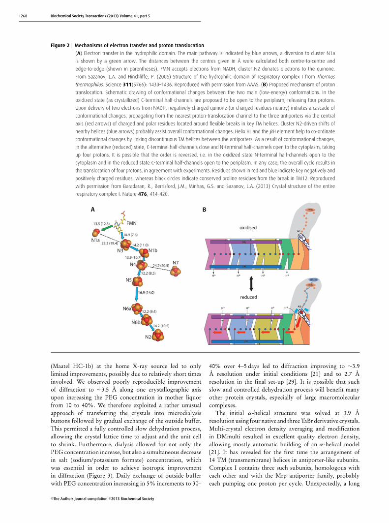
Figure 2 Mechanisms of electron transfer and proton translocation
(A) Electron transfer in the hydrophilic domain. The main pathway is indicated by blue arrows, a diversion to cluster N1a
is shown by a green arrow. The distances between the centres given in Å were calculated both centre-to-centre and
edge-to-edge (shown in parentheses). FMN accepts electrons from NADH, cluster N2 donates electrons to the quinone.
From Sazanov, L.A. and Hinchliffe, P. (2006) Structure of the hydrophilic domain of respiratory complex I from Thermus
thermophilus. Science 311(5766): 1430–1436. Reproduced with permission from AAAS. (B) Proposed mechanism of proton
translocation. Schematic drawing of conformational changes between the two main (low-energy) conformations. In the
oxidized state (as crystallized) C-terminal half-channels are proposed to be open to the periplasm, releasing four protons.
Upon delivery of two electrons from NADH, negatively charged quinone (or charged residues nearby) initiates a cascade of
conformational changes, propagating from the nearest proton-translocation channel to the three antiporters via the central
axis (red arrows) of charged and polar residues located around flexible breaks in key TM helices. Cluster N2-driven shifts of
nearby helices (blue arrows) probably assist overall conformational changes. Helix HL and the βH element help to co-ordinate
conformational changes by linking discontinuous TM helices between the antiporters. As a result of conformational changes,
in the alternative (reduced) state, C-terminal half-channels close and N-terminal half-channels open to the cytoplasm, taking
up four protons. It is possible that the order is reversed, i.e. in the oxidized state N-terminal half-channels open to the
cytoplasm and in the reduced state C-terminal half-channels open to the periplasm. In any case, the overall cycle results in
the translocation of four protons, in agreement with experiments. Residues shown in red and blue indicate key negatively and
positively charged residues, whereas black circles indicate conserved proline residues from the break in TM12. Reproduced
with permission from Baradaran, R., Berrisford, J.M., Minhas, G.S. and Sazanov, L.A. (2013) Crystal structure of the entire
respiratory complex I. Nature 476, 414–420.
(Maatel HC-1b) at the home X-ray source led to onlylimited improvements, possibly due to relatively short timesinvolved. We observed poorly reproducible improvementof diffraction to ∼3.5 A along one crystallographic axisupon increasing the PEG concentration in mother liquorfrom 10 to 40%. We therefore exploited a rather unusualapproach of transferring the crystals into microdialysisbuttons followed by gradual exchange of the outside buffer.This permitted a fully controlled slow dehydration process,allowing the crystal lattice time to adjust and the unit cellto shrink. Furthermore, dialysis allowed for not only thePEG concentration increase, but also a simultaneous decreasein salt (sodium/potassium formate) concentration, whichwas essential in order to achieve isotropic improvementin diffraction (Figure 3). Daily exchange of outside bufferwith PEG concentration increasing in 5% increments to 30–
40% over 4–5 days led to diffraction improving to ∼3.9A resolution under initial conditions [21] and to 2.7 Aresolution in the final set-up [29]. It is possible that suchslow and controlled dehydration process will benefit manyother protein crystals, especially of large macromolecularcomplexes.
The initial α-helical structure was solved at 3.9 Aresolution using four native and three TaBr derivative crystals.Multi-crystal electron density averaging and modificationin DMmulti resulted in excellent quality electron density,allowing mostly automatic building of an α-helical model[21]. It has revealed for the first time the arrangement of14 TM (transmembrane) helices in antiporter-like subunits.Complex I contains three such subunits, homologous witheach other and with the Mrp antiporter family, probablyeach pumping one proton per cycle. Unexpectedly, a long
C©The Authors Journal compilation C©2013 Biochemical Society

Bioenergetics in Mitochondria, Bacteria and Chloroplasts 1269
Figure 3 Improvement of diffraction properties by dehydration in
microdialysis buttons
Crystals of the membrane domain of E. coli complex I (from initial
conditions [21]) were transferred into microdialysis buttons (illustrated)
and the external buffer was exchanged daily, with increasing PEG 4000
concentrations and decreasing salt concentrations in various orders, as
indicated by arrows. The optimal process was to change both of these
parameters simultaneously (diagonal arrow). Anisotropic diffraction
limits (in Å along the a*, b* and c* crystallographic axes) achieved
after each treatment are shown.
amphipathic helix was found to straddle nearly the entiredomain, joining the antiporter subunits together. It wastempting to suggest that it may also drive conformationalchanges in analogy to a coupling rod from a steam engine,although accumulating structural and functional evidenceindicates that the main driving force may be the centralhydrophilic axis ([28] and see below). A similar helix wasobserved in the low-resolution (∼6 A) X-ray analysis ofcomplex I from Yarrowia lipolytica [30]. We then usedthe E. coli backbone structure for molecular replacementto determine the architecture of the entire T. thermophilusenzyme, at 4.5 A resolution [21]. One of the major surpriseswas the location of Fe–S cluster N2, which donates electronsto quinone: it is approximately 25–30 A away from themembrane surface, suggesting that, unprecedentedly, quinonehas to come out of the membrane in order to be reduced.
Structure of E. coli membrane domainThe next step was to determine the atomic structure ofthe membrane domain of the E. coli complex I [29].Here, the crucial improvement came from identifying adifferent detergent mixture: CYMAL-7 as primary detergentwith cholate and NDSB-256 or nonyl glucoside as thesecondary/tertiary detergents. In this case, crystals formed inspace group P1 with two molecules per ASU and diffractedstrongly, up to 2.7 A resolution (anisotropically, with thecomplete dataset collected to 3.0 A). This was achievedonly after dehydration in microdialysis buttons as describedabove. In order to solve the structure an additional phasingfrom SeMet (selenomethionine) derivatives was essential. Thepositions of selenium atoms were also essential markers inderiving the fold, as there were no homologues of any ofthe subunits in the PDB. For SeMet labelling, a feedback-inhibition method [31] was applied during wild-type cellgrowth on minimal medium with 0.6% malate, optimized for
maximal complex I yield. Despite the challenges presentedby anomalous data collection in the P1 space group, severalselenium MAD (multi-wavelength anomalous dispersion)datasets were obtained using a low intensity beam and 360–720◦ rotations of the crystal. Positions of 160 seleniumatoms in the ASU were determined using phases calculatedwith a previously built polyalanine backbone model. Datafrom several native, selenium and TaBr P1 and P212121
datasets were then used in DMmulti to produce excellentquality electron density. A solvent-flipped map calculatedin CNS (also with extreme phase extension) using only theselenium MAD dataset was also of good quality, with betterside-chain density for some residues. The whole processfrom initial purification attempts to structure solution was amajor undertaking: ∼80 000 crystallization conditions tested,∼1000 crystals frozen and tested at the synchrotrons, and ∼80datasets collected.
The structure revealed a novel fold for the antiportersubunits, with two inverted pseudo-screw symmetry-related5-TM repeats containing half of a proton channel each. Thetwo halves are connected in the middle of the membrane,forming a single channel. Uniquely, lysine residues ratherthan the usual carboxy groups appear to be the centralprotonable residues of the proton pumps.
Structure of the entire T. thermophilus complexIn the meantime, crystals of the intact T. thermophiluscomplex were optimized to diffract up to 3.2 A resolution.Two key developments were necessary for this improvement,both rather counterintuitive. First, we increased the primarydetergent (TDM) concentration in protein solution toapproximately 4% (w/v). This is very high compared withthe common notion of using detergent for crystallizationof membrane proteins at approximately 1–2× CMC. Ithas to be said that for all of our crystallizations ofthe membrane domain or the entire complex, we havea high starting concentration of detergent as a result ofconcentrating the protein after the final gel-filtration column.Because the detergent concentrates even on a 100 kDacut-off concentrator, usually the final concentration ofdetergent in ∼20 mg/ml protein solution is, as we determined,approximately 1–1.5%. Colorimetric assays show that theratio of complex I to bound detergent is ∼4:1 (w/w), whichstill leaves at least 0.5% free detergent; much higher than theCMC. However, when we tried to reduce this free detergentconcentration in the protein solutions, crystallization wasnegatively affected or crystals did not appear. One possibilityis that, to ensure full solubility of such large hydrophobicproteins, an excess of free detergent is needed (that is alsothe case on gel-filtration columns, which we run at up to0.5% detergent in the buffer). When we increased detergentconcentration in the intact complex I solutions in steps toeven higher values, crystals were bigger (rods became thicker)with the optimum at approximately 4% TDM. At theseconcentrations, we start to approach the phase-separationboundary, and so it is possible that increased local proteinconcentrations lead to bigger crystals. The second important
C©The Authors Journal compilation C©2013 Biochemical Society
1270 Biochemical Society Transactions (2013) Volume 41, part 5
development was to decrease PEG 4000 concentration inharvest solutions to ∼9% (w/v), even though the crystalsgrew against 19–24% PEG. Unlike E. coli membrane domaincrystals, these crystals do not tolerate dehydration by anyof the methods. When we used 20% or higher PEGconcentrations in earlier harvesting, crystals often startedto bend and diffraction was hit-and-miss. The reason isthat protein solution contains 25% glycerol, and so afterequilibration against the reservoir, the drop becomes biggerthan expected due to the hygroscopic nature of glycerol. As aresult, the final PEG concentration in the drop is much lowerthan expected. We found that 9% PEG in the harvest solutionwas optimal to mimic the conditions in the drop, preservingthe shape and diffraction properties of the crystals. (As a sidenote, attempts to match glycerol in the protein/drop and inthe reservoir solutions did not lead to any crystal growth.)
Since these crystals are nearly perfectly twinned, due tomodel bias it was problematic to use the relatively distal E.coli membrane domain model (∼30–35% sequence similarity)for the structure solution. We therefore crystallized theisolated membrane domain from T. thermophilus complexI. The intact complex was split at low pH (4.0), removingsubunits Nqo1–Nqo3 and Nqo15. The remaining hydro-philic subunits were lost upon crystallization. Conditions,identified after many rounds of optimization, included∼1% UDM as the primary detergent and 5 mM CHAPSas the secondary detergent, with PEG 300 as precipitant.Crystals were rectangular in shape (∼100 μm×100 μm×500–700 μm) and diffracted to ∼3.3 A resolution (Figure 1C),with relatively minor improvements upon dehydration byaddition of 30% PEG 3350 into the harvest solution. Otherdehydration approaches did not improve the diffractionfurther. These crystals were not twinned and formed in P1space group with packing resembling that of the E. colimembrane domain. They contained subunit Nqo8, in contrastwith E. coli, where the corresponding subunit (NuoH) foundat the interface of the two main domains was lost uponcrystallization. Thus the T. thermophilus membrane domainstructure contained the full complement of seven subunitsand 64 TM helices. Initial attempts at the structure solutionby MR (molecular replacement) with the E. coli model wereproblematic, mostly due to significant shifts in the positionsof some TM helices, especially in the distal antiportersubunit Nqo12. We therefore used the Rosetta_MR methodin PHENIX for one cycle, which shifted most helices intotheir positions (further cycles in Rosetta resulted in toodivergent models, possibly due to limited resolution of thedata). This brought Rfree down from the initial 50% to 46%and allowed further successful refinement in CNS usingDEN (Deformable Elastic Network) restrains (or Refmacwith Jelly body restrains) to Rfree of 36–37%. This (anddensity modification in DM) led to improved electron densityso that subunit Nqo8 could be modelled and the structurerebuilt/refined.
We then used this new structure of the membrane domainto solve the structure of the entire complex by MR, togetherwith the previous hydrophilic domain structure (PDB code
3I9V). Some of the features at the interface of the two maindomains were not well resolved since the conformation herechanges upon connection/disconnection of the domains. Itwas important therefore to obtain the iron peak wavelengthdatasets from two rare crystals with relatively low twinfraction (∼0.4). Jelly body refinement with iron SAD (single-wavelength anomalous dispersion) target function in Refmacresulted in a much improved electron density for missingfeatures, including a novel hydrophilic subunit Nqo16. Thisprotein is essential for crystal contacts, but is not neededfor activity and may play the role of an assembly factor.Understandably, arriving at the structure of entire complextook even more effort than for individual subcomplexes, with∼3000 crystals tested at the synchrotrons and ∼200 datasetscollected. The final structure contains 4780 residues, nine Fe–S clusters and one FMN molecule, and, with 64 TM helices, isthe largest (asymmetric) membrane protein complex solvedto date.
Conclusions and implications for themechanism of complex ISeveral common themes emerge from our experience withcrystallization of large membrane protein complexes. (i) Itis usually necessary to use a mixture of a primary anda secondary or even a tertiary detergent as an additive,presumably to optimize properties of the micelle and makeit compatible with crystal contacts. The primary detergentmust be relatively mild so that it preserves the fragileprotein complex intact (this should be tested by gel-filtrationchromatography). The concentration of this detergent (free)in the protein solution does not have to be near its CMC; onthe contrary, it can be beneficial to have it at approximately1–2%. The secondary/tertiary detergent can be quite harsh(but not necessarily) and can be used at approximately 1×CMC (although higher and lower concentrations can andshould be tested). (ii) The addition of native or artificiallipids to the protein can be beneficial in some cases, butnot in others, and has to be evaluated on a case-by-casebasis, including the optimization of the lipid mixture. (iii)Dehydration of crystals either by a common method (inthe drop by increasing precipitant concentrations or byusing a free mounting system on a beamline) or by themicrodialysis method that we described can significantlyimprove the diffraction properties. This appears to work bestif the initial diffraction is poor (∼6–8 A resolution) and maynot help if it is already reasonable (by the membrane proteinstandards, i.e. 3–4 A resolution). (iv) For crystal harvesting(if not dehydrating), it is important to match exactly theconditions in the drop; they may not be as expected dueto glycerol and other compounds from the protein solution.When cryoprotectant needed to be added, with our samples25% ethylene glycol was consistently a better performerthan glycerol. (v) Modern software and careful refinementof heavy atom parameters, combined with aggressive densitymodification/extension, can lead to high-quality electron-density maps even from weak anomalous/isomorphoussignal. It is important to use NCS and all available sources of
C©The Authors Journal compilation C©2013 Biochemical Society
Bioenergetics in Mitochondria, Bacteria and Chloroplasts 1271
phasing information, which can be combined by multi-crystaldensity averaging/modification.
The structures revealed several remarkable features uniqueto complex I, likely to be related to the unusual mechanism ofcoupling between electron transfer and proton translocation.These include: a long, enclosed and out-of-membranequinone reaction chamber; central flexible hydrophilic axisof charged and polar residues (sitting on the breaks in TMhelices) extending in the middle of the membrane from thequinone cavity all the way to the tip of the membranedomain; four copies of proton channels each formed fromtwo connected half-channels (three in antiporters and one atthe interface with the hydrophilic domain); lysine residuesrather than carboxy groups as central residues in antiporters;connecting elements on the both sides of the membranedomain, etc. The mechanism which we propose (Figure 2B)involves long-range conformational changes in the membranedomain, driven, through the central hydrophilic axis, by theredox reactions around cluster N2 and electrostatic inter-actions with a negatively charged area around the quinoneheadgroup. In this two-state (one stroke) mechanism, thehalf-channels would be open to the opposite sides of themembrane in the oxidized and reduced states of the enzyme.Thus the next challenge is to crystallize the reduced form ofthe complex, in order to verify these proposals.
Acknowledgements
We thank the European Synchrotron Radiation Facility (Grenoble),
the Swiss Light Source (Villigen) and the Diamond Light Source
(Didcot) for the provision of synchrotron radiation facilities.
Funding
This work was funded by the Medical Research Council.
References1 Walker, J.E. (1992) The NADH–ubiquinone oxidoreductase (complex I) of
respiratory chains. Q. Rev. Biophys. 25, 253–3242 Yagi, T. and Matsuno-Yagi, A. (2003) The proton-translocating
NADH–quinone oxidoreductase in the respiratory chain: the secretunlocked. Biochemistry 42, 2266–2274
3 Brandt, U. (2006) Energy converting NADH:quinone oxidoreductase(complex I). Annu. Rev. Biochem. 75, 69–92
4 Sazanov, L.A. (2007) Respiratory complex I: mechanistic and structuralinsights provided by the crystal structure of the hydrophilic domain.Biochemistry 46, 2275–2288
5 Sazanov, L.A. (ed.) (2012) A Structural Perspective on RespiratoryComplex I: Structure and Function of NADH:Ubiquinone Oxidoreductase,Springer, New York
6 Galkin, A.S., Grivennikova, V.G. and Vinogradov, A.D. (1999) H+ /2e−
stoichiometry in NADH–quinone reductase reactions catalyzed by bovineheart submitochondrial particles. FEBS Lett. 451, 157–161
7 Galkin, A., Drose, S. and Brandt, U. (2006) The proton pumpingstoichiometry of purified mitochondrial complex I reconstituted intoproteoliposomes. Biochim. Biophys. Acta 1757, 1575–1581
8 Ripple, M.O., Kim, N. and Springett, R. (2013) Mammalian complex Ipumps 4 protons per 2 electrons at high and physiological proton motiveforce in living cells. J. Biol. Chem. 288, 5374–5380
9 Moser, C.C., Farid, T.A., Chobot, S.E. and Dutton, P.L. (2006) Electrontunneling chains of mitochondria. Biochim. Biophys. Acta 1757,1096–1109
10 Watt, I.N., Montgomery, M.G., Runswick, M.J., Leslie, A.G. and Walker, J.E.(2010) Bioenergetic cost of making an adenosine triphosphate moleculein animal mitochondria. Proc. Natl. Acad. Sci. U.S.A. 107, 16823–16827
11 Vinogradov, A.D. (1998) Catalytic properties of the mitochondrialNADH–ubiquinone oxidoreductase (complex I) and the pseudo-reversibleactive/inactive enzyme transition. Biochim. Biophys. Acta 1364,169–185
12 Schapira, A.H. (1998) Human complex I defects in neurodegenerativediseases. Biochim. Biophys. Acta 1364, 261–270
13 Murphy, M.P. (2009) How mitochondria produce reactive oxygenspecies. Biochem. J. 417, 1–13
14 Dawson, T.M. and Dawson, V.L. (2003) Molecular pathways ofneurodegeneration in Parkinson’s disease. Science 302, 819–822
15 Balaban, R.S., Nemoto, S. and Finkel, T. (2005) Mitochondria, oxidants,and aging. Cell 120, 483–495
16 Carroll, J., Fearnley, I.M., Skehel, J.M., Shannon, R.J., Hirst, J. and Walker,J.E. (2006) Bovine complex I is a complex of 45 different subunits. J. Biol.Chem. 281, 32724–32727
17 Balsa, E., Marco, R., Perales-Clemente, E., Szklarczyk, R., Calvo, E.,Landazuri, M.O. and Enriquez, J.A. (2012) NDUFA4 is a subunit ofcomplex IV of the mammalian electron transport chain. Cell Metab. 16,378–386
18 Yip, C.Y., Harbour, M.E., Jayawardena, K., Fearnley, I.M. and Sazanov, L.A.(2011) Evolution of respiratory complex I: “supernumerary” subunits arepresent in the α-proteobacterial enzyme. J. Biol. Chem. 286,5023–5033
19 Efremov, R.G. and Sazanov, L.A. (2012) The coupling mechanism ofrespiratory complex I: a structural and evolutionary perspective. Biochim.Biophys. Acta 1817, 1785–1795
20 Efremov, R.G. and Sazanov, L.A. (2011) Respiratory complex I: ‘steamengine’ of the cell? Curr. Opin. Struct. Biol. 21, 532–540
21 Efremov, R.G., Baradaran, R. and Sazanov, L.A. (2010) The architecture ofrespiratory complex I. Nature 465, 441–445
22 Sazanov, L.A., Carroll, J., Holt, P., Toime, L. and Fearnley, I.M. (2003) Arole for native lipids in the stabilization and two-dimensionalcrystallization of the Escherichia coli NADH–ubiquinone oxidoreductase(Complex I). J. Biol. Chem. 278, 19483–19491
23 Baranova, E.A., Holt, P.J. and Sazanov, L.A. (2007) Projection structure ofthe membrane domain of Escherichia coli respiratory complex I at 8 Åresolution. J. Mol. Biol. 366, 140–154
24 Hinchliffe, P., Carroll, J. and Sazanov, L.A. (2006) Identification of a novelsubunit of respiratory complex I from Thermus thermophilus.Biochemistry 45, 4413–4420
25 Hinchliffe, P. and Sazanov, L.A. (2005) Organization of iron–sulfur clustersin respiratory complex I. Science 309, 771–774
26 Sazanov, L.A. and Hinchliffe, P. (2006) Structure of the hydrophilicdomain of respiratory complex I from Thermus thermophilus. Science311, 1430–1436
27 Berrisford, J.M. and Sazanov, L.A. (2009) Structural basis for themechanism of respiratory complex I. J. Biol. Chem. 284,29773–29783
28 Baradaran, R., Berrisford, J.M., Minhas, G.S. and Sazanov, L.A. (2013)Crystal structure of the entire respiratory complex I. Nature 494, 443–448
29 Efremov, R.G. and Sazanov, L.A. (2011) Structure of the membranedomain of respiratory complex I. Nature 476, 414–420
30 Hunte, C., Zickermann, V. and Brandt, U. (2010) Functional modules andstructural basis of conformational coupling in mitochondrial complex I.Science 329, 448–451
31 Van Duyne, G.D., Standaert, R.F., Karplus, P.A., Schreiber, S.L. and Clardy,J. (1993) Atomic structures of the human immunophilin FKBP-12complexes with FK506 and rapamycin. J. Mol. Biol. 229, 105–124
Received 12 August 2013doi:10.1042/BST20130193
C©The Authors Journal compilation C©2013 Biochemical Society