a link between the systems: functional differentiation and ...abcwest/pmwiki/cafe/kurth et...
TRANSCRIPT

ORIGINAL ARTICLE
A link between the systems: functional differentiationand integration within the human insula revealed by meta-analysis
Florian Kurth • Karl Zilles • Peter T. Fox •
Angela R. Laird • Simon B. Eickhoff
Received: 2 December 2009 / Accepted: 21 April 2010 / Published online: 29 May 2010! Springer-Verlag 2010
Abstract Whether we feel sympathy for another, listen toour heartbeat, experience pain or negotiate, the insular
cortex is thought to integrate perceptions, emotions,
thoughts, and plans into one subjective image of ‘‘ourworld’’. The insula has hence been ascribed an integrative
role, linking information from diverse functional systems.
Nevertheless, various anatomical and functional studies inhumans and non-human primates also indicate a functional
differentiation of this region. In order to investigate this
functional differentiation as well as the mechanisms of thefunctional integration in the insula, we performed activa-
tion-likelihood-estimation (ALE) meta-analyses of 1,768
functional neuroimaging experiments. The analysisrevealed four functionally distinct regions on the human
insula, which map to the social-emotional, the sensorimo-
tor, the olfacto-gustatory, and the cognitive network of thebrain. Sensorimotor tasks activated the mid-posterior and
social-emotional tasks the anterior-ventral insula. In the
central insula activation by olfacto-gustatory stimuli wasfound, and cognitive tasks elicited activation in the ante-
rior-dorsal region. A conjunction analysis across these
domains revealed that aside from basic somatosensory andmotor processes all tested functions overlapped on the
anterior-dorsal insula. This overlap might constitute a
correlate for a functional integration between differentfunctional systems and thus reflect a link between them
necessary to integrate different qualities into a coherent
experience of the world and setting the context for thoughtsand actions.
Keywords Cognitive ! Social-emotional ! Sensory !BrainMap
Introduction
Experiencing the warmth of a hot cup is likely to make us
feel an interpersonal warmth towards others so that we willinterpret their intentions as friendlier and kinder (Williams
and Bargh 2008). We can also direct awareness to normally
unconscious sensory stimuli from within our body, as weare able to detect our heartbeat and compare it to external
rhythms (Critchley et al. 2004). Finally it has been shown
that patients suffering from borderline personality disordercannot integrate a feeling of trust into an economic
exchange game and hence trend to undermine cooperation
and prevent a successful outcome (King-Casas et al. 2008).
Electronic supplementary material The online version of thisarticle (doi:10.1007/s00429-010-0255-z) contains supplementarymaterial, which is available to authorized users.
F. Kurth ! K. ZillesC. & O. Vogt Institute of Brain Research, University Dusseldorf,Dusseldorf, Germany
F. Kurth (&) ! K. Zilles ! S. B. EickhoffInstitute for Neuroscience and Medicine (INM-2),Research Center Julich, Julich, Germanye-mail: [email protected]
P. T. Fox ! A. R. LairdResearch Imaging Center, University of Texas Health ScienceCenter at San Antonio, Texas, USA
K. Zilles ! S. B. EickhoffJARA - Translational Brain Medicine, Julich, Germany
S. B. EickhoffDepartment of Psychiatry and Psychotherapy,RWTH Aachen University, Aachen, Germany
123
Brain Struct Funct (2010) 214:519–534
DOI 10.1007/s00429-010-0255-z

At first these three examples seem quite unrelated. How-
ever, all of them rely on the integration of information fromdifferent functional systems in the brain, and throughout
all these scenarios, the insular cortex has been discussed
as a neural correlate for such integration. This integrativerole between cognitive, sensorimotor, social-emotional, and
olfacto-gustatory systems has moreover been hypothesized
in various other studies analyzing links between sensation,emotion, and cognition (Chen 2007; Craig 2002; Dolan
2002; Frith and Singer 2008; Johansen-Berg and Matthews2002). The essential role of the insula in the integration
between major functional systems has recently led to the
proposal that an insular role in function might constitute acorrelate of awareness (Craig 2009).
In spite of this evidence for an integrative role of the
human insula, cytoarchitectonic and connectivity data innon-human primates indicate a set of anatomically differ-
ent insular regions (Augustine 1996; Mesulam and Mufson
1985). Mirroring this anatomical data, several functionalimaging studies in humans also suggest a functional dif-
ferentiation of this region. For example, it was reported that
sensory processing is located on the posterior aspect of theinsula while olfactory- as well as emotion-elicited pro-
cessing takes place at more anterior locations (Dolan 2002;
Dupont et al. 2003; Ostrowsky et al. 2002; Shelley andTrimble 2004; Wager and Barrett 2004). This differentia-
tion has recently been corroborated by first meta-analyses
on insular activations (Mutschler et al. 2009; Wager and
Barrett 2004). These analyses, however, were more limited
in their scopes and could hence not sufficiently address theissue of differentiation and integration in the human insula
across a wide range of functional conditions. Thus,
although there is already evidence for a functional differ-entiation in the human insula, its organization and the
relationship to the aforementioned integrative role remain
largely elusive. More comprehensive data on functionaldifferentiation and functional integration in this region,
however, will be an important precondition for under-standing the role of the insular lobe and potentially also
contribution of its malfunction in neuropsychiatric
disorders.In order to investigate functional integration and dif-
ferentiation within the insular cortex and its role in
different functional systems, we applied a large-scalemeta-analysis on 1,768 functional imaging experiments
from 13 functional categories, which were frequently
reported to activate the insular cortex. We use the term‘‘functional systems’’ to refer to the functional networks
which are employed in cognitive, social-emotional, sen-
sorimotor, or olfacto-gustatory processing (Cabeza andNyberg 2000; Corbetta et al. 2008; Kandel et al. 1991;
Phillips et al. 2003). The term ‘‘functional categories’’
summarizes the tested ‘‘brain functions’’ from the analyzedexperiments like attention, empathy, or interoception. For
an overview of the tested functional categories and their
included experiments, please see Table 1. To address both,
Table 1 All 13 categories as defined for analysis
Category Papers Subjects Experiments Included paradigms
Emotion 83 1,383 195 All paradigms that elicited emotion in the subjects such as induction, imagination or recall ofown happiness, fear, anxiety, anger, sadness, or disgust
Empathy 46 657 120 All paradigms in which the subjects had to judge emotions in faces or attend to pain in others.This thus involved feeling with the emotions in others, not the more cognitive aspects of‘‘theory of mind’’
Olfaction 12 175 31 All olfactory stimuli, no stimuli that elicited trigeminal afferents, no gustatory stimuli
Gustation 13 162 31 All gustatory tasks. Subjects had to taste tastants compared to a tasteless baseline. No odors
Interoception 43 638 105 All visceral sensation, hunger/thirst, sexual arousal, air hunger, changing core temperature orurge to void
Pain 46 573 79 All painful stimuli applied to subjects by temperature, electrical or physical stimulation orhyper-sensitation. Pain was applied to different parts of the subject’s body
Somatosensation 34 421 46 All somatosensation, no painful or visceral stimuli. Again, different modalities and bodyparts
Motion 64 731 125 All motor tasks, including movement of face, mouth and tongue, and hands/feet irrespectiveof side
Attention 125 1,918 264 Spatial attention, sorting/matching, Stroop/Flanker/Simon/gonogo/Switching tasks, cues
Language 129 1,704 297 Semantic, syntactic, phonological, orthographic decisions and listening to language
Speech 51 709 130 All motor speech
Workingmemory
96 1,537 213 All short term memory, working memory, n-back tasks, encoding, and recall
Memory 69 1,188 131 Memory tasks, recall of information learned previous to the experiment
Sum 811 11,796 1,768
520 Brain Struct Funct (2010) 214:519–534
123

a functional integration as well as a functional differenti-
ation within the insula, two different analyses were applied.One was a conventional meta-analysis which yielded all
regions involved in a certain category. These regions
included multimodal integration regions that were acti-vated in multiple different categories. The other analysis
was a modified meta-analysis, which yielded only regions
that showed specific effects in a certain category. Thus,multimodal integration regions were not considered. This
enabled a differentiation between multimodal integrationand specific processing of different functional categories.
To further investigate the insular role in different func-
tional systems, we subsumed familial categories to fourfunctional domains (see Table 2), which reflect these
described functional systems.
Methods
We searched the BrainMap database (Fox et al. 2005; Laird
et al. 2005b; Laird et al. 2009) and Pubmed for functional
neuroimaging experiments investigating paradigms per-taining to 13 functional categories (c.f. Table 1) that are
hypothesized to involve the insular cortex. Included studies
were normal mapping fMRI or PET experiments (asopposed to the analysis of, e.g., age and gender effects) in
healthy subjects and did not comprise pharmacological
trials or those involving clinical populations. All studieswere whole brain studies. For each category, we only
included those contrasts, which clearly aimed at the iso-
lation of the respective function against a high or low levelbaseline. Thus, for example, ‘‘button pressing versus rest’’
was considered to be a motor task, while ‘‘button pressing
as a response to attention cues versus random buttonpressing’’ was considered to test for attentional responses.
Consequently, studies focusing on some kind of functional
integration between the categories or multiple componentsdue to unspecific contrasts were not included. All studies
obtained from Pubmed were added to the BrainMap data-
base prior to further analysis. By these means, we collecteda total of 1,768 experiments from 811 papers, assessing
together a population of 11,796 subjects as a basis for the
current study (Table 1). A referenced tabular register of allincluded studies can be found in supplement #1.
Since we hypothesized both a functional integration and
a functional differentiation within the insula, the analysishad to serve two different aims. To investigate a functional
integration, it had to identify those regions, which were
involved in multiple different categories. This was assessedby a conventional meta-analysis for each category which
mapped all involved regions, including multimodal inte-
gration regions. Subsequently, integration regions wereidentified by calculating the overlap between the regions
which resulted from the conventionally analyzed catego-
ries. To achieve the second aim of the analysis, we cal-culated a modified meta-analysis, which identified those
regions that were more active in a certain functional cat-
egory than in a random selection of tasks. Consequently, itenabled the mapping of those areas which show specific
effects to a certain category. Multimodal integration areas,
which are active in multiple functional categories, weretherefore not incorporated in this second, modified analy-
sis. This mapping of specific effects thus enabled aninvestigation of a functional differentiation.
Analysis
For each category the reported coordinates for functional
activations were analyzed for topographic convergenceusing the activation likelihood estimation (ALE) method.
The idea behind this approach is that reported foci are not
treated as single points but as localization probability dis-tributions centered at the given coordinates, which were
modeled by three-dimensional Gaussean functions (Laird
et al. 2005a). Thus, the reported maxima are assigned aspatial profile representing the probability for their true
location based on an empirical model for the spatial
uncertainty associated with neuroimaging results. As thisuncertainty (and thus the applied FWHM) is directly
dependent on the number of subjects, it was calculated
individually for each experiment based on empirical esti-mates of between-subject variability (see Eickhoff et al.
2009). Subsequently, an ‘activation likelihood estimate’
(ALE), given by the union of the probabilities associatedwith the different foci, was calculated for each voxel.
Conventional ALE analysis mapping
The ALE map for any given category indicates the con-
vergence of the individual studies of that category at eachvoxel but does not yet allow distinguishing random con-
vergence (noise) from true clustering of reported activa-
tion. To this end, the significance of the convergence acrossindividual experiments reflected by the ALE map was
statistically evaluated to identify those regions, where ALE
scores were higher than could be expected by chance. Thenull-hypothesis, against which a particular ALE map was
tested, represented the case of no convergence between the
single experiments of the respective category (Eickhoffet al. 2009), that is, no spatial association between the
different individual experiments. To assess this hypothesis,
an independently sampled random voxel was drawn fromeach experiment, and an ALE score was calculated from
these and recorded. This procedure was iterated 2 9 1010
times (1 9 105 times for each of the *200,000 voxel pervolume) to generate a null-distribution, against which the
Brain Struct Funct (2010) 214:519–534 521
123

ALE map can then be compared (Eickhoff et al. 2009).
ALE probability maps were then thresholded at p\ 0.05(cluster level corrected for multiple comparisons).
Mapping the specific functional processing
We also tested whether the convergence across experi-
ments contained in a particular category was higher thanacross an equally large random sample of experiments
from the BrainMap database. By this means we were ableto account for the presence of multimodal integration areas
that integrate and relay information from different func-
tional categories. These areas consequently show no spe-cific effects for a certain category and are thus not
incorporated in this analysis. The null hypothesis, against
which the ALE maps were tested, represented the case thatno region showed a higher convergence between the tested
experiments than across an equally large random sample of
neuroimaging experiments. To assess this hypothesis, weused all experiments of the BrainMap database (altogether
7,156 at the time of calculation). From this sample, the
same number of experiments as in the subset of studies thatis being tested was drawn randomly. An ALE map was
then calculated across this randomly drawn subset of
experiments and stored. This procedure was again iterated105 times to generate the null-distribution, against which
the actual ALE map was tested. The resulting ALE prob-
ability maps were then thresholded at p\ 0.05 (clusterlevel corrected for multiple comparisons).
Evaluation and interpretation
For the identification of multimodal integration regions, we
used the results from the conventional ALE, as theycomprised all regions involved in a category. Since these
multimodal integration regions are not specific for one
certain category but involved in several, we calculated theoverlap between all regions. To this means, we calculated
two different conjunctions. The first conjunction comprised
all 13 categories and assumed that there was an area whichwas active in all categories. The second conjunction
comprised all categories except somatosensation and
motion. This second conjunction was calculated as theanalyzed somatosensory and motor tasks did not need
cognitive or emotional evaluation. It could therefore be
assumed, that these somatosensory and motor tasks wouldbe executed without any need for a functional integration
with other brain systems.
For the investigation of a functional differentiation it hasto be kept in mind that different functional categories can
be grouped to the same functional systems. For example,
both attention and working memory tasks employ—amongother regions—roughly a fronto-parietal network, which is
commonly found to be activated in cognitive tasks (Cabeza
and Nyberg 2000; Corbetta et al. 2008; Kandel et al. 1991).This cognitive network differs from a social-emotional
network, which is employed by processing emotional
stimuli and empathy (Phillips et al. 2003). To investigate afunctional differentiation, we sorted familial categories to
four functional domains (c.f. Table 2), which represent the
respective functional systems. For each functional domaina modified analysis was calculated and the results mapped
on the insula. Thus, we were able to identify those regionsthat were specifically activated by processing a certain
domain.
For evaluation of insular activation a macroanatomicalmask of the insular cortex in MNI-space was created and
used for masking the resulting activation from the ALE
meta-analysis. The mask comprised the gray matter of theinsular cortex, which is limited by the anterior, superior
and inferior limiting sulci, the extreme capsule, and the
CSF (Mesulam and Mufson 1985; von Economo andKoskinas 1925; Zilles 2004). Activations within this mask
were then projected onto a single subject template, from
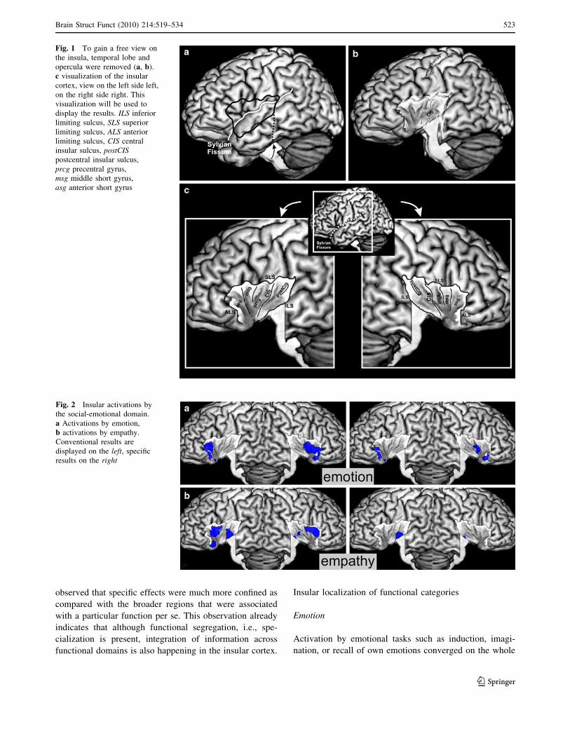
which temporal lobe and operculum were removed to gaina free view on the insular cortex (see Fig. 1). Thus, the
topographic relationships of the resulting activations to
each other and to anatomical landmarks of the insularcortex could be investigated. Activations were also
assigned histologically using the SPM Anatomy Toolbox
(Eickhoff et al. 2005). The latter approach was important inorder to eliminate activation of, e.g., the parietal operculum
spilling over into the insular mask.
Results
As outlined above, we investigated the functional organi-
zation of the human insula by conventional ALE analysis
of activations associated with different cognitive, sensori-motor, and social-emotional processes (cf. Table 2).
Moreover, by using the modified variant of the ALE
algorithm, we could delineate those insular regions, whichwere specific to a particular process. Throughout all anal-
yses (Figs. 2, 3, 4, 5, maxima provided in Table 3), we
Table 2 The investigated categories were subsumed to four func-tional domains, which represent functional systems
Domain Category
Social-emotional Emotion and empathy
Olfacto-gustatory Olfaction and gustation
Cognitive Attention, language, speech,working memory, memory
Sensorimotor Interoception, somatosensation,pain, motion
522 Brain Struct Funct (2010) 214:519–534
123
observed that specific effects were much more confined as
compared with the broader regions that were associated
with a particular function per se. This observation alreadyindicates that although functional segregation, i.e., spe-
cialization is present, integration of information across
functional domains is also happening in the insular cortex.
Insular localization of functional categories
Emotion
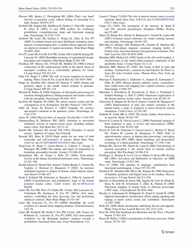
Activation by emotional tasks such as induction, imagi-nation, or recall of own emotions converged on the whole
Fig. 1 To gain a free view onthe insula, temporal lobe andopercula were removed (a, b).c visualization of the insularcortex, view on the left side left,on the right side right. Thisvisualization will be used todisplay the results. ILS inferiorlimiting sulcus, SLS superiorlimiting sulcus, ALS anteriorlimiting sulcus, CIS centralinsular sulcus, postCISpostcentral insular sulcus,prcg precentral gyrus,msg middle short gyrus,asg anterior short gyrus
Fig. 2 Insular activations bythe social-emotional domain.a Activations by emotion,b activations by empathy.Conventional results aredisplayed on the left, specificresults on the right
Brain Struct Funct (2010) 214:519–534 523
123

anterior insula. It extended onto the central part on the right
hemisphere. After testing for specific effects evoked byemotional processing, only the anterior-ventral insula
bilaterally and a small cluster on the right central region
remained significant (see Fig. 2a).
Empathy
Activation by paradigms tapping into empathy like
judging emotions in faces was observed on the anterior-dorsal insula bilaterally, on the left anterior-ventral
Fig. 3 Insular activations bythe chemical sensory domain.a Activations by olfaction,b activations by gustation.Conventional results aredisplayed on the left, specificresults on the right
Fig. 4 Insular activations bythe sensorimotor domain.a Activations by interoception,b activations by pain,c activations bysomatosensation, d activationsby motion. Conventional resultsare displayed on the left,specific results on the right
524 Brain Struct Funct (2010) 214:519–534
123

region, and bilaterally on the posterior ventral part. Whentesting for specific effects of empathic processing we
found significant effects in the posterior ventral part of
the insula, in particular on the left hemisphere (seeFig. 2b).
Olfaction
Activation by olfactory stimulation was found on the
anterior dorsal insula on both sides, extending onto thecentral insula on the right hemisphere. This right central
cluster also remained significant after testing for specific
effects, while left hemispheric effects were not seen anymore (see Fig. 3a).
Gustation
We observed bilateral activation by gustatory stimuli onthe anterior dorsal insula as well as on the dorsal mid-
insula. In particular on the right hemisphere, these activa-
tions comprised the whole anterior insula. When tested forspecific effects of gestation, activation was only observed
on the right anterior and the bilateral mid-dorsal insula (see
Fig. 3b).
Interoception
Activation by interoceptive tasks such as listening to one’s
own heartbeat or suppressing the urge to void was found on
Fig. 5 Insular activations bythe cognitive domain.a Activations by attention,b activations by language,c activations by speechd activations by workingmemory, e activations bymemory. Conventional resultsare displayed on the left,specific results on the right
Brain Struct Funct (2010) 214:519–534 525
123

Table 3 Insular maxima for each analyzed category
Category Conventional analysis Specific analysis
x y z T x y z T
Emotion -31 24 -4 6.13 -31 24 -6 5.77
-30 12 10 2.25 28 17 -15 4.42
42 15 -3 4.96 42 8 -6 3.32
39 7 0 4.54 44 10 -4 3.03
28 17 -15 4.42
Empathy -40 15 3 5.38 -35 -11 -7 4.69
-31 16 -19 4.04
-41 -7 4 3.25
39 19 3 5.11
37 8 7 4.76
46 -6 -1 3.37
Olfaction -30 16 5 4.10 42 13 -4 5.96
42 13 -4 5.96
Gustation -35 20 5 3.67 -33 0 12 3.07
-36 10 11 3.60 42 26 -6 4.68
-33 0 11 3.09 44 9 1 4.15
44 24 -5 4.79 39 12 -7 4.10
44 9 2 4.17 47 7 6 3.22
40 12 -6 4.12
Interoception -43 -3 6 5.94 -43 -3 6 5.94
-38 15 7 4.99 41 2 3 5.18
41 2 3 5.18
38 15 7 4.76
35 -13 9 3.56
Pain -39 -18 13 6.73 -39 -18 13 6.73
-44 2 7 6.66 -44 2 7 6.66
34 -20 14 5.58 42 9 -4 3.97
Somatosensation -37 -11 5 3.50 -37 -2 -4 3.34
-39 0 -4 3.46
-38 -4 1 3.10
41 21 -5 3.39
Motion -29 12 9 3.85 – – – –
-41 -13 13 3.57
Attention -35 18 7 6.35 – – – –
-33 18 -5 6.22
36 19 3 7.98
Language -33 20 3 8.13 – – – –
-35 22 2 8.13
36 20 -1 7.84
Speech -34 20 7 8.13 -34 20 7 8.13
36 18 5 5.10
Working memory -30 24 3 8.13 -32 24 4 8.13
-33 20 1 8.13 -32 20 1 8.13
36 21 1 7.78 -34 21 1 8.12
34 21 1 7.69
39 21 1 7.57
Memory -32 22 3 6.27 35 21 0 7.07
36 17 -7 5.56
Results by the conventional analysis are given on the right, results by the specific analysis on the left. All co-ordinates are given in MNI space
526 Brain Struct Funct (2010) 214:519–534
123

the central and anterior dorsal insula bilaterally. Among
these effects, those observed in the central insula werebilaterally shown to be specific to interoceptive processing
(see Fig. 4a).
Pain
Extended activation by painful stimuli was found on vir-tually the entire insula and extended onto the parietal
operculum. Interestingly, even after testing for specificeffects, extended activation on the central and posterior
insula was observed (see Fig. 4b).
Somatosensation
Somatosensory stimuli evoked activation on the left centralregion of the insula and its right anterior dorsal part. As
most studies examined somatosensory stimulation of the
right hand, however, this lateralization must be seen cau-tiously and should warrant further investigation. Testing
for specificity revealed only activation on the left central
insula (see Fig. 4c).
Motor
Activation by active motor tasks was found bilaterally on
the central insula, close to the representation of somato-
sensory stimuli reported above but more bilateral.Observed activation after testing for specific effects might
have been rather due to spillover effects from opercular
areas and basal ganglia, as maxima of activation werelocated in these directly neighboring structures rather than
in the insula. This observation indicates that the insular
region identified by the conventional analysis might notonly be specifically engaged in motor tasks, but also in
other functional categories (see Fig. 4d).
Attention
Convergent activation by tasks tapping into attentionalprocesses, e.g., Stroop-, Simon-, Go/NoGo-tasks, or spatial
attention, was found bilaterally on the anterior-dorsal
insula. However, there was no significant effect to be foundwhen testing for activation that is specific to attentional
tasks, indicating that regions sustaining these functions are
also engaged in other cognitive or sensory processes aswell (see Fig. 5a).
Language
Activations by language processing, e.g., lexical decision
making or semantic judgments, were found on the anterior
dorsal insula bilaterally. After testing for specific effects,
no maximum of activation was found on the insula. Rather,we only observed spillover effects from the left frontal
operculum on the anterior dorsal insula on this hemisphere
(see Fig. 5b).
Speech
Activations by active (overt) speech were located on the
anterior and posterior parts of the dorsal insula bilaterally.However, it has to be noted that activation on the posterior
insula seems to be caused by spillover effects, in particular
from the neighboring primary auditory cortex (areas Te1.0and TE1.1, see Morosan et al. 2001). This interpretation is
in line with the observation that among the implicated
regions, only the left anterior insula remained significantafter testing for specific effects (see Fig. 5c).
Working memory
Activations by working memory tasks, such as n-back or
Sternberg paradigms, were found bilaterally on the ante-rior-dorsal insula. Moreover, activation in these regions
also remained significant after testing for specific effects
(see Fig. 5d).
Memory
Activations by episodic or short-term memory retrieval
were found bilaterally on the anterior-dorsal insula in a
similar location as the activations associated with workingmemory. However, in contrast to the above, only the
effects on the right anterior-dorsal insula were specific to
memory paradigms (see Fig. 5e).
Functional integration
All analyses revealed a considerably smaller extent of
activation when comparing the results from the conven-
tional and the modified ALE. This indicates that theinsular cortex is engaged in functional integration
between different functional categories. To map those
regions where different categories are integrated (i.e.where results from the conventional ALE overlap), we
calculated two conjunction analyses over the single cat-
egories (see ‘‘Methods’’). The first conjunction over all13 analyzed categories revealed no overlap throughout
the whole brain. The second analysis over all investi-
gated categories except somatosensation and motionrevealed an overlap on the anterior-dorsal insula (see
Fig. 6). No other overlap was observed throughout the
whole brain.
Brain Struct Funct (2010) 214:519–534 527
123

Functional differentiation
To investigate a functional differentiation of the insula the13 investigated functional categories were sorted into four
functional domains (cf. Table 2, methods). The modified
analysis of every functional domain revealed a functionalsegregation of the insular cortex (see Fig. 7). The mid-
posterior insula was activated by tasks from the sensori-
motor domain. This region extended from posterior insulato the posterior short insular gyrus. The anterior-ventral
insula was specific to social-emotional functions, i.e.,
emotional processing and empathy. This region was
located around the insular pole bilaterally. The anterior-
dorsal insula processed tasks from the cognitive domain. Itcomprised the dorsal part of the anterior and middle short
gyrus. Finally, for the olfacto-gustatory domain a region on
the right middle insular gyrus was revealed to be specific.
Discussion
The present results indicate a functional map of the humaninsula based on a large-scale coordinate-based meta-anal-
ysis of published functional neuroimaging studies. Using
Fig. 6 Overlap of allfunctional categories exceptsomatosensation and motion.All other eleven categoriesoverlap on the anterior-dorsalpart of the insula (a), indicatingfunctional integration betweenthem. b Throughout the wholebrain the anterior-dorsal insulawas the only region thatparticipated in the processingof all eleven categories
Fig. 7 Functionaldifferentiation of the insulaby functional domains. Redsensorimotor, green cognitive,yellow chemical sensory,blue social-emotional
528 Brain Struct Funct (2010) 214:519–534
123

this approach, we could show a differentiation of the
insular cortex into four functional regions. These regionswere defined by a sensorimotor, a cognitive, a social-
emotional, and an olfacto-gustatory domain. Throughout
the whole brain activations by these domains reflected thepreviously described social-emotional, cognitive, and sen-
sorimotor networks as well as to a network processing
olfacto-gustatory stimuli (Corbetta et al. 2008; Kandelet al. 1991; Phillips et al. 2003). Apart from this functional
differentiation, a conjunction analysis of investigatedfunctional categories revealed the anterior dorsal insula to
be involved in the processing of all investigated categories
except somatosensation and motion. This finding points theanterior-dorsal insula to act as a multimodal integration
region.
A functional differentiation of the insular cortex wasalready indicated by anatomical data reported in non-
human primates. Cytoarchitectonically, the non-human
primate insula can be divided into an anterior-basalagranular, allocortical part, a posterior granular part, and a
dysgranular part which is located between these and covers
the anterior-dorsal and central insula (Augustine 1996;Mesulam and Mufson 1982). This parcellation is also
reflected by differences in connectivity as investigated by
invasive tracing studies in these species. In monkeys, theanterior-basal part shows dense connections to limbic areas
like the amygdala, periamygdaloid, and entorhinal cortices
as well as to the temporal pole (Fudge et al. 2005; Hoistadand Barbas 2008; Mesulam and Mufson 1985; Stefanacci
and Amaral 2002). Furthermore, the anterior insula also
participates in a structurally connected network formedtogether with the orbitofrontal, entorhinal, and piriform and
olfactory cortex, which is engaged in olfactory and gusta-
tory processing (Augustine 1996; Mesulam and Mufson1985). Interestingly, the mid-dorsal insula also receives
input from the thalamic taste area (Craig 2002; Pritchard
et al. 1986), which matches well with the present findingsof gustatory activation in this part. The mid-posterior
regions, finally, are densely connected to primary and
secondary sensory and motor areas, while the anteriordorsal areas are preferably connected to frontal regions
(Augustine 1996; Friedman et al. 1986; Mesulam and
Mufson 1985).This separation into an anterior-basal region, which is
closely linked to the social-emotional and the olfacto-
gustatory system, a mid-posterior insular region, which isclosely connected to sensorimotor areas and an anterior
dorsal part, which is more closely connected to frontal
association areas matches well with the present results. Themid-posterior insula, which is connected to somatosensory
and motor cortices in monkeys, was found to process the
sensorimotor domain in the present study. Equally, theanterior-basal region, which in monkeys is connected
to limbic areas like the amygdala, periamygdaloid, and
entorhinal cortices, was observed to process the socio-emotional domain and the anterior-dorsal insula with its
connections to frontal association areas was activated by
the cognitive domain. Finally, the reported connectivitybetween anterior-dorsal insula and frontal regions matches
well the identification of a cognitive insular region, as
frontal areas are regularly reported to serve workingmemory, attention, and other cognitive tasks (Cabeza and
Nyberg 2000). However, it must be taken into account thatcross-species comparisons are problematic, as they disre-
gard the obvious and considerable differences between the
brains. A comparison to non-human tracing data is unfor-tunately the best reference for insular connectivity avail-
able at the present, as there is currently no technique which
allows a reliable delineation the connections of insularregions in the human brain. In future, more recent imaging
and analysis techniques will hopefully help to unravel
connectivity patterns of the human insula and allow acomparison with that of other species.
In good accordance with the anatomical findings in non-
human primates, the anterior insula and in particular itsanterior-basal parts have been discussed in the processing of
emotion and empathy (Adolphs 2002; Dolan 2002; Frith and
Singer 2008; Lamm and Singer 2010; Phillips et al. 2003;Singer and Lamm 2009). In particular, it has been proposed
that this region is involved in the generation and mediation
of feeling states as a response to environmental stimuli andaffective states (Dolan 2002; Phillips et al. 2003). In
empathy-related processing it is involved in the recognition
of emotions in faces (Phillips et al. 2003) and the feeling ofpain in others (Frith and Singer 2008; Singer et al. 2006).
Finally, the anterior basal insula was hypothesized to pro-
vide a link between the mirror neuron system and emotionalprocessing, thus enabling a matching between own and
observed emotions (Iacoboni and Dapretto 2006). This focus
on the anterior basal part is supported by electrophysiolog-ical findings. Preoperative direct recordings in patients
suffering from drug-refractory epilepsy revealed that a role
in the categorization of facial expressions is confined to thisregion (Krolak-Salmon et al. 2003). The findings from the
present study corroborate these results, as they show an
involvement of the anterior insula and in particular theanterior-basal part in the processing of emotions.
Different functional studies reported insular activation
in olfactory and gustatory tasks (Kringelbach et al. 2004;Poellinger et al. 2001; Royet and Plailly 2004; Small et al.
1999). These studies show that the anterior insula processes
interactions between emotion, memory and olfaction(Poellinger et al. 2001), habituation to odorants (Royet and
Plailly 2004), and several tasks testing taste processing
(Kringelbach et al. 2004; Small et al. 1999). Most inter-estingly, the insula was reported to be part of a network
Brain Struct Funct (2010) 214:519–534 529
123

involved, especially in integration of taste and flavor (Rolls
2006; Small and Prescott 2005), supporting an insular rolein olfacto-gustatory processing. Intraoperative direct stim-
ulations of the anterior insula also led to gustatory and
olfactory sensations (Penfield and Faulk 1955). It is inter-esting to note though, that these sensations were also
elicited as well near the basal part of the precentral gyrus
and on the anterior part. However, on the anterior part,insular stimulation was accompanied by descriptions like
‘‘bad taste’’ or feelings of fear, while more posterior rathersensations in the mouth, salivation or increased gastric
motility were observed. This matches well the hypothesis
that the insular taste area is located rather in the moreposterior parts in humans (Small 2010, Craig 2010), while
the more anterior parts will be more engaged in an inte-
gration with odors and emotions.The anterior-dorsal insula has repeatedly been impli-
cated in several cognitive tasks. For example, it processes
both working memory and attention tasks with additiveBOLD effects for both tasks (Mayer et al. 2007; Soros et al.
2007). It was furthermore assigned to be a part of the
inferior frontoparietal network, which responds to behav-ioral relevant rather than to expected stimuli (Corbetta
et al. 2008) and reported to play a role in language
processing (McCarthy et al. 1993; Price 2000; Rieckeret al. 2000). These findings might implicate an abstract
role in extracting and processing task-relevant and salient
information.The mid-posterior insula, finally, has been repeatedly
demonstrated to be involved in different somato- and vis-
cerosensory stimuli. In particular it responds to painful,visceral and somatosensory stimulation (Chen 2007;
Dupont et al. 2003; Ostrowsky et al. 2002; Peyron et al.
2000; Peyron et al. 2002; Shelley and Trimble 2004). Inaddition to this sensory processing, movement was elicited
by electrical stimulation of this region in humans (Showers
and Lauer 1961), which indicates a role in sensorimotorprocessing of this region. Other reports of direct stimula-
tion of this region reported interoceptive and somatic
sensations, changes in gastric motility, breathing or heartrate as well as the sensation or urge of movement (Penfield
and Faulk 1955). Interestingly, these findings also match
reports from frontotemporal dementia (Seeley et al. 2008).Seeley et al. showed that early changes in this disease,
which present with cognitive and emotional impairments,
appear in the anterior insula among other regions. Only atlater stages of this disease, when the patients show
impairments in executive functions, these changes will
comprise the posterior parts.It is important to note though that all results from the
current analysis reflect the convergence across a large
number of studies. Along with this they map the commonneuronal substrates between different experiments on the
same topic, for example, the shared neural substrate of
different emotions in various experimental setups. Theinference one can draw from these results is that observed
regions are involved in the process, which underlies all -or
at least most- investigated experiments. While we can thussay that the anterior insula is involved in the processing of
emotions in general, any inference from these results on a
specific emotion in a specific context must be regardedwith extreme caution. The fact that in any given individual
study only a sub-category of all emotions is investigated ina particular experimental context (this might be ‘‘rating a
disgusting taste in comparison to a memorized reference’’)
will lead to conclusions which may diverge from the cur-rent results. Therefore, although this analysis can reliably
identify a general functional differentiation of the insula, it
cannot predict or reflect effects observed in a specificexperimental task.
The described converging evidence for a functional
segregation of the insula was confirmed and extended inthe current meta-analysis, which showed a reliable differ-
entiation of the human insula into functional specific
regions. Still, in spite of this ample evidence for functionalspecification within the insula, this region has also
repeatedly been implicated as the anatomical substrate for
the behaviorally essential integration of information fromdifferent functional systems. Sensory stimuli, for example,
have an immediate effect on emotions (Chen 2007; Craig
2002; Williams and Bargh 2008), which is in particularreflected by the James-Lange theory and Damasio’s
‘‘somatic marker’’ hypothesis (Damasio 1994; James 1894;
Lange 1885). These hypotheses state that interoceptiveinformation and emotion are directly depending on another
and even cause each other. Recently, the human insula
has emerged as the prime candidate for sustaining thisintegration, as it was reported to be directly involved in
processing the reciprocal influence of interoception and
emotion (Craig 2002; Critchley et al. 2004; Critchley2005). Moreover, not only integration between interocep-
tion and emotion, but also between cognitive tasks and
sensation as well as emotion was described to involve thisregion. For example, the insula has been reported to be
involved in processing attention to sensory stimuli like
attention to touch or comparing the own heartbeat to anexternal rhythm (Critchley et al. 2004; Johansen-Berg and
Matthews 2002). Also, it is involved in distinguishing
between self and other, which was assigned to this regionusing the rubber hand illusion (Ehrsson et al. 2004; Karnath
and Baier 2010; Tsakiris et al. 2007). Finally, a convincing
theory of how interoception, emotion/empathy and thecognitive evaluation of risk and uncertainty may be linked
within the anterior insula cortex was recently brought up
(Singer et al. 2009). In this theory the insula was describedto process current feeling states that are closely linked to
530 Brain Struct Funct (2010) 214:519–534
123

interoception, predicted feeling states and a prediction
error, which allows for an evaluation and adaption of theown prediction. This combination of different modalities
makes the insula a highly integrative region.
On the background of the described functional differ-entiation of the insula, the question of how this functional
integration is implemented arises. We would argue that
there may be two complementary answers to this: On theone hand the different areas or zones are extremely well
interconnected as it was shown in tracing studies in non-human primates (Augustine 1996). These dense connec-
tions may represent a structural basis for a rapid flow of
information between the functional systems. On the otherhand, the present study also revealed an overlap of all
categories (except for basic somatosensory and motor
processing) on the anterior-dorsal insula. This overlapprobably reflects a shared role in information processing
between the systems. This shared role might reflect a
multimodal integration between the different categoriesrelaying information between the different functional sys-
tems on the one hand and on the other hand a basic func-
tional role that all categories have in common. In thefollowing, we will discuss this shared role and its impli-
cations for this region. Still, in particular, the combination
of this shared functional role and the close connectivitybetween insular domains gives this region a unique posi-
tion for regulating flow and integration of information
between the systems.It must be mentioned here that an interpretation of the
observed overlap is subject to a limitation that cannot be
solved within the scope of this paper, namely the questionof what constitutes an independent cognitive process. It is
well known that, for example, pain is not a solely sensory
process, but has also an important emotional component(Chen 2007). The same will go for interoception, which
may be regarded a sensory category but is an important
component in the generation and interpretation of emotionsand empathy (Damasio 1994; James 1894; Lange 1885).
The same goes in some way for nearly all categories, i.e.,
they always have some potential aspect of ambiguity.Unambiguous classifications, however, are probably
impossible to define without a full knowledge of the
intrinsic ontology of cognitive processes, i.e., the buildingblocks of brain organization. The question will now be, if
this potential ambiguity of the categories led to the mapped
overlap in the insula, or if this mapped overlap in the insulacaused the possible ambiguity of the categories. That is, is
the overlap spurious or does it reflect more basic processes
that are shared among categories. By a careful selection ofstudies, which avoided ambiguity between different cate-
gories as much as possible, we attempted to avoid this
possibly artificial overlap. Although a definite answer tothis problem cannot be given here, a review of literature
points the anterior dorsal insula to be involved in a mul-
titude of different studies (e.g. Craig 2009).In the following, we will assume that there is an overlap
in the mapping of the categories in the anterior-dorsal
insula, as observed in this study. This region was recentlyproposed to integrate momentary images of own feelings,
the sensory environment, and the motivational, hedonic
and social interaction between these into one whole rep-resentation of the sentient self (Craig 2009). In this con-
cept, the anterior-dorsal insula was proposed to constitutethe final stage of a hierarchical processing of information in
the insular cortex––starting with pure sensory information
posterior and after integrating emotional and cognitivevaluation ending with a full representation of the sentient
self in the anterior parts. This representation, which thus
constitutes an integration of salient information from allfunctional systems led subsequently to the suggestion that
the anterior-dorsal insula might be a potential neural cor-
relate of awareness (Craig 2009). This described hierarchyof processing also explains the finding that the functional
overlap between different functional systems on the insular
cortex did not include basic somatosensory and motortasks. The pure somatosensory and motor tasks that were
analyzed in this study do not have the need for cognitive or
social-emotional evaluation. Hence, they do not have aneed for integration. Thus, it can be expected that they do
not elicit activation in the anterior-dorsal multimodal
integration region, but rather in the posterior part of theinsula.
However, the shared role which probably underlies the
observed overlap between different functional categoriescan also be due to a basic functional role that all cate-
gories have in common. This basic functional role might
be a general role in task processing, like starting, updat-ing, and ending of a given task. This was indicated by an
fMRI study investigating ten different functional para-
digms, including different language, sensorimotor andcognitive tasks, in the same set of subjects (Dosenbach
et al. 2006). Using a conjunction analysis, the authors
found only the dACC/msFC and the bilateral anterior-dorsal insula to be active across all different paradigms.
This result, which is confirmed and extended by our
current data, was interpreted as evidence for a central roleof the anterior-dorsal insula in initiating, updating, and
maintaining tasks. That is, as an alternative to the theory
outlined above, this view regards the anterior-dorsalinsula not primarily as a region integrating information
from different functional systems into a putative correlate
of awareness (Craig 2009). Rather, it regards this overlapbetween the different processing as a task-set region
responsible for maintaining the task-set which is neces-
sary to perform any task in the scanner, or––to put it tothe extreme––as the smallest common denominator. These
Brain Struct Funct (2010) 214:519–534 531
123

two hypotheses may be regarded as two potential inter-
pretations of the observed overlap across many functionaldomains in this region, which, however, cannot be finally
differentiated, from the current data.
A few last points may be presented here, which will stillneed further investigation. We have compared our findings
to invasive tracing studies in non-human primates. While
there are analogies between the results from this study andthese tracing studies, any comparison assumes that human
and non-human insula are matchable. This assumption canbe regarded critically, as the insula differs considerably
between species (Craig 2009; Craig 2010; Rose 1928). A
cytoarchitectonic mapping study of the posterior humaninsula supported that notion, as this region proved to be far
more complex than studies from non-human primates
report (Kurth et al. 2009). Unfortunately, the areas mappedin this study comprise just a very limited part of the insular
cortex, limiting all comparisons to imaging data. As there
are currently ongoing further mapping studies, a compre-hensive comparison of insular structure and function will
become feasible in the future.
Another important point may be the finding that variableoccurrences of lateralization and localization were found
on the insula with respect to the current study. To give
some examples, Jabbi et al. (2007) reported a representa-tion of disgust on the left anterior central region, Royet
et al. (1999) did not report activation from a complex
olfactory task on the insula, and Qureshy et al. (2000)reported activation from another complex olfactory task on
the left insula. Partly, these differences in localization can
be explained by the fact that these studies did not onlyinvestigate basic emotional or olfactory processing. How-
ever, if we extrapolate the results from the cytoarchitec-
tonic mapping of the posterior insula (Kurth et al. 2009),we will expect a multitude of different areas in this region.
Thus, the here presented functional differentiation might be
rather a framework for a more generalized concept of theinsular role. Its detailed functioning and structure–function
relationships will certainly remain topics of further
research.
Conclusion
Using a large-scale meta-analysis, we revealed a functional
differentiation as well as a functional integration within thehuman insula. The identified regions are involved in the
processing of different functional systems. This differen-
tiation is also reflected in anatomical connectivity studiesin monkeys, thus corroborating the present observations.
The observed functional integration in the anterior-dorsal
insula might constitute a link between these differentfunctional systems and a correlate for the relaying of
information between them. Indeed, this might be inter-
preted as a representation of a sentient self and even as aneural correlate of self-awareness. However, the observed
overlap of activations by multiple different tasks might also
represent a smallest common denominator, which is a task-set region for initiating, updating, and maintaining a
function. Still, as these interpretations do not totally dis-
qualify each other––a task set region can also be a multi-modal integration region and vice versa––they rather
describe extreme interpretations. To characterize thisintriguing role of the anterior-dorsal insula subsequent
studies will be necessary.
Acknowledgments This research was funded by the NationalInstitute of Biomedical Imaging and Bioengineering, the NationalInstitute of Neurological Disorders and Stroke, and the NationalInstitute of Mental Health. K.Z. acknowledges further funding by theHelmholtz Alliance ‘‘Systembiologie’’ the ‘‘Human Brain Model’’.A.R.L. and P.T.F. were supported by the Human Brain Project of theNIMH (R01-MH074457-01A1).
Conflict of interest statement None.
References
Adolphs R (2002) Neural systems for recognizing emotion. Curr OpinNeurobiol 12:169–177
Augustine JR (1996) Circuitry and functional aspects of the insularlobe in primates including humans. Brain Res Brain Res Rev22:229–244
Cabeza R, Nyberg L (2000) Imaging cognition II: an empirical reviewof 275 PET and fMRI studies. J Cogn Neurosci 12:1–47
Chen LM (2007) Imaging of pain. Int Anesthesiol Clin 45:39–57Corbetta M, Patel G, Shulman GL (2008) The reorienting system of
the human brain: from environment to theory of mind. Neuron58:306–324
Craig AD (2002) How do you feel? Interoception: the sense ofthe physiological condition of the body. Nat Rev Neurosci3:655–666
Craig AD (2009) How do you feel––now? The anterior insula andhuman awareness. Nat Rev Neurosci 10:59–70
Craig AD (2010) The sentient self. Brain Struc Func 214(5–6). doi:10.1007/s00429-010-0248-y (this issue)
Critchley HD (2005) Neural mechanisms of autonomic, affective, andcognitive integration. J Comp Neurol 493:154–166
Critchley HD, Wiens S, Rotshtein P, Ohman A, Dolan RJ (2004)Neural systems supporting interoceptive awareness. Nat Neuro-sci 7:189–195
Damasio AR (1994) Descartes’ error: emotion, reason and the humanbrain. Putnam’s Sons, New York
Dolan RJ (2002) Emotion, cognition, and behavior. Science298:1191–1194
Dosenbach NU, Visscher KM, Palmer ED, Miezin FM, Wenger KK,Kang HC, Burgund ED, Grimes AL, Schlaggar BL, Petersen SE(2006) A core system for the implementation of task sets.Neuron 50:799–812
Dupont S, Bouilleret V, Hasboun D, Semah F, Baulac M (2003)Functional anatomy of the insula: new insights from imaging.Surg Radiol Anat 25:113–119
532 Brain Struct Funct (2010) 214:519–534
123
Ehrsson HH, Spence C, Passingham RE (2004) That’s my hand!Activity in premotor cortex reflects feeling of ownership of alimb. Science 305:875–877
Eickhoff SB, Stephan KE, Mohlberg H, Grefkes C, Fink GR, AmuntsK, Zilles K (2005) A new SPM toolbox for combiningprobabilistic cytoarchitectonic maps and functional imagingdata. Neuroimage 25:1325–1335
Eickhoff SB, Laird AR, Grefkes C, Wang LE, Zilles K, Fox PT(2009) Coordinate-based activation likelihood estimation meta-analysis of neuroimaging data: a random-effects approach basedon empirical estimates of spatial uncertainty. Hum Brain Mapp30:2907–2926
Fox PT, Laird AR, Fox SP, Fox PM, Uecker AM, Crank M, Koenig SF,Lancaster JL (2005) BrainMap taxonomy of experimental design:description and evaluation. Hum Brain Mapp 25:185–198
Friedman DP, Murray EA, O’Neill JB, Mishkin M (1986) Corticalconnections of the somatosensory fields of the lateral sulcus ofmacaques: evidence for a corticolimbic pathway for touch.J Comp Neurol 252:323–347
Frith CD, Singer T (2008) The role of social cognition in decisionmaking. Philos Trans R Soc Lond B Biol Sci 363:3875–3886
Fudge JL, Breitbart MA, Danish M, Pannoni V (2005) Insular andgustatory inputs to the caudal ventral striatum in primates.J Comp Neurol 490:101–118
Hoistad M, Barbas H (2008) Sequence of information processing foremotions through pathways linking temporal and insular corticeswith the amygdala. Neuroimage 40:1016–1033
Iacoboni M, Dapretto M (2006) The mirror neuron system and theconsequences of its dysfunction. Nat Rev Neurosci 7:942–951
Jabbi M, Swart M, Keysers C (2007) Empathy for positiveand negative emotions in the gustatory cortex. Neuroimage 34:1744–1753
James W (1894) Physical basis of emotion. Psychol Rev 1:516–529Johansen-Berg H, Matthews PM (2002) Attention to movement
modulates activity in sensory-motor areas, including primarymotor cortex. Exp Brain Res 142:13–24
Kandel ER, Schwartz JH, Jessell TM (1991) Principles of neuralscience. Appleton & Lange, East Norwalk
Karnath HO, Baier B (2010) Right insula for our sense of limbownership and self-awareness of actions. Brain Struc Func214(5–6). doi:10.1007/s00429-010-0250-4 (this issue)
King-Casas B, Sharp C, Lomax-Bream L, Lohrenz T, Fonagy P,Montague PR (2008) The rupture and repair of cooperation inborderline personality disorder. Science 321:806–810
Kringelbach ML, de Araujo I, Rolls ET (2004) Taste-relatedactivity in the human dorsolateral prefrontal cortex. Neuroimage21:781–788
Krolak-Salmon P, Henaff MA, Isnard J, Tallon-Baudry C, Guenot M,Vighetto A, Bertrand O, Mauguiere F (2003) An attentionmodulated response to disgust in human ventral anterior insula.Ann Neurol 53:446–453
Kurth F, Eickhoff SB, Schleicher A, Hoemke L, Zilles K, Amunts K(2009) Cytoarchitecture and probabilistic maps of the humanposterior insular cortex. Cereb Cortex. doi:10.1093/cercor/bhp208
Laird AR, Fox PM, Price CJ, Glahn DC, Uecker AM, Lancaster JL,Turkeltaub PE, Kochunov P, Fox PT (2005a) ALE meta-analysis: controlling the false discovery rate and performingstatistical contrasts. Hum Brain Mapp 25:155–164
Laird AR, Lancaster JL, Fox PT (2005b) BrainMap: the socialevolution of a human brain mapping database. Neuroinformatics3:65–78
Laird AR, Eickhoff SB, Kurth F, Fox PM, Uecker AM, Turner JA,Robinson JL, Lancaster JL, Fox PT (2009) ALE meta-analysisworkflows via the Brainmap database: progress towards aprobabilistic functional brain atlas. Front Neuroinform 3:23
Lamm C, Singer T (2010) The role of anterior insular cortex in socialemotions. Brain Struc Func 214(5–6). doi:10.1007/s00429-010-0251-3 (this issue)
Lange CG (1885) The mechanism of the emotion. In: Rand B(ed) The classical psychologist. Houghton Mifflin, Boston,pp 672–685
Mayer JS, Bittner RA, Nikolic D, Bledowski C, Goebel R, Linden DE(2007) Common neural substrates for visual working memoryand attention. Neuroimage 36:441–453
McCarthy G, Blamire AM, Rothman DL, Gruetter R, Shulman RG(1993) Echo-planar magnetic resonance imaging studies offrontal cortex activation during word generation in humans. ProcNatl Acad Sci USA 90:4952–4956
Mesulam MM, Mufson EJ (1982) Insula of the old world monkey. I.Architectonics in the insulo-orbito-temporal component of theparalimbic brain. J Comp Neurol 212:1–22
Mesulam MM, Mufson EJ (1985) The insula of Reil in man andmonkey. Architectonics, connectivity and function. In: Peters A,Jones EG (eds) Cerebral cortex. Plenum Press, New York, pp179–226
Morosan P, Rademacher J, Schleicher A, Amunts K, Schormann T,Zilles K (2001) Human primary auditory cortex: cytoarchitec-tonic subdivisions and mapping into a spatial reference system.Neuroimage 13:684–701
Mutschler I, Wieckhorst B, Kowalevski S, Derix J, Wentlandt J,Schulze-Bonhage A, Ball T (2009) Functional organization ofthe human anterior insular cortex. Neurosci Lett 457:66–70
Ostrowsky K, Magnin M, Ryvlin P, Isnard J, Guenot M, Mauguiere F(2002) Representation of pain and somatic sensation in thehuman insula: a study of responses to direct electrical corticalstimulation. Cereb Cortex 12:376–385
Penfield W, Faulk ME Jr (1955) The insula; further observations onits function. Brain 78:445–470
Peyron R, Laurent B, Garcia-Larrea L (2000) Functional imaging ofbrain responses to pain. A review and meta-analysis (2000).Neurophysiol Clin 30:263–288
Peyron R, Frot M, Schneider F, Garcia-Larrea L, Mertens P, BarralFG, Sindou M, Laurent B, Mauguiere F (2002) Role ofoperculoinsular cortices in human pain processing: convergingevidence from PET, fMRI, dipole modeling, and intracerebralrecordings of evoked potentials. Neuroimage 17:1336–1346
Phillips ML, Drevets WC, Rauch SL, Lane R (2003) Neurobiology ofemotion perception I: the neural basis of normal emotionperception. Biol Psychiatry 54:504–514
Poellinger A, Thomas R, Lio P, Lee A, Makris N, Rosen BR, KwongKK (2001) Activation and habituation in olfaction––an fMRIstudy. Neuroimage 13:547–560
Price CJ (2000) The anatomy of language: contributions fromfunctional neuroimaging. J Anat 197:335–359
Pritchard TC, Hamilton RB, Morse JR, Norgren R (1986) Projectionsof thalamic gustatory and lingual areas in the monkey, Macacafascicularis. J Comp Neurol 244:213–228
Qureshy A, Kawashima R, Imran MB, Sugiura M, Goto R, Okada K,Inoue K, Itoh M, Schormann T, Zilles K, Fukuda H (2000)Functional mapping of human brain in olfactory processing:a PET study. J Neurophysiol 84:1656–1666
Riecker A, Ackermann H, Wildgruber D, Dogil G, Grodd W (2000)Opposite hemispheric lateralization effects during speaking andsinging at motor cortex, insula and cerebellum. Neuroreport11:1997–2000
Rolls ET (2006) Brain mechanisms underlying flavour and appetite.Philos Trans R Soc Lond B Biol Sci 361:1123–1136
Rose M (1928) Die Inselrinde des Menschen und der Tiere. J PsycholNeurol 37:467–624
Royet JP, Plailly J (2004) Lateralization of olfactory processes. ChemSenses 29:731–745
Brain Struct Funct (2010) 214:519–534 533
123

Royet JP, Koenig O, Gregoire MC, Cinotti L, Lavenne F, Le BD,Costes N, Vigouroux M, Farget V, Sicard G, Holley A,Mauguiere F, Comar D, Froment JC (1999) Functional anatomyof perceptual and semantic processing for odors. J CognNeurosci 11:94–109
Seeley WW, Crawford R, Rascovsky K, Kramer JH, Weiner M,Miller BL, Gorno-Tempini ML (2008) Frontal paralimbicnetwork atrophy in very mild behavioral variant frontotemporaldementia. Arch Neurol 65:249–255
Shelley BP, Trimble MR (2004) The insular lobe of Reil––itsanatamico-functional, behavioural and neuropsychiatric attri-butes in humans––a review. World J Biol Psychiatry 5:176–200
Showers MJ, Lauer EW (1961) Somatovisceral motor patterns in theinsula. J Comp Neurol 117:107–115
Singer T, Lamm C (2009) The social neuroscience of empathy. AnnN Y Acad Sci 1156:81–96
Singer T, Seymour B, O’Doherty JP, Stephan KE, Dolan RJ, Frith CD(2006) Empathic neural responses are modulated by theperceived fairness of others. Nature 439:466–469
Singer T, Critchley HD, Preuschoff K (2009) A common role ofinsula in feelings, empathy and uncertainty. Trends Cogn Sci13:334–340
Small DM (2010) Taste representation in the human insula. BrainStruc Func 214(5–6). doi:10.1007/s00429-010-0266-9 (this issue)
Small DM, Prescott J (2005) Odor/taste integration and the perceptionof flavor. Exp Brain Res 166:345–357
Small DM, Zald DH, Jones-Gotman M, Zatorre RJ, Pardo JV, Frey S,Petrides M (1999) Human cortical gustatory areas: a review offunctional neuroimaging data. Neuroreport 10:7–14
Soros P, Marmurek J, Tam F, Baker N, Staines WR, Graham SJ(2007) Functional MRI of working memory and selectiveattention in vibrotactile frequency discrimination. BMC Neuro-sci 8:48
Stefanacci L, Amaral DG (2002) Some observations on cortical inputsto the macaque monkey amygdala: an anterograde tracing study.J Comp Neurol 451:301–323
Tsakiris M, Hesse MD, Boy C, Haggard P, Fink GR (2007) Neuralsignatures of body ownership: a sensory network for bodily self-consciousness. Cereb Cortex 17:2235–2244
von Economo C, Koskinas GN (1925) Die Cytoarchitectonik derHirnrinde des erwachsenen Menschen. Springer, Berlin
Wager TD, Barrett LF (2004) From affect to control: functionalspecialization of the insula in motivation and regulation.Available online via: PsycExtra http://www.columbia.edu/cu/psychology/tor/
Williams LE, Bargh JA (2008) Experiencing physical warmthpromotes interpersonal warmth. Science 322:606–607
Zilles K (2004) Architecture of the human cerebral cortex. Regionaland laminar organization. In: Paxinos G (ed) The human nervoussystem, 2nd edn. Elsevier, San Diego, pp 997–1055
534 Brain Struct Funct (2010) 214:519–534
123