a handbook of the amazonian species of anopheles...
TRANSCRIPT

Mosquito Systematics
I
Vol. 13(l) 1981 1
A HANDBOOK OF THE AMAZONIAN SPECIES OF
ANOPHELES (NYSSORHYNCHUS) (DIPTERA: CULICIDAE)’
BY
Michael E. Faran and Kenneth J. Linthicum2
CONTENTS
INTRODUCTION .............. .2 MATERIAL AND M’E’iHiXk : : : : : : : : : : : .............. .3
SYSTEMATICS .............................. .4
TAXONOMICCHARACTERS ........................ .5 Adult Females ............................ .5
Male Genitalia ............................ .6
Fourth Stage Larvae ...... ;
BIONOMICS .................................. : i
MEDICAL IMP&TAN& : : : : : : : : : : ................ .9
PLATES l-5 ILLUSTRATED KEYSTO THE AMAZdNiAN &E&s &*ANOPHE;;ES ’ * ’ ’ ’ ’
. 11
(NYSSORHYNCHUS). ........................ 16 DISCUSSIONS, BIONOMICS, MEDICAL IMPORTANCE AND DISTRIBUTION OF SPECIES
1. Anopheles (Nys.) argyritarsis ..................... 34 2. Anopheles (Nys.) darlingi ...................... 35
3. Anopheles (Nys.) allopha ...................... 37
4. Anopheles (Nys.) braziliensis ..................... 39
5. Anopheles (Nys.) oswaldoi ...................... 4 1
6. Anopheles (Nys.) galvaoi ...................... 42 7. Anopheles (Nys.) evansi ....................... 43
8. Anopheles (Nys.) aquasalis ...................... 45
9. Anopheles (Nys.) ininii ....................... 47 10. Anopheles (Nys.) rangeli ...................... 48 11. Anopheles (Nys.) nuneztovari .................... 49
’ This research was supported by the Medical Entomology Project, Smithsonian Institu- tion, U.S. Army Medical Research and Development Command Research Contract DAMD- 17- 74C-4086 and the Mosquitoes of Middle America project, University of California, Los Angeles, U.S. Army Medical Research and Development Contract DA-49-l 93-MD-2478.
2 Captain, Me dical Service Corps, U.S. Army, Department of Entomology, Walter Reed Army Institute of Research, Washington, DC 200 12.

12. Anopheles (Nys.) strodei ...................... 5 1
13. Anopheles (Nys.) rondoni ...................... 53 14. Anopheles (Nys.) benarrochi ..................... 54
15. Anopheles (Nys.) triannulatus .................... 55
ACKNOWLEDGMENTS ........................... 56 REFERENCES .............................. 57 PLATES 6-24 ............................... 63
PLATES
1. Map of Amazonia ........................... 11 2. Morphology of adult: general ....................... 12
3. Morphology of adult: thorax, legs, abdomen ................. 13
4. Morphology of male genitalia: oswaldoi ................... 14
5. Morphology of larva: oswaldoi ...................... 15
6. Distribution of argyritarsis, darlingi, lanei, pictipennis and sawyeri ......... 63
7. Distribution of albitarsis, allopha and braziliensis ............... 64
8. Distribution of albimanus, triannulatus, strodei, rondoni and benarrochi ...... 65 9. Distribution of oswaldoi, evansi, aquasalis, ininii and anomalophyllus ....... 66
10. Distribution of galvaoi, rangeli, trinkae and nuneztovari ............. 67
11. Hypothesized phylogenetic tree of Argyritarsis Section ............. 68
12. Hypothesized phylogenetic tree of Albimanus Section ............. 69
13. Anopheles (Nyssorhynchus) argyritarsis: larva ................ 70
14. Anopheles (Nyssorhynchus) darlingi: larva .................. 7 1
15. Anopheles (Nyssorhynchus) allopha: larva .................. 72
16. Anopheles (Nyssorhynchus) braziliensis: larva. ................ 73
17. Anopheles (Nyssorhynchus) evansi: larva .................. 74 18. Anopheles (Nyssorhynchus) aquasalis: larva ................. 75 19. Anopheles (Nyssorhynchus) ininii: larva ................... 76 20. Anopheles (Nyssorhynchus) rangeli: larva .................. 77 21. Anopheles (Nyssorhynchus) nuneztovari: larva ................ 78 22. Anopheles (Nyssorhynchus) strodei: larva .................. 79 23. Anopheles (Nyssorhynchus) benarrochi: larva. ................ 80 24. Anopheles (Nyssorhynchus) triannulatus: larva ................ 8 1
INTRODUCTION
Malaria is currently highly endemic throughout most of Amazonia. The primary anophe- line vectors of this disease in Amazonia are members of the subgenus Nyssorhynchus, the most important of which is Anopheles darlingi. Unfortunately, many of the species in this subgenus are morphologically extremely similar, so that species identification is often very difficult. Pri- marily for this reason and because of numerous taxonomic changes, previous keys are often unre- liable for the differentiation of the species of Nyssorhynchus occurring in this region. We have attempted to reconcile some of these difficulties in this handbook by (1) providing keys and illus- trations for the adult females, male genitalia and larvae for the species of Nyssorhynchus occur- ring in Amazonia, (2) discussing differentiating taxonomic characters for each currently recog- nized Amazonian species and (3) providing a brief summary of the bionomics and medical impor- tance of each species.

Mosquito Systematics Vol. 13(l) 1981
This handbook and its keys pertain only to those species of Nyssorhynchus known to exist in Amazonia: argyritarsis, darlingi, allopha, braziliensis, oswaldoi, galvaoi, evansi, aquasalis, ininii, rangeli, nuneztovari, strodei, rondoni, benarrochi and triannulatus. The other species of Nyssorhynchus occurring outside of Amazonia occasionally appear in the general discussion of the taxonomic characters, bionomics, medical importance and distribution maps, but have been omitted from the keys. These latter species are presented only to give an overview of the subge- nus. Although trinkae occurs along the eastern margin of Amazonia (pl. lo), this species present- ly is not known to occur in the tropical forest of Amazonia and, therefore, is not included.
MATERIAL AND METHODS
The information and many of the illustrations presented in the handbook have been ex- tracted (often verbatim or in toto) and consolidated from two recent revisions, one on the Albi- manus Section (Faran 1980) and the other on the Argyritarsis Section3. For a complete list of the pertinent literature, and for a detailed discussion of the material and methods, systematics, and descriptions of the species in the subgenus Nyssorhynchus, refer to these two papers.
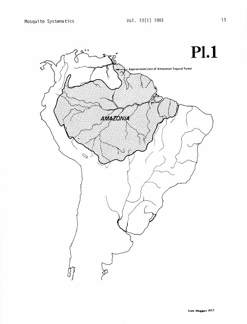
Amazonia, as represented by the tropical forest, is delimited for the purposes of this handbook as including northern Brazil, the Guianas, the eastern margins of Venezuela, Colombia, Ecuador and Peru, and northern Bolivia (pl. 1; Meggers 1977); it is sometimes referred to in this paper as simply the “Amazon.”
The majority of the material for this study was collected (or acquired) by the project Mosquitoes of Middle America (MOMA), University of California, Los Angeles (UCLA) and the Medical Entomology Project (MEP), Smithsonian Institution, Washington, DC.
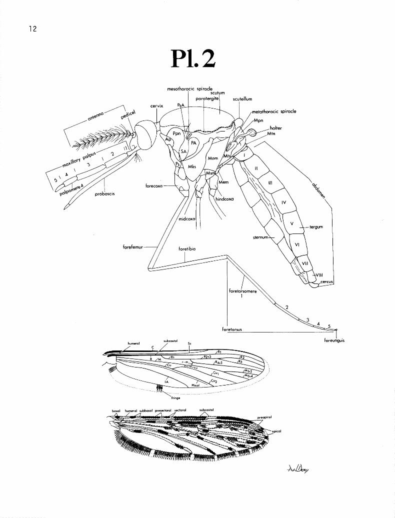
TERMINOLOGY. The terminology and abbreviations are largely those of Harbach and Knight ( 1980). The form of presentation is essentially that of Belkin ( 1962). Regarding the chaetotaxy of the immatures, branching for those setae considered to be taxonomically important is usually listed as 2 sets of figures. The first set of numbers after the seta represents the frequen- cy of at least 75% of the observations. The set of numbers following this “75% range” is the en- tire range of all observations; if the 75% range and the entire range are the same, then only one set of figures is listed. Following Harbach and Knight ( 1980) a number of new terms are used for the adult thorax, palpus, antenna, tarsi and male genitalia, and the larval spiracular plate. In addition, a few special terms are used for the wing spots of the adult, the ventral and dorsal lobes of the claspette (ventral and dorsal claspettes) of the male genitalia, and abdominal tergal plates of the larva. These terms are listed in the chapter on taxonomic characters and illustrated on plates 2-5.
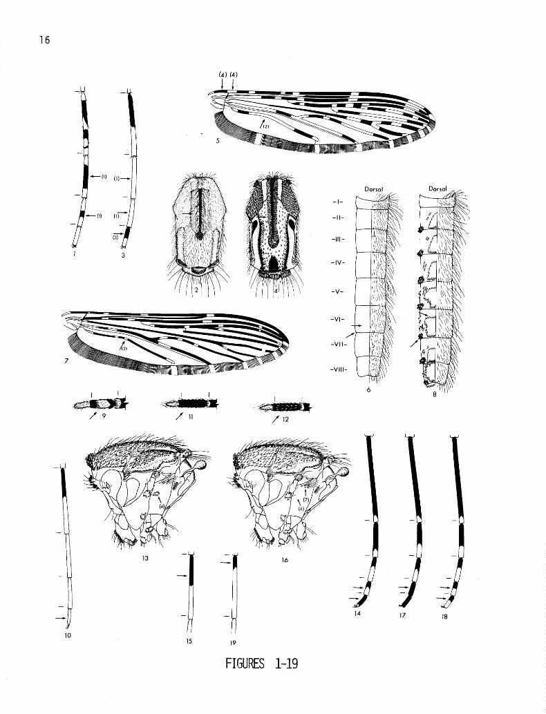
KEYS. Illustrated identification keys are given for the adult females, male genitalia and fourth stage larvae. For each couplet in the key, it is very important to read the entire first half of the couplet before proceeding to the second half. In the figures, a number in parentheses associated with an arrow indicates to which couplet the structure pertains; these parenthesized numbers are used when different portions of a single figure are used for more than one couplet.
DISCUSSIONS. The beginning of this section for each species pertains to the important differentiating characters for the adult female, male genitalia and larva. When pertinent to this handbook, affinities, evolutionary trends, and intrapopulational and geographical variations are presented.
3 Linthicum, Kenneth J. 1978. A revision of the Argyritarsis Section of the subgenus Nyssorhynchus of Anopheles. Ph.D. dissertation. University of California, Los Angeles. 409 p.

BIONOMICS. Unless otherwise stated, those data on the habitat of the immatures have been extracted from the MOMA field collection records on file at the Smithsonian Institution. Addition- al information about the natural history of each species has been taken from the literature.
MEDICAL IMPORTANCE. A brief summary of the medical importance of each species is included following the bionomics. This section is only a summary, and does not in any way attempt to be an inclusive discussion of the literature. It pertains primarily to malaria and what is known concerning the vector capacity of each species.
DISTRIBUTION. The known distribution is given for each species and is also outlined on plates 6- 10. On these plates, a solid line broken by a dotted line implies uncertainty regarding the distribution of that particular species within the area bordered by the dotted line (for example: pl. 8, rondoni; pl. 10, galvaoi).
FIGURES and PLATES. The drawings in the key are referred to as figures (fig.) to distin- guish them from the other drawings in the handbook that are designated as plates (~1.). In addition to the figures in the key to the larvae, the complete illustration for the fourth stage larva of each species is included following the distribution maps (except for oswaldoi, pl. 5).
SYSTEMATICS
Plates 11, 12
The subgenus Nyssorhynchus has been subdivided into two sections, the Argyritarsis Section and the Albimanus Section (Faran 1980). The 2 sections are closely allied to each other and togeth- er form a tight, well-defined taxon. The entire subgenus is restricted to the Neotropics except for albimanus which extends into the Nearctic. The Albimanus Section is distinguished from the Argy- ritarsis Section in the adults primarily by the basal dark band on hindtarsomere 5, and in the male genitalia by the variously developed, fused ventral lobes of the claspette (ventral claspette). Only on the basis of these two characters can these sections be readily differentiated. The Myzorhynch- ella group has been excluded from consideration because of the paucity of material available for study, and because of its uncertain taxonomic position.
The Argyritarsis Section includes 8 valid species that we have separated into 2 groups, the Albitarsis Group and Argyritarsis Group. The more ancestral Argyritarsis Group has been separated into 4 subgroups: the Argyritarsis Subgroup composed of argyritarsis and sawyeri, and the mono- typic Lanei, Pictipennis and Darlingi Subgroups. We have divided the Albitarsis Group into 2 sub groups, the monotypic Braziliensis Subgroup and the Albitarsis Subgroup composed of 2 species, albitarsis and allopha. We no longer consider allopha to be a junior synonym of albitarsis but a sep- arate, distinct species (see discussion section for allopha).
Faran (1980) divided the Albimanus Section into 2 groups, the monotypic Albimanus Group and the Oswaldoi Group. The Oswaldoi Group has been separated into 2 subgroups, the monotypic Triannulatus Subgroup, and the Oswaldoi Subgroup composed of 12 species, which is further separated into the Oswaldoi Series and the Strodei Series. Fat-an (1980) referred to these latter 2 taxa as the Oswaldoi Complex and the Strodei Complex. Within the Oswaldoi Series, 2 sep- arate phyletic lines are discernible on the basis of the structure of the ventral lobe of the claspette of the male genitalia. One line is composed of oswaldoi, galvaoi, evansi, aquasalis, ininii and possibly the relict anomalophyllus, and the other by rangeli, trinkae and nuneztovari. It is important to mention that evansi is now considered to be the senior synonym of noroestensis (Faran 1981), and no longer a nomen dubium (Faran 1980), or the senior synonym of strodei (Knight and Stone 1977). The Strodei Series contains strodei, rondoni and benarrochi. An. strodei and rondoni are very close- ly related, but their relationship to benarrochi is difficult to determine as benarrochi is the most

Mosquito Systetnatics Vol. 13(l) 1981
derived species in the section. An. hcnurrochi has been placed in the Strodei Series only because of the dcrivcd similarity ot’ its malt genitalia with those ofslrmfei and rcmdoni.
TAXONOMIC CHARACTERS
The following is a summary of the pertinent characters used for species identification. The most reliable characters for species identification are in the male genitalia and the larva. The exter- nal morphology of the adult female, particularly in the case of the Oswaldoi Subgroup, is very simi- lar interspecifically and usually variable intraspecifically. For this reason, the key to adult females may not always be entirely reliable when used by itself. It is highly recommended that one refer to the species discussions when attempting to identify any species in this subgroup, and to correlate those data with the information given on the bionomics and distribution. As has been emphasized by Belkin ( 1962), it is best to examine more than one specimen. To be certain of an identification, the immatures should be individually reared, and slides prepared of their exuviae and of the genitalia of the corresponding males to permit the correlation of characters in the different life stages. Where morphologically similar species occur sympatrically it is doubly important to use more than one de- velopmental stage. Lastly, it must be emphasized that not all the populations from every locality where each species exists were examined. Thus, the range of variation reported is conservative, and although sufficient in most cases, may be exceeded in some individuals. Also, as indicated by Arnell (1973), the illustrations of the larvae show only the modal condition of the setal branching and can- not represent the variation that occurs within a species.
Adult Females Plates 2, 3
The important differentiating characters in the adult female are the (1) maxillary palpo- meres 45, (2) presence or absence of scales on mesanepimeron, (3) number of setae on posterior margin of scutellum, (4) banding patterns of legs, (5) relative lengths, and presence or absence of wingspots, (6) dark caudolateral scale tufts of abdomen and (7) presence or absence of scales on first abdominal sternum.
The new terminology (Harbach and Knight 1980) used in the keys, discussions and illustra- tions for the head, thorax and legs is listed here after the old terminology, and is illustrated in plates 2 and 3.
Old Name New Name anterior pronotum an teprono turn
meron mesomeron mesepimeron mesanepimeron
palpal segment palpomere postnotum mesopostnotum
posterior pronotum postpronotum propleuron proepisternum
spiracular area prespiracular area sternopleuron mesokatepisternum tarsal segment tarsomere
torus pedicel The scale banding patterns in the fore- and midtarsus, as in the hindtarsus, are important diagnos-
tic characters. The dorsal surface of the fore- and midtarsomere 1 is predominantly dark, speckled with light scales and with a light apical band usually less than 0.1 the length of the tarsomere. The
5

6
ventral surfaces of the fore- and midtarsomeres 1,2 are light; the ventral surfaces of fore- and mid- tarsomeres 3-5 are often speckled with dark scales, varying from completely light to dark. Foretar- somere 1 has a white to golden apical band in some species. Foretarsomere 2 has a narrow to wide, light-scaled band in apical 0.15-0.95. Foretarsomere 3 is light scaled in apical 0.1-0.9. Foretarso- meres 4,5 are from dark to predominantly light. Midtarsal markings are generally as in the foretar- sus except that the ventral surface usually is darker, and the apical light bands, when present, are white to golden, usually darker than on the foretarsus. The dorsal surfaces of midtarsomeres 2,3 are from completely dark to light in apical 0.4. Midtarsomere 4 is usually dark, occasionally with an apical light band (ininii). Midtarsomere 5 is from completely dark to completely light. Hindtar- somere 1 is predominantly dark with a speckling of light scales on the ventral surface, a longitudinal light streak on the anterior surface and sometimes a yellow to white band at the apex. Hindtarso- mere 2 is highly variable, with a dark band in basal 0.05-0.90. Hindtarsomeres 3,4 are completely white or with a dark basal band present on either one (rondoni, uncommon variants) or both tarso- meres (uncommon variants). Hindtarsomere 5 is from completely white to dark in about basal OS.
The wing venation and wing spots are illustrated in plate 2. The special terminology used for the wing spots is modified from Zavortink (1973). Vein C usually has a basal, humeral, subba- sal, presectoral, sectoral, subcostal and preapical light-scaled spot; the subbasal, presectoral and subcostal light spots are sometimes absent. Vein Rs-R2+a is variable, more or less predominantly dark, with 3 or 4 dark spots, 2 or 3 light spots and with or without an extra subcostal light spot. Veins R, and R3 have 2 or 3 light spots. Vein R 4+5 has 2 small to moderately large dark spots, one subcostal, the other preapical. Vein M has a subcostal dark spot that in some species reaches the furcation; M may or may not have a sectoral dark spot of variable length. Vein MI+* has 2 dark spots, and M3+4 has 1 dark spot. The base of vein Cu is light with a small to medium sectoral dark spot usually not reaching furcation. Vein Cur is predominantly light, with usually 3 small, dark spots, 2 toward base in sectoral-subcostal region and 1 preapical. Vein Cu, is light except for a small to moderate preapical dark spot. Vein A is predominantly light, with 2 dark spots, 1 subbasal and 1 subcostal. The apical light fringe spot is conspicuous and is small to large; fringe of remainder of wing is largely dark with light areas where veins intersect wing margin except occasionally for R,; usually there is an additional moderately long, light fringe spot present between base of wing and apex of vein A.
Male Genitalia Plate 4
The subgenus Nyssorhynchus is characterized by the fusion of the ventral lobes of the clasp- ette to form a single median structure, and by the single parabasal seta. The claspette in Nysso- rhynchus is divided into a dorsolateral lobe (dorsal lobe of Harbach and Knight 1980) and an apical- ly fused, membranous, mesoventral lobe (ventral lobe of Harbach and Knight 1980). To avoid pos- sible confusion, which could occur because of the various uses of the term “lobe” in reference to the structure of the ventral lobe of the claspette, we shall refer to the ventral and dorsal lobes of the claspette simply as the “ventral cIaspette” and the “dorsal claspette” respectively. The species in this subgenus have 2 accessory setae and a single internal seta.
The new terminology (Harbach and Knight 1980) used in the keys, discussions and illustra- tions for the male genitalia is listed here after the old terminology, and is illustrated in plate 4.
Old Name New Name accessory spine accessory seta basal apodeme apodeme of gonocoxite
clasper gonostylus in ternal spine in ternal seta
parabasal spine parabasal seta

Mosquito Systematics
sidepiece tergomedial bristle
DORSAL CLASPETTE. In
Vol. 13(l) 1981
gonocoxite tergomedial seta
the Argyritarsis Section the dorsal claspette possesses reliable characters for species delineation. The dorsal seta (leaflet) of the dorsal claspette of darlingi and braziliensis has a well-developed basomesal projection, while that of all the other species in the section is without a basomesal projection.
VENTRAL CLASPETTE. The fused ventral claspette best delimits the taxa within the sub- genus and has been used most often to divide the subgenus into different groups, subgroups and ser- ies. All the species in the Argyritarsis Section lack spicules on the ventral claspette, whereas all the species in the Albimanus Section, except for albimanus and triannulatus, possess spicules. “Spic- ules” is used here to denote the vestiture present on the ventral claspette rather than “setae” previ- ously used in Faran ( 1980), as these processes lack the aveolus characteristic of true setae. The vari- ous components of the ventral claspette used in the key and the discussions, such as the basal lob- ules, preapical plate, refringent structure, membranous area, median sulcus and mesal cleft, are illus- trated in plate 4.
PHALLOSOME. We believe that primitively in Nyssorhynchus the aedeagus had sclerotized, long, serrated, subapical leaflets. This condition is present in all species in the Argyritarsis Group and in anomalophyllus of the Albimanus Section. In all other species sclerotized, serrated leaflets are absent. However, in several species of the Oswaldoi Series remnants of leaflets still occur as un- serrated, membranous, basolateral expansions of the apex of the aedeagus. Wherever width of aede- agus is used, it refers to the width of the aedeagus at the base of the apex (subapex where lateral sclerotizations extend ventromedially to form an incomplete tube; referred to as the ventromesal subtriangular projections). Length of apex of aedeagus refers to distance from the subapical, collar- like, ventromesal subtriangular projection to apex of aedeagus.
Fourth Stage Larvae Plate 5
Many characters in the larvae of Nyssorhynchus correlate with characters in the adult and male genitalia to clearly define the separate taxa. The chaetotaxy is very important for species iden- tification.
HEAD. The important setae are 2,3-C, and the length and branching of 4-C. Setae 2,3-C may be (1) of subequal or unequal lengths and (2) single and simple, barbed or plumose with branches short to long and occasionally dendritic. The relationship between the approximation of setae 2-C (inner clypeals) and setae 3-C (outer clypeals) is given by the clypeal index, which is the distance between the insertions of seta 2-C and seta 3-C on one side of the head divided by the dis- tance separating insertions of setae 2-C (pl. 5).
ANTENNA. Seta 1-A in the ancestral condition is small; it is large and independently de- rived in benarrochi and ininii.
THORAX. Seta 1-P in the ancestral condition is plumose and multibranched with filiform branches; in the derived state, 1-P is palmate with lanceolate branches. Other important characters are ( 1) presence or absence of common sclerotized tubercle for setae 1,2-P or 1-3-P, (2) approxima- tion of setae 1-P from opposite sides and (3) seta 3-T filiform or with lanceolate branches.
ABDOMEN. Primitively, seta 1-I has filiform branches; this condition is present only in ar- gyritarsis and darlingi. Seta 1-I in sawyeri may represent an intermediate condition with very weak- ly developed lanceolate leaflets. In all other species seta l-1 has well-developed lanceolate leaflets. Important characters for species identification include (1) size and number of branches of setae 1 l- I, 13-I-IV, and O-II, (2) location and size of seta 5-I,II, (3) width of leaflets of palmate seta I-II-VII and (4) in the Argyritarsis Section, the varied development of the multiple tergal plates on segments I-VIII. In the Argyritarsis Section the median tergal plate is usually moderately large, strongly scler-

8
otized and located on or near the anterior margin of segments I-VIII. Accessory median and acces- sory submedian tergal plates are present caudad of the median tergal plate, and are usually subspher- ical or ovoid, and small (only about 0.05 the area of the median tergal plate). The accessory median tergal plates are ( 1) oriented longitudinally on the midline 0.1 the length of the segment from the anterior margin, (2) always absent on segment I, (3) single or absent on segment II, (4) single (argyr- itarsis, darlingi, braziliensis) or sometimes double (allopha) on segment III and (5) single and with- out lobes (argyritarsis) or bilobed (allopha, darlingi, braziliensis) or trilobed (allopha, braziliensis) or with 2,3 round plates (allopha) on segments IV-VII. The accessory submedian tergal plates are (1) usually in pairs with one immediately on each side of midline located 0.5 length of segment from anterior margin of segment, (2) present or absent in argyritarsis, (3) present on segments I-VII of allopha, braziliensis and darlingi as 1 or 2 pairs.
SPIRACULAR LOBE. In Faran ( 1980) the term lateral arm of the “spiracular apparatus” was used to denote the lateral arm of the “median plate of the spiracular apparatus” of Harbach and Knight (1980). We are following the latter authors’ usage of the term “median plate.” In the ances- tral condition the lateral arm of the median plate of the spiracular apparatus is either nonexistent or short. There has been an independent increase in the size of the lateral arm in triannulatus and, to a lesser degree, in ininii. The dentition of the pecten is important as a group character in the Argyri- tarsis Section and is a secondary key character in species diagnosis in the Albimanus Section.
ANAL SEGMENT. In the Albimanus Section there appears to be a trend for the insertion of seta 1-X to migrate from a dorsal position within the saddle, to a position on or near the ventral margin of the saddle. In the most derived case (oswaldoi) seta 1-X is not inserted on the saddle at all, but ventrad of it. Seta 1-X is moderately long to long in all species except benarrochi. The anal gills are short in aquasalis, and in all other Amazonian species of Nyssorhynchus, they are moderate- ly long to very long.
BIONOMICS
The immature stages of the subgenus Nyssorhynchus are found predominantly in ground water. They occur in a variety of habitats such as ponds, lakes, stream and river margins, seepage and drainage areas, ditches, flooded meadows and pastures, reservoirs, swamps, ground pools, bor- row pits, and animal and vehicle tracks. An. aquasalis is the only species primarily restricted to the coast; this species preferentially occurs in brackish water such as in mangrove swamps and coastal ground pools. However, aquasalis is capable of living in fresh water and often is collected several kilometers from the coast. An. argyritarsis, although usually found in ground pools, occasionally is collected in artificial containers such as tin cans and animal watering troughs. An. triannulatus, ininii and allopha commonly are found in lakes, ponds or large ground pools. An. triannulatus is the only species clearly shown often to be closely associated with a specific plant; it is usually col- lected in or between the rosette crowns of Pistia stratiotes Linnaeus. An. allopha is always found with some vegetation such as green algae, Eichhornia spp., Ceratophyllum spp. and Salvinia spp. An. darlingi is often collected in mats of Ceratophyllum spp. along the margins of rivers and canals in partial or well-shaded areas. An. evansi and braziliensis are usually collected in forests or areas of secondary vegetation. An. oswaldoi, ininii and triannulatus adults are usually collected in the interi- or of forests, although the larvae may be collected from ground pools in interspersed secondary growth areas. An. rondoni occurs in ditches, puddles, flooded meadows, etc., in southern Brazil and northern Argentina, and is not reported further north than the southern margin of upper Amazonia.
Regarding altitudinal distribution, strodei has been collected at the highest elevations (1600 m) for any species of Nyssorhynchus, although it also occurs at lower elevations. An. rangeli and, to a lesser extent, benarrochi are principally found at intermediate elevations (200-1000 m), such as

Mosquito Systematics Vol. 13(l) 1981
in the upper Amazon and the llano plateau region of Colombia, extending south to Mato Gross0 and Bolivia. An. aquasalis normally occurs on or near the coast usually at elevations less than 400 m. The remainder of the Amazonian species are found at low to intermediate elevations.
Host preference studies for several species indicate that these mosquitoes feed predominant- ly on large mammals such as dogs, cats, cattle, pigs, goats, donkeys and man; occasionally some feed on fowl. In all known cases, the species feed readily on man when given the opportunity (ininii and galvaoi have not been studied in this respect). The adults are active either crepuscularly or nocturn- ally except for those of triannulatus and braziliensis, which may be somewhat diurnal. Most of the species are exophilic and zoophilic except, in certain instances, for aquasalis, nuneztovari, darlingi and allopha.
MEDICAL IMPORTANCE
An. darlingi is a very efficient vector of malaria in northern and northeastern Brazil as well as in numerous other areas in South America. Wherever this species occurs along with malaria, dar- Zingi females are almost always found naturally infected. An. darlingi is highly endophagous and anthropophilic. In addition to malaria, this species has also been suspected of being a vector of hu- man filariasis. It has transmitted Wuchereria bancrofti (Cobbold) in the laboratory and has been collected naturally infected with this parasite.
An. allopha, although usually not a primary vector of malaria, may act as a secondary vector (Galvao 1940). This species will readily attack man and at times is very endophilic. It has been ex- perimentally infected with malaria parasites and has been collected naturally infected.
An. braziliensis, like allopha, is not a primary vector of malaria but is capable of malaria transmission under the proper ecological conditions (Deane, Causey and Deane 1948). Normally this species is exophilic and zoophilic; however, in some regions of Amazonia when domestic ani- mals are absent, braziliensis will readily enter houses and bite man. It has been collected naturally infected with Plasmodium spp. An. argyritarsis is generally considered not to be a primary vector of malaria but may be important when it occurs at high densities. Although it is rarely found inside houses and rarely attacks man, argyritarsis has been found naturally infected with malaria parasites.
An. aquasazis is a primary vector of malaria in the Lesser Antilles, and in Trinidad and Toba- go. Along the coast of Brazil, the Guianas and possibly Venezuela, it is always a potential vector but usually only important when it occurs in large numbers. An. aquasalis feeds readily on man and is commonly collected in houses. In the past it has been an important vector of malaria in coastal Brazil.
An. nuneztovari is a primary vector of malaria in western Venezuela and northern Colombia, and is a probable vector in Suriname; in some areas where it occurs in Venezuela, spleen indices have been close to 100% (Gabaldon and Guerrero 1959). In Venezuela and Colombia the vector poten- tial of nuneztovari has been reported to depend on the density of the nearby vegetation surrounding regions of habitation, which may be correlated with the vector’s greater life expectancy and density in the forest (Hamon, Mouchet et al. 1970). In the Amazon basin nuneztovari has not been report- ed as being important as a vector of malaria.
An. triannulatus does not seem to be an important vector of malaria. This species was impli- cated as a possible vector during an epidemic at a boys’ school near Maracay, Venezuela (Benarroch 193 I), and once was found to have a natural oocyst infection (Gabaldon and Cova Garcia 1946b). Several investigators have experimentally infected triannulatus with Plasmodium vivax (Grassi and Feletti) and P. falciparum (Welch); however, it is much more refractory to infection than albimanus.
According to Correa ( 1938), strodei transmitted malaria at the Fazenda Santa Alice, Sao Paula, Brazil; he reported a natural infection rate of 1.2%. Other Brazilian workers have experi-
9

10
mentally infected strodei with Plasmodium vivax, although there have not been any other reports implicating strodei as a vector.
Very little is known regarding the vector potential of rangeli, evansi, rondoni and oswaldoi. An. evansi was found naturally infected once in Ribeira, Sao Paulo, Brazil by Correa and Ramos ( 1942b). Lucena ( 1940) reported finding oswaldoi var. metcaij? naturally infected in Pontesinha, Brazil; however, Lucena may have been studying aquasalis rather than evansi. An. oswaldoi has been experimentally infected with P. vivax and P. falciparum (Fonseca and Fonseca 1942; Roze- boom 1942). An. rangeli has been suspected of transmitting malaria in Ecuador (Forattini 1962), but it has never been shown to be naturally infected. An. rondoni was investigated in Jujuy, Argen- tina, during the malaria season by Davis and Shannon (1928), and it was not found naturally infect- ed nor was it possible to infect it experimentally with P. falciparum, P. vivax or P. malariae (Grassi and Feletti) in 3 different experiments. Nevertheless, Shannon and Del Ponte (1927) stated that Davis was able to infect rondoni in other experiments. Nothing is known about the vector potential of ininii or galvaoi.
ABBREVIATIONS FOR ADULT PLATES 2 AND 3
A anterior Mtn metanotum AP antepronotum Mtpn metapostnotum L lower Mts me tepisternum Mam mesanepimeron PA postspiracular area Mem me tameron PPn postpronotum Mks mesokatepistemum Ps proepistemum Mpn mesopostnotum PsA prespiracular area Msm mesomeron Mtm metepimeron
A antenna C cranium M mesothorax Mx maxilla P prothorax
SA U
subspiracular area upper
ABBREVIATIONS FOR LARVAL PLATE 5
PT pecten S spiracular lobe SAP spiracular apparatus T metathorax
Mosquito Systematics Vol. 13(l) 1981
e Limit of Amozonion Tropicoi Forest
from Muggers 1977
11
12
PI.2 mesothoracic spiracle
metathoracic spiracle
humeral subcortol
/ SC
I foretarsus
basal humtral subbasal tmsectoral sectorol
I foreunguls
pical

Mosquito Systematics Vol. 13(l) 1981 13
knob
THORAX - LATERAL
median anterior promontory
scutal fossalP
THORAX- DORSAL SCALING
THORA;iT;ERSAL nORSAL - ABDOMEN - VENTRAL
Caudolateral
scale tuft.
P1.3 LEGS
FORE- MID- HIND-

14
P1.4
refringent struct
tergomedial seta
ventral lobe of claspette (ventral claspette)
internal seta oswaldoi
ventromesal
subtriangular projection
anterior apode
phallosome

Mosquito Systematics Vol. 13(l) 1981
clypeal index =I diatance between Z-C and 3-C on one side
distance separating eetae 2-C
NYSSORHYNCHUS
n plate of epiracular apparatus
15
-t6
- ‘d - M -(O (I)-+
- -
~ /
- (0 (li-- -
GT
1 3
/
-I-
-II-
-Ill-
-IV-
-V-
18
10
FIGURES l-19

Mosquito Systematics Vol. 13(l) 1981 17
1.
ILLUSTRATED KEYS TO THE AMAZONIAN SPECIES OF ANOPHELES (NYSSORHYNCHUS)
FEMALES (6. galvaoi and 13. rondoni not included; see species discussions)
Hindtarsomeres 3 and 4 not entirely white (fig. 1); acrostichal and dorsocentral areas at most with scattered scales (fig. 2); wings variable . . . . . . . . . . . . . . . . . . . . . . Subgenera Anopheles, Lophopodomyia, Kerteszia and Stethomyia
Hindtarsomeres 3 and 4 entirely white, except in unusual variants (fig. 3); acrostichal and dorsocentral areas with numerous scales (fig. 4); wings with distinct light and dark spots (fig. 5) (Subgenus Nyssorhynchus) . . . . . . . . . . . . . . . . . .2
Abdominal terga II-VII without obvious scales or caudolateral scale tufts (fig. 6); vein 1A more than 0.5 dark (fig. 7) . . . ‘0’. . . . . . . . Myzorhynchella group
At least some of abdominal terga II-VII with obvious scales and caudolateral scale tufts (fig. 8); vein 1A 0.5 or more light (fig. 5) (Albimanus and Argyritarsis Sections) . . 3
Hindtarsomere 5 with a basal dark band (fig. 3); palpomere 4 with at least some white or cream on mediolateral surface (fig. 9) (Albimanus Section) . . . . . . . . . . 4
Hindtarsomere 5 entirely white (fig. 10); palpomere 4 dark (fig. 1 l), or with light and dark scales intermingled on mediolateral surface (fig. 12) (Argyritarsis Section) . . 11
Anterior mesanepimeron (Mam) with a conspicuous patch of light scales (fig. 13); foretar- somere 4 with a light band in apical 0.40-0.65 (fig. 14); foretarsomere 5 predominantly dark (fig. 14); hindtarsomere 2 dark in basal 0.4-0.7 (fig. 15); vein C humeral light spot small, 0.5-1.3 length of basal dark spot (fig. 5); small species . . . . 15. triannukiztus
Anterior mesanepimeron (Mam) without a patch of light scales (fig. 16); foretarsomere 4 predominantly dark (fig. 17), or if light in more than apical 0.3 then foretarsomere 5 about 0.5 or more apically light (fig. 18) and/or hindtarsomere 2 less than 0.4 basally dark (fig. 19); vein C humeral light spot large, greater than 1.5 length of basal dark spot (fig. 20) except in nuneztovari; moderately large to large species . . . . . . . . 5

18
24
- - I - - 26 - I - -
28
FIGURES 20-N

Mosquito Systematics Vol. 13(l) 1981 19
X4). Hindtarsomere 2 with basal dark band usually less than 0.25 length of tarsomere (fig. 21); vein C humeral light spot greater than 1.5 length of basal dark spot (fig. 20) . . . . 6
Hindtarsomere 2 with basal dark band usually equal to or greater than 0.25 length of tar- somere (fig. 22), if less than 0.25 then vein C humeral light spot less than 1.5 length of Cbasaldarkspot(fig.23) . . . . . . . . . . . . . . . . . . . . . . .7
h(5). Foretarsomere 4 all light to rarely more than 0.3 basally dark (fig. 24); midtarsomere 4 with a light band in apical 0.15-0.25 (fig. 25); foretarsomeres 3-5 predominantly cream to white, dark scales often present only on dorsobasal surface of tarsomere (fig. 24); foretarsomere 2 light in apical 0.35-0.55 (fig. 24); foretarsomere 3 light in apical 0.70- 0.86(fig. 24) . . . . . . . . . . . . . . . . . . . . . . . . . 9. ininii
Foretarsomere 4 at least 0.3 basally dark to all dark (fig. 26); midtarsomere 4 all dark (fig. 27); dark basal bands on foretarsomeres 3-5 almost completely encircling each tar- somere, dark scales usually absent from ventral surface (fig. 26); foretarsomere 2 light in apical 0.20-0.45 (fig. 26); foretarsomere 3 light in apical 0.50-0.85 (fig. 26) . . . . . . . . . . . . . . . . . . . . . . . . . . . . . . . . 5. oswaldoi
7(5). Subcostal light spot of vein C usually greater than 0.5 (0.45-1.00) length of subcostal dark spot (fig. 28); upper mesanepimeron (Mam) often with l-4 light obovate scales (fig. 29); hindtarsomere 2 usually dark in basal 0.25-0.35 (fig. 22); humeral light spot of vein C usually large, 1.8-3.5 ( 1 J-3.7) length of basal dark spot (fig. 28) . 10. rangeli
Subcostal light spot of vein C almost always less than 0.5 length of subcostal dark spot (fig. 20); upper mesanepimeron (Mam) usually without light scales (fig. 16); hindtarso- mere 2 and humeral light spot of vein C variable , . . . . . . . . . . . . . . 8
8(7). Hindtarsomere 2 dark in about basal half, 0.40-0.55 (0.3-0.6) (fig. 30); light wing spots at least on veins C and R light cream to yellowish, not white . . . . . . 8. aqua&is
14. benarrochi
Hindtarsomere 2 dark in less than basal 0.40 (fig. 22), or if greater than 0.40 then light wing spots white, not light cream to yellowish . . . . . . . . . . . . . . 9
9@). Vein C humeral light spot less than 2.0 (0.7-1.7) length of basal dark spot (fig. 23) . . . . . . . . . . . . . . . . . . . . . . . . . . . . . . 11. nuneztovari
Vein C humeral light spot equal to or greater than 2.0 length of basal dark spot (fig. 20) . . . . . . . . . . . . . . . . . . . . . . . . . . . . . . . . . 10

20
36
32 34 - -
-J -1
- -
I I
t
39 44
42
Dorsal
Dorsal
- -
-11 - - 46 49
FIGURES 31-50

Mosquito Systematics Vol. 13(l) 1981
1 o(9). Light scales on wing (at least anterior veins) and coxae (usually) gray to cream to yellow, not white; foretarsomere 5 cream, gray or golden in apical 0.30-0.50 (fig. 3 1); midtar- somere 5 gray to cream in about apical 0.5 (fig. 32) . . . . . . . . . . 7. evansi
Light scales on wing and coxae usually white or very light cream; foretarsomere 5 usually golden to brown (fig. 33), occasionally differentiated into 0.5 dark, 0.5 light; midtarso- mere 5 usually cream in less than apical 0.3 (fig. 34) . . . . . . . . . 12. strodei
1 l(3). Sternum I completely bare, without white scales (fig. 35); scutellum usually with more than 12 large, dark se tae on posterior margin (fig. 36) . . . . . . . . . . . . 12
Sternum I with 2 longitudinal rows of white scales (fig. 37); scutellum usually with fewer than 12 large, dark setae on posterior margin (fig. 38) . . . . . . . . . . . 13
12(11). Anterior mesanepimeron (Mam) with a distinct patch of light scales (fig. 39); upper mes- anepimeron (Mam) without scales (fig. 39); vein C with basal dark spot greatly en- larged, about 4.0 of humeral light spot (fig. 40); vein R3 with 3 dark spots (fig. 40); palpomere 4 with scattered white scales on mediolateral surface (fig. 4 1) . 2. darlingi
Anterior mesanepimeron (Mam) bare (fig. 42); upper mesanepimeron (Mam) with a line of light scales (fig. 42); vein C with basal dark spot not greatly enlarged, at most equal to humeral light spot (fig. 43); vein R3 with 2 dark spots (fig. 43); palpomere 4 with- out scattered light scales on mediolateral surface (fig. 44) . . . . . . 1. argvritarsis
13(11). Caudolateral scale tufts well developed on abdominal segment II (fig. 45); hindtarsomere 2 with basal 0.3-0.4 dark (fig. 46); vein C with or without a small presectoral light spot (fig. 47); vein R3 with 3 dark spots (fig. 47); abdominal terga II-IV with relatively few light scales (fig. 45) . . . . . . . . . . . . . . . . . . . . . 4. braziliensis
21
Caudolateral scale tufts absent on abdominal segment II (fig. 48); hindtarsomere 2 with basal 0.45-0.90 dark (fig. 49); vein C never with presectoral light spot (fig. 50); vein R3 with 1 or 2 dark spots (fig. 50); abdominal terga II-IV with numerous light scales (fig. 48) . . . _ . . . . . . . . . . . . . . . . . . . . . . . . 3. allopha

Mosquito Systematics Vol. 13(l) 1981 23
MALE GENITALIA (13. rondoni not included; see species discussion)
1. Ventral claspette not apically fused into single structure (fig. 5 1); gonocoxite with more than one parabasal seta, or if one then longer than 0.3 length of gonocoxite (fig. 52) or not arising from prominent tubercle; gonocoxite with or without accessory seta (fig. 52) . . . . . . Subgenera Anopheles, Lophopodomyia, Kerteszia and Stethomyia
Ventral claspette apically fused into a single structure (fig. 53); gonocoxite with one para- basal seta of less than 0.3 length of gonocoxite arising from a prominent tubercle and with 2 accessory setae (fig. 54) (Subgenus Nyssorhynchus) . . . . . . . . . . . 2
x0. Ventral claspette with spicules (fig. 53) ................... 3
Ventral claspette without spicules (fig. 55) ................. 11
3(2). Ventral claspette with apex rugose or deeply striated, and moderately to strongly expand- ed laterally into rounded or pointed lobe, spicules on lateral margins not extending toward apex as far as level of apical margin of preapical plate (fig. 56); apex of aedea- gus without leaflets (fig. 57) . . . . . . . . . . . . . . . . . . . . . .4
Ventral claspette with apex neither rugose nor deeply striated nor laterally expanded, spicules extending to apex or at least as far as level of apical margin of preapical plate (fig. 58); apex of aedeagus with or without leaflets . . . . . . . . . . . . . . 5
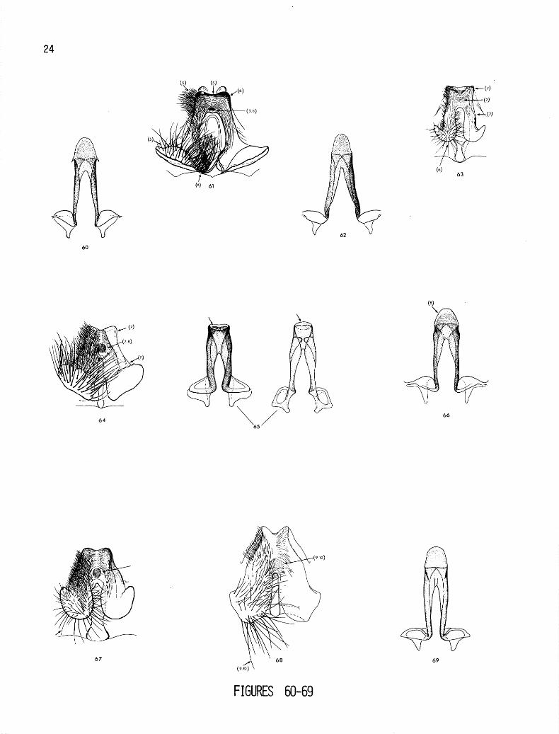
4(3)* Ventral claspette small, about 0.33 length of gonocoxite, apex moderately expanded lat- erally, apicolateral margins sharply angled and moderately pointed (fig. 53); basal lob- ule of ventral claspette small, narrow, curving mesad, with spicules along basal margin short, about equal to or slightly longer than width of aedeagus (figs. 53, 59); preapical plate of ventral claspette heavily sclerotized (fig. 53) . . . . . . . 14. benarrochi
Ventral claspette large, about 0.5 length of gonocoxite, apex strongly expanded laterally, apicolateral margin produced into large, rounded lobe with basal and lateral margins convex and apical margin weakly concave (fig. 56); basal lobule of ventral claspette large, with spicules along basal margin long, about 2.0-3.5 width of aedeagus (figs. 56, 57); preapical plate of ventral claspette weakly to moderately sclerotized (fig. 56) . . . . . . . . . . . . . . . . . . . . . . . . . . . . . . . . 12. strodei
24
FIGURES 60-69

Mosquito Systematics Vol. 13(l) 1981 25
X3). Ventral claspette strongly conoid, with a very small median sulcus at apex, spicules on lateral margins (exclusive of basal lobule) short and extending toward apex only as far as level of apical margin of preapical plate, spicules along basal margin of basal lobule long about 2.0-3.0 width of aedeagus, preapical plate very large, crescent shaped and heavily sclerotized (figs. 5 8, 60) . . . . . . . . . . . . . . . . . . 9. ininii
Ventral claspette not strongly conoid, either truncate or with a moderately small to large median sulcus at apex, spicules on lateral margins extending to or nearly to apex, spic- ules along basal margin of basal lobule short to long, preapical plate small to large (fig. 61)................................6
6(5). Ventral claspette with a concentration of long spicules about 1.5 width of aedeagus on basomesal margin of basallobuledirected caudally into mesa1 cleft, wide at apex (width 0.4-0.5 length of claspette) with abruptly angled, rounded lateral margins, preapical plate small, oval and heavily sclerotized (figs. 6 1, 62) . . . . . . . . . 10. rangeli
Ventral claspette without spicules concentrated on basomesal margin of basal lobule, shape and preapical plate varied (fig. 63) . . . . . . . . . . . . . . . . . . 7
7W. Ventral claspette with apex moderately broad to broad, width at apex about 0.5-0.6 length of claspette, lateral margins of claspette not tapering appreciably medially toward apex, preapical plate moderately small and semicircular to oval (fig. 63) . . . . . . . . . . . . . . . . . . . . . . . . . . . . . . 1 I. nuneztovari
Ventral claspette with apex moderately narrow to narrow, width at apex about 0.3 length of claspette, lateral margins of claspette tapering toward apex, preapical plate variously developed (fig. 64) . . . . . . . . . . . . . . . . . . . . . . . . . . 8
g(7). Aedeagus truncate or weakly rounded at apex (fig. 65); preapical plate of ventral claspette moderately large, semicircular to oval, heavily sclerotized (fig. 64) . . . . . 7. evansi
Aedeagus well rounded at apex (fig. 66); preapical plate of ventral claspette variously developed . . . . . . . . . . . . . . . . . . . . . . . . . . . . .9
9W. Ventral claspette with spicules along basal margin of basal lobule moderately short about equal to or slightly longer than width of aedeagus, preapical plate moderately small, circular to oval and weakly to occasionally strongly sclerotized (figs. 66, 67) . . . . . . . . . . . . . . . . . . . . . . . . . . . . . . . . . 8. aquasalk
Ventral claspette with spicules along basal margin of basal lobule long about 2.0 or more width of aedeagus, preapical plate large and moderately to strongly sclerotized (figs. 68,69). . . . . . . . . . . . . . . . . . . . . . . . . . . . . . 10

26
71 73 72
81
FIGURES 70-85

Mosquito Systematics vol. 13(l) 1981 27
1 o(9). Ventral claspette with spicules along basal margin of basal lobule about 2.0 width of aede- agus, preapical plate large, usually crescent shaped and moderately to strongly sclero- tized (figs. 68,69) . . . . . . . . . . . . . . . . . . . . . 5. oswaldoi
Ventral claspette with spicules along basal margin of basal lobule very long about 3.0 width of aedeagus, preapical plate strongly sclerotized, large and circular to semicircu- lar with small basolateral projections (figs. 70, 71) . . . . . . . . . . 6. galvaoi
1 l(2). Aedeagus with a pair of strong, sclerotized subapical leaflets (fig. 72) ....... 12
Aedeagus without subapical leaflets (fig. 73) ................ 14
12( 11). Aedeagus hook-like at apex (fig. 74), or if not hooked then ventromesal subtriangular projection of aedeagus located about 0.5 from base of aedeagus (fig. 75) . . . . . . . . . . . . . . . . . . . . . . . . . . . . . . Myzorhynchella group
Aedeagus not hook-like at apex, ventromesal subtriangular projection subapical immedi- ately basad of subapical leaflets (fig. 72) . . . . . . I . . . . . . . . . 13
13( 12). Ventral claspette with apex broad and truncate, median sulcus small, often indistinct (fig. 55); apex of aedeagus egg shaped, longer than wide (fig. 72); dorsal leaflet of dorsal claspette with distinct basomesal projection (fig. 76) . . . . . . . . . 2. darlingi
Ventral claspette with apex narrow, median sulcus well developed (fig. 77); apex of aedea- gus broadly rounded, width 1.5 of length (fig. 78); dorsal leaflet of dorsal claspette without a basomesal projection (fig. 79) . . . . . . . . . . . . . 1. argvritarsis
14(11). Apex of ventral claspette expanded laterally into large, auriculate lobe (fig. 80); apex of aedeagus about 1.5 as long as wide (fig. 73) . . . . . . . . . . 15. triannukztus
Apex of ventral claspette not expanded laterally into large, auriculate lobe (fig. 81); apex of aedeagus as wide as or wider than long (fig. 82) . . . . . . . . . . . . . 15
15( 14). Ventral claspette with lateral margins slightly expanded laterally from apex to about 0.5 from base (fig. 8 1); dorsal leaflet of dorsal claspette with a distinct basomesal projec- tion (fig. 83) . . . . . . . . . . . . . . . . . . . . . . . 4. braziliensis
Ventral claspette with lateral margins strongly expanded laterally from apex to about 0.5 from base (fig. 84); dorsal leaflet of dorsal claspette without a prominent basomesal projection (fig. 85) . . . . . . . . . . . . . . . . . . . . . . 3. allopha

28
89 90
93 1-P
/ ’ 98 100
102
1101 103
FIGURES 86-104

Mosquito Systematics Vol. 13(l) 1981 29
LARVAE (6. galvaoi and 13. rondorti not included)
1. Setae l-III-VII with filiform branches (fig. 86) or lanceolate leaflets with notched margins (fig. 87) . . . . . . . . . . Subgenera Anopheles, Lophopodomyia, Stethomyia
Setae l-III-VII with smooth-margined lanceolate leaflets (fig. 88) (Subgenera Kerteszia, Nyssorhynchus) . . . . . . . . . . . . . . . . . . . . . . . . . . . 2
2(l). Setae 5-7-C single or forked (fig. 89) . . . . . . . . . . . . . Subgenus Kerteszia
Setae S-7-C plumose (fig. 90) (Subgenus Nyssorhynchus) . . . . . . . . . . . . 3
3~2). Setae 6-IV-VI branched (fig. 91) . . . . . . . . . . . . . Myzorhynchella group
Setae 6-IV-VI single (fig. 92) (Albimanus and Argyritarsis Sections) . . . . . . . . 4
4(3). Seta 1-P plumose or branched with filiform branches, never palmate (fig. 93); lateral arms of median plate of spiracular apparatus not projecting free laterally (fig. 94) . . 5
Seta 1-P always palmate with lanceolate branches (fig. 95); lateral arms of median plate of spiracular apparatus projecting free laterally, short to long (fig. 96) . . . . . . 6
X4). Seta 13-S very strongly developed, long, about 2.2-2.5 length of saddle (fig. 97); setae 2-C widely spaced, clypeal index about 1.5 (fig. 98) . . . . . . . . . . . 2. darlingi
Seta 13-S not strongly developed, never long (fig. 99); setae 2-C closely approximated, clypeal index greater than 4.0 (fig. 100) . . . . . . . . . . . . . 1. argyritarsis
6(4). Setae 2-C closely approximated, clypeal index 2.5 or greater (fig. 100) ....... 7
Setae 2-C widely separated, clypeal index less than 2.5 (fig. 98) .......... 8
7@). Seta 1-P often with blunt-tipped leaflets (fig. 101); 4-C usually single, long, extending to near or beyond insertion of 2-C (fig. 102); l-3-P or 1,2-P sharing common tubercle (fig. 101). . . . . . . . . . . . . . . . . . . . . . . . . . . 4. braziliensis
Seta 1-P with acuminate leaflets (fig. 103); 4-C l-4 branched, small to moderately small, usually not extending to near or beyond insertion of 2-C (fig. 104); l-3-P not sharing common tubercle (fig. 103), rarely 1,2-P share common tubercle . . . . 12. strodei

105 108
113 117
“t; 1. -_- 32 ?3
. 0
C
116
i-P 109
114 118
1 115
119
106
111
122 125
124 121
FIGUfES 105425

Mosquito Systematics Vol. 13(7) 7981
8(6).
9W.
lO(9).
Seta 11-I large, usually 5-7 branched (3-7) (fig. 1 OS); 13-I large, usually 3 branched (2-4) (fig. 1 OS); lateral arm of median plate of spiracular apparatus long, directed laterally (fig. 106); 1-P with 1 S-20 ( 13-20) very narrow to narrow lanceolate leaflets, leaflets not overlapping with leaflets of 1-P of opposite side (fig. 107) . . . 15. trianrzulatus
Seta 11-I moderately large, 2-4 branched (fig. 108); 13-I small to moderately large, usual- ly more than 3 branched (fig. 108); lateral arm of median plate of spiracular apparatus very short to moderately long (except long in i&ii) (fig. 96); 1-P with usually 9-l 6 (8- 18) narrow to broad lanceolate leaflets (fig. log), if more than 16 branched then leaf- lets overlapping with leaflets of 1-P of opposite side (fig. 110) . . . . . . . . . 9
Seta 3-C (and usually 2-C) plumose in about apical half, with distinct, moderately long to long branches (fig. 111) . . . . . . . . . . . . . . . . . . . . . . . 10
Setae 2,3-C single, and simple or barbed (fig. 112) . . . . . . . . . . . . . . 12
Seta 1-A long, at least 2.0 width of antenna at point of insertion (fig. 113); 13-IV small, usually lo- 13 branched (6- 13) (fig. 114); 1-X moderately short, as long as or slightly longer than saddle (fig. 115); 3-C with moderately long branches, 2-C single and barbed (fig. 116) . . . . . . . . . . . . . . . . . . . . . . . . 14. benarrochi
Seta 1-A short to moderately short, always less than 2.0 width of antenna at point of in- sertion (fig. 117); 13-IV moderately large, usually 5-7 branched (3-8) (fig. 118); 1-X longer than saddle (fig. 119); 2,3-C with branches subequal in length or branches of 3-C slightly longer than those of 2-C (fig. 111) . . . . . . . . . . . . . . 11
ll(10). Seta 1-X inserted on saddle, on or near ventral margin (fig. 120); anal gills usually short, about 0.5 length of anal segment (fig. 120); 2,3-C with simple branches, rarely dendri- tic (fig. 111); lateral arm of median plate of spiracular apparatus very short (fig. 12 1); pecten with shorter median teeth mixed medium and short (fig. 122) . .8. aqua&is
Seta 1-X not inserted on saddle (fig. 123); anal gills long, as long as or longer than anal segment (fig. 123); 2,3-C with usually dendritic branches (fig. 124); lateral arm of me- dian plate of spiracular apparatus moderately long (fig. 96); pecten with shorter medi- an teeth mostly subequal (fig. 125) . . . . . . . . . . . . . . . 5. oswaldoi

32
ii
i,r -_ 3- I 2 2 P -3
4 4
C
126 127 128
130 133 132 134
13-v
P o” 1
137
129 131
138
FIGURES X26-140

Mosquito Systernatics Vol. 13(l) 1981 33
12(9). Set a 4-C single or 2-4 forked, moderately long to long, usually extending to near or be- yond insertion of 2-C (fig. 126), if moderately long (0.30-0.60 length of 3-C) then 13- V 4-6 branched (4-7) (fig. 127) . . . . . . . . . . . . . . . II. nuneztovari
Seta 4-C variously branched, short to moderately long, usually not extending to base of 2-C (fig. 104), if moderately long (0.30-0.45 length of 3-C) then 13-V almost always 3 branched (3-S) at least on one side (fig. 128) . . . . . . . . . . . . . . 13
13( 12). Seta 1-A long, about 2.0 width of antenna at point of insertion (fig. 129); lateral arm of median plate of spiracular apparatus long, directed laterally (fig. 130) . . . 9. ininii
Seta 1-A short, as long as or slightly longer than width of antenna at point of insertion (fig. 13 1); lateral arm of median plate of spiracular apparatus short to moderately short, directed caudolaterally (fig. 12 1) . . . . . . . . . . . . . . . . . 14
14(13). Seta 1-P with 14-19 (14-22) leaflets overlapping with leaflets of 1-P of opposite side (fig. 110); 3-C 0.65-0.75 length of 2-C (fig. 132) . . . . . . . . . . . . 3. allopha
Seta 1-P with 8-14 leaflets not overlapping with leaflets of 1-P of opposite side (fig. 133); 3-C 0.8-1.0 length of 2-C (fig. 134) . . . . . . . . . . . . . . . . . . . 15
15( 14). Seta 5-I short, inserted 0.75-l .OO its length from lateral margin of abdomen (fig. 135); 2-111 relatively short, 1.5-2.0 length of leaflets of l-111 (fig. 136); 2,13-W moderately short, slightly longer than leaflets of l-IV (fig. 137) . . . . . . . . . IO. rangeli
Seta 5-I moderately short, inserted less than 0.75 its length from lateral margin of abdo- men (fig. 138); 2-111 longer than 2.0 length of leaflets of l-111 (fig. 139); 2,13-IV mod- erately long, about 1.5 length of leaflets of l-IV (fig. 140) . . . . . . . . 7. evansi

34
I. Anopheles (Nyssorhynchus) argyritumis Robineau-Desvoidy Plates 6, 11, 13
1827
DISCUSSION. An. argyritarsis can be distinguished in the female by ( 1) terga II-IV entirely covered by cream scales, (2) basal dark spot of vein C 0.4-l .O length of humeral light spot, (3) hind- tarsomere 2 dark in basal 0.4 or less, (4) foretarsomere 1 dark in basal 0.92-0.94, (5) frontal tuft of head with 8 or 9 pairs of long setae, (6) interorbital space moderately wide, about 0.6-0.8 width of pedicel of antenna, (7) anterior mesanepimeron (Mam) never with scales, (8) upper mesanepimeron (Mam) always with a line of light scales, (9) vein R3 with 2 dark spots and ( 10) palpomere 4 with- out scattered light scales on mediolateral surface; in the male genitalia by ( 1) ventral claspette with narrow apex and rounded apical lobes, median sulcus at apex well developed, mesa1 cleft only about 0.15 length of claspette, and refringent structure weakly developed, (2) apex of aedeagus broadly rounded, width 1.5 length, (3) dorsal leaflet of dorsal claspette without a basomesal projection, (4) sternum IX without distinct anterior apodeme, (5) parabasal seta with short basal tubercle, spine 2.8 length of its tubercle and (6) apodeme of gonocoxite about 0.3 length of gonocoxite; and in the larva by (1) seta 1-P plumose with filiform branches, (2) setae 3-T and 1-I not palmate, usually with filiform branches, (3) seta 8-C usually double or triple, (4) accessory submedian tergal plates of ab- domen usually not completely absent, (5) collar moderately narrow, about 0.35 mm, (6) seta 3-C about 0.65 length of 2-C, (7) seta 4-C extending just to base of 2-C (8) seta 13-S not strongly devel- oped, never long, (9) setae 2-C closely approximated, clypeal index greater than 4.0 and ( 10) lateral arm of median plate of spiracular apparatus not projecting free laterally.
BIONOMICS. The immature stages of argyritarsis have been collected in the following habi- tats: stagnant ponds, swamps and marshes, drainage ditches, rain pools, wet meadows, forest streams, ground pools, animal tracks, springs, artificial containers such as tin cans and animal water troughs, rock holes, and river and stream margins. These sites were usually in full sun or partial shade, rarely in deeply shaded areas. They contained some type of grassy vegetation and, to a lesser extent, green algae. The water was clear or turbid, but not obviously polluted or brackish. The sites were most often in areas of secondary growth such as plantations, cultivated fields, pastures and sunny forest clearings. Reports by other workers, in general, are in agreement with collection records of MOMA.
An. argyritarsis occurs predominantly at low to intermediate elevations. According to Root (1926) argyritarsis in Brazil is mainly a highland and interior species, although it is also present in the foothills of the coastal plain. Galvao (1940) has reported that the larvae are incapable of survi- ving large variations in pH and temperature. The adults are exophilic and crepuscular, being most active in the evening and in the early morning hours. Galvao, Lane and Correa (1937) stated that in April only a single specimen of argyritarsis was captured in a house in Novo Oriente, Sao Paulo, Brazil. In Palmeiras, Bahia, Brazil, argyritarsis is very rarely found in houses and appears to be in- different to humans (Pinto 1939).
MEDICAL IMPORTANCE. In general argyritarsis is not considered to be an important vec- tor of malaria in either Central and South America, or in the Lesser Antilles. Several attempts to experimentally infect argyritarsis have been unsuccessful (Darling 1910; Benarroch 193 1). Similar- ly, a number of workers have examined the salivary glands and midguts of field-caught argyritarsis and have not found Plasmodium infections (Stephens 192 1; Davis 1927; Benarroch 193 1; Godoy and Pinto 1923; Earle 1936). However, according to Pinto (1939), Paterson in 1911, and Neiva and Barbara in 19 17 demonstrated the transmission of P. vivax by argyritarsis in Argentina. In the state of Rio de Janeiro, Brazil, Boyd (1926) found mature oocysts and sporozoites in 8% of the females of argyritarsis from Porto das Caixas and Itamby, and Davis (in Boyd 1926) found oocysts in 3.6% of those from Sant’Anna. Unfortunately, until 1926 when Root described darlingi, and for several years thereafter, darlingi was continuously mistaken for argyritarsis. An. darlingi has since been

Mosquito Systematics Vol. 13(l) 1981
shown to be a very efficient vector. It is most likely, particularly in Rio de Janeiro, that the reports of argyritarsis being naturally infected as well as endophilic actually pertain to darlingi.
DISTRIBUTION (pl. 6). An. argyritarsis is the most widespread species in the Argyritarsis Section. It is usually found at elevations up to 100 m, often in interior plains, but also along the margins of coastal foothills. An. argyritarsis occurs as far north as the state of Guerrero, in the Sier- ra Madre de1 Sur of Mexico, and throughout most of Central America. An. argyritarsis is the only species in the Argyritarsis Section to extend into the Lesser Antilles, being found throughout the Windward Islands and in the Leewards north to Montserrat. In South America, argyritarsis occurs in Colombia, Venezuela, the Guianas, Brazil, Bolivia, Paraguay, Uruguay and the northern provinces of Argentina. West of the Andes argyritarsis extends as far south as the Department of Narino in Colombia.
2. Anopheles (Nyssorhynchus) darlingi Root 1926 Plates 6, 11, 14
DISCUSSION. An. darlingi can be distinguished from all other species in the Argyritarsis Section in the female by (1) anterior mesanepimeron (Mam) always with a distinct patch of light scales, (2) upper mesanepimeron (Mam) never with a line of scales, (3) interorbital space narrow, less than 0.3 width of pedicel of antenna, (4) palpomere 4 with scattered white scales on mediolat- era1 surface, (5) scales of scutum predominantly yellowish, (6) vein C with basal dark spot greatly enlarged, about 4.0 length of humeral light spot and (7) vein R3 with 3 dark spots; in the male geni- talia by (1) ventral claspette with apex broad and truncate, median sulcus small, often indistinct, (2) dorsal leaflet of dorsal claspette with a well-developed basomesal projection, (3) tergum VIII covered with light golden scales, (4) sternum IX not emarginated on anterior border, (5) aedeagus at least 0.5 length of gonocoxite and (6) apex of aedeagus egg shaped, longer than wide, with large sclerotized leaflets and distinct, heavily sclerotized, collarlike, subapical, ventromesal subtriangular projections; and in the larva by (1) seta 13-S very strongly developed, about 2.2-2.5 length of sad- dle, (2) setae 2-C widely spaced, clypeal index about 1.5, (3) seta 4-C double, (4) seta 1-P with fili- form branches arising at apex of a short shaft, (5) seta 3-T palmate, with broad lanceolate leaflets, (6) seta 1-I palmate with lanceolate leaflets arising at apex of a median shaft, (7) seta 1-A arising about 0.35-0.45 from base of antenna, (8) median tergal plate of abdominal segment VIII very large, about 0.60-0.65 width of segment, (9) accessory median tergal plates of abdominal segments III-VII with distinct lobes and (10) pecten without distinct serrations on teeth.
An. darlingi is easily recognized in nearly all cases by several unique characters. In the adult, the presence of a white scale patch on the anterior mesanepimeron and the greatly enlarged basal dark spot of vein C immediately identify darlingi. No other species in the Argyritarsis Section exhi- bits either of these characters; triannulatus in the Albimanus Section is the only other species of Nyssorhynchus with a white scale patch on the anterior mesanepimeron. The male genitalia of darl- ingi is distinguished in the dorsal claspette by the presence of a well-developed basomesal projection on the dorsal leaflet, in the ventral claspette by its truncated apex, and in the aedeagus by the pres- ence of large sclerotized leaflets and by its distinct, heavily sclerotized, subapical, ventromesal sub- triangular projections forming a subapical collar. The dorsal leaflet of the dorsal claspette of all other species in the Argyritarsis Group is without a basomesal projection, and the apex of the ven- tral claspette of these species is never truncate. The aedeagus of the species in the Albitarsis Group never has subapical leaflets. The presence of distinct, heavily sclerotized, collarlike, subapical, ven- tromesal subtriangular projections in darlingi is unique in the Argyritarsis Section. The larva of darl- ingi is characterized by the extremely long seta 13-S arising from a large, conspicuous tubercle.
BIONOMICS. The immatures of darlingi have been collected in streams and ponds with mud bottoms, ground pools, and swamps. Most of the immatures were in partially shaded areas.
35

36
All the sites contained grassy or floating vegetation and sometimes green algae. The water was clear, never turbid or polluted. The sites were usually in arcas of secondary growth such as plantations or cultivated fields.
Root (1926) collected darlingi at the type-locality in patches of Ceratophyllum sp. along the margins and in inlets of a small river and canal that had “rather rapid currents.” In other places he found the larvae “ in mats of surface vegetation in lagoons with almost no current, in small pools full of vegetation and, on one occasion, in a large, bare, muddy road-pool.” Pinto (1939) states that Davis collected larvae in debris along the margin of a pool in the state of Parana, Brazil. Shannon (193 1) collected darlingi in large swamps and marshes, always in areas open to sunlight in the state of Bahia, Brazil. Barrett0 (1938) found larvae in calm water along the margins of still bays of large rivers. The larvae have been found associated with Cupressus glauca that was growing along the mar- gins of reservoirs. Barrett0 observed that the immatures were usually in areas shaded by trees. He did find some larvae in small collections of water such as springs, wells, mud puddles, animal foot- prints and roadside ditches, exposed to the sun during most of the day. Shannon (1933) found only a few larvae of darlingi in the Amazon and concluded that this species occurs in large bodies of still water (igapos) along tributaries of the Amazon River. Galvao, Lane and Correa ( 1937) encoun- tered very few larvae of darlingi (as darlingi paulistensis) in Novo Oriente, Sao Paulo, Brazil, despite the large number of adults found in houses. Stage and Giglioli ( 1947) stated that in Guyana darlingi occurs on the coast in large bodies of fresh water such as irrigation canals, rice fields, flooded cane fields and pastures; while in the interior, it is found in seepage swamps, forest streams and rainwater pools.
An. darlingi is usually found in regions characterized by elevated relative humidity and high annual rainfall, as it seems to be vulnerable to dry seasons. It is not present in much of northeastern Brazil where the dry season is long and drought often occurs (Deane, L.M., Causey and Deane 1946). Root (1926) observed in the state of Rio de Janeiro that the species did not become abundant until rather late in the rainy season, and that the increase coincided with the onset of cooler weather.
During May in the state of Rio de Janeiro, Root (1926) observed that in Porto das Caixas, darlingi was as abundant inside houses as albitarsis, and in houses in Sant’Anna, it was more com- mon than either albitarsis or “tarsimaculatus.” He concluded that darlingi was definitely an endo- philic species. A number of workers have verified that when a bait animal is used as a form of mos- quito control outside houses, more specimens of darlingi are still found inside the houses than on the bait animal. When compared to “tarsimaculatus, ” pseudopunctipennis, strodei, triannulatus and punctimacula, darlingi is by far the most common anopheline inside houses (Davis 193 1; Davis and Kumm 1932; Shannon 1933). Galvao, Lane and Correa ( 1937) recorded the percentages of anophelines found in houses in Novo Oriente, Sao Paulo, and found that in March 1936, 100% of the anophelines inside houses were darlingi, in February 1937, 9 1% in April 1937, 82%, and in May 1937,80%.
Deane and Damasceno ( 1948) stated that the postfeeding resting sites in houses are the ver- tical surfaces; 99% of the adults they studied in Brazil clung to walls in houses, and most were with- in 2 m of the floor. The adults of darlingi have been observed to disperse as far as 200-l 500 m (Davis and Kumm 1932; Deane 1947). Recent behavioral studies concerning darlingi by Roberts, Alecrim, Tavaris and McNeil1 (198 1) at Floresta, Amazonas, Brazil, a field site along the Ituxi River, indicate that darlingi feeds readily indoors and outdoors. Preliminary results indicate a bimodal peak in the biting activity of darlingi, the largest peak occurring about 30 minutes after sunset and another smaller peak just before sunrise; a few females have been observed to feed throughout the day and night both inside and outside the house. The majority of engorged and unengorged females were consistently found resting on the ceiling inside their study house, although a large minority were collected resting on the walls. In 2 nights of human biting collections at a site 10 m from the house, darlingi was the only mosquito collected; whereas at a site 20 m from the house 7 other culi- cid species were caught, 5 of which were other anophelines. Essentially only darlingi was biting man
Mosquito Systematics Vol. 73(l) 1981
in and immediately around the house. Charlwood and Wilkes (1979) also observed a pronounced peak in biting activity of darlingi at dawn and dusk (by nulliparous females).
Essentially all reports in the literature indicate that darlingi prefers humans to domestic animals. This evidently is correct, although almost all the adults caught by the MOMA project have been lured to and collected off of domestic animal bait, mainly burros, indicating that darlingi is also attracted to large mammals other than man.
MEDICAL IMPORTANCE. According to L.M. Deane, Causey and Deane (1946Xdarlingi is “the most efficient indigenous malaria vector in north and northeast Brazil.” Horsfall(l955) has stated that most observers agree that darlingi is “the most satisfactory component in the insect Por- tion of the reservoir for human plasmodia in Neotropica.” An. darlingi is a very serious vector throughout its range because of its domestic habits and its great susceptibility to plasmodial infec- tion. Almost all examinations of darlingi in nature have yielded either oocysts in the gut or sporo- zoites in the salivary glands (Benarroch 193 1; Davis 193 1; Davis and Kumm 193 2; Shannon 1933; Correa and Ramos 1942b; Correa 1943 ; Floch and Abonnenc 1943 ; Kenney 1946; Floch 1954). Charlwood and Wilkes ( 1979) applied the Polovodova aging technique (see Gillies 1958) to females of darlingi collected in Aripuana, Mato Grosso, Brazil, and reported that 1.5% of the females had oviposited at least 4 times and were old enough to be potential vectors of malaria.
Foote and Cook (1959) stated, “Malaria would become a relatively unimportant disease in South America with the disappearance of darlingi.” This species probably also contributes to the endemic malaria in the extreme southern part of Mexico as well as the northern part of Central America. It is a vector of malaria in the southeastern jungle, Savannah plateau and on the coast in Venezuela. East of the Andes, in Ecuador and probably in Peru, and in the plains of northern Boli- via, darlingi is the principal vector of malaria. In Brazil and the Guianas this species is widespread in the lowlands, particularly along waterways, and again is the primary vector of malaria.
An. darlingi has been suspected by several workers to be a vector of human filariasis. Davis ( 193 1) collected 200 specimens of darlingi in houses in Belem, Para, Brazil, and found that 14 con- tained filarial larvae. Giglioli (1948) reported that Wuchereria bancrofti readily invades the tissues of darlingi in Brazil and Guyana. The mosquitoes were given an infective blood meal, and in 13 days 74% showed infection, and 9% had mature worms in their proboscises. In Brazil, Causey, Deane et al. ( 1945) also found darlingi infected with filarial worms in nature.
DISTRIBUTION (pl. 6). An. darlingi is widely distributed. It is usually found at lower elevations along large rivers in the interior, but it may exist in coastal areas and occasionally at high- er elevations. An. darlingi has been reported to occur as far north as the state of Chiapas, Mexico, near the Gulf of Mexico, at an elevation of 60 m or less (Vargas and Martinez Palacios 1950, 1955). It occurs throughout Belize, Honduras and Guatemala; however, it has not been reported elsewhere in Central America. In South America darlingi has been reported east of the Andes in Colombia, Venezuela, Bolivia, Peru, Ecuador, Brazil, the Guianas and Argentina. West of the Andes it extends as far south as the Department of Choco in Colombia. The discontinuity in the distribution of darl- ingi is not understood. Perhaps the species was introduced into northern Central America and has not dispersed throughout the rest of Central America. Or possibly, although unlikely, darlingi is present throughout the remainder of Central America but has never been collected.
3. Anopheles (Nyssorhynchus) allopha (Peryassu 192 1) Plates 7, 11, 15
37
DISCUSSION. An. allopha is distinguished from other species in the Albitarsis Group in the female by ( 1) hindtarsomere 2 with basal 0.45-0.90 dark, (2) vein C with presectoral light spot al- ways absent, (3) interorbital space moderately wide, about 0.6-0.7 width of pedicel of antenna, (4) frontal tuft of head usually composed of 3 to 5 setae, (5) clypeus moderately broad, about 2.0

38
width of pedicel of antenna, (6) caudolateral scale tufts absent on abdominal segment II, dark brown to black on segments III-VIII, (7) vein R3 with 1 or 2 dark spots and (8) abdominal terga II- IV with numerous light scales; in the male genitalia by (1) sternum IX always less than 0.10 length of gonocoxite, (2) ventral claspette with lateral margins strongly expanded laterally from apex to about 0.5 from base then tapering toward base, (3) median sulcus of apex of ventral claspette dis- tinct, moderately shallow, (4) refringent structure and transparent membranous area of ventral claspette absent, (5) gonocoxite with 4,5 tergomedial setae, (6) dorsal leaflet of dorsal clasp&e without a basomesal projection and (7) tergum VIII vested with creamy and silvery scales; and in the larva by ( 1) clypeal index usually about 1.3, (2) seta 3-C about 0.65-0.75 length of seta 2-C (3) accessory median tergal plates on abdominal segments IV-VII single and bilobed or trilobed or with 2,3 round plates, (4) seta 12-C with 5 branches, (5) seta 1 -P with 14-l 9 ( 14-22) leaflets overlapping with leaflets of opposite l-P, (6) seta 6-T usually triple, (7) 1-I with 17-l 9 branches, (8) seta 3-IV triple, (9) seta 4-111 3,4 branched, ( 10) seta 5-IV,V usually 5-7 branched and ( 11) seta 7-IV 4,5 branched.
The populations of allopha we examined do not exhibit great geographical variation. The only consistent variation appears in the adult, although reared larval and pupal material from the en- tire geographical range of allopha has not been available for study. The adults from southern Brazil show a slight increase in the length of the basal dark band on hindtarsomere 2 and the number of dark scales on vein C of the wing.
We recognize 2 species of what has been called albitarsis. One species, allopha, occurs throughout most of Brazil, regions of Paraguay, Bolivia, Colombia, the Guianas, Trinidad, Venezue- la, Panama, Costa Rica and Guatemala. The other, albitarsis, occurs in Argentina, Uruguay, regions of Paraguay and southern Brazil. An. allopha is distinguished from albitarsis in the female by the combination of (1) small to moderate size, (2) hindtarsomere 2 usually with basal 0.4-0.6 dark, (3) vein C never with all of the light spots absent, although they may be reduced to just a few scales, (4) interorbital space moderately wide, usually about 0.6-0.7 width of diameter of pedicel and (5) caud- olateral scale tufts usually dark brown on proximal abdominal segments and black on distal seg- ments; in the male genitalia by the combination of characters listed above for allopha and dorsal claspette with base of pedicel truncate, not rounded; in the pupa (not otherwise treated in this handbook) by the combination of ( 1) small to moderate size, (2) length of paddle about 1.7 of width, (3) seta 2-P always single, (4) length of male genital lobes about 2.6 of width, (5) seta 9-C either single or double, (6) minor distal branches of 1-I (float hair) with several nonuniform melanic patches, (7) seta 2-11 with 4-6 branches, (8) seta 5-11 weakly developed, double and (9) seta 9-V usu- ally about 2.0 length of seta 9-IV; and in the larva by the combination of characters listed above for allopha.
BIONOMICS. The immatures of allopha have been collected from the following habitats: large ground pools, small stream pools, swampy shores of lakes, stagnant ground pools, turbid marshy depressions, small road puddles and small ponds. Most of the immatures were found ex- posed to full sunlight, rarely were they in partial shade and never in deep shade. Most of the sites contained abundant grassy and herbaceous vegetation. The water was usually clear, and the majority of the sites had a muddy bottom. Most sites occurred in areas of secondary growth such as in open savannahs or along roads.
Root ( 1926) collected larvae of allopha (as albitarsis) associated with aquatic vegetation such as green algae, Eichhornia spp., Ceratophyllum spp., Salvinia spp. and AzoUa spp., and usually not in bare, muddy pools. He stated that allopha showed a preference for large bodies of water such as large ponds, marshes, bays and overflows of rivers. To a lesser extent the larvae were collected in small pools and puddles. According to Kumm, Komp and Ruiz ( 1940), and Amett ( 1950) larvae have been found among algal filaments and emergent vegetation in ponds in direct sunlight. Deane, Causey and Deane (1948) reported that the immature sites are principally marshes, grassy spots along the margins of rivers and lagoons rich in organic matter. Forattini (1962) stated that Cova

Mosquito Systematics vol. 13(l) 1981
Garcia found the larvae in brackish water in Venezuela. The adults can be either exophilic or endophilic. In the state of Bahia, Brazil, Davis and
Kumm ( 1932) reported that allopha (as albitarsis) represented 62% of all anophelines caught fecd- ing on a horse used as bait. In Panama, Curry (1934) and Rozeboom (1937, 1938, 1942) stated that allopha (as albitarsis) ignores man and does not enter houses; this apparently is also true in Venezuela, Guyana and Trinidad (Rozeboom 1942). On the other hand, in some regions of Brazil allopha is endophilic and readily enters houses. According to L.M. Deane, Causey and Deane (1946), in many parts of eastern Rio Grande do Norte, Paraiba, Pernambuco, Alagoas and in the Amazon basin, allopha (as albitarsis domesticus) can be captured in large numbers inside houses both during the day and night. The reasons for the difference in adult female activity patterns in different geographic areas is not understood. According to Horsfall ( 1955), females feed on what- ever animal is available. Downs, Gillette and Shannon (1943) found that allopha will feed freely on man.
MEDICAL IMPORTANCE. An. allopha is not a primary vector of malaria throughout most of its range. There is no doubt that allopha will readily attack man under the proper conditions, although only along the coast of Brazil does allopha enter houses and become a potential malaria vector. The percentage of females of allopha that can be experimentally infected with Plasmodium is well below that of the females of darlingi. According to Galvao (1940), Boyd observed in the Baixada Fluminense, Rio de Janeiro, that 6.2% of the naturally occurring allopha had oocysts, and 2.8% had sporozoites.
In Panama, where allopha is not a malaria vector, Rozeboom (1938) found 4 specimens of allopha (as albitarsis) that were infected with P. falciparum, but the oocysts failed to mature. Roze- boom suggested the possibility that allopha was introduced into Panama, and that this population is resistant to infection by the Panamanian strain of P. falciparum. Probably the principal reason it is not a malaria vector in Panama is its reluctance to attack man (Rozeboom 1938). L.M Deane, Cau- sey and Deane (1946) observed in Ceara, Brazil, that allopha (as albitarsis) constituted 97.6% of the anophelines captured outdoors and only 4.3% of those in houses. They found that during a malaria epidemic in Ceara ( 1938-l 93 9), in which 24% of the human population was infected, none of the 3 14 specimens of allopha examined were positive for Plasmodium. On Marajo Island, Para, Brazil, in the malarious village of Cachoeira they dissected 224 specimens of allopha and found all negative for oocysts and sporozoites. Further inland, in a non-malarious village they examined 1493 females of allopha and found only 2 infected with oocysts and none with sporozoites.
DISTRIBUTION (pl. 7). An. allopha is a tropical species normally found at low elevations, but it has been reported to occur as high as 400 m in the Serra do Cabral in the state of Minas Ger- ais, Brazil. It probably occurs as far north as Guatemala, and Forattini (1962) reports it from Hon- duras. It also occurs in Costa Rica and Panama. In South America it is not known how far south allopha extends east of the Andes. All specimens examined from the state of Sao Paulo, Brazil, and from Paraguay were allopha, not albitarsis. An. albitarsis has been reported from Bolivia, but whether this is actually allopha or albitarsis we have not determined. Elsewhere in South America, allopha occurs in the northern coastal areas of Brazil, the Guianas, Colombia, Venezuela and Trini- dad. West of the Andes allopha extends as far south as the Department of Valle Del Cauca, Colom- bia.
4. Anopheles (Nyssorhynchus) braziliensis (Chagas 1907) Plates 7, 11, 16
DISCUSSION. An. braziliensis can be distinguished from other species in the Albitarsis Group in the female by (1) caudolateral scale tufts of abdomen well developed on segments II-VIII, (2) abdominal terga II-IV with relatively few scales, usually with a medial dark scale patch surround-
39

40
ed by golden scales, (3) vein C sometimes with a small presectoral light spot, usually represented by only a few white scales, (4) vein R3 with 3 dark spots and (5) hindtarsomere 2 with basal 0.3-0.4 dark; in the male genitalia by ( 1) ventral claspette with lateral margins tapering toward narrow apex, apex with moderately shallow median sulcus, (2) preapical plate of ventral claspette weakly devel- oped, (3) dorsal claspette always with distinct basomesal projection on dorsal leaflet, (4) parabasal seta of gonocoxite about 2.5 length of basal tubercle, (5) tergum VIII vested with golden scales, (6) sternum VIII with patch of golden scales on anterior margin and 2,3 rows of long setae along apical margin and (7) sternum IX with anteromesal border not emarginated, posterior border slightly emar- ginated; and in the larva by ( 1) setae 2-C moderately closely approximated, clypeal index about 2.7, (2) seta 4-C long, usually single and extending to near or beyond insertion of 2-C, (3) pecten teeth alternating one long and 2,3 short, (4) seta 1-P often with blunt-tipped leaflets not overlapping leaf- lets of adjacent l-P, (5) setae l-3-P or 1,2-P sharing a common tubercle, (6) seta 13-P 5-9 branched, (7) abdominal median tergal plates about 0.20-0.25 width of segment, (8) accessory median tergal plates of abdominal segments II-VII always single and often lobed and (9) integument of head with heavily pigmented band extending anteriorly from collar along arms of frontoclypeal suture to seta S-C, continuing anteriorly to seta 3-C.
The adults, male genitalia and larvae of braziliensis can easily be distinguished from those of other species in the Albitarsis Group. As in all species examined, the larval chaetotaxy is somewhat variable. Not all individuals possess blunt-tipped leaflets on seta l-P, although the bases of setae 1-P are always widely spaced so that the leaflets from the setae on opposite sides do not overlap. Seta 4-C of the head usually is long and single, but in some populations from Brazil this seta is frequently apically branched. The teeth of the pecten are remarkably uniform and are reliable for species iden- tification throughout this species’ range.
BIONOMICS. The immatures of braziliensis have been found in clear ponds and clear stag- nant swamps with mud bottoms, ponds in cultivated fields and a flooded borrow pit along a stream with stagnant water. Most of the immatures were in full sun or in partial shade, never in entirely shaded areas. All the aquatic habitats contained some type of vegetation; they usually had grassy margins with emergent and floating vegetation, and algae. The water was usually clear, but occa- sionally turbid. Most sites were in areas of secondary growth such as pastures or forest clearings. Other workers have collected braziliensis in similar habitats. Root (1926) found it at Lassance, Minas Gerais, Brazil, in large and small ground pools, ponds, marshes and seepage areas, all with con- siderable vegetation. Galvao (1940) collected the larvae in the city of Sao Paulo, Brazil, in well-lit shallow pools that had a large number of erect emergent plants and abundant algae. In Lassance, Shannon and Davis (1930) found braziliensis in spring water at the head of a spring-fed swamp. Deane, Causey and Deane (1948) stated that in northeastern Brazil the larvae of braziliensis (as pes- soai) are principally collected in clear, slow-moving water containing grasses and algae, and usually exposed to the sun.
An. braziliensis is exophagous in some regions, rarely entering houses and preferring to feed on animals; elsewhere it is frequently seen inside houses. In northeastern Brazil, Deane, Causey and Deane ( 1948) reported that only 15 of 527 (2.%) adults of braziliensis (as pessoai) captured were from inside houses. In Ceara and Paraiba, Brazil, they found that 0.4% of the anophelines collected inside houses were braziliensis, whereas it represented over 20% of the anophelines found outside. The same authors in Amazonia often collected braziliensis inside houses where it constituted 50% to 100% of the anophelines examined. It has been suggested that the two different behavior patterns for braziliensis in the various regions might be a result of the different lifestyles of the people who live in these areas. The people in northeastern Brazil always have a number of domestic animals near their houses while the people in Amazonia do not. An. braziliensis feeds outside on these do- mestic animals in northeastern Brazil, whereas in Amazonia selection pressure has resulted in brazil- iensis being endophagous (Deane, Causey and Deane 1948).
Where braziliensis is common it is active both during the day and night. Deane, Causey and

Mosquito Systematics Vol. 13(l) 1981
Deane ( 1948) investigated the activity pattern of braziliensis (as pessoai) and concluded that the time of maximum activity is in the evening. Nevertheless, they often observed it feeding in the mid- dle of the day when sunlight is most intense.
MEDICAL IMPORTANCE. It is not believed that braziliensis is a primary vector of human malaria, but it is possible that this species could act as a secondary vector. As has been mentioned, braziliensis may, under the proper conditions (in the absence of domestic animals), enter houses in great numbers and may become involved in malaria transmission. Deane, Causey and Deane ( 1948) found that one of 122 females of braziliensis (as pessoai) dissected from Tamucuri, Para, Brazil, had sporozoites. In this particular area conditions were optimal for it to transmit malaria. The preva- lence of endemic malaria was very high, and it was probably maintained by darlingi. The inhabi- tants showed a high gametocyte rate, domestic animals were very rare, and braziliensis was observed “breeding” very close to houses where subsequently it was encountered biting. Apparently when conditions are such that braziliensis may enter houses, it can occasionally transmit malaria. If how- ever, those conditions are not met, braziliensis would probably play a very small role in the trans- mission of malaria.
DISTRIBUTION (pl. 7). An. braziliensis apparently is rarely found along the coastal plain of South America, but it is distributed throughout the interior in northern South America east of the Andes. To the south, it extends at least as far as the state of Sao Paulo, Brazil, and into parts of Bolivia. It occurs throughout Colombia, Venezuela, the Guianas and Trinidad. It is not found on any of the islands in the Antilles, nor does it occur anywhere in Central America.
5. Anopheles (Nyssorhynchus) oswaldoi (Peryassu 1922) Plates 4, 5, 9, 12
DISCUSSION. An. oswaldoi can be distinguished from other Amazonian species of the Albimanus Section in the female by the combination of ( 1) foretarsomeres 2 and 3 cream to white in apical 0.20-0.45 and 0.50-0.85 respectively, (2) foretarsomere 4 all dark to more than 0.3 dark basally, (3) dark basal bands on foretarsomeres 3-5 almost completely encircling each tarsomere, dark scales usually absent from ventral surface, (4) midtarsomere 4 all dark, (5) hindtarsomere 2 less than 0.25 dark basally and (6) vein C with a small, basal dark spot, humeral light spot 1.6-3.0 (l.l- 3.8) length of basal dark spot; in the male genitalia by the combination of (1) basal lobule of ventral claspette very large, expanded laterally, with long spicules along basal margin about 2.0 width of aedeagus, (2) lateral and ventral surfaces of ventral claspette (exclusive of basal lobules) with spic- ules equal to or slightly less than width of aedeagus, (3) preapical plate of ventral claspette large, usually crescent shaped, moderately to heavily sclerotized and (4) aedeagus usually without leaflets; and in the larva by the combination of (1) setae 2,3-C plumose with usually dendritic branches, setae 2-C widely spaced, clypeal index about 1.65, (2) seta 4-C moderately short, (3) setae 8,9-C moderately large, length about 1.5 distance separating setae 5-C (4) lateral arm of median plate of spiracular apparatus moderately long, (5) median teeth of pecten mostly subequal, (6) seta 1-X not inserted on saddle and (7) anal gills long, as long as or longer than anal segment.
An. oswaldoi is most closely related to evansi, galvaoi and aquasalis. The male genitalia of these species are very similar in general appearance. The shape of the preapical plate and the length of the spicules on the ventral claspette of galvaoi and evansi, and the truncate aedeagus of evansi are the only characters which differentiate the male genitalia of these species from oswaldoi. There is also considerable overlap in the adult females of oswaldoi and evansi, which sometimes makes spe- cies identification difficult. The larvae of aquasalis and evansi are similar to those of oswaldoi, dif- fering mainly in the characters given above. Although oswaldoi may occur sympatrically, in the broad sense, with aqumalis and evansi, it is ecologically isolated from these species, occurring in fresh, usually well-shaded water in the interior of forests.
41

42
BIONOMICS. The immature stages ofoswaldoi are found in the interior, usually in or on the margins of tropical forests, frequently along roads, in cultivated fields or grasslands adjacent to forested areas. They occur in permanent or temporary ground pools, margins of ponds or lakes, swamps, and stream-side pools; the sites most often are found in deeply to partially shaded areas, less often in full sun. The immature stages are commonly collected in the grassy margins of pools with muddy bottoms. Algae and abundant flotage often are present. The water is always fresh and may be turbid or clear.
The adults of oswaldoi are largely restricted to the forest. They are exophilic and zoophilic. Of the 6470 adults of oswaldoi Deane, Causey and Deane (1948) captured in Para, Brazil, 83 or 1.3% were from inside houses. Although primarily zoophilic, oswaldoi feeds freely on man inside the forest such as in the Mojinga Swamp in Panama or in the forest of French Guiana (Curry 1932; Rozeboom 194 1; Floch and Abonnenc 1947). Deane, Causey and Deane ( 1948) reported that the peak in biting activity of oswaldoi was between 1800 and 1900 h.
MEDICAL IMPORTANCE. An. oswaldoi has been experimentally infected with Plasmodi- urn vivax and P. falciparum by,Fonseca and Fonseca ( 1942) in the state of Sao Paulo, Brazil, and by Rozeboom (1942) in Trinidad; however, it has never been found naturally infected or otherwise im- plicated in transmitting malaria. Deane, Causey and Deane (1948) dissected 540 females from the northeast of Brazil and found none infected with Plasmodium. At present, oswaldoi does not seem to be a vector of malaria.
DISTRIBUTION (pl. 9). An. oswaldoi is distributed throughout South America east of the Andes as far south as the northern provinces (Formosa, Misiones, Salta, Tucuman) of Argentina. It occurs throughout most of Colombia, Venezuela, the Guianas, Brazil, Paraguay and Bolivia; oswal- doi also occurs in Ecuador and Peru east of the Andes. Northward, this species extends into Pana- ma and Costa Rica. It is also found in Trinidad, but not in Tobago or any other islands of the Antil- les. It is not certain how far south oswaldoi occurs west of the Andes.
6. Anopheles (Nyssorhynchus) galvaoi Causey, Deane & Deane 1943 Plates 10, 12
DISCUSSION. An. galvaoi can be distinguished from other Amazonian species of the Albi- manus Section in the male genitalia by the combination of ( 1) ventral claspette with basal lobule very large, covered with very long, uniformly distributed spicules about 3.0 width of aedeagus, (2) ventral and lateral surfaces of ventral claspette (exclusive of basal lobules) with short spicules about 0.5 width of aedeagus, (3) preapical plate of ventral claspette heavily sclerotized, circular to semi- circular with small basolateral projection and (4) apex of aedeagus rounded, about as wide as long (possibly shorter than that of oswaldoi), without leaflets. The female of galvaoi is apparently very similar to those of aquasalis and benarrochi. Since there were only 4 damaged specimens available, we were unable to differentiate the female ofgalvaoi from those of the latter 2 species. Conse- quently, we have not included galvaoi in the key to females. Forattini (1962) states that Correa and Ramalho can distinguish galvaoi from aquasalis on the basis of the relative lengths of the dark basal spot and humeral light spot of vein C. Forattini reports that in aquasalis the basal dark spot is about 0.5 the length of the Bz (humeral) spot; whereas in galvaoi, the basal dark spot is clearly less than 0.5 the length of the humeral light spot. For aquasalis this is not always true. Because aquasah and galvaoi are not sympatric, there should be no problem in recognizing aquasalis.
only by The male genitalia of galvaoi is very similar to those of oswaldoi and evansi, galvaoi differing (1) longer spicules on basal lobule of ventral claspette and shorter spicules on ventral and
lateral surfaces of the fused portion of ventral claspette when compared to oswaldoi, (2) circular or semicircular shape of preapical plate when compared to oswaldoi and (3) rounded apex of the aede- agus in contrast to evansi.

Mosquito Systematics Vol. 13(l) 1981 43
The larva of galvaoi was unavailable for this handbook; however, the larva has been de- scribed by M.P. Deane, Causey and Deane (1946). From their description and key it seems that galvaoi most closely resembles nunez tovari (as goeldii), evansi (as noroestensis) and rangeli. With re- gard to the larva, these investigators stated, “. . . it [galvaoi] can usually be separated from A. goel- dii in which the posterior clypeal hairs [setae 4-C] are longer and simple or divided far from the base. In A. goeldii, A. noroestensis and A. rangeli the anterior clypeal hairs [setae 2,3-C] have even less numerous and shorter branches, and the leaflets of the abdominal palmate tufts of segments 5 to 7 [ seta l-V-VII] are pointed at the tips.” In their key, galvaoi is distinguished from evansi and ran- geli by palmate setae 1-V-W possessing apically truncate leaflets.
BIONOMICS. Very little is known about the natural history of galvaoi. Deane, Causey and Deane (1948) collected the larvae in puddles containing grass and algae exposed to the sun. Forat- tini ( 1962) reports that the larvae are found in similar habitats to those of rondoni, in puddles and marshes. The females are said to be crepuscular, and they have been collected on animal bait but not inside houses (Deane, Causey and Deane 1948).
MEDICAL IMPORTANCE. An. galvaoi is of no known medical importance. DISTRIBUTION (pl. 10). An. galvaoi was described from the territory of Acre, Brazil. This
species has been reported also to occur in Brazil in the following territories or states: Amazonas, Rondonia, Mato Grosso, Para, Sao Paula, Rio de Janeiro (?) (Carvalho and Rachou 195 1); Goias (?) (Mattos and Xavier 1965); and Bahia (?) and Parana (?) (Rachou and Ricciardi 195 1). In Amazonia, Deane, Causey and Deane (1948) collected galvaoi at Rio Branco, Rio Zinho and Brasileia (Acre), and at Guajara Mirim (Rondonia). Garcia and Ronderos (1962) recordgalvaoi from Paraguay.
7. Anopheks (Nyssorhynchus) evansi (Brethes 1926) Plates 9, 12, 17
DISCUSSION. An. evansi can be distinguished from other Amazonian species of the Albi- manus Section in the female by the combination of (1) palpomere 4 often with a dark ventral stripe, (2) hindtarsomere 2 with adark band in basal 0.25-0.35 (0.2-0.4), (3) foretarsomere 3 with a light band in apical 0.55-0.80, (4) foretarsomere 5 cream, gray or golden in apical O-3-0.5, (5) midtarso- mere 5 usually cream in less than apical 0.3, (6) light wing spots (at least on anterior veins) and coxae (usually) gray to cream to yellow, humeral light spot of vein C usually greater than 2.0 length of basal dark spot, (7) subcostal light spot of vein C moderately large, about 0.3GO.45 length of subcostal dark spot, (8) sectoral and preapical dark spots of vein M moderately large to large and (9) apical light fringe spot moderately large to large and conspicuous; in the male genitalia by the (1) aedeagus truncate or very weakly rounded at apex, subequal to length of ventral claspette, (2) basal lobule of ventral claspette large, expanded laterally, with spicules along basal margin long about 1.5- 1.8 width of aedeagus, (3) apex of ventral claspette narrow, width 0.20-0.36 length of claspette and (4) preapical plate of ventral claspette moderately large, semicircular to oval and heavily sclerotized; and in the larva by the combination of (1) setae 2,3-C barbed in about apical half, setae 2-C widely spaced, clypeal index about 1.7 (1 .O-2.0), (2) seta 4-C 3,4 branched (2-5), moderately short to mod- erately long, 0.30-0.45 length of 3-C (3) seta 14-P 8,9 branched (7-lo), with long branches, (4) seta 13-W 3,4 branched, moderately long, about 1.5 length of leaflets of l-IV, (5) seta 2-T long, extend- ing beyond caudal margin of thorax, (6) seta 2-111 4-6 branched, large, more than 2.0 length of leaf- lets of l-111, (7) seta 2-IV moderately long, about 1.5 length of leaflets of l-IV, and 2-V very long, 3.0-4.0 length of leaflets of l-V, (8) seta 5-I moderately short, inserted less than 0.75 its length from lateral margin, (9) seta 13-V very large, about 3.0-4-O length of leaflets of l-V, almost always 3 branched (3-5) at least on one side of larva and ( 10) lateral arm of median plate of spiracular appa- ratus short to moderately short, directed caudolaterally. An. evansi is difficult to diagnose in all stages except for the male genitalia. The larve is almost identical with that of rangeli except for the

44
position of setae 4-C and S-I,II, and the relative lengths of several of the abdominal setae. The adult is like those of strodei and anomalophyllus except for the coloration of the scales of the thorax, coxae and wing, and the banding pattern of the foretarsus. Locality data are very useful in distin- guishing evansi from other Amazonian species of Nyssorhynchus, since evansi is not believed to OC-
cur throughout most of the Amazon. It is known only along the southern and southeastern margins of Amazonia.
An. evansi is most closely allied to oswaldoi and galvaoi, and, to a somewhat lesser extent, to aquasa& The male genitalia differ from those of galvaoi and oswaldoi primarily in the shape of the apex of the aedeagus, and from that of oswaldoi in the shape of the preapical plate (see discussion section for oswaldoi). The basal band of hindtarsomere 2 of the adult is the main character separa- ting evansi from aquasalis, galvaoi and oswaldoi. Occasionally, females of evansi at the variational extremes (with either very small or very large dark bands on hindtarsomere 2) cannot be distin- guished from the adults of the latter species. Ecologically, evansi occupies a different habitat than either aquasalis or oswaldoi; it is found in fresh water in secondary growth areas, usually not in for- ested areas or coastal brackish water habitats.
BIONOMICS. The immatures of evansi have been collected in both permanent and tempor- ary water habitats in drainage ditches, small ground pools, potholes and along stream margins. The immatures are usually found in fresh water, either exposed to the sun or in partial shade. The water is frequently turbid and brownish. One collection from the state of Rio de Janeiro, Brazil, was from a flooded grassy yard highly contaminated with pig and chicken feces. The immatures have been collected in regions of secondary growth such as in cultivated areas; but they have not been collec- ted in the forest, although they may occur along the margins of forested areas. Additional larval habitats reported in the literature are muddy puddles, rock holes and wells (Deane, Causey and Deane 1948), and marshes and swamps (Galvao 1940).
Most reports indicate that evansi is not very anthropophilic or endophilic. Of the 1342 fe- males collected in northeastern Brazil by Deane, Causey and Deane (1948), only 53 or 3.9% were collected in houses. Deane, Causey and Deane (1948) stated that evansi (as noroestensis) was most abundant during the rainy season (June 194 1) in Ceara, Brazil. In 2 days in June, they collected 447 females on animal bait, representing 96.9% of the anophelines captured. During the dry season in December over a 2 day period, they collected only 12 adults, whichwere 35.3% of the anophelines caught. In host preference studies made between 1730 and 1930 h in June, these investigators col- lected 38 females on a horse and only one on a man. Biting peaked around sundown, and there was another small peak of activity at dawn, although some individuals could be captured throughout the night.
MEDICAL IMPORTANCE. An. evansi does not seem to be important as a vector of malaria. Correa and Ramos ( 1942a, 1942b) dissected 24 females of “metcalfi” (presently a synonym of evansi) that were collected inside houses in Ribeira, Sao Paulo, Brazil, and found 2 (8.3%) with an oocyst infection of Plasmodium. We are not certain that the specimens these authors reported as metcalfi were actually evansi; however, a photograph of the head capsule of the larva of “‘metcal”” in Correa and Ramos (1942b) corresponds to that of evansi, as does the habitat in which these mos- quitoes were collected. Deane, Causey and Deane ( 1948), after studying evansi in many different localities in the northeast of Brazil, stated that all indications are that this species is not important in the transmission of malaria. Forattini (1962) reports that it is possibly a secondary vector. An. evansi (as noroestensis) from Guaratingueta and Guaruja, Sao Paulo, Brazil, has been experimentally infected with Plasmodium vivax by Fonseca and Unti (1943).
DISTRIBUTION (pl. 9). An. evansi is distributed throughout central and southeastern South America. Its northernmost limits are the southern margins of Amazonia and the northeastern states of Brazil(Maranhao, Ceara). In the west, evansi extends to the eastern slope of the Andes. An. evansi is reported to occur as far south as the provinces of Chaco, Corrientes, Formosa, Mision- es, Salta, Santa Fe and Tucuman in Argentina (Garcia and Ronderos 1962).

Mosquito Systematics Vol. 13(l) 1981 45
8. Anopheles (Nyssorhynchus) aquasalk Curry 193 2 Plates 9, 12, 18
DISCUSSION. An. aquasalis can be distinguished from the other Amazonian species in the Albimanus Section in the female (except for occasionally benarrochi) by the combination of (1) palpomere 4 usually without a distinct ventral stripe of dark scales, (2) light scales on coxae light cream to grayish, not white, (3) foretarsomere 3 highly variable, light in apical 0.4-0.7, (4) hindtar- somere 2 brown in basal 0.40-0.55 (0.3-0.6), (5) vein C with humeral light spot large, 1.5-3.5 length of basal dark spot, and with basal dark spot usually about 0.5 its length from humeral crossvein, (6) subcostal light spot on vein C usually less than 0.5 of subcostal dark spot, (7) apical light fringe spot small to moderate, (8) dark spots on posterior veins distinct and (9) light wing spots at least on veins C and R light cream to yellowish, not white; in the male genitalia by the combination of (1) apex of ventral claspette moderately narrow, width at apex about 0.3 length of claspette, (2) basal lobule of ventral claspette broad with uniformly distributed spicules, spicules along basal margin equal to or slightly longer than width of aedeagus and usually not reflexed, (3) lateral and ventral surfaces of ventral claspette (exclusive of basal lobules) with short spicules 0.5 or less width of aedeagus and (4) preapical plate of ventral claspette moderately small, oval to circular and weakly to occasionally strongly sclerotized; and in the larva by the combination of (1) setae 2,3-C plumose with 7-l 2 sim- ple, rarely dendritic branches, setae 2-C widely spaced, clypeal index about 1.67, (2) seta 4-C l-3 branched, short to moderately short, (3) seta 8C slightly longer than distance separating insertion of setae 5-C (4) pecten with median teeth mixed medium and short, (5) lateral arm of median plate of spiracular apparatus very short, (6) saddle with ventral margin irregular, seta 1-X inserted on ven- tral margin at base of indentation or on saddle near ventral margin and (7) anal gills usually short, about 0.5 length of anal segment, rarely longer. The adults of aquasalis are easily distinguished in most cases from all other species in the Oswaldoi Subgroup except for benarrochi. Even so, aquasa- lis can usually be distinguished from benarrochi by the characters given in the above discussion and by its geographical distribution. Because of the paucity of adult females of benarrochi available for study, an attempt has not been made to separate aquasalis from the former in the key to the adult females. We believe that separation of these 2 species in the key would lead to misidentification.
The closest allies of aquasalis are oswaldoi, galvaoi and evansi. As previously mentioned, the male genitalia of these 4 species are very similar, differing primarily in the shape and sclerotization of the preapical plate, the length of spicules on the ventral claspette, and, in the case of evansi, the shape of the aedeagus. An. aqumalis does not share as many characters with galvaoi, oswaldoi and evansi as these latter share with each other, but the larvae of all 4 are similar.
BIONOMICS. An. aquasalis is primarily a brackish water mosquito, even though it has been collected from a wide variety of habitats in both fresh and brackish water. It is more or less restric- ted to the coast and areas that are influenced by tides, although on certain occasions it has been col- lected considerable distances inland. Deane, Causey and Deane ( 1948) collected aquasalis 60 km from the coast in the state of Pemambuco, Brazil, in a brackish water lake, Lagoa Salgada. In the Amazon basin, the same authors collected aquasalis 140 km from the coast on the right and left banks of the Rio Para. Recently, adults that resemble aquasalis have been collected at numerous localities in the Maraba area, Para, Brazil (Roberts, Hoch and Peterson 198 1). Since these latter collections were made about 500 km from the coast in fresh water, and the immatures have not been studied, we are reluctant to state that these adults are definitely aquasazis. Interestingly, in this region these adults are endophilic with an outdoor to indoor ratio of 3.9 (D.R. Roberts, per- sonal communication). Lucena (1946) collected aquasalis 252 km from the coast at Jupi in north- eastern Brazil, at an altitude of 750 m, in an area in which the soil had a high salt concentration. In all these instances, except possibly in the Maraba area, salt seemed to be essential for the presence

46
of aquasalis inland from the coast. The immatures have been collected in mangrove swamps, brackish marshes, flooded mead-
ows, rice fields, sugarcane plantations, ditches, fresh water ponds, pools along stream margins, tem- porary and semipermanent ground pools, large artificial containers, and crab holes. Although there seems to be a preference for brackish water, many of the specimens examined came from fresh wa- ter but usually no farther than 5-8 km from the coast at elevations of 200-300 m; its occurrence in fresh water is particularly common in the Lesser Antilles. Most of the collections were from aquatic habitats in full sun or partial shade, but some were in deep shade, such as in mangrove swamps and in crab holes. The larvae seem to be able to tolerate moderate amounts of pollution. Frequently aquasaCs is associated with mats of algae.
The adults of aquasalis are crepuscular, usually active from dusk to a few hours after sun- down. The adult females feed readily on man and on domestic animals, such as cattle, horses, don- keys, dogs, sheep and pigs. They rest inside houses on the walls, normally less than one meter above the floor (Deane and Damasceno 1948). Throughout vast stretches of northeastern Brazil and in some areas of the Guianas and Lesser Antilles, it is the only anopheline that occurs in large numbers inside houses; however, it is reported that less than 10% stay in the house for 24 hours at a time (Deane, Causey and Deane 1948). Feeding preference studies indicate that aquasalis is more zoo- philic than anthropophilic, except possibly in the Lesser Antilles. Nevertheless, when domestic ani- mals are removed from an area, which has happened in many places because of mechanization, man usually becomes the primary host; under these conditions aquasalis becomes a major health threat (Downs, Gillette and Shannon 1943 ; Giglioli 1959, 1963 ; Hamon,Mouchet et al. 1970). The adults of aquasalis are considered to be strong fliers capable of migrating considerable distances.
MEDICAL IMPORTANCE. An. aquasalis has been a major vector of malaria along the coast of Brazil south to the state of Parana, and in the Lesser Antilles. It is believed not to be a vector in Panama. In Venezuela and the Guianas, aquasalis has played a role in residual malaria transmission (Floch 1956; Gabaldon and Guerrero 1959; Giglioli 1959; Hamon, Mouchet et al. 1970). An. aqua- salis has been extremely important as a malaria vector in Trinidad and Tobago, and in northeastern Brazil. It has been incriminated as the major vector of malaria in Grenada (Earle 1936; Root and Andrews 1938), St. Lucia (Earle 1936) and Guadeloupe (Fauran 1962). It has been found naturally infected with Plasmodium in Brazil (Fonseca and Fonseca 1942; Coutinho 1943 ; Correa and Ramos 1944; Deane, L.M. Causey and Deane 1946), Trinidad and Tobago (Downs, Gillette and Shannon 1943), Grenada and St. Lucia (Earle 1936). In all other areas where it is important as a potential vector, it is considered to be an effective vector only when it occurs in large numbers. Thus, its vec- tor effectiveness is dependent on adult density (Deane, L.M., Causey and Deane 1946; Forattini 1962; Senior-White 195 l), and to some extent, on the presence of alternate hosts that serve to de- crease the number of females feeding on man.
DISTRIBUTION (pl. 9). An. aquasalis is primarily restricted to the coast, coastal lowlands and coastal waterways influenced by tides, except in the islands of the Lesser Antilles and possibly in Par-a, Brazil. It occurs from the state of Parana, Brazil, north along the coast, west through the Guianas, Venezuela and Colombia, and into Central America predominantly along the Atlantic side of Panama and Costa Rica to Nicaragua. On the Pacific side of South America, aquasalis is reported to occur as far south as the province of El Oro, Ecuador, on the shores of the Gulf of Guayaquil. An. aquasah is present in Trinidad, Tobago and the Lesser Antilles as far north as the Leeward Is- lands of Antigua and St. Kitts, but it does not occur in the Greater Antilles, Virgin Islands or the Bahama Islands.

Mosquito Systematics Vol. 13(l) 1981
9. Anopheles (Nyssorhynchus) ininii Seneve t & Abonnenc 193 8 Plates 9, 12, 19
DISCUSSION. An. ininii can be distinguished from other Amazonian species of the Albima- nus Section in the female by the combination of (1) palpomere 4 with a dark basal band extending distad on the ventral surface to form a distinct, dark, ventral stripe usually not reaching apical dark ring, (2) foretarsomeres 3-5 predominantly cream to white with dark scales often present only on dorsobasal surface of tarsomere, tarsomeres 2 and 3 light in apical 0.45 (0.3 5-0.55) and 0.75 (0.70- 0.86) respectively, tarsomere 4 often all light cream to golden brown or with a small, dark, basal area rarely longer than 0.3 length of tarsomere, tarsomere 5 from about 0.5 basally dark to all cream, (3) midtarsomere 4 with a cream band in apical 0.15-0.25, and tarsomere 5 from completely cream to 0.5 basally dark, (4) hindtarsomere 2 with a dark band in basal 0.17-o. 26, (5) wing spots white to very light cream, (6) humeral light spot on vein C about 2.0-3.3 (1.5-3.3) of basal dark spot and (7) subcostal light spot on vein C 0.35-0.70 of subcostal dark spot; in the male genitalia by the combination of (1) ventral claspette strongly conoid, with a very small median sulcus at apex, nar- row and rounded at apex, width at apex about 0.3 length of claspette, (2) basal lobule of ventral claspette very large, expanded laterad and usually bent ventrally at base so that spicules on basal margin project caudally, (3) spicules along basal margin of basal lobule very strong and long, 2.0-3.0 width of aedeagus, spicules distributed uniformly over surface of lobule, (4) spicules on lateral and ventral surfaces of ventral claspette (exclusive of basal lobules) short, about 0.5 width of aedeagus, extending toward apex only as far as level of apical margin of preapical plate, (5) preapical plate heavily sclerotized, very large, crescent shaped and (6) apex of aedeagus with or without very small, membranous leaflets; and in the larva by the combination of ( 1) setae 2,3-C single and barbed in about apical half, setae 2-C widely spaced, clypeal index about 1.25, seta 3-C about 0.8-0.9 length of 2-C (2) seta 4-C short, not extending to base of 2C, (3) setae 8,9-C 6-8 branched (5-l 0), strong- ly developed, 9-C weakly plumose with moderately long shaft, subequal to 2.0 distance separating insertion of setae 5-C and 6-C (4) seta 1-A dendritic, 7-9 branches (6-lo), long, about 2.0 width of antenna at point of insertion, (5) seta 14-P 7-l 0 branched (7-13) from moderately long shaft, (6) seta 13-IV 3,4 branched, larger than 13-I-111, (7) lateral arm of median plate of spiracular apparatus long, directed laterally, appearing truncate on lateral margin, (8) seta 1-X inserted on saddle near ventral margin, never on ventral margin at base of indentation and (9) anal gills very long, usually 2.0 or more length of saddle.
The females of oswaldoi and ininii are very much alike in all characters except for palpomere 4 and the banding patterns of the foretarsus. It is impossible to differentiate these 2 species on the basis of the wing or hindtarsomere 2. Ecologically, ininii occurs sympatrically with oswaldoi, the immatures being found in many of the same habitats, although ininii usually occurs in more open areas than oswaldoi.
BIONOMICS. Very little is known about the bionomics of ininii, particularly for the adults. Almost all the information relating to the larval habitats discussed below came from data recorded on collection forms by J.F. Reinert while in Para, Brazil, in 1974. The immatures of ininii are found along grassy margins of ponds, lakes, reservoirs, temporary or permanent ground pools, and animal or wheel tracks, in full sun or occasionally in partial shade. The water is fresh, clear, and with algae and emergent vegetation, although the vegetation is usually scanty in the larval habitat. The general environment in Para was characterized as secondary scrub. The type material of ininii was collected in an artificial reservoir in St. Elie, French Guiana (Senevet and Abonnenc 193 8). Floch and Abonnenc ( 1947, 195 1) reported that ininii occurs in dense vegetation with An. (Nys.) braziliensis, darlingi, nuneztovari, oswaldoi and triannulatus. Floch and Abonnenc ( 195 1) stated that in 9 years only 6 specimens were collected.
47

48
MEDICAL IMPORTANCE. Nothing is known about the adult behavior or the medical im- portance of ininii.
DISTRIBUTION (~1. 9). An. ininii has been collected only in French Guiana and in the state of Para, Brazil.
10. Ano@zeZes (Nyssorhynchus) rangeli Gabaldon, Cova Garcia & Lopez 1940 Plates 10, 12, 20
DISCUSSION. An. rangeli can be distinguished from the other Amazonian species of the Albimanus Section in the female by the combination of ( 1) upper mesanepimeron (Mam) often with l-4 light obovate scales, (2) foretarsomere 2 with a light band in apical 0.30-0.45 and tarsomere 3 with a light band in apical 0.7 (0.60-0.85), (3) hindtarsomere2 with abrown band in basal 0.28 (0.24-0.35), (4) wing with several large light spots, humeral light spot of vein C about 1.8-3.5 (1 .O- 3.7) length of basal dark spot, (5) subcostal light spot of vein C usually greater than 0.5 (0.45-1.00) length of subcostal dark spot, (6) preapical light spot of vein C 0.40-0.65 length of preapical dark spot, (7) presectoral and sectoral dark spots of vein R usually small, (8) vein M predominantly light with sectoral dark spot often indistinct and (9) apical light fringe spot large, conspicuous and usual- ly undivided; in the male genitalia by ( 1) ventral claspette large, apex truncate and wide, width at apex 0.4-0.5 length of claspette, apex with abruptly angled, rounded lateral margins, (2) basal lob- ule of ventral claspette very large, usually curved ventrally, with long spicules along basal margin about 1.5 width of aedeagus, (3) long spicules along basomesal margin of basal lobule concentrated and directed caudally into mesa1 cleft, (4) preapical plate of ventral claspette small, oval, heavily sclerotized, convex apically, flat or slightly concave basally, occasionally with small basomesal pro- jection and (5) apex of aedeagus with or without very small, membranous or weakly sclerotized leaf- lets, apex slightly longer than wide; and in the larva by the combination of (1) setae 2,3-C single, and simple or barbed, setae 2-C widely spaced, clypeal index about 1.67, (2) seta 4-C short, not ex- tending to base of 2-C (3) setae 8,9-C 4-6 (2-6) and 5-7 branched respectively, dendritic, moderate- ly large, (4) palmate seta 1-P with 9-13 (8-14) long, broad leaflets, (5) seta 2-111 3-5 branched (3-6), relatively short, 1.5-2.0 length of leaflets of I-III, (6) seta 2-IV single, moderately short, slightly longer than leaflets of l-IV, (7) seta 5-I,11 short, inserted 0.75-1.00 its length from lateral margin of abdomen, (8) seta 13-IV 3 branched (3-5), moderately short, slightly longer than leaflets of l-IV, (9) lateral arm of median plate of spiracular apparatus short and (10) seta 1-X inserted on saddle near ventral margin of saddle or on ventral margin at base of indentation, rarely inserted just ventrad of ventral margin.
An. rangeli forms the sister group of nuneztovari and trinkae. It differs from these 2 species in the female usually by ( 1) the absence of a ventral dark line on palpomere 4, (2) presence of a few light scales on upper mesanepimeron and (3) presence of a small basal dark spot and a large subcos- tal light spot on vein C. The male genitalia of these 3 species are very similar, rangeli differing only in the (1) presence of a concentration of long spicules along the basomesal margin of the basal lob- ule, (2) development of the preapical plate and (3) to a lesser extent, structure of the apex of the ventral claspette. The larva of evansi is very much like that of rangeli and can be distinguished only by the characters given in the key. Locality data are extremely useful in distinguishing the larva of rangeli from evansi, since the distributions of the 2 species do not overlap, except possibly in Bolivia and southwestern Brazil.
There seems to be no significant variation among the populations we have studied. Intra- populational variation seems as great as or greater than interpopulational variation. As in all the species in the Albimanus Section, there is considerable variation in the adult female in the relative lengths of the wing spots and in the tarsal banding.

Mosquito Systematics Vol. 13(l) 1981
BIONOMICS. The immatures of rangeli occur in fresh water in marshy depressions, tempor- ary ground pools, animal and wheel tracks, ditches, stream margins, and lakes. They are usually found in full sun or partial shade, and usually not in the forest proper but in open glades, meadows, and scrub or grassland areas. There is frequently abundant vegetation in the aquatic habitat. An. rangeli occurs in lowlands and at higher elevations; the highest recorded collection we have was made at 950 m above sea level.
The adults are predominantly exophilic. Rey and Renjifo ( 1950) collected 24 14 anophe- lines inside houses from September to November near Cucuta, Colombia, of which only 0.62% were rangeli. Of the 3722 adults of rangeli collected by Deane, Causey and Deane (1948), only 25 or 0.7% were from inside houses. In another study by the latter investigators, 13 1 hours of collecting in houses resulted in only 5 specimens of rangeli; however, in less than 41 hours collecting outside, 1391 individuals were captured. The majority of the specimens were collected on horses or bulls, although many were biting man. In Boca de Acre, Brazil, in a collection made at sundown simul- taneously from a cow and a man, 5 females of rangeli were collected from the cow and 8 from the man. Elliott ( 1972) reported that in Peru the peak hours of biting by rangeli were 1800-2000 h and 0400-0600 h.
MEDICAL IMPORTANCE. Very little is known about the vector capacity of rangeli. It does not seem to be a vector of malaria, although Forattini (1962) stated that it has been suspected of transmitting malaria in Ecuador. Deane, Causey and Deane (1948) dissected 363 females from Rio Branco, Acre, Brazil, and found none infected with Plasmodium. Rey and Renjifo (1950) did not find rangeli naturally infected in the Cucuta area of Colombia during a malaria epidemic in which Plasmodium falciparum ( 18%), P. vivax (5 5%) and P. malariae (27%) were present in the hu- man population.
DISTRIBUTION (pl. 10). An. rangeli occurs in the upper Amazon and Orinoco basins, Co- lombia, Venezuela, Ecuador (Morona Santiago,Napo, Pastaza), and south through eastern Peru and into northern Bolivia (Beni, Santa Cruz).
Il. Anopheles (Nyssorhynchus) nuneztovari Gabaldon 1940 Plates 10, 12, 21
DISCUSSION. An. nuneztovari can be distinguished from other Amazonian species of the Albimanus Section in the female by the combination of ( 1) integument dark brown to very dark brown with considerable contrast between light and dark regions, and light wing spots usually cream at least on anterior veins, (2) usually ventral surface of palpomere 4 with row of dark scales, (3) foretarsomere 2 with a light band in apical O-2-0.4 (0.15-0.44) and tarsomere 3 with a light band in apical 0.55 (0.40-0.75), (4) foretarsomere 4 with a small, apical cream band, (5) hindtarsomere 2 with a dark band in basal 0.25-0.32 (0.20-0.32) (6) large dark wing spots distinct, humeral light spot of vein C 0.7-1.3 (0.7-l .7) of basal dark spot, (7) subbasal dark spot of vein C usually longer than subbasal light spot, (8) subcostal light spot of vein C 0.20-0.55 of subcostal dark spot, usually less than 0.5 and (9) subcostal and apical dark spots of vein R4+5 and dark spots of Mr+* and Ma+4 all very dark and conspicuous; in the male genitalia by the combination of ( 1) ventral claspette moderately short, 0.25-O-40 length of sidepiece, width at apex about 0.50-0.60 (0.50-0.64) length of claspette, and apex appearing either truncate or with median sulcus, apicolateral margins abrupt- ly angled, lateral margins of claspette not tapering appreciably medially toward apex, (2) basal lob- ule of ventral claspette moderately expanded laterally, spicules along basal margin moderately long, slightly longer than to 1.5 width of aedeagus but never 2.0 width, spicules distributed evenly over basal surface and radiating in different directions, not concentrated along basomesal margin as in rangeli, (3) ventral and lateral surfaces of ventral claspette (exclusive of basal lobules) with short spicules about 0.5 width of aedeagus, (4) preapical plate of ventral claspette moderately small, semi-
49

50
circular to oval, and weakly to moderately heavily sclerotized and (5) length of aedeagus 1.33-l .60 ( 1.3 I- I .89) length of ventral claspette, apex of aedeagus usually wider than long, moderately round- ed and with or without very small, membranous leaflets; and in the larva by the combination of (1) setae 2,3-C single and barbed, setae 2-C widely spaced, clypeal index 1.0-1.3, seta 3C 0.75-0.90 length of Z_C, (2) seta 4-C single or 2-4 forked, moderately long, 0.30-0.60 length of 3-C, usually ex- tending to near or beyond base of 2-C (3) palmate seta 1-P with 9-12 (9-l 5) pigmented, moderately broad leaflets, (4) seta O-II-VII 5-8 branched (4-l 0), large, very conspicuous, O-II subequal to or longer than leaflets of l-11, (5) seta 13-IV 4-6 branched (3-6), moderate, equal to or slightly longer than leaflets of l-IV, 13-V 4-6 branched (4-7) and (6) seta 1-X inserted on saddle near ventral mar- gin, on ventral margin at base of indentation, or just ventrad of ventral margin.
Unfortunately, the adult females of nuneztovari, trinkae and rangeli can sometimes be con- fused due tointraspecific variability and the paucity of reliable differentiating characters. An. trinkae is currently not known to exist in the tropical forest of Amazonia so it is necessary only to distinguish nuneztovari from rangeli in this region.
BIONOMICS. The immatures of nuneztovari are found in open marshy areas, ponds and lakes (often in the grassy margins), small to large, permanent or temporary ground pools, animal and wheel tracks, and along stream margins; they are found in fresh water exposed to the full sun or in partial shade. Aquatic vegetation may be abundant, and algae are often present. An. nuneztovari is collected in the interior or in clearings within the forest, and in areas of secondary growth (scrub), such as around villages.
Elliott (1968, 1972) studied the relationship between the biting activity of nuneztovari in- doors and outdoors with regard to human activity at 5 localities in Colombia: El Pescado, Rio Fue- go, Puerto Reyes, Turbo and Las Aranas. During months of highest adult density, biting reached a peak shortly before midnight, indoor biting being equal to or slightly greater than outdoor. In months of low density biting peaked an hour or so earlier than during the high density season, bi- ting being slightly greater outdoors than indoors. Biting activity was unimodal, not showing early evening or morning peaks. Elliott found that nuneztovari usually rests one meter high or less on the walls inside houses, although occasionally (15%) above this height. Because it feeds inside while most people sleep, it is effectively endophilic.
In 1950 Rey and Renjifo reported that in Norte de Santander, Colombia, nuneztovari was abundant inside houses between 2200-2400 h. Gabaldon (1972) stated that before spraying the in- side walls of houses with DDT in Venezuela, nuneztovari was very endophilic, remaining in the hous- es and resting on the walls and ceiling after taking a blood meal. Spraying of insecticides, however, selected for “intense exophilism.” An. nuneztovari is still anthropophilic, but immediately upon taking a blood meal, the females leave the house, thereby avoiding a lethal dose of insecticide. Ga- baldon stated that, even though strongly exophilic, “A. nuneztovari in Venezuela, for example, maintains a human blood preference of around SO%, and a man-biting rate of more than 100 during a night indoors.” Gabaldon believes that this intense exophilism has been largely responsible for re- fractory malaria in Venezuela.
Panday (1977) reported a unimodal distribution of bitmg activity of nuneztovari in Suri- name occurring from 1800-l 900 h, the peak beginning at the termination of twilight ( 1830 h, sun- set was fixed at 1800 h). Daily collections on human bait at 1800-l 900 h (July-December 1976) from 15 m inside the forest resulted in the capture of 13,824 females of nuneztovari.
In Para, Brazil, the females seem to be primarily exophagous, unlike nuneztovari in Colom- bia and Venezuela. Deane, Causey and Deane (1948) reported that of the 21,967 females of nunez- tovari collected, only 4 11 or 1.9% were captured inside houses. In 29 hours collecting inside houses in Maraba, Para, one specimen of nuneztovari was found; however, in 3 1 hours outside on animal bait, 978 females were captured. Feeding preference studies, comparing a horse and a man as bait, indicate that nuneztovari feeds freely on man outdoors. Recent studies, conducted from March 1975 to April 1976 by the U.S. Army Medical Research Unit-Belem (USAMRU) in Palestina (100
Vol. 13(l) 1987 Mosquito Systematics
km SW of Maraba, Para), also indicate that nuneztovari is exophilic and most active at sunset (D.R. Roberts, personal communication). In this region, nuneztovari was the dominant anopheline cap- tured in landing and resting collections.
MEDICAL IMPORTANCE. An. nuneztovari is a major vector of malaria in western Venezu- ela and northern Colombia. Gabaldon and Guerrero (1959) stated that in some areas where nunez- tovari was transmitting malaria in Venezuela, spleen indices were close to 100%. They also found that in areas not close to the jungle, malaria disappeared when the local inhabitants took chloro- quine; however, in regions near forest, chloroquine failed to stop transmission. Hamon, Mouchet et al. ( 1970) also indicated that the importance of nunez tovari depends on the amount and density of vegetation around houses. Vector density is reduced in areas where vegetation has been cleared around houses.
In Suriname, Panday (1977) reparted that nuneztovari may have been the principal vector of Plasmodium falciparum in recent epidemics. He stated that An. ~Nys.) darlingi, thought to be the primary vector of malignant malaria, has not been captured in the epidemic regions. In these same areas nuneztovari has been collected in great numbers.
An. nuneztovari has not been reported to be an important vector of malaria in Amazonia other than possibly in Suriname. Recently in the Maraba area of Para, Brazil, D.R. Roberts (person- al communication) found both P. falciparum and P. vivax in the local human population in the ab- sence of a recognized primary vector. Roberts said that in August 1975, 4 teenage members of a colonist family in Gleba 36 (a colonization site) became ill with malaria (3 with P. falciparum and one with P. vivax; all denied recent travel from their homestead). Although nuneztovari occurs in high densities in this region, the entomological and/or epidemiological data are insufficient to impli- cate this species as a vector. Several other anophelines have been collected in this general locality, such as ininii, oswaldoi, strodei, triannulatus, ?aquasalis, allopha, braziliensis, An. (Ano. ) mattogros- sensis and An. (Ano.) sp. None of these species has been considered a primary vector with the ex- ception of ?aquasalis which occurs in relatively low numbers. For these reasons, nuneztovari, a known or suspected vector in other countries of northern South America, should be carefully inves- tigated as a potential vector in Para, Brazil.
DISTRIBUTION (pl. 10). An. nuneztovari occurs throughout most of Amazonia; it is also found in northern Colombia and Venezuela, and eastern Panama. It is not known how far south it occurs in Colombia and Venezuela, nor how far west in Amazonia. Cerqueira’s report ( 1943) of goeldii from Bolivia may refer to trinkae or nuneztovari.
12. Anopheles (Nyssorhynchus) strodei Root 1926 Plates 8, 12, 22
DISCUSSION. An. strodei can be distinguished from the other Amazonian species of the Albimanus Section in the female (except for occasionally evansi) by the combination of (1) palpo- mere 4 often with a narrow brown stripe on ventral surface, (2) light scales on coxae usually white or very light cream, (3) foretarsomeres 2 and 3 cream to white in apical 0.25 (0.1 S-0.35) and 0.5 (0.25-0.80) respectively, (4) foretarsomere 5 usually entirely golden to brown or with a few light apical scales, rarely with apical 0.5 lighter than basal 0.5, (5) midtarsomere 5 usually with a small cream band in less than apical 0.3, (6) hindtarsomere 2 brown in basal 0.35-0.45 (0.3-O. 5), (7) light wing spots usually white, occasionally very light cream on vein C, (8) basal dark spot of vein C small, humeral light spot 2.0-4.0 (1.2-4.1) of basal dark spot, (9) subcostal light spot of vein C 0.25- 0.50 of subcostal dark spot and ( 10) abdomen with dark caudolateral tuft scales large and cuneate; in the male genitalia by (1) ventral claspette large, about 0.5 length of gonocoxite, (2) apex of ven-
51

52
tral claspette very wide, width at apex OS-O.6 length of claspette, without spicules, more or less truncate, rugose or deeply striated, strongly expanded laterally into large rounded lobe that is con- vex on basal and lateral margins and weakly concave on apical margin, (3) spicules along basal mar- gin of basal lobule of ventral claspette long, 2.0-3.5 width of aedeagus, (4) spicules on lateral mar- gins of ventral claspette extending toward apex only to base of apicolateral lobes, (5) preapical plate of ventral claspette moderate, oval to circular, weakly to moderately sclerotized, moderately well defined and (6) apex of aedeagus without leaflets, slightly longer than wide; and in the larva by the combination of (1) setae 2,3-C single and barbed, setae 2-C closely approximated, clypeal index about 3.0-4.0, (2) seta 4-C l-4 branched, small to moderately small, usually not extending to near or beyond insertion of 2-C (3) collar moderately thin dorsomedially, heavily pigmented, (4) setae 1,2-P rarely sharing common tubercle, palmate seta 1-P with 13-l 7 narrow, acuminate leaflets, 2-P 16-23 branched ( 16-24) (5) seta 1 -M 3 l-35 branched, ovate in outline, apical branches much short- er than lateral, (6) seta 13-V very large, extending beyond caudal margin of segment and (7) seta l- X inserted on saddle near ventral margin, or rarely on ventral margin at base of indentation. Fore- tarsomere 5 in the adults is usually of uniform color, which is unique in the Oswaldoi Subgroup; however, occasionally it is 0.5 apically light, 0.5 basally dark.
The closest ally to strodei is rondoni. The male genitalia of strodei and rondoni are almost identical, except that in rondoni the setae on the ventral claspette do not extend toward the apex as far as the base of the apicolateral lobes.
BIONOMICS. The immatures of strodei occur predominantly in ground pools; in addition, the immatures have been collected from animal tracks, ponds, lakes, swamps, stream margins, pot- holes, marshy depressions, ditches, seepage areas and rock holes. An. strodei is found only in fresh water, usually in full sun or partial shade but occasionally in deep shade. It occurs over a wide range of elevations, from near the coast to altitudes of 1600 m (Unti 194 1). It is commonly found in mountainous areas, plains and plateaus in the interior. The immatures usually are associated with abundant vegetation such as grasses and algae.
The adult females of strodei are exophilic, feeding predominantly on mammals other than humans. They only rarely enter houses to feed. In Brazil, Deane, Causey and Deane (1948) collec- ted 1895 adults of strodei of which only one was from inside a house. Kumm, Komp and Ruiz (1940) in Costa Rica and Rozeboom ( 1938) in Panama occasionally found strodei inside houses, but usually strodei showed a preference for animals and fed outside. The only exception is a paper in which Correa (1938) reported a very high density of strodei inside houses at the Fazenda Santa Alice, Sao Paulo, Brazil; 165 females of strodei were collected inside houses, which represented 95.3% of all the anophelines captured. In host preference studies using a human and a horse as bait in Alagadico, Brazil, 1942, Deane, Causey and Deane ( 1948) collected 17 females of strodei on the human and 23 on the horse during the crepuscular hours.
MEDICAL IMPORTANCE. An. strodei does not seem to be an important vector of malaria. It has been incriminated only once as transmitting malaria, and that was by Correa (1938) at the Fazenda Santa Alice, Sao Paulo, Brazil. In that study Correa dissected 163 females collected inside houses and found 2 of them (1.2%) naturally infected with oocysts. Galvao and Lane (1937), Gal- vao ( 1938), and Fonseca and Unti ( 1943) have easily and successfully experimentally infected stro- dei with Plasmodium vivax.
DISTRIBUTION (~1. 8). An. strodei is widely distributed throughout Central America and much of South America east of the Andes. Its northernmost limit is reported to be the state of Vera- CTUZ, Mexico, on the Gulf of Mexico; on the Pacific side it is reported from as far north as the bor- der of Costa Rica and Nicaragua. In South America, it is found in northern and eastern Colombia, Venezuela, the Guianas, Brazil, Bolivia, Paraguay and northern Argentina. An. strodei occurs along the eastern slope of the Andes in Colombia, Bolivia and northern Argentina; it is presumably also found in eastern Ecuador and Peru, although there are no records. It is not known if it occurs on the Pacific slope of the Andes, although it is reported to occur in the Magdalena River drainage sys-

Mosquite Systematics vol. 13(l) 1981
tern in northern Colombia. The southernmost limit east of the Andes is in the province of Buenos Aires, Argentina. An. strodei does not occur on any of the Caribbean islands including Trinidad and Tobago.
I3. Anopheles (Nyssorhynchus) rondoni (Neiva & Pinto 1922) Plates 8, 12
DISCUSSION. We have not included rondoni in the keys because it is reportedly very rare in Amazonia, and because no material was available from this region for study (see below and distri- bution section). The specimens of rondoni we examined from Argentina, Bolivia and Paraguay can be distinguished from other Amazonian species in the Albimanus Section in the female by (1) pre- scutellar space large, subtriangular, very dark brown to black, (2) foretarsomere 3 light in apical 0.3- 0.5, (3) hindtarsomere 2 with a large dark band in basal 0.65085, (4) hindtarsomere 3 with a dark band in basal 0.20-0.35, (5) vein C predominantly dark, subbasal, presectoral and sectoral dark spots fused into a very long, dark spot, occasionally with a few, interspersed, light scales, apical dark spot relatively large, (6) vein Rz with large preapical dark spot and (7) the preapical dark spot of vein M extending uninterrupted onto M1+2; and in the male genitalia by the combination of (1) ventral claspette similar to strodei, except that spicules usually on basal lobule only, not extending toward apex to base of apicolateral lobe, (2) apex of ventral claspette strongly expanded laterally into large rounded apicolateral lobe which appears slightly heavier than in strodei and (3) preapical plate mod- erate, circular to oval, very weakly sclerotized and ill defined.
An. rondoni is the sister species of strodei. The male genitalia of these 2 species are almost identical with the exception of the characters mentioned above. Although we did not have the OP- portunity to study the larva of rondoni, several authors (Davis 1933 ; Galvao 1940; Correa and Ra- mos 1943) report that the larva is similar to that of strodei, differing only in that rondoni has (1) fewer branches on seta 1-P (1 l-14), (2) fewer branches on palmate seta l-I-VII (20-24) and (3) few- er teeth on pecten.
BIONOMICS. Most of this discussion is from the work of Davis and Shannon (1928) on the bionomics of rondoni in and around the city of Ledesma, in the villages of Canitas and Calilegua, Jujuy, Argentina, from March 1926 until May 1927. Davis and Shannon first discovered the imma- tures of rondoni along the margin of a reservoir in Ledesma. They soon found that a shallow ditch adjacent to a reedy swamp behind the reservoir was a highly favored aquatic habitat of rondoni. Out of that ditch 100 larvae were collected from which 75 adults emerged: 72 were rondoni, 2 were An. (Ano.) pseudopunctipennis and one was An. (Nys.) “‘tarsimaculatus. ” Later Davis and Shannon encountered the immatures of rondoni in clear water, such as in ditches, drying pools along the edges of swamps, a puddle in a road and in heavy growths of grass in a flooded meadow; the aquatic habitats sometimes had considerable detritus, but not algae. The type-specimens were collected on the right margin of the Paraguay River, and later material was collected on the left margin of the S. Lourenco River.
The adults are usually active early in the evening. On one occasion 120 females were collec- ted on a horse, between 1730 and 1800 h, flight commencing while it was still daylight (Davis and Shannon 1928). The females are commonly found inside houses (Shannon and Del Ponte 1927; Da- vis and Shannon 1928; Pinto 1939). Of 1266 anophelines collected in houses in the area of Ledes- ma, 118 or 9.3% were rondoni. Davis and Shannon concluded that rondbni is not as domestic as pseudopunctipennis, “but still is found in houses in appreciable numbers.”
MEDICAL IMPORTANCE. An. rondoni has never been incriminated as a vector of malaria. During the malaria season in Jujuy, Davis and Shannon ( 1928) dissected 88 individuals and all were negative. In 3 different experiments, Davis and Shannon unsuccessfully attempted to infect rondoni with Plasmodium falciparum, P. vivax and P. malariae. Shannon and Del Ponte ( 1927) reported that
53
54
Davis was able to infect rondoni experimentally; however, we have not found any other reference to that work.
DISTRIBUTION (pl. 8). An. rondoni occurs primarily in southern and southeastern South America. In Amazonia it has been reported from southern Amazonas, Rondonia and Acre near the Brazil-Bolivian border (Deane, Causey and Deane 1948; Cerqueira 196 1). In other states of Brazil, rondoni has been reported from Goias, Mato Grosso, Parana, Sao Paulo, Santa Catarina and Rio Grande do Sul. In Bolivia it is known in the departments of Santa Cruz, Beni and Tarija (Cerqueira 1943; Gabaldon and Cova Garcia 1952). The southern distributional limit is in northern Argentina where it has been reported in the provinces of Chaco, Corrientes, Formosa, Misiones, Salta, Santa Fe and Tucuman (Garcia and Ronderos 1962).
14. Anopheles (Nyssorhynchus) benarrochi Gabaldon, Cova Garcia & Lopez 1941 Plates 8, 12, 23
DISCUSSION. The adult female of benarrochi is very similar to that of aquasaks and, to a lesser extent, to that of strodei. In order to correctly identify these 3 species, the characters given below must be correlated and compared with those given in the discussion sections of the latter 2 species. It must be emphasized that the number of specimens of benarrochi available for study was not large. For this reason great care must be taken in identifying the adult female. An. benarrochi can be distinguished from the other Amazonian species of the Albimanus Section in the female (ex- cept for occasionally aquasaks) by the combination of (1) palpomere 4 usually with a row of brown scales on ventral surface, (2) light scales on coxae usually white, (3) foretarsomere 3 with a white band in apical 0.5-0.8, (4) hindtarsomere 2 brown in basal 0.40-0.55 (0.36-0.55), (5) light wing spots cream on anterior veins and white on posterior veins, (6) vein C with humeral light spot about 2.5 or more of basal dark spot, (7) vein R with presectoral and sectoral dark spots moderately small, (8) vein M mostly white, sectoral dark spot present or absent, (9) apical light fringe spot large, un- broken or broken by a few dark scales and ( 10) dark spots on veins Ra, M1+2 and M3+4, and Cu sub- costal dark spot and A subbasal dark spot often indistinct; in the male genitalia by the combination of (1) ventral claspette small, about 0.33 length of gonocoxite, apex wide, width at apex about 0.5 length of claspette, (2) apex of ventral claspette rugose, moderately expanded laterally with apico- lateral margin sharply angled and moderately pointed, appearing truncate or with a median sulcus, (3) spicules on ventral claspette extending toward apex to or nearly to base of apicolateral lobe, (4) preapical plate of ventral claspette small, circular, heavily sclerotized and (5) basal lobules of ventral claspette narrow, curving mesad, with spicules along basal margin about equal to or slightly longer than width of aedeagus; and in the larva by the combination of (1) seta 2-C single and barbed, 3-C plumose with moderately long branches in about apical 0.5, setae 2-C widely spaced, clypeal index about 1.4, (2) seta 4-C small, (3) collar moderately narrow dorsomedially, heavily pigmented, (4) seta 1-A of antenna 5-9 branched, long, at least 2.0 width of antenna at point of insertion, (5) setae 1,2-P not sharing a common sclerotized tubercle, (6) seta 13-IV 1 O-13 branched (6-13), small, (7) lateral arm of median plate of spiracular apparatus moderately long and thick and (8) seta 1-X mod- erately short, as long as or slightly longer than saddle, inserted on saddle, on or very near ventral margin.
The closest allies of benarrochi are rondoni and strodei, with which it shares in the male gen- italia the nonspiculose, laterally expanded apicolateral lobes and the rugose apical margin of the ven- tral claspette.
BIONOMICS. Very little is known about the natural history of benarrochi. The immatures have been collected in stagnant ground pools, abandoned wells and small streams, either exposed to the full sun or partial shade, and in water containing a large amount of organic material (Deane, Causey and Deane 1948; Cerqueira 196 1). An. benarrochi is not found in the lower Amazon basin,

Mosquito Systematics Vol. 13(l) 1981
and no place is it encountered in great abundance (Deane, Causey and Deane 1948). The females rarely enter houses, and they feed primarily on animals. Of 545 adults of benar-
rochi collected by Deane, Causey and Deane (1948), only 46 or 8.4% were from inside houses. Elli- ott (1972) reports that the peak hours of bitingfor the females in Peru are between 1800-2000 h and 0400-0600 h, which correlates with the observation of Deane, Causey and Deane ( 1948) that benar- rochi is crepuscular.
MEDICAL IMPORTANCE. An. benarrochi has never been implicated as a vector of malaria. Deane, Causey and Deane (1948) dissected 3 1 blood-fed females and found none infected with Plas- modium.
DISTRIBUTION (pl. 8). An. benarrochi is limited primarily to the Orinoco basin and the eastern versant of the Andes, including the llanos plateau region of Colombia, parts of the upper Amazon in Brazil (Rondonia, Acre, Amazonas) and Loreto, Peru.
15. Anopheles (Nyssorhynchus) triannulutus (Neiva & Pinto 1922) Plates 8, 12, 24
DISCUSSION. The monotypic Triannulatus Subgroup can be distinguished from other Am- azonian species of the Albimanus Section in the female by ( 1) anterior mesanepimeron (Mam) with a conspicuous patch of silver to white, cuneate scales, (2) palpomere 4 with mediolateral surface having a reduced number of light scales, (3) foretarsomere 4 with a light band in apical 0.55 (O.40- 0.65), (4) foretarsomere 5 predominantly dark, occasionally with a few apical golden scales, (5) hindtarsomere 2 dark in basal 0.5 (O/l-0.7), (6) vein C with large dark spots, light spots occasionally obsolete, humeral light spot small, 0.5- 1.3 length of basal dark spot, apical dark spot moderately large and conspicuous and (7) vein M with sectoral dark spot usually very large, often 0.7 length of vein; in the male genitalia by (1) ventral claspette without spicules, apicolateral margins produced into 2 large, striated, auriculate, laterally projecting lobes, (2) preapical plate of ventral claspette small, oval, heavily sclerotized and (3) aedeagus long, subequal to length of ventral claspette, with apex about 1.5 as long as wide; and in the larva by the combination of (1) setae 2-C single, and sim- ple or barbed, widely spaced, clypeal index about 1.3, (2) collar narrow dorsomedially, heavily pig- mented, (3) palmate seta 1-P with 15-20 ( 13-20) very narrow to narrow, lanceolate leaflets, leaflets not overlapping with leaflets of 1-P of opposite side, 2-P 16-23 branched, arising from a small pig- mented tubercle, (4) seta 14-P 11-14 branched (9-15) from an elongated, flattened shaft, (5) seta 11-I 5-7 branched (3-7), large, and 13-I 2-4 branched, large, (6) seta 13-II&IV 4-6 branched (3-7), large, (7) tergal plate of segment VII large, about 2.0 size of tergal plate VI and (8) lateral arm of median plate of spiracular apparatus long, laterally directed.
BIONOMICS. The immatures of triannulatus are most commonly collected in permanent ponds, lakes, canals, slow streams or river margins, ditches, and swamps, either exposed to the full sun or partial shade. Occasionally triannulatus is collected in semipermanent and temporary rock holes, small ground pools, and animal tracks. The immatures are found in clear, fresh water, never brackish, and are usually associated with Pistia stratiotes. Other commonly associated aquatic plants are species of Eichhornia, Azolla, Utricularia, Jussiaea, Elodea and Salvinia.
The adult females are primarily exophilic and zoophilic. They are rarely found inside hous- es. In French Guiana, Floch and Abonnenc (1944) collected 2 102 anophelines in houses, of which 6 were triannulatus. Deane, Causey and Deane (1948) reported that of 15,583 specimens of trian- nulatus collected, only 714 or 4.6% were taken in houses. They stated that triannulatus feeds out- side, although it will readily feed on man. Rozeboom (1938) in Panama found that only 0.5% of the anophelines in houses were triannulatus, and the females preferred to feed on pigs rather than humans. In contrast, Gabaldon (1949) reported collecting a large number of triannulatus inside a house in the Rio Apure region of Venezuela. Hill (1934) collected triannulatus in the area around
55

56
Maracay, Venezuela, in stables, houses, dairy farms, and a farm and an orphanage where malaria epi- demics were a yearly occurrence. He performed precipitin tests on 262 blood-engorged females and found 14 had fed on man, 173 on horses and 38 on cattle; 3 7 did not react to the test. He conclu- ded that triannulatus “definitely prefers animal blood.”
MEDICAL IMPORTANCE. Throughout most of its range, triannulatus does not seem to be important as a vector of malaria. Only once triannulatus was found with a natural oocyst infection and that was in Cojedes, Venezuela (Gabaldon and Cova Garcia 1946a). Benarroch ( 193 1) incrimi- nated triannulatus as a possible vector of malaria at a boys’ school near Maracay, Venezuela, as it was the most common species present during a malaria epidemic. Hill (1934) stated that when pre- sent in a high density, “it is probable that this species can act as a malaria transmitter.”
Several investigators (Bonne and Bonne-Wepster 1925; Rozeboom 1935; Floch and Abon- nenc 1944; Fonseca and Unti 1943) have experimentally infected triannulatus with Phsmodium vi- vax and P. falciparum. In comparing the susceptibility of triannulatus to infection with P. vivax and P. falciparum with that of albimanus, Rozeboom ( 193 5) found a larger percentage of specimens of triannulatus refractory to infection. Because of the feeding behavior and low susceptibility to plas- modial infection, triannulatus is probably not a serious health threat, except possibly when present in very large numbers.
DISTRIBUTION (pl. 8). An. triannulatus is widely distributed throughout South America east of the Andes as far south as the Argentine provinces of Chaco, Corrientes, Formosa, Jujuy, Sal- ta, Santa Fe, Tucuman and Missiones. East of the Andes, it occurs throughout Brazil, Paraguay, most of Bolivia, the Guianas, Colombia, Venezuela, Ecuador and Peru. On the Pacific side of the Andes, triannulatus extends as far south as Tumbes, Peru. It is found in Central America as far north as central Nicaragua.
ACKNOWLEDGMENTS
We would like to express out deep gratitude to the late John N. Belkin for his support and guidance. We are greatly indebted to Ronald A. Ward of Walter Reed Army Institute of Research, Washington, DC, Thomas J. Zavortink, University of San Francisco, San Francisco, and Sandra J. Heinemann for editing and criticizing the manuscript. We thank Gale Munro for the difficult and frustrating job of typing the preliminary drafts of the manuscript and Sandra J. Heinemann for pre- paring the text copy for lithoprinting. Also we are very grateful to Ann L. Dery, Nobuko Kitamura, L. Margaret Kowalczyk, Young Sohn and Vichai Malikul for preparation of the preliminary and fi- nal drawings. I /
We would like to extend our sincerest appreciation to Pablo Cova Garcia and Arnold0 Ga- baldon, John F. Reinert and Donald R. Roberts, James B. Kitzmiller, Richard F. Darsie, Jr. and Roy S. Panday for valuable specimens from Venezuela, Brazil, Colombia, El Salvador and Suriname respectively. Also we would like to thank the following individuals and institutions that have so generously loaned specimens for this study: Paul H. Arnaud, California Academy of Sciences, San Francisco; Henry S. Dybas, Field Museum of Natural History, Chicago; Peter F. Mattingly and Graham B. White, the British Museum (Natural History), London; L.L. Pechuman, Cornell Universi- ty, Ithaca; Selwyn S. Roback, Academy of Natural Sciences, Philadelphia; Milan Trpis, Johns Hop- kins University, Baltimore; and Pedro W. Wygodzinsky, American Museum of Natural History, New York.
During our field studies we have been assisted by many people. We would especially like to thank Nelson Papavero of the Museo de Zoologia, Sao Paulo; Oscar de Souza Lopes and his staff of the Instituto Adolf0 Lutz, Sao Paulo; Oswald0 P. Forattini of the Faculdade de Saude Publica, Sao Paulo; Aluizo Prata and Wilson Alecrim of the Universidade de Brasilia, Brasilia; Hector Dourado,

Mosquito Systematics Vol. 13(l) 1981
Bernardino Claudio and Nelson Ferreira Fe of the Hospital Molestais Tropicais, Manaus; and Donald R. Roberts, K.M. McNeilland Willis Reid of U.S. Army Medical Research Unit, Brasilia.
REFERENCES
Arnell, J. Hal 1973. Mosquito Studies (Diptera, Culicidae). XxX11. A revision of the genus Haemagogus.
Contrib. Am. Entomol. Inst. (Ann Arbor) lO(2): l-l 74. Arnett, Ross H., Jr.
1950. Notes on the distribution, habits, and habitats of some Panama culicines (Diptera: Culicidae). J. N.Y. Entomol. Sot. 58:99-l 15.
Barretto, Mauro P. 1938. Observacoes sobre a ecologia do A. darlingi Root 1926, var. paulistensis Galvao, Lane
& Correia 1937. Rev. Biol. Hyg. 9: 116-132. Belkin, John N.
1962. The mosquitoes of the South Pacific (Diptera, Culicidae). Berkeley, Univ. Calif. Press. 2 vol. 608 p., 412 plates.
Benarroch, Elias I. 193 1. Studies on malaria in Venezuela. Am. J. Hyg. 14:690-693.
Bonne, Cornelis and J. Bonne-Wepster 1925. Mosquitoes of Surinam. R. Colon. Inst. Amst., Afd. Trop. Hyg. 13. 558 p.
Boyd, Mark F. 1926. Studies of the epidemiology of malaria in the coastal lowlands of Brazil, made before
and after the execution of control measures. Am. J. Hyg., Monogr. Ser. 5. 26 1 p. Carvalho, Francisco Fiere de and R. G. Rachou
195 1. Consideracoes sobre a malaria no Estado do Rio de Janeiro corn especial referencia ao Vale do Rio Pirai. Rev. Bras. Malariol. Doencas Trop. 3:473-485.
Causey, Ottis R., M.P. Deane, 0. da Costa and L.M. Deane 1945. Studies on the incidence and transmission of filaria, Wuchereria bancrofti, in Belem.
Am. J. Hyg. 41: 143-149. Cerqueira, Nelson L.
1943. Lista dos mosquitos da Bolivia (Diptera, Culicidae). Mem. Inst. Oswald0 Cruz Rio de J. 39: 15-36.
196 1. Distribuicao geografica dos mosquitos da amazonia (Diptera, Culicidae, Culicinae). Rev. Bras. Entomol. 10: 111-168.
Charlwood, J.D. and T.J. Wilkes 1979. Studies on the age-composition of samples of Anopheles darlingi Root (Diptera:
Culicidae) in Brazil. Bull. Entomol. Res. 69:337-342. Correa, Renato R.
1938. 0 Anopheles (NJ strodei Root, 1926 coma provavel vetor de malaria. Rev. Biol. Hyg. 9: 104-109.
1943. OS vetores ,de malaria no Estado de Sao Paulo. Arq. Hig. Saude Publica (Sao Paulo) 8( 19): 121-132. ’
Correa, Renato R. and A.S. Ramos 1942a. OS anofelinos da regiao meridional do Estado de Sao Paulo. Arq. Hig. Saude Publica
(Sao Paulo) 7( 15): 3 7-5 7.
57

58
1942b. Do encontro do A. (N.) darlingi Root, 1926, e do A. oswaldoi var. metcalfi Galvao e Lane, 1937, naturalmente infetados corn OS parasitas malaricos, na regiao sul do Es- tado de Sao Paulo. Arq. Hig. Saude Publica (Sao Paula) 7( 15):379-387.
1943. Nota sistematica sobre Anopheles (N.) rondoni (Neiva e Pinto, 1922). Dwricao do ovo (Diptera, Culicidae). Arq. Hig. Saude Publica (Sao Paula) 8( 19): 135137.
1944. OS anofelinos da Ilha de Santo Amaro. Arq. Hig. Saude Publica (Sao Paula) 9(22): 9-l 6.
Coutinho, Jose de Oliveira 1943. Contribuicao para o estudo do Anopheles (NJ tarsimaculatus no Distrito Federal,
Brasil. Arq. Hig. (Rio de J.) 13:65-77. Curry, Delferes P.
1932. Some observations on the Nyssorhynchus group of the Anopheles (Culicidae) of Pan- ama. Am. J. Hyg. 15:566-572.
1934. Breeding of anopheline mosquitoes among aquatic vegetation of Gatun Lake, accom- panied by periodic long flights of A. albimanus Wied. South. Med. J. 27:644-65 1.
Darling, Samuel T. 19 10. Factors in the transmission and prevention of malaria in the Panama Canal Zone.
Ann. Trop. Med. Parasitol. 4: 179-224. Davis, Nelson C.
1927. Anopheles pseudopunctipennis as a malaria transmitter in northern Argentine Repub- lic. Am. J. Trop. Med. 7: 167-176.
,’
193 1. A note on the malaria-carrying anophelines in Belem, Para and in Natal, Rio Grande do Norte, Brazil. Riv. Malariol. 10:43-S 1.
1933. Notes on some South American mosquitoes. Ann. Entomol. Sot. Am. 26:277-295. Davis, Nelson C. and H.W. Kumm
1932. Further incrimination of Anopheles darlingi Root as a transmitter of malaria. Am. J. Trop. Med. 12: 93-95.
Davis, Nelson C. and R.C. Shannon 1928. The habits of Anopheles rondoni in the Argentine Republic. Am. J. Hyg. 8448-456.
Deane, Leonidas M. 1947. Observacoes sobre a malaria na Amazonica brasiliera. Rev. Braz. Serv. Espec. Saude
Publica 1: I-60. Deane, Leonidas M., O.R. Causey and M.P. Deane
1946. An illustrated key by adult female characteristics for identification of thirty-five spe- cies of Anophelini from Northeast and Amazon regions of Brazil, with notes on the malaria vectors (Diptera, Culicidae). Am. J. Hyg., Monogr. Ser. 18: l-l 8.
1948. Notas sobre a distribuicao e a biologia dos anofelinos das regioes Nordestina e Ama- zonica do Brasil. Rev. Braz. Serv. Espec. Saude Publica 1:827-965.
Deane, Leonidas M. and R.G. Damasceno 1948. Altura de pouso das femeas de Anopheles darlingi e de Anopheles aquasalis nas pare-
das internas das casas. Rev. Braz. Serv. Espec. Saude Publica 2: 50 l-508. Deane, M-P., O.R. Causey and L.M. Deane
1946. An illustrated key by larval characteristics for the identification of thirty-two species of Anophelini from the Northeast and Amazon regions of Brazil, with descriptions of two larvae. Am. J. Hyg., Mongr. Ser. 18: 35-50.
Downs, Wilbur G., H.P.S. Gillette and R.C. Shannon 1943. A malaria survey of Trinidad and Tobago, British West Indies. J. Natl. Malariol. SOC.,
Suppl. 2( 1). 44 p.

Mosquito Systematics Vol. 13(l) 1981 59
Earle, Walter C. 1936. The relative importance of Anopheles tarsimaculatus, Anopheles argyritarsis, and
Anopheles pseudopunctipennis as vectors of malaria in the Windward group of the West Indies. Am. J. Trop. Med. 16:459469.
Elliott, R. 1968. Studies on man-vector contact in some malarious areas in Colombia. Bull. W H 0 38:
239-253. 1972. The influence of vector behavior on malaria transmission. Am. J. Trop. Med. Hyg.
21:755-763. Faran, Michael E.
1980. Mosquito Studies (Diptera, Culicidae). XXXIV. A revision of the Albimanus Section of the subgenus Nyssorhynchus of Anopheles. Contrib. Am. Entomol. Inst. (Ann Ar- bor) 15(7): 1-215.
198 1. Synonymy of Anopheles (Nyssorhynchus) noroestensis with An. (Nys.) evansi, with a description of the male genitalia of the lectotype of An. (Nys.) evansi (Diptera: Culi- cidae). Mosq. Syst. 13 : 86-91 .
Fauran, Pierre 1962. II.-Compte rendu d’une etude entomologique effectuee en Guadeloupe. Arch. Inst.
Pasteur Guadeloupe 196 1: 70-89. Floch, Herve
1954. Exophilie des Anopheles et transmission residuelle du paludisme. Arch. Inst. Pasteur Guyane Fr., Publ. 346. 7 p.
1956. La lutte antipaludique en Guyane Francaise. Notre huitieme campagne de pulverisa- tions d’insecticides a effet remanent dans les habitations. Arch. Inst. Pasteur Guyane Fr., Publ. 404. 80 p.
Floch, Herve and E. Abonnenc 1943. Sur le role de A. darlingi Root 1926 dans la transmission du paludisme en Guyane
Francaise. Inst. Pasteur Guyane Territ. Inini, Publ. 7 1. 10 p. 1944. A. triannulatus Neiva et Pinto en Guyane Francaise. Morphologie des oeufs et infec-
tion experimentale par P. falciparum et P. vivax. Inst. Pasteur Guyane Territ. Inini, Publ. 77. 7 p.
1947. Distribution des Anopheles en Guyane Francaise. Inst. Pasteur Guyane Territ. Inini, Publ. 144. 9 p.
195 1. Anopheles de la Guyane Francaise. Arch. Inst. Pasteur Guyane Territ. Inini, Publ. 236. 91 p.
Fonseca, J.A.B. da and F. da Fonseca 1942. Transmissao da malaria humana por anofelinos da serie tarsimaculatus. Mem. Inst.
Butantan (Sao Paulo) 16:93-l 24. Fonseca, J.A.B. da and 0. Unti
1943. Infeccao experimental de anofelinos de regioes indenes a malaria. Folia Clin. Biol. 15:43-52.
Foote, Richard H. and D.R. Cook 1959. Mosquitoes of medical importance. U.S. Dep. Agric., Agric. Handb. 152. 158 p.
Forattini, Oswald0 P. 1962. Entomologia Medica. Vol. 1. Sao Paulo, Fat. Hig. Saude Publica. 662 p.
Gabaldon, Arnold0 1949. Malaria incidence in the West Indies and South America. In Boyd, Mark F., ed.
Malariology. Phila., W.B. Saunders Co. 1: 764-787. 1972. Difficulties confronting malaria eradication. Am. J. Trop. Med. Hyg. 2 1: 634-639.

60
Gabaldon, Arnold0 and P. Cova Garcia 1946a. Zoogeografia de 10s anofelinos en Venezuela: I. LOS dos vectores principales. Tijer-
etazos Sobre Malar. 10: 19-32. 19&b. Zoogeografia de 10s anofelinos en Venezuela: II. LOS vectores secundarios y 10s no
vectores. Tijeretazos Sobre Malar. lo:78127 1952. Zoogeografia de 10s anofelinos en Venezuela. IV. Su position en la region Neotro-
pica y observaciones sobre las especies de esta region. Rev. Sanid. Assist. SOC. 17: 171- 209.
Gabaldon, Arnold0 and L. Guerrero 1959. An attempt to eradicate malaria by weekly administration of pyrimethamine in areas
of out-of-doors transmission in Venezuela. Am. J. Trop. Med. Hyg. 8: 433-439. Galvao, A.L. Ayroza
1938. Sobre a infeccao experimental do Anopheles strodei pelo Plasmodium vivax. Rev. Biol. Hyg. 9: 133-l 34.
1940. Contribuicao ao conhecimento dos anofelinos do grupo Nyssorhynchus de Sao Paul0 e regioes vizinhas (Diptera, Culicidae). Arq. Zool. Estado Sao Paulo 1:399-484.
Galvao, A.L. Ayroza and J. Lane 1937. Notas sobre OS Nyssorhynchus de S. Paulo. IV. Sobre a infeccao experimental do
Anopheles albitarsis e A. strodei da cidade de Sao Paulo, pelo Plasmodium vivax. Folia Clin. Biol. 3: 65-69.
Galvao, A.L. Ayroza, J. Lane and R.R. Correa 1937. Notas sobre OS Nyssorhynchus de S. Paulo. V. Sobre OS Nyssorhynchus de Novo
Oriente. Rev. Biol. Hyg. 8:37-45. Garcia, Miguel and R.A. Ronderos
1962. Mosquitos de la Republica Argentina. I. Tribu Anophelini (Diptera-Culicidae- Culi- cinae). An. B. Aires Corn. Invest. Cient., B. Aires (Prov.) 3: 103-2 12.
Giglioli, George 1948. The transmission of Wuchereria bancrofti by Anopheles darlingi in the American trop-
ics. Am. J. Trop. Med. 28:71-85. 1959. Fifteen years experience in malaria eradication in British‘Guiana. Maintenance poli-
cies and residual problems. Riv. Parassitol. 20: 279-288. 1963. Ecological change as a factor in renewed malaria transmission in an eradicated area.
A localized outbreak of A. aquasalis-transmitted malaria on the Demerara River estu- ary, British Guiana, in the fifteenth year of A. darlingi and malaria eradication. Bull. W H 0 29: 13 1-145.
Gillies, M.T. 1958. A modified technique for the age-grading of populations of Anopheles gambiae.
Ann. Trop. Med. Parasitol. 52:261-273. Godoy, Alcides and C. Pinto
1923. Estudos sobre malaria. Braz.-Med. 37: 29-33. Hamon, Jacques J., J. Mouchet, J. Brengues and G. Chauvet
1970. Problems facing anopheline vector control. Vector ecology and behavior, during, and after application of control measures. Misc. Publ. Entomol. Sot. Am. 7: 28-4 1.
Harbach, Ralph E. and K.L. Knight 1980. Taxonomists’ glossary of mosquito anatomy. Plexus Publ., Inc., Marlton, NJ. 415 p.
Hill, Rolla B. 1934. Feeding habits of some Venezuelan Anopheles. Am. J. Trop. Med. 14:425-429.
Horsfall, William R. 1955. Mosquitoes. Their bionomics and relation to disease. Ronald Press, NY. 723 p.

Mosquito Systematics Vol. 13(l) 1981
Kenney, Michael 1946. Inadequate house screening as a factor in malaria transmission. J. Natl. Malariol. SOC.
5: 223-227. Knight, Kenneth L. and A. Stone
1977. A catalog of the mosquitoes of the world (Diptera: Culicidae). Ed. 2. Entomol. SOC. Am. (Thomas Say Found., vol. 6), College Park, MD. 611 p.
Kumm, Henry W., W.H.W. Komp and H. Ruiz 1940. The mosquitoes of Costa Rica. Am. J. Trop. Med. 20:385442.
Lucena, Durval T. de 1940. OS Nyssorhynchus de Pemambuco. II. Anopheles (Nyssorhynchus) oswaldoi var.
metcalfi Galvao and Lane, 1937 muito provavel vetor da malaria em Pontesinha. Folia Med., Rio de J. 1940: 176-l 77.
1946. Esboco ecologico do Anopheles (N.) tarsimaculatus Goeldi, 1905 no nordeste brasil- eiro. An. Sot. Biol. Pernambuco 6( 1):3-40.
Mattos, Synezio S. and S.H. Xavier 1965. Distribuicao geografica dos culicineos do Brasil (Diptera, Culicidae). I-Estado do
Goias. Rev. Bras. Malariol. Doencas Trop. 17: 269-291. Meggers, Betty J.
1977. Vegetational fluctuation and prehistoric cultural adaptation in Amazonia: some ten- tative correlations. World Archaeol. 8: 287-303.
Panday, Roy S. 1977. Anopheles nuneztovari and malaria transmission in Surinam. Mosq. News 37: 728-
737. Pinto, Cesar
1939. Disseminacao da malaria pela aviacao; biologia do Anopheles gambiae e outros ano- felineos do Brasil. Mem. Inst. Oswald0 Cruz Rio de J. 34:293-430.
Rachou, Rene G. and I. Ricciardi 195 1. Contribuicao ao conhecimento da distribuicao geografica dos anofelinos no Brasil:
Estado do Parana (distribuicao por municipios e localidades). Rev. Bras. Malariol. Doencas Trop. 3:423447.
Rey, Hernando and S. Renjifo 1950. Anopheles (N) nunez-tovari infectado en la naturaleza con Plasmodium sp. Rev.
Acad. Colomb. Cienc. Exactas Fis. Nat. 7: 534-538. Roberts, Donald R., W.D. Alecrim, A.M. Tavaris and K.M. McNeil1
198 1. Observations on the vector ecology of Anopheles darlingi Root in Amazonia, Brazil. (In preparation.)
Roberts, Donald R., A.L. Hoch and N.E. Peterson 198 1. A multidisciplinary program of infectious disease surveillance along the transamazon
highway in Brazil. IV. Entomological surveillance. Bull. Pan. Am. Health Organ. (In press.)
Root, Francis M. 1926. Studies on Brazilian mosquitoes. I. The anophelines of the Nyssorhynchus group.
Am. J. Hyg. 6:684-717. Root, Francis M. and J. Andrews
1938. Malaria and anopheline survey of Grenada, B.W.I. Am. J. Hyg. 27549579. Rozeboom, Lloyd E.
1935. Infection of Anopheles bachmanni, Petrocchi, with Plasmodium vivax, Grassi and Feletti, and observations on the bionomics of the mosquito. Am. J. Trop. Med. 15: 521-528.
61

62
1937. On Anopheles albitarsis Lynch Arribalzaga in Panama. South Med. J. 30: 590-59 1. 1938. The role of some common anopheline mosquitoes of Panama in the transmission of
malaria. Am. J. Trop. Med. 18:289-302. 194 1. Distribution and ecology of the Anopheles mosquitoes of the Caribbean region. Am.
Assoc. Adv. Sci. Symp. Hum. Malar., p. 98-107. (Publ. 15). 1942. Subspecific variations among neotropical Anopheles mosquitoes, and their impor-
tance in the transmission of malaria. Am. J. Trop. Med. 22: 23 5-255. Senevet, Georges and E. Abonnenc
1938. Quelques anophelines de la Guyane Francaise. Arch. Inst. Pasteur Alger. 16486-5 12. Senior-White, Ronald A.
195 1. Studies on the bionomics of Anopheles aquasalis Curry, 1932 (contd.). Part II. Indi- an J. Malariol. 5:465-5 12.
Shannon, Raymond C. 193 1. The environment and behavior of some Brazilian mosquitoes. Proc. Entomol. Sot.
Wash. 33: l-27. 1933. Anophelines of the Amazon Valley. Proc. Entomol. Sot. Wash. 3 5: 117- 143.
Shannon, Raymond C. and N.C. Davis 1930. Observations on the Anophelini (Culicidae) of Bahia, Brazil. Ann. Entomol. Sot.
Am. 23:467-505. Shannon, Raymond C. and E. Del Ponte
1927. Informe de una investigation preliminar sobre 10s anofeles de1 Rio Alto Parana, en la Argentina. Rev. Inst. Bacterial. Malbran, B. Aires 4:706-723.
Stage, H.H. and G. Giglioli 1947. Observations on mosquito control in the Caribbean area. Part II. British Guiana.
Mosq. News 7:73-76. Stephens, John W.W.
1921. Malaria on a Venezuelan oilfield. Ann. Trop. Med. Parasitol. 15:435-444. Unti, Qvidio
194 1. Anofelinos do Vale do Rio Paraiba. Anopheles (Nyssorhynchus) strodei Root, 1926 corn a descricao de tres variedadas novas. Trab. Sao Paulo Serv. Profil. Malar. 33 :3-l 8.
Vargas, Luis and A. Martinez Palacios 1950. Estudio taxonomico de 10s mosquitos anophelinos de Mexico. Mex., D.F., Seer.
Salubr. Asist. 143 p. 1955. Distribution de 10s anofelinos de Mexico. Rev. Inst. Salubr. Enferm. Trop. 15:8 l-
1973. Mosquito Studies (Diptera, Culicidae). XXIX. A review of the subgenus Kerteszia of Anopheles. Contrib. Am. Entomol. Inst. (Ann Arbor) 9(3): l-54.
123. Zavortink, Thomas J.

Mosquito Systematics Vol. 13(l) 1981 63
NYSSORHYNCHUS
NORTH & SOUTH AMERICA

64
Group
NORTH & SOUTH AMERICA
KILOMETERS 0

Mosquito Systematics Vol. 13(l) 1981 65
NYSSORHYNCHUS
-IL:-,,.., ,rroP c of
lie---- - . ..)/
il$ijza rn-.: -a? ‘,,:
.._ z Triannulatus Subgroup
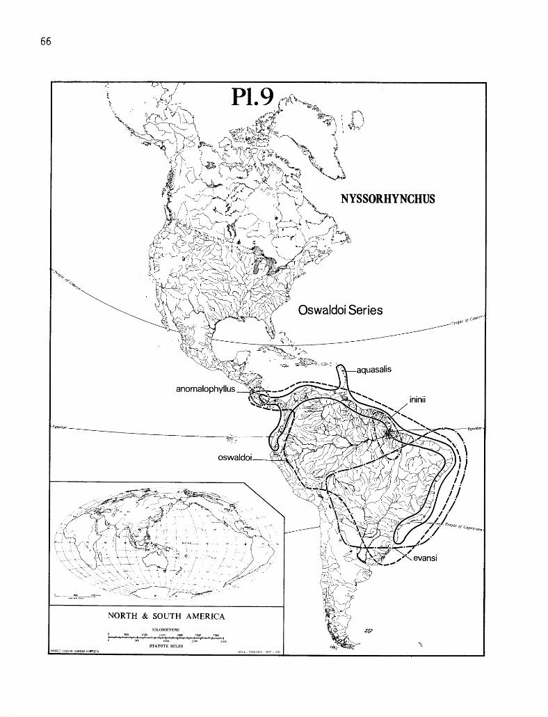
66
YNCHUS
Oswaldoi Series
NORTH & SOUTH AMERICA I
KILOMETERS

Mosquito Systematics Vol. 13(l) 1981 67
NYSSORHYNCHUS
Oswaldoi Ser
NORTH & SOUTH AMERICA
KILOMETERS
ies
/---
nuneztovari /
\ galvaoi

Pl. 11 Argyritarsis Section
Hypothesized phylogenetic tree
of the Argyritarsis Section
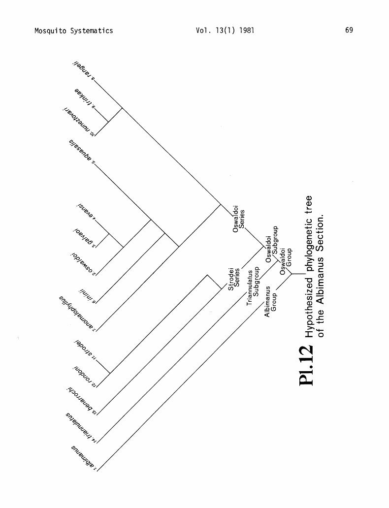
Mosquito Systematics Vol. 13(l) 1981 69

70
NYSSORHYNCHUS PI. 13

Mosquito Systematics Vol. 13(l) 1981 71

72

Mosquito Systematics Vol. 13(l) 1981 73
NYSSORHYNCHUS


' Mosquito Systematics Vol. 13(l) 1981 75
T NYSSORHYNCHUS
! I2
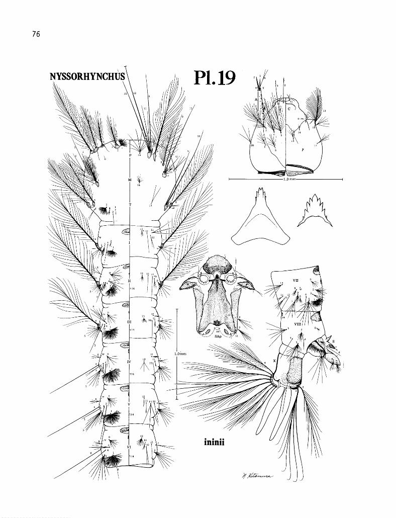
76

Mosquito Systematics Vol. 13(?) 1981 77
\ J NYSSORHYNCHUS 12 ,
l.Omm
I

PI.21

Mosquito Systematics Vol. 13(l) 1981
NYSSORHYNCHUS
79

80
NYSSORHYNCHUS

Mosquito Systematics
hi,, NYSSORHYNCHUS :;
Vol. 13(l) 1981
P1.24 \\
triannulatus
81