a genome-wide screen for sporulation-defective mutants · pdf filea genome-wide screen for...
TRANSCRIPT

MUTANT SCREEN REPORT
A Genome-Wide Screen for Sporulation-DefectiveMutants in Schizosaccharomyces pombeEsma Ucisik-Akkaya,* Janet K. Leatherwood,† and Aaron M. Neiman*,1
*Department of Biochemistry and Cell Biology, Stony Brook University, Stony Brook, New York 11794-5215, and†Department of Molecular Genetics and Microbiology, Stony Brook University, Stony Brook, New York 11794-5222
ABSTRACT Yeast sporulation is a highly regulated developmental program by which diploid cells generatehaploid gametes, termed spores. To better define the genetic pathways regulating sporulation, a systematicscreen of the set of ~3300 nonessential Schizosaccharomyces pombe gene deletion mutants was per-formed to identify genes required for spore formation. A high-throughput genetic method was used tointroduce each mutant into an h90 background, and iodine staining was used to identify sporulation-defective mutants. The screen identified 34 genes whose deletion reduces sporulation, including 15 thatare defective in forespore membrane morphogenesis. In S. pombe, the total number of sporulation-defectivemutants is a significantly smaller fraction of coding genes than in S. cerevisiae, which reflects the differentevolutionary histories and biology of the two yeasts.
KEYWORDS
knockoutcollection
erp2erp5foresporemembrane
Ascospore formation in yeast is a response to nutrient deficiency(Tomar et al. 2013). In Schizosaccharomyces pombe, cells exit mitosisto differentiate into spores when they encounter the lack of a nitrogensource (Tanaka and Hirata 1982; Egel 1989; Shimoda and Nakamura2004b). First, haploid cells of opposite mating types fuse to formdiploid zygotes. These diploids then immediately undergo meiosisto generate four haploid nuclei. During the course of meiosis, thesenuclei become packaged into daughter cells, termed spores. Spores arecreated by a specialized form of cell division that occurs withoutcleavage of the mother cell (Shimoda 2004a). Each of the four haploidnuclei produced by meiosis are packaged into daughter cells byenvelopment within newly synthesized membranes called foresporemembranes (Yoo et al. 1973; Shimoda and Nakamura 2004b). Fore-spore membrane formation initiates on meiotic spindle pole bodies(SPBs) early in meiosis II and as meiosis proceeds, each foresporemembrane expands to engulf the associated nucleus (Shimoda2004a; Nakase et al. 2008). Closure of the forespore membranearound a nucleus completes cell division, and these cells then matureinto spores by deposition of spore wall material (Yoo et al. 1973). All
of these events occur within the cytoplasm of the original mothercell, which is referred to as the ascus.
Mutants defective in meiosis and sporulation have been identifiedin S. pombe in a number of different screens. Originally spo mutantswere found by direct screening for sporulation defects (Bresch et al.1968; Kishida and Shimoda 1986). More recently targeted mutagenesisof genes whose expression is sporulation-induced has identified addi-tional genes involved in both processes (Gregan et al. 2005; Martin-Castellanos et al. 2005). Although these screens have defined manygenes involved in sporulation, these screens were not saturating and soadditional genes likely remain to be identified.
The process of sporulation is similar in S. pombe and in the bud-ding yeast Saccharomyces cerevisiae, although there appears to be onlylimited conservation of the specific genes involved in the process(Shimoda 2004a). Systematic screening of the S. cerevisiae knockoutcollection has proven to be a valuable approach, identifying hundredsof genes required for sporulation (Deutschbauer et al. 2002; Enyenihiand Saunders 2003; Marston et al. 2004). Sporulation-defectivemutants in S. cerevisiae can be divided into several broad categories:(1) genes required for aspects of cell physiology necessary to supportsporulation, for example mitochondrial function or autophagy;(2) genes required for progression through meiotic prophase to theinitiation of spore development; and (3) genes required for sporeassembly, per se, for instance genes involved in growth of the pro-spore membrane (the S. cerevisiae equivalent of the forespore mem-brane) or for spore wall formation (Neiman 2005).
To obtain a more comprehensive list of genes required forsporulation in S. pombe, we undertook a genome-wide systematicscreen of the S. pombe haploid deletion set (~3300 strains in total).
Copyright © 2014 Ucisik-Akkaya et al.doi: 10.1534/g3.114.011049Manuscript received March 13, 2014; accepted for publication April 8, 2014;published Early Online April 9, 2014.This is an open-access article distributed under the terms of the CreativeCommons Attribution Unported License (http://creativecommons.org/licenses/by/3.0/), which permits unrestricted use, distribution, and reproduction in anymedium, provided the original work is properly cited.1Corresponding author: Department of Biochemistry and Cell Biology, Stony BrookUniversity, Stony Brook, New York 11794-5215. E-mail: [email protected]
Volume 4 | June 2014 | 1173

In S. pombe, nitrogen starvation induces haploid cells of oppositemating types (h+ and h2) to mate and then undergo meiosis andspore formation. Strains that carry the h90 allele at the mat1 locusare homothallic, meaning the cells switch mating types during mitoticgrowth so that both the h+ and h2 mating types are present in col-onies originally derived from a single cell. Diploids generated by h90
strains are therefore completely homozygous because they are a resultof self-mating. This greatly simplifies the detection of meiotic andsporulation mutants because meiosis is normally induced only indiploid cells. The haploid deletion set was constructed in an h+ matingtype background. Therefore, it was necessary to introduce h90 intoeach deletion strain to enable the creation of homozygous mutantdiploids. After these mutants were exposed to conditions that promotesporulation, iodine staining was used as an initial screen to determinewhether spores were present (Garcia et al. 2006). Secondary screensincluded direct observation of asci by phase contrast microscopy andexamination of fluorescent markers for the forespore membrane andSPBs. Our screen identified .90% of the previously known sporula-tion-defective mutants present in the collection, suggesting that thescreen has identified the majority of non-essential genes required forspore formation. Among the novel sporulation genes are membranetrafficking proteins, signaling proteins, transcription factors, and met-abolic enzymes. These results provide a wealth of information forfuture investigations.
MATERIALS AND METHODS
Yeast strains and cultureStandard media and growth conditions were used unless otherwisenoted (Forsburg and Rhind 2006). For synthetic medium containingG418, pombe glutamate medium (PMG) was used (Sabatinos andForsburg 2010). Genotypes of the strains used in this study are listedin Table 1. Strain EAP20, which was used to introduce h90, as well asgenes encoding tagged versions of psy1+ and sid4+ (markers for theforespore membranes and SPBs, respectively) into the knockout col-lection, was constructed in several steps. First, a spontaneous cyclo-heximide resistant mutant of strain JLP18 (EAP3) was selected byplating cells on YES plates containing 10 mg/L of cycloheximide(Sigma-Aldrich Co.). EAP11 was generated by transforming EAP3with SphI-digested pEA4, which targets integration of the S. cerevisiaeURA3 gene adjacent to the his5+ locus. his5+ is tightly linked to mat1,which contains the h90 allele, and the Ura+ phenotype can then beused to follow the h90 allele in subsequent crosses. Next, an allele ofthe SPB gene sid4+ fused to a gene encoding the fluorescent proteintdTomato (sid4+-tdTomato::hphMX6) was introduced by crossing
EAP11 with strain 843 (Doyle et al. 2009) to generate EAP16. Tointroduce a marker for the forespore membrane, a strain [FY12295;(Nakase et al. 2008)] carrying a green fluorescent protein (GFP)-tagged allele of psy1+ was crossed to EAP16, generating EAP19.Finally, EAP19 was backcrossed to EAP16 to generate a segregant,EAP20, which carries the marked h90 locus, both fluorescent proteingene fusions, and cycloheximide resistance.
PlasmidspEA4, which contains the S. pombe his5+ gene in pRS306 (Sikorskiand Hieter 1989), was constructed by polymerase chain reaction(PCR) amplification of a 1.3-kb fragment including his5+ and its59 and 39 regions from genomic DNA using EAO11 (59-GTTCTTGGTACCGAGCGTGCTCAGTTTTCTATG-39) and HJO274 (59-GTTGTTGAA TTCTTACAACACTCCCTTCGTGCTTGGG-39) oli-gonucleotides. The PCR product was engineered to contain KpnI andEcoRI sites at its 59 and 39 ends, respectively, and was cloned intosimilarly digested pRS306.
pEA18, which expresses wsc1+-mTagBFP under control of thespo13 promoter, was constructed in three steps. First, a yeast codon-optimized form of mTagBFP without a stop codon was PCR amplifiedfrom pRS426 Spo2051–91-mTagBFP (Lin et al. 2013) using EAO44 (59-GTTCTTCATATGGTTCTTGTTCCATGGATGTCTGAGGAGTTGATAAAGG-39) and EAO46 (59-GTTCTTGGATCCCTTGTTCTTGCGGCCGCGTTCAACTTGTGACCCAACTTTG-39) oligos and clonedas a NdeI/BamHI fragment into similarly digested pREP42x (Forsburg1993) creating pEA13. Second, overlap PCR was used to constructa Pspo13-wsc1+ fusion. A ~500-bp fragment of the spo13 promoterregion and the wsc1+ open reading frame lacking the stop codonwere amplified using the oligonucleotide pairs EAO47 (59-GTTCTTCTGCAGGGCACTCTGTAATTGTAAG-39) and EAO48 (59-GAGGAATTTAAAAAGACCATAGATCTTGTTTCAATTTTTTTTCCTTTCC-39), and EAO49 (59-GGAAAGGAAAAAAAATTGAAA-CAAGATCTATGGTCTTTTTAAATTCCTC-39) and EAO50 (59-GTTCTTCCATGGGTTCAAATTTGTGACACGC), respectively. These PCRproducts were mixed and used as template in a reaction with EAO47and EAO50 to yield a ~1.6-kb spo13pr-wsc1+ fusion fragment. Thisproduct was digested with PstI and NcoI and cloned into similarlydigested pEA13 to replace the nmt1 promoter of pREP42x in front ofmTagBFP creating pEA17. Finally, pEA18 was created by amplifyingmTagBFP with its stop codon from pRS426 Spo2051–91-mTagBFP usingEAO44 and EAO45 (59-GTTCTTGGATCCCTTGTTCTTGCGGCCGCTTAGTTCAACTTGTGACCCAACTTTG-39), digested with NcoIand NotI, and cloned into similarly digested pEA17.
n Table 1 Strains used in this study
Name Genotype Source
JLP18 h90 ura4-D18 leu1-32 his3-127 This studyEAP3 h90 ura4-D18 leu1-32 his3-127 cyhR This studyEAP11 h90 his5::URA3::his5+ ura4-D18 leu1-32 his3-127 cyhR This study843 h90 myo51+-GFP::kanMX6 sid4+-tdTomato::hphMX6 ura4-D18 leu1-32 (Doyle et al. 2009)EAP16 h90 his5::URA3::his5+ sid4+-tdTomato::hphMX6 ura4-D18 leu1-32
his3-127 cyhRThis study
FY12295 h90 spo15::ura4+ ura4-D18 leu1+::GFP-psy1+ (Nakase et al. 2008)EAP19 h90 leu1::GFP-psy1+-LEU2 sid4+-tdTomato::hphMX6 ura4-D18 leu1-32 This studyEAP20 h90 his5::URA3::his5+ leu1+::GFP-psy1+ sid4+-tdTomato::hphMX6
ura4-D18 leu1-32 cyhRThis study
Bioneer deletion set h+ ade6-M210 ura4-D18 leu1-32 geneXD::kanMX4 (Kim et al. 2010)deletion mutants after outcrosses h90 his5::URA3::his5+ leu1+::GFP-psy1+ sid4+-tdTomato::hphMX6
ura4-D18 leu1-32 cyhR geneXD::kanMX4This study
1174 | E. Ucisik-Akkaya et al.
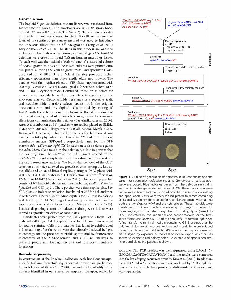
Genetic screenThe haploid S. pombe deletion mutant library was purchased fromBioneer (South Korea). The knockouts are in an h+ strain back-ground (h+ ade6-M210 ura4-D18 leu1-32). To examine sporula-tion, each mutant was crossed to strain EAP20 and a modifiedform of the synthetic gene array method was used to introducethe knockout alleles into an h90 background (Tong et al. 2001;Baryshnikova et al. 2010). The steps in this process are outlinedin Figure 1. First, strains containing individual geneXD::kanMX4deletions were grown in liquid YES medium in microtiter dishes.To each well was then added 1/10th volume of a saturated cultureof EAP20 grown in YES and the mixed cultures were pinned ontoME plates, allowing the cells to grow, mate, and sporulate (Fors-burg and Rhind 2006). Use of ME at this step produced higherefficiency sporulation than other media (data not shown). Thepatches were then replica plated to YES plates supplemented with200 mg/L Geneticin (G418; USBiological Life Sciences, Salem, MA)and 10 mg/L cycloheximide. Combined, these drugs select forrecombinant haploids from the cross. Geneticin selects for theknockout marker. Cycloheximide resistance is a recessive trait,and cycloheximide therefore selects against both the originalknockout strain and any diploid cells created by mating ofEAP20 with the deletion strain. Inclusion of this step is essentialto prevent a background of diploids heterozygous for the knockoutallele from contaminating the patches (Baryshnikova et al. 2010).After 3 d incubation at 31�, patches were replica plated to EMM2plates with 200 mg/L Hygromycin B (Calbiochem, Merck KGaA,Darmstadt, Germany). This medium selects for both uracil andleucine prototrophy, which are linked to h90 and the foresporemembrane marker GFP-psy1+, respectively, and for the SPBmarker sid4+-tdTomato::hphMX6. In addition it also selects againstthe ade6-M210 allele found in the deletion set. It is important thatthe resulting strain be ade6+ as the red pigment created by theade6-M210 mutant complicates both the subsequent iodine stain-ing and fluorescence analyses. We found that removal of the G418selection at this step allowed the growth of cells lacking the knock-out allele and so an additional replica plating to PMG plates with200 mg/L G418 was performed. G418 selection is more efficient onPMG than EMM2 (Benko and Zhao 2011). The resulting patchesconsist of h90 haploid deletion mutants harboring sid4+-tdTomato::hphMX6 and GFP-psy1+. These patches were then replica plated toSPA plates to induce sporulation, incubated at 25� for 3 d, and theninverted over a Petri dish of iodine crystals for 2-3 min (Sabatinosand Forsburg 2010). Staining of mature spore wall with iodinevapor produces a dark brown color (Meade and Gutz 1975).Patches displaying absent or reduced staining with iodine werescored as sporulation-defective candidates.
Candidates were picked from the PMG plates to a fresh PMGplate with 200 mg/L G418, replica plated to SPA, and then retestedfor iodine staining. Cells from patches that failed to exhibit goodiodine staining after the retest were then directly analyzed by lightmicroscopy for the presence of visible spores and by fluorescencemicroscopy of the Sid4-tdTomato and GFP-Psy1 markers toevaluate progression through meiosis and forespore membraneformation.
Barcode sequencingIn construction of the knockout collection, each knockout incorpo-rated “uptag” and “downtag” sequences that provide a unique barcodefor each knockout (Kim et al. 2010). To confirm the identity of themutants identified in our screen, we amplified the uptag region for
each one. This PCR product was then sequenced using EAO62 (59-GGGCGACAGTCACATCATGC39-) and the results were comparedwith the list of uptag sequences given by Kim et al. (2010). In addition,the meu14 and mfr1 deletions were also analyzed by PCR-amplifica-tion of the loci with flanking primers to distinguish the knockout andwild-type alleles.
Figure 1 Outline of generation of homothallic mutant strains and thescreen for sporulation defective mutants. Genotypes of cells at eachstage are boxed. Blue indicates genes from the deletion set strains,and red indicates genes derived from EAP20. These two strains werefirst mixed in liquid and then spotted onto ME plates to allow matingand sporulation. Cells were then replica plated to plates containingG418 and cycloheximide to select for recombinant progeny containingboth the geneXD::kanMX4 and the cyhR alleles. These haploids weretransferred to minimal medium containing hygromycin to select forthose segregants that also carry the h90 mating type (linked toURA3, indicated by the underline) and harbor markers for the fore-spore membrane (GFP-psy1+) and the SPB (sid4+-tdTomato::hphMX6).A final transfer to minimal medium containing G418 ensures that thedeletion alleles are still present. Meiosis and sporulation were inducedby replica plating the patches to SPA medium and spore formationwas assayed by exposure of the cells to iodine vapor, which causesspores to exhibit a red colony color. An example of sporulation pro-ficient and defective patches is shown.
Volume 4 June 2014 | S. pombe Sporulation Mutants | 1175

MicroscopyImages were collected on a Zeiss Observer Z.1 microscope andprocessed using Zeiss Axiovision or Zen software.
Acetone resistance assaysSpore wall function was tested using an acetone resistance assaymodified from (Smith 2009). Wild-type spores are resistant to ace-tone, whereas unsporulated cells or cells with defective spore walls arekilled. The wild-type and the knockout strains were first incubated onPMG plates with 200 mg/L G418 at 31� for 2 d. Cells were then replicaplated to SPA plates and incubated at 25� for 3 d to allow for matingand sporulation, and then replica-plated onto YES plates. An acetone-soaked filter paper (Whatman #3, 1003-090) was placed on a glassPetri dish and inverted above the YES plate to expose the patches toacetone vapor for 15 min. These were then incubated at 31� for 3 dbefore being photographed.
RESULTS AND DISCUSSION
Isolation of sporulation-defective mutantsUsing a series of selective steps diagrammed in Figure 1, we con-structed h90 homothallic derivatives of each deletion strain in theBioneer S. pombe haploid deletion collection, at the same time in-troducing fluorescent markers for the SPBs and the forespore mem-brane. Because h90 strains are able to undergo mating type switching,h90 cells can be induced to self-mate and create homozygous diploidsthat then proceed through meiosis and sporulation. The ability of thestrains in the deletion set to form spores was then assayed by exposureto iodine, which produces a dark brown stain in patches containingspores.
Eighty-five candidates passed the initial screen as well as a retest.In addition to sporulation-defective mutants, the assay of decreasediodine staining might also identify knockouts that cause h2-specificmating defects, that is, mutants that are unable to mate with h+
haploids. The deletion strain background is h+ and these cells aretherefore able to mate with h- cells present in the h90 background inthe initial cross. However, these cells will be unable to self-mate oncein the h90 background and so will not produce spores. Similarly, as theURA3 marker is integrated approximately 10 cM from the mat1 locuscontaining the h90allele (Egel 2004), recombination between URA3and the mat locus can produce URA3 h+ haploids that would slipthrough the selection procedure and these would also fail to sporulate.To test for such false positives, the 85 strains were assayed for theirmating types by replica-plating to h+ and h2 tester strains followed byiodine staining to examine whether diploids formed that could spor-ulate. Strains that are h90 are expected to mate to both h2 and h+ cells.Three of the candidates mated only to the h+ tester strains, and thirty-three mutants mated only to the h2 tester, demonstrating that a mat-ing defect is indirectly responsible for the absence of spores. Of thestrains that mated only to the h2 tester, two were deletions in mam1(M-factor transporter) and mam2 (P-factor receptor), both of whichare known to produce an h2 specific sterility (Kitamura and Shimoda1991; Christensen et al. 1997). The remaining mutants we suspectwere simply h+ strains that leaked through the selection process.The strains with mating defects were not analyzed further.
The remaining candidates were sporulated and examined by phasecontrast microscopy to determine the frequency of spore formation inthe culture. Those strains in which no spores were detected were alsoexamined by fluorescence microscopy of the Sid4-tdTomato and GFP-Psy1 markers to look at progression through meiosis and foresporemembrane formation, respectively. Based on these microscopy
assays the mutants can be divided into four classes: (1) reducedfrequency of zygotes, suggesting that the sporulation defect is sec-ondary to a mating defect; (2) near wild-type frequency of zygotesand spores, suggesting a defect in formation of the iodine-reactivelayer of the spore wall; (3) no spores and no forespore membraneformation; and (4) no spores with abnormal forespore membraneformation (Table 2).
To confirm the identity of the deleted gene in the knockout strains,we used PCR to amplify the unique uptag region for many of thedeletions (Kim et al. 2010). These PCR products were then sequencedand compared with the published lists to confirm the identity of theknockouts. For 48 knockouts for which we obtained sequences, 32matched the published barcodes. The knockouts that did not producethe expected barcode sequence are listed in Table 3. In three cases, thebarcode sequence found corresponded to that of known sporulation-defective mutants, suggesting that the identification by the barcodesequence, rather than position in the collection, is correct. In all casesof misidentification, the expected knockout and the actual one arefound in different plates within the collection. These errors are, there-fore, unlikely to have been caused by cross-contamination during ourhandling of the collection as different plates were processed at differ-ent times. Although this is a small sample, the surprisingly high errorrate (33%) highlights the need for confirmation of knockout identitywhen using this collection.
To test the effectiveness of our screen, we culled from the literaturea list of previously identified mutants that block spore formation.Several of the original spo mutants proved to be hypomorphic allelesof essential genes (Nakase et al. 2001; Nakamura-Kubo et al. 2003)and so are not present in our deletion set; however we identified 13known sporulation-defective mutants listed as present in the collec-tion (Table 4). Amplification and barcode sequencing confirmed thepresence of nine of these at the correct location in the collection andanother two mutants were identified at different locations. Of these 11mutants, 10 were identified in the screen. This yield suggests that thescreen has identified ~90% of the sporulation-defective mutants pres-ent in the collection.
Classes of genes required for positive iodinestaining phenotype
Genes required for zygote formation: For mutants that formed somelevel of visible spores, the frequency of zygote formation and of sporeformation were examined by light microscopy (Table 5). Mutants thatdisplay bilateral mating defects, that is, are able to mate with the h+
and h2 tester strains but are unable to self-mate to produce zygoteswould pass the mating tests described above and show reduced spor-ulation. For seven mutants, zygote formation was reduced greater thanthreefold from that seen in a wild-type h90 strain, indicative of a bi-lateral mating defect. Thus, the primary defect in these mutants islikely to be in the mating process or response to nitrogen starvationrather than in spore formation, per se. The two genes in this class withthe strongest phenotype were prm1+ and cyp9+. Consistent with ourinterpretation, prm1+ encodes an integral membrane protein recentlyshown to be necessary for conjugation (Sun et al. 2013; Curto et al.2014). These results reveal a previously unknown role for cyp9+ in themating reaction.
Genes required for spores to be iodine-reactive: Mutants in sevenadditional genes formed zygotes at near normal frequency anddisplayed at most modestly reduced spore formation relative to wildtype. Because strains in this class form significant numbers of spores,
1176 | E. Ucisik-Akkaya et al.

their loss of staining may reflect defects in generation of the iodinereactive alpha-glucan component of the spore wall (Garcia et al.2006). It is noteworthy that a number of mutants known to disruptassembly of the beta-glucan or chitosan layers of the spore wall werepresent in the collection but were not found in our screen, probablybecause those mutants that effect beta-glucan or chitosan do not alteriodine staining (Liu et al. 2000).
Two of the genes in this class, php3+ and php5+, encode subunits ofthe CCAAT-binding transcription complex (McNabb et al. 1997;Mercier et al. 2006). Although previous reports have implicated thiscomplex in induction of transcription during nitrogen starvation andin the activity of meiotic recombination hotspots, no requirement forthese genes in spore formation has been reported (Nakashima et al.
2002; Steiner et al. 2011). This work suggests that transcriptional in-duction by this complex of as yet unidentified genes is important forproper spore formation.
Genes required for entry into meiosis or for the initiation offorespore membrane assembly: The five genes identified in this classwere previously known. Three of the genes are required for entry intomeiosis. mei2+ encodes an RNA-binding protein that is required forpremeiotic DNA synthesis as well as progression into meiosis I (Watanabeet al. 1988; Watanabe and Yamamoto 1994). mei3+ is essential for theinitiation of meiosis since it encodes a protein that binds and inhibits themeiosis-inhibitory protein kinase Pat1 during sporulation(McLeod andBeach 1988). The transcription factor that is encoded by mei4+ is
n Table 2 Genes identified in the sporulation-defective screen
Gene Gene ID Commentsa
Class 1. Genes required for zygote formation (n = 7)atg12+ SPAC1783.06c Autophagy-associated ubiquitin-like
modifiercyp9+ SPCC553.04 Predicted cyclophilin family peptidyl-prolyl
cis-trans isomerasemmd1+ SPAC30C2.02 Predicted deoxyhypusine hydroxylaseprm1+ SPAP7G5.03 Integral membrane protein important for
cell2cell fusionSPBC1711.12 Predicted oxidized protein hydrolaseSPBC18E5.08 Predicted N-acetyltransferaseSPBC146.02 Sequence orphan
Class 2. Genes required for spores to be iodine-reactive (n = 7)fsc1+ SPAC22H12.05c Fasciclin domain proteinlcf2+ SPBP4H10.11C Long-chain-fatty-acid-CoA ligasemam3+ SPAP11E10.02c Cell agglutination proteinmcl1+ SPAPB1E7.02c DNA polymerase alpha accessory factorphp3+ SPAC23C11.08 CCAAT-binding factor complex subunitphp5+b SPBC3B8.02 CCAAT-binding factor complex subunitrik1+ SPCC11E10.08 Silencing protein
Class 3. Genes required for entry into meiosis or for the initiationof forespore membrane assembly (n = 5)mei2+ SPAC27D7.03c RNA-binding protein required for meiosismei3+ SPBC119.04 Required for the initiation of meiosismei4+ SPBC32H8.11 Transcription factor regulating meiotic
gene expressionmug79+ (spo7+) SPAC6G9.04 Meiotic spindle pole body componentspo15 SPAC1F3.06c Meiotic spindle pole body component
Class 4. Genes that are essential for the proper formation andthe maturation of the forespore membrane (n = 15)csn1+ SPBC215.03c COP9/signalosome complex subunitcsn2+ SPAPB17E12.04c COP9/signalosome complex subunitcdt2+ SPAC17H9.19c COP9/signalosome associated factorerp2+ SPAC17A5.08 ER exit receptor for secretory cargoerp5+ SPBC16E9.09c ER exit receptor for secretory cargomes1+ SPAC5D6.08c Meiotic APC/C regulatorspe2+ SPBP4H10.05c S-adenosylmethionine decarboxylase
proenzyme(spe3+) SPBC12C2.07c Predicted spermidine synthasespn2+ SPAC821.06 Septinspo3+ SPAC607.10 Required for spore formationspo4+ SPBC21C3.18 Kinase required for spore formationspo5+ SPBC29A10.02 Meiotic RNA-binding proteintpp1+ SPAC19G12.15c Trehalose-6-phosphate phosphatase
SPAC6C3.06c Predicted P-type phospholipid flippaseSPCC1739.04c Sequence orphan
ER, endoplasmic reticulum; APC/C, Anaphase Promoting Complex/Cyclosome.a
Descriptions are based on PomBase (Wood et al. 2012) (www.pombase.org).b
Knockout not confirmed by barcode sequencing.
Volume 4 June 2014 | S. pombe Sporulation Mutants | 1177

a regulator necessary for the expression of many sporulation-inducedgenes (Horie et al. 1998). The remaining two genes in this class,mug79+/spo7+ and spo15+, both encode components of the meiotic SPBnecessary for the SPB to catalyze the coalescence of secretory vesicles intoa forespore membrane (Ikemoto et al. 2000; Nakamura-Kubo et al. 2011).
Genes that are essential for the proper formation and thematuration of the forespore membrane: Mutants in Class 4 genesprogress through meiosis and initiate forespore membrane growth,but the membranes display morphological defects and no spores arevisible by light microscopy. There were 15 genes identified in thiscategory, of which five (spo3+, spo4+, spo5+, mes1+, spn2+) were pre-viously shown to be required for sporulation (Nakamura et al. 2001,2002; Izawa et al. 2005; Kasama et al. 2006; Onishi et al. 2010).Among the 10 genes in this class not previously associated with spor-ulation defects, two encode subunits of the COP9 signalosome (csn1+
and csn2+) and one encodes a reported interacting partner of thesignalosome (cdt2+) (Mundt et al. 1999; Liu et al. 2005). Several otherCOP9 subunits are present in the collection but were not found to beiodine-negative in our screen. Thus, the Csn1 and Csn2 subunits ofthe signalosome may be specifically required for sporulation. A similar
difference in function between Csn1/Csn2 and other COP9 subunitsin sensitivity to DNA damage has been noted previously (Mundt et al.2002). Also in this class are spe2+ and SPBC12C2.07c (spe3+), genesthat encode enzymes involved in consecutive steps in spermidinesynthesis (Tabor and Tabor 1985; Chattopadhyay et al. 2002). Thispathway has also been shown to be required for sporulation inS. cerevisiae (Cohn et al. 1978), suggesting a conserved requirementfor spermidine for spore formation in fungi.
Three of the mutants in this class have predicted functions withinthe secretory pathway. SPAC6C3.06c encodes a predicted phospho-lipid flippase orthologous to the NEO1 gene of S. cerevisiae. Neo1 islocalized to the endosome and to the Golgi and has been implicated inmembrane trafficking (Hua and Graham 2003; Wicky et al. 2004).The erp2+ and erp5+ genes encode two S. pombe members of the p24protein family. The p24 proteins are a highly conserved family ofintegral membrane proteins that act as cargo receptors and shuttlebetween the endoplasmic reticulum (ER) and the Golgi (Strating andMartens 2009). In particular, they play an important role in cargoselection and packaging into COPII vesicles at ER exit sites (Stratingand Martens 2009). Consistent with the similar phenotypes of botherp2 and erp5 deletions, studies in S. cerevisiae suggest that the four
n Table 3 Gene deletions that do not have the correct barcode
Gene Gene ID Barcode Information
atg15D SPAC23C4.16c Matches with spo5atp10D SPAC4G8.11c No matchatp14D SPBC29A3.10c No matchctf1D SPBC3B9.11c No matchlsk1D SPAC2F3.15 Matches with mei4mei4D SPBC32H8.11 No matchmfr1D SPBC1198.12 Matches with SPAC17H9.14c mfr1 knockout is not present
as determined by PCR with flanking primers,scd1D SPAC16E8.09 Matches with mei4spo5D SPBC29A10.02 No matchspo6D SPBC1778.04 No match
SPBC15C4.06c No matchapq12D SPBC428.04 Matches with cyp9
SPBC21H7.06c Matches with cyp9SPAC139.01c Matches with nrd1SPBC23G7.06c Matches with nrd1SPBC1711.08 Matches with nrd1
n Table 4 Known sporulation-defective genes listed in the S. pombe haploid deletion set
Gene Comment Phenotype in Our Study
spo3+ Confirmed by barcode sequence Sporulation defectspo4+ Confirmed by barcode sequence Sporulation defectspo5+ Knockout found at different position in the collectiona Sporulation defectspo6+ Not presenta n.d.b
mug79+/spo7+ Confirmed by barcode sequence Sporulation defectspo15+ Confirmed by barcode sequence Sporulation defectmei2+ Confirmed by barcode sequence Sporulation defectmei3+ Confirmed by barcode sequence Sporulation defectmei4+ Knockout found at different position in the collectiona Sporulation defectmes1+ Confirmed by barcode sequence Sporulation defectmfr1+ Not presenta n.d.meu14+ Knockout is present as determined both by PCR with flanking
primers and by barcode sequenceNormal sporulation
cdt2+ Confirmed by barcode sequence Sporulation defect
n.d., not determined; PCR, polymerase chain reaction.a
See Table 3.
1178 | E. Ucisik-Akkaya et al.

family members function in a single complex (Hirata et al. 2013).Knockouts of the other family members in S. pombe, emp24+
(SPCC24B10.17.1) and erv25+ (SPAC23H4.03c.1), were not presentin the collection, though we expect mutants in these genes woulddisplay a similar sporulation defect. We predict that the p24 familyis necessary for the exit of some protein(s) from the ER so that thecargo protein(s) can be transported through the secretory pathway tothe forespore membrane and contribute to proper membrane growth.
erp2 and erp5 mutants do not cause a general block toER exitIn S. cerevisiae, a different class of ER cargo receptor, encoded by theERV14 and ERV15 genes, is required for proper formation of theprospore membrane (the budding yeast equivalent of the foresporemembrane) during sporulation (Powers and Barlowe 1998; Nakanishiet al. 2007). Although these genes are not essential for vegetativegrowth, their deletion creates a general block to ER exit of integralmembrane proteins during sporulation (Nakanishi et al. 2007). Be-cause ERV14 deletion mutants have small, abnormal forespore mem-branes similar to erp2D and erp5D mutants, we hypothesized that,parallel to the S. cerevisiae ER cargo receptors, erp2+ and erp5+ mightbecome essential for ER exit of integral membrane proteins inS. pombe sporulation. The GFP-Psy1 reporter is localized to the fore-spore membrane in erp2D and erp5D cells, however this does notprovide a strong test of a role for erp2+ and erp5+ in ER exit asPsy1 is relocalized from the plasma membrane to the forespore mem-brane via the endosome (Kashiwazaki et al. 2011). Therefore, to testa possible general role for erp2+ and erp5+ in ER exit, the strains weretransformed with a plasmid carrying an integral plasma membraneprotein, wsc1+, fused with mTagBFP and placed under control of thesporulation-specific protein spo13 promoter (Nakase et al. 2008).When expressed in a wild-type strain, Wsc1-mTagBFP localized tothe forespore membrane (Figure 2). In erp2D and erp5D mutantsWsc1-mTagBFP colocalized with GFP-Psy1 in the abnormal fore-spore membranes and no additional BFP fluorescence from the ERwas seen, indicating that transport of Wsc1-mTagBFP is unaffected inthe mutants (Figure 2). If loss of erp2 or erp5 cause forespore mem-brane defects indirectly by limiting the exit of some cargo from theER, this is likely an effect on some specific cargo protein(s) and notdue to a more general block in transport.
The lcf2+ and mcl1+ gene products may contribute tospore wall functionThe spore wall provides the cell with resistance to environmentalstresses such as acetone vapor (Egel 1977). To examine spore wallfunction we tested mutants in Class 2 for resistance to acetone (Smith2009). Two of the mutants, lcf2D andmcl1D, showed strong sensitivityto acetone exposure (Figure 3). This stress-sensitivity is striking asthese mutants show near-normal levels of sporulation. This resultsuggests a structural defect in the spore walls of these mutants, pre-sumably in the alpha-glucan component of the spore wall. The mcl1+
gene encodes a polymerase alpha accessory protein, so its effect on thespore wall is likely indirect (Williams and McIntosh 2005). lcf2+
Figure 2 Wsc1-mTagBFP localization in erp2 and erp5 mutants. Wild-type, erp2D, or erp5D cells expressing Pspo13-wsc1+-mTagBFP andGFP-psy1+ were imaged after 24-hr incubation on SPA plates. Scalebar = 2 microns.
n Table 5 Mating and sporulation efficiency of different mutants
Gene Gene ID Classa % of Zygotesb (SD) % of Sporulationc (SD)
WT 67.0 (4.0) 76.7 (5.5)cyp9D SPCC553.04 1 ,0.5 n.d.prm1D SPAP7G5.03 1 ,0.5 n.d.
SPBC1711.12 1 9.0 (2.2) 21.3 (1.2)SPBC18E5.08 1 9.8 (2.4) 87.7 (2.5)SPBC146.02 1 14.8 (5.0) 3.3 (0.6)
atg12D SPAC1783.06c 1 19.0 (8.6) 47.7 (6.8)mmd1D SPAC30C2.02 1 21.5 (11.2) 71.0 (8.9)fsc1D SPAC22H12.05c 2 27.3 (12.6) 52.0 (4.4)mcl1D SPAPB1E7.02c 2 38.5 (8.3) 56.3 (12.5)php3D SPAC23C11.08 2 38.5 (6.6) 43.0 (6.6)mam3D SPAP11E10.02c 2 39.5 (4.8) 76.3 (6.7)lcf2D SPBP4H10.11C 2 43.8 (11.5) 58.5 (6.1)rik1D SPCC11E10.08 2 57.8 (7.1) 85.3 (3.9)
SD, standard deviation; n.d., not determined.a
Class 1 = Genes required for zygote formation; Class 2 = Genes required for spores to be iodine-reactive.b
The average of at least three experiments. At least 100 cells were counted in each experiment.c
The average of at least three experiments. At least 100 asci were counted in each experiment.
Volume 4 June 2014 | S. pombe Sporulation Mutants | 1179
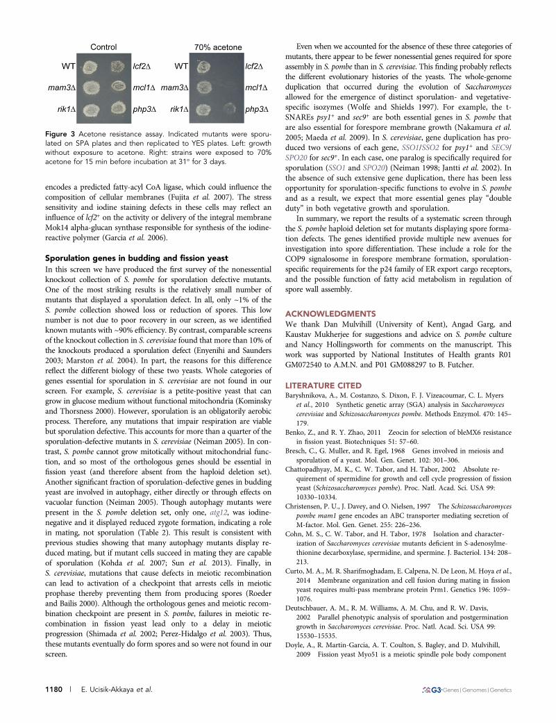
encodes a predicted fatty-acyl CoA ligase, which could influence thecomposition of cellular membranes (Fujita et al. 2007). The stresssensitivity and iodine staining defects in these cells may reflect aninfluence of lcf2+ on the activity or delivery of the integral membraneMok14 alpha-glucan synthase responsible for synthesis of the iodine-reactive polymer (Garcia et al. 2006).
Sporulation genes in budding and fission yeastIn this screen we have produced the first survey of the nonessentialknockout collection of S. pombe for sporulation defective mutants.One of the most striking results is the relatively small number ofmutants that displayed a sporulation defect. In all, only ~1% of theS. pombe collection showed loss or reduction of spores. This lownumber is not due to poor recovery in our screen, as we identifiedknown mutants with ~90% efficiency. By contrast, comparable screensof the knockout collection in S. cerevisiae found that more than 10% ofthe knockouts produced a sporulation defect (Enyenihi and Saunders2003; Marston et al. 2004). In part, the reasons for this differencereflect the different biology of these two yeasts. Whole categories ofgenes essential for sporulation in S. cerevisiae are not found in ourscreen. For example, S. cerevisiae is a petite-positive yeast that cangrow in glucose medium without functional mitochondria (Kominskyand Thorsness 2000). However, sporulation is an obligatorily aerobicprocess. Therefore, any mutations that impair respiration are viablebut sporulation defective. This accounts for more than a quarter of thesporulation-defective mutants in S. cerevisiae (Neiman 2005). In con-trast, S. pombe cannot grow mitotically without mitochondrial func-tion, and so most of the orthologous genes should be essential infission yeast (and therefore absent from the haploid deletion set).Another significant fraction of sporulation-defective genes in buddingyeast are involved in autophagy, either directly or through effects onvacuolar function (Neiman 2005). Though autophagy mutants werepresent in the S. pombe deletion set, only one, atg12, was iodine-negative and it displayed reduced zygote formation, indicating a rolein mating, not sporulation (Table 2). This result is consistent withprevious studies showing that many autophagy mutants display re-duced mating, but if mutant cells succeed in mating they are capableof sporulation (Kohda et al. 2007; Sun et al. 2013). Finally, inS. cerevisiae, mutations that cause defects in meiotic recombinationcan lead to activation of a checkpoint that arrests cells in meioticprophase thereby preventing them from producing spores (Roederand Bailis 2000). Although the orthologous genes and meiotic recom-bination checkpoint are present in S. pombe, failures in meiotic re-combination in fission yeast lead only to a delay in meioticprogression (Shimada et al. 2002; Perez-Hidalgo et al. 2003). Thus,these mutants eventually do form spores and so were not found in ourscreen.
Even when we accounted for the absence of these three categories ofmutants, there appear to be fewer nonessential genes required for sporeassembly in S. pombe than in S. cerevisiae. This finding probably reflectsthe different evolutionary histories of the yeasts. The whole-genomeduplication that occurred during the evolution of Saccharomycesallowed for the emergence of distinct sporulation- and vegetative-specific isozymes (Wolfe and Shields 1997). For example, the t-SNAREs psy1+ and sec9+ are both essential genes in S. pombe thatare also essential for forespore membrane growth (Nakamura et al.2005; Maeda et al. 2009). In S. cerevisiae, gene duplication has pro-duced two versions of each gene, SSO1/SSO2 for psy1+ and SEC9/SPO20 for sec9+. In each case, one paralog is specifically required forsporulation (SSO1 and SPO20) (Neiman 1998; Jantti et al. 2002). Inthe absence of such extensive gene duplication, there has been lessopportunity for sporulation-specific functions to evolve in S. pombeand as a result, we expect that more essential genes play “doubleduty” in both vegetative growth and sporulation.
In summary, we report the results of a systematic screen throughthe S. pombe haploid deletion set for mutants displaying spore forma-tion defects. The genes identified provide multiple new avenues forinvestigation into spore differentiation. These include a role for theCOP9 signalosome in forespore membrane formation, sporulation-specific requirements for the p24 family of ER export cargo receptors,and the possible function of fatty acid metabolism in regulation ofspore wall assembly.
ACKNOWLEDGMENTSWe thank Dan Mulvihill (University of Kent), Angad Garg, andKaustav Mukherjee for suggestions and advice on S. pombe cultureand Nancy Hollingsworth for comments on the manuscript. Thiswork was supported by National Institutes of Health grants R01GM072540 to A.M.N. and P01 GM088297 to B. Futcher.
LITERATURE CITEDBaryshnikova, A., M. Costanzo, S. Dixon, F. J. Vizeacoumar, C. L. Myers
et al., 2010 Synthetic genetic array (SGA) analysis in Saccharomycescerevisiae and Schizosaccharomyces pombe. Methods Enzymol. 470: 145–179.
Benko, Z., and R. Y. Zhao, 2011 Zeocin for selection of bleMX6 resistancein fission yeast. Biotechniques 51: 57–60.
Bresch, C., G. Muller, and R. Egel, 1968 Genes involved in meiosis andsporulation of a yeast. Mol. Gen. Genet. 102: 301–306.
Chattopadhyay, M. K., C. W. Tabor, and H. Tabor, 2002 Absolute re-quirement of spermidine for growth and cell cycle progression of fissionyeast (Schizosaccharomyces pombe). Proc. Natl. Acad. Sci. USA 99:10330–10334.
Christensen, P. U., J. Davey, and O. Nielsen, 1997 The Schizosaccharomycespombe mam1 gene encodes an ABC transporter mediating secretion ofM-factor. Mol. Gen. Genet. 255: 226–236.
Cohn, M. S., C. W. Tabor, and H. Tabor, 1978 Isolation and character-ization of Saccharomyces cerevisiae mutants deficient in S-adenosylme-thionine decarboxylase, spermidine, and spermine. J. Bacteriol. 134: 208–213.
Curto, M. A., M. R. Sharifmoghadam, E. Calpena, N. De Leon, M. Hoya et al.,2014 Membrane organization and cell fusion during mating in fissionyeast requires multi-pass membrane protein Prm1. Genetics 196: 1059–1076.
Deutschbauer, A. M., R. M. Williams, A. M. Chu, and R. W. Davis,2002 Parallel phenotypic analysis of sporulation and postgerminationgrowth in Saccharomyces cerevisiae. Proc. Natl. Acad. Sci. USA 99:15530–15535.
Doyle, A., R. Martin-Garcia, A. T. Coulton, S. Bagley, and D. Mulvihill,2009 Fission yeast Myo51 is a meiotic spindle pole body component
Figure 3 Acetone resistance assay. Indicated mutants were sporu-lated on SPA plates and then replicated to YES plates. Left: growthwithout exposure to acetone. Right: strains were exposed to 70%acetone for 15 min before incubation at 31� for 3 days.
1180 | E. Ucisik-Akkaya et al.

with discrete roles during cell fusion and spore formation. J. Cell Sci. 122:4330–4340.
Egel, R., 1977 Selective spore survival during replica-plating of fission yeast.Arch. Microbiol. 112: 109–110.
Egel, R., 1989 Mating-type genes, meiosis and sporulation, pp. 31–73 inMolecular Biology of the Fission Yeast, edited by A. Y. Naism, P. Young,and B. F. Johnson, et al. Academic Press, San Diego, CA.
Egel, R., 2004 Fission yeast in general genetics, pp. 1–12 in The MolecularBiology of Schizosaccharomyces pombe: Genetics, Genomics and Beyond,edited by R. Egel. Springer-Verlag, Berlin.
Enyenihi, A. H., and W. S. Saunders, 2003 Large-scale functional genomicanalysis of sporulation and meiosis in Saccharomyces cerevisiae. Genetics163: 47–54.
Forsburg, S. L., 1993 Comparison of Schizosaccharomyces pombe expressionsystems. Nucleic Acids Res. 21: 2955–2956.
Forsburg, S. L., and N. Rhind, 2006 Basic methods for fission yeast. Yeast23: 173–183.
Fujita, Y., S. Mita, H. Ohtsuka, and H. Aiba, 2007 Identification of a fattyacyl-CoA synthetase gene, lcf2+, which affects viability after entry into thestationary phase in Schizosaccharomyces pombe. Biosci. Biotechnol. Bio-chem. 71: 3041–3047.
Garcia, I., V. Tajadura, V. Martin, T. Toda, and Y. Sanchez, 2006 Synthesisof alpha-glucans in fission yeast spores is carried out by three alpha-glucan synthase paralogues, Mok12p, Mok13p and Mok14p. Mol.Microbiol. 59: 836–853.
Gregan, J., P. K. Rabitsch, B. Sakem, O. Csutak, V. Latypov et al.,2005 Novel genes required for meiotic chromosome segregation areidentified by a high-throughput knockout screen in fission yeast. Curr.Biol. 15: 1663–1669.
Hirata, R., C. Nihei, and A. Nakano, 2013 Isoform-selective oligomer for-mation of Saccharomyces cerevisiae p24 family proteins. J. Biol. Chem.288: 37057–37070.
Horie, S., Y. Watanabe, K. Tanaka, S. Nishiwaki, H. Fujioka et al., 1998 TheSchizosaccharomyces pombe mei4+ gene encodes a meiosis-specific tran-scription factor containing a forkhead DNA-binding domain. Mol. Cell.Biol. 18: 2118–2129.
Hua, Z., and T. R. Graham, 2003 Requirement for Neo1p in retrogradetransport from the Golgi complex to the endoplasmic reticulum. Mol.Biol. Cell 14: 4971–4983.
Ikemoto, S., T. Nakamura, M. Kubo, and C. Shimoda, 2000 S. pombesporulation-specific coiled-coil protein Spo15p is localized to the spindlepole body and essentail for its modification. J. Cell Sci. 113: 545–554.
Izawa, D., M. Goto, A. Yamashita, H. Yamano, and M. Yamamoto,2005 Fission yeast Mes1p ensures the onset of meiosis II by blockingdegradation of cyclin Cdc13p. Nature 434: 529–533.
Jantti, J., M. K. Aalto, M. Oyen, L. Sundqvist, S. Keranen et al.,2002 Characterization of temperature-sensitive mutations in the yeastsyntaxin 1 homologues Sso1p and Sso2p, and evidence of a distinctfunction for Sso1p in sporulation. J. Cell Sci. 115: 409–420.
Kasama, T., A. Shigehisa, A. Hirata, T. T. Saito, T. Tougan et al., 2006 Spo5/Mug12, a putative meiosis-specific RNA-binding protein, is essential formeiotic progression and forms Mei2 dot-like nuclear foci. Eukaryot. Cell5: 1301–1313.
Kashiwazaki, J., Y. Yamasaki, A. Itadani, E. Teraguchi, Y. Maeda et al.,2011 Endocytosis is essential for dynamic translocation of a syntaxin 1orthologue during fission yeast meiosis. Mol. Biol. Cell 22: 3658–3670.
Kim, D.-U., J. Hayles, D. Kim, V. Wood, H.-O. Park et al., 2010 Analysis ofa genome-wide set of gene deletions in the fission yeast Schizosacchar-omyces pombe. Nat. Biotechnol. 28: 617–623.
Kishida, M., and C. Shimoda, 1986 Genetic mapping of eleven spo genesessential for ascospore formation in the fission yeast Schizosaccharomycespombe. Curr. Genet. 10: 443–447.
Kitamura, K., and C. Shimoda, 1991 The Schizosaccharomyces pombe mam2gene encodes a putative pheromone receptor which has a significant homol-ogy with the Saccharomyces cerevisiae Ste2 protein. EMBO J. 10: 3743–3751.
Kohda, T. A., K. Tanaka, M. Konomi, M. Sato, M. Osumi et al.,2007 Fission yeast autophagy induced by nitrogen starvation generates
a nitrogen source that drives adaptation processes. Genes Cells 12: 155–170.
Kominsky, D. J., and P. E. Thorsness, 2000 Expression of the Saccharo-myces cerevisiae gene YME1 in the petite-negative yeast Schizosaccharo-myces pombe converts it to petite-positive. Genetics 154: 147–154.
Lin, C. P., C. Kim, S. O. Smith, and A. M. Neiman, 2013 A highly re-dundant gene network controls assembly of the outer spore wall inS. cerevisiae. PLoS Genet. 9: e1003700.
Liu, C., M. Poitelea, A. Watson, S. H. Yoshida, C. Shimoda et al.,2005 Transactivation of Schizosaccharomyces pombe cdt2+ stimulatesa Pcu4-Ddb1-CSN ubiquitin ligase. EMBO J. 24: 3940–3951.
Liu, J., X. Tang, H. Wang, and M. Balasubramanian, 2000 Bgs2p, a 1,3-beta-glucan synthase subunit, is essential for maturation of ascospore wallin Schizosaccharomyces pombe. FEBS Lett. 478: 105–108.
Maeda, Y., J. Kashiwazaki, C. Shimoda, and T. Nakamura, 2009 TheSchizosaccharomyces pombe syntaxin 1 homolog, Psy1, is essential in thedevelopment of the forespore membrane. Biosci. Biotechnol. Biochem.73: 339–345.
Marston, A. L., W. H. Tham, H. Shah, and A. Amon, 2004 A genome-widescreen identifies genes required for centromeric cohesion. Science 303:1367–1370.
Martin-Castellanos, C., M. Blanco, A. E. Rozalen, L. Perez-Hidalgo, A. I.Garcia et al., 2005 A large-scale screen in S. pombe identifies sevennovel genes required for critical meiotic events. Curr. Biol. 15: 2056–2062.
McLeod, M., and D. Beach, 1988 A specific inhibitor of the ran1+ proteinkinase regulates entry into meiosis in Schizosaccharomyces pombe. Nature332: 509–514.
McNabb, D. S., K. A. Tseng, and L. Guarente, 1997 The Saccharomycescerevisiae Hap5p homolog from fission yeast reveals two conserved do-mains that are essential for assembly of heterotetrameric CCAAT-bindingfactor. Mol. Cell. Biol. 17: 7008–7018.
Meade, J. H., and H. Gutz, 1975 A new type of mutation in Schizosac-charomyces pombe: vegetative iodine reaction. Genetics 80: 711–714.
Mercier, A., B. Pelletier, and S. Labbe, 2006 A transcription factor cascadeinvolving Fep1 and the CCAAT-binding factor Php4 regulates gene ex-pression in response to iron deficiency in the fission yeast Schizosac-charomyces pombe. Eukaryot. Cell 5: 1866–1881.
Mundt, K. E., J. Porte, J. M. Murray, C. Brikos, P. U. Christensen et al.,1999 The COP9/signalosome complex is conserved in fission yeast andhas a role in S phase. Curr. Biol. 9: 1427–1430.
Mundt, K. E., C. Liu, and A. M. Carr, 2002 Deletion mutants in COP9/signalosome subunits in fission yeast Schizosaccharomyces pombe displaydistinct phenotypes. Mol. Biol. Cell 13: 493–502.
Nakamura, T., M. Nakamura-Kubo, A. Hirata, and C. Shimoda, 2001 TheSchizosaccharomyces pombe spo3+ gene is required for assembly of theforespore membrane and genetically interacts with psy1(+)-encodingsyntaxin-like protein. Mol. Biol. Cell 12: 3955–3972.
Nakamura, T., M. Nakamura-Kubo, T. Nakamura, and C. Shimoda,2002 Novel fission yeast Cdc7-Dbf4-like kinase complex required forthe initiation and progression of meiotic second division. Mol. Cell. Biol.22: 309–320.
Nakamura, T., J. Kashiwazaki, and C. Shimoda, 2005 A fission yeast SNAP-25 homologue, SpSec9, is essential for cytokinesis and sporulation. CellStruct. Funct. 30: 15–24.
Nakamura-Kubo, M., T. Nakamura, A. Hirata, and C. Shimoda, 2003 Thefission yeast spo14+ gene encoding a functional homologue of buddingyeast Sec12 is required for the development of forespore membranes.Mol. Biol. Cell 14: 1109–1124.
Nakamura-Kubo, M., A. Hirata, C. Shimoda, and T. Nakamura, 2011 Thefission yeast pleckstrin homology domain protein Spo7 is essential forinitiation of forespore membrane assembly and spore morphogenesis.Mol. Biol. Cell 22: 3442–3455.
Nakanishi, H., Y. Suda, and A. M. Neiman, 2007 Erv14 family cargo re-ceptors are necessary for ER exit during sporulation in Saccharomycescerevisiae. J. Cell Sci. 120: 908–916.
Nakase, Y., T. Nakamura, A. Hirata, S. M. Routt, H. B. Skinner et al., 2001 TheSchizosaccharomyces pombe spo20+ gene encoding a homologue of
Volume 4 June 2014 | S. pombe Sporulation Mutants | 1181

Saccharomyces cerevisiae Sec14 plays an important role in foresporemembrane formation. Mol. Biol. Cell 12: 901–917.
Nakase, Y., M. Nakamura-Kubo, Y. Ye, A. Hirata, C. Shimoda et al.,2008 Meiotic spindle pole bodies acquire the ability to assemble thespore plasma membrane by sequential recruitment of sporulation-specificcomponents in fission yeast. Mol. Biol. Cell 19: 2476–2487.
Nakashima, A., M. Ueno, T. Ushimaru, and M. Uritani, 2002 Involvementof a CCAAT-binding complex in the expression of a nitrogen-starvation-specific gene, isp6+, in Schizosaccharomyces pombe. Biosci. Biotechnol.Biochem. 66: 2224–2227.
Neiman, A. M., 1998 Prospore membrane formation defines a develop-mentally regulated branch of the secretory pathway in yeast. J. Cell Biol.140: 29–37.
Neiman, A. M., 2005 Ascospore formation in the yeast Saccharomycescerevisiae. Microbiol. Mol. Biol. Rev. 69: 565–584.
Onishi, M., T. Koga, A. Hirata, T. Nakamura, H. Asakawa et al., 2010 Roleof septins in the orientation of forespore membrane extension duringsporulation in fission yeast. Mol. Cell. Biol. 30: 2057–2074.
Perez-Hidalgo, L., S. Moreno, and P. A. San-Segundo, 2003 Regulation ofmeiotic progression by the meiosis-specific checkpoint kinase Mek1 infission yeast. J. Cell Sci. 116: 259–271.
Powers, J., and C. Barlowe, 1998 Transport of Axl2p depends on Erv14p, anER-vesicle protein related to the Drosophila cornichon gene product.J. Cell Biol. 142: 1209–1222.
Roeder, G. S., and J. M. Bailis, 2000 The pachytene checkpoint. TrendsGenet. 16: 395–403.
Sabatinos, S. A., and S. L. Forsburg, 2010 Molecular genetics of Schizosac-charomyces pombe, pp. 759–795 in Methods in Enzymology, edited byJ. Weissman, C. Guthrie, and G. R. Fink. Academic Press, San Diego.
Shimada, M., K. Nabeshima, T. Tougan, and H. Nojima, 2002 The meioticrecombination checkpoint is regulated by checkpoint rad+ genes in fis-sion yeast. EMBO J. 21: 2807–2818.
Shimoda, C., 2004a Forespore membrane assembly in yeast: coordinatingSPBs and membrane trafficking. J. Cell Sci. 117: 389–396.
Shimoda, C., and T. Nakamura, 2004b Control of late meiosis and asco-spore formation, pp. 311–326 in The Molecular Biology of Schizosac-charomyces pombe: Genetics, Genomics and Beyond, edited by R. Egel.Springer-Verlag, Berlin.
Sikorski, R. S., and P. Hieter, 1989 A system of shuttle vectors and yeasthost strains designed for efficient manipulation of DNA in Saccharomycescerevisiae. Genetics 122: 19–27.
Smith, G. R., 2009 Genetic analysis of meiotic recombination in Schizo-saccharomyces pombe. Methods Mol. Biol. 557: 65–76.
Steiner, W. W., P. A. Davidow, and A. T. Bagshaw, 2011 Important char-acteristics of sequence-specific recombination hotspots in Schizosacchar-omyces pombe. Genetics 187: 385–396.
Strating, J. R., and G. J. Martens, 2009 The p24 family and selectivetransport processes at the ER-Golgi interface. Biol. Cell 101: 495–509.
Sun, L. L., M. Li, F. Suo, X. M. Liu, E. Z. Shen et al., 2013 Global analysis offission yeast mating genes reveals new autophagy factors. PLoS Genet. 9:e1003715.
Tabor, C. W., and H. Tabor, 1985 Polyamines in microorganisms. Micro-biol. Rev. 49: 81.
Tanaka, K., and A. Hirata, 1982 Ascospore development in the fissionyeasts Schizosaccharomyces pombe and S. japonicus. J. Cell Sci. 56: 263–279.
Tomar, P., A. Bhatia, S. Ramdas, L. Diao, G. Bhanot et al., 2013 Sporulationgenes associated with sporulation efficiency in natural isolates of yeast.PLoS ONE 8: e69765.
Tong, A. H., M. Evangelista, A. B. Parsons, H. Xu, G. D. Bader et al.,2001 Systematic genetic analysis with ordered arrays of yeast deletionmutants. Science 294: 2364–2368.
Watanabe, Y., and M. Yamamoto, 1994 S. pombe mei2+ encodes an RNA-binding protein essential for premeiotic DNA synthesis and meiosis I,which cooperates with a novel RNA species meiRNA. Cell 78: 487–498.
Watanabe, Y., Y. Lino, K. Furuhata, C. Shimoda, and M. Yamamoto,1988 The S. pombe mei2+ gene encoding a crucial molecule for com-mitment to meiosis is under the regulation of cAMP. EMBO J. 7: 761–767.
Wicky, S., H. Schwarz, and B. Singer-Kruger, 2004 Molecular interactionsof yeast Neo1p, an essential member of the Drs2 family of aminophos-pholipid translocases, and its role in membrane trafficking within theendomembrane system. Mol. Cell. Biol. 24: 7402–7418.
Williams, D. R., and J. R. McIntosh, 2005 Mcl1p is a polymerase alphareplication accessory factor important for S-phase DNA damage survival.Eukaryot. Cell 4: 166–177.
Wolfe, K. H., and D. C. Shields, 1997 Molecular evidence for an ancientduplication of the entire yeast genome. Nature 387: 708–713.
Wood, V., M. A. Harris, M. D. McDowall, K. Rutherford, B. W. Vaughanet al., 2012 PomBase: a comprehensive online resource for fission yeast.Nucleic Acids Res. 40: D695–D699.
Yoo, B. Y., G. B. Calleja, and B. F. Johnson, 1973 Ultrastructural changes ofthe fission yeast (Schizosaccharomyces pombe) during ascospore forma-tion. Arch. Microbiol. 91: 1–10.
Communicating editor: C. S. Hoffman
1182 | E. Ucisik-Akkaya et al.