a case report of fatal disseminated fungal sepsis in a
TRANSCRIPT

CASE REPORT Open Access
A case report of fatal disseminated fungalsepsis in a patient with ARDS andextracorporeal membrane oxygenationStefanie Prohaska1* , Philipp Henn1, Svetlana Wenz2, Leonie Frauenfeld2, Peter Rosenberger1 andHelene A. Haeberle1
Abstract
Background: With the following report we want to present an unusual case of a patient suffering from acuterespiratory distress syndrome with early discovery of bacterial pathogens in bronchoalveolar liquid samples thatdeveloped a fatal undiscovered disseminated fungal infection.
Case presentation: A 67-year-old man was admitted to our university hospital with dyspnea. Progressiverespiratory failure developed leading to admission to the intensive care unit, intubation and prone positioning wasnecessary. To ensure adequate oxygenation and lung protective ventilation veno-venous extracorporeal membraneoxygenation was established. Despite maximal therapy and adequate antiinfective therapy of all discoveredpathogens the condition of the patient declined further and he deceased. Postmortem autopsy revealed Mucorand Aspergillus mycelium in multiple organs such as lung, heart and pancreas as the underlying cause of hisdeterioration and death.
Conclusion: Routine screening re-evaluation of every infection is essential for adequate initiation anddiscontinuation of every antiinfective therapy. In cases with unexplained deterioration and unsuccessful samplingthe possibility for diagnostic biopsies should be considered.
Keywords: Mucormycosis, ARDS, ECMO
IntroductionARDS may be caused by a variety of conditions but inimmunocompromised patients it is mainly due to infec-tion. In this patient the pattern of infection by Pneumo-cystis jiroveci, Staphylococcus aureus/MSSA, Candidadubliniensis, Cytomegalovirus and Legionella pneumo-phila reflecst the compromised immune function. Themortality of immunosuppressed patients suffering ARDSis increased [1] regardless of the severity of the disease.Polimicrobial infections are seen regularly in
immunocompromised critically ill patients. Fungal coin-fections were described in children [2] and adults suffer-ing ARDS [3, 4] due to viral infections. Pneumocystisjiroveci is often found in immunocompromised patients[5], as is the reactivation of Cytomegalovirus [6].The differentiation of fungal contamination or infec-
tion in non-hematological patients may be challenging.Risk factors for fungal infections or coinfections in non-hematological ICU-patients are numerous, but not suit-able as a distinguishing factor between infection andcontamination. Diagnostics and first line treatment ofthe most common invasive fungal infections are listed inTable 1.
© The Author(s). 2020 Open Access This article is licensed under a Creative Commons Attribution 4.0 International License,which permits use, sharing, adaptation, distribution and reproduction in any medium or format, as long as you giveappropriate credit to the original author(s) and the source, provide a link to the Creative Commons licence, and indicate ifchanges were made. The images or other third party material in this article are included in the article's Creative Commonslicence, unless indicated otherwise in a credit line to the material. If material is not included in the article's Creative Commonslicence and your intended use is not permitted by statutory regulation or exceeds the permitted use, you will need to obtainpermission directly from the copyright holder. To view a copy of this licence, visit http://creativecommons.org/licenses/by/4.0/.The Creative Commons Public Domain Dedication waiver (http://creativecommons.org/publicdomain/zero/1.0/) applies to thedata made available in this article, unless otherwise stated in a credit line to the data.
* Correspondence: [email protected] of Anesthesiology and Intensive Care Medicine, Intensivstation39, Tübingen University Hospital, Eberhard-Karls-University, Hoppe-Seyler-Str.3, 72076 Tübingen, GermanyFull list of author information is available at the end of the article
Prohaska et al. BMC Anesthesiology (2020) 20:107 https://doi.org/10.1186/s12871-020-01031-9

The reported single cases about mucormycosis in-creased lately. Most cases were described in patientswith malignancies, organ transplantation, HIV or DM(recently reviewed in [12]). Recently Jiang and coworkerssuggested the liquid-based cytopathology to identifymucorales promptly in samples obtained by bronchialbrushes [13]. Much like with conventional cultures, theresult may be difficult to interpret due to overgrowth.
Case presentationA 67-year-old man with progressive dyspnea over 2 dayswas presented to the emergency department. Due to re-spiratory insufficiency he required intubation and initi-ation of mechanical ventilation and was thereforedirectly admitted to the ICU. The patient had a historyof high-dose steroid therapy (dexamethasone 24mg/day)for 5 weeks prior because of a spinal (suspected ependy-moma presenting with spinal bleeding and paraplegia).His body temperature peaked at 40.4 °C approximately 2h after admittance to the ICU. Leukocyte counts werenormal but C-reactive protein (CRP) and Procalcitonin
(PCT) levels were elevated (CRP 45.05 mg/ml, PCT 5.59ng/ml). Several blood and BAL samples were taken formicrobiological diagnosis. Anti-infective therapy wasstarted with Piperacillin/Tazobactam and Clarithromycinin accordance with the local standard. On the followingday CT scan showed bipulmonary infiltrates and nosigns of pulmonary embolism (Fig. 1). At this point ad-equate ventilation required high driving pressures(paO2/FiO2 77, pressure control ventilation, Pmax 32mbar). Tidal volume goals were calculated with 6 ml/kgbody weight. Prone positioning for about 19 h signifi-cantly improved the patient’s oxygenation and ventila-tion settings (paO2/FiO2 207, pressure controlventilation, Pmax 24 mbar). On the second day we re-ceived the first results from the bronchoalveolar lavage.PCR for Pneumocystis jiroveci, Legionella species andCytomegalovirus was positive. PCT levels peaked at63.02 ng/ml and CRP levels at 56.18 mg/ml whileleukocyte counts were remaining within normal range.Anti-infective therapy was changed to Primaquine, Clin-damycin, Ganciclovir and Levofloxacin. Results from
Table 1 most common invasive fungal infections [7] with additional diagnostics and first line treatment [8–11]
Diagnostics Treatment
Candida spp. Direct microscopy and histopathology, cultur Blood culturesB-D-GlucanSerum-Mannan/ anti-Mannan (in Candidaemia)
Echinocandins
Aspergillus spp. CT (chest)Galactomannan (Serum, BAL)
IsacuconazolVoriconazol
Mucorales CT (chest) Surgical debridementLipos. Amphotericin BPosaconazol (salvage treatment)
Fig. 1 CT scan of the lung on day 1 after admission
Prohaska et al. BMC Anesthesiology (2020) 20:107 Page 2 of 7
PCR and cultures 2 days later showed Pneumocystisjiroveci, Staphylococcus aureus/MSSA, Candida dubli-niensis, Cytomegalovirus and Legionella pneumophila.Blood tests showed signs of a disseminated Cytomegalo-virus infection with 13.800 copies per ml CMV DNA.Renal replacement therapy was started. On day 5 afteradmission to the ICU, the patient’s condition was rapidlydeteriorating (FiO2/paO2 50–60, pressure control venti-lation, PEEP 14 mbar, Pmax 31, paCO2 66, pH 7.12, BE− 7.8). Due to continuing severe septic shock (Lactat 8.3mmol/l, Norepinephrine 1.5 μg/kg/min) and persistentrisk of hypoxemia after interdisciplinary discussionextracorporal veno-venous oxygenation was established.In cases of septic shock extracorporal veno-arterial oxy-genation is often limited due to higher heart time vol-umes in sepsis and developing harlequin phenomenonwith insufficient systemic oxygenation. In these cases aveno-veno-arterial ECMO might be an option if the car-diac function is sufficient. After the start of veno-venousECMO therapy, the patient stabilized slowly and thelactate levels decreased. There was no need for anadditional arterial cannulation.
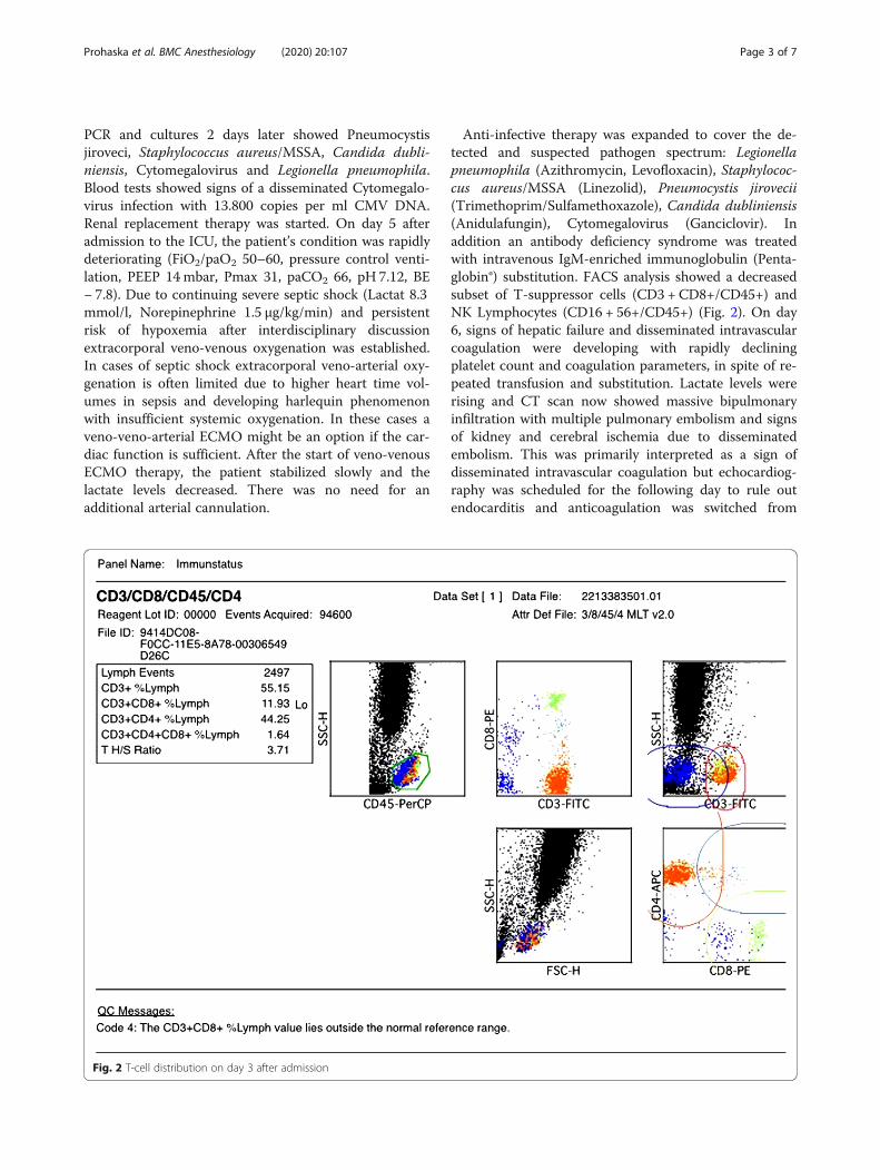
Anti-infective therapy was expanded to cover the de-tected and suspected pathogen spectrum: Legionellapneumophila (Azithromycin, Levofloxacin), Staphylococ-cus aureus/MSSA (Linezolid), Pneumocystis jirovecii(Trimethoprim/Sulfamethoxazole), Candida dubliniensis(Anidulafungin), Cytomegalovirus (Ganciclovir). Inaddition an antibody deficiency syndrome was treatedwith intravenous IgM-enriched immunoglobulin (Penta-globin®) substitution. FACS analysis showed a decreasedsubset of T-suppressor cells (CD3 + CD8+/CD45+) andNK Lymphocytes (CD16 + 56+/CD45+) (Fig. 2). On day6, signs of hepatic failure and disseminated intravascularcoagulation were developing with rapidly decliningplatelet count and coagulation parameters, in spite of re-peated transfusion and substitution. Lactate levels wererising and CT scan now showed massive bipulmonaryinfiltration with multiple pulmonary embolism and signsof kidney and cerebral ischemia due to disseminatedembolism. This was primarily interpreted as a sign ofdisseminated intravascular coagulation but echocardiog-raphy was scheduled for the following day to rule outendocarditis and anticoagulation was switched from
Fig. 2 T-cell distribution on day 3 after admission
Prohaska et al. BMC Anesthesiology (2020) 20:107 Page 3 of 7
Heparin to Argatroban. Blood samples were sent to anextern laboratory to rule out HIT. Based on the impairedhemeostasis with severe thrombopenia (14.000/μl), biop-sie on ECMO was abandoned and echocardiographypostponed.On day 7 after the initiation of anti-infective therapy
Candida glabrata, Pneumocystis jiroveci, Cytomegalo-virus and Legionella pneumophila were still present inBAL cultures. Even though MSSA was not detected any-more, Flucloxacillin was added to cover all bases andAnidulafungin was changed to Voriconazol.Still, there were no signs of improvement. The signs of
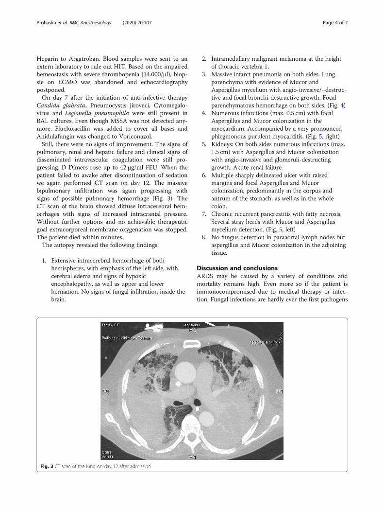
pulmonary, renal and hepatic failure and clinical signs ofdisseminated intravascular coagulation were still pro-gressing. D-Dimers rose up to 42 μg/ml FEU. When thepatient failed to awake after discontinuation of sedationwe again performed CT scan on day 12. The massivebipulmonary infiltration was again progressing withsigns of possible pulmonary hemorrhage (Fig. 3). TheCT scan of the brain showed diffuse intracerebral hem-orrhages with signs of increased intracranial pressure.Without further options and no achievable therapeuticgoal extracorporeal membrane oxygenation was stopped.The patient died within minutes.The autopsy revealed the following findings:
1. Extensive intracerebral hemorrhage of bothhemispheres, with emphasis of the left side, withcerebral edema and signs of hypoxicencephalopathy, as well as upper and lowerherniation. No signs of fungal infiltration inside thebrain.
2. Intramedullary malignant melanoma at the heightof thoracic vertebra 1.
3. Massive infarct pneumonia on both sides. Lungparenchyma with evidence of Mucor andAspergillus mycelium with angio-invasive/−destruc-tive and focal bronchi-destructive growth. Focalparenchymatous hemorrhage on both sides. (Fig. 4)
4. Numerous infarctions (max. 0.5 cm) with focalAspergillus and Mucor colonization in themyocardium. Accompanied by a very pronouncedphlegmonous purulent myocarditis. (Fig. 5, right)
5. Kidneys: On both sides numerous infarctions (max.1.5 cm) with Aspergillus and Mucor colonizationwith angio-invasive and glomeruli-destructinggrowth. Acute renal failure.
6. Multiple sharply delineated ulcer with raisedmargins and focal Aspergillus and Mucorcolonization, predominantly in the corpus andantrum of the stomach, as well as in the wholecolon.
7. Chronic recurrent pancreatitis with fatty necrosis.Several stray herds with Mucor and Aspergillusmycelium detection. (Fig. 5, left)
8. No fungus detection in paraaortal lymph nodes butaspergillus and Mucor colonization in the adjoiningtissue.
Discussion and conclusionsARDS may be caused by a variety of conditions andmortality remains high. Even more so if the patient isimmunocompromised due to medical therapy or infec-tion. Fungal infections are hardly ever the first pathogens
Fig. 3 CT scan of the lung on day 12 after admission
Prohaska et al. BMC Anesthesiology (2020) 20:107 Page 4 of 7

thought of, but fungal coinfections were described inchildren suffering ARDS due to viral infection [2]. Riskfactors for fungal infections in ICU-patients are numer-ous, including immunosuppressive drugs and thedifferentiation of contamination or infection in non-hematological patients is challenging.In this case the decision to deescalate and stop the
therapy was based on the CT scan of the brain. The
scan showed diffuse intracerebral hemorrhages withsigns of increased intracranial pressure. The situationwas evaluated and deemed to be infaust. With theknowledge of the autopsy findings, the overall situ-ation of the mucor infection must now also beregarded as hopeless. Our initial discussion and deci-sion to establish ECMO therapy was based upon thefacts known at the time.
Fig. 4 lung tissue, macroscopy (left) and Grocott-Gomori methenamine silver stain (right)
Fig. 5 Pancreas tissue (left) and Myocard tissue (right)
Prohaska et al. BMC Anesthesiology (2020) 20:107 Page 5 of 7

Although the reported single cases about mucormyco-sis increased lately, they are rare. Most cases weredescribed in patients with malignancies, organ trans-plantation, HIV or DM (recently reviewed in [12]). Ourpatient was affected by none of these diseases but hadreceived corticoid therapy and was thereforeimmunocrompromised.There is some evidence, that mucormycosis and asper-
gillosis may be linked to previous antifungal therapy [14]although mainly in patients with hematological disor-ders. Therefore the onset of antifungal therapy is an im-portant issue. Based on the expert opinion of theEuropean Society of Anesthesia Intensive Care ScientificSubcommittee [15] the decision pathway in this case wascorrect. Initially the colonization index was < 0.5; can-dida score < 3. PCR- and Serum-tests were negative forfungal infection. 1,3-Beta-D-Glucan was not applied.However, the interpretation of this marker in patientsinfected by pneumocystis may be challenging [16]. Theserological test (Platelia®; Bio Rad; München) did notprove positive Aspergillus-Galaktomannan-Antigen until2 days before the patient’s death. In addition diagnosisof pulmonary mucormycosis by conventional culturemay be difficult due to overgrowth. Microscopical exam-ination of BAL may lead to misinterpretation due tocontamination. Histopathological examination may be avalid option, although it is of risk in patients with antic-oagulation and/or disseminated intravascular coagula-tion [8, 17]. Recently Jiang and coworkers suggested theliquid-based cytopathology to identify mucoralespromptly in samples obtained by bronchial brushes,which could be a less invasive method to detect this in-fection promptly [13].Considering and even re-considering frequent risk fac-
tors of fungal infections, e.g. mucormycosis and aspergil-lus, might be more fruitful than pursuing the question ofhow to provide evidence of the pathogen. There is noevidence but according to data obtained in the few hun-dred known cases of mucormycosis, history of poorlycontrolled diabetes in combination with impaired cell-mediated immune function including neutropenia aremainly the issue. Recent data suggests that T cells mayplay an important role in host defense to fungal disease[18, 19]. Like in our patient, lymphopenia may be an im-portant indicator for the application of frequent fungalscreening and fungal prophylaxis.Routine screening before starting an antifungal
prophylaxis and frequent re-evaluation of every infectionare essential for adequate initiation and discontinuationof every fungal therapy especially with patients at highrisk for fungal infections. All patients receiving immuno-suppressive therapy, for whatever reason, must be in-cluded in this group. In case of assumed mucor infectionthe decision for biopsy should be taken into account for
ARDS patients with progressive lung inflammation ofunknown origin when all standard samples fail to pro-vide an edaquate explanation for the patients status,since the risk to die due to mucor may outweigh the riskof fatal bleeding due to the biopsy. But this decisionneeds to be based on a detailed risk/benefit analysis foreach patient.
AbbreviationsARDS: Acute respiratory distress syndrome; BAL: Bronchoalveolar lavage;CMV: Cytomegalovirus; DNA: Desoryribonucleic acid; CRP: C-reactive protein;CT: Computed tomography; DIC: Disseminated intravascular coagulation;DM: Diabetes mellitus; ECMO: Extracorporeal membrane oxygenation;FACS: Fluorescence activated cell sorting; FiO2: Fraction of inspired oxygen;paO2: Arterial partial pressure of oxygen; HIT: Heparin inducedthrombocytopenia; HIV: Human immunodeficiency virus; ICU: Intensive careunit; IgM: Immunoglobulin M; INR: International normalized ratio;MSSA: Methicillin sensitive Staphylococcus aureus; PCR: Polymerase chainreaction; PCT: Procalcitonin; Pmax: Peak pressure
AcknowledgementsThe authors would like to thank all their colleges for their support and help.In addition we would like to thank the Department of Radiology for theaccess to the CT scan and their help to create those special figures.
Authors’ contributionsPS: Preparation of the manuscript, preparation of Figs. HP: Collection of data,review of manuscript. WS: Preparation of figures, review of manuscript. FL:Preparation of figures, review of manuscript. RP: review of manuscript. HHA:review of manuscript. All authors read and approved the final manuscript.
FundingWe acknowledge support by Deutsche Forschungsgemeinschaft and OpenAccess Publishing Fund of University of Tübingen to cover article-processingcharges.
Availability of data and materialsAdditional clinical data is available on request. Please contact thecorresponding author for any additional clinical data. This case reportcontains five figures.All figures have been uploaded with the manuscript.
Ethics approval and consent to participateNot applicable.
Consent for publicationSince the Patient died, written consent to publish was obtained from hiswife and legal representative. Consent was given on August 23rd 2019.
Competing interestsThe authors are not aware of any competing interests concerning thispublication.
Author details1Department of Anesthesiology and Intensive Care Medicine, Intensivstation39, Tübingen University Hospital, Eberhard-Karls-University, Hoppe-Seyler-Str.3, 72076 Tübingen, Germany. 2Department of Pathology, Tübingen UniversityHospital, Eberhard-Karls-University, Tübingen, Germany.
Received: 10 September 2019 Accepted: 3 May 2020
References1. Cortegiani A, Madotto F, Gregoretti C, Bellani G, Laffey JG, Pham T, Van
Haren F, Giarratano A, Antonelli M, Pesenti A, Grasselli G. Investigators LS,the ETG. Immunocompromised patients with acute respiratory distresssyndrome: secondary analysis of the LUNG SAFE database. Crit Care. 2018;22:157.
Prohaska et al. BMC Anesthesiology (2020) 20:107 Page 6 of 7

2. Phung TTB, Suzuki T, Phan PH, Kawachi S, Furuya H, Do HT, Kageyama T, TaTA, Dao NH, Nunoi H, Tran DM, Le HT, Nakajima N. Pathogen screening andprognostic factors in children with severe ARDS of pulmonary origin.Pediatr Pulmonol. 2017;52:1469–77.
3. Alobaid K, Alfoudri H, Jeragh A. Influenza-associated pulmonary aspergillosisin a patient on ECMO. Med Mycol Case Rep. 2020;27:36–8.
4. Vanderbeke L, Spriet I, Breynaert C, Rijnders BJA, Verweij PE, Wauters J.Invasive pulmonary aspergillosis complicating severe influenza:epidemiology, diagnosis and treatment. Curr Opin Infect Dis. 2018;31:471–80.
5. Avino LJ, Naylor SM, Roecker AM. Pneumocystis jirovecii Pneumonia in theNon-HIV-Infected Population. Ann Pharmacother; 2016.
6. Kotton CN, Kumar D, Caliendo AM, Huprikar S, Chou S, Danziger-Isakov L,Humar A, Group TTSICC. The Third International Consensus Guidelines onthe Management of Cytomegalovirus in Solid-organ Transplantation.Transplantation. 2018;102(6):900–31.
7. Bongomin F, Gago S, Oladele RO, Denning DW. Global and multi-NationalPrevalence of fungal diseases—estimate precision. J Fungi (Basel). 2017;3(4):57.
8. Cornely OA, Cuenca-Estrella M, Meis JF, Ullmann AJ. European Society ofClinical Microbiology and Infectious Diseases (ESCMID) fungal infectionstudy group (EFISG) and European Confederation of Medical Mycology(ECMM) 2013 joint guidelines on diagnosis and management of rare andemerging fungal diseases. Clin Microbiol Infect. 2014;20(Suppl 3):1–4.
9. Cornely OA, Bassetti M, Calandra T, Garbino J, Kullberg BJ, Lortholary O,Meersseman W, Akova M, Arendrup MC, Arikan-Akdagli S, Bille J, CastagnolaE, Cuenca-Estrella M, Donnelly JP, Groll AH, Herbrecht R, Hope WW, JensenHE, Lass-Floerl C, Petrikkos G, Richardson MD, Roilides E, Verweij PE, ViscoliC, Ullmann AJ, Group EFIS. ESCMID guideline for the diagnosis andmanagement of Candida diseases 2012: non-neutropenic adult patients.Clin Microbiol Infect. 2012;18:19–37.
10. Pappas PG, Kauffmann CA, Andes DR, Clancy CJ, Marr KA, Ostrosky-ZeichnerL, Reboli AC, Schuster MG, Vazques JA, Walsh TJ, Zaoutis TE, Sobel JD.Clinical Practice Guideline for the Management of Candidiasis: 2016 Updateby the Infectious Diseases Society of America. Clin Infect Dis. 2016;62:e1–50.
11. Ullmann AJ, Aquado JM, Arikan-Akdagli S, Denning DW, Groll AH, Lagrou K,Lass-Floerl C, Lewis RE, Munoz P, Verweij P, Warris A, Ader F, Akova M,Arendrup MC, Barnes RA, Beigelman-Aubry C, Blot S, Bouza E, BrüggemannRJM, Buchheidt D, Cadranel J, Castagnola E, Chakrabarti A, Cuenca-EstrellaM, Dimopoulos G, Fortun J, Gangneux JP, Garbino J, Heinz WJ, Herbrecht R,Heussel CP, Kibbler CC, Klimko N, Kullberg BJ, Lange C, Lehrnbecher T,Löffler J, Lortholary O, Maertens J, Marchetti O, Meis JF, Pagano L, Ribaud P,Richardson M, Roilides E, Ruhnke M, Sanguinetti M, Sheppard DC, Sinko J,Skiada A, Vehreschild MJGT, Viscoli C, Cornely OA. Diagnosis andmanagement of Aspergillus diseases: executive summary of the 2017ESCMID-ECMM-ERS guideline. Clin Microbiol Infect. 2018;24:e1–e38.
12. Yamin HS, Alastal AY, Bakri I. Pulmonary Mucormycosis over 130 years: acase report and literature review. Turk Thorac J. 2017;18:1–5.
13. Jiang X, Yang T, Li Q, Zhu X, Su X, Li J, Jiang Y. Liquid-based cytopathologytest: a novel method for diagnosing pulmonary Mucormycosis in bronchialbrushing samples. Front Microbiol. 2018;9:2923.
14. Guinea J, Escribano P, Vena A, Munoz P, Martinez-Jimenez MDC, Padilla B,Bouza E. Increasing incidence of mucormycosis in a large Spanish hospitalfrom 2007 to 2015: epidemiology and microbiological characterization ofthe isolates. PLoS One. 2017;12:e0179136.
15. O'Leary RA, Einav S, Leone M, Madach K, Martin C, Martin-Loeches I.Management of invasive candidiasis and candidaemia in critically ill adults:expert opinion of the European Society of Anaesthesia Intensive CareScientific Subcommittee. J Hosp Infect. 2018;98:382–90.
16. Lahmer T, da Costa CP, Held J, Rasch S, Ehmer U, Schmid RM, Huber W.Usefulness of 1,3 Beta-D-Glucan detection in non-HIV Immunocompromisedmechanical ventilated critically ill patients with ARDS and suspectedPneumocystis jirovecii pneumonia. Mycopathologia. 2017;182:701–8.
17. Combes A, Hajage D, Capellier G, Demoule A, Lavoue S, Guervilly C, Da SilvaD, Zafrani L, Tirot P, Veber B, Maury E, Levy B, Cohen Y, Richard C, Kalfon P,Bouadma L, Mehdaoui H, Beduneau G, Lebreton G, Brochard L, FergusonND, Fan E, Slutsky AS, Brodie D, Mercat A, Eolia Trial Group R, Ecmonet.Extracorporeal membrane oxygenation for severe acute respiratory distresssyndrome. N Engl J Med. 2018;378:1965–75.
18. Arens C, Kramm T, Decker S, Spannenberger J, Brenner T, Richter DC,Weigand MA, Uhle F, Lichtenstern C. Association of Immune Cell Subtypes
and Phenotype with Subsequent Invasive Candidiasis in Patients withAbdominal Sepsis. Shock. 2019;52(2):191–97.
19. Zhang J, Cui N, Long Y, Wang H, Han W, Li Y, Xiao M. Prospectiveevaluation of lymphocyte subtyping for the diagnosis of invasive candidiasisin non-neutropenic critically ill patients. Int J Infect Dis. 2019;78:140–7.
Publisher’s NoteSpringer Nature remains neutral with regard to jurisdictional claims inpublished maps and institutional affiliations.
Prohaska et al. BMC Anesthesiology (2020) 20:107 Page 7 of 7