a case of double primary cancers of uterine endometrium ... · endometrium and bladder with unusual...
TRANSCRIPT

WWW.KJOG.ORG 543
A CASE OF DOUBLE PRIMARY CANCERS OF UTERINE ENDOMETRIUM AND BLADDER WITH UNUSUAL HISTOLOGYSul Ah Kim, MD, Dong Choon Park, MD, PhDDepartment of Obstetrics and Gynecology, St. Vincent’s Hospital, The Catholic University of Korea, Suwon, Korea
A case of a moderate differentiated endometrial carcinoma of the uterus with a synchronous poorly differentiated bladder cancer is reported. A review of the literature revealed that simultaneous presentation of primary endometrial and bladder neoplasm is rare and usually related to low-stage bladder lesions in contrast to our case with undifferentiated and the deep myometrial invasion of bladder lesion. A 79-year-old woman with endometrial cancer stage IB was performed of total abdominal hysterectomy with bilateral salpingo-oophorectomy, pelvic and para-aortic lymph nodes dissection and adjuvant concurrent cisplatin-radiation therapy. After treatment, she complained intermittent gross hematuria. She was performed the bladder mucosal biopsy and fi nally diagnoses with poorly differentiated carcinoma of bladder. She received transurethral resection of bladder tumor alone without total cystectomy or any other adjuvant treatment due to her refusal. Her condition is tolerable except intermittent hematuria and anemia.
Keywords: Double primary cancer; Endometrial neoplasms; Bladder cancer
CASE REPORT
Received: 2011. 5. 9. Revised: 2011. 6.20. Accepted: 2011. 7.29.Corresponding author: Dong Choon Park, MD, PhD Department of Obstetrics and Gynecology, St. Vincent’s Hospital, Th e Catholic University of Korea, 93 Gi-dong, Paldal-gu, Suwon 442-836, KoreaTel: +82-31-249-8221 Fax: +82-31-254-7481E-mail: [email protected]
Th is is an Open Access article distributed under the terms of the Creative Commons Attribution Non-Commercial License (http://creativecommons.org/licenses/by-nc/3.0/) which permits unrestricted non-commercial use, distribution, and reproduction in any medium, provided the original work is properly cited.
Copyright © 2011. Korean Society of Obstetrics and Gynecology
Korean J Obstet Gynecol 2011;54(9):543-547http://dx.doi.org/10.5468/KJOG.2011.54.9.543pISSN 2233-5188 · eISSN 2233-5196
In the case of synchronous detection of neoplasms of two organs, the distinction between double primary tumors with metastasis to the other organs based on conventional clinicopathologic criteria may be diffi cult. The distinction between metastatic and double pri-mary tumors is signifi cant since it affects staging and prognosis [1].We reported the rare case of endometrium-bladder double primary cancer, which showed the presence of features of poorly differen-tiated carcinoma of the bladder along with deep invasion of the myometrium, contrary to early stage of endometrial cancer.
Case Report
A 79-year-old (gravid 3, para 3) woman was admitted in our de-partment with a 3-month history of irregular vaginal spotting and bleeding accompanied by frequency. In the past history, other than total hip replacement surgery received 15 years ago, special fi nd-ings were not observed. In pelvic examination, compared to her ages the uterus was large and soft. In sonogram the endometrial thickness was 2.4 cm. She was performed endometrial biopsy and diagnosed moderately differentiated endometrioid carcinoma. Magnetic resonance imaging confi rmed the existence of nonho-mogeneous endometrial tumor which invaded the myometrium
more than half (Fig. 1A). There was no evidence of retroperitoneal lymph node or liver involvement. Her clinical stage was endome-trial cancer Ib. Complete blood count, renal and liver function tests were unremarkable. Serum tumor markers were elevated (CA-125, 65.3 IU/mL). At laparotomy, the uterus was enlarged with endo-metrial tumor measuring 2.5×2.7 cm and a diffusely enlarged uterus were discovered. Both ovaries appeared normal as did the omentum and the other pelvic and abdominal viscera. Total hys-terectomy and bilateral salpingo-oophorectomy as well as pelvic and paraaortic lymphadenectomy were performed. Histopatho-logic examination disclosed moderately differentiated endometrioid

WWW.KJOG.ORG544
KJOG Vol. 54, No. 9, 2011
Fig. 1. (A) Pelvic magnetic resonance imaging. About 2.8×2.6×2.4 cm sized T2 in-termediate signal intensity mass (arrow) within the endometrial cavity and thinning of myometrium, especially right side body and fundus. (B) Pelvic computed tomogra-phy. Irregular lobulated tumor (arrow) around right posterior inferior wall of the uri-nary bladder with suspicious adjacent perivascular retroperitoneal space infi ltration along the right internal iliac vessels.
A
B
Fig. 2. In the uterus, typical endometrioid adenocarcinoma invading myometrium is noted (A). The lesion in the urinary bladder is morphologically dif-ferent from that of the uterus, and composed of highly pleomorphic malignant cells without forming glandular structure (B). Immunohistologically, these tumor cells show positive reaction to cytokeratin (CK)-7 (C) and vimentin (D), but negative reaction to CK20 (E). So, for the lesion in the urinary bladder, poorly differentiated carcinoma or carcinosarcoma is suspected. Magnifi cation, ×200.
A B
C D E
WWW.KJOG.ORG 545
Sul Ah Kim, et al. Double primary cancer of uterine endometrium and bladder with unusual histology
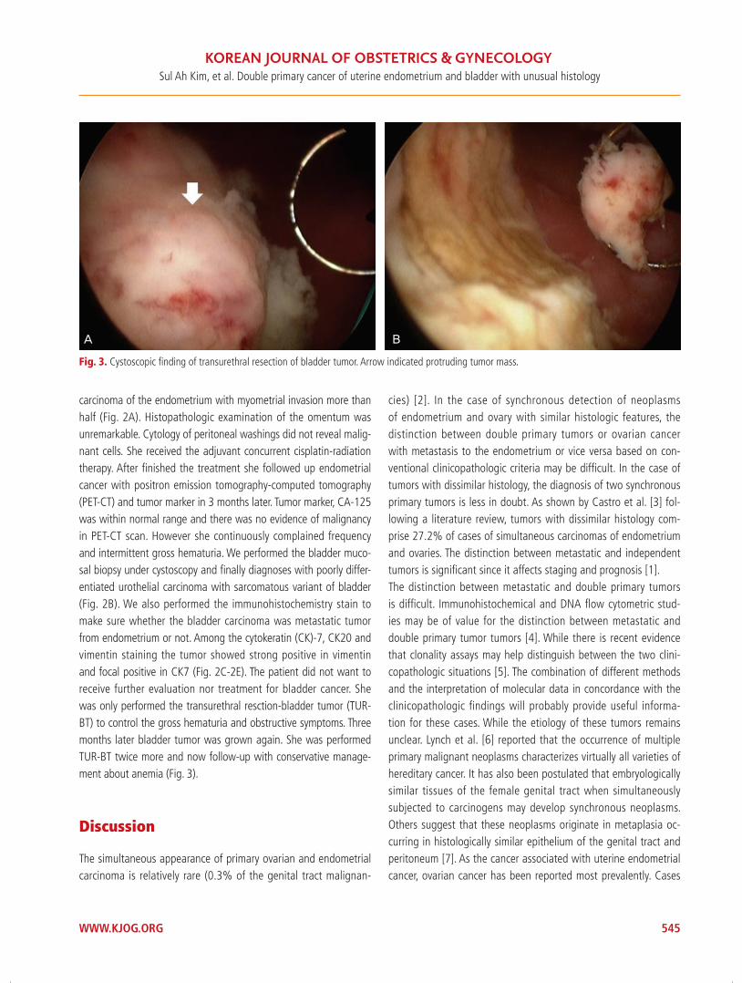
carcinoma of the endometrium with myometrial invasion more than half (Fig. 2A). Histopathologic examination of the omentum was unremarkable. Cytology of peritoneal washings did not reveal malig-nant cells. She received the adjuvant concurrent cisplatin-radiation therapy. After fi nished the treatment she followed up endometrial cancer with positron emission tomography-computed tomography (PET-CT) and tumor marker in 3 months later. Tumor marker, CA-125 was within normal range and there was no evidence of malignancy in PET-CT scan. However she continuously complained frequency and intermittent gross hematuria. We performed the bladder muco-sal biopsy under cystoscopy and fi nally diagnoses with poorly differ-entiated urothelial carcinoma with sarcomatous variant of bladder (Fig. 2B). We also performed the immunohistochemistry stain to make sure whether the bladder carcinoma was metastatic tumor from endometrium or not. Among the cytokeratin (CK)-7, CK20 and vimentin staining the tumor showed strong positive in vimentin and focal positive in CK7 (Fig. 2C-2E). The patient did not want to receive further evaluation nor treatment for bladder cancer. She was only performed the transurethral resction-bladder tumor (TUR-BT) to control the gross hematuria and obstructive symptoms. Three months later bladder tumor was grown again. She was performed TUR-BT twice more and now follow-up with conservative manage-ment about anemia (Fig. 3).
Discussion
The simultaneous appearance of primary ovarian and endometrial carcinoma is relatively rare (0.3% of the genital tract malignan-
cies) [2]. In the case of synchronous detection of neoplasms of endometrium and ovary with similar histologic features, the distinction between double primary tumors or ovarian cancer with metastasis to the endometrium or vice versa based on con-ventional clinicopathologic criteria may be diffi cult. In the case of tumors with dissimilar histology, the diagnosis of two synchronous primary tumors is less in doubt. As shown by Castro et al. [3] fol-lowing a literature review, tumors with dissimilar histology com-prise 27.2% of cases of simultaneous carcinomas of endometrium and ovaries. The distinction between metastatic and independent tumors is signifi cant since it affects staging and prognosis [1].The distinction between metastatic and double primary tumors is diffi cult. Immunohistochemical and DNA fl ow cytometric stud-ies may be of value for the distinction between metastatic and double primary tumor tumors [4]. While there is recent evidence that clonality assays may help distinguish between the two clini-copathologic situations [5]. The combination of different methods and the interpretation of molecular data in concordance with the clinicopathologic findings will probably provide useful informa-tion for these cases. While the etiology of these tumors remains unclear. Lynch et al. [6] reported that the occurrence of multiple primary malignant neoplasms characterizes virtually all varieties of hereditary cancer. It has also been postulated that embryologically similar tissues of the female genital tract when simultaneously subjected to carcinogens may develop synchronous neoplasms. Others suggest that these neoplasms originate in metaplasia oc-curring in histologically similar epithelium of the genital tract and peritoneum [7]. As the cancer associated with uterine endometrial cancer, ovarian cancer has been reported most prevalently. Cases
Fig. 3. Cystoscopic fi nding of transurethral resection of bladder tumor. Arrow indicated protruding tumor mass.
A B

WWW.KJOG.ORG546
KJOG Vol. 54, No. 9, 2011
such as our case that uterine endometrial cancer is associated with bladder cancer are very rare. Even more the presence of poor differentiation in the bladder carcinoma and the deep myometrial invasion found in our case is unusual.Therefore, until now, the standardized guideline for its diagnosis or treatments has not been established yet, and it is treated by the methods suitable to each cancer. Since the incidence is very low, treatment responses or prognostic factors have not been reported yet. However, according to the reports, the prognosis in most cases of double primary malignant tumor is surprisingly good. It is reported that the prognosis of the “highly malignant” tumor does not worsen as a result of a second malignant cancer [8] and furthermore the prognosis of double primary carcinoma of uterine corpus and ovary is rather good [9,10]. The precise mechanism of good prognosis of double primary cancer has not been revealed yet. It just is speculated that in double primary cancer, only one cancer is diagnosed, and during the test procedure, another can-cer may be detected and thus it is detected rather early, hence, the prognosis is good. Engine [11] suggested prognosis of multiple primary cancer more comprehensively. According to his report, gender, age, and the in-terval of two primary tumors were prognostic factors. In our case, the prognosis is speculated to be poorer. Because the fi rst tumor was detected after the age of 50 years, and the interval of the two primary cancers was less than 2 years. Furthermore the differentia-tion of bladder cancer was poor and it progressed rapidly. Patients with endometrial cancer should be carefully and regularly followed up by monitoring et every anatomic site, especially the breast, stomach, and colon, in order that the development of a second primary carcinoma can be detected as early as possible, and not be overlooked in examinations. Additional risk factors for endometrial carcinoma with multiple malignant neoplasms include: menopause occurring after age fi fty-one; obese women with body mass index higher than 32; reproductive period lon-ger than 37 years [12]. Our case was a patient with early stage uterine endometrial cancer of which possibility of the metastasis to the bladder is very low. Endometrial cancer was treated and during follow-ups by general tests, bladder cancer was diagnosed. During postsurgical radiation therapy, our patient presented with intermittent hematuria, nevertheless, it was misdiagnosed as side effects of radiation therapy, and thus the diagnosis was delayed. In addition, our patient received total hip replacement surgery and her condition was inappropriate to diagnose masses in the blad-der by imaging study. After uterine endometrial cancer treatments, the recurrence of uterine endometrial cancer was followed up by
PET- CT, hence, bladder cancer was overlooked. The patient was old and did not want to receive the total cystecto-my, and thus only conservative management was performed with TUR-BT. Contrary to our expectations, her condition is tolerable except intermittent hematuria.
References
1. Sheu BC, Lin HH, Chen CK, Chao KH, Shun CT, Huang SC. Syn-chronous primary carcinomas of the endometrium and ovary. Int J Gynaecol Obstet 1995;51:141-6.
2. Eisner RF, Nieberg RK, Berek JS. Synchronous primary neo-plasms of the female reproductive tract. Gynecol Oncol 1989;33:335-9.
3. Castro IM, Connell PP, Waggoner S, Rotmensch J, Mundt AJ. Synchronous ovarian and endometrial malignancies. Am J Clin Oncol 2000;23:521-5.
4. Prat J, Matias-Guiu X, Barreto J. Simultaneous carcinoma in-volving the endometrium and the ovary. A clinicopathologic, immunohistochemical, and DNA fl ow cytometric study of 18 cases. Cancer 1991;68:2455-9.
5. Prat J. Clonality analysis in synchronous tumors of the female genital tract. Hum Pathol 2002;33:383-5.
6. Lynch HT, Harris RE, Lynch PM, Guirgis HA, Lynch JF, Bardawil WA. Role of heredity in multiple primary cancer. Cancer 1977;40:1849-54.
7. Ayhan A, Yalcin OT, Tuncer ZS, Gürgan T, Küçükali T. Synchro-nous primary malignancies of the female genital tract. Eur J Obstet Gynecol Reprod Biol 1992;45:63-6.
8. Schröcksnadel H, Fuith LC, Hetzel H. Multiple cancers in gyne-cologic oncology. Geburtshilfe Frauenheilkd 1988;48:710-4.
9. Chen F, Shen K, Lang JH, Huang HF, Wu M. Clinical features and prognostic of double primary carcinoma of uterine corpus and the ovary. Zhonghua Yi Xue Za Zhi 2005;85:1257-60.
10. Papathanasiou K, Tolikas A, Dovas D, Kostopoulou E, Fragke-dakis N, Tzafettas J. Simultaneously detected primary malig-nant tumors of ovary and endometrium with unusual histol-ogy. Int J Gynecol Cancer 2005;15:1191-4.
11. Engin K. Cancers in multiple primary sites. Int Surg 1994;79:33-7.
12. Studziński Z, Branicka D. The coexistence of endometrial can-cer with second primary malignant neoplasms. Ginekol Pol 1999;70:186-92.

WWW.KJOG.ORG 547
Sul Ah Kim, et al. Double primary cancer of uterine endometrium and bladder with unusual histology
초기 자궁내막암과 진행된 방광암의 원발성 악성종양 1예
가톨릭대학교 성빈센트병원 산부인과
김슬아, 박동춘
두 기관 이상의 원발암을 갖는 예는 흔하지 않다. 자궁내막암과 동반된 방광암은 대부분이 낮은 임상병기를 보이며 예후 또한 비교적 좋
은 편으로 보고된다. 그러나 일반적인 예와는 달리 저자는 중등도의 분화도를 보이는 자궁내막암과 드물게 동반된 분화도가 매우 나쁜 방
광암을 경험하였기에 문헌 고찰과 더불어 보고하는 바이다. 자궁내막암 1기의 본 환자는 전자궁절제 및 양측 난소난관절제술, 골반 및 대
동맥 임파절절제를 시행받은 후 완전 관해를 보여 추적 관찰하던 중 혈뇨의 증상을 호소, 시행한 방광경하 조직검사상 분화도가 매우 나
쁜 방광암이 진단되었던 예로 환자가 추가적인 검사나 수술적 치료를 반대하여 transurethral resection of bladder tumor로 대증적인 치료
를 하였다.
중심단어: 이중원발암, 자궁내막암, 방광암