a. · 176 coelom comparison figure 27.6 a. endoderm digestive cavity pseudocoelom coelom from...
TRANSCRIPT
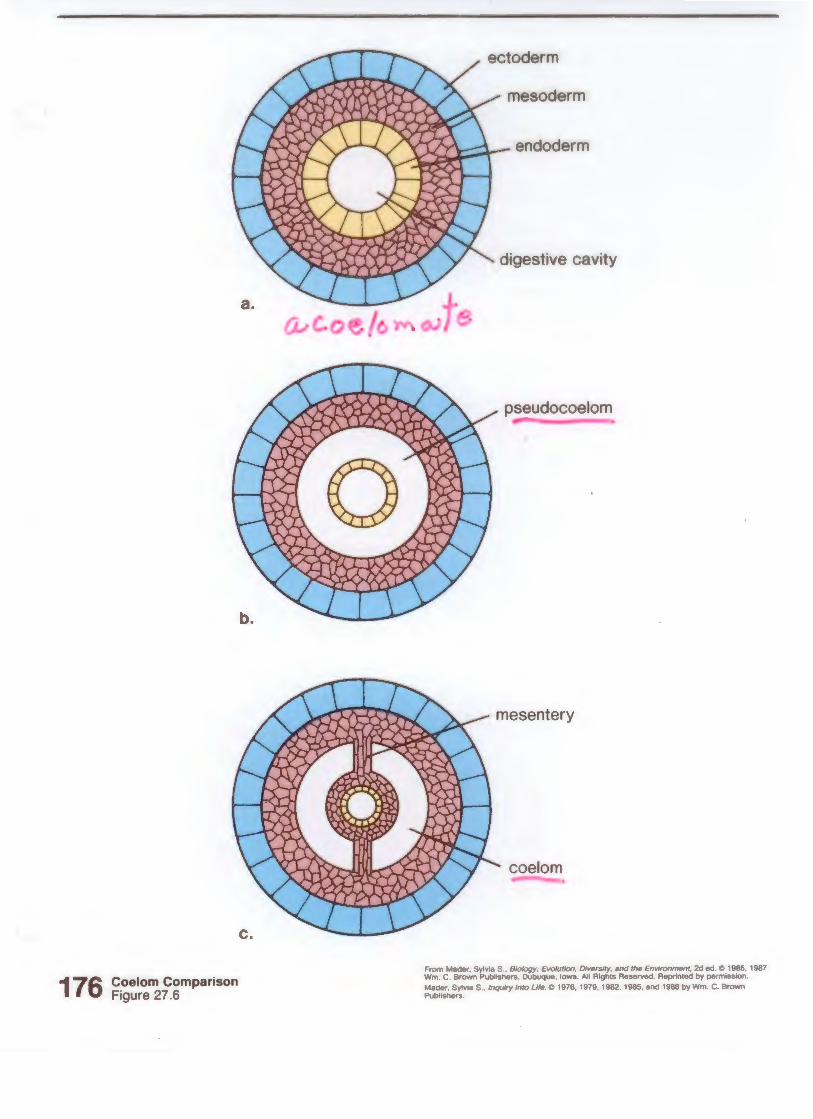
176 Coelom Comparison Figure 27.6
a.
endoderm
digestive cavity
pseudocoelom
coelom
From Mader. Sylv1a S .. Biology: Evolullon. Dwersity, and the Env,onment. 2d ed. tO 1985, 1987 Wm . C. Brown Publishers , Dubuque , Iowa. All Rights Reserved. Reprinted by perm1ssion .
Mader, Sylv1a S., lnqwry Into Ufe. tO 1976, 1979, 1982, 1985, and 1988 by Wm. C. Brown Publishers
a.
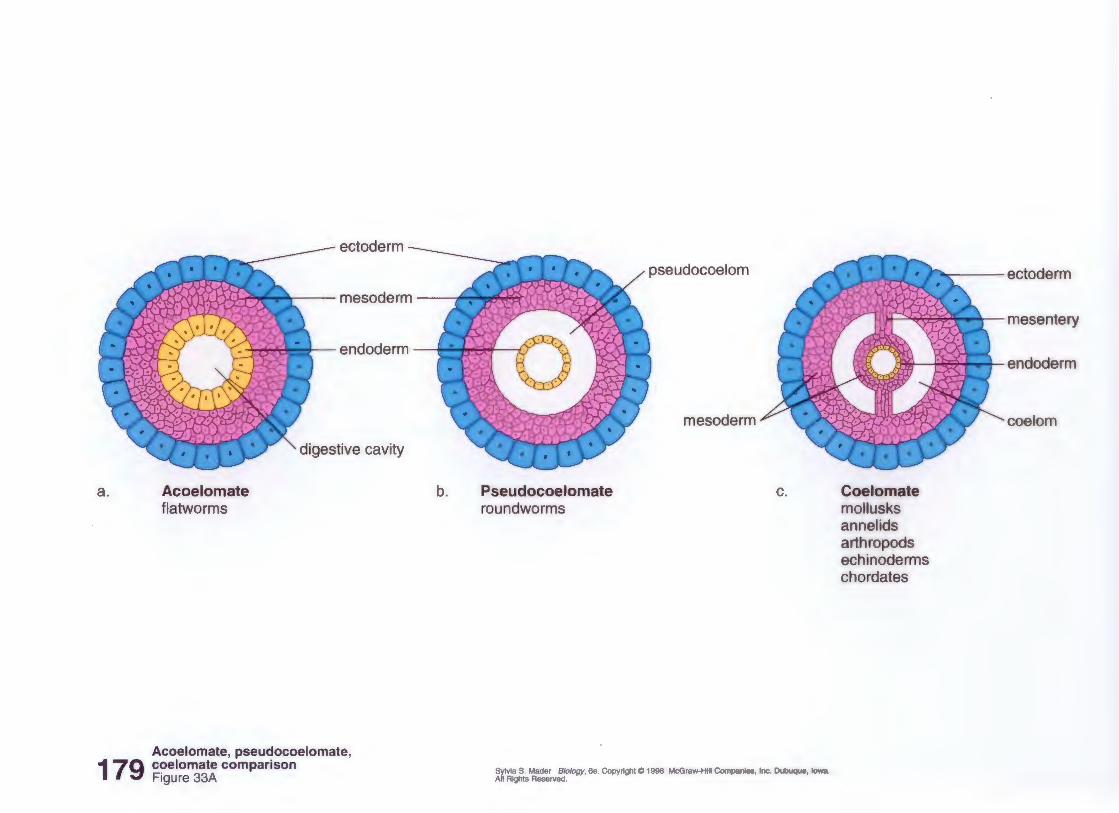
179
ectoderm
pseudocoelom
..----mesoderm JIF~((.
endoderm ~
Acoelomate flatworms
digestive cavity
Acoelomate, pseudocoelomate, coelomate comparison Figure 33A
b. Pseudocoelomate roundworms
mesoderm
c.
Sylvia S. Mader Biology, 6e. Copyright@ 1998 McGraw-Hill Companies, Inc. Dubuque, Iowa. All Rights Reserved.
,.....,. ectoderm
J~ Y~X:D:> .LW-.. '11 mesentery
Coelomate mollusks annelids arthropods echinoderms chordates
endoderm
coelom
7 "p
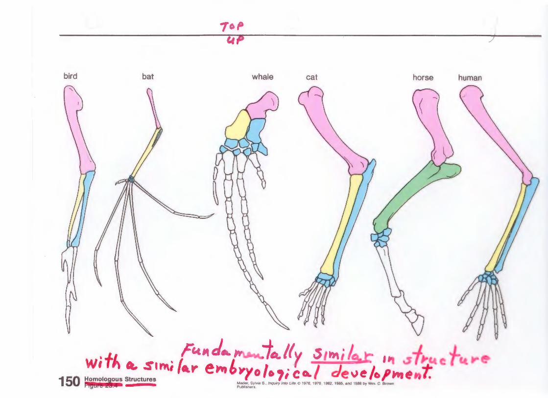
bird bat whale cat horse human
. Fc.t"d ... ~"ftJt.lfy .s,"";IQ,r ,._ .s WtfJ.. a. Sl rfl.i (-.r- e-rr.£-ryo /o ,; CA./ c!~.~.~~.~~-,
150 Homolol ous Structures Mader, Sylvoa s., lnqUify Into Life. e> 1976, 1979. 1982· • r PubliShers. IQS: 6 ~ . I
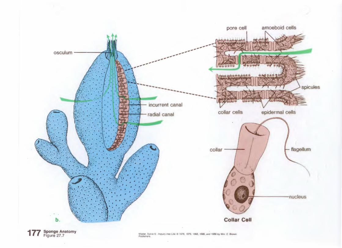
177
osculum ,<JfAIJIII\\t--
b.
Sponge Anatomy Figure 27.7
--------,.-'
,.-' ,.-'
,.-'
pore cell
------------------
-------------------------
collar cells
collar _ __,._
amoeboid cells
epidermal cells
flagellum
'---1~-----nucleus
Collar Cell
Mader, Sylv1a S., lnqwry Into Life. Q 1976, 1979. 1982, 1985, and 1988 by Wm. C Brown Publishers
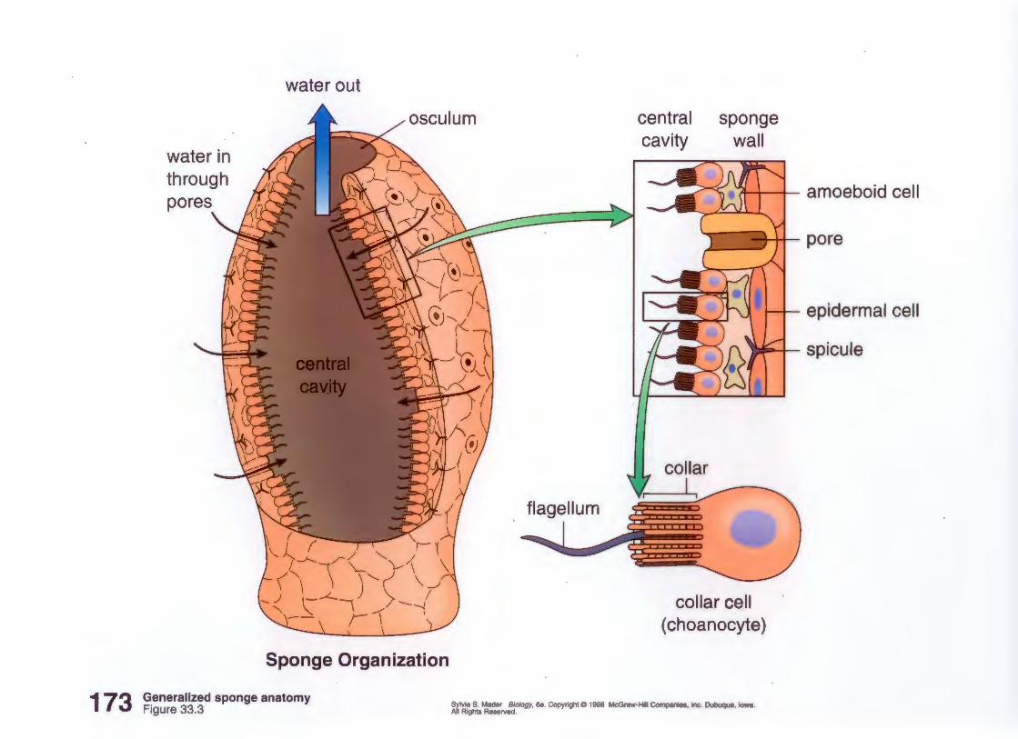
173
water out
central sponge
water in cavity wall
through , /ll ..__ - ~ ...... ,~ '-- \. \. 1 ~ ~ ......... w ' 1 amoeboid cell
Sponge Organization
Generalized sponge anatomy Figure 33.3
pore
epidermal cell
M" 1 spicule
flagellum
collar cell ( choanocyte)
Sylvia S. Mader Biology, 6e . Copyright@ 1998 McGraw-Hill Companies, Inc. Dubuque, Iowa. All Rights Reserved.
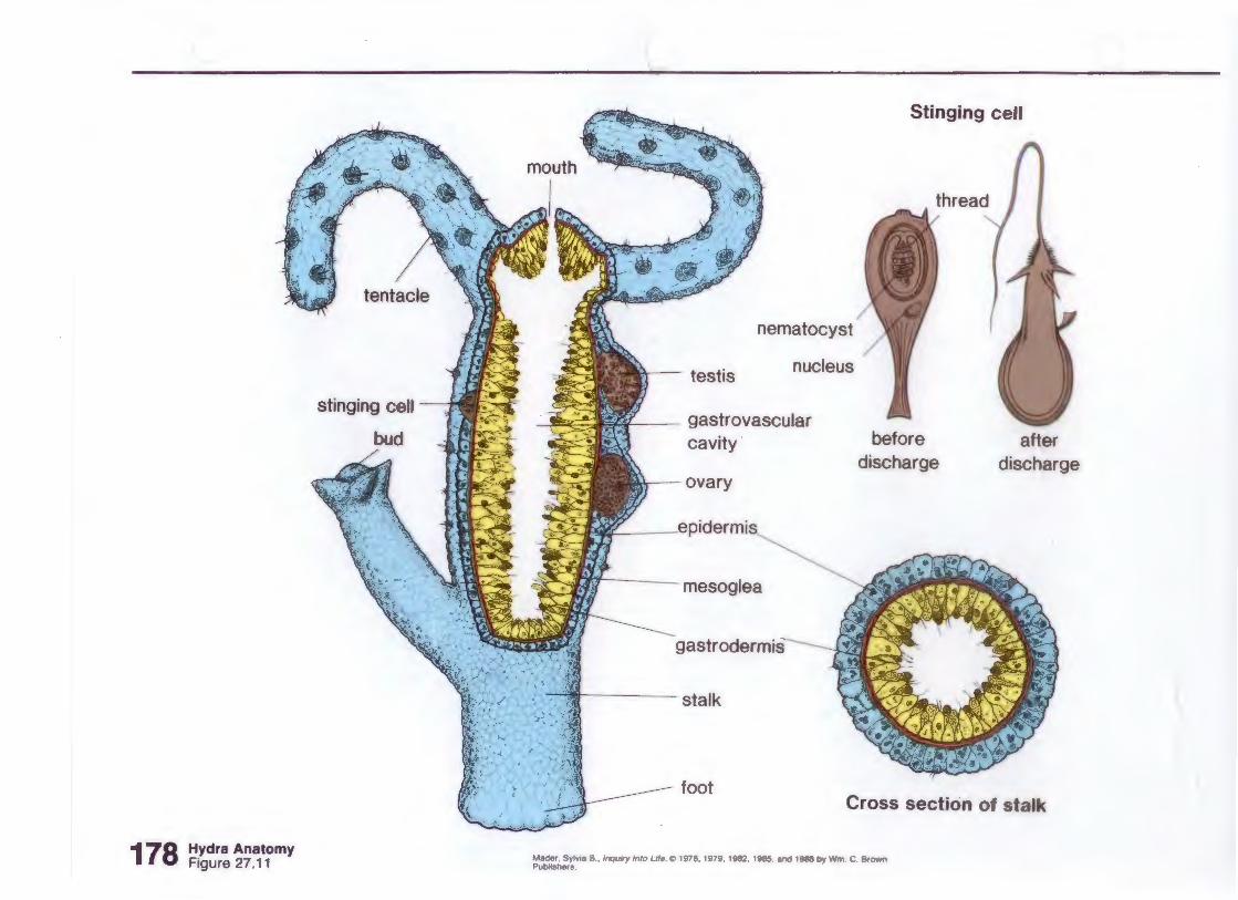
178 Hydra Anatomy Figure 27.11
tentacle
stinging cell
Stinging cell
nematocyst
nucleus
gastrovascular cavity
ta.l. ovary
~mesoglea
·'afil stalk
foot
before discharge
after discharge
Cross section of stalk
Mader. Sylvoa S., /nqulfy lnlo Life. 00 1976. 1979. 1982, 1985. and 1988 by Wm. C. Brown Publishers.
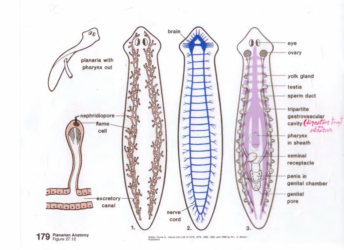
,_/ (1!~~~') Planaria with
Pharynx out · /1oi ~;~_I
nephridiopore --J;:J~
lfame 1'\ ~ l;~l
ceu bts:l "'"i''b' I
~ ~}~f/ ---excretory ~· §
canal ) ~
179 Ptanarian Anatomy
Figure 27. 12
bra
) ~ I~ f
l:t 1: I I " I I ' I
"""· ..... ' . -ry '"' ,, .. ""· ""· '"'· '"' ·~ '"' " ... ' ~-. Publishers
\ta- ;"'al____ ~eye ovary
Yolk gland
testis
-sperm auct
I 'iril lw-T"- triParrtte
~astrova.
~avity (di
Pharynx . . ' m sheath
seminal
receptacle
Penis in
genital chamber
genita
ore
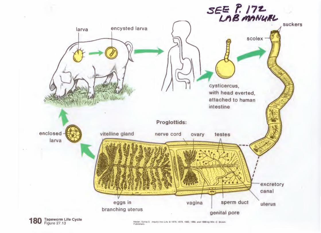
larva encysted larva
180 Tapeworm Life Cycle Figure 27.13
vitelline gland
eggs in branching uterus
Proglottids:
nerve cord ovary
vagina
cysticercus,
with head everted, attached to human intestine
testes
sperm duct
genital pore
Mader. Sylv1a S , /nqwry Into We. C> 1976. 1979, 1982. 1985. and 1988 by Wm . C Brown Publ•shers .
I
#I, suckers
excretory canal
uterus
@AI _o -o< (X)(D -o::J o-L "< 0 :::r -t::J 3' ~ (D ::J "' .. ~~ - · 0 ., -., 0 Q<e
~"< "' 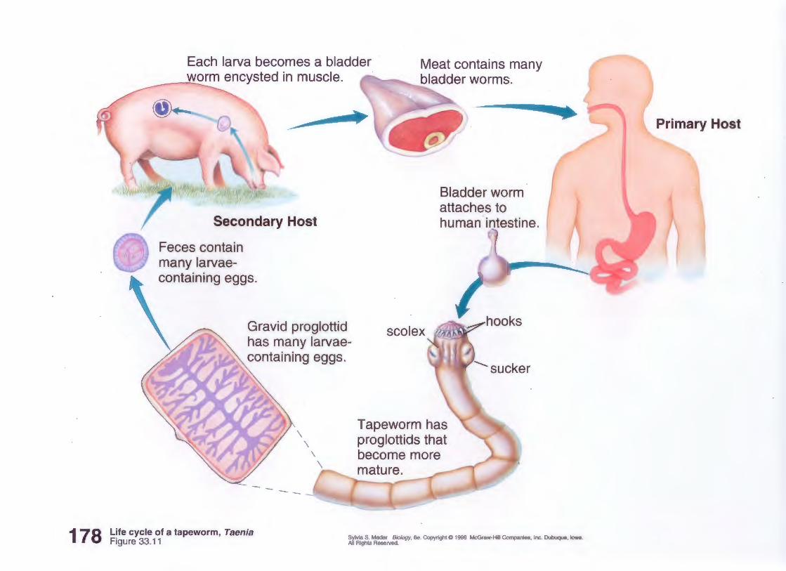
178
Each larva becomes a bladder _ ....... w_..._2rm encysted in muscle.
Meat contains many bladder worms.
-
Secondary Host
Feces contain many larvae-containing eggs.
Gravid proglottid has many larvaecontaining eggs.
\ \
1'
Bladder worm attaches to human intestine.
-...sucker
Tapeworm has proglottids that become more mature.
.. --
Life cycle of a tapeworm, Taenia Figure 33.11 Sylvia S. Mader Biology, 6e. Copyright@ 1998 McGraw-Hill Companies, Inc. Dubuque, Iowa.
All Rights Reserved.
Primary Host
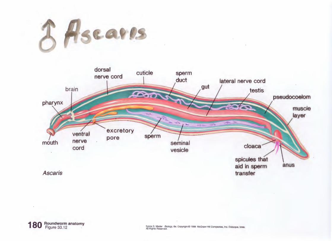
Ascaris
brain
nerve cord
180 Roundworm anatomy Figure 33.12
dorsal nerve cord
pore
cuticle
lateral nerve cord
testis
spicules that aid in sperm transfer
Sylvia S. Mader Biology, 6e. Copyright@ 1998 McGraw-Hill Companies, Inc. Dubuque, Iowa. All Rights Reserved.
pseudocoelom
anus
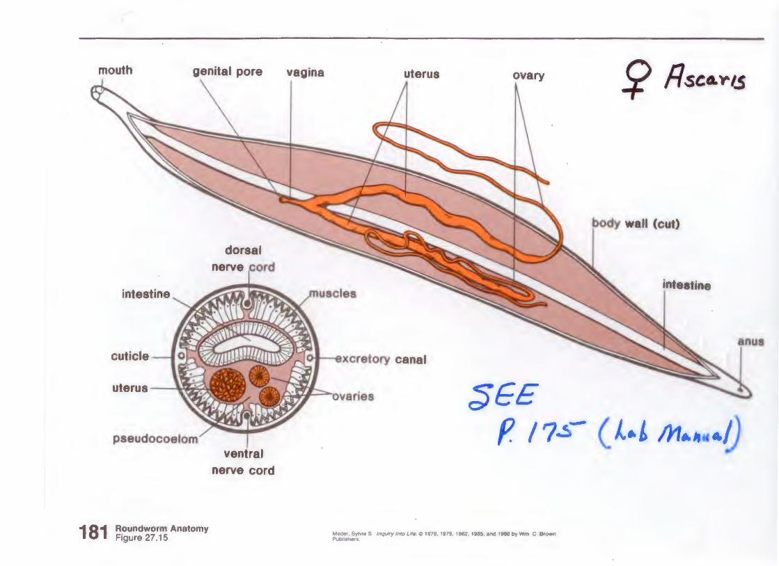
mouth genital pore
181
dorsal
cuticle
uterus \.~~
pseudocoelom
Roundworm Anatomy Figure 27.15
ventral nerve cord
vagina uterus ovary
ro • Avcretory canal
~ta'l =-nvs:.ries 3E
Mader. Sylvta S , Jnqwry Into Life. CC> 1976, 1979, 1982. 1985, and 1988 by Wm . C Brown Publishers
S? fl St:.O. 'Y' 1.$
body II Ccut)
intestine
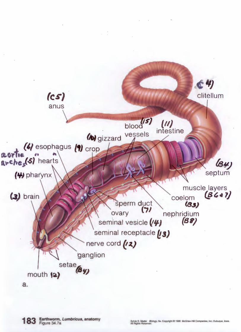
a.
(cs} anus
fs~) septum
muscle layers coelom (§" ~ 1)
perm duct ~ (8_,)
ovary (iJ nephridium sem.inal vesicle~~) (8')
seminal receptacle ~3)
nerve cord ~ ~)
~anglion
mouth t setae~
·) {B'I)
1 83 Earthworm, Lumbricus, anatomy Figure 34.7a
Sylvia S Mader Biology, 6e. Copyright 0 1998 McGraw-Hill Companies, Inc. Dubuque, Iowa. All Rights Reserved.
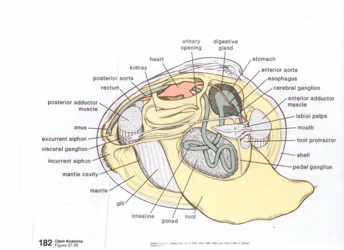
posterior adductor muscle
anus 1 'i'" 1
excurrent siphon
visceral ganglion
incurrent siphon
mantle cavity
182 Clam Anatomy Figure 27.20
urinary opening
digestive gland
anterior aorta
esophagus
cerebral ganglion
anterior adductor muscle
labial palps
·.·:. ... .. I ~·· ~·. ···· ·· ··· ., · .. 1 mouth ....__,_, ", . ..... h I Il l
?~llfT111P~ 1 ; ' i 1 foot protractor ' \ _ . JT u 1 ; I II
pedal ganglion
Mader Sylvoa S. lnqu1ry Into L1/e C> 1976, 1979. 1982. 1985. and 1988 by Wm. C. Brown Publisher~ .
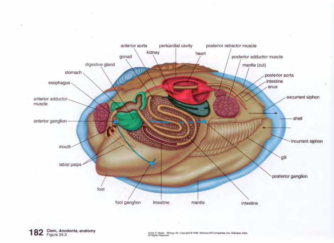
stomach
esophagus
anterior adductor muscle
anterior ganglion • ·•
182 Clam, Anodonta, anatomy Figure 34.3
anterior aorta pericardia! cavity posterior retractor muscle
foot ganglion
I kidney
intestine mantle intestine
Sylvia S. Mader Biology, 6e. Copyright C 1998 McGraw-Hill Companies, Inc. Dubuque, Iowa. All Rights Reserved.
posterior aorta
excurrent siphon
• shell
incurrent siphon
posterior ganglion

• ... ..
a.
antenna
compound eye
antennule
mouth parts
nephridiopore
claw third fourth
mouth oviduct opening
anus /
tel son
swimmerets uropod
;.,
s~co~d walking . · leg
walking · walking fifth
walking ·~,
feg leg leg
b.
cerebral ganglion
183
,,
esophagus
Crayfish Anatomy Figure 27.26
heart pericardia! sinus
digestive gland
stomach nerve cord anus
Mader. Sylv1a S .. lnqwry Into Life. (C) 1976. 1979. 1982, 1985, and 1988 by Wm . C. Brown Publishers
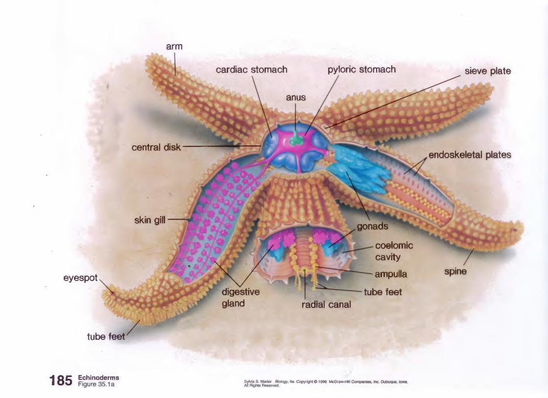
arm
central disk---
skin gill~
eyes pot
tube feet/
185 Echinoderms Figure 35.1 a
anus
----tube feet
Sylvia S. Mader Biology, 6e. Copyright© 1998 McGraw-Hill Companies, Inc. Dubuque, Iowa. All Rights Reserved.
sieve plate
endoskeletal plates
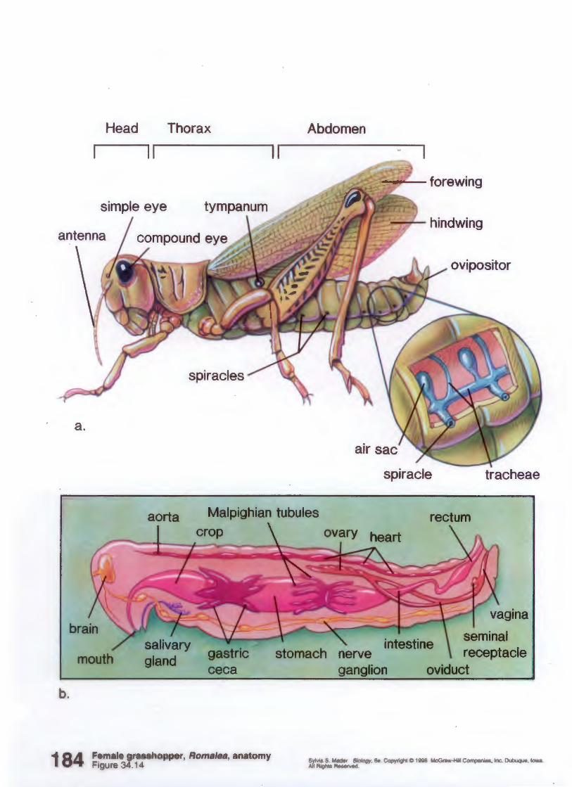
a.
b.
Head Thorax Abdomen
II
~· · forewing
·~hindwing
ovipositor
spiracles
spiracle tracheae
aorta Malpighian tubules
ceca
Female grasshopper, Romalea, anatomy Figure 34.14
stomach intestine seminal
receptacle oviduct
Sylvia S. Mader Biology, 6e. Copyright C 1998 McGraw-Hill Companies , Inc. Dubuque, Iowa. All Rights Reserved .
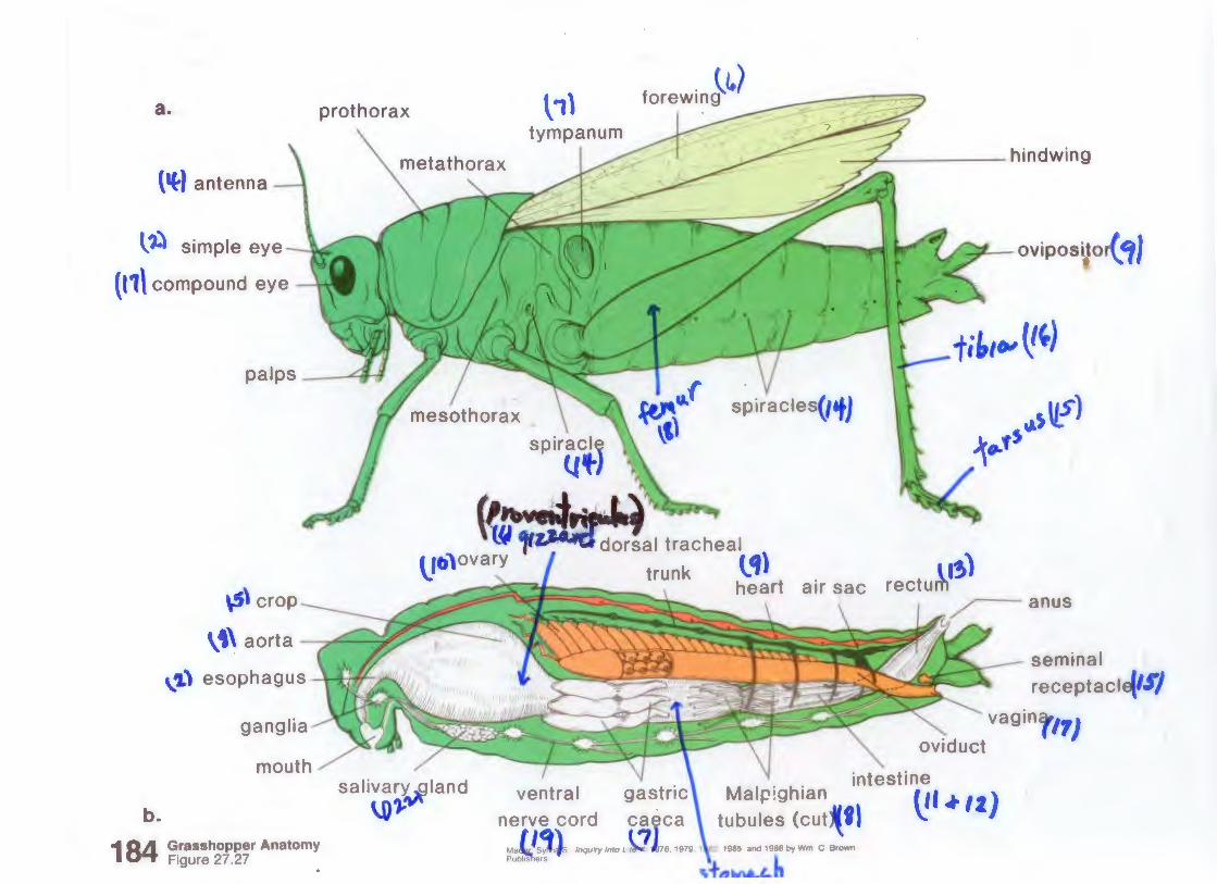
a. prothorax
(~) antenna
t,:) simple eye --.J.looC
{11\ compound eye
palps <a tt
\S\ crop
\•\ aorta 7' ~
\.'1) esophagus ' l i N ..
salivar~land
b. \.01-=
184 Grasshopper Anatomy Figure 27.27
~1\ forewin}~)
tympanum --+------hindwing
~ ovipositor( Cf}
fi~IOI ~~)
_,.s u_.r) i~fS
rectu~l~) ~anus
" oviduct
seminal receptacl
vagin ')
intestine ventral ga~tric\ Malpighian 111 ~ ll)
nerve cord caeca tubules (cut~fl \: <!'l /nqwry/ntoL/~176,1979.1 ~2.1965.and1968byWm C Brown
,=f-~MLL.b
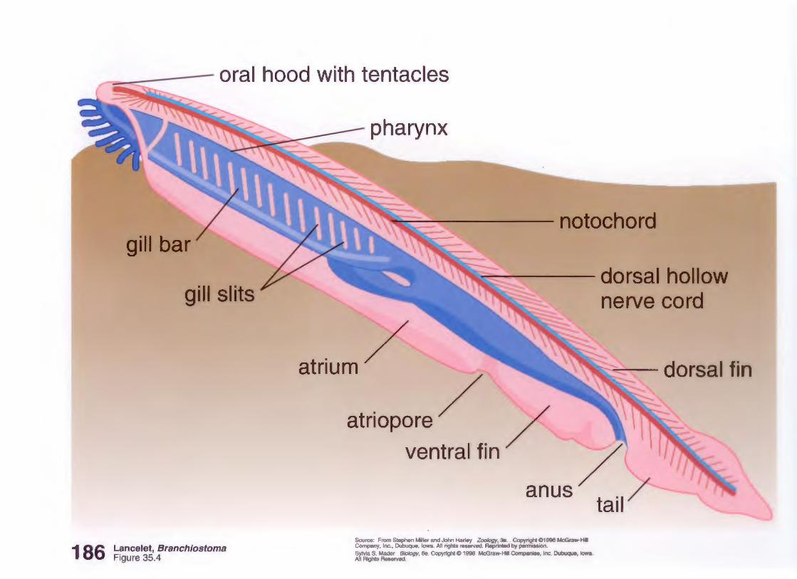
,-- ~ - oral hood with tentacles
gill bar
186 Lancelet, Branchiostoma Figure 35.4
~ -~ notochord
~~ ~- ~ dorsal hollow
atriopore ventral fin
Source: From Stephen Miller and John Harley Zoology, 3e .. Copyright C>1996 McGraw-Hill Company, Inc., Dubuque, Iowa. All rights reserved. Reprinted by permission. Sylvia S. Mader Biology, 6e. Copyright C> 1998 McGraw-Hill Companies, Inc. Dubuque. Iowa. All Rights Reserved.
nerve cord
, ~ dorsal fin
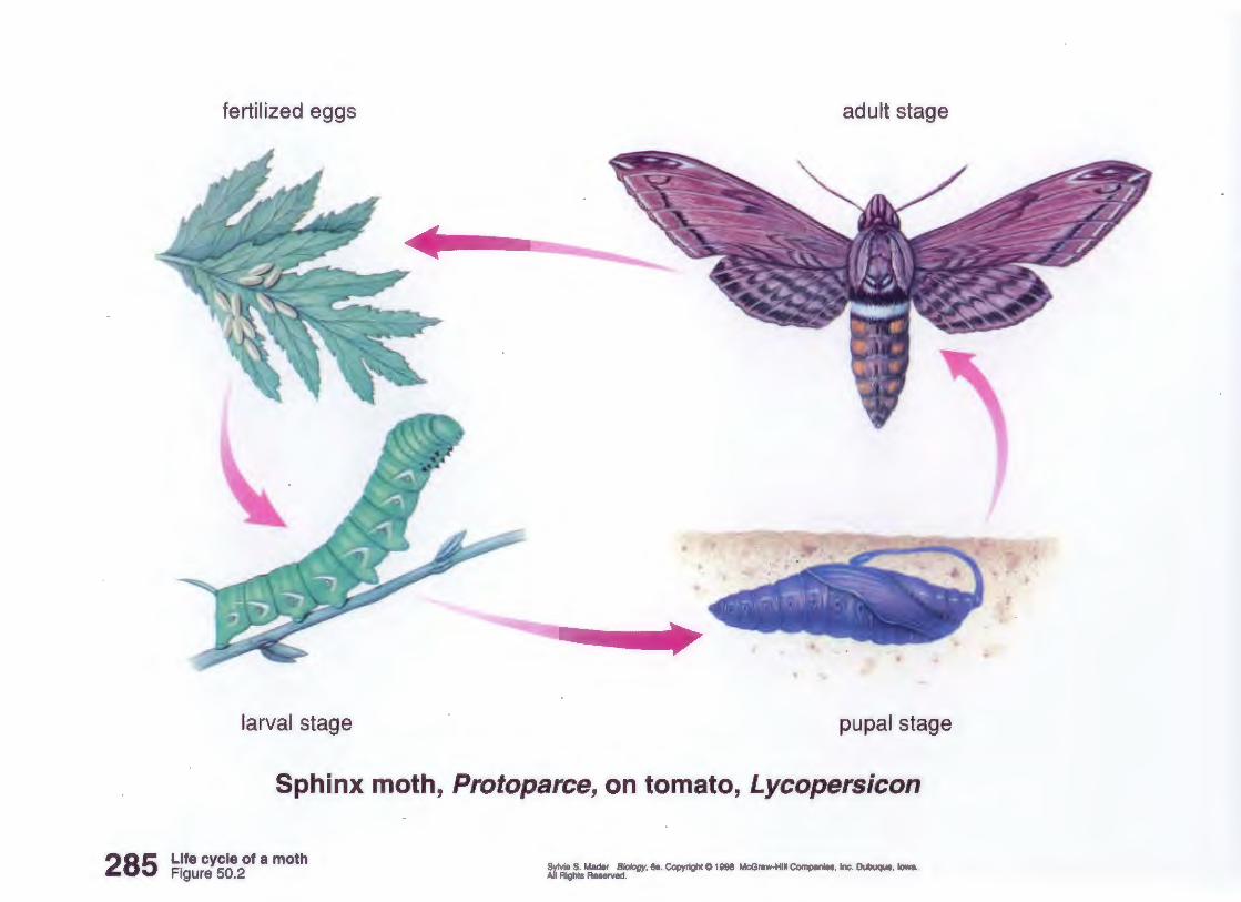
285
fertilized eggs adult stage
•
larval stage pupal stage
Sphinx moth, Protoparce, on tomato, Lycopersicon
Life cycle of a moth Figure 50.2
Sylvia S. Mader Biology, 6e. Copyright C 1998 McGraw-Hill Companies, Inc. Dubuque, Iowa. All Rights Reserved.
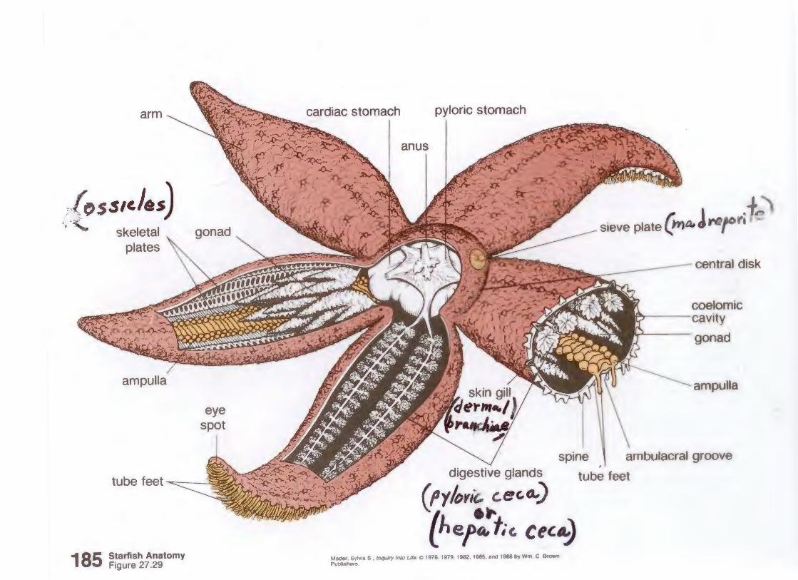
:':{~ss1cles) .. , skeletal
plates
185 Starfish Anatomy Figure 27.29
eye spot
~~cardiac stomach pyloric stomach
anus
~ sieve plate
central disk
coelomic lff"'f"',.----cavity
gonad
ampulla
spine . ' ambulacral groove digestive glands
(py/~;rit.. c.ec.a..)
'l ·" lhera.fic. cec~ Mader. Sylv1a S .. /nqwry Into Life C> 1976 1979. 1982. 1985. and 1988 by Wm C Brown Publishers.
tube feet