a-15-year experience of paediatric systemic lupus ... · lft liver function test ln lupus nephritis...
TRANSCRIPT

A-15-year experience of Paediatric Systemic Lupus
Erythematosus (pSLE) in Hospital Universiti Sains Malaysia
By
Dr. Jamil Mohammed Allamani Ali
Dissertation Submitted In Partial Fulfillment of the Requirement for the
Degree of Master of Medicine (Paediatric)
UNIVERSITI SAINS MALAYSIA
2014
A-15-YEAR EXPERIENCE OF PAEDIATRIC SYSTEMIC LUPUS ERYTHEMATOSUS (pSLE) IN HOSPITAL UNIVERSITI SAINS MALAYSIA
Dr. Jamil Mohammed Allamani
MMed Paediatrics
Department of Paediatrics
School of Medical Sciences, Universiti Sains Malaysia
Health Campus, 16150 Kelantan, Malaysia
Introduction: Systemic lupus erythematosus (SLE) is a multisystem autoimmune
disorder, relatively rare and under-reported in paediatric age group, affecting females
in the majority of cases. The disease manifestations and clinical outcomes vary
depending on which system being involved and treatment response. Its etiology is
complex and involves an interaction between genetic, hormonal and environmental
factors. Diagnosis is made clinically based on certain clinical and laboratory criteria
made by American College of Rheumatology (ACR).
Objectives: The aims of the study were to describe the demography, clinical and
laboratory manifestations, and outcomes of children with SLE and to determine the
risk factors associated with the development of renal failure and death among SLE
children admitted to Hospital USM in the period between 1996 and 2010.
Patients and Methods: In this cross sectional study we retrospectively reviewed the
folders of 51 children over a period of 15 years; all patients were below 18 years of
age and fulfilled the SLE criteria outlined by American College of Rheumatology.
Results: The median age was 12 years with female predominance, male to female
ratio was 1:10. Majority of our patients have positive family history and the
commonest clinical manifestation was involvement of haematological and renal
systems. One quarter of the cohort developed lupus nephritis some requiring acute
dialysis but none necessitating long term dialysis or progressed to end stage renal
disease (ESRD). Death complication was observed in almost 30% of the cases,
majority due to sepsis.
Conclusion: The sociodemography, clinical profile, laboratory findings and outcome
of childhood SLE in Kelantan are not much different from other studies done abroad.
Lupus nephritis was found to be a risk factor for development of renal failure in this
group of patients whereas infection was the leading cause of death; however, the other
studied factors associated with these outcomes among our SLE children need further
larger studies to be concluded.
Dr Mohamad Ikram Ilias: Supervisor
AP Dr Nik Zainal Abidin Nik Ismail: Co-Supervisor
Professor Dr Hans Van Rostenberghe: Co-Supervisor
Dr Azriani Berahim @ Abd Rahman: Statistician


i
ACKNOWLEDGEMENT
In the name of Allah, the Most Gracious and Most Merciful, I would like to thank Him
for His grace in completing the thesis. All the praise is with Thee.
I would like to express my special thank you and deepest appreciation to my main
supervisor, Dr. Mohamad Ikram Ilias, my co-supervisors, Associate Professor Dr. Nik
Zainal Abidin bin Nik Ismail and Professor Dr. Hans Van Rostenberghe. Their
continuing guidance, constructive criticisms, encouragement and patience had made a
tremendous help in me completing this write-up. I would like to extend my appreciation
to Dr Azriani Berahim@Abd Rahman, Lecturer in Community Medicine Department,
HUSM who was helping me in statistical analysis and interpretation of the results.
Special thanks go to all lecturers, superiors, staffs and colleagues in Hospital USM that
helped me throughout my journey to be a pediatrician.
Not to forget, many thanks to staffs at the record office Hospital USM for their crucial
role in tracing all the folders during data collection for this dissertation.
Finally I would like to thank my wife for her continuous support throughout the course
of this study as well as my children (Mohammed, Wisam and Sahar).
ii
TABLE OF CONTENTS PAGE
ACKNOWLEDGEMENT i
TABLE OF CONTENTS ii
LIST OF TABLES vi
LIST OF FIGURES vii
ABBREVIATIONS viii
ABSTRAK x
ABSTRACT xii
CHAPTER 1: INTRODUCTION
1.1 Systemic Lupus Erythematosus (SLE) 1
1.2 Aetiology and Pathogenesis of SLE 3
1.3 Clinical manifestations and diagnosis 6
1.4 Complications of SLE 10
1.5 Management of SLE 11
1.5.1 General measures 11
1.5.2 Medications 12
1.5.2.1 Glucocorticoids 12
1.5.2.2 Immunosuppressive/Cytotoxic agents 14
1.5.2.3 Antimalarial agents 15
1.5.3 Follow up and monitoring 15
1.6 Place of study 18
1.7 Rationale of this study 19
iii
CHAPTER 2: STUDY OBJECTIVES AND HYPOTHESES
2.1 General objective 20
2.2 Specific objectives 20
2.3 Study hypotheses 20
CHAPTER 3: METHODOLOGY
3.1 Study design 21
3.2 Study population 21
3.3 Inclusion criteria 21
3.4 Exclusion criteria 22
3.5 Sampling frame 22
3.6 Sample size calculation 22
3.7 Sampling method 24
3.8 Ethical approval 24
3.9 Research tool 24
3.10 Data collection 25
3.11 Statistical analysis 27
3.12 Definitions/Operational terms 27
3.13 Flow chart of the study 29
CHAPTER 4: RESULTS
4.1 Sociodemographic characteristics of children with SLE 31
4.2 Clinical manifestations of SLE 35
4.3 Complications of SLE 37
iv
4.4 Laboratory findings in SLE 39
4.5 Renal biopsy in SLE 42
4.6 Univariable analysis to determine the factors associated
with renal failure among children with SLE 44
4.7 Multivariable analysis (Factors associated with acute
renal failure in SLE children) 46
4.8 Univariable analysis to determine the factors associated
with death among children with SLE 48
4.9 Multivariable analysis (Factors associated with death
in SLE children) 50
CHAPTER 5: DISCUSSION
5.1 Sociodemographic characteristics of children with SLE 53
5.2 Clinical profiles of SLE 55
5.3 Complications of SLE 56
5.4 Laboratory findings of SLE 57
5.5 Factors associated with renal failure and death
among SLE children 58
CHAPTER 6: LIMITATIONS AND CONCLUSION
6.1 Limitations 60
6.2 Conclusion 61

v
CHAPTER 7: RECOMMENDATION AND FUTURE WORKS 62
REFERENCES 64
APPENDIX
I: Proforma form 69
II: Ethical approval letter 72
III: HUSM Director Approval 73
VI: Multivariable logistic regression tables 74
V: ROC Curve 75

vi
LIST OF TABLES
Table 1: SLICC classification criteria
Table 2: Cases excluded from further review
Table 3: Demographic characteristics of SLE patients (1)
Table 4: Demographic characteristics of SLE patients (2)
Table 5: Frequency of missing laboratory data
Table 6: Laboratory findings of SLE children at presentation
Table 7: Renal biopsy and lupus nephritis classification for SLE children
Table 8: Associated factors for acute renal failure among children with SLE by
simple logistic regression
Table 9: Factors associated with renal failure in SLE children using multiple
logistic regression
Table 10: Associated factors for death among children with SLE by
Simple Logistic Regression
Table 11: Factors associated with death in SLE children using Multiple Logistic
Regression

vii
LIST OF FIGURES
Figure 1: Natural history of systemic lupus erythematosus
Figure 2: Diagnostic criteria for SLE at presentation
Figure 3: Complications of SLE

viii
ABBREVIATIONS
ACR American College of Rheumatology
AIHA Autoimmune hemolytic anemia
ALT Alanine aminotransferase
ANA Antinuclear antibody
Anti-Sm Anti-smooth muscle
ARF Acute renal failure
aSLE Adult systemic lupus erythematosus
AST Aspartate aminotransferase
BP Blood pressure
C3, C4 Complement 3, complement 4
CNS Central nervous system
CRP C-reactive protein
DCT Direct Coombs test
DIL Drug-induced lupus
dsDNA Double-stranded DNA
EBV Epstein Bar Virus
ERA-EDTA European Renal Association – European Dialysis and Transplant Association
ESR Erythrocyte sedimentation rate
ESRD End stage renal disease
EULAR European League Against Rheumatism
FBC Full blood count
FBP Full blood picture
GFR Glomerular filtration rate

ix
HD Haemodialysis
Hib Hemophilus influenza b
HLA Human leukocyte antigen
HRPZ II Hospital Raja Perempuan Zainab II
HUSM Hospital Universiti Sains Malaysia
IQR Interquartile range
KDIGO Kidney Disease Improving Global Outcomes
JIA Juvenile idiopathic arthritis
LFT Liver function test
LN Lupus nephritis
MCTD Mixed connective tissue disease
PD Peritoneal dialysis
pSLE Paediatric systemic lupus erythematosus
RBCs Red blood cells
RF Rheumatoid factor
RFT Renal function test
SD Standard deviation
SLE Systemic lupus erythematosus
SLICC Systemic Lupus International Collaborating Clinics
UK United Kingdom
Urine PCI Urine protein creatinine index
UV light Ultraviolet light
x
ABSTRAK
Pengenalan
Systemic lupus erythematosus ialah penyakit autoimune yang jarang bagi peringkat
umur kanak-kanak. Kebanyakan pesakit adalah dalam kalangan perempuan yang
berumur 5 hingga 15 tahun. Namun penyakit ini sangat jarang bagi kanak-kanak
berumur bawah dari 5 tahun. Nisbah bagi lelaki kepada perempuan adalah 1 kepada 10.
Objektif
Objektif kajian ini adalah untuk mengenalpasti faktor sosiodemografi, profil klinikal,
keputusan makmal, komplikasi dan kesan terhadap kanak-kanak yang menghidap
penyakit SLE di HUSM, Kelantan. Selain itu ia juga bertujuan untuk mengkaji faktor-
faktor yang menyumbang kepada penyakit buah pinggang dan kematian di kalangan
kanak-kanak.
Kaedah
Kajian telah dijalankan secara retrospektif di HUSM kelantan. Kajian ini melibatkan
semua pesakit yang baru di sahkan menghidap penyakit SLE. Semua pesakit adalah
berumur 18 tahun atau kebawah dari yang memenuhi kriteria kajian bermula dari
Januari 1996 sehingga Disember 2010.
xi
Keputusan
Terdapat seramai 51 kanak-kanak yang memenuhi kriteria kajian dengan median umur
adalah pada umur 12 tahun. Kanak-kanak perempuan adalah paling ramai disahkan
menghidap SLE dengan nisbah 1 dalam 10. Majoriti pesakit adalah berasal dari
kawasan bandar dan lebih dari separuh pesakit ( 78 % ) mempunyai sejarah keluarga
yang menghidap penyakit autoimun. Tanda-tanda klinikal yang paling banyak ditemui
iaitu 60 peratus dalam kalangan pesakit adalah manifestasi buah pinggang dan
haematologi. Manakala 56 peratus lagi adalah manifestasi immunologi. Antinuclear
antibody (ANA) adalah positif bagi 96 peratus pesakit. Komplikasi yang biasa
dikalangan pesakit adalah Lupus Nephritis (LN) dimana ia melibatkan 60.8 peratus dan
separuh dari mereka menghidap Lupus Nephritis Class IV. Lupus Nephritis yang teruk
boleh menyebabkan pesakit menghidap Acute Renal Failure (ARF) dan 27% pesakit
memerlukan dialisis kerana uraemia dan fluid overload. 14 pesakit iaitu 27.5 peratus
telah meninggal dunia dan kebanyakannya adalah disebabkan kerana jangkitan kuman.
Konklusi
Sosiodemografi, profil klinikal, keputusan makmal dan kesan-kesan SLE di kalangan
kanak-kanak di Kelantan adalah hampir sama dengan kajian-kajian yang dilakukan di
luar negara. Walaubagaimnapun faktor-faktor penting yang menyebabkan pesakit-
pesakit SLE mendapat penyakit buah pinggang dan meningal dunia adalah tidak dapat
disahkan di dalam kajian ini.
xii
ABSTRACT
Introduction
Systemic lupus erythematosus is an uncommon multisystemic autoimmune disorder,
relatively rare in paediatric age group. It is predominantly affecting females in the age
group 5 to 15 years. It is very rare in children below 5 years old. The male to female
ratio is 1 in 10.
Objectives
The objectives of this study were to evaluate the sociodemographic, clinical profiles,
laboratory findings, complications and outcome of children with SLE in Hospital USM
(HUSM), Kelantan and to explore the factors associated with development of renal
failure and death among those patients.
Methodology
This study was conducted via retrospective (cross sectional) record review in HUSM,
Kelantan. It involved all newly diagnosed SLE cases aged 18 years or less who met all
the study criteria from January 1996 till December 2010.
Results
There were 51 children included in this study with the median age at diagnosis of 12
years old with female predominance, male to female ratio of 1:10. The majority of

xiii
patients came from urban areas and family history of autoimmune diseases was seen in
more than half of the patients (78%). The most common clinical findings in our patients
were renal and haematological manifestations (60% of patients) followed by
immunological findings (56%). Antinuclear antibodies (ANA) were positive in 96% of
patients. The most common encountered complication was lupus nephritis (LN) in
60.8% of patients; half of them have lupus nephritis class IV. Lupus nephritis was
complicated by ARF in some patients; 27% needed acute dialysis for their uraemia and
fluid overload. Fourteen patients in this study (27.5%) passed away. Infection was the
most common cause of death in pSLE.
Conclusion
The sociodemography, clinical profile, laboratory findings and outcome of childhood
SLE in Kelantan are not much different from other studies done abroad. Lupus nephritis
was found to be a risk factor for development of renal failure in this group of patients
whereas infection was the leading cause of death; however, the other studied factors
associated with these outcomes among our SLE children need further larger studies to
be concluded.

1
CHAPTER 1
INTRODUCTION
1.1 Systemic Lupus Eyrthematosus (SLE)
Systematic Lupus Erythematosus (SLE) is the commonest autoimmune disorder with
renal involvement. About 10-15% of all SLE patients are diagnosed in childhood
(Stichweh et al 2004). Childhood-onset SLE patients have been reported to differ from
their adult-onset counterparts in that they have more nephritis, more central nervous
system involvements, and have been reported to have a poorer outcome. There have
also been reports of ethnic differences in the incidence and severity of pediatric SLE
(pSLE) patients, indicating that African Americans and Hispanic patients are more
prone to the disorder and have more severe symptoms than Caucasian patients(Tucker
LB et al 1995, and Alareon GS 2001 ). However, most of these reports were small
case-series and limited by referral bias as they came from tertiary pediatric centers.
Systemic Lupus Erythematosus (SLE) is a multisystem autoimmune disease
characterized by widespread inflammation of blood vessels and connective tissues and
by the presence of antinuclear antibodies (ANAs), especially antibodies to the native
double stranded DNA (Anti-dsDNA). Its clinical manifestation is variable and its
natural history is unpredictable. Incidence of juvenile SLE varies by location and
ethnicity. There is a wide range of variation in the natural history of SLE among
different ethnic and geographical groups.

2
Studies in juvenile SLE have estimated the incidence at 0.28 to 0.9/100,000 per year
(Malleson P et al 1996, Kaipiainen-Seppanen 1996, and Fujikawa S et al 1997).
The course of SLE disease is characterized by periods of flare and remission, and
inflammation that can result in irreversible tissue damage, as well as premature death
(Brunner HI et al 2002). Despite numerous reports of SLE in adults, in the recent years
there have been few reports on the clinical and laboratory features of pediatric SLE at
presentation (Hiraki et al 2008). Although adult and pediatric SLE share clinical
features, it has been suggested that children have a more severe and aggressive disease
course and may present different signs and symptoms at onset; in fact about one third of
patients with pSLE may have non classical manifestations at presentation that may be
responsible for the major diagnostic delay in this age group (Brunner HI et al 2008).
Most of the studies of pSLE reported to date have been limited by the relatively small
sample sizes compared with studies of adult SLE (aSLE). There has been no large
comprehensive, single-center cohort study in pSLE examining the clinical presentation,
laboratory features, autoantibody profile, disease progression, and accrual of damage.
Most studies have been limited by sample size or have focused on only one or two
aspects of pSLE, with the majority of studies focusing on either renal or CNS disease
(Levy DM et al 2003, Alsaeid K et al 2004, Balkaran BN et al 2004, Beresford et al
2005, and Houghton KM et al 2006).

3
Worldwide estimated incidence of SLE in children under 18 years old is 15-17%, with a
peak incidence at 10-14 years with female predominance; the disease is rare in children
below 5 years old (White P 1994).
1.2 Aetiology and Pathogenesis of SLE
The aetiology of SLE is largely unknown, but it involves genetic, hormonal, and
environmental factors.
Genetic factors – the evidence of genetic susceptibility to SLE in humans is based on
familial aggregation. The prevalence of SLE is estimated to be 2.6-3.5% in first-degree
relatives of SLE probands compared with 0.3-0.4% in relatives of match controls.
Moreover, an increase in concordance rate is observed in monozygotic twins (24-56%)
compared to dizygotic twins (2-5%). The risk of developing the disease in siblings of
SLE patients is 10-40 times higher than that in the general population.
Hormonal factors – the female predominance in SLE is particularly strong during
childbearing age, suggesting that hormonal differences may contribute to the increased
risk of the disease. Reduced androgen levels, increased estradiol and prolactin levels
have been reported in SLE patients.
Environmental factors – beside genetic and hormonal factors studies have shown that
other factors such as toxic and infectious factors are suspected to induce SLE. The fact
that seroconversion against EBV was observed in 99% of children with SLE as

4
compared to only 70% of their controls is consistent with but does not in itself
establishes EBV as a causative factor in SLE. Photosensitivity is a common feature of
SLE since specific lupus lesions occur on sun-exposed areas and the disease is
aggravated by sun exposure in 40-70% of patients. The current theory is that the UV
light induces apoptosis of keratinocytes, which develop small surface blebs containing
lupus auto- antigens such as Ro particle. Similarly drugs such as chlorpromazine or
environmental toxins such as silica dust have been shown capable of inducing
apoptosis.
SLE is an autoimmune disease characterized by B cell hyperreactivity, producing
multitude of different auto- antibodies against self antigens such as dsDNA,
nucleosome, anticardiolipin etc. The complement system plays an important role in the
onset as well as the effect phase of SLE. The autoantibodies and self antigens form
immune complexes which over activate complement, causing inflammation and multi-
organ damage, including skin, joints, CNS, kidney etc (Siegert et al 1997, and
Fremeaux Bacchi et al 2002).
The aetiology of SLE is complex and relates to interactions between environmental,
genetic, and endocrine influences. It is more common in certain ethnic groups (Asian,
Hispanic, South -East Asian, African, and African American), and strongly associated
with HLA haplotypes. Lupus occurs much more commonly after puberty and in
females, this suggests an important role of estrogens, which is supported by animal
models. Known environmental influences include sun exposure and certain medications.
5
The end result to these interactions is the production of multiple auto-antibodies that is
both organ-specific and directed against a host of nuclear and cytoplasmic antigens.
The pathogenesis and etiology of SLE remains unclear. It is well recognized that SLE
predominantly affects females, especially in the reproductive age. An increase in
frequency of SLE among females is believed to be related to the effect of endogenous
sex hormones. In fact, supporting evidence is provided by the literature showing the
ratio of female to male s much lower in prepubertal children and after menopause,
although a female predominance remains (Ho CT et al 1998, and Alsaeid K et al 2004).
An administration of exogenous estrogens exacerbates SLE, while androgens have a
protective role (Roubinian et al 1997, and Carlsten H et al 1993).
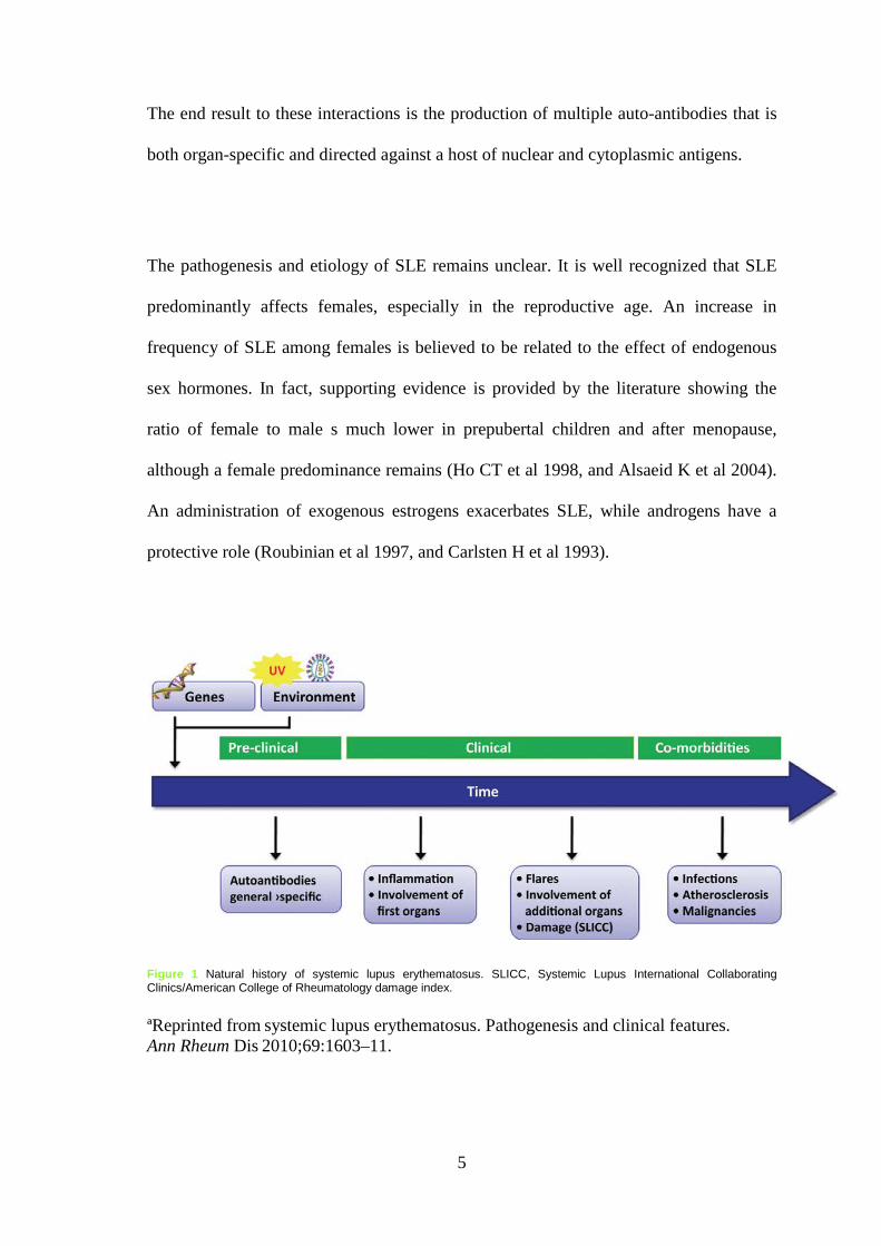
Figure 1 Natural history of systemic lupus erythematosus. SLICC, Systemic Lupus International Collaborating Clinics/American College of Rheumatology damage index. ªReprinted from systemic lupus erythematosus. Pathogenesis and clinical features. Ann Rheum Dis 2010;69:1603–11.

6
1.3 Clinical manifestations and diagnosis
The clinical manifestations of SLE reflect the degree of systemic inflammation as well
as the organ system (s) affected. Systemic manifestations, both at the time of diagnosis
as well as a time of disease exacerbations, frequently include fever, anorexia, lethargy,
weight loss and fatigue. The diagnosis is based on the presence of multisystem
involvement with compatible laboratory abnormalities. The presence of 4 of 11
classification criteria for SLE has both a very high sensitivity and specificity for the
diagnosis of pediatric SLE. (Steinlin et al 1995)
According to American College of Rheumatology (ACR) revised SLE criteria in 1997,
there are 11 criteria for SLE; presence of 4 out of these 11 criteria is diagnostic. These
criteria are:
1) Malar rash
2) Discoid rash
3) Photosensitivity
4) Oral ulcers
5) Arthritis: nonerosive, involvement of 2 or more joints characterized by
tenderness, swelling and effusion
6) Serositis: pleuritis or pericarditis
7) Renal disorder: persistent proteinuria or cellular casts in the urine
8) Neurologic disorder: seizures or psychosis
9) Haematological disorder: haemolytic anemia, leukopenia, lymphopenia, or
thrombocytopenia
10) Immunologic disorder: positive LE preparation; anti-DNA antibody or
antibodies to sm nuclear antigen, or false positive serologic tests for syphilis
11) Antinuclear antibody

7
Clinical manifestations at onset are diverse; it may range from isolated mild skin rash to
severe multi-organ involvement. Initial symptoms may be insidious; however, it may
present as an acute and even fatal disease. The disease course is progressive and may
lead to significant morbidity and mortality if left untreated. A high level of suspicion
and the proper use of laboratory tests may aid the treating physician in reaching an early
diagnosis and proper management. (Lehman T 1995).
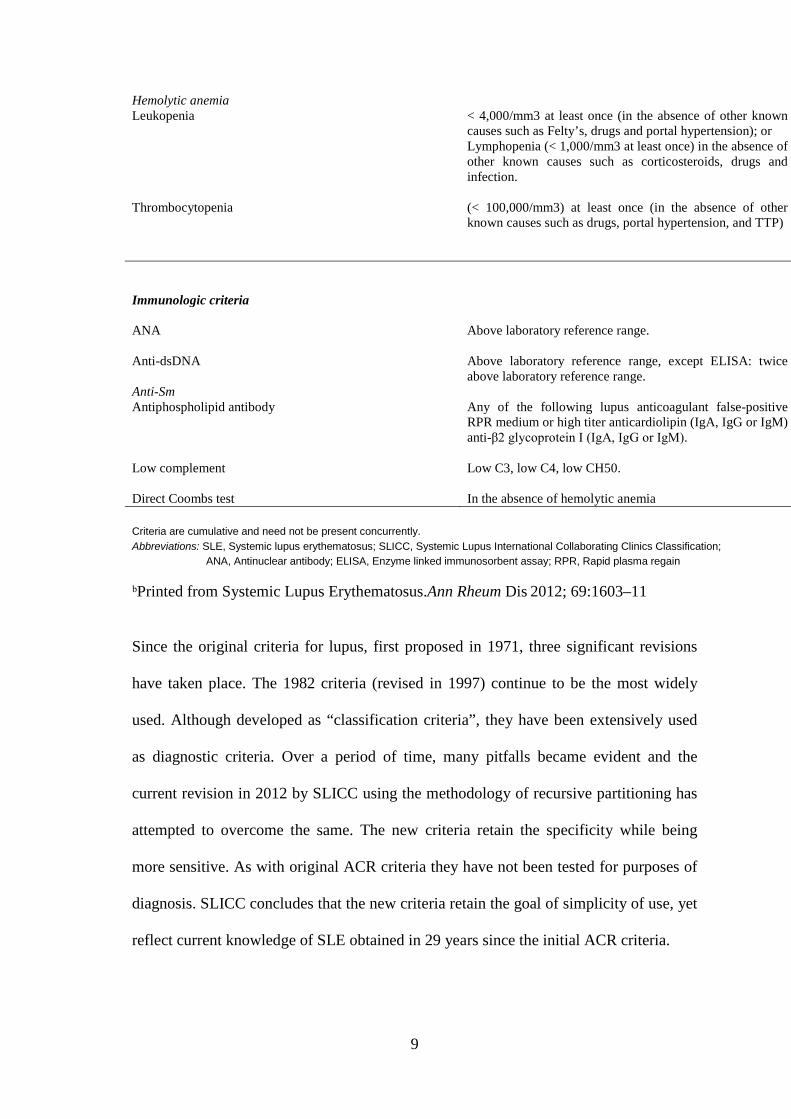
The Systemic Lupus International Collaborating Clinics (SLICC) Classification 2012
criteria were derived using the technique of “recursive partitioning” to derive a
relatively simple classification rule. The diagnosis of SLE is as following:
Classify a patient as having SLE if
• The patient satisfies four of the criteria listed in table 1 including at least one
clinical criterion and one immunologic criterion; or
• The patient has biopsy-proven nephritis compatible with SLE and with ANA or
anti-dsDNA antibodies. (Petri M et al 2012).

8
Table 1 SLICC classification criteria
Clinical and immunologic criteria used in the SLICC classification criteria
Clinical criteria Acute cutaneous lupus
Including lupus malar rash (do not count if malar discoid); bullous lupus; toxic epidermal necrolysis variant of SLE; maculopapular lupus rash; photosensitive lupus rash in the absence of dermatomyositis; or subacute cutaneous lupus (nonindurated psoriaform and/or annular polycyclic lesions that resolve without scarring, although occasionally with postinflammatory depigmentation or telangiectasia).
Chronic cutaneous lupus Including classical discoid rash; localized (above the neck); generalized (above and below the neck); hypertrophic (verrucous) lupus; lupus panniculitis (profundus); mucosal lupus; lupus erythematosus tumidus; chillblains lupus; discoid lupus/lichen planus overlap.
Oral ulcers Palate, buccal, tongue or nasal ulcers in the absence of other causes, such as vasculitis, Behçet’s, infection (herpes), inflammatory bowel disease, reactive arthritis and acidic foods.
Nonscarring alopecia Diffuse thinning or hair fragility with visible broken hairs in the absence of other causes such as alopecia areata, drugs, iron deficiency and androgenic alopecia.
Synovitis Involving two or more joints, characterized by swelling, effusion or tenderness in two or more joints, and 30 minutes or more of morning stiffness.
Serositis Typical pleurisy for more than 1 day or pleural effusions or pleural rub; typical pericardial pain (pain with recumbency improved by sitting forward) for more than 1 day or pericardial effusion or pericardial rub or pericarditis by electrocardiography in the absence of other causes, such as infection, uremia and Dressler’s pericarditis.
Renal Urine protein/creatinine (or 24-hour urine protein) representing 500 mg of protein/24 hour or red blood cell casts.
Neurologic Seizures; psychosis; mononeuritis multiplex in the absence of other known causes such as primary vasculitis; myelitis; peripheral or cranial neuropathy in the absence of other known causes such as primary vasculitis, infection and diabetes mellitus; acute confusional state in the absence of other causes, including toxic-metabolic, uremia, drugs
9
Hemolytic anemia Leukopenia < 4,000/mm3 at least once (in the absence of other known
causes such as Felty’s, drugs and portal hypertension); or Lymphopenia (< 1,000/mm3 at least once) in the absence of other known causes such as corticosteroids, drugs and infection.
Thrombocytopenia
(< 100,000/mm3) at least once (in the absence of other known causes such as drugs, portal hypertension, and TTP)
Immunologic criteria ANA
Above laboratory reference range.
Anti-dsDNA Above laboratory reference range, except ELISA: twice above laboratory reference range.
Anti-Sm Antiphospholipid antibody Any of the following lupus anticoagulant false-positive
RPR medium or high titer anticardiolipin (IgA, IgG or IgM) anti-β2 glycoprotein I (IgA, IgG or IgM).
Low complement Low C3, low C4, low CH50.
Direct Coombs test In the absence of hemolytic anemia Criteria are cumulative and need not be present concurrently. Abbreviations: SLE, Systemic lupus erythematosus; SLICC, Systemic Lupus International Collaborating Clinics Classification; ANA, Antinuclear antibody; ELISA, Enzyme linked immunosorbent assay; RPR, Rapid plasma regain ᵇPrinted from Systemic Lupus Erythematosus.Ann Rheum Dis 2012; 69:1603–11
Since the original criteria for lupus, first proposed in 1971, three significant revisions
have taken place. The 1982 criteria (revised in 1997) continue to be the most widely
used. Although developed as “classification criteria”, they have been extensively used
as diagnostic criteria. Over a period of time, many pitfalls became evident and the
current revision in 2012 by SLICC using the methodology of recursive partitioning has
attempted to overcome the same. The new criteria retain the specificity while being
more sensitive. As with original ACR criteria they have not been tested for purposes of
diagnosis. SLICC concludes that the new criteria retain the goal of simplicity of use, yet
reflect current knowledge of SLE obtained in 29 years since the initial ACR criteria.

10
1.4 Complications of SLE
The morbidity and mortality associated with SLE is still considerable despite
improvement in initial immunosuppressive therapy of active disease. There is still much
to learn about the long term complications of this disease. Patients require lifelong
follow up by physicians aware of the broad range of conditions that may ensue. The
lupus patients and their families need to understand why this is important and their own
role in modifying lifestyle factors that increase the risk of complications.
The common comorbidities of SLE include renal toxicity, infection, neuropsychiatric
issues, ophthalmic complications and drug side effects. Other complications are rare in
paediatric age group such as coronary artery disease, osteoporosis, and malignancies.
Because of paucity of data in paediatric SLE, a little is known about its long term
outcome, and optimal management.
Studies published around 1980 found that about 80% of patients survived 5 year and
about 60% of patients survived 10 year. More recent studies have shown that 5year
survival is now nearer 90–95% and that 70–85% of patients survives 10 year (Trager et
al 2001). In most studies, patients with renal involvement have had a poorer prognosis
than those without renal disease. Nevertheless, survival has shown improvement in
those with renal disease presenting to a UK centre between 1976 and 1986 (81% 10
year survival), compared with those presenting between 1963 and 1975 (56% 10 year
survival) [(Bono et al 1999)].

11
In one adult SLE study it was observed that the commonest cause of death has been
infection, both in early and late deaths. Active SLE contributes to about a third of early
deaths but less commonly to late deaths. However, deaths related to acute and chronic
vascular disease including sudden death are more common in those dying more than
5 years after diagnosis. However, there is more to prognosis than just death. There is
considerable morbidity associated with more prolonged survival after the diagnosis of
SLE (Abu Shakra et al 1995).
Having improved therapy for active lupus disease, the challenge is now to understand
and prevent the long-term complications of this disease, whether they are due to effects
of the disease itself, the therapies used, or co-morbid disease (perhaps with associated
underlying disease mechanisms or linked genetic predisposition).
1.5 Management of SLE
The treatment of SLE is mainly based on the use of traditional drugs, such as
corticosteroids, antimalarials, azathioprine, and cyclophosphamide. Because of paucity
of data in paediatric SLE, a little is known about its optimal management, however, it
has the same principles as for adult SLE.
1.5.1 General measures
All SLE patients need education, counseling, and support due to complexity and
unpredictability of the disease process. Patient education programs for SLE patients and
their families are designed to provide information, knowledge, and social support with
an emphasis on enhancing self-management skills (Robbins et al 1997).

12
Patients need advice regarding physical measures, including minimizing sun exposure,
using sunscreen, and exercising regularly. Diet management for prevention of obesity,
osteoporosis, and hyperlipidemia is of particular importance. Routine health
maintenance, including regular ophthalmologic examinations and dental care (especially
for those on corticosteroids and antimalarials), is very important in this chronic systemic
disease. Preventive measures such as immunizations (e.g., Hepatitis B, hemophilus
influenza (Hib), and pneumococcal vaccine) or use of hormone replacement therapy (in
adolescent girls) should be also considered (American College of Rheumatology (ACR)
guidelines 1999).
1.5.2 Medications
The first line treatment of SLE is the use of steroids. As our knowledge on the
mechanisms of immune response increases, new drugs that can interfere with T and B
cell interaction, immune-complex deposition and cytokine activation have been
developed and some of these drugs are still under investigation in SLE. Although initial
data regarding efficacy are encouraging, caution must be taken before these drugs are
considered as the treatment of choice for specific SLE manifestations (Marta Moska et
al 2001).
1.5.2.1 Glucocorticoids
Glucocorticoids are used for refractory manifestations of SLE, as well as for severe
organ-threatening disease. High-dose daily therapy (40-60mg/day) improves survival
13
among patients with severe forms of SLE nephritis , but is associated with virtually
universal undesirable side effects.
The treatment of active SLE nephritis, cerebritis, or thrombocytopenia may require high
doses of prednisolone, or intravenous pulses up to 1 gm of methylprednisolone per day
for 3 consecutive days. Studies of monthly high-dose intravenous methylprednisolone
(in addition to daily oral glucocorticoids) have shown a positive effect on severe SLE
nephritis, although the therapy is not as effective as intermittent intravenous
cyclophosphamide added to oral glucocorticoids (Gourley MF et al 1996).
Even with aggressive therapy, some patients with proliferative lupus nephritis (LN) will
have a progressive decline in renal function leading to end-stage renal disease (ESRD).
Clinical risk factors for progression, evident at the time of initial presentation, include
an elevated serum creatinine, hypertension, nephrotic range proteinuria, anemia with a
hematocrit below 26 percent, and black and Hispanic race and ethnicity (Contreras et al
2005 and Siso et al 2010).
The likelihood of a successful initial outcome is greater if therapy for LN is initiated
relatively early in the course of the disease. Delaying therapy because of presumed mild
disease can be associated with increased glomerular injury, progressive tubulointerstitial
fibrosis, glomerulosclerosis, and therefore a lesser response to immunosuppressive
drugs (Faurschou et al 2006).
14
1.5.2.2 Immunosuppresive/Cytotoxic agents
Immunosuppressive therapy for proliferative lupus nephritis (LN) consists of
Induction and maintenance phases. A number of immunosuppressive/cytotoxic
medications have been used to treat SLE, this includes azathioprine, cyclophosphamide,
methotrexate, cyclosporine, etc. the choice of the drug will depend on the nature and
severity of the condition, as well as individual preference. For example, for patients
with particularly severe arthritis, methotrexate may be preferred as the first cytotoxic
medication, whereas for SLE nephritis, azathioprine or cyclophosphamide may be
chosen first.
For initial induction therapy in patients with diffuse or moderate to severe focal
proliferative lupus nephritis (LN), it is recommended to use immunosuppressive therapy
with either an intravenous cyclophosphamide- or MMF-based regimen. Glucocorticoids
are also given as part of the induction regimen. This recommendation is in agreement
with both the KDIGO and EULAR/ERA-EDTA guidelines (Bertasias GK et al 2012).
In a series of long-term studies (more than 20 years follow up) in patients with SLE
nephritis, treatment with glucocorticoids plus cyclophosphamide for > 2 years appears
to be superior than glucocorticoids and azathioprine, and both seem superior to
glucocorticoid alone in preventing renal failure in these patients (Boumpas DT et al
1995).

15
Some patients with severe disease ( renal or extra renal) respond well over both the
short term and long term to glucocorticoid alone, or they require only a few months of
treatment with cytotoxic agents plus glucocorticoids to achieve long term improvement.
There is evidence that cytotoxic agents plus low-dose steroids prevent scarring in the
kidney better than do glucocortecoids alone (Hahn BH et al 2012).
1.5.2.3 Antimalarial agents (e.g., Hydroxychloroquine)
Antimalarial agents are useful for skin and joint manifestations of SLE, for preventing
flares, and for other constitutional symptoms of the disease (Wallace DJ et al 1994,
Petri M et al 1994, and Drake LA et al 1996). They may also reduce fatigue and
decrease levels of low density lipoproteins.
1.5.3 Follow up and monitoring
Lifelong follow up is required for all patients. It is the cornerstone of managing SLE to
detect flares of disease early and to institute prompt, appropriate therapy. The frequency
of these evaluations will depend on the activity, severity, and extent of the SLE, the
response to treatment, and the type of treatment, including the need for toxicity
monitoring. SLE disease activity can be monitored by specific clinical features (such as
arthritis, serositis, etc.) or laboratory features (e.g., anti-dsDNA antibodies and
complement level), or by using other disease activity indices.

16
At routine follow up visits, full blood count (FBC), renal function test (RFT), and
urinalysis should be obtained. Patients with lupus nephritis should have a urinalysis, 24-
hour urine protein,serum electrolytes and creatinine measurements on regular basis till
stable (Bootsma H et al 1995).
Renal biopsy is indicated for diagnostic purposes in patients in whom nephritis is
suspected. Thus, patients with persistent urinary sediment abnormalities such as
hematuria and pyuria without adequate explanation (infection, menstrual period, stone),
patients who have urinary casts, or patients who have increased serum creatinine levels
should have a renal biopsy. The prognostic value of renal biopsy, although
controversial, has been demonstrated, particularly for patients with normal serum
creatinine levels (Boompas DT et al 1995, and Gladman DD et al 1996). Patients with
proliferative glomerular lesions and chronic changes found on renal biopsy are at a
higher risk for end-stage renal disease and death than patients who do not demonstrate
these changes. Patients with deteriorating renal function, or patients not responding to
conventional therapy, may need a renal biopsy to outline a course of treatment (Esdaile
et al 1998 and Markowitz et al 2007).
International Society of Nephrology/Renal Pathology Society 2003 has categorized
lupus nephritis (LN) into 6 main classes as follows:
• Class I Minimal mesangial LN
• Class II Mesangial proliferative LN

17
• Class III Focal LN (<50% of glomeruli)
III (A): active lesions
III (A/C): active and chronic lesions
III (C): chronic lesions
• Class IV Diffuse LN (≥50% glomeruli)
Diffuse segmental (IV-S) or global (IV-G) LN
IV (A): active lesions
IV (A/C): active and chronic lesions
IV (C): chronic lesions
• Class V Membranous LN • Class VI Advanced sclerosing LN (90% globally sclerosed glomeruli
Without residual activity)

18
1.6 PLACE OF STUDY
Hospital Universiti Sains Malaysia (HUSM) in Kubang Kerian is the tertiary care centre
in Kelantan. Hospital USM is also one of the main teaching hospitals for the
undergraduates and postgraduate students in Malaysia. It offers 723 beds for inpatients.
This hospital provides multiple clinical sub-specialities, together with clinical and non-
clinical support services.
The weather in Kelantan in general is hot and dry. Over the East Coast states including
Kelantan, the months with maximum rainfall are November, December and January
while June and July are the driest months in most districts (http://www.met.gov.my).
The population here are predominantly Malays (95%), followed by other races like
Chinese, Indian, Siamese and Orang Asli. The population estimates based on the
adjusted population and housing census of Malaysia in 2010 was around 1.6 million.

19
1.7 RATIONALE OF STUDY
At present very limited studies have been published on paediatric SLE worldwide. This
study aimed to provide local data for paediatric SLE and it was conducted to serve as a
baseline for subsequent similar studies on SLE in Malaysia. Even though the cohort was
derived from only one state and may not represent the whole population size, the data
and results in this study will help to focus on specific risk factors and at risk groups,
hence provide early intervention and hopefully better prognosis.

20
CHAPTER TWO
OBJECTIVES AND HYPOTHESIS
2.1 OBJECTIVES
2.1.1 General objective:
To study the demographic characteristics, clinical and laboratory manifestations,
and outcome of children with SLE admitted to HUSM between 1996 and 2010.
2.1.2 Specific objectives:
1. To describe the demographic characteristics, clinical manifestations,
laboratory characteristics and the outcome of all children with SLE
admitted to HUSM from 1996 to 2010
2. To determine the factors associated with renal failure in that group of
children with SLE.
3. To explore the risk factors associated with death among paediatric SLE
patients admitted to HUSM at that period of time.
2.3 Study hypotheses
Certain factors are associated with the development of renal failure and death
among patients with SLE such as age at presentation, gender, positive family
history, number of relapses, and hypertension at presentation, lupus nephritis,
cerebral lupus, and proteinuria at presentation.

21
CHAPTER THREE
METHODOLOGY
3.1 STUDY DESIGN
This was a cross sectional study conducted retrospectively via hospital record review.
This study was done in Hospital USM in Kubang Kerian. This hospital is the main
referral center for other district hospitals in Kelantan as well as bordering district, Besut
in Terengganu.
3.2 STUDY POPULATION
The reference population was pediatric patients in Kelantan admitted to HUSM and
diagnosed as having SLE, whereas the source population was the newly diagnosed
paediatric SLE patients who were diagnosed and treated in Hospital USM in the period
between 1996 and 2010.
3.3 INCLUSION CRITERIA
All children aged 18 years and below who were diagnosed to have SLE (diagnosis
based on revised SLE criteria made by ACR, 1997) and were admitted to Paediatric
Medical wards in the period from January/1st/1996 to December/31st/2010.

22
3.4 EXCLUSION CRITERIA
The exclusion criteria were those with missing or untraceable case note. Patients who
had the following conditions were also excluded:
- Drug-induced lupus
- Mixed connective tissue disease
- Overlap syndrome
3.5 SAMPLING FRAME
All children aged 18 years and below who were diagnosed to have SLE (diagnosis on
discharge) and were admitted to Paediatric medical wards in the period from
January/1996 to December/2010 who fulfilled the inclusion and exclusion criteria.
3.6 SAMPLE SIZE CALCULATION
For specific objectives 1 sample size was calculated using single proportion formula
based on previous study is as follows:
Calculation of sample size:
Single proportion formula,
n = (Z/∆) ² P (1 - P)
Z = 1.96
Precision value (∆) = 0.05
P = the Prevalence

23
For specific objective 1 we used proportion of SLE patients with nephritis = 0.86
(Supavekin et al 2005).
The sample size calculation for objectives 1 was as following:
- n = (1.96) /0.05)² x 0.86(1-0.86) = 185
Note: adding 20% missing rate for the calculated sample size, the actual sample size for
objective 1 will be 222.
For specific objective 2, sample size was calculated for the variable sex using PS
software as follows:
α= level of significant = 0.05
m= the ratio between SLE patients with and without renal failure = 1
Po = percentage of males among SLE patients = 8.9% (Carein et al 2011)
P₁ = expected percentage of males with renal failure = 70%
From the calculation, the sample size for objective 2 (n) was 9
For patients with and without renal failure as a total 9+9 = 18, adding 20% missing rate
(n) was 22
For specific objective 3, sample size was calculated for infection as a risk factor for
death using PS software as follows:
α= level of significant = 0.05
m= the ratio between SLE patients who alive and those died = 2 (those who alive are
twice more than those who died)
Po = percentage of infection among SLE patients = 34.5% (Carein et al 2011)
P₁ = expected percentage of infection among those who died = 77%

24
From the calculation, the sample size for objective 3 (n) was 15; adding 20% missing,
the total sample size was (15 X 2 of alive sample) + (15 of dead sample) + 20% = 54
The sample size chosen for this study was the sample size for objective 1 = 222
3.7 SAMPLING METHOD
All patients who fulfilled the inclusion and exclusion criteria were included in the study.
3.8 ETHICAL APPROVAL
Approval for the study was obtained from the Human Research Ethics Committee USM
on 29th January 2012 [(reference: USMKK/PPP/JEPeM [244.4.(1.2)]. Confidentiality
was maintained during data collection whereby each case was given a code number.
The name and the code number were recorded in a separate list to avoid identification of
patient’s name. Permission from Hospital Director was obtained as well (appendix II).
3.9 RESEARCH TOOL
Patient’s proforma consisting of demographic data, diagnostic criteria of SLE at the
time of presentation, other clinical manifestations at the time of diagnosis, laboratory
investigations at the time of diagnosis, complications that had occurred throughout the
course of the disease, renal biopsy details and the specific management given based on
the classes of lupus nephritis, medications given with their total duration and side
effects were included, as well as other management modalities such as dialysis.