7.1 life is cellular - beachwood city schools 7 1 - 7 2 cell… · 7.1 life is cellular mrs....
TRANSCRIPT

The key to every
biological problem must
finally be sought in the
cell, for every living
organism is, or at some
time has been, a cell.
E.B. Wilson, 1925
7.1 Life is Cellular
Mrs. Baldessari
Biology
1
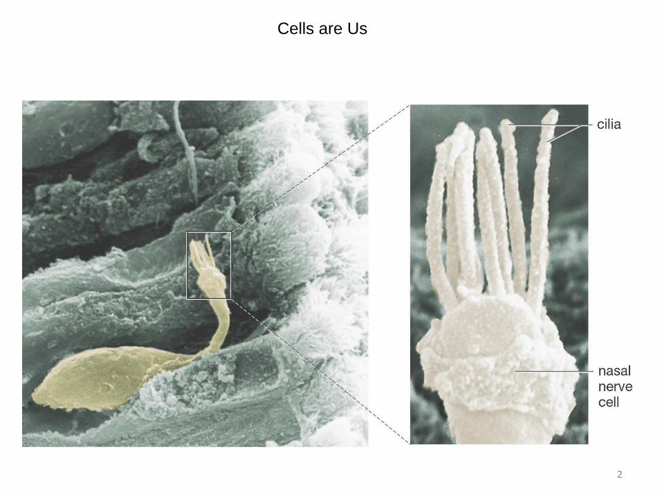
Cells are Us
2
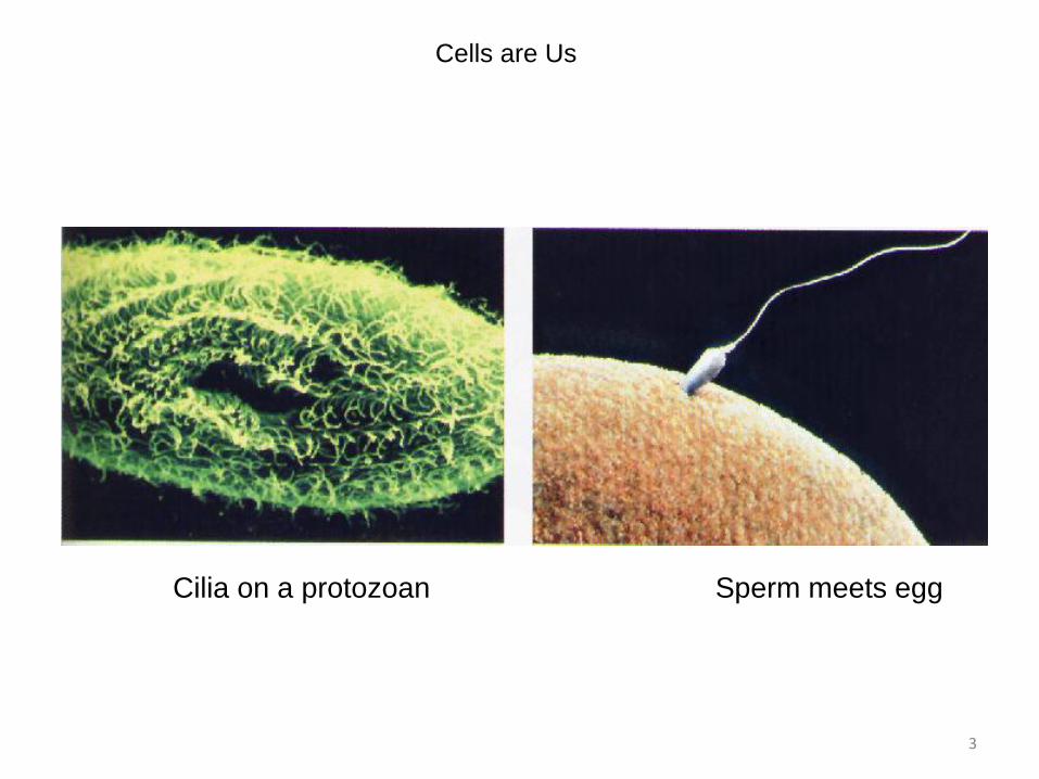
Cells are Us
Cilia on a protozoan Sperm meets egg
3

Lesson Overview Life Is Cellular
THINK ABOUT IT
What’s the smallest part of any living thing that still counts as being
―alive?‖
Can we just keep dividing living things into smaller and smaller parts,
or is there a point at which what’s left is no longer alive?
As you will see, there is such a limit. The smallest living unit of any
organism is the cell.
4
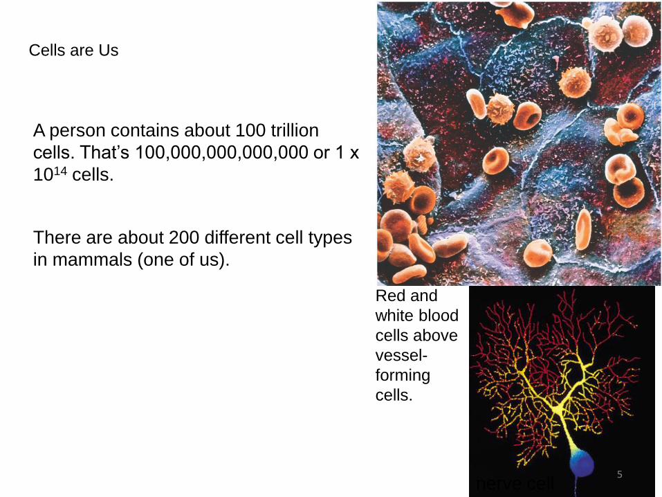
Cells are Us
A person contains about 100 trillion
cells. That’s 100,000,000,000,000 or 1 x
1014 cells.
There are about 200 different cell types
in mammals (one of us).
nerve cell
Red and
white blood
cells above
vessel-
forming
cells.
5

CELL SIZE
• Smallest cell in the human body is the sperm cell
• Largest cell in the human body is the egg cell
6
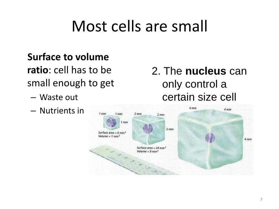
Most cells are small
1. Surface to volume ratio: cell has to be small enough to get
– Waste out
– Nutrients in
2. The nucleus can
only control a
certain size cell
7

Lesson Overview Life Is Cellular
The Discovery of the Cell
What is the cell theory?
The cell theory states:
- All living things are made up of cells.
- Cells are the basic units of structure
and function in living things.
- New cells are produced from existing
cells.
8

Early Microscopes
• Prototypes were developed in the late 1500’s by European eyeglass makers
• It was not until the
mid-1600s that
scientists began to
use microscopes to observe living things.
Early compound microscope 17th century 9

In 1665, Robert Hooke used an early compound microscope to look at cork
• Observed that cork was made up of thousands of hollow chambers
• Dubbed them cells since they looked like the monatsery’s tiny rooms called “cellula”.
• Today we know that living cells are not empty chambers, but contain a huge array of working parts, each with its own function.
10

Anton von Leewenhoek
• Late 1600’s- Dutch textile salesman
• Created different types of microscopes
• Discovered over 5,000 types of microscopic life
• Lenses were able to magnify up to 300X
• .
FIRST person to
OBSERVE and
DESCRIBE
MICROSCOPIC
ORGANISMS
and LIVING
CELLS
11

Lesson Overview Life Is Cellular
Early Microscopes
Anton van
Leeuwenhoek
examined pond water
and other things,
including a sample
taken from a human
mouth. He drew the
organisms he saw in
the mouth—which
today we call bacteria.
12
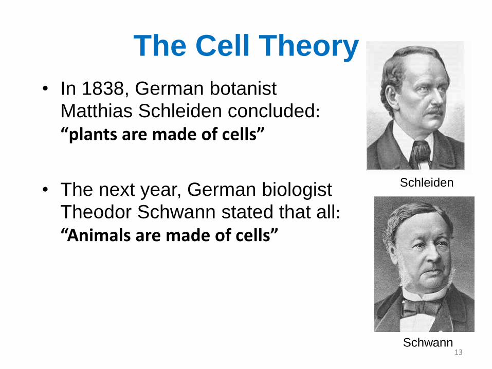
The Cell Theory
• In 1838, German botanist
Matthias Schleiden concluded: “plants are made of cells”
• The next year, German biologist Theodor Schwann stated that all: “Animals are made of cells”
Schleiden
Schwann 13

Cell Theory
1. All living things are made of cells.
2. Cells are the basic unit of structure and function
3. New cells come from preexisting cells.
*cells are different shapes and sizes based on their function
In 1855, German physician Rudolf Virchow concluded
that new cells could be produced only from the division of
existing cells.
14

Lesson Overview Life Is Cellular
Exploring the Cell
How do microscopes work?
Most microscopes use lenses to magnify the image of an object by focusing
light or electrons.
15

A Sense of Scale and Abundance – Bacteria on the Head of a Pin
16

1. There are
1) Light Microscope
•Allows light to pass through a specimen and
uses two lenses to form an image.
•The first set of lenses, located just above the
specimen, produces an enlarged image of the
specimen.
•The second set of lenses magnifies this image
still further.
•Because light waves are diffracted, or
scattered, as they pass through matter, light
microscopes can produce clear images of
objects only to a magnification from 40 – 1,000
times depending on objective being used
Exploring the Cell- 3 major types of
microscopes
17

Lesson Overview Life Is Cellular
1) Light Microscopes
• magnification from 40 – 1,000
• Eyepiece magnifies 10X
• Total magnification calculation: multiply the eyepiece by the objective being used. Example- eyepiece (10X) times low power objective (4X) = 40X
• Used to magnify objects that light can pass through.
• Uses slides
18
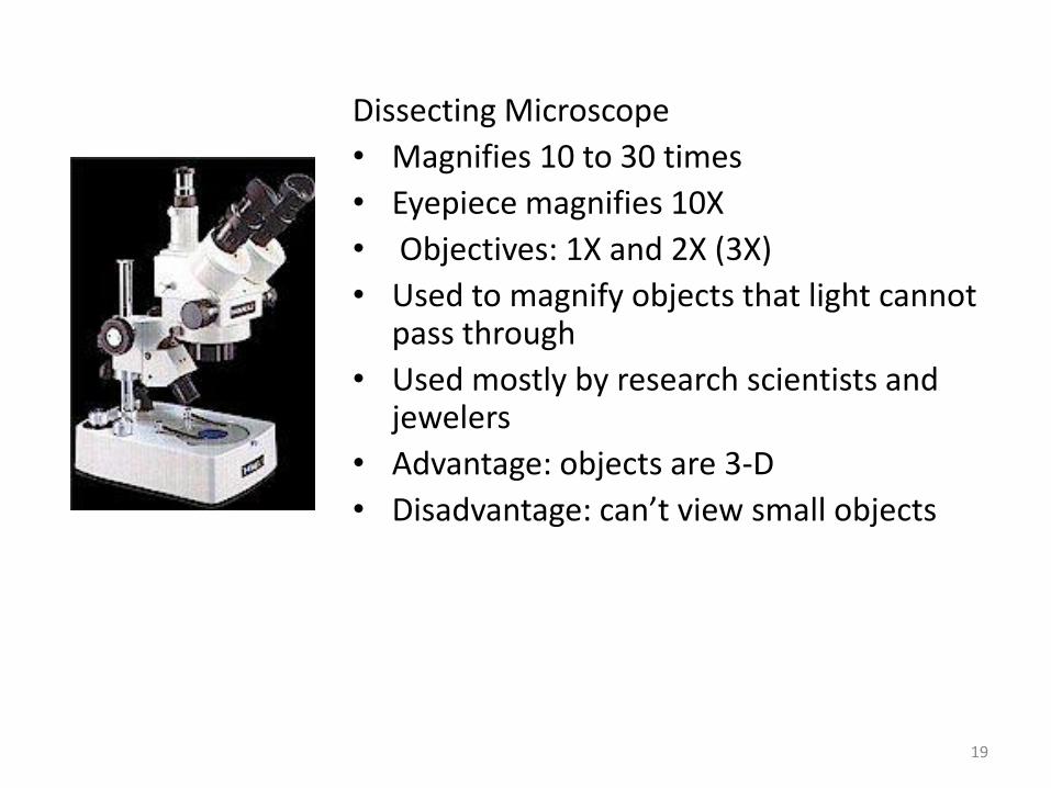
Dissecting Microscope
• Magnifies 10 to 30 times
• Eyepiece magnifies 10X
• Objectives: 1X and 2X (3X)
• Used to magnify objects that light cannot pass through
• Used mostly by research scientists and jewelers
• Advantage: objects are 3-D
• Disadvantage: can’t view small objects
19

Lesson Overview Life Is Cellular
Light Microscopes and Cell Stains
A problem with light microscopy is that most
living cells are nearly transparent, making it
difficult to see the structures within them.
Using chemical stains or dyes can usually
solve this problem. Some of these stains are
so specific that they reveal only compounds
or structures within the cell.
20

Lesson Overview Life Is Cellular
Light Microscopes and Cell Stains
Some dyes give off light of a particular color when
viewed under specific wavelengths of light, a property
called fluorescence.
Fluorescent dyes can be attached to specific molecules
and can then be made visible using a special
fluorescence microscope.
Fluorescence microscopy makes it possible to see and
identify the locations of these molecules, and even to
watch them move about in a living cell.
21
Lesson Overview Life Is Cellular
2) Electron Microscopes
Light microscopes can be used to see cells and cell structures as small as
1 millionth of a meter. To study something smaller than that, scientists need
to use electron microscopes.
Electron microscopes use beams of electrons, not light, that are focused by
magnetic fields.
Electron microscopes offer much higher resolution than light microscopes.
There are two major types of electron microscopes: transmission and
scanning.
22

Electron Microscope -Two types: Transmission and Scanning
• Uses electrons to illuminate objects
• Can magnify from 30,000 to 9 million times
• Mostly large institutions have them
• Costly to own and maintain
• Can only be used to look at dead specimens
• Used for cytology, forensics, and virology
23

Lesson Overview Life Is Cellular
Transmission Electron Microscopes
Transmission electron microscopes make it possible to
explore cell structures and large protein molecules.
Because beams of electrons can only pass through thin
samples, cells and tissues must be cut first into ultra thin
slices before they can be examined under a
transmission electron microscope.
Transmission electron microscopes produce flat, two-
dimensional images.
24

• Transmission Electron Microscope
•TEM- thin slices need to be made to have clear images,
images are 2-D
•Useful for studying internal structures
25

Lesson Overview Life Is Cellular
Scanning Electron Microscopes
In scanning electron microscopes, a pencil-like beam of
electrons is scanned over the surface of a specimen.
Because the image is of the surface, specimens viewed
under a scanning electron microscope do not have to be
cut into thin slices to be seen.
Scanning electron microscopes produce three-
dimensional images of the specimen’s surface.
26

•SEM- samples do not need to be cut, are in 3-D
• Useful for studying external
structure
27

Lesson Overview Life Is Cellular
Electron Microscopes
Because electrons are easily scattered by molecules in
the air, samples examined in both types of electron
microscopes must be placed in a vacuum in order to be
studied.
Researchers chemically preserve their samples first and
then carefully remove all of the water before placing
them in the microscope.
This means that electron microscopy can be used to
examine only nonliving cells and tissues.
28
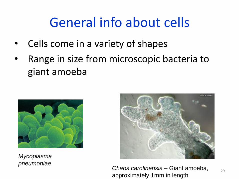
General info about cells
• Cells come in a variety of shapes
• Range in size from microscopic bacteria to giant amoeba
Chaos carolinensis – Giant amoeba,
approximately 1mm in length
Mycoplasma
pneumoniae 29

General info about cells
c. All contain DNA
d. All have a cell membrane- an outer, flexible barrier
Cell
membrane Nucleus-
containing
DNA
30
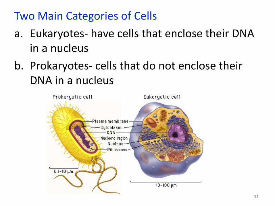
Two Main Categories of Cells
a. Eukaryotes- have cells that enclose their DNA in a nucleus
b. Prokaryotes- cells that do not enclose their DNA in a nucleus
31

Lesson Overview Life Is Cellular
Prokaryotes and Eukaryotes
How are prokaryotic and eukaryotic cells different?
Prokaryotic cells do not separate their genetic material within a nucleus.
In eukaryotic cells, the nucleus separates the genetic material from the rest of
the cell.
32

Prokaryotes and Eukaryotes
• Organisms whose cell contain a nucleus and other membrane bound organelles are called EUKARYOTES
• Organisms whose cells never contain (or lack) a nucleus and other membrane bound organelles are called PROKARYOTES
33

A rat liver cell (with color enhancement to show organelles) 34

It’s Crowded In There
An artist’s conception of the cytoplasm - the region of a
cell that’s not in the nucleus or within an organelle. 35

A micrograph showing
cytoskeleton (red),
ribosomes (green), and
membrane (blue)
It’s Crowded In There
36

Two Fundamentally Different Types of Cells
37
Lesson Overview Life Is Cellular
Prokaryotes and Eukaryotes
Cells fall into two broad categories, depending on whether
they contain a nucleus.
The nucleus is a large membrane-enclosed structure that contains the
cell’s genetic material in the form of DNA. The nucleus controls many of the
cell’s activities.
38

Lesson Overview Life Is Cellular
Prokaryotes and Eukaryotes
Eukaryotes are cells that enclose their DNA in nuclei.
Prokaryotes are cells that do not enclose DNA in nuclei.
39

Prokaryotes • Are generally smaller than eukaryotic cells
• Have no nucleus
• Carry out all of life’s processes
• Ex: bacteria
Bacillus anthracis 40

Eukaryotes a. Are generally larger and more complex than
prokaryotes
b. Contain dozens of membrane bound structures that are specialized
c. Nucleus separates DNA from rest of cell
41

Eukaryotes
• Come in a variety of shapes and size
• Ex: protists, fungi, plant, and animal cells
42

Lesson Overview Life Is Cellular
Eukaryotes • Eukaryotic cells are generally larger and more complex
than prokaryotic cells.
• Most eukaryotic cells contain dozens of structures and
internal membranes. Many eukaryotes are highly
specialized.
• There are many types of eukaryotes: plants, animals,
fungi, and organisms commonly called ―protists.‖
43

Us vs. Them -
Eukaryotes
and
Prokaryotes
44

7.2 Cell Structure
Cell organization
• Organelles- “tiny organs”; the specialized structures within the cell that perform a function
45

Animal vs Plant cells
46

Animal and Plant Cells Have More
Similarities Than Differences
47

The cell as a factory
• The different organelles of a cell can be compared to a living version of a modern factory
• Cells, like factories follow instructions to produce products
48

Cytoplasm
Cytoplasm- the portion of the cell outside of the nucleus; the fluid that fills the entire cell
• Makes up about 70% of the cells volume.
• Most of the cells chemical reactions occur here
49

The
Nucleus
•Control center of
the cell
•Surrounded by a
nuclear envelope with
pores
•DNA is in the form
of chromatin which
condenses down
into chromosomes
•Prokaryotic cells lack
a nucleus, but they do
have DNA
50

Lesson Overview Life Is Cellular
The Nucleus
Most nuclei also contain a small,
dense region known as the nucleolus.
The nucleolus is where the assembly
of ribosomes begins.
51

Organelles that Store, Clean-up, and Support
1. Vacuoles- large sac-like, membrane enclosed structures - store water, salts, proteins, carbohydrates
2. Lysosomes- the “clean-up crew”; small organelles filled with enzymes
3. Cytoskeleton-The framework of the cell ; a network of protein filaments; made up of microfilaments and microtubules; some parts help in transport of materials
a. Centrioles - Are located near the nucleus; help organize cell division; not found in plant cells
52

Vacuoles
• large sac-like, membrane enclosed structures
• Function- store water, salts, proteins, carbohydrates
• Larger in plant cells
• Found in some unicellular
organisms and animal cells
• Ex: Paramecium
Contractile vacuole in paramecium cell
53
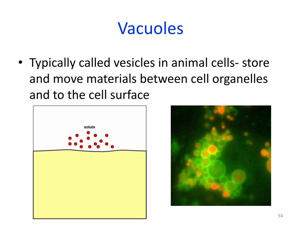
Vacuoles
• Typically called vesicles in animal cells- store and move materials between cell organelles and to the cell surface
54

Lesson Overview Life Is Cellular
Vacuoles and Vesicles
• saclike, membrane-enclosed structures
that store materials such as water, salts,
proteins, and carbohydrates
• Larger in plant cells
55

Lysosomes- the “clean-up crew”; small organelles filled with enzymes
• Break down many types of materials
Lipids
Carbohydrates
Proteins
Cellular debri
• Found mostly in animal cells
56

The Lysosome
Recycling cellular components
Functions:
Digesting food or cellular
invaders
57

Lesson Overview Life Is Cellular
Lysosomes
• Lysosomes are small organelles filled with enzymes that function as the cell’s cleanup crew. Lysosomes perform the vital function of removing ―junk‖ that might otherwise accumulate and clutter up the cell.
• Found mostly in white blood cells
• Have been linked to diseases, such as Tay Sach’s
• Tay Sach’s is a disorder that is caused by a genetic defect that prevents the formation of an essential enzyme that breaks down lipids
• These lipids build up in the body and can cause nerve damage; prognosis is not good
58

The Lysosome
This
bacterium
about to be
eaten by an
immune
system cell
will spend the
last minutes of
its existence
within a
lysosome.
59

Many Diseases are Caused by Lysosome Malfunction
60

Cytoskeleton
• The framework of the cell ; a network of protein filaments
• Some parts help in transport of materials
• Aids the cell in movement
• Made up of microfilaments and microtubules
61

The name is misleading.
The cytoskeleton is the
skeleton of the cell, but it’s
also like the muscular
system, able to change
the shape of cells in a
flash.
Maintains the shape of
the cell
Aids in movement
The Cytoskeleton
An animal cell cytoskeleton
62

A Cytoskeleton Gallery
63

The Cytoskeleton in Action
Cilia on a protozoan Beating sperm tail at fertilization
Smoker’s cough is due to destruction of cilia linking the airways. 64

Cytoskeleton
• Microfilaments- threadlike structures made up of a protein called actin
• Help cell maintain shape and allow it to move
• Assembly and dissasembly are responsible for amoeboid movement
65

Cytoskeleton
• Microtubules- are hollow structures made up of proteins known as tubulins
• Important for cells in maintaining shape, as well as for cell division
• Help build projections from the cell surface
66

Centrioles
• Are located near the nucleus
• Function- help organize cell division
• Not found in plant cells
67

Organelles That Build Proteins
1. Ribosomes- are small pieces of RNA and protein; the worktable for making proteins
2. Endoplasmic Reticulum- Protein synthesis (about half the cell’s proteins are made here); Protein movement (transport)
3. Golgi Apparatus-Proteins produced in the rough ER move here for packaging
68
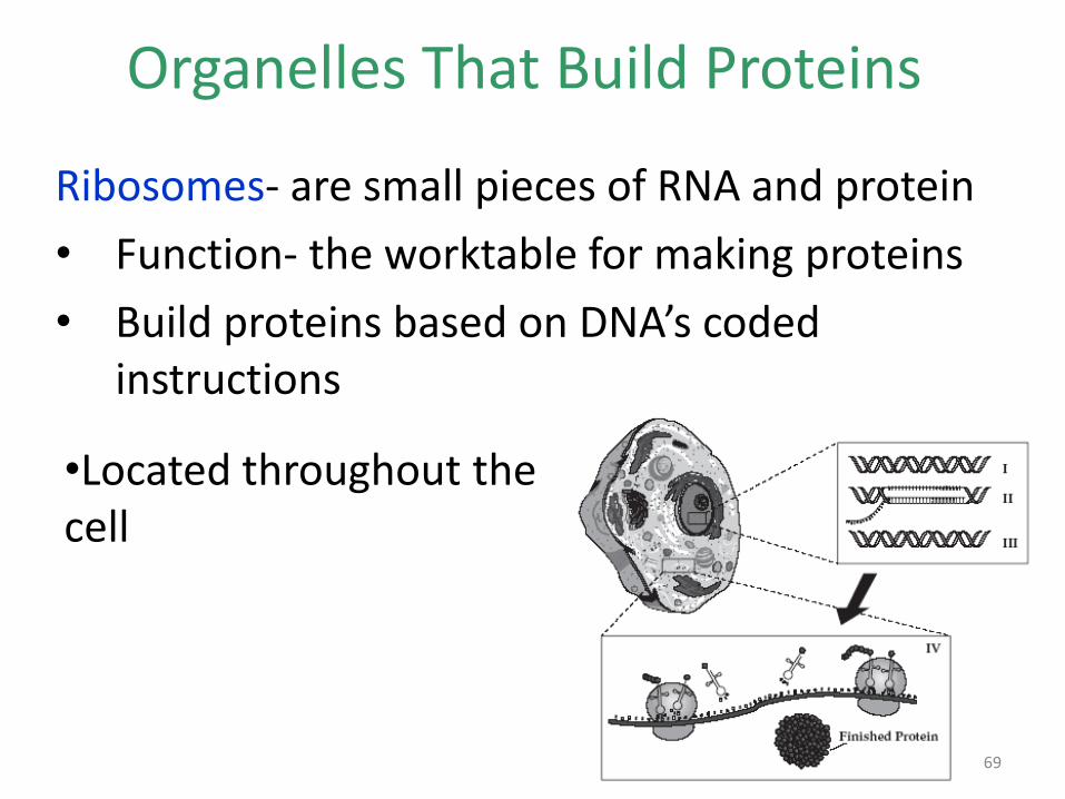
Organelles That Build Proteins
Ribosomes- are small pieces of RNA and protein
• Function- the worktable for making proteins
• Build proteins based on DNA’s coded instructions
•Located throughout the cell
69

Ribosomes and the Endoplasmic Reticulum
70

Ribosome
• Ribosomes Are Not Surrounded by a membrane. They are the site of PROTEIN SYNTHESIS (Production or Construction) in a cell.
71

Cystic Fibrosis
Click here to see the article. 72

Endoplasmic reticulum- “ER”
• Found in eukaryotic cells only
• Internal membrane system responsible for assembling lipids for the cell membrane
• Also assembles proteins for and other materials that are exported from the cell
• Two types- rough and smooth
73

The Rough Endoplasmic Reticulum
Protein movement
(transport)
Protein synthesis
(about half the cell’s
proteins are made
here).
Studded with ribosomes
74

The Rough Endoplasmic Reticulum • Rough ER- builds proteins; contains ribosomes
on its surface
• Rough ER chemically modifies the proteins made by the ribosomes
• Produces proteins that will eventually be secreted out of the cell
75

Smooth ER • IS NOT Covered with RIBOSOMES and
processes LIPIDS and CARBOHYDRATES.
• The Smooth ER is involved in the synthesis of steroids in gland cells, the regulation of calcium levels in muscle cells, and the breakdown of toxic substances by liver cells.
76

• Smooth ER- known as smooth because there are no ribosomes on its surface
• Contains enzymes that allow it to synthesize membrane and detoxify drugs
• Highly concentrated in liver cells
Right: rough er; Left: smooth er
Human liver cell 77
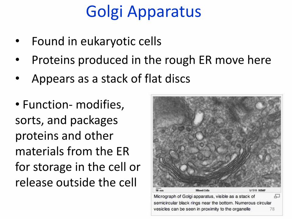
Golgi Apparatus
• Found in eukaryotic cells
• Proteins produced in the rough ER move here
• Appears as a stack of flat discs
• Function- modifies, sorts, and packages proteins and other materials from the ER for storage in the cell or release outside the cell
78

Golgi Apparatus
• Processing, Packaging and Secreting Organelle of the Cell that is made of flattened SAC
79
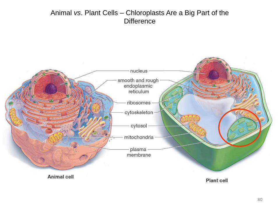
Animal vs. Plant Cells – Chloroplasts Are a Big Part of the
Difference
80

Cells In a Leaf
81

Two Other Unique Features of Plant Cells
The central
vacuole
may occupy
90% of a
plant cell.
82

Organelles that Capture and Release Energy
1. Chloroplasts - Found in plant cells, algae, and some bacteria; Capture light energy and convert it into food that contains chemical energy ; Where photosynthesis occurs
2. Mitochondria-Found in all eukaryotic cell; “powerplant” of the cell; Converts the chemical energy stored in food into compounds that are more convenient for the cell to use (ATP)
83

Chloroplasts
• Found in plant cells, algae, and some bacteria
• Capture light energy and convert it into food that contains chemical energy
• Where photosynthesis occurs
84

The Chloroplast
Think of the chloroplast as the solar panel of the plant cell.
Only plants have chloroplasts; they perform photosynthesis 85

Chloroplast Structure
• 2 membranes surround the organelle
• Stacks of membranes called grana are where chlorophyll occurs
• Chlorophyll- green pigment
86
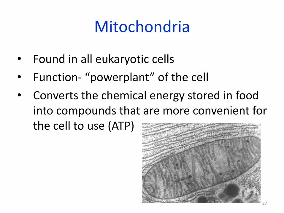
Mitochondria
• Found in all eukaryotic cells
• Function- “powerplant” of the cell
• Converts the chemical energy stored in food into compounds that are more convenient for the cell to use (ATP)
87

The Mitochondrion
Think of the mitochondrion as the
powerhouse of the cell.
Both plant and animal cells
contain many mitochondria.
88

Mitochondrial Diseases
89

Mitochondria and Health
90
Lesson Overview Life Is Cellular
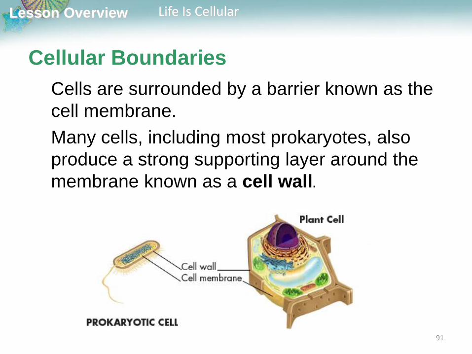
Cellular Boundaries
Cells are surrounded by a barrier known as the
cell membrane.
Many cells, including most prokaryotes, also
produce a strong supporting layer around the
membrane known as a cell wall.
91

Lesson Overview Life Is Cellular
Cell Membranes
All cells contain a cell membrane that
regulates what enters and leaves the cell
and also protects and supports the cell.
92

Cell membrane • The Cell Membrane is a complex barrier
separating the cell from it's external environment. The "Selectively Permeable" Membrane regulates what passes into and out of the cell.
93

Lesson Overview Life Is Cellular
Cell Membranes
The composition of nearly all cell
membranes is a double-layered sheet
called a lipid bilayer, which gives cell
membranes a flexible structure and forms
a strong barrier between the cell and its
surroundings.
94
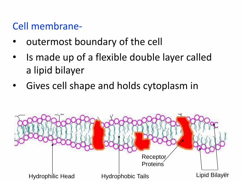
Cell membrane-
• outermost boundary of the cell
• Is made up of a flexible double layer called a lipid bilayer
• Gives cell shape and holds cytoplasm in
Hydrophobic Tails Hydrophilic Head Lipid Bilayer
Receptor
Proteins
95

Cell membrane-
• Selective Permeability- some substances can cross over the cell membrane, some cannot
• Gets in: Oxygen, water, and Carbon Dioxide •Does Not Get in: Sodium, Potassium, Calcium, Chlorine, proteins (need active support), most large molecules
96

You will not be tested on the material
presented on the next 7 slides covering lipids and fluid mosaic model
– these slides do not appear in you powerpoint – this material is covered
in Honors Bio ad AP bio – I am presenting it for those who like to
understand how things work.
97

Lesson Overview Life Is Cellular
The Properties of Lipids that control how
a cell membrane works
Many lipids have oily fatty acid chains attached to chemical
groups that interact strongly with water.
The fatty acid portions of such a lipid are hydrophobic, or
―water-hating,‖ while the opposite end of the molecule is
hydrophilic, or ―water-loving.‖
98

Lesson Overview Life Is Cellular
The Properties of Lipids
When such lipids are mixed with water, their
hydrophobic fatty acid ―tails‖ cluster together
while their hydrophilic ―heads‖ are attracted to
water. A lipid bilayer is the result.
99

Lesson Overview Life Is Cellular
The Properties of Lipids
The head groups of lipids in a bilayer are
exposed to water, while the fatty acid tails
form an oily layer inside the membrane
from which water is excluded.
100

Lesson Overview Life Is Cellular
The Fluid Mosaic Model
Most cell membranes contain protein molecules
that are embedded in the lipid bilayer.
Carbohydrate molecules are attached to many
of these proteins.
101

Lesson Overview Life Is Cellular
The Fluid Mosaic Model
Because the proteins embedded in the lipid bilayer can move around and
―float‖ among the lipids, and because so many different kinds of molecules
make up the cell membrane, scientists describe the cell membrane as a
―fluid mosaic.‖
102

Lesson Overview Life Is Cellular
The Fluid Mosaic Model
Some of the proteins form channels and pumps that help to move material
across the cell membrane.
Many of the carbohydrate molecules act like chemical identification cards,
allowing individual cells to identify one another.
103

Lesson Overview Life Is Cellular
The Fluid Mosaic Model Although many substances can cross biological membranes, some
are too large or too strongly charged to cross the lipid bilayer.
If a substance is able to cross a membrane, the membrane is said to
be permeable to it.
A membrane is impermeable to substances that cannot pass across
it.
Most biological membranes are selectively permeable, meaning that
some substances can pass across them and others cannot.
Selectively permeable membranes are also called semipermeable
membranes.
104
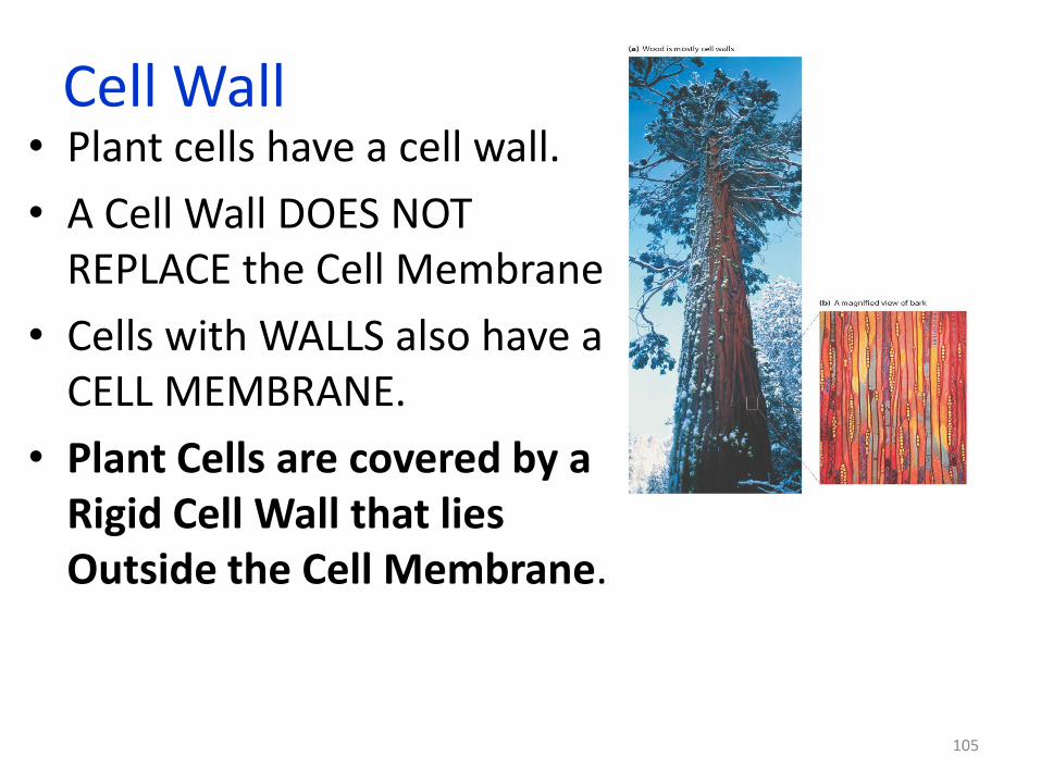
Cell Wall • Plant cells have a cell wall.
• A Cell Wall DOES NOT REPLACE the Cell Membrane
• Cells with WALLS also have a CELL MEMBRANE.
• Plant Cells are covered by a Rigid Cell Wall that lies Outside the Cell Membrane.
105
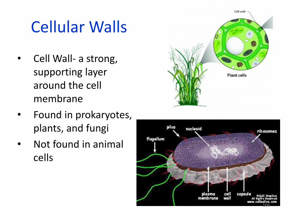
Cellular Walls
• Cell Wall- a strong, supporting layer around the cell membrane
• Found in prokaryotes, plants, and fungi
• Not found in animal cells
106

• Function- supports, shapes, and protects the cell
• Porous enough to allow water, O2, and CO2 to pass through
• In plants, it’s made up of cellulose
Right: products made from
cellulose
107
Cellular Walls

The Central Vacuole
Controls Turgor Pressure
Flaccid – no water
Turgid – full of water 108

Cellular Anatomy
109

Plants vs Animals
• Chloroplasts
• Cell Wall
• Centrioles
Both
•Nucleus Cytoskeleton
•Nucleolus Vacuoles
•Mitochondria Ribosomes
•Golgi Apparatus Lysosomes
•ER Cytoplasm
110

Cell Structure Review
You must know the similarities and differences between plant and animal cells
• You must be able to locate the various organelles in a cell diagram
• You must know the function of each organelle
111
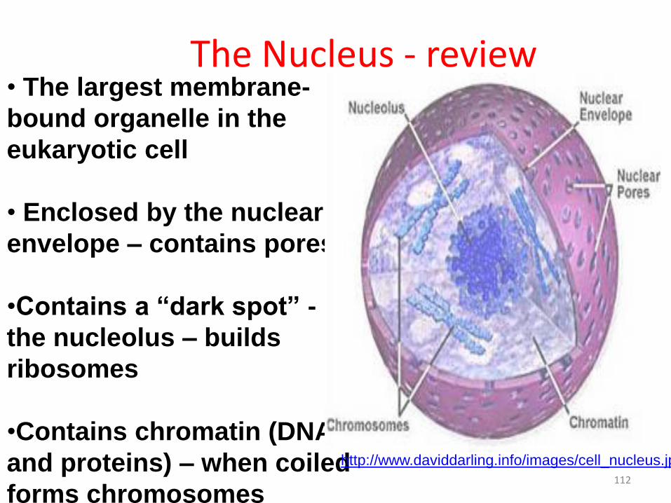
The Nucleus - review • The largest membrane-
bound organelle in the
eukaryotic cell
• Enclosed by the nuclear
envelope – contains pores
•Contains a “dark spot” -
the nucleolus – builds
ribosomes
•Contains chromatin (DNA
and proteins) – when coiled
forms chromosomes
http://www.daviddarling.info/images/cell_nucleus.jpg
112

Plasma Membrane - review • Composed of a phospholipid bilayer with
embedded proteins
• Provides a barrier that controls what enters and leaves the cell.
• All cells, both prokaryotic and eukaryotic, contain plasma membranes.
113

Cell Wall - review • Both prokaryotic and eukaryotic cells can have a cell
wall.
• Composed of cellulose in plant cells.
• A rigid, protective covering that provides structural support.
http://home.earthlink.net/~dayvdanls/ptcytok.
GIF
114

Cytoplasm - review
• Cell fluid which surrounds the organelles and contains many dissolved solutes.
• Site of many cellular chemical reactions.
http://www.jenningsk12.net/WE/peimann/Science/Cells/cell_amoeba.jpe
115

Vacuole - review
• Bag-like storage structure
• Can take up to 90% of the volume of a plant cell
• Stores food, water, wastes, or other substances
• Animal cell vacuoles are much smaller
http://student.nu.ac.th/u46410320/vacuole%5B1%5
D.jpeg
116

Mitochondria - review • The site of cellular
respiration.
• Provides energy for the cell by producing a molecule called ATP.
• Has a highly folded inner membrane that provides increased surface area for chemical reactions.
http://micro.magnet.fsu.edu/cells/mitochondria/images/mitochondriafigure1.jpg
117

Chloroplast - review • Site of photosynthesis.
• Contains the green pigment chlorophyll.
• Has a highly folded inner membrane that provides increased surface area for chemical reactions.
http://www.helpsavetheclimate.com/chloroplast1.gif
118
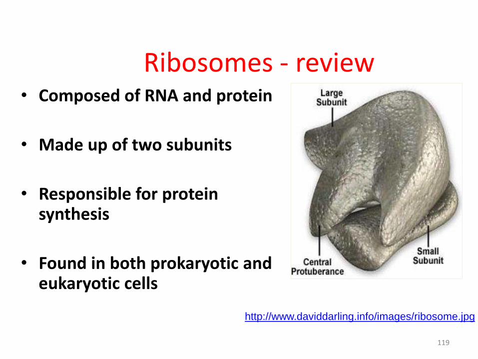
Ribosomes - review • Composed of RNA and protein
• Made up of two subunits
• Responsible for protein synthesis
• Found in both prokaryotic and eukaryotic cells
http://www.daviddarling.info/images/ribosome.jpg
119

Other Organelles - review • Other organelles help to
carry out cell functions.
• ER – makes lipids and helps build new membrane.
• Golgi receives and modifies proteins and then ships them to new locations in the cell.
• Lysosomes digest worn out cell parts and food.
Golgi
Apparatus
Transport
Vesicle
Endoplasmic
Reticulum
Lysosome
http://www.answers.com/topic/endomembrane-system-diagram-no-text-nucleus-png
120