α7 nicotinic receptor modulation alters glutamate release
TRANSCRIPT

α7 Nicotinic Receptor Modulation Alters
Glutamate Release: Implications for
Cognitive Deficits in Schizophrenia Undergraduate Research Thesis
Presented in partial fulfillment of the requirements for graduation with honors research distinction in the
undergraduate colleges of The Ohio State University
By
Brian A. Upton
The Ohio State University
April 2015
Project Advisor: Professor John P. Bruno, Department of Neuroscience


Upton 3
Contents List of Figures ................................................................................................................................. 5
Abstract ........................................................................................................................................... 7
Introduction ..................................................................................................................................... 9
Research Overview ....................................................................................................................... 17
Mesolimbic Stimulation Assay ................................................................................................. 17
Aims .......................................................................................................................................... 17
Hypothesis ................................................................................................................................. 18
Experimental Design ................................................................................................................. 18
Methods......................................................................................................................................... 19
Microelectrode Array: Second-by-Second Measurements of Glutamate ................................. 19
Sensor Calibration: in vitro ....................................................................................................... 20
Experimental Subjects ............................................................................................................... 21
Implantation of Microelectrode and Infusion Cannulae ........................................................... 21
In vivo Recordings and Drug Administration ........................................................................... 22
Histology ................................................................................................................................... 22
Data Analysis ............................................................................................................................ 23
Results ........................................................................................................................................... 25
AVL-3288 Results..................................................................................................................... 25
Effects of AVL-3288 on basal glutamate .............................................................................. 25
Effects of AVL-3288 on evoked glutamate ........................................................................... 25
PNU-120596 Results ................................................................................................................. 27
Effects of PNU-120596 on basal glutamate .......................................................................... 27
Effects of PNU-120596 on evoked glutamate ....................................................................... 28
Discussion ..................................................................................................................................... 31
References ..................................................................................................................................... 41
Figures........................................................................................................................................... 51


Upton 5
List of Figures Figure 1: enzyme detection scheme. ............................................................................................. 51
Figure 2: calibration of the MEA. ................................................................................................. 51
Figure 3: sample placement of MEA and cannula. ....................................................................... 53
Figure 4: sample tracing of low-dose NMDA after vehicle and low-dose AVL-3288 ................ 55
Figure 5: sample tracing of high-dose NMDA after vehicle and high-dose AVL-3288 .............. 55
Figure 6: AVL-3288 Group Data .................................................................................................. 57
Figure 7: sample tracing of low-dose NMDA after vehicle and high-dose PNU-120596............ 59
Figure 8: PNU-120596 Group Data. ............................................................................................. 59


Upton 7
Abstract Cognitive deficits in schizophrenia are thought to be caused, in part, by disrupted prefrontal
cholinergic and glutamatergic transmission. Activation of the α7 nicotinic acetylcholine receptor
(α7nAChR) has been shown to increase prefrontal glutamate as well as rescue failing
performance in cognitive tasks in rodents and primates. Intra-accumbens stimulation with
NMDA dose-dependently increases prefrontal acetylcholine, which in turn increases prefrontal
glutamate via α7nAChR activity. Using NMDA stimulation as an assay to examine the
potentiation of glutamate release as a function of the amount of acetylcholine released in the
PFC, the potentiation profiles of two α7nAChR positive allosteric modulators (PAMs), AVL-
3288 and PNU-120596, were assessed at varying levels of PAM and stimulation. Second-by-
second measurements with a glutamate-sensitive microelectrode in the PFC in awake rats reveal
that only an appropriate combination of the dose of NMDA and dose of PAM consistently
potentiates prefrontal glutamate release. However, at other concentrations of NMDA and PAM,
the effect on mesolimbic stimulation varied greatly from significant potentiation to a reduction of
glutamate release. Furthermore, the potentiation profile for the type I and type II PAMs differed
significantly, possibly due to differences in receptor desensitization in the presence of these two
drugs. Overall, these results demonstrate the importance of dose of α7nAChR PAMs in
modulating neurotransmitter systems, and PAMs as a potential therapeutic in treating cognitive
deficits of schizophrenia as effects were dependent on activity of the orthosteric ligand.

Upton 9
Introduction Schizophrenia is a chronic brain disorder disrupting thought, perception, and behavior.
Schizophrenia is prevalent in roughly 1% of the population and has an economic burden
exceeding $62 billion/year in the United States alone (Perala et al., 2007; Wu et al., 2005).
Diagnosis typically occurs after the first psychotic episode, occurring in late adolescence to early
adulthood in men and throughout early adulthood in women, although prodromal symptoms are
often present before this episode (Faraone et al. 1994; White et al., 2006). While the age of the
first psychotic episode varies between men and women, both are affected in equal numbers (Saha
et al., 2005). Not only does schizophrenia affect equal numbers across sex, culture, and
socioeconomic status, the symptoms of the disorder also appear to be conserved across these
demographic factors as well (Saha et al., 2006; Karagianis et al., 2009).This similar presentation
is not to say that schizophrenia is a homogeneous disorder, but rather that schizophrenia is a
worldwide problem with significant psychosocial and economic impact.
Schizophrenia is a heterogeneous disorder composed of symptoms from three symptom
clusters: positive, negative, and cognitive symptoms. Positive symptoms include features of the
disease that are present in patients that are not present in the general population. These symptoms
include hallucinations, delusions, and disorganized thought and speech (American Psychiatric
Association, 2013). Similarly, negative symptoms of schizophrenia include features that are
absent in schizophrenia but present in the general population, including flattened affect,
avolition, and anhedonia (American Psychiatric Association, 2013). Lastly, schizophrenia
presents with cognitive deficits, or deficits in thought processes (Kerns et al., 2008). Aspects of
executive functioning are impaired in schizophrenia, particularly in working memory, cognitive

Upton 10
flexibility, and attentional processing. Current antipsychotic medications are effective at
addressing some, but not all, of the symptoms associated with schizophrenia.
The first antipsychotic, chlorpromazine (trade name Thorazine), was originally developed
as an anesthetic agent, yet successfully reduced many of the positive symptoms associated with
schizophrenia (Ban, 2007). The success of chlorpromazine led to the development of similar
drugs, known as first generation, or typical, antipsychotics. This class of antipsychotic
medication exerts its antipsychotic effects as D2 antagonists (Kapur & Mamo, 2003).
Additionally, these medications have activity on the cholinergic, histaminergic, and adrenergic
systems, which caused a variety of side effects such as dry mouth, weight gain, and sedation. The
most debilitating and possibly permanent side effects of typical antipsychotics are
extrapyramidal side effects, which are caused by high D2 occupancy leading to a dopamine
hypofunction in extrapyramidal pathways, similar to what is seen in Parkinson’s disease
(Tuppurainen et al., 2010). Extrapyramidal side effects involve movement difficulties, including
dystonia, tremors, akathisia, bradykinesia, and tardive dyskinesia. To reduce these side effects,
second generation, or atypical, antipsychotics were developed that bind more transiently to the
D2 receptor, allowing for more endogenous dopamine binding, and additionally serve as 5-HT2A
antagonists producing antipsychotic effects (Seeman 2002). While atypical antipsychotics had a
smaller side effect profile than typical antipsychotics, they were no more efficacious than some
typical antipsychotics in their antipsychotic properties (Lieberman & Stroup 2011). Both classes
of drugs are capable at reducing many of the positive and negative symptoms; however, neither
sufficiently address cognitive symptoms and subsequently, cognitive symptoms remain untreated
(Keefe, 2007a; 2007b).

Upton 11
Cognitive deficits of schizophrenia result in a poorer quality of life and are more
debilitating to daily function than positive or negative symptoms (Swartz et al., 2007). Cognitive
function is often the best predictor of long-term functional outcome in the treatment of
schizophrenia (Green et al., 2000). Presumably, improving cognitive functioning in patients with
schizophrenia will improve their treatment outcome and quality of life. Poor cognitive function
may predict a poor prognosis because poor cognitive function is a barrier to employment and a
risk factor for the discontinuation of treatment (Rosenheck et al., 2006; Robinson et al. 2002). In
addition to a personal decline in quality of life, cognitive symptoms contribute to the social and
economic burden of the disease; therefore, it is imperative to improve treatments aimed at
ameliorating the cognitive deficits associated with schizophrenia.
Many aspects of cognition are mediated via a distributed neural network comprised of
cortical and subcortical structures. The prefrontal cortex (PFC), serving as a source of top-down
control, is important in all aspects of higher-order cognition (Miller, 2000). The PFC plays
important roles in goal-directed behavior, personality expression, error detection, and problem
solving. Disruptions in prefrontal activity can lead to deficits in tasks designed to measure
working memory, cognitive flexibility and attentional processing (Demakis, 2004). It should
serve as no surprise that the prefrontal cortices of individuals with schizophrenia appear
disrupted. The brains of patients with schizophrenia display hypofrontality, or decreased
prefrontal activity, both at rest and during cognitive task performance (Berman et al., 1992).
Individuals with schizophrenia also display deficits in tasks dependent on prefrontal activity,
such as the Stroop task and Wisconsin card sorting test (Barch et al., 2004; Haut et al., 1996). In
longitudinal studies, patients who are later diagnosed with schizophrenia have reduced prefrontal
gray matter at the time of their first episode (Hirayasu et al., 2001; Molina et al., 2006). Thus,

Upton 12
prefrontal irregularities may be involved in the cognitive deficits present in schizophrenia as
similar deficits are seen in both patients with schizophrenia as well as frontal lobe damage.
Glutamate, the primary excitatory neurotransmitter in the CNS, is important for cognitive
performance. Disrupted prefrontal glutamate transmission can lead to impaired performance in a
variety of cognitive tasks, including tasks designed to measure working memory, cognitive
flexibility, attentional processing, and sensory gating (Umbrecht et al. 2000). Similarly,
enhancing glutamate transmission in the PFC may lead to increased cognitive performance.
Genetic risk factors in schizophrenia, including altered expression of catechol-o-methyl
transferase (COMT), dysbindin, neuregulin, disrupted in schizophrenia 1 (DISC1), regulator of
G-protein signaling 4 (RGS4), metabotropic glutamate receptor-3 (GRM3), and G72, have been
linked to disruptions in glutamate transmission (Harrison & Weinberger 2004). Additionally,
transcript expression for vesicular glutamate transporter-1 (VGLUT1) is down-regulated in the
prefrontal cortex of individuals with schizophrenia, likely negatively affecting glutamate
transport and release (Eastwood & Harrison, 2004). Furthermore, the activity of excitatory amino
acid transporters (EAATs) appears disrupted in schizophrenia. Glial EAATs (i.e. EAAT1 and
EAAT2) are down-regulated, possibly leading to an increase in autoreceptor activation and
compensatory up-regulation of neuronal EAATs (i.e. EAAT3), which together, lead to a decrease
in synaptic glutamate and increase in extrasynaptic glutamate (Bauer et al. 2008). Accordingly,
group II metabotropic glutamate receptor expression is increased in the prefrontal cortex in
schizophrenia (Gupta et al. 2005). Altered expression of the NMDA receptor, a glutamate
receptor important in learning and memory, has been noted in the dorsolateral PFC and anterior
cingulate cortex in schizophrenic brains (Kristiansen et al., 2006). Antagonism of the NMDAR is
capable of producing psychotic symptoms in non-diseased individuals and exacerbates psychotic

Upton 13
symptoms in schizophrenic individuals (Krystal et al. 1994; Lahti et al., 1995). Additionally,
NMDA blockade produces cognitive deficits in humans and animals and is frequently used as an
animal model for cognitive deficits in schizophrenia (Krystal et al., 1994; Blot et al., 2013).
Taken together, disruptions of glutamate transmission in the PFC may lead to glutamatergic
hypofunction and underlie cognitive deficits seen in the disorder; therefore, methods of
enhancing prefrontal glutamate may have pro-cognitive effects and serve to ameliorate the
cognitive deficits seen in schizophrenia.
Prefrontal glutamate can be modulated bi-directionally via the α7 nicotinic acetylcholine
receptor (α7nAChR; Konradsson-Geuken et al., 2010). The α7nAChR is a homopentameric
heteroreceptor, located pre- and postsynaptically, that gates cations (i.e. calcium, sodium,
potassium)(Dajas-Bailador & Wonnacott, 2004). Calcium ion influx through the α7nAChR
initiates a signal cascade known as the extracellular signal-regulated kinase 1/2 (ERK1/2)
pathway (also known as the mitogen-activated protein kinase (MAPK) pathway), which is a
transduction mechanism that has implications for learning and memory (Gubbins et al., 2010).
Thus, presynaptic α7nAChR activity enhances neurotransmitter release both by causing
depolarization at the axon terminal as well as affecting gene expression. In this manner,
acetylcholine and other direct agonists of the α7nAChR are able to enhance glutamate
transmission, while antagonism of the α7nAChR decreases glutamate transmission (Bortz et al.,
2013; Wu et al., 2010). Similarly, drugs that increase α7nAChR activity in the PFC have pro-
cognitive effects in rodents and result in an increased performance in a variety of cognitive tasks,
while drugs that reduce α7nAChR activity can produce cognitive deficits (Redrobe et al., 2009;
Thomsen et al., 2009; Castner et al., 2011; Alexander et al., 2012). Therefore, increasing

Upton 14
α7nAChR activity may serve as a target for cognitive-enhancing therapeutics in schizophrenia by
enhancing several neurotransmitter systems, including glutamate.
In schizophrenia, α7nAChR activity appears disrupted. Single nucleotide polymorphisms
in the promoter region of the CHRNA7 gene, the gene that encodes the α7 protein, are a risk
factor in the development of schizophrenia (Leonard et al., 2002). In individuals who develop
schizophrenia, a decrease in both α7 protein, and in some populations, decreases in mRNA
expression have been reported (Guan et al., 1999; Matthew et al., 2007). Additionally, kynurenic
acid, a tryptophan metabolite, is an endogenous antagonist to the α7nAChR and has been found
to be elevated in cerebrospinal fluid and postmortem brains of patients with schizophrenia
(Hilmas et al., 2001; Linderholm et al., 2012; Schwarcz et al., 2001). Given the importance of
α7nAChR activity in cognition, decreased α7nAChR activity may contribute to the cognitive
deficits and glutamate hypofunction seen in schizophrenia.
Nicotine, the primary drug found in tobacco products, is a direct agonist of the α7nAChR
and has limited pro-cognitive effects. Interestingly, between 60 and 90% of patients with
schizophrenia smoke cigarettes (Leonard et al., 2001). This staggering correlation has led some
to believe that increased nicotine consumption is a form of self-medication to alleviate cognitive
deficits that are not managed with current treatment regimens (Leonard et al., 2007).
Unfortunately, nicotine is not an ideal therapeutic for treating cognitive deficits for a variety of
reasons: the pro-cognitive effects of nicotine are short-lived, as the α7nAChR receptor is easily
desensitized; nicotine is not selective to the α7nAChR, leading to many side effects; and many
negative health effects of nicotine products (Papke et al. 2009). As a result, direct α7nAChR
agonists have been developed which can reduce cognitive symptoms in animal models of
schizophrenia (Hauser et al., 2009). Additionally, pro-cognitive effects α7nAChR agonists have
Upton 15
been demonstrated in human populations (Olincy et al., 2006). The tendency for the α7nAChR
receptor to desensitize has prompted investigation into low-dose agonists, which have shown
benefits in animal models of cognitive deficits (Werkheiser et al., 2011). While selective
α7nAChR agonists, particularly at low doses, may have advantages over the use of nicotine, they
may still be limited to characteristics inherent to direct agonists.
There are two primary fundamental limitations of direct agonists as
pharmacotherapeutics. First, direct agonists bind to the receptor and cause a response when
present in a system. This indiscriminant activation occurs with or without afferent activity,
leading to the tendency to cause false alarms at the receptor. Second, direct agonist activity does
not preserve the temporal integrity of endogenous neurotransmission, which may be important to
many neural processes underlying cognitive performance. In other words, neural connections
lose the degree-of-freedom afforded by the ability to selectively activate and deactivate the
receptor as is biologically-relevant. Instead, direct agonists reduce this complexity to an “always
on” signal while the agonist is present in the brain. Due to the disruptions of native spatial and
temporal signaling caused by direct agonism, a more subtle method of enhancing receptor
activity is needed that maintains the complexity of the neural system.
Positive allosteric modulators (PAMs) are chemicals that enhance receptor activity in the
presence of an orthosteric ligand, while in the absence of an orthosteric ligand, have no intrinsic
receptor activity. Unlike orthosteric agonists, which bind to the same location as the endogenous
neurotransmitter, positive allosteric modulators bind at a different site, termed allosteric site.
Binding at the allosteric site allows PAMs to alter the receptors configuration and, therefore,
activity at the orthosteric site. In this manner, PAMs are able to enhance receptor activity in the
presence of an orthosteric ligand; however, binding to the allosteric site in the absence of

Upton 16
orthosteric activity does not result in a receptor response. In the case of α7nAChRs, PAMs do
not cause cation influx in the absence of orthosteric activity.
α7nAChR PAMs are now being investigated as pro-cognitive therapeutics with potential
in ameliorating deficits in diseases such as schizophrenia. Several α7nAChR PAMs have been
used either alone or in conjunction with drugs that increase α7nAChR orthosteric activity to
ameliorate cognitive deficits and sensory gating deficits in several animal models of
schizophrenia (Hurst et al., 2005; Ng et al., 2007; Thomsen et al., 2011; McLean et al., 2012;
Callahan et al., 2013). Additionally, α7nAChR PAMs have been shown to potentiate evoked
excitatory amino acid release in vitro and evoked prefrontal dopamine release in vivo (Zwart et
al., 2002; Livingstone et al., 2010). α7nAChR PAMs fall into two categories: type I and type II.
The PAM types differ in that type I PAMs affect peak current, whereas type II PAMs affect both
peak current and current decay (Grønlien et al., 2007). That is, both types increase the peak
amount of cation influx, but while type I PAMs allow the receptor to naturally desensitize into a
closed configuration, type II PAMs prevent the receptor from entering this desensitized state and
allow the receptor to remain open with continued cation influx (Williams et al., 2011a).
Furthermore, only type II PAMs have been shown to reactivate desensitized α7nAChRs (Hurst et
al., 2005; Grønlien et al., 2007). While interest has been growing in the use of α7 PAMs for a
variety of neurological disorders, limited research has been published characterizing effects on
neurotransmitter systems in vivo.

Upton 17
Research Overview
Mesolimbic Stimulation Assay In order to investigate the effect of PAMs, the drug must be applied in the presence of the
neurotransmitter that is modulated, as PAMs have no effect on receptor activity in the absence of
orthosteric activity. Both task- and chemical-induced activity in the nucleus accumbens (NAc)
have been previously shown to cause increases in both prefrontal cholinergic and glutamatergic
activity (Neigh et al., 2004; Zmarowski et al., 2007; Bortz et al., 2014). Similarly, NMDA
infusion into the anterior shell of the nucleus accumbens dose-dependently increases prefrontal
choline, a selective α7nAChR agonist, and glutamate (Alkondon et al., 1997; Bortz et al.,
unpublished data). NMDA-evoked glutamate release is dependent on α7nAChR activity, as
antagonism of the α7nAChR abolishes this evoked release (Bortz et al., unpublished data).
Therefore, differing concentrations of NMDA infused into the accumbens cause different
concentrations of acetylcholine to be released in the prefrontal cortex. These varying
concentrations of acetylcholine activate the α7nAChR, facilitating glutamate release dose-
dependently as a function of the concentration of acetylcholine. Thus, with this assay, the effects
of α7nAChR PAM can be assessed in the presence of endogenously released acetylcholine
without disrupting the integrity of cholinergic transmission in the PFC. Additionally, this assay is
a model of top-down cognitive processing and is capable of rescuing failing performance during
the distractor portion of sustained attention tasks (St. Peters et al., 2011).
Aims The goal of this experiment is to assess the ability of different doses of both type I and
type II α7nAChR PAMs, AVL-3288 and PNU-120596 respectively, in potentiating glutamate
release in vivo as a function of choline release in awake, freely moving rats.

Upton 18
Hypothesis
If cholinergic activity is required for PAMs to enhance activity at the orthosteric site of the
α7nAChR, then each PAM will potentiate prefrontal glutamate release as a result of mesolimbic
stimulation, but not under basal conditions.
Experimental Design Animals were tested on three consecutive test days, receiving the same concentration of
NMDA stimulation on each day. Prior to NMDA stimulation, animals were systemically injected
with either AVL-3288 or PNU-120596. Dose of PAM was manipulated between test days so that
the effect of either high-dose PAM, low-dose PAM, or vehicle on a constant level of stimulation
could be measured. Each subject only received one type of PAM for experiments. The effects of
each dose of PAM were compared to the glutamate release on the vehicle test day. Using this set-
up, an increase or decrease in glutamate release from the vehicle test day in the presence of PAM
could be observed as a potentiation or attenuation, respectively, within subject.

Upton 19
Methods
Microelectrode Array: Second-by-Second Measurements of Glutamate Self-referencing, ceramic-based, multisite microelectrode arrays (MEA) were used for
the detection of in vivo glutamate release in awake, freely-moving rats (Quanteon, LLC,
Nicholasville, KY)(see figure 1). Each MEA consists of two pairs of platinum recording
channels (15 x 333µm each) separated 100µm vertically and 30µm horizontally. The highly-
selective enzyme, glutamate oxidase (GluOx, 2%, 1 unit/µL, 100nL) was cross-linked to the first
pair of recording channels using glutaraldehyde (0.125%) and bovine serum albumin (BSA; 1%).
GluOx oxidizes glutamate, producing α-ketoglutarate, ammonia, and hydrogen peroxide (H2O2).
H2O2, which is electrically active at the applied voltage, serves as the reporting molecule for
glutamate. H2O2 is oxidized at the platinum recording site due to an electric potential of +0.7V
relative to a Ag|AgCl reference electrode, placed distant from the recording site. The oxidation
of H2O2 produces a current, which is amplified and recorded using the FAST-16 mkII recording
system (Quanteon, LLC, Nicholasville, KY). This amperometric design allows for sub-second
temporal resolution of the MEA. By calibrating each MEA in vitro prior to implantation, the in
vivo concentration of glutamate can be derived from the recorded current. The second pair of
recording channels, coated in only glutaraldehye and BSA but lack GluOX, is not glutamate-
sensitive and serves as a background, or sentinel, channel, recording signals that are not
glutamate-driven. The glutamate-sensitive channels are self-referenced with the sentinel
channels, isolating the signal derived exclusively from glutamate (Burmeister & Gerhardt, 2001;
Day et al., 2006; Rutherford et al., 2007). In order to minimize the signal from oxidizable
interferents, such as dopamine or ascorbic acid, m-phenylenediamine dihydrochloride (mPD)
was electroplated onto the recording sites prior to sensor calibration and implantation. mPD

Upton 20
serves as a physical barrier, preventing larger molecules, such as catecholamines and ascorbic
acid, from interacting with the recording site, while smaller molecules, such as H2O2, are not
excluded (Wahono et al., 2012). The exclusion barrier in conjunction with the self-referencing
technique allow each MEA to be highly selective for glutamate with a limit of detection less than
0.5µM. MEAs can remain selective and sensitive to glutamate for up to seven days of chronic
recording (Rutherford et al., 2007).
Sensor Calibration: in vitro Immediately prior to sensor implantation, the microelectrodes were calibrated in vitro in
a solution of PBS (0.05 M, 40 mL; pH 7.4; 37°C; +0.7 V). After baseline was established,
channel signals were recorded at a rate of 1 Hz following the addition of ascorbic acid (250 µM),
glutamate (3 x 20 µM), dopamine (2 mM), and hydrogen peroxide (8.8 µM) (see fig. 2). The
effectiveness of the mPD barrier was assessed by observing no change in measurement on any of
the four channels during the addition of ascorbic acid or dopamine, both of which can be found
in high concentrations in vivo and are electrically active at the sensor’s applied voltage (Lowry &
O’Neill, 1992). When glutamate was added, an increase in current was observed on the two
GluOx channels. The slope (nA/µM glutamate) was determined from these three glutamate
additions in order to relate the measured current to the glutamate concentration when used in
vivo. The final addition of hydrogen peroxide created a current observable on all four channels to
ensure all channels are equally sensitive to the reporting molecule. In order to qualify for use,
every sensor must have met the following criteria: (i) no difference greater than 20 pA during
background current between all four channels, (ii) linear response to increase in glutamate
concentration with a coefficient of determination (r2) value greater than 0.998, (iii) high
sensitivity, determined by a slope greater than -0.003 nA/µM glutamate, (iv) a limit of detection

Upton 21
<0.5 µM, (v) high selectivity of glutamate to ascorbic acid and dopamine (>50:1), and (vi)
greater than 80% but less than 125% similarity to hydrogen peroxide for channel pairings.
Experimental Subjects All measurements were done in adult male Wistar rats (body weight 280-420 g; Charles
River Laboratory: Wilmington, MA). Animals were housed in individual plastic cages with corn
cob bedding (Harlan Teklad, Madison, WI) in a temperature (72°) and humidity (30-70%)
controlled room on a 12:12 light:dark cycle. All animals had access to food and water ad libitum.
All procedures involving animals were approved by The Ohio State University Institutional
Animal Care and Use Committee in accordance with the NIH Guide for the Care and Use of
Laboratory Animals. As such, all efforts were made to minimize animal suffering, to reduce the
number of animals used, and to consider alternatives to in vivo techniques.
Implantation of Microelectrode and Infusion Cannulae Animals were anesthetized using isoflorane gas (2%, 0.6 L/min) and positioned with a
stereotax. An MEA was placed unilaterally between infralimbic and prelimbic cortices of the
medial prefrontal cortex (in mm from bregma: AP +2.7, ML ±0.65, DV -4.0; hemispheres
counterbalanced; see fig. 3A and 3B). A steel guide cannula used for intracoritcal infusions of
NMDA was placed in the anterior shell of the nucleus accumbens, ipsilateral to the MEA (in mm
from bregma: AP +0.4, ML ±0.70, DV -6.4; see fig. 3C and 3D). The infusion cannula extended
1.0 mm farther than the guide cannula and when not in place, a dummy cannula was inserted,
extending 0.7 mm past the tip of the guide cannula. The Ag|AgCl reference electrode was placed
in the contralateral parietal lobe, distant from the MEA. The atlas of Paxinos and Watson (1998)
was used to determine all coordinates. The sensor was secured using three stainless steel screws

Upton 22
mounted to the skull and dental acrylic (DuraLay Inlay Pattern Resin). Animals were not tested
until the second day after surgery to allow time to recover.
In vivo Recordings and Drug Administration The day following implantation surgery, the rats habituated to their testing environment,
a wooden test box (16” width, 16” length, 24” height) with corn cob bedding inside a Faraday
cage, but were not connected to the sensor’s preamplifier. On the following days, the animals
were placed into the testing box and connected to the preamplifier and a baseline recording was
established for a minimum of two hours before the infusion of any drugs. Prior to NMDA
infusion, rats were pretreated with an intraperitoneal injection of AVL-3288 (1 or 3 mg/kg) or
PNU-120596 (3 or 9 mg/kg) or vehicle (dimethyl sulfoxide (DMSO), 5% by volume; Solutol®,
8% by weight). Thirty minutes after PAM pretreatment, artificial cerebrospinal fluid (aCSF; pH
7.1-7.4) was infused into the NAc to serve as a positive control as well as to clear the cannula
and guide cannula prior to drug administration. Ten minutes after aCSF infusion, NMDA (0.05
or 0.30 µg/0.5 µL aCSF) was infused into the NAc. A Hamilton PB600-1 manual dispenser was
used to deliver aCSF and NMDA through the cannula over two seconds. Dose of PAM was
manipulated within subjects between test days, while the dose of NMDA was consistent between
test days and was only manipulated between subjects. The dose order of PAMs was
counterbalanced among animals.
Histology All MEA and cannula placement was verified post mortem. Following the third test day,
rats were given an overdose of pentobarbital sodium and transcardially perfused with heparinized
saline (0.2%) followed by formalin (10%). Brains were removed and were placed in formalin for
a minimum of three days at 4°C. One day prior to sectioning, the brains were moved to a sucrose

Upton 23
solution (30%) and maintained at 4°C. The sucrose solution serves as a cryoprotectant to prevent
artifacts as a result of freezing the tissue samples. Brains were sectioned into coronal slices
(50µm) using a cryostat (-25°C), mounted on gelatin-coated slides, and stained using cresyl
violet. Slices were examined under a light microscope to determine sensor and cannula
placement. If MEA placement into the mPFC or cannula placement into the anterior NAcSh
could not be verified, the data from the animal was excluded from analysis (see figure 3 for
sample MEA and cannula placement).
Data Analysis Measurements derived from the MEA recording and the FAST-16 data file included: (i)
basal glutamate levels, (ii) maximum peak concentration (uM) of glutamate, (iii) the latency
(second) of onset of the effect from the time of drug infusion, and (iv) T80, the time in seconds
from maximum peak amplitude to 80% decay of signal (a measure of glutamate clearance). The
signal derived exclusively by the oxidation of glutamate was isolated using a self-referencing
procedure between the glutamate-sensitive recording channel and the adjacent background
sentinel channel (Burmeister and Gerhardt, 2001; Day et al., 2006; Rutherford et al., 2007). All
dependent measures will be analyzed by analysis of variance (ANOVA) using the SPSS 18
statistics program (V19, IBM Corporations, Armonk, NY). In all ANOVAs, the Huynh-Feldt
correction will be utilized to reduce type I errors associated with repeated measure ANOVAs
(Vasey and Thayer, 1987). Statistical significance was defined as P < 0.05.


Upton 25
Results
AVL-3288 Results
Effects of AVL-3288 on basal glutamate
Basal glutamate, measured after IP injection but before aCSF infusion, did not vary
between AVL-3288 dose (F2,28=3.134, p=0.068) or NMDA dose (F1,9=1.031, p=0.395) and there
was no interaction between the dose combinations (F4,18=0.321, p=0.860). These results indicate
that systemic injection of AVL-3288 does not affect basal glutamate release, i.e. in the absence
of stimulation.
Effects of AVL-3288 on evoked glutamate
The type I α7nAChR PAM, AVL-3288, was injected systemically 40 minutes prior to
mesolimbic-stimulation with NMDA. The dose of NMDA (0.05µg and 0.30µg) was held
constant across test days and was administered in the presence of vehicle, low-dose, and high-
dose of AVL-3288. With this setup, effects of dose of AVL-3288 will be presented as a percent
change from the vehicle test day. In this manner, a positive percent change signifies that more
glutamate was released in the presence of AVL-3288 with the same level of stimulation (i.e.
concentration of NMDA), while a negative percent change signifies that less glutamate was
released in the presence of AVL-3288 than on the vehicle-treated day.
Two-way (AVL dose x NMDA dose) omnibus ANOVA revealed a significant effect on
the percent change in glutamate release between AVL-3288 dose (F2,28=9.128, p=0.001) and an
AVL dose-NMDA dose interaction (F4,28=5.008, p=0.04). Subsequent one-way ANOVA on dose
of AVL-3288 (vehicle, 1 mg/kg, 3 mg/kg) revealed a significant difference between AVL doses

Upton 26
within the 0.30 NMDA group (F2,14=11.873, p=0.001), but not within the 0.05 NMDA group
(F2,17=0.494, p=0.620). Within the high-dose of NMDA group, percent change in glutamate
release was significantly potentiated by 1 mg/kg AVL-3288 compared to the vehicle response
(F1,9=5.932, p=0.41) with an average percent change of 84.70 ± 34.78%. Conversely, the percent
change in glutamate release was significantly attenuated by 3 mg/kg AVL-3288 with respect to
vehicle response (F1,9=20.575, p=0.002) with an average percent change of -64.24 ± 14.16%.
The percent change of glutamate release also varied between 1 mg/kg and 3 mg/kg AVL-3288
(F1,9=15.732, p=0.004). An additional one-way ANOVA on dose of NMDA (aCSF, 0.05µg,
0.30µg) revealed an interaction within the 1mg/kg AVL-3288 treated group (F2,16=5.661,
p=0.016) but not within the 3mg/kg AVL-3288 treated group (F2,16=1.953, p=0.179). 1 mg/kg
AVL-3288 potentiated glutamate release more after 0.05µg compared to aCSF (F1,11=17.471,
p=0.002) and more after 0.30µg compared to aCSF (F1,10=7.280, p=0.024). Thus, 1 mg/kg AVL-
3288 produced a percent change of 24.12 ± 5.77% after 0.05µg stimulation. Lastly, additional
two-way (AVL dose x NMDA dose) ANOVAs did not reveal any variation among latency to
peak onset, T50, T80, and peak duration (all p-values >0.1).
Fig. 4 demonstrates representative line tracings for an animal receiving 0.05µg/0.5 µL
NMDA in the presence of vehicle and 1 mg/kg AVL3288. Time is represented on the X-axis and
glutamate concentration is represented on the Y-axis. Infusions of aCSF or NMDA into the
nucleus accumbens are denoted with arrows. The top line tracing is the signal produced from the
glutamate-sensitive channels, whereas the line tracing beneath is from the sentinel channels,
which lack GluOx. The bottom tracing is the self-referenced tracing, which represents the signal
derived solely from the oxidation of glutamate and is obtained by subtracting the sentinel signal
from the glutamate signal. This sample tracing from an individual animal demonstrates an

Upton 27
increase of 2.83µM glutamate from low-dose NMDA stimulation during the vehicle test day.
However, when pretreated with 1 mg/kg AVL-3288, the low-dose NMDA produced a
potentiated response of 3.46 µM, a percent change of 22%.
Fig. 5 is a representative tracing from an animal receiving high-dose NMDA stimulation
and vehicle and high-dose AVL-3288 injections. For simplicity, only the self-reference tracing is
shown. After vehicle injection, the high-dose of NMDA produced a 9.44 µM increase in
glutamate. When pretreated with the high-dose of AVL-3288, the high dose NMDA response
was attenuate and only 7.16µM was evoked, a percent change of -24%.
Overall, AVL-3288 differentially modulates NMDA-evoked glutamate release in the PFC
as a function of dose of NMDA. At the low-dose of NMDA, the low-dose of AVL-3288
significantly potentiates glutamate release, but at the high-dose of AVL-3288, glutamate was not
potentiated. At the high-dose of NMDA, glutamate release was significantly potentiated with the
low-dose of AVL-3288, but significantly attenuated at the high-dose of AVL-3288. Group data
for the dose combinations is presented in Fig. 6 as a percent change from vehicle test days.
Absolute increases of glutamate from aCSF, low-dose NMDA, and high-dose NMDA on vehicle
test days are presented in the inset graph for reference to percent change.
PNU-120596 Results
Effects of PNU-120596 on basal glutamate
Basal glutamate, measured after IP injection but before aCSF infusion, did not vary
between PNU-120596 dose (F2,30=1.704, p=0.207) or NMDA dose (F1,15=0.580, p=0.572) and
there was not an interaction between the dose combinations (F2,30=0.332, p=0.804). These results

Upton 28
indicate that systemic injection of PNU-120596 does not affect basal glutamate release, i.e. in the
absence of stimulation.
Effects of PNU-120596 on evoked glutamate
The type II α7nAChR PAM, PNU-120596, was injected systemically 40 minutes prior to
mesolimbic-stimulation with NMDA. Similar to AVL-3288 experiments, dose of NMDA
(0.05µg or 0.30µg) was held constant between test days in the presence of vehicle, 3 mg/kg, and
9 mg/kg PNU-120596. Data will be presented as a percent change from NMDA-evoked
glutamate release after vehicle treatment.
Two-way (PNU dose x NMDA dose) omnibus ANOVA revealed a significant effect on
the percent change in glutamate release between PNU-120596 dose (F2,30=11.43, p<0.001) and
NMDA dose (F1,15=7.260, p=0.006) and an AVL dose-NMDA dose interaction (F4,30=7.020,
p<0.001). Subsequent one-way ANOVA on dose of PNU-120596 (vehicle, 3 mg/kg, 9 mg/kg)
revealed a significant difference between PNU doses within the 0.05 NMDA group (F2,17=9.128,
p=0.003), but not within the 0.30 NMDA group (F2,17=0.822, p=0.458). Within the low-dose
NMDA group, 9 mg/kg PNU-120596 significantly potentiated glutamate release more than both
vehicle (F1,11=17.404, p=0,002) and 3 mg/kg PNU-120596 (F1,11=6.958, p=0.025); however, 3
mg/kg PNU-120596 did not significantly potentiate glutamate release over vehicle (F1,11=1.153,
p=0.308). 9 mg/kg PNU-120596 potentiated 0.05µg by 211.95 ± 50.80% when compared to
glutamate evoked by 0.05µg after vehicle pretreatment. An additional one-way ANOVA on dose
of NMDA (aCSF, 0.05µg, and 0.30µg) revealed an effect of the 9 mg/kg PNU-120596
(F2,17=9.827, p=0.002), but not 3 mg/kg PNU-120596 (F2,17=0.917, p=0.421) on percent change
of evoked glutamate release. 9 mg/kg PNU-120596 significantly potentiated glutamate release
after 0.05µg NMDA more than after aCSF (F1,11=17.404, p=0.002) and 0.30µg NMDA

Upton 29
(F1,11=7.788, p=0.019). Lastly, two-way (PNU dose x NMDA dose) ANOVAs did not reveal any
variation among latency to peak onset, T50, T80, and peak duration (all p-values >0.7).
Fig. 7 demonstrates a representative self-tracing for low-dose NMDA stimulation after
the pretreatment of vehicle and 9 mg/kg PNU-120596. The glutamate response of 1.72µM on the
vehicle test day was potentiated to 4.27µM on the high-dose PNU-120596 test day, a percent
change of 148%.
Overall, PNU-120596 differentially modulates NMDA-evoked glutamate release in the
PFC as a function of dose of NMDA. At the low-dose of NMDA, the high-dose of PNU-120596
significantly potentiates glutamate release, whereas at the low-dose of PNU-120596, glutamate
was not potentiated. Interestingly, at the high-dose of NMDA, glutamate release was not
potentiated with the low-dose of PNU-120596 or high-dose of PNU-120596 nor was it
attenuated at either dose. Group data for the dose combinations is presented in Fig. 8 as a percent
change from vehicle test days. Absolute increases of glutamate from aCSF, low-dose NMDA,
and high-dose NMDA on vehicle test days are presented in the inset graph for reference to
percent change.


Upton 31
Discussion Several lines of evidence indicate that the signal measured by the MEA is glutamate-
derived. Reducing the applied potential from +0.7V to +0.2V eliminates the sensor’s ability to
record glutamate as the reporting molecule, H2O2, is not electrically active at this lower potential
(Pomerleau et al., 2003; Day et al., 2006). In this circumstance, glutamate is being oxidized, but
the H2O2 produced is unable to be reduced to generate a signal. While such experiments establish
that the potential must be at large enough to reduce H2O2 in order to measure glutamate, they do
not establish that the H2O2 is produced from the oxidation of glutamate. Although GluOx is a
highly selective enzyme, further determination of the signal as glutamate-derived can be
accomplished by altering glutamate kinetics. Application of a glutamate uptake inhibitor, TBOA,
causes an increase in glutamate signal and clearance time (Pomerleau et al., 2003; Bortz et al.,
2013). These increases in signal and clearance time reflect extracellular glutamate persisting due
to the inhibition of glutamate transporters. Similarly, enhancing glutamate transporters with
ceftriaxone leads to a decrease in clearance time as glutamate is cleared more rapidly (Bortz et
al., 2013). Furthermore, stimulating mGluR2/3, a glutamate autoreceptor, decreases glutamate
signal as less glutamate is released and antagonism of the mGluR2/3 results in an increase of
glutamate signal because more glutamate is released (Hascup et al., 2010). Together, this
evidence suggests that the MEA used in the experiments for this study was capable of measuring
glutamate release in vivo.
In addition to measuring glutamate in vivo, the MEA measures glutamate that is
neuronally-derived. Local application of ω-conotoxin (MCVIIC) blocks the influx of calcium
through calcium channels, inhibiting exocytocic release of neurotransmitters at the axon terminal
and decreasing basal glutamate measured with the MEA (Hascup et al., 2010). Unlike glutamate

Upton 32
measured via microdialysis, glutamate measured with the MEA is tetrodotoxin (TTX)-sensitive
(Day et al., 2006; van der Zeyden et al., 2008; Hascup et al., 2010). TTX blocks sodium
channels, preventing action potentials; so, insensitivity to TTX indicates that the signal is not
neuronally-derived, whereas a signal that is neuronally-derived would be affected by this
inhibition. This discrepancy between microdialysis and the MEA TTX-sensitivity is likely due to
differing amount of damage caused by either structure. The microdialysis probe causes more
tissue damage and gliosis than the MEA, leading to more microglial and astrocytic activation
(van der Zeyden et al., 2008; Hascup et al., 2009). Blocking the cysteine-glutamate exchanger,
an antiporter located primarily on glia, with (S)-4-carboxyphenylglycine (CPG) reduces
glutamate measured by microdialysis, but not glutamate measured by the MEA (Hascup et al.,
2010). Therefore, due to increased glial activation by the microdialysis probe, microdialysis
techniques may measure non-neuronal pools of glutamate, whereas the MEA measures from
neuronal pools with no detectable glutamate measurements from cysteine-glutamate transporters.
Using an MEA that measures neuronally-derived glutamate, the results from this study
demonstrate for the first time the ability of α7nAChR PAMs to potentiate glutamate release in
vivo and additionally, in awake, freely-moving rats. Previous studies investigating PAMs have
been limited to in vitro measurements or in vivo recordings of dopamine in anesthetized animals.
Many in vitro study findings may have limited implications as it has recently been demonstrated
that the type II PAMs PNU-120596 and SB-206553 are considerably less potent at physiological
temperatures than at the room temperature, which is the temperature that the majority of in vitro
experiments occurred (Sitzia et al., 2011). This study demonstrates that in vivo, i.e. physiological
temperatures, both AVL-3288 and PNU-120596 were capable of modulating neurotransmitter
release. The effect of increased temperature on α7nAChR activity is more pronounced at higher

Upton 33
agonist concentration, as the percent decrease in current amplitude as a result of increased
temperature is more significant at maximal concentrations than at EC50 (Jindrichova et al., 2012).
Additionally, maximal concentration of agonist caused a more rapid desensitization at
physiological temperatures than at room temperature, whereas temperature had no effect on
desensitization from agonist at EC50 (Jindrichova et al., 2012). Two distinct desensitized states
have been proposed for the α7nAChR, one that is destabilized by PNU-120596 and one that is
not destabilized by PNU-120596 (Williams et al., 2011a). This ability to destabilize a
desensitized receptor configuration may underlie the capability for type II PAMs to block
desensitization. However, the second desensitized state proposed by Williams et al. was
insensitive to PNU-120596. Greater free energy at physiological temperatures may favor the
PNU-120596-insensitive desensitized state over the PNU-120596-sensitive desensitized state and
contribute to reduced efficacy of type II PAMs at physiological temperatures. The existence of
multiple desensitized states may also contribute to differences demonstrated in vivo between the
type I PAM, AVL-3288, and the type II PAM, PNU-120596, at the higher dose combinations in
this study.
Glutamate signal was attenuated with high levels of NMDA-stimulation in the presence
of the high-dose of AVL-3288. This may reflect rapid receptor desensitization as a result of
interaction between a high concentration of acetylcholine and a high concentration of AVL-
3288. These results are not surprising as AVL-3288 is a type I PAM and, therefore, should have
no effect on receptor desensitization. Notably, AVL-3288 was originally developed by screening
GABAA PAMs to find an initial template for an α7nAChR PAM and was then modified to
increase selectivity to the α7nAChR and enhance bioavailability (Ng et al., 2007). While AVL-
3288 does not have activity at other nicotinic receptors, e.g. α4β2 and α3β4, the PAM still has a

Upton 34
low affinity as a GABAA PAM (Ng et al., 2007). Activity at this receptor may contribute to
glutamate attenuation, however, is likely not the sole cause of the response. If GABAA activity
alone was sufficient to significantly attenuate the glutamate response, then a significant
attenuation should have been observed when the low-dose of NMDA-stimulation was given in
the presence of the high dose of AVL-3288. Because the high-dose of AVL-3288 was not
sufficient to attenuate glutamate, the high-dose must interact with the higher NMDA-stimulation
to reduce the recorded signal. While α7nAChR desensitization is a plausible and likely
explanation of the phenomenon, increased GABA activity cannot be excluded as GABA is
primarily an inhibitory neurotransmitter and can reduce glutamate transmission. Although it has
not been reported, if mesolimbic stimulation with NMDA dose-dependently increases GABA as
well as choline and glutamate, then positive allosteric modulation of GABAA could lead to
increased inhibitory effects at higher levels of NMDA-stimulation. Although, if GABA activity
does not vary between levels of NMDA-stimulation, then the increased attenuation observed at
higher concentrations of NMDA-stimulation is more likely desensitization or some other
interaction between the dose of AVL-3288 and the increased stimulation. When tested on
oocytes, i.e. in the absence of GABA, AVL-3288 exhibited an attenuated response in the
presence of continued stimulation, indicating that desensitization of the α7nAChR in the
presence of AVL-3288 occurs independent of its GABA activity. While the extent of the
interaction of GABA and AVL-3288 in vivo is not yet known, a clear interaction occurs between
the high-dose combination of AVL-3288 and NMDA stimulation that results in an attenuation of
evoked glutamate release and this attenuation likely involves α7nAChR desensitization.
In contrast to AVL-3288, PNU-120596 did not cause a significant attenuation in
glutamate release at any dose-combinations tested. Although, at higher doses of NMDA, the

Upton 35
ability of PNU-120596 to potentiate glutamate was diminished; however, the signal was not less
than after vehicle pretreatment. This result is consistent with a PNU-120596-insensitive
desensitized state (Williams et al., 2011a). An alternate explanation to the decreased
effectiveness of PNU-120596 is open channel block by large cations. While the α7nAChR gates
cations, large cations, such as choline, are too large to flow through the channel pore and
consequently, can block the influx of smaller cations, such as Ca2+
or Na+ (Kalappa & Uteshev,
2013). By preventing PNU-120596-sensitive desensitization, the kinetics of the α7nAChR is
altered and the channel stays open for longer durations (Hurst et al., 2005; Szabo et al., 2014).
The increased duration of channel opening increases the probability that the channel will be
blocked by large cations, resulting in open-channel inhibition. Additionally, PAMs generally
have a saturability of their effects due to a limited degree of cooperativity between orthosteric
and allosteric binding sites, producing diminishing returns with increased orthosteric-allosteric
saturation (Christopolous, 2002; Uteshev, 2014a). Together, these mechanisms may contribute to
the diminished ability of PNU-120596 to potentiate glutamate release, despite the ability of
PNU-120596 to prevent rapid desensitization.
Behaviorally, the lack of potentiation at high levels of stimulation with PNU-120596 may
not be relevant. PAMs may be most advantageous when enhancing signal that has low salience.
If the signal is strong, i.e. high salience, the signal likely will not need to be further enhanced and
positive modulation may not be necessary; therefore, the differential ability to potentiate high
levels of stimulation between types of PAMs may not cause changes in cognitive performance.
More importantly, both types of PAMs demonstrate the ability to potentiate low-dose stimulation
in vivo. While enhancing low levels of α7nAChR activity may be essential in enhancing
glutamate release in order to ameliorate cognitive deficits, PAMs may also increase the risk of

Upton 36
inappropriately activating the receptor in the presence of a subthreshold stimulus. This false
alarm would be the result of potentiating a signal that would have not otherwise generated a
response. Indeed, PAMs can enable receptor activity in the presence of concentrations of
acetylcholine that did not previously induce activity (Hurst et al., 2005). This fine line between
enhancing cholinergic function in a state of hypofunction and avoiding false alarms may limit the
therapeutic advantage of using PAMs over orthosteric agonists. This is not to say that PAMs will
not have advantages over orthosteric agonists, but rather PAMs will inherently cause some
inappropriate receptor activation, although likely to a lesser extent than orthosteric agonists. This
effect is because PAMs require a minimal amount of orthosteric activity in order to produce a
response, even if the afferent activity is subthreshold in the absence of PAM, whereas orthosteric
agonists will indiscriminately activate the receptor, even in the absence of any afferent activity.
Thus, the ability to potentiate highly salient stimuli, or lack thereof, may not influence PAM
efficacy in behavioral tasks; although, PAMs can potentiate stimuli with low salience, but may
be limited in their advantage over orthosteric agonists due to their ability to potentiate
subthreshold stimuli.
The differential effect on rapid desensitization between the two types of PAMs may also
affect the therapeutic advantage of PAMs. By blocking one form of receptor desensitization,
PNU-120596 allows for an increased receptor response and more calcium entry into the neuron.
As such, the potentiation of glutamate by PNU-120596 was far more robust than the potentiation
measured in the presence of AVL-3288. While these results are intriguing, it will be interesting
to see which type of PAM produces more advantageous results in behavioral tasks. While
increased glutamate release is generally construed as beneficial, too much can cause excitotoxic
damage. Thus, desensitization of the α7nAChR may serve to protect the brain from destructive

Upton 37
levels of stimulation. At room temperature, PNU-120596 was cytotoxic to cell lines and this
damage was dependent on α7nAChR activity (Ng et al., 2007). However, when tested at
physiological temperatures, PNU-120596 did not demonstrate excitotoxicity (Hu et al., 2009;
Sitzia et al., 2011). Although PNU-120596 likely does not decrease cell viability in vivo likely
due to increased temperature, AVL-3288 did not cause damage to cells at any temperature
measured, including at room temperature where the drugs and receptor have significantly
increased activity (Ng et al., 2007). Therefore, type II PAMs may have more potential to
exacerbate excitotoxic events than type I PAMs due to differential effects on rapid
desensitization. Receptor desensitization is also important for agonist-induced α7nAChR up-
regulation and by blocking desensitization, PNU-120596 prevents this up-regulation of the
receptor (Thomsen & Mikkelsen, 2012). However, increased α7nAChR expression did not
correlate with cognitive improvements in these experiments, although localization of receptors
on the surface versus within the cell was not determined (Thomsen et al., 2011). In addition to
these differences between type I and type II PAMs based on receptor desensitization, the results
presented here demonstrate differential effects of type I and type II PAMs in evoking glutamate
release at high-dose combinations. Further behavioral studies are needed to determine if these in
vivo differences produce different results in behavioral tasks and if the inhibition of
desensitization is more or less beneficial to cognitive performance.
If the goal of PAMs is to enhance endogenous cholinergic activity, then type I PAMs
better preserve the nature and integrity of cholinergic transmission. While type II PAMs may
produce more robust effects, they also alter α7nAChR kinetics and allow the channel to be open
for a longer duration. Type I PAMs, conversely, have no effect on receptor kinetics and as such,
receptor dynamics with low concentrations of acetylcholine and type I PAM are similar to

Upton 38
receptor dynamics with higher concentrations of acetylcholine (Ng et al., 2007). Regardless of
the effect of receptor kinetics, both types of PAM have demonstrated pro-cognitive effects in
sensory gating, working memory, spatial memory, short-term memory, and cognitive flexibility
(Hurst et al. 2005; Ng et al., 2007; Timmerman et al., 2007; Dunlop et al., 2008; Faghih et al.,
2009; McLean et al., 2012). Despite the promising effects of α7nAChR PAMs in preclinical
studies, the type II PAM, JNJ-39393406, failed to reduce sensory gating deficits in patients with
schizophrenia when used as an adjunct to antipsychotic treatment (Winterer et al., 2013). In this
phase II trial, the PAM was administered acutely. Several preclinical studies have failed to
demonstrate an improvement in cognitive performance when administering a PAM alone and
acutely (Thomsen et al., 2011; Callahan et al. 2013). It may be that some of the PAMs,
particularly type II, require multiple administrations before pro-cognitive effects can be observed
and that immediate facilitation of α7nAChR activity may not always be sufficient to enhance
cognitive performance. Although they may not always be effective when administered alone,
PAMs can be used in conjunction with other drugs that increase orthosteric activity at the
α7nAChR to enhance cognitive performance (Callahan et al., 2013). α7 PAMs increase the
potency of both acetylcholine and direct agonists, enabling the effective dose to be lowered when
these drugs are used together. Lowering the effective dose increases the therapeutic index (also
referred to as therapeutic window), making drugs safer while reducing side effect profiles
(Williams et al., 2011b). Additionally, while the orthosteric binding side is highly conserved
throughout evolution, the allosteric site is less conserved, and therefore, has greater variability in
the binding domain between receptor subtypes (Yang et al., 2012). This variation allows for
highly selective PAMs. Orthosteric agonists can be less selective to an individual receptor,
however, when used in conjunction with a PAM, can effectively become more selective. PAMs

Upton 39
will enhance the orthosteric activity at the target receptor, allowing for a reduced dose of agonist
required to be effective. Because PAMs generally do not have affinity to other receptors,
orthosteric activity at the non-target receptors is not enhanced and the reduced dose of agonist
results in a decreased activity at these non-target receptors (Uteshev, 2014b). In other words, as a
result of positive allosteric modulation of the target receptor, a lower dose of an orthosteric
agonist can be used, reducing activity at receptors with similar orthosteric binding sites. Thus,
PAMs may be used to reduce the side effects of orthosteric agonists and have benefits in
treatment whether alone, or as a supplement to more direct therapeutics.
α7nAChR PAMs have been demonstrated preclinically to improve performance in a
variety of tasks, either alone or in conjunction with other orthosteric agonists. Two classes of α7
PAMs were demonstrated in this study to significantly potentiate glutamate release during low
levels of stimulation. During high levels of mesolimbic stimulation, the two classes had
differential effects: the type I PAM produced either a potentiation or an attenuation of glutamate
release at the low- and high-dose of PAM, respectively, whereas the type II PAM did not have an
effect at either dose of PAM. The potentiation profiles of both types of PAM demonstrate the
importance of the interaction between the dose of PAM and the level of stimulation as either
PAM has varying effects depending on these interactions. Collectively, these results demonstrate
the ability of α7nAChR PAMs to potentiate glutamate release in vivo and therefore, support
further investigation into the use of α7nAChR PAMs in the treatment of cognitive deficits
associated with schizophrenia.


Upton 41
References Alexander, K.S., Wu, H.Q., Schwarcz, R., & Bruno, J.P. (2012). Acute elevations of brain
kynurenic acid impair cognitive flexibility: normalization by the alpha7 positive
modulator galantamine. Psychopharmacology, 220, 627-637.
Alkondon, M., Pereira, E.F.R, Cortes, W.S., Maelicke, A., & Albuquerque, E.X. (1997). Choline
is a selective agonist of α7 nicotinic acetylcholine receptors in the rat brain neurons.
European Journal of Neuroscience, 9, 2734-2742.
American Psychiatric Association. (2013). Schizophrenia Spectrum and Other Psychotic
Disorders. In Diagnostic and Statistical Manual of Mental Disorders (5th ed.).
doi:10.1176/appi.books.9780890425596.dsm02
Ban, T.A. (2007). Fifty years chlorpromazine: a historical perspective, Neuropsychiatric Disease
and Treatment, 3(4), 495-500.
Barch, D.M., Carter, C.S., & Cohen, J.D. (2004). Factors influencing Stroop performance in
schizophrenia. Neuropsychology, 18(3), 477-484.
Bauer, D., Gupta, D., Harotunian, V., Meador-Woodruff, J.H., & McCullumsmith, R.E. (2008).
Abnormal expression of glutamate transporter and transporter interacting molecules in
prefrontal cortex in elderly patients with schizophrenia. Schizophrenia Research, 104,
108-120.
Berman, K.F., Torrey, E.F., Daniel, D.G., & Weinberger, D.R. (1992). Regional cerebral blood
flow in monozygotic twins discordant and concordant for schizophrenia. General
Archives of Psychiatry, 49, 927-934.
Blot, K., Bai, J., & Otani, S. (2013). The effect of non-competitive NMDA receptor antagonist
MK-801 on neuronal activity in rodent prefrontal cortex: an animal model for cognitive
symptoms of schizophrenia. Journal of Physiology, 107, 448-451.
Bortz, D.M., Jørgensen, C.V., Mikkelsen, J.D., & Bruno, J.P. (2014). Transient inactivation of
the ventral hippocampus in neonatal rats impairs the mesolimbic regulation of prefrontal
glutamate release in adulthood. Neuropharmacology, 84, 19-30.
Bortz, D.M., Mikkelsen, J.D., & Bruno, J.P. (2013). Localized infusions of the partial alpha 7
nicotinic receptor agonist SSR180711 evoke rapid and transient increases in prefrontal
glutamate release. Neuroscience, 255, 55-67.
Burmeister, J.J. & Gerhardt, G.A. (2001). Self-referencing ceramic-based multisite
microelectrodes for the detection and elimination of interferences from the measurement
of L-glutamate and other analytes. Anal. Chem., 73, 1037-1042.
Callahan, P.M., Hutchings, E.J., Kille, N.J., Chapman, J.M., & Terry, A.V. (2013). Positive
allosteric modulator of alpha 7 nicotinic acetylcholine receptors, PNU-120596 augments
the effects of donepezil on learning and memory in aged rodents and non-human
primates. Neuropharmacology, 67, 201-212.

Upton 42
Castner, S.A., Smagin, G.N., Piser, T.M., Wang, Y., Smith, J.S., Christian, E.P., Mrzljak, L., &
Williams, G.V. (2011). Immediate and sustained improvements in working memory after
selective stimulation of α7 nicotinic acetylcholine receptors. Biological Psychiatry, 69,
12-18.
Christopoulos, A. (2002). Allosteric binding sites on cell-surface receptors: novel target for drug
discovery. Nature Reviews: Drug Discovery, 1, 198-210.
Dajas-Bailador, F. & Wonnacott, S. (2004). Nicotinic acetylcholine receptors and the regulation
of neuronal signalling. Trends in Pharmacological Sciences, 25(6), 317-324.
Day, B.K., Pomerleau, F., Burmeister, J.J., Huettl, P., & Gerhardt, G.A. (2006). Microelectrode
array studies of basal and potassium-evoked release of L-glutamate in the anesthetized rat
brain. Journal of Neurochemistry, 96, 1626-1635.
Demakis, G.J. (2004). Frontal lobe damage and tests of executive processing: a meta-analysis of
the category test, Stroop test, and trail-making test. Journal of Clinical and Experimental
Neuropsychology, 26(3), 441-450.
Dunlop, J., Lock, T., Jow, B., Sitzia, F., Grauer, S., Jow, F., Kramer, A., Bowlby, M.R., Randall,
A., Kowal, D., Gilbert, A., Comery, T.A., LaRocque, J., Soloveva, V., Brown, J., &
Roncarati, R. (2009). Old and new pharmacology: Positive allosteric modulation of the
α7 nicotinic acetylcholine receptor by the 5-hydroxytryptamine2B/C receptor antagonist
SB-206553 (3,5-dihydro-5-methyl-N-3-pyridinylbenzo[1,2-b:4,5-b’]dipyrrole-1-(2H)-
carboxamide). Journal of Pharmacology and Experimental Therapeutics, 328, 766-776.
Eastwood, S.L. & Harrison, P.J. (2005). Decreased expression of vesicular glutamate transporter
1 and complexin II mRNAs in schizophrenia: further evidence for a synaptic pathology
affecting glutamate neurons. Schizophrenia Research, 73, 159-172.
Faghih, R., Gopalakrishnan, S.M., Gronlien, J.H., Malysz, J., Briggs, C.A., Wetterstrand, C.,
Ween, H., Curtis, M.P., Sarris, K.A., Gfesser, G.A., El-Kouhen, R., Robb, H.M., Radek,
R.J., Marsh, K.C., Bunnelle, W.H., & Gopalakrishnan, M. (2009). Discovery of 4-(5-(4-
chlorophenyl)-2-methyl-3-propionyl-1H-pyrrol-1-yl)benzenesulfonamide (A-867744) as
a novel positive allosteric modulator of the α7 nicotinic acetylcholine receptor. Journal of
Medicinal Chemistry, 52(10), 3377-3384.
Faraone, S. V., Chen, W. J., Goldstein, J. M., & Tsuang, M. T. (1994). Gender differences in age
at onset of schizophrenia. British Journal of Psychiatry, 164, 625-629.
Green, M.F., Kern, R.S., Braff, D.L., & Mintz, J. (2000). Neurocognitive deficits and functional
outcome in schizophrenia: are we measuring the “right stuff”?. Schizophrenia Bulletin,
26(1), 119-136.
Grønlien, J.H., Håkerud, M., Ween, H., Thorin-Hagene, K., Briggs, C.A., Gopalakrishnan, M., &
Malysz, J. (2007). Distinct profiles of α7 nAChR positive allosteric modulation revealed
by structurally diverse chemotypes. Molecular Pharmacology, 72(3), 715-724.

Upton 43
Guan, Z.Z., Zhang, X., Blennow, K., & Nordberg, A. (1999). Decreased protein level of
nicotinic receptor α7 subunit in the fronal cortex from schizophrenic brain. NeuroReport,
10, 1779-1782.
Gubbins, E.J., Gopalakrishnan, M., & Li, J. (2010). α7 nAChR-mediated activation of MAP
kinase pathways in PC12 cells. Brain Research, 1328, 1-11.
Gupta, D.S., McCullumsmith, R.E., Beneyto, M., Haroutunian, V., Davis, K.L. & Meador-
Woodruff, J.H. (2005). Metabotropic glutamate receptor protein expression in the
prefrontal cortex and striatum in schizophrenia. Synapse, 57, 123-131.
Harrison, P.J. & Weinberger, D.R. (2005). Schizophrenia genes, gene expression, and
neuropathology: on matter of their convergence. Molecular Psychiatry, 10, 40-68.
Hascup, E.R., af Bjerkén, S., Hascup, K.N., Pomerleau, F., Huettl, P., Strömberg, I., & Gerhardt,
G.A. (2009). Histological studies of the effects of chronic implantation of ceramic-based
microelectrode arrays and microdialysis probes in rat prefrontal cortex. Brain Research,
1291, 12-20.
Hascup, E.R., Hascup, K.N., Stephens, M., Pomerleau, F., Huettl, P., Gratton, A. & Gerhardt,
G.A. (2010). Rapid microelectrode measurements and the origin and regulation of
extracellular glutamate in rat prefrontal cortex. Journal of Neurochemistry, 115, 1608-
1620.
Hauser, T.A., Kucinski, A., Jordan, K.G., Gatto, G.J., Wersinger, S.R., Hesse, R.A., Stachowiak,
E.K., Stachowiak, M.K., Papke, R.L., Lippiello, P.M., & Bencherif, M. (2009). TC-5619:
An alpha7 neuronal nicotinic receptor-selective agonist that demonstrates efficacy in
animal models of the positive and negative symptoms and cognitive dysfunction of
schizophrenia. Biochemical Pharmacology, 78, 803-812.
Haut, M.W., Cahill, J., Cutlip, W.D., Stevenson, J.M., Makela, E.H., & Bloomfield, S.M. (1996).
On the nature of Wisconsin Card Sorting Test performance in schizophrenia. Psychiatry
Research, 65, 15-22.
Hilmas, C., Pereira, E.F.R., Alkondon, M., Rassoulpour, A., Schwarcz, R., & Albuquerque, E.X.
(2001). The brain metabolite kynurenic acid inhibits α7 nicotinic receptor activity and
increases non-α7 nicotinic receptor expression: physiopathological implications. The
Journal of Neuroscience, 21(19), 7463-7473.
Hirayasu, Y., Tanaka, S., Shenton, M.E., Salisbury, D.F., DeSantis, M.A., Levitt, J.J., Wible, C.,
Yurgelun-Todd, D., Kikinis, R., Jolesz, F.A., & McCarley, R.W. (2001). Prefrontal gray
matter volume reduction in first episode schizophrenia. Cerebral Cortex, 11 (4), 374-381.
Hu, M., Gopalakrishnan, M., & Li, J. (2009). Positive allosteric modulation of α7 neuronal
nicotinic acetylcholine receptors: lack of cytotoxicity in PC12 cells and rat primary
cortical neurons. British Journal of Pharmacology, 158, 1857-1864.
Hurst, R.S., Hajós, M., Raggenbass, M., Wall, T.M., Higdon, N.R., Lawson, J.A., Rutherford-
Root, K.L., Berkenpas, M.B., Hoffmann, W.E., Piotrowski, D.W., Groppi, V.E.,

Upton 44
Allaman, G., Ogier, R., Bertrand, S., Bertrand, D., & Arneric, S.P. (2005). A novel
positive allosteric modulator of the α7 neuronal nicotinic acetylcholine receptor: in vitro
and in vivo characterization. The Journal of Neuroscience, 25(17), 4396-4405.
Jindrichova, M., Lansdell, S.J., & Millar, N.S. (2012). Changes in temperature have opposing
effects on current amplitude in α7 and α4β2 nicotinic acetylcholine receptors. PLos One,
7(2), e32073, 1-8.
Kalappa, B.I. & Uteshev, V.V. (2013). The dual effect of PNU-120596 on α7 nicotinic
acetylcholine receptor channels. European Journal of Pharmacology, 718, 226-234.
Kapur, S. & Mamo, D. (2003). Half a century of antipsychotics and still a central role for
dopamine D2 receptors. Progress in Neuro-Psychopharmacology & Biological
Psychiatry, 27, 1081-1090.
Karagianis, J., Novick, D., Pecenak, J., Haro, J.M., Dossenback, M., Treuer, T., Montgomery,
W., Walton, R., & Lowry, A.J. (2009). Worldwide-schizophrenia outpatient health
outcomes (W-SOHO): baseline characteristics of pan-regional observational data from
more than 17,000 patients. The International Journal of Clinical Practice, 63 (11), 1578-
1588.
Keefe, R.S.E, Bilder, R.M., Davis, S.M., Harvey, P.D., Palmer, B.W., Gold, J.M., Meltzer, H.Y.,
Green, M.F., Capuano, G., Stroup, T.S., McEvoy, J.P., Swartz, M.S., Rosenheck, R.A.,
Perkins, D.O., Davis, C.E., Hsiao, J.K., & Lieberman, J.A. (2007a). Neurocognitive
effects of antipsychotic medications in patients with chronic schizophrenia in the CATIE
trial. Archives of General Psychiatry, 64, 633-647.
Keefe, R.S.E., Sweeney, J.A., Gu, H., Hamer, R.M., Perkins, D.O., McEvoy, J.P., & Lieberman,
J.A. (2007b). Effects of olanzapine, quetiapine, and resperidone on neurocognitive
function in early psychosis: a randomized, double-blind 52-week comparison. American
Journal of Psychiatry, 164, 1061-1071.
Kerns, J.G. Nuechterlein, K.H., Braver, T.S., & Barch, D.M. (2008). Executive functioning
component mechanisms and schizophrenia. Biological Psychiatry, 64, 26-33.
Konradsson-Geuken, Å., Wu, H.Q., Gash, C.R., Alexander, K.S., Campbell, A., Sozeri, Y.,
Pellicciari, R., Schwarcz, R., & Bruno, J.P. (2010). Cortical kynurenic acid bi-
directionally modulates prefrontal glutamate levels as assessed by microdialysis and rapid
electrochemistry. Neuroscience, 169, 1848-1859.
Kristiansen, L.V., Beneyto, M., Haroutunian, V., & Meador-Woodruff, J.H. (2006). Changes in
NMDA receptor subunits and interacting PSD proteins in dorsolateral prefrontal and
anterior cingulate cortex indicate abnormal regional expression in schizophrenia.
Molecular Psychiatry, 11, 737-747.
Krystal, J.H., Karper, L.P., Seibyl, J.P., Freeman, G.K., Delaney, R., Bremner, J.D., Heninger,
G.R., Bowers, M.B., & Charney, D.S. (1994). Subanesthetic effects of the
noncompetitive NMDA antagonist, ketamine, in human. Archives of General Psychiatry,
51, 199-214.

Upton 45
Lahti, A.C., Koffel, B., LaPorte, D., & Tamminga, C.A. (1995). Subanesthetic doses of ketamine
stimulate psychosis in schizophrenia. Neuropsychopharmacology, 13, 9-19.
Leonard, S. Adler, L.E., Benhammou, K., Berger, R., Breese, C.R., Drebing, C., Gault, J., Lee,
M.J., Logel, J. Olincy, A., Ross, R.G., Stevens, K., Sullivan, B., Vianzon, R., Virnich,
D.E., Waldo, M., Walton, K., & Freedman, R. (2001). Smoking and mental illness.
Pharmacology, Biochemistry and Behavior, 70, 561-570.
Leonard, S., Gault, J., Hopkins, J., Logel, J., Vianzon, R., Short, M., Drebing, C., Berger, R.,
Venn, D., Sirota, P., Zerbe, G., Olincy, A., Ross, R.G., Adler, L.E., & Freedman, R.
(2002). Association of promoter variants in the α7 nicotinic acetylcholine receptor
subunit gene with an inhibitory deficit found in schizophrenia. Archives of General
Psychiatry, 59, 1085-1096.
Leonard, S., Mexal, S., & Freedman, R. (2007). Smoking, genetics and schizophrenia: evidence
for self medication. Journal of Dual Diagnosis, 3, 43-59.
Lieberman, J.A. & Stroup, T.S. (2011). The NIMH-CATIE schizophrenia study: What did we
learn?. American Journal of Psychiatry, 168(8), 770-775.
Linderholm, K.R., Skogh, E., Olsson, S.K., Dahl, M.L., Holtze, M., Engberg, G., Samuelsson,
M., & Erhardt, S. (2012). Increased levels of kynurenine and kynurenic acid in the CSF
of patients with schizophrenia. Schizophrenia Bulletin, 38(3), 426-432.
Livingstone, P.D., Dickinson, J.A., Srinivasan, J., Kew, J.N.C., & Wonnacott, S. (2010).
Glutamate-dopamine crosstalk in the rat prefrontal cortex is modulated by alpha7
nicotinic receptors and potentiated by PNU-120596. Journal of Molecular Neuroscience,
40, 172-176.
Lowry, J.P. & O’Neill, R.D. (1992). Homogenous mechanism of ascorbic acid interference in
hydrogen peroxide detection at enzyme-modified electrodes. Analytical Chemistry, 64(4),
453-456.
Matthew, S.V., Law, A.J., Lipska, B.K., Dávila-García, M.I., Zamora, E.D., Mitkus, S.N.,
Vakkalanka, R., Straub, R.E., Weinberger, D.R., Kleinman, J.E., & Hyde, T.M. (2007).
α7 nicotinic acetylcholine receptor mRNA expression and binding in postmortem human
brain are associated with genetic variation in neuregulin 1. Human Molecular Genetics,
16(23), 2921-2932.
McLean, S., Idris, N.F., Grayson, B., Gendle, D.F., Mackie, C., Lesage, A.S., Pemberton, D.J., &
Neill, J.C. (2012). PNU-120596, a positive allosteric modulator of α7 nicotinic
acetylcholine receptors, reverses a sub-chronic phencyclidine-induced cognitive deficit in
the attentional set-shifting task in female rats. Journal of Psychopharmacology, 26(9),
1265-1270.
Miller, E.K. (2000). The prefrontal cortex and cognitive control. Nature Reviews Neuroscience,
1, 59-65.

Upton 46
Molina, C., Sanz, J., Sarramea, F., Luque, R., Benito, C., & Palomo, T. (2006). Dorsolateral
prefrontal and superior temporal volume deficits in first-episode psychoses that evolve
into schizophrenia. European Archives of Psychiatry and Clinical Neuroscience, 256,
106-111.
Neigh, G. N., Arnold, H. M., Rabenstein, R. L., Sarter, M., & Bruno, J. P. (2004). Neuronal
activity in the nucleus accumbens is necessary for performance-related increases in
cortical acetylcholine release. Neuroscience, 123, 635-645.
Ng, H.J., Whittemore, E.R., Tran, M.B., Hogenkamp, D.J., Broide, R.S., Johnstone, T.B., Zheng,
L., Stevens, K.E., & Gee, K.W. (2007). Nootropic α7 nicotinic receptor allosteric
modulator derived from GABAA receptor modulators. PNAS, 104(19), 8059-8064.
Olincy, A., Harris, J.G., Johnson, L.L., Pender, V., Kongs, S., Allensworth, D., Ellis, J., Zerbe,
G.O., Leonard, S., Stevens, K.E., Stevens, J.O., Martin, L., Adler, L.E., Soti, F., Kem,
W.R., & Freedman, R. (2006). Proof-of-concept trial of an α7 nicotinic agonist in
schizophrenia. Archives of General Psychiatry, 63, 630-638.
Papke, R.L., Kem, W.R., Soti, F., López-Hernández, G.Y., & Horenstein, N.A. (2009).
Activation and desensitization of nicotinic α7-type acetylcholine receptors by
benzylidene anabaseines and nicotine. Journal of Pharmacology and Experimental
Therapeutics, 329(2), 791-807.
Paxinos, G. & Watson, C. (1998). The rat brain in stereotaxic coordinates (4th
Ed.). San Diego,
CA: Academic Press.
Perala, J., Suvisaari, J., Saarni, S, I., Kuoppasalmi, K., Isometsä, E., Pirkola, S., Partonen, T.,
Tuulio-Hendriksson, A., Hintikka, J., Kieseppä, T., Härkänen, Y., Koskinen, S., &
Lönnqvist, K. (2007) Lifetime prevalence of psychotic and bipolar I disorders in a
general population. Archives of General Psychiatry, 64, 19-28.
Pomerleau, F., Day, B.K., Huettl, P., Burmeister, J.J., & Gerhardt, G.A. (2003). Real time in vivo
measures of L-glutamate in the rat central nervous system using ceramic-based multisite
microelectrode arrays. Annals New York Academy of Sciences, 1003, 454-457.
Redrobe, J.P., Nielsen, E.Ø., Christensen, J.K., Peters, D., Timmermann, D.B., & Olsen, G.M.
(2009). α7 nicotinic acetylcholine receptor activation ameliorates scopolamine-induced
behavioral changes in a modified continuous Y-maze task in mice. European Journal of
Pharmacology, 602, 58-65.
Robinson, D.G., Woerner, M.G., Alvir, J.M.J., Bilder, R.M., Hinrichsen, G.A., & Lieberman,
J.A. (2002). Predictors of medication discontinuation by patients with first-episode
schizophrenia and schizoaffective disorder. Schizophrenia Research, 57, 209-219.
Rosenheck, R., Leslie, D., Keefe, R., McEvoy, J., Swartz, M., Perkins, D., Stroup, S., Hsiao,
J.K., & Lieberman, J. (2006). Barriers to employment for people with schizophrenia.
American Journal of Psychiatry, 163, 411-417.

Upton 47
Rutherford, E. C., Pomerleau, F., Huettl, P., Strömberg, I., & Gerhardt, G. A. (2007). Chronic
second-by-second measures of L-glutamate in the central nervous system of freely
moving rats. Journal of Neurochemistry, 102, 712-722.
Saha, S., Chant, D., Welham, J., & McGrath, J. (2005). A systematic review of the prevalence of
schizophrenia. PLoS Medicine, 2(5), 413-433.
Saha, S., Welham, J., Chant, D., & McGrath, J. (2006). Incidence of schizophrenia does not vary
with economic status of the country. Social Psychiatry and Psychiatric Epidemiology, 41,
338-340.
Schwarcz, R., Rassoulpour, A., Wu, H.Q., Medoff, D., Tamminga, C.A., & Roberts, R.C. (2001).
Increased cortical kynurenate content in schizophrenia. Biological Psychiatry, 50, 521-
530.
Seeman, P. (2002). Atypical antipsychotics: mechanism of action. Canadian Journal of
Psychiatry, 47(1), 27-38.
Sitzia, F., Brown, J.T., Randall, A.D., & Dunlop, J. (2011). Voltage- and temperature-dependent
allosteric modulation of α7 nicotinic receptors by PNU120596. Frontiers in
Pharmacology, 2(81), 1-9.
St. Peters, M., Demeter, E., Lustig, C., Bruno, J. P., & Sarter, M. (2011). Enhanced control of
attention by stimulating mesolimbic-corticopetal cholinergic circuitry. The Journal of
Neuroscience, 31(26), 9760-9771.
Swartz, M.S., Perkins, D.O., Stroup, T.S., Davis, S.M., Capuano, G., Rosenheck, R.A.,
Reimherr, F., McGee, M.F., Keefe, R.S.E., McEvoy, J.P., Hsiao, J.K., & Lieberman, J.A.
(2007). Effects of antipsychotic medications of psychosocial functioning in patients with
chronic schizophrenia: findings from the NIMH CATIE study. American Journal of
Psychiatry, 164, 428-436.
Szabo, A.K., Pesti, K., Mike, A., & Vizi, E.S. (2014). Mode of action of the positive modulator
PNU-120596 on α7 nicotinic acetylcholine receptors. Neuropharmacology, 81, 42-54.
Thomsen, M.S. & Mikkelsen, J.D. (2012). Type I and II positive allosteric modulators
differentially modulate agonist-induced up-regulation of α7 nicotinic acetylcholine
receptors. Journal of Neurochemistry, 123, 73-83.
Thomsen, M.S., Christensen, D.Z., Hansen, H.H., Redrobe, J.P., & Mikkelsen, J.D. (2009). Α α7
nicotinic acetylcholine receptor activation prevents behavioral and molecular changes
induced by repeated phencyclidine treatment. Neuropharmacology, 56, 1001-1009.
Thomsen, M.S., El-Sayed, M., & Mikkelsen, J.D. (2011). Differential immediate and sustained
memory enhancing effects of alpha7 nicotinic receptor agonists and allosteric modulators
in rats. PLoS ONE, 6(11), e27014, 1-9.
Timmerman, D.B., Grønlien, J.H., Kohlhaas, K.L., Nielsen, E.Ø., Dam, E., Jørgensen, T.D.,
Ahring, P.K., Peters, D., Holst, D., Christensen, J.K., Malysz, J., Briggs, C.A.,
Gopalakrishnan, M., & Olsen, G.M. (2007). An allosteric modulator of the α7 nicotinic

Upton 48
acetylcholine receptor possessing cognition-enhancing properties in vivo, Journal of
Pharmacology and Experimental Therapeutics, 323(1), 294-307.
Tuppurainen H., Kuikka J.T., Viinamäki H., Husso M., & Tiihonen J. (2010) Extrapyramidal
side-effects and dopamine D2/3 receptor binding in substantia nigra. Nordic Journal of
Psychiatry, 64(4), 233–238.
Umbrecht, D., Schmid, L., Koller, R., Vollenweider, F.X., Hell, D., & Javitt, D.C. (2000).
Ketamine-induced deficits in auditory and visual context-dependent processing in healthy
volunteers. Archives of General Psychiatry, 57, 1139-1147.
Uteshev, V.V. (2014a). Allosteric modulation as a therapeutic alternative to orthosteric
activation in nicotinic acetylcholine receptors. Calcium Signaling, 1(1), 24-35.
Uteshev, V.V. (2014b). The therapeutic promise of positive allosteric modulation of nicotinic
receptors. European Journal of Pharmacology, 727, 181-185.
van der Zeyden, M., Oldenziel, W.H., Rea, K., Cremers, T.I., & Westerink, B.H. (2008).
Microdialysis of GABA and glutamate: analysis, interpretation and comparison with
microsensor. Pharmacology, Biochemistry and Behavior, 90, 135-147.
Vasey, M.W. & Thayer, J.F. (1987). The continuing problem of false positives in repeated
measures ANOVA in psychophysiology: a multivariate solution. Psychophysiology,
24(4), 479-486.
Wahono, N., Qin, S., Oomen, P., Cremers, T.I.F., de Vries, M.G., & Westerink, B.H.C. (2012).
Evaluation of permselective membranes for optimization of intracerebral amperometric
glutamate biosensors. Biosensors and Bioelectronics, 33, 260-266.
Werkheiser, J.L., Sydserff, S., Hubbs, S.J., Ding, M., Eisman, M.S., Perry, D., Williams, A.J.,
Smith, J.S., Mrzljak, L., & Maier, D.L. (2011). Ultra-low exposure to alpha-7 nicotinic
acetylcholine receptor partial agonists elicits an improvement in cognition that
corresponds with an increase in alpha-7 receptor expression in rodents: implications for
low dose clinical efficacy. Neuroscience, 186, 76-87.
White, T., Anjum, A., & Schulz, S.C. (2006). The Schizophrenia Prodrome. American Journal of
Psychiatry, 163 (3), 376-380.
Williams, D.K., Wang, J., & Papke, R.L. (2011a). Investigation of the molecular mechanism of
the α7 nicotinic acetylcholine receptor positive allosteric modulator PNU-120596
provides evidence for two distinct desensitized states. Molecular Pharmacology, 80(6),
1013-1032.
Williams, D.K., Wang, J., & Papke, R.L. (2011b). Positive allosteric modulators as an approach
to nicotinic acetylcholine repcetor-targeted therapeutics: advantages and limitations.
Biochemical Pharmacology, 82, 915-930.
Winterer, G., Gallinat, J., Brinkmeyer, J., Musso, F., Kornhuber, J., Thuerauf, N., Rujescu, D.,
Favis, R., Sun, Y., Franc, M.A., Ouwerkerk-Mahadevan, S., Janssens, L., Timmers, M.,

Upton 49
& Streffer, J.R. (2013). Allosteric alpha-7 nicotinic receptor modulation and P50 sensory
gating in schizophrenia: A proof-of-mechanism study. Neuropharmacology, 64, 197-204.
Wu, E.Q., Birnbaum, H.G., Shi, L., Ball, D.E., Kessler, R.C., Moulis, M., & Aggarwal, J. (2005).
The economic burden of schizophrenia in the United States in 2002. Journal of Clinical
Psychiatry, 66(9), 1122-1129.
Wu, H.Q., Pereira, E.F.R, Bruno, J.P., Pellicciari, R., Albuquerque, E.X., & Schwarcz, R.
(2010). The astrocyte-derived α7 nicotinic receptor agonist kynurenic acid controls
extracellular glutamate levels in the prefrontal cortex. Journal of Molecular
Neuroscience, 40, 204-210.
Yang, J.S., Seo, S.W., Jang, S., Jung, G.Y., & Kim, S. (2012). Rational engineering of enzyme
allosteric regulation through sequence evolution analysis. PLos Computational Biology,
8(7), e1002612, 1-10.
Zmarowski, A., Sarter, M., & Bruno, J. P. (2007). Glutamate receptors in nucleus accumbens
mediate regionally selective increases in cortical acetylcholine release. Synapse, 61, 115-
123.
Zwart, R., Filippi, G.D., Broad, L.M., McPhie, G.I., Pearson, K.H., Baldwinson, T., & Sher, E.
(2002). 5-hydroxyindole potentiates human α7 nicotinic receptor-mediated responses and
enhances acetylcholine-induced glutamate release in cerebellar slices.
Neuropharmacology, 42, 347-384.


Upton 51
Figures
Figure 1: enzyme detection scheme and photomicrograph of recording channels on MEA. Glutamate is oxidized by
GluOx on glutamate-sensitive channels producing H2O2 and α-ketoglutarate. H2O2 is then reduced, generating a
measurable current. Glutamate is not oxidized on sentinel channels. mPD on all channels prevents dopamine and
ascorbic acid from generating a signal. Using this exclusion barrier and self-reference technique, glutamate can be
measured selectively with sub-second resolution.
Figure 2: a representative in vitro calibration of the MEA immediately prior to implantation. Glutamate-sensitive
channels are represented by the black upper tracing whereas sentinel channels are below in blue. Time is represented
on the x-axis (in seconds) and current is represented on the y-axis (in nA). Three additions of glutamate produce
three equal increases in current. The slope of these additions is used to relate current and concentration of glutamate
(nA/µM Glu). No change in current is detected on the sentinel channel after glutamate additions. Additionally, no
response is observed after the addition of ascorbic acid or DA on either channel. Lastly, an addition of H2O2 is
observed equally on all channels indicated equal sensitivity of all channels to the reporting molecule.

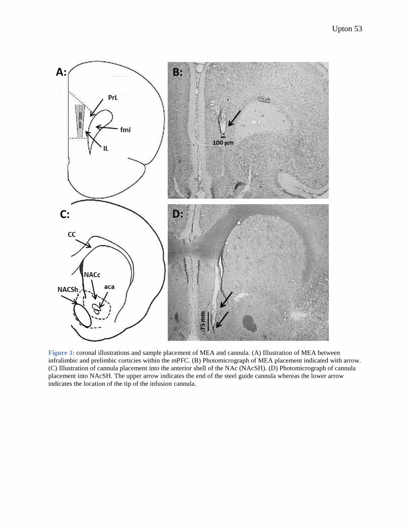
Upton 53
Figure 3: coronal illustrations and sample placement of MEA and cannula. (A) Illustration of MEA between
infralimbic and prelimbic corticies within the mPFC. (B) Photomicrograph of MEA placement indicated with arrow.
(C) Illustration of cannula placement into the anterior shell of the NAc (NAcSH). (D) Photomicrograph of cannula
placement into NAcSH. The upper arrow indicates the end of the steel guide cannula whereas the lower arrow
indicates the location of the tip of the infusion cannula.


Upton 55
Figure 4: sample tracing from a single animal receiving low-dose of NMDA stimulation after both vehicle and low-
dose AVL-3288. The top tracing (red) is from a glutamate-sensitive channel (GluOx) and the middle tracing (blue)
is from a sentinel channel. The bottom tracing is the self-referenced tracing obtained by subtracting the sentinel
channel from the GluOx channel. In this representative example, the response of 0.05µg NMDA during vehicle
treatment was potentiated in the presence of 1 mg/kg AVL-3288 from 2.83µM to 3.46µM glutamate, a percent
change of 22%.
Figure 5: sample self-referenced tracing from a single animal receiving high-dose of NMDA stimulation after both
vehicle and high-dose AVL-3288. 3 mg/kg AVL-3288 attenuated glutamate release from 9.44µM to 7.16µM
glutamate, a percent change of -24%.


Upton 57
Figure 6: AVL-3288 interacts with mesolimbic stimulation in evoked glutamate release. AVL-3288 data is
presented as the percent change from NMDA-evoked glutamate after vehicle treatment. Absolute increases of
glutamate during NMDA stimulation is reported in the inset graph. 1 mg/kg AVL-3288 significantly potentiated
0.05µg NMDA-evoked glutamate release. When pretreated with 3 mg/kg AVL-3288, 0.05µg NMDA-evoked
glutamate release was potentiated whereas 0.30µg NMDA-evoked glutamate release was attenuated.


Upton 59
Figure 7: sample self-referenced tracing from a single animal receiving low-dose of NMDA stimulation after both
vehicle and high-dose PNU-120596. 9 mg/kg AVL-3288 potentiated glutamate release from 1.72µM to 4.27µM
glutamate, a percent change of 148%.
Figure 8: PNU-120596 interacts with mesolimbic stimulation in evoked glutamate release. PNU-120596 data is
presented as the percent change from NMDA-evoked glutamate after vehicle treatment. Absolute increases of
glutamate during NMDA stimulation is reported in the inset graph. 9 mg/kg AVL-3288 significantly potentiated
0.05µg NMDA-evoked glutamate release. Glutamate release was not significantly potentiated or attenuated after
0.30µg NMDA stimulation at either dose of PNU-120596.