5 the biology and biochemistry of cellular receptors for
TRANSCRIPT
5 The Biology and Biochemistry of Cellular Receptors for Enveloped Viruses
5.1
5.2
5.3
504
5.5
KATHRYN V. HOLMES
Introduction
Characteristics of mammalian cell membranes 5.2.1 Structure 5.2.2 Chemical composition
Biology of receptors for enveloped viruses
Biochemical characterization of virus receptors 5.4.1 Aggregation of uninfected cells by virions or YAPs 5.4.2 Rosetting of uninfected cells with virus infected cells 504.3 Binding of virions or YAPs to cells
page
5.404 Binding of virions or YAPs to isolated plasma membranes 5.4.5 Binding of enveloped virions or YAPs to components
of solubilized plasma membranes 5.4.6 Binding of enveloped virions or YAPs to artificial
membranes
Summary
References
87 87 87 90
93
95 97 97 98
109
109
110
110
111
K. Lonberg-Holm et al. (eds.), Virus Receptors© Chapman and Hall 1981

Acknowledgements I thank Drs A. 1. Bednarz-Prashad, 1. N. Behnke, A. Smith, W. Brandt, 1. Richter, M. Dubois-Dalcq, and L. S. Sturman for providing data in advance of publication. I am grateful to the many investigators whose figures have been used to illustrate this article, to Drs L. S. Sturman, E. DoBer, R. K. Holmes, V. C. Hascall, and L. A. Caliguiri for their thoughtful comments on the manuscript and to Drs K. LonbergHolm and L. Philipson for excellent editorial advice.
Virus Receptors Part 2 (Receptors and Recognition, Series B, Volume 8) Edited by K. Lonberg-Holm and L. Philipson Published in 1981 by Chapman and Hall, 11 New Fetter Lane, London EC4P 4EE © Chapman and Hall

5.1 INTRODUCTION
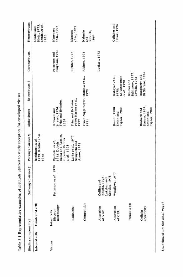
The plasma membrane of eukaryotic cells is a complex and delicate structure which has been the subject of intensive investigation. Diverse molecules on the plasma membrane which can serve as receptors for the many groups of enveloped viruses have been studied, but only a few receptors have been partially characterized biochemically. The purpose of this essay is to correlate current knowledge about the eukaryotic plasma membrane with the scattered data which are available on plasma membrane receptors for enveloped viruses. After reviewing the structural and biochemical properties of the plasma membrane which are of particular importance to the study of virus receptors, I will discuss the biological significance of some of the unique interactions which may occur between plasma membrane receptors and enveloped viruses, and review the studies designed to isolate and/or characterize these receptors. The limitations of current experimental methods explain our limited understanding of virus-receptor interactions. This review therefore emphasizes the advantages and disadvantages of different techniques for studying virus receptors. Rather than providing an encyclopedic review, only a few examples have been chosen to illustrate different approaches. Table 5.1 is a brief summary of the techniques that have been used to analyze receptors for different groups of enveloped viruses.
5.2 CHARACTERISTICS OF MAMMALIAN CELL MEMBRANES
5.2.1 Structure
Although the cell surface molecules which serve as receptors for some bacterial viruses have been identified (Bassford et at., 1977), the chemical composition of receptors for enveloped animal viruses is largely undetermined. Characterization of receptors for bacterial viruses was facilitated by detailed knowledge of the chemical composition of bacterial membranes and by the availability of cellular mutants which lack, or have defects in, specific membrane components (Lindberg, 1973; Picken and Beacham, 1977). In contrast, studies on animal virus receptors have been hampered both by limited information on the biochemical composition of the plasma membrane and by the lack of cellular mutations in specific membrane components. Since mammalian cells are diploid, genes coding for altered membrane components are probably incompletely expressed. The elegant studies on the identification of bacterial receptors can serve only in part as models for the analysis of receptors for animal viruses. Because of fundamental structural and functional
87
Tab
le 5
.1 R
epre
sent
ativ
e ex
ampl
es o
f m
etho
ds u
tili
zed
to s
tudy
rec
epto
rs f
or e
nvel
oped
vir
uses
Bin
ding
co
mp
on
en
tst
Infe
cted
cel
ls
Vir
ions
Un
infe
cted
cel
ls
Inta
ct c
ells
E
lect
ron
m
icro
sco
py
Rad
iola
bel
Co
mp
etit
ion
Alt
erat
ion
o
fVA
P
Alt
erat
ion
o
fCR
U
Pse
ud
oty
pes
Cel
lula
r sp
ecif
icit
y
(co
nti
nu
ed o
n th
e n
ext
page
)
Ort
ho
my
xov
irus
es:j
: P
aram
y xo
viru
ses ~
Ban
kh
urs
t et
aI.,
19
79
; R
enti
er e
t al
., 1
97
8
Pat
ters
on
et
al.,
19
79
D
jald
etti
et
al.,
19
78
; D
uboi
sD
alcq
an
d R
enti
er,
in p
ress
; R
enti
er
eta
l.,
19
78
Col
lins
an
d
Kni
ght,
19
78
; L
aksh
mi
and
S
chu
lze,
19
78
Was
sile
wa,
19
77
Lar
ke e
tal.
, 1
97
7
Sek
iguc
hi a
nd
A
sano
, 1
97
8
Alp
havi
ruse
s R
etro
viru
ses
§
Bir
dwel
l an
d
Str
auss
, 1
97
4;
Fri
es a
nd
Hel
eniu
s,
19
79
Fri
es a
nd
Hel
eniu
s,
19
79
; M
arke
r et
al.,
19
77
Co
ron
a vir
uses
Pat
ters
on
an
d
Bin
gh
am,
19
76
Ric
hte
r, 1
97
6
Fri
sch-
Nig
gem
eyer
, M
old
ow
et
al.,
Ric
hte
r, 1
97
6
1971
1
97
9
Bra
nd
t, 1
98
0
Sm
ith
an
d
Tig
no
r, 1
98
0
Bir
dwel
l an
d
Str
auss
, 1
97
4;
Sm
ith
an
d
Tig
no
r, 1
98
0
Bis
haye
e et
al.,
19
78
; K
aly
anar
aman
et
al.,
19
78
Bes
mer
an
d
Bal
tim
ore
, 1
97
7;
Zav
ada,
19
72
Do
ug
her
ty a
nd
D
i S
tefa
no
, 1
96
9 L
uck
ert,
197
2
Her
pesv
irus
es
Jon
dal
an
d
Kle
in,
19
73
, Jo
nd
al e
t al
., 1
97
6
Men
enze
s et
al.
, 1
97
6
Men
ezes
et
al.,
19
77
Nah
mia
s an
d
Kib
rick
, 1
96
4
Gal
lahe
r an
d
Ho
we,
19
76

Tab
le 5
.1
Rep
rese
ntat
ive
exam
ples
of
met
ho
ds
to s
tud
y r
ecep
tors
for
env
elop
ed v
irus
es (
co
nti
nu
ed
)
Bin
din
g c
om
po
nen
ts t
Vir
ion
s
Iso
late
d
VA
P
Inta
ct c
ells
G
enet
ics
Oth
er
Iso
late
d
pla
sma
mem
bra
nes
So
lub
iliz
ed
mem
bra
ne
com
po
nen
ts
Art
ific
ial
mem
bra
nes
Inta
ct c
ells
So
lub
iliz
ed
mem
bra
ne
com
po
nen
ts
art
ho
rny
xo
vir
use
s :t.
Ly
les
and
L
and
sber
ger
, 1
97
6
Mar
ches
i e
t a
l.,
19
72
Sh
aro
m e
t a
l.,
19
76
Par
amy
xo
vir
use
s ~
Fan
an
d S
efto
n,
19
78
; F
uch
s e
t a
l., 1
97
8;
Lyl
es a
nd
L
and
sber
ger
, 1
97
6
Hay
wo
od
, 1
97
5
and
19
78
Alp
hav
iru
ses
Hel
eniu
s e
t a
l.,
19
78
; Y
asu
i e
tal.
,19
71
Fri
es a
nd
H
elen
ius,
19
79
; H
elen
ius
et
al.
, 1
97
8;
Mo
on
ey
et
al.,
1
97
5;
Yas
ui
and
No
zim
a, 1
97
1
Ret
rov
iru
ses
~ C
oro
na
viru
ses
Oie
et
at.
, 1
97
8;
Ru
dd
le
et
al.
, 1
97
8
McG
rath
et
al.
, 1
97
8;
McG
rath
an
d W
eiss
man
, 1
97
9
Bis
hay
ee e
t a
l.,
19
78
; M
o1do
w
et
al.
, 1
97
7
Mo
ldo
w e
t a
l.,
19
77
a an
d b
Bis
hay
ee e
t a
l.,
19
78
; D
eLar
co
and
To
dar
o,
19
76
; F
ow
ler
eta
l.,1
97
9;
Mo
ldo
w e
t a
l.,
19
79
Kal
yan
aram
an
et
al.
, 1
97
8
Bis
wal
et
al.
, 1
96
6
Her
pes
vir
use
s
Men
ezes
et
al.
, 1
97
6
Nak
ajo
et
al.
, 1
97
9
Th
is t
able
has
bee
n c
on
stru
cted
to
ill
ust
rate
a v
arie
ty o
f ap
pro
ach
es t
hat
hav
e b
een
use
d t
o s
tud
y r
ecep
tors
fo
r en
vel
op
ed v
irus
es.
Fo
r m
ost
gro
up
s o
f en
vel
op
ed v
iru
ses
on
ly a
lim
ited
nu
mb
er o
f th
ese
app
roac
hes
hav
e b
een
use
d.
t In
eac
h e
xam
ple
th
e v
iru
s at
tach
men
t p
rote
in (
VA
P)
is p
rese
nt
in t
he
form
lis
ted
in
the
firs
t co
lum
n a
nd
th
e ce
llu
lar
rece
pto
r u
nit
(C
RU
) is
pre
sen
t in
th
e fo
rm l
iste
d i
n th
e se
con
d c
olu
mn
. :t.
See
Ch
apte
r 4
~ S
ee e
ha p
ter
3 ~
See
Ch
apte
r 1
0

90 Virns Receptors - Animal Virnses
differences between the membranes of prokaryotic and eukaryotic cells there may be profound differences in the biology and biochemistry of their interactions with viruses.
The plasma membranes of mammalian cells are specialized organelles which perform many functions essential for interaction with the micro-environment. The flexible and fluid membranes are constantly in motion. Structural specializations of cell membranes including microvilli, pinocytic ruffles, pinocytic vesicles, coated pits, desmosomes, tight junctions, and gap junctions form and disappear in response to complex regulatory mechanisms (Morre et al., 1979). Regions of the plasma membrane may be highly specialized for particular cellular functions such as attachment, secretion, and adsorption. Movements of the different molecular species in the plasma membrane are being analyzed in many laboratories (Gershon, 1978; Singer et al., 1978; Wolfe et aI., 1979). In metabolically active cells new molecules are inserted into the plasma membrane while other membrane molecules and fragments of membranes may be destroyed or shed into the medium (Morre et aI., 1979). Although the structure and composition of the plasma membrane undergo major changes during development, during the cell cycle, and in response to cellular injury, these changes are all directed by the genome of the cell.
5.2.2 Chemical composition
Molecules on the cell surface include structural components, enzymes, cellular recognition factors, and receptors for hormones, neurotransmitters, and regulatory molecules. These membrane components are composed of simple or complex lipids, proteins, or proteoglycans. The general organization of these components in the membrane has been discussed in detail elsewhere (Borstein et al., 1978; Singer, 1974; Singer et al., 1978) and will only be summarized here.
(a) Proteoglycans Proteoglycarts are macromolecules associated with the plasma membrane of animal cells and with the ground substance of connective tissues. Proteoglycans consist of a core protein covalently bonded to one or more chains of a glycosaminoglycan such as chondroitin sulfate, keratin sulfate, heparin sulfate or heparin (Hascall et al., 1976; Perkins et al., 1979). These glycosaminoglycans are po1yanionic polysaccharide chains of repeating disaccharide units which contain a hexosamine and sulfate esters and/or carboxylate groups. The most thoroughly studied proteoglycan is that of cartilage (Hascall, 1977; Hascall and Heinegard, 1980) which has molecular weights above 106 daltons. Approximately 100 chains of chondroitin sulfate and 30 to 60 chains of keratin sulfate are covalently bonded to a core protein with a molecular weight of 200 000. The core protein is noncovalently bound to a small link protein and
.to a strand of hyaluronic acid, a glycosaminoglycan (Faltz et al., 1978). Proteoglycans from different sources may differ in structure, but are all very large molecules which tend to aggregate. The negative charge of cells and their Ca2+ binding activity
Cellular Receptors for Enveloped Viruses 91
are due in part to the aggregates of polyanionic sulfated proteoglycans associated with the plasma membrane (Vannucchi et al., 1978). The extent to which proteoglycans and glycosaminoglycans can affect membrane binding phenomena such as cellcell recognition and virus-cell interactions is undetermined. Although it first appeared that there were too few different proteoglycans to account for diverse and specific membrane interactions. a variety of proteoglycans has now been distinguished (Glimeliusetal., 1978; Vogel and Dolde, 1979; Ward and Packham, 1979). These molecules may participate in cell membrane receptor interactions via either the carbohydrate or protein moiety. Although interactions of enveloped viruses with glycosaminoglycans or proteoglycans have received little attention, it is clear that they do occur. For example, the binding of the glycosaminoglycan heparin to herpes simplex virions prevents infection of susceptible cells (Nahmias and Kibrick, 1964: Takemoto and Fabisch, 1964), and preformed cellular proteoglycans can become associated with newly synthesized enveloped viruses such as orthomyxoviruses (Com pans and Pinter, 1975). paramyxoviruses (Pinter and Compans, 1975) and corona viruses (Sturman, 1980).
An intriguing hypothesis is that virion-associated proteoglycans could spontaneously aggregate with similar molecules on the cell membrane. This could augment other virion-membrane interactions and might be one mechanism of host controlled modification of virions. As new techniques for the study of the synthesis and transport of proteoglycans in cell cultures are developed (Glimelius et aI., 1978; Kimura et al., 1978: Yanagishita and Hascall, 1980), they can be applied to the analysis of the role of proteoglycans in virus attachment.
(b) Lipids The lipid composition of the plasma membrane is described in Chapter 7. Although much of the lipid bilayer may be masked by glycoproteins or other molecules on the surface of the plasma membrane, there may be regions where the lipids are exposed and could be utilized for binding of enveloped virions. Because the lipid components of the plasma membrane of many cell types are similar, enveloped viruses which bind to specific lipid receptors might be expected to exhibit a broad host range.
Glycolipids from different cell types may be distinguished by their carbohydrate moieties. However, on a single cell type the same carbohydrate side chains can be carried on glycoproteins as well as on glycolipids. Therefore, if virus receptor activity were associated only with the carbohydrate moiety, then efforts to purify a single macromolecular virus receptor molecule might fail.
(c) Glycoproteins Recent studies on the synthesis, glycosylation and intracellular transport of membrane glycoproteins have used as paradigms the synthesis of viral glycoproteins in infected cells (Lenard, 1978: Rothman et al., 1978) as well as other models (Lingappa et al., 1978; Turco and Robbins, 1978; Wolfe et aI., 1979). Following polypeptide synthesis in the rough endoplasmic reticulum and a series of glycosylation steps at

92 Virus Receptors - Animal Viruses
this site and in the Golgi apparatus, glycoproteins are inserted into the plasma membrane. Glycoproteins on the cell surface are heterogeneous because glycosylation is not always complete and because they may be further modified by proteolytic cleavage. Under normal growth conditions there is significant loss of glycoproteins from the membrane. Some may be shed from the membrane in large amounts (Vitetta et al., 1974), others may be released from the cell by exocytosis on small vesicles pinched off from the membrane, and still others may re-enter the cell by endocytosis to be recycled or destroyed (Loor et al., 1972). In addition, some of the glycoproteins released from the cell may bind to the membranes of other cells. These dynamic processes may affect efforts to identify or isolate a glycoprotein which functions as a virus receptor.
The amount of a single glycoprotein species in the membrane of a eukaryotic cell is usually limited. This is due both to the low ratio of surface area to volume in eukaryotic cells and to the large number of different glycoproteins expressed on the surface of a single cell type. Thus, although a glycoprotein may be detectable on the cell surface by sensitive immunological techniques, it may be difficult to isolate in sufficient quantity for biochemical characterization. A few glycoproteins present in large amounts in cell membranes have been isolated and characterized (Hughes and Nairn, 1978; Marchesietal., 1976; Nakajo etal., 1979, Yamada and Olden, 1978), and new methods have been devised for the analysis of glycoproteins present in limited amounts (Kulczycki et al., 1979; Lotan and Nicolson, 1979; Vitetta et al., 1977).
The identification of a cell surface glycoprotein as a receptor unit for an enveloped virus may not define the specificity of virus attachment. Because membrane glycoproteins may be large molecules with multiple sites for the attachment of different carbohydrate side chains, the same glycoprotein could have several different binding sites, each specific for one virus. The different sites on the glycoprotein could also have different affinities for the same virus and/or require different ionic conditions for binding. The situation may be even more complex if the specific receptor determinant is on a carbohydrate side chain which is also present on a different polypeptide or a glycolipid (Haywood, 1978). Oearly biochemical studies on the determinants of the receptors for enveloped viruses will require identification of the specific portions of the molecule involved in binding. This could be approached by using monoclonal antibodies directed against cell surface antigens or by cross-linking the receptor moiety to the virus attachment protein (V AP) with a cleavable crosslinking agent.
Identification of new components of the plasma membrane is potentially of great significance to the study of receptors for enveloped viruses. Conversely, identification of cell surface molecules by means of their capacity to bind viruses can lead to characterization of cell surface molecules not detected by other methods. The normal functions of such cell surface molecules might then be determined. In addition, virus binding could be used as a marker for cells which might be otherwise indistinguishable from closely related cell types.
Cellular Receptors for Enveloped Viruses 93
5.3 BIOLOGY OF RECEPTORS FOR ENVELOPED VIRUSES
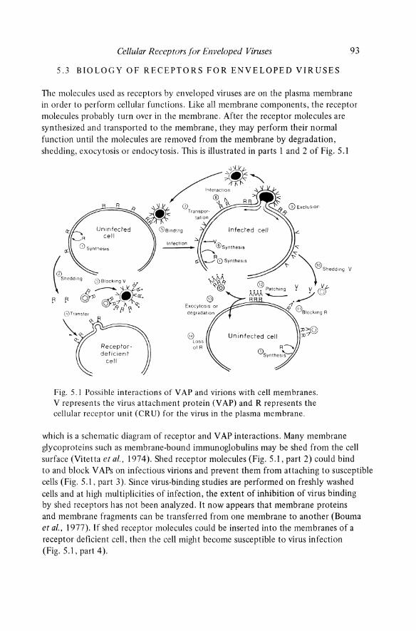
The molecules used as receptors by enveloped viruses are on the plasma membrane in order to perform cellular functions. Like all membrane components, the receptor molecules probably turn over in the membrane. After the receptor molecules are synthesized and transported to the membrane, they may perform their normal function until the molecules are removed from the membrane by degradation, shedding, exocytosis or endocytosis. TIlis is illustrated in parts 1 and 2 of Fig. 5.1
8 Loss of R
Fig. 5.1 Possible interactions of VAP and virions with cell membranes. V represents the virus attachment protein (V AP) and R represents the cellular receptor unit (CRU) for the virus in the plasma membrane.
which is a schematic diagram of receptor and V AP interactions. Many membrane glycoproteins such as membrane-bound immunoglobulins may be shed from the cell surface (Vitetta et at., 1974). Shed receptor molecules (Fig. 5.1, part 2) could bind to and block V APs on infectious virions and prevent them from attaching to susceptible cells (Fig. 5.1, part 3). Since virus-binding studies are performed on freshly washed cells and at high multiplicities of infection, the extent of inhibition of virus binding by shed receptors has not been analyzed. It now appears that membrane proteins and membrane fragments can be transferred from one membrane to another (Bouma et aI., 1977). If shed receptor molecules could be inserted into the membranes of a receptor deficient cell, then the cell might become susceptible to virus infection (Fig. 5.1, part 4).
94 Virus Receptors - Animal Viruses
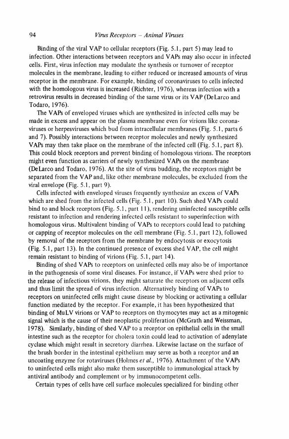
Binding of the viral V AP to cellular receptors (Fig. 5.1, part 5) may lead to infection. Other interactions between receptors and V APs may also occur in infected cells. First, virus infection may modulate the synthesis or turnover of receptor molecules in the membrane, leading to either reduced or increased amounts of virus receptor in the membrane. For example, binding of coronaviruses to cells infected with the homologous virus is increased (Richter, 1976), whereas infection with a retrovirus results in decreased binding of the same virus or its V AP (DeLarco and Todaro, 1976).
The V APs of enveloped viruses which are synthesized in infected cells may be made in excess and appear on the plasma membrane even for virions like coronaviruses or herpesviruses which bud from intracellular membranes (Fig. 5.1, parts 6 and 7). Possibly interactions between receptor molecules and newly synthesized V APs may then take place on the membrane of the infected cell (Fig. 5.1, part 8). This could block receptors and prevent binding of homologous virions. The receptors might even function as carriers of newly synthesized V APs on the membrane (DeLarco and Todaro, 1976). At the site of virus budding, the receptors might be separated from the V AP and, like other membrane molecules, be excluded from the viral envelope (Fig. 5.1, part 9).
Cells infected with enveloped viruses frequently synthesize an excess of V APs which are shed from the infected cells (Fig. 5.1, part 10). Such shed V APs could bind to and block receptors (Fig. 5.1, part 11), rendering uninfected susceptible cells resistant to infection and rendering infected cells resistant to superinfection with homologous virus. Multivalent binding of V APs to receptors could lead to patching or capping of receptor molecules on the cell membrane (Fig. 5.1, part 12), followed by removal of the receptors from the membrane by endocytosis or exocytosis (Fig. 5.1, part 13). In the continued presence of excess shed V AP, the cell might remain resistant to binding of virions (Fig. 5.1, part 14).
Binding of shed V APs to receptors on uninfected cells may also be of importance in the pathogenesis of some viral diseases. For instance, if V APs were shed prior to the release of infectious virions, they might saturate the receptors on adjacent cells and thus limit the spread of virus infection. Alternatively binding of V APs to receptors on uninfected cells might cause disease by blocking or activating a cellular function mediated by the receptor. For example, it has been hypothesized that binding of MuLV virions or YAP to receptors on thymocytes may act as a mitogenic signal which is the cause of their neoplastic proliferation (McGrath and Weissman, 1978) .. Similarly, binding of shed V AP to a receptor on epithelial cells in the small intestine such as the receptor for cholera toxin could lead to activation of adenylate cyclase which might result in secretory diarrhea. Likewise lactase on the surface of the brush border in the intestinal epithelium may serve as both a receptor and an uncoating enzyme for rotaviruses (Holmes et aI., 1976). Attachment of the YAPs to uninfected cells might also make them susceptible to immunological attack by antiviral antibody and complement or by immunocompetent cells.
Certain types of cells have cell surface molecules specialized for binding other

Cellular Receptors for Enveloped Viruses 95
molecules. These receptors could also serve as virus receptors. For instance, hepatocytes and macrophages (Stahl et al., 1978) have cell surface receptors for circulating plasma glycoproteins. These receptors recognize the terminal and subterminal sugars in the carbohydrate moiety of the glycoprotein. If a V AP shared the same carbohydrate determinants, it would bind to the cell via the cellular receptor for glycoproteins. Although this binding might not lead to infection, this illustrates how specific tissue tropisms of viruses could be determined by the distribution of receptors for normal host molecules.
On B lymphocytes which are capable of secreting antibody directed against a particular YAP, anti-YAP expressed on the membrane may serve as receptor. Virions which bind to this specific antibody on the membrane might stimulate the lymphocyte and/or infect it. A virus which could bind tD and kill B lymphocytes capable of making anti-V AP antibody might prevent the formation of an adequate titer of neutralizing antibody which in turn might lead to persistent infection or lack of protection against reinfection.
Many types of cells including macrophages have a cell surface receptor for the Fc portion of IgG that is bound to an antigen (Bourgois et aI., 1977). Such cells could bind virions which were complexed with low levels of antibody and infection could be initiated. Dengue virus complexed with small amounts of antibody can initiate infection via the Fc receptor on monocytes. When human monocytes are treated with trypsin, specific receptors for dengue virus are removed but cellular F c receptors are retained. Such trypsin-treated cells are more susceptible to infection with dengue virus complexed with a small amount of antibody than to the virus itself (Brandt, 1979). This observation may have important implications for the pathogenesis of the dengue shock syndrome (Halstead and O'Rourke, 1977).
Many of these speculations on the biology of receptors will only be testable when we know more about the chemical composition of receptors.
5.4 BIOCHEMICAL CHARACTERIZA nON OF VIR US RECEPTORS
Attempts have been made to identify the molecule(s) in the cell membrane which serve as receptors for enveloped viruses and to determine the mechanism of the initial interaction of virions with cells. In analyzing these results it should be remembered that cellular receptor units (CRU) may be complexes of several molecules rather than single molecules. The determinant for a CRU may be found on several different membrane components. Initial binding of virions to cells by means of a CRU does not necessarily lead to virus infection, as replication may be blocked at penetration or subsequent steps. TIle same virus may bind to different cell types by means of different CRUs. For example, a hemagglutinating virus may bind to a component of the erythrocyte membrane not present on the membranes of fibroblasts which support virus replication. ~imilarly, although Sindbis virus can replicate in both

Tab
le 5
.2 E
ffec
t o
f en
zym
e tr
eatm
ent
of c
ells
on
atta
chm
ent
of
coro
navi
ruse
s an
d al
phav
irus
es.
Fig
ures
sho
w p
erce
ntag
e o
f co
ntro
l bi
ndin
g o
f gr
adie
nt-p
urif
ied
radi
olab
eled
vir
ions
to
sha
m-t
reat
ed c
ells
Vir
us
Mou
se h
epat
itis
vir
us (
A59
str
ain:
at
tach
men
t in
1 h
, 37
°C)*
Sin
dbis
vir
us (
avir
ulen
t st
rain
EgA
r 33
9;
atta
chm
ent
in 3
h,
4°C
)t
Cel
l ty
pe
L2
mur
ine
fibr
obla
sts
Aed
es
albo
pict
us
mos
quit
o ce
lls
CE
R
non-
neur
al
Sin
dbis
vir
us (
neur
ovir
ulen
t st
rain
SaA
r 86
; A
edes
at
tach
men
t in
3 h
, 4°
C)t
al
bopi
ctus
* R
icht
er (
1976
) t
Sm
ith
and
Tig
nor
(198
0)
mos
quit
o ce
lls
CE
R
non-
neur
al
Try
psin
P
rona
se
62
35
100
100
lOa
a
100
lOa
93
57
Enz
yme
Bro
mel
ain
Pro
tein
ase
K
Neu
ram
inid
ase
28
N.D
. 98
N.D
. lO
a 10
0
N.D
. 11
54
N.D
. 74
10
0
N.D
. 67
80

Cellular Receptors for Enveloped Viruses 97
mammalian and mosquito cell lines, the receptors on these cells differ in susceptibility to proteolytic enzymes (Table 5.2; Smith and Tignor, 1980).
The studies on characterization of receptors for enveloped viruses are discussed according to the strategies used to analyze the receptors. Table 5.1 is not comprehensive but has been included to provide an overview of the technology involved. Not all approaches have been used for each group of enveloped viruses. Indeed, several groups have been omitted since the nature of their receptor is uncertain. Because each method has advantages and limitations, it may be necessary to study a virus receptor by several techniques to characterize it adequately.
Studies on binding of virions or isolated V APs have usually led to similar conclusions, with a few notable exceptions. The isolation and characterization of V APs are discussed in Chapter 3.
5.4.1 Aggregation of uninfected cells by virions or V APs
Hemagglutination is the classic example of this type of virus-cell interaction (Lonberg-Holm and Philipson, 1974). Viruses which can bind to receptors on erythrocyte membranes can cause the cells to agglutinate. The erythrocytes may be from a species in which the virus cannot replicate, and the receptors on erythrocyte and host cells may differ biochemically. Nevertheless, the hemagglutination technique was used to demonstrate that orthomyxoviruses and paramyxoviruses bind to receptors which contain N-acetyl neuraminic acid (NANA) and that the enzyme neuraminidase on the viral envelope could cleave NANA from the receptor and inactivate the receptor (Chapter 4).
Cells other than erythrocytes can also be agglutinated by enveloped viruses. The paramyxovirus NDV can agglutinate rat thoracic duct lymphocytes and this agglutination is also prevented by prior treatment with neuraminidase (Woodruff and Woodruff, 1972). NDV can also aggregate platelets (Larke et al., 1977). These effects of virus binding may be significant in the pathogenesis of myxovirus infection (Rott, 1979). Cultured cells may also be aggregated by enveloped viruses. Although respiratory syncytial virus does not hemagglutinate, it will aggregate HeLa cells in spinner culture (Holmes and Gerin, unpublished observation).
5.4.2 Rosetting of uninfected cells with virus infected cells
The discovery that receptors for Epstein Barr (EB) virus are present on human B lymphocytes but not T lymphocytes was made simply by allowing lymphocytes to attach to chronically infected cells (Jondal and Klein, 1973). Since B lymphocytes adhered to viral antigens on the infected cells, purification of virions was not required to demonstrate the receptor activity of B cells. This method provides a rapid identification of cell types carrying receptors. Hemadsorption of erythrocytes to cultures of virus infected cells is a long recognized variation of this technique (Rentier et at., 1978).

98 Virus Receptors - Animal Viruses
The sensitivity of the rosetting technique depends upon the many V AP-containing regions in the membrane of the infected cell which bind to the receptors on the uninfected cell more firmly than single virions as in agglutination reactions. It is essential to demonstrate that the uninfected cells are adhering to the viral attachment polypeptide (V AP) on the infected cells. Antibodies against V AP should inhibit the binding in the same way as the rosetting of human peripheral blood lymphocytes with measles-infected cells is inhibited by anti-measles antibody (Bankhurst et al., 1979).
The rosetting technique is useful to demonstrate the presence of receptor sites, and inhibition of rosetting by chemicals, enzymes, extracts of solubilized membranes (Bankhurst et al., 1979), or other viruses can provide information about the chemical composition of the receptor.
5.4.3 Binding of virions or V APs to cells
Most studies of virus receptors have used variations of this method. A criticism common to all of these studies is that purified preparations of enveloped viruses contain non-infectious virions which may interact differently with the cell than do infectious virions. For enveloped viruses the ratio of non-infectious to infectious virions may vary from> 100: 1 to 1: 1, but it is assumed that non-infectious virions bind to receptors in the same way as infectious virions. Furthermore, some of these methods require high virion/cell ratios, in which case virus attachment may not be the same as at low multiplicities. Therefore it is important to characterize the virion preparation with regard to uniformity of virion structure and ratio of physical particles to infectious units.
(a) Methods used to detect binding of virions or VAPs (i) Electron microscopy. Binding of enveloped virions to cells has been detected by several electron microscopic techniques (Figs. 5.2 and 5.3). With transmission electron microscopy (TEM), high r.lUltiplicities are used to increase the probability of finding in a thin section more than one virion attached to the cell membrane. Such studies revealed that initial attachment was by means of the viral peplomers. Bound virions have been observed on the smooth cell membrane and on microvilli projecting from it (Fries and Helenius, 1979; Menezes et at., 1977; Patterson et al., 1979). TEM showed that rhabdoviruses may attach to specialized areas in the plasma membrane which develop into coated pits (Fig. 5.2a) (Simpson et al., 1969). Alphaviruses (Fries and Helenius, 1979) and orthomyxoviruses (Patterson et al., 1979) also attach to coated pits. Negative staining has been used to show binding of enveloped virions to artificial membranes such as liposomes (Fig. 5.2b) (Haywood, 1975 and 1978). Freeze etch studies show changes in the distribution of intramembranous particles (IMP) within the plasma membrane after virus binding (Fig. 5.2c) (Bachiet al., 1973; Sekiguchi and Asano, 1978; Sharom et al., 1976). These may result from migration of multiple CRUs under a multivalent virion to

Cellular Receptors for Enveloped Viruses 99
form a cellular receptor site (CRS). Rapid freezing of cells with adsorbed virions, followed by sublimation of ice and preparation of a surface replica, can be used to
show virions adsorbed to membranes (Fig. S.2d) (Bachi et al., 1973). The pseudoreplica technique has yielded a surface view of the binding of very large numbers of togaviruses to the plasma membrane (Fig. S.2e and f) (Birdwell and Strauss, 1974) and demonstrated fusion of measles virions with the cell membrane (Fig. S.3a) (Dubois-Dalcq and Rentier, 1980). Most ultrastructural studies do not allow quantitation of the number of receptor sites per cell. Although scanning electron microscopy (SEM) permits examination of the entire upper surface of the cell, it has not yet been exploited for virus receptor studies (Djaldetti et al., 1978). SEM can demonstrate penetration of virions during binding studies. When L2 cells in spinner culture were saturated with a coronavirus at 37°C, few virions remained on the plasma membrane but large ruffles of membrane were induced during virus penetration (Fig. S.3b) (Richter, 1976). Fig. S.3(c) and (d) show that the distribution of virions adsorbed to the surface of infected cells can easily be analyzed by SEM
(Richter, 1976).
Oi) Infectivity. Infectivity can be used to detect virus binding in several ways. Frequently virus binding is assayed by loss of infectivity from the inoculum. However, inactivation during virus elution may complicate interpretation of these studies. The presence of a receptor may be inferred when synthesis of viral components or virions can be detected (Dougherty and DiStefano, 1969), although the presence of a receptor on a cell membrane does not guarantee that all subsequent steps in virus replication will be carried out. Infectivity may be used to demonstrate that two viruses do not share the same receptor. If cells infected with one virus show homologous interference but not interference with a different virus, then the virions may utilize different receptors on the cell membrane.
An ingenious application of infectivity assays for the presence of receptors makes use of the formation of pseudotypes during mixed infection with two different viruses. nle rhabdovirus VSV can attach to, and cause productive lytic infection in many different vertebrate cells. [n contrast, the moderate replication of retroviruses is more difficult to detect. During mixed infection, pseudotype virions are formed which have the VSV envelope and the retrovirus genome, or the retrovirus envelope and the VSV genome (Besmer and Baltimore, 1977). Virions with the VSV envelope can be eliminated by treatment with anti-VSV antibody or
by heat treatment of virions produced by a VSV mutant with a temperature-sensitive defect in the virus attachment protein G (Zavada, 1972). Receptors for the retrovirus V AP can then be assayed by the ability of pseudotype virions with the retrovirus envelope and the VSV genome to form VSV plaques on cell monolayers (Besmer and Baltimore, 1977). In this way it was shown that the N-B tropism of MuLV coded for by the Fv-I gene of the mouse is not expressed at the level of the receptor on the plasma membrane (Chapter 10). Similarly, pseudotypes with envelopes from a leukemia virus and genomes from a sarcoma virus can be identified


Cellular Receptors for Enveloped Viruses 101
Fig. 5.2 Binding of enveloped virions to membranes. (a) Transmission electron microscopy of a thin section of an L cell showing
adsorption of a vesicular stomatitis virion. After 5 min at 37°C, the VSV virion is attached to a coated pit on the plasma membrane. The virions enter the cell by viropexis at these specialized membrane sites. (x 119 000). Reproduced with permission, R.W. Simpson, Virology, and Academic Press (Simpson et aI., 1969).
(b) Interaction of a Sendai virion with a liposome composed of phosphatidylcholine, cholesterol and gangliosides. V APs on the viral envelope bind to the liposome at 4°C, and at 37°C the liposome flows around the virion in a process resembling phagocytosis (x 109 000). Reproduced with permission, A. Haywood, The Journal of General Virology, and Cambridge University Press from the article: Phagocy tosis of Sendai Virus by Model Membranes (Haywood, 1975).
(c) Freeze-etch preparation of the membranes of two human erythrocytes agglutinated by Sendai virus. The inner fracture face of the overlying cell has been broken away to reveal the outer fracture face of the lower cell. After 10 min at 37° C, intra membrane particles had clustered together and some areas which might represent fusion of the membranes were observed (arrow) (x 50000). Reproduced with permission, C. Howe, The Journal of Virology and the American Society for Microbiology (Bachi et al., 1973).
(d) Sendai virions adsorbed to the surface of an erythrocyte at 4°C followed by reaction with ferritin labeled anti-viral antibody. In this surface replica of freeze-etched cells, ferritin antibody particles can be seen on the virion envelope but not on the external surface of the cell (x 37 400). Reproduced with permission, C. Howe, The Journal of Virology,and the American Society for Microbiology (Bachi et al., 1973).
(e) Surface replica showing Sindbis virions adsorbed at 4°C to the surface of unfixed chicken embryo fibroblasts. The clustering of virions shown here and in Fig. 5.2(f) did not occur when virions were adsorbed to prefixed cells, suggesting that virus receptors can diffuse laterally in the unfixed plasma membrane even at 4°C (x 13 300). Reproduced with permission. 1. Strauss, Tile Journal of Virology, and The American Society for Microbiology (Birdwell and Strauss, 1974).
(f) Sindbis virions adsorbed at 4°C to the surface of unfixed chicken embryo fibroblasts. A surface replica of two overlapping cells A 'and B shows numerous spherical virions adsorbed to the plasma membranes. These images suggest that there are> 5 x 10⁵ Sindbis receptor sites per cell (x 8700). Reproduced with permission, 1. Strauss, The Journal of Virology, and The American Society for Microbiology (Birdwell and Strauss, 1974).


Cellular Recepturs fur Enveluped Viruses
by their ability to transform cells. Thus, a transformation assay could utilize viral pseudotypes to study membrane receptors and host ranges for leukemia viruses (Chapter 10).
103
(iii) Radiolabel. Purified, radioactively labeled virions or V APs have been used to
study binding of enveloped viruses to cells. Attachment of virions or V APs can be followed both by loss of label from the inoculum and by binding of label to the cells. Studies of the binding of radiolabeled enveloped virions to cells have sometimes shown that the percent of label which binds to cells is much less than the percent of infectivity lost from the inoculun} (Richter, 1976). TIlis suggests that some labeled virions either do not bind and are not infectious or elute rapidly from cells and cannot re-attach, while the labeled virions which do bind to cells include the infectious virions.
TIle peplomeric glycoproteins which are the V APs have been isolated from many viruses including orthomyxoviruses (Collins and Knight, 1978), paramyxoviruses (Nagi et at., 1976; Scheid and Choppin, 1974), rhabdoviruses (Kelley et al., 1972), coronaviruses (Sturman et at., 1980), alphaviruses (Helenius and Soderlund, 1973; Simons et al., 1973), and retroviruses (Strand and August, 1976). It is important to determine the physical state of the isolated V APs since V APs isolated by detergent
Fig. 5.3 Binding of enveloped virions to membranes. (a) Measles virions (arrows) fusing with the plasma membrane of Vero
cells, 7 min after virus inoculation. This is a surface replica of cells dried by the critical point technique. The viral envelope is recognized by the presence of ridges which are peplomers assembled over the helical nucleocapsid. Although most of the cell surface is covered with small particles, fewer particles are present at the site of viral entry (x 36 900). Courtesy of Dr Monique Dubois-Dalcq.
(b) L2 cell from spinner culture after saturation with coronavirions at 37°C. Cells were dried by the critical point technique and examined by scanning electron microscopy. Few virions can be identified among the numerous microvilli on the cell surface. Large ruffles of membrane were induced by virus attachment and may be associated with viral penetration (x 6200). Courtesy of Dr J. M. Richter.
(c) Membrane of an uninfected L2 cell from monolayer culture. Scanning electron microscopy shows a moderate number of microvilli on the cell membrane (x 9100). Courtesy of Dr J. M. Richter. (Richter, 1976).
(d) Coronavirions adsorbed to the membrane of an L2 cell 9 hours after infection at a multiplicity of infection of 3 PFU per cell. Scanning electron microscopy shows numerous virions (arrows) attached to the plasma membrane and to microvilli. TEM studies show that the virions bud into the rough endoplasmic reticulum. After release by exocytosis, numerous virions readsorb to the membrane of infected cells (x 9100). Courtesy of Dr l.M. Richter
(Richter, 1976).
104
20 (a)
0
.. -'0 5
(c) " E a.
4 u
c 0
'.c: 3 u
~ ~ 2 ~ ':;; ;:: u <1!
,2 'U
<1! tr
0
Virus Receptors - Animal Virnses
20 40 60 Minutes
20 60 Fraction number
E a. ~ 2 ., S
" L
:J , I
'"
100
90
_ 80 0? ~ 70 'U § 60
..8 50
(b)
4 6 8 10 12 Fraction number
(d)
5 10 15 Liposomes (J-Il)
14
20
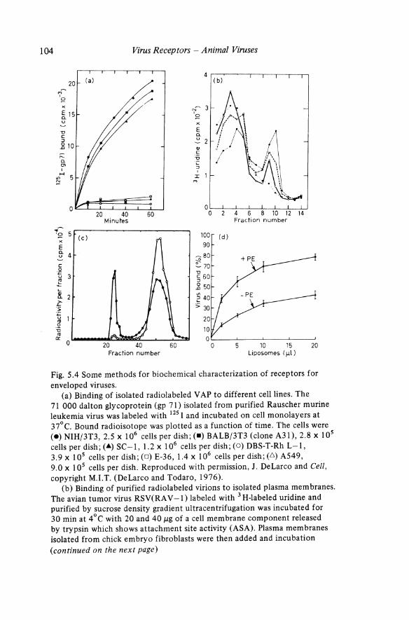
Fig. 5.4 Some methods for biochemical characterization of receptors for enveloped viruses.
(a) Binding of isolated radiolabeled V AP to different cell lines. The 71 000 dalton glycoprotein (gp 71) isolated from purified Rauscher murine leukemia virus was labeled with 125 I and incubated on cell monolayers at 37°C. Bound radioisotope was plotted as a function of time. The cells were (e) NIH/3T3, 2.5 x 10⁶ cells per dish; (.) BALB/3T3 (clone A31), 2.8 x 10⁵ cellsperdish;(.&)SC-l, 1.2 x 10⁶ cellsperdish;(O)DBS-T-RhL-I, 3.9 x 10⁵ cells per dish; (0) E-36, 1.4 x 10⁶ cells per dish; (.6) A549, 9.0 x 10⁵ cells per dish. Reproduced with permission, J. DeLarco and Cell, copyright M.LT. (DeLarco and Todaro, 1976).
(b) Binding of purified radiolabeled virions to isolated plasma membranes. The avian tumor virus RSV(RA V -1) labeled with 3 H-labeled uridine and purified by sucrose density gradient ultracentrifugation was incubated for 30 min at 4°C with 20 and 40 J,1g of a cell membrane component released by trypsin which shows attachment site activity (ASA). Plasma membranes isolated from chick embryo fibroblasts were then added and incubation (continued on the next page)

Cellular Receptors for Enveloped Viruses 105
solubilization and purified by density gradient centrifugation or column chromatography may be in the form of micelles or rosettes rather than monomeric glycoproteins (Helenius and von Bonsdorff, 1976; Helenius and Soderlund, 1973; Lenard, 1978; Simons et al., 1973 and 1978; Sturman et al., 1980). Binding of isolated, radiolabeled retrovirus V APs has been used as an indicator of the presence of receptors for the virus (Fig. 5.4a) (DeLarco and Todaro, 1976; Moldow et al., 1979). Apparently the V APs bind to the receptors via the tips of the peplomers rather than via their membrane-anchored ends. However, inactivation of V AP may occur during radiolabeling and purification since < 10% of the purified, labeled glycoproteins attach to susceptible cells (Moldow et al., 1979).
Fig. 5.4 (continued) continued for 30 min at 4°C. Virus binding to membranes was assayed by sedimentation on discontinuous sucrose gradients. (e-e) virus + CEF membranes; (0--0) virus + 20 /lg soluble attachment site; (0-0) virus + 40 /lg soluble attachment site; (e- -e) virus only. Reproduced with permission C. F. Moldow, The Journal of General Virology and Cambridge University Press from the article: A vian Tumour Virus Interactions with Chicken Fibroblast Membranes: Partial Characterization of Initial Attachment Site Activity. (Moldow et al., 1977b).
(c) Binding of isolated radiolabeled YAP to fractionated cell membrane. Membrane factions were prepared from homogenized Kirsten murine sarcoma virus-transformed murine cells (KA 31). Microsomal membranes which pelleted at 100000 g were incubated with 125 I-labeled gp 70 isolated from Rauscher murine leukemia virus. The membrane-receptor complex was precipitated with polyethylene glycol and chromatographed on a column of Sepharose CL-6B (Pharmacia). Samples were run without (e) and with (0) excess unlabeled gp 70 as a control. The peak at the void volume in fraction 23 to 27 represented 125 I-label in gp 70 bound to membranes and the peak at fractions 44 to 60 represented free 125 I-labeled gp 70. Reproduced with permission from V.S. Kalyanaraman, The Journal of Virology and the American Society for Microbiology. (Kalyanaraman et al., 1978).
(d) Binding of purified radiolabeled virions to liposomes. Sindbis virus labeled with a mixture of 3 H-Iabeled amino acids was purified by ammonium sulfate precipitation and sucrose density gradient ultracentrifugation, and incubated at 23°C with different quantities of liposomes containing one of the following molar mixtures: dipalmitolphosphatidylethanolamine-sphingomyelinphosphatidylserine-cholesterol (I: I :0.2: 1.5) (+ PE) or sphingomyelinphosphatidylserine-cholesterol (2: 0.22: 1.5) (-PE). The entire mixture was ultra centrifuged on a discontinuous gradient which was frozen and cu t into sections. The per cent virus bound was the radiolabel in the liposomal fraction gradient divided by the total radioactivity in the gradient, x 100. Reproduced with permission] .M. Dalrymple, The Journal of Virology, and the American Society for Microbiology. (Mooney et al., 1975).
106 Virus Receptors - AnimaZ Viruses
(iv) Other methods used to detect binding of virions or VAPs to cells. A variety of other direct and indirect techniques have been used to detect binding of virions to cell surface receptors. Binding of rhodamine- or fluorescein-labeled MuLV virions to thymic lymphoma cells could be detected with the fluorescence-activated cell sorter (McGrath et aZ., 1978; McGrath and Weissman, 1979). Binding of virions to cells has been detected by the development of cellular susceptibility to immune cytolysis by anti-viral antibody and complement (Fan and Sefton, 1978). Binding and penetration of paramyxoviruses may be associated with leakiness of the cell membrane and rapid ion fluxes (Frisch -Niggemeyer, 1971). Other chemical and physical changes in the cell membrane may also be detected in association with virus attachment (Levanon et aZ., 1977; Lyles and Landsberger, 1976).
(b) Characterization of cellular receptors (i) Number of receptors. Estimates of the number of cell membrane receptors for enveloped viruses vary greatly. Electron microscopy suggested that fibroblasts may have > 105 receptors for Sindbis virions (Birdwell and Strauss, 1974). In contrast, the coronavirus MHV apparently binds < 103 virions per cell at saturation (Richter, 1976). Approximately 5 x 10⁵ molecules per cell of the gp71 YAP of Rauscher MuLV bind to NIH/3T3 cells (DeLarco and Todaro, 1976). The number of detectable receptor sites may depend on many factors including cell type, temperature of incubation and whether the ligand was multivalent virions or monovalent YAP. However, in all systems studied there appears to be a maximum of 106 V AP receptors per cell (Bishayee et aZ., 1978; DeLarco and Todaro, 1976; Moldow et aZ., 1979). Thus, the amount of available receptor on the cell surface is quite limited, as discussed above (Section 5.2.2(b )).
(ii) Competition. Competition studies can demonstrate whether two different viruses share a common receptor. Herpes simplex virions types 1 and 2 which fail to compete with each other for binding may recognize different cell membrane receptors (Vahlne et aZ., 1979). Competition between virions and purified YAPs or between V APs of different viruses has also been studied. The binding of 125 I-labeled gp 70-71 (V AP) of Rauscher MuLV to NIH/3T3 cells is inhibited by prior incubation of the cells with homologous RMuLV virions demonstrating that the YAP binds to the same receptor as the virions (DeLarco and Todaro, 1976). In contrast, prior incubation of the cells with the xenotropic virus AT -124 does not inhibit binding of the RMuLV YAP, suggesting that these two viruses do not share a common receptor. Similarly, the isolated YAPs of Sernliki Forest virus (Fries and Helenius, 1979) have been shown to compete with whole virions for CRUs on intact cells at neutral pH.
The interference between two viruses (Section 5.4.3(a) above) has been used to analyze the specificity of cell surface receptors for retroviruses (Steck and Rubin, 1966). Cells infected with ecotropic viruses failed to bind the V AP of ecotropic RMuLV whereas cells infected with a xenotropic virus bound RMuLV as well as did uninfected cells. Thus, cellular receptors are specific for ecotropic and xenotropic

Cellular Receptors for Enveloped Viruses 107
murine retroviruses. Analysis of the binding to cells of isolated radiolabeled gp 70 V APs from type C and type D primate retroviruses shows that these viruses share a common receptor (Moldow et al .. 1979). Therefore, differences in susceptibility to infection of different cell lines to which V AP binds probably occur after virus attachment.
Information about the characteristics of the receptor may also be obtained by allowing a variety of chemical compounds to compete with virions (Gallaher and Howe, 1976). For most enveloped viruses, simple sugars and disaccharides do not compete with virus binding. However, large or complex carbohydrate containing molecules like fetuin, ovomucoid or heparin do compete with myxoviruses (Springer et al .. 1969) or herpes viruses (Nahmias and I(jbrick, 1964: Takemoto and Fabisch, 1964). Inhibition of alphavirus binding by polyphosphatidylinositides (FrischNiggemeyer. 1971) may suggest that the receptor contains phosphatidylinositol (see Section 5.4.6).
When purified membrane receptors for enveloped viruses are isolated, they should be able to compete with intact cells for binding virions or YAPs. However, it may be difficult to obtain sufficient receptor to demonstrate this competition.
(iii) Genetics of expression of receptors for enveloped viruses. Although genetic analysis has been informative for studies of receptors of bacteriophages, genetic studies on expression of receptors for animal viruses have been limited. Naturally occurring species-specific and tissue-specific receptors are known to exist for many virus groups (Wise, 1975), but the genetics of receptor expression have not been characterized in detail. Somatic cell hybridization has permitted the identification of the murine chromosome which codes for the receptor for MuLV. The binding of RMuL V gp 71 to different hybrid clones derived by fusion of susceptible mouse cells and resistant Chinese hamster cells was correlated with the segregation of chromosomes in the clones. The ability to bind RMuLV segregated with mouse chromosome 5 and was expressed only in clones which produced 2 enzyme markers specific for the presence of mouse chromosome 5 (Oie et al., 1978; Ruddle et al., 1978).
(iv) Modification of cellular receptors. Information about the biochemical characteristics of cellular receptors has been obtained by chemical alterations of cells. Pretreatment of the cells with neuraminidase is a classic example of receptor modification in order to inhibit binding of orthomyxoviruses or paramyxoviruses. Receptors for many enveloped viruses can be inactivated by treatment of the cells with proteolytic enzymes, suggesting that the receptors contain proteins or glycoproteins (Table 5.2) (Bishayee et a!.. 1978; Gallaher and Howe, 1976: Kalyanaraman et al., 1978; Richter, 1976; Smith and Tignor, 1980). However, proteolysis of cells can lead to a variety of indirect effects including changes in shape, release of membrane associated fibronectin (Hughes and Nairn, 1978; Perkins et a!., 1979; Wolfe et al .. 1979: Yamada and Olden. 1978) and proteoglycans, changes in the relationships

108 Virus Recep tors - Animal Viruses
between molecules within the membrane, and enhanced secretion of proteases (Werb and Aggeler, 1978). Nevertheless, this type of evidence has implicated cellular glycoproteins as receptors for herpesviruses (Gallaher and Howe, 1976), coronaviruses (Richter, 1976), alphaviruses (Smith and Tignor, 1980), and retroviruses (Kalyanaraman et al., 1978). Cells treated with proteases may regain their receptor activity after a few hours, and this regeneration of receptor activity may require synthesis of new cellular protein{s) (Richter, 1976). Regeneration of receptors for alphaviruses apparently does not require protein synthesis (Smith and Tignor, 1980).
(v) Correlation of receptor with functions of membrane components. One of the most important questions about virus receptors concerns their normal cellular functions. Virions could attach to membrane molecules which normally function as tissue-specific antigens or as receptors for drugs, hormones, and immunoglobulins, etc. Therefore, studies with compounds that specifically interact with and inhibit known receptors or antigens on the cell membrane can be informative. EBV virus receptors on human B lymphocytes appear to be closely associated with the receptors for the complement components C3b or C3d (Jondal et al., 1976). The receptors for EBV and complement could be co-capped with anti-EBV or anti-complement antibodies (Jondal et al., 1976). The well characterized glycoprotein glycophorin has been shown to act as a receptor for influenza virus on the erythrocyte membrane (Marchesi et al., 1972). The interaction of influenza or NDV viruses with murine lymphocytes can alter the pattern of migration of recirculating lymphocytes (Woodruff and Woodruff, 1976). At concentrations of 10-7 M, the alpha adrenergic antagonist phenoxybenzamine specifically inhibits attachment of a neurovirulent but not of an avirulent strain of Sindbis virus to a rat neural cell line (Smith and Tignor, 1979). This suggests that the neurovirulent virus may bind to a receptor for a neurotransmitter on nerve cells. Many known cell membrane receptors for toxins, drugs, and hormones have not yet been studied for interference with virus binding. Studies in which well-defined cell membrane antigens are masked by specific antibodies prior to binding of virions or V APs might aid in characterization of the virus receptor{s). Conversely, binding of virions or V APs to a cell surface molecule may perturb its cellular function and thus lead to its identification.
To identify the cell surface antigen which acts as a receptor for Semliki Forest virus, radiolabeled membranes with bound Semliki Forest virus V APs were solubilized, then complexes containing both the V AP and the solubilized receptor were immunoprecipitated and analyzed by polyacrylamide gel electrophoresis. The resulting membrane polypeptides were enriched in HLA antigens. While this does not prove that SFV binds to HLA antigens, it is an intriguing possibility (Helenius et al., 1978).
The molecular weight and other characteristics of receptors may be determined if the receptor can be radiolabeled and isolated in association with V AP. Murine lymphocyte membranes were labeled with 125 I and MuLV was allowed to adsorb. A detergent-solubilized V AP-receptor complex was specifically immunoprecipitated with anti-YAP antibody, and the 125 I-labeled receptor was shown by polyacrylamide

Cellular Receptors for Enveloped Viruses 109
gel electrophoresis to have a molecular weight of 23 000 daltons (Bednarz-Prashad and Holmes, 1977). Similar immunoprecipitation studies could be done to determine whether known cell membrane antigens were present in the V AP-receptor complex.
5.4.4 Binding of virions or YAPs to isolated plasma membranes
Moldow et al.,(l977b) demonstrated by ultracentrifugation that avian retroviruses bound specifically to isolated membranes of chicken embryo fibroblasts (Fig. S Ab). Receptors on these membranes could be saturated with excess virus. In addition, cellular receptors could be competed out with a cell membrane component released from the membranes by trypsin. The biochemical characteristics of this solubilized receptor activity have not been determined.
Similarly, membrane fractions from murine cells were shown to bind 125 I -labeled gp 70-71 of RMuL V. The receptor-V AP complex could be isolated by chromatography (Fig. SAc) (Kalyanaraman et al., 1978). The specificity of the V AP-receptor interaction was shown by competition with unlabeled RMuLV virions. The biochemical characteristics of this receptor have also not been determined.
5.4.5 Binding of enveloped virions or V APs to components of solubilized plasma membranes
Although it is possible to demonstrate binding of virions to solubilized plasma membrane components, these studies have not led to identification of the virus receptors except for the glycophorin receptor for influenza virus solubilized from erythrocyte membranes (Marchesi and Andrews, 1971; Marchesi et aI., 1972 and 1976). Although similar treatment of chicken embryo fibroblasts with lithium diiodosalicylate did release a membrane component which had receptor activity for avian oncornaviruses (Moldow et al., 1977a), biochemical characterization of this receptor has not been achieved.
Binding of a solubilized receptor to V APs may be inefficient because on the whole membrane, high affinity binding may require multiple identical receptor molecules; or the receptor may be a complex containing several different components such as protein, lipid and carbohydrate which are separated during solubilization. Further, 'solubilized' membrane components may not be truly dispersed. For example (Bednarz-Prashad and Holmes, unpublished observation), we solubilized 125 I-labeled lymphocytes with the nonionic detergent NP40, removed much of the detergent with Biorad SM2 beads, incubated the extract with MuLV virions and analyzed the receptor-virion interaction by sucrose density gradient ultracentrifugation. The radiolabeled cell surface molecules that cosedimented with intact virions contained a mixture of labeled polypeptides similar to those in the whole plasma membrane. Apparently the receptor activity was present in mixed micelles, and this method of solubilization of the membrane did not lead to separation of components.
110 Virus Receptors - Animal Viruses
Therefore it might be useful for virologists to apply a method which has recently been used to characterize a cell membrane glycoprotein that binds to diphtherial toxin and is a putative toxin receptor (Proia et al., 1979). Radiolabeled cell membrane components from receptor-bearing cells were solubilized with a non-ionic detergent and fractionated by lentil lectin affinity chromatography to separate a subclass of labeled glycoproteins. These were then incubated with diphtherial toxin, and the glycoprotein-toxin complex was specifically precipitated with anti-toxin. A single toxin-binding glycoprotein was identified by polyacrylamide gel electrophoresis. Thus, successful isolation of virus receptors may also depend on subfractionation of cell membrane components prior to incubation with the virus or YAP.
5.4.6 Binding of enveloped virions or V APs to artificial membranes
In an effort to develop a model system for virus-membrane interactions, several investigators have studied the binding of virions or V APs to artificial membranes or liposomes. Haywood (l97 5) demonstrated that a paramyxovirus could bind to liposomes which contained glycolipids but no proteins (Fig. 5.2 b) and that the viral envelope fused with the liposomes (Haywood, 1978). Thus, glycolipid alone could function as a Sendai virus receptor. Similarly, radiolabeled Sin db is virus, an alphavirus, was shown to bind at acid pH to liposomes prepared from the lipids of sheep erythrocytes (Mooney et al., 1975). Manipulation of the liposome preparation showed that under these conditions, phospholipids (Fig. 5.4d) and cholesterol were essential for virus binding and that a polypeptide receptor was not essential. This is in agreement with the observations that phosphatidylinositol can inhibit binding of alpha viruses (Frisch-Niggemeyer, 1971), that alphavirus hemagglutination occurs at acid pH, and that isolated V AP binds to cells at an acid pH. However, this interaction of Sindbis virus with membrane lipids may be different from the interaction which takes place between virions and susceptible cells at neutral pH or above. Helenius et al. (l978) showed that if the histocompatibility antigens HLA-A and HLA-B were inserted into liposomes, they could interact directly with SFV virions and compete with cells for binding of virions. Virions bind to cells with a neutral pH optimum (Fries and Helenius, 1979) and treatment of cells with proteases can remove binding activity for SFV. Thus, initial binding of the virion to the plasma membrane may be via glycoprotein receptors. Subsequently in the lysosomes at acid pH, virions may interact with lipids in the membranes.
5.5 SUMMARY
Table 5.1 shows a brief summary of the techniques which have been used to study the receptors for many groups of enveloped viruses. In addition, several new methods can be applied to the study of virus receptors. Reversible cross-linking agents may be

Cellular Receptors for Enveloped Viruses 111
used to bind V AP to a receptor (Hennache and Boulanger, 1977). Purification by immunoprecipitation followed by dissociation may reveal not only the molecular species of the receptor but also information on adjacent molecules on the cell membrane. Also, the development of monospecific hybridoma antibodies directed against components of the plasma membrane may facilitate identification of virus receptors by blocking the receptors with anti-cellular antibody. As new techniques for analysis of molecules in the membranes of eukaryotic cells are developed, they may prove applicable to the study of receptors for enveloped viruses.
The analysis of receptors for viruses can also provide fundamental new information about the composition of cell membranes. For example, bacteriophages have been used to identify important components of the outer membrane of E. coli (Chai and Foulds, 1978; Datta et al., 1977). In the same way, different animal viruses may serve as probes for the characterization of yet unknown molecules on the membranes of animal cells.
REFERENCES
Bachi, T., Aguet, M. and Howe, C. (1973),1. Virol., 11,1004-1012. Bankhurst, A.D., Maki, D., Sanchez, M. and McLaren, L. (1979), Infect. Immunity,
24. 65-70. Bassford. P.l., lr., Diedrich. D.L., Schnaitman, c.L. and Reeves, P. (1977), J. Bact.,
131,608-622. Bednarz-Prashad, A.J. and Holmes, K.V. (I 977), A bst. A m. Soc. Microbiol., 332. Bednarz-Prashad, A.1. and Holmes, K. V. (1980), unpublished observation. Besmer, P. and Baltimore, D. (1977), J. Virol., 21,965-973. Bingham, R.W., Madge, M.H. and Tyrrell, D.A.J. (1975),J. Gen. Virol., 28,381-390. Birdwell, C.R. and Strauss, J.H. (1974), J. Virol., 14,672-678. Bishayee, S., Strand, M. and August, J.T. (1978), Arch. Biochem. Biophys., 189,
161-171. Biswal, N., Nazerian, K. and Cunningham, C.H. (1966), Am. J. Vet. Res., 27,
1157-1167. Borstein, P., Duksin, D., Balian, G., Davidson, J .M. and Crouch, E. (1978),
Ann. N. Y. Acad. Sci., 312,93-105. Bouma, S.R., Drisland, F.W. and Huestis, W.H. (1977), J. bioi. Chem., 252,
6759-6763. Bourgois, A., Abney, E.K. and Parkhouse, R.M.E. (1977), Eur. J. Immunol., 7,
691-695. Brandt, W. (1979), personal communication. Chai, T.-J. and Foulds, 1. (1978),1. Bact., 135, 164-170. Collins, J.K. and Knight, C.A. (1978), J. Viral., 26,457-467. Collins, 1.K. and Knight, C.A. (1978),1. Viral., 27, 164-171. Compans, R.W. and Pinter, A. (1975), Virology, 66,151-160. Datta, D.B., Arden, B. and Henning, U. (1977),1. Bact., 131,821-829. DeLarco, J. and Todaro, G.J. (1976), Cell, 8,365-371.
112 Virus Receptors - Animal Viruses
Djaldetti, M., Fishman, P., Bessler, H. and Apostolov, K. (1978), Exp. Bemat., 6, 321-326.
Dougherty, R.M. and DiStefano, H.S. (1969), Prog. Med. Virol., 11,154-184. Dubois-Dalcq, M. and Rentier, B. (1980), Progr. Med. Virol., in press. Faltz, L.L., Caputo, C.B., Kimura, J.H., Schrode, J. and Hascall, V.C. (1978),
J. bioi. Chem., 254, 1381-1387. Fan, D.P. and Sefton, B.M. (1978), Cell, 15, 985-992. Fowler, A.K., Reed, C.D., Riggs, C.W., Twardzik, D.R., Weislow, O.S. and Hellman, A.
(1979),Infect. Immunity, 24,647-655. Fries, E. and Helenius, A. (1979), Eur. J. Biochem., 97, 213-220. Frisch-Niggemeyer, W. (1971), Acta. Virol., 15, 119-125. Fuchs, P., Spiegelstein, M., Haimsohn, M., Gitelman, J. and Kohn, A. (1978),
J. cell Physiol., 95, 223-234. Gallaher, W.R. and Howe, C. (1976),Immunol. Communic., 5,535-552. Gershon, N.D. (1978), Proc. natn. Acad. Sci. U.S.A., 75, 1357-1360. Glimelius, B., Vorling, B., Westermark, B. and Wasteson, A. (1978), Exp. cell Res.,
117,179-189. Halstead, S.B. and O'Rourke, E.J. (1977),J. expo Med., 146,201-217. Hascall, V.C. (l977),J. Supramolec. Struct., 7, 101-120. Hascall, V.c. and Heinegard, (1980), Structure of Cartilage Proteoglycans in
Glycoconjugate Research, 1, 341-374. Hascall, V.c., Oegema, T.R. and Brown, M. (1976),J. bioi. Chem., 251, 3511-3519. Haywood, A.M. (1975), J. Gen. Virol., 29,63-68. Haywood, A.M. (1978), Ann. N. Y. A cad. Sci., 275-280. Helenius, A. and von Bonsdorff, C.H. (1976), Biochim. biophys. Acta, 436,
895-899. Helenius, A., Morein, B., Fries, E., Simons, K., Robinson, P., Schirrmacher, V.,
Terhorst, C. and Strominger, J.L. (1978), Proc. natn. A cad. Sci. U.S.A., 75, 3846-3850.
Helenius, A. and Soderlund, H. (1973), Biochim. biophys. Acta, 307,287-300. Hennache, B. and Boulanger, P. (1977), Bioch.em. J., 16.6,237-247. Holmes, LH., Schnagl, R.D., Rodger, S.M., Ruck, B.J., Gust, LD., Bishop, R.F.
and Barnes, G.L. (1976), Lancet, 1387-1388. Holmes, K. V. and Gerin, J. (1980), unpublished observation. Hughes, R.C. and Nairn, C. (1978), Ann. N.Y. Acad. Sci., 312, 192-206. Jondal, M. and Klein, G. (1973),J. expo Med., 138, 1365-1378. Jondal, M., Klein, G., Oldstone, M.B.A., Bokish, V. and Yefenof, E. (1976),
Scand. J. Immunol., 5,401-410. Kalyanaraman, V.S., Sarngadharan, M.G. and Gallo, R.C. (1978),J. Virol., 28.
686-696. Kelley, M.J., Emerson, S.V. and Wagner, R.R. (1972), J. Virol., 10, 1231-1235. Kimura, J .H., Hardingham, T.E., Hascall, V.C. and Solursh, M. (1978), J. bioI. Chem.,
254,2600-2609. Kulczycki, Jr. A., Hempstead, B.L., Hofman, S.L., Wood, E.W. and Parker, C.W.
(I 979),J. Bioi. Chem., 254,3194-3200. Lakshmi, M.V. and Schulze, LT. (1978), Virology, 88, 314-324.

Cellular Receptors for Enveloped Viruses 113
Larke, R.P.B., Turpie, A.G.A., Scott, S. and Chernesky, M.A. (1977), Lab. Invest., 37,150-157.
Lenard, 1. (1978), Ann. Rev. Biophys. Bioeng., 7,139-165. Levanon, A., Kohn, A. and Inbar, M. (1977), J. Viral., 22,353-360. Lindberg, A.A. (1973), Ann. Rev. Microbiol., 27, 205-241. Lingappa, V.R., Katz, F.N., Lodish, H.F. and Blobe1, E. (1978), J. bioi. Chem., 253,
8667-8670. Lonberg-Ho1m, K. and Philipson, L. (1974), Monog. Viral .. 9. Loor, F., Forni, L. and Pernis, B. (1972), Eur. J. Immunol., 2, 203-212. Lotan, R. and Nicolson, G. (1979), Biochim. biophys. Acta, 559,329-376. Luckert, P.D. (1972), Am. 1. vet. Res .. 33,987-994. Lyles, D.S. and Landsberger, F.R. (1976),Prac. natn. A cad. Sci. U.S.A., 73,
3497-3501. Marchesi, V.T. and Andrews, E. (1971), Science, 174, 1247. Marchesi, V.T., Tillack, T.W. and Scott, R.E. (1972), J. expo Med., 135, 1209. Marchesi, V.T., Furthmayr, H. and Tomita, M. (1976), Ann. Rev. Biochem., 45,
667-698. Marker, S.c., Connelly, D. and Jahrling, P.B. (1977),J. Virol., 21,981-985. McGrath, M.S., Decleve, A., Lieberman, M., Kaplan, H.S. and Weissman, I.L. (1978),
J. Virol., 28,819-827. McGrath, M.S. and Weissman, I.L. (1978), In: Differentiation of Normal and Neoplastic
Hematopoeitic Cells, Cold Spring Harbor, Cold Spring Harbor Laboratory. McGrath, M.S. and Weissman, I.L. (1979), Cell, 17,65-75. Menezes, J., Jondal, M., Leibold, W. and Dorval, G. (1976), Infect. Immunity, 13,
303-310. Menezes, 1., Seigneurin, J.M., Patel, P., Bourkas, A. and Lenoir, G. (1977),
J. Viral., 22,816-821. Moldow, C.F., Kauffman, R.S., Devare, S.G. and Stephenson, J.R. (1979), Virology,
98,373-384. Moldow, C.F., McGrath, M. and Peterson, C. (1977a), Proc. Soc. expo Bioi. Med.,
154,201-205. Mo1dow, C.F., Volberding, P., McGrath, M. and Lee, J.A. (1977b),J. Gen. Viral.,
37,385-398. Mooney, J.J., Dalrymple, J.M., A1ving, C.R. and Russell, P.K. (1975),1. Viral., 15,
225-231. Morn~, D.1., Kartenbeck, J. and Franke, W.W. (1979), Biochim. biophys. Acta,
559,71-152. Nagai, Y, Klenk, H.-D. and Rott, R. (1976), Virology, 72,494-508. Nahmias, A.1. and Kibrick, S. (1964),1. Bact., 87, 1060-1066. Nakajo, S., Nakaya, K. and Nakamura, Y. (1979), Biochim. biophys. Acta, 579,
88-94. Ng, M.H., Lui, S.W.L. and Chan, K.H. (1978), Arch. Viral., 58,221-233. Die, H.K., Gazdar, A.F., Lalley, P.A., Russell, E.K., Minna, J.D., DeLarco, G.1.
and Francke, U. (1978), Nature, 274, 60-62. Patterson, S. and Bingham, R.W. (1976), Arch. Viral., 52, 191-200. Patterson, S., Oxford, I.S. and Dourmaskin, R.R. (1979), J. Gen. Viral., 43,
223-229.
114 Virus Receptors - Animal Viruses
Perkins, M.E., Ji, T.H. and Hynes, R.O. (1979), Cell, 16, 941-952. Picken, R.N. and Beacham, I.R. (1977),1. Gen. Microbiol., 102,305-318. Pinter, A. and Compans, R.W. (1975),1. Virol., 16, 859-866. Proia, R.L., Hart, D.A., Holmes, R.K., Holmes, K.V. and Eide1s, L. (1979),
Proc. natn. A cad. Sci. U.S.A., 76,685-689. Rentier, B., Hooghe-Peters, E.L. and Dubois-Da1cq, M. (1978),1. Virol., 28,
567-577. Richter, J .M. (1976), Thesis, University of Texas South western Medical School
at Dallas, Dallas, Texas. Rothman, J.E., Katz, F.N. and Lodish, H.F. (1978), Cell, 15, 1447-1454. Rott, R. (1979), Arch. Virol., 59, 285-298. Ruddle, N.H., Conta, B.S., Leinwand, L., Kozak, C., Ruddle, F., Besmer, P. and
Baltimore, D. (1978), J. expo Med., 451-465. Scheid, A. and Choppin, P.W. (1974), Virology, 62, 125-133. Sekiguchi, K. and Asano, A. (1978), Proc. natn. Acad. Sci. U.S.A., 75, 1740-1744. Sharom, F.J., Barratt, D.G., Thede, A.E. and Grant, C.W.M. (1976), Biochim. biophys.
Acta, 455,485-492. Simpson, R.W., Hauser, R.E. and Dales, S. (1969), Virology, 37,285-290. Simons, K., He1enius, A. and Garoff, H. (1973), J. mol. Bioi., 80, 119-133. Simons, K., He1enius, A., Leonard, K., Sarvas, M. and Gething, M.l. (1978),
Proc. natn. A cad. Sci. U.S.A., 75, 5306-5310. Singer, S.J. (1974), Ann. Rev. Biochem., 43, 803-833. Singer, S.J., Ash, J.F., Bourguignon, L.Y.W., Heggeness, M.H. and Louvard, D.
(1978), J. Supramolec. Struct., 9,373-389. Smith, A. and Tignor, G.H. (1979), personal communication. Smith, A. and Tignor, G.H. (1980), Arch. Virol., in press. Springer, G.F., Schwick, H.G. and Fletcher, M.A. (1969), Proc. natn. Acad. Sci.
U.S.A., 64, 634-64l. Stahl, P.D., Rodman, J .S., Miller, M.J. and Schlesinger, P.H. (1978), Proc. natn.
A cad. Sci. U.S.A., 75, 139~-1403. Steck, F.T. and Rubin, H. (1966), Virology, 29, 628-64l. Strand, M. and August, J.T. (1976),1. bioi. Chem., 251, 559-564. Sturman, L.S. (1980), Abst. Am. Soc. Microbiol., p. 267. Sturman, L.S., Holmes, K.V. and Behnke, J.N. (1980),J. Virol., 33,449-462. Takemoto, K.K. and Fabisch, P. (1964), Proc. Soc. expo bioi. Med., 116, 140-144. Turco, S.J. and Robbins, P.W. (1978), Ann. N. Y. Acad. Sci., 312,392-398. Vah1ne, A., Svennerho1m, B. and Lycke, E. (1979), J. Gen. Virol., 44, 217-225. Vannucchi, S., delRosso, M., Cella, c., Urbano, P. and Chiarugi, V. (1978),
Biochem. J., 170, 185-187. Vitetta, E.S., Grundke-Iqba1, I., Holmes, K.V. and Uhr, J.W. (1974), J. expo Med.,
139,862-867. Vitetta, E.S., Uhr, J.W., Klein, J., Pazderka, F., Moticka, E.J., Ruth, R.F. and
Capra, J.D. (1977), Nature, 270, 535-536. Vogel, K.G. and Do1de, J. (1979), Biochim. biophys. Acta, 552, 194-200. Ward, J.V. and Packham, M.A. (1979), Biochim. biophys. Acta, 583,196-207. Wassilewa, L. (1977), Arch. Virol., 54, 299-305.

Cellular Receptors for Enveloped Viruses
Werb, Z. and Aggeler, 1. (1978), Pruc. natn. Acad. Sci. U.S.A., 75, 1839-1843. Wise, John A. (1975), Ann. N. Y. Acad. Sci., 266,255-258.
115
Wolfe, 1., Mautner, V., Hogan, B. and Tilly, R. (1979), Exp. Cell Res., 118,63-71. Woodruff, 1.F. and Woodruff, J.J. (1970), Celilmmunol., 3, 333-354. Woodruff, I.F. and Woodruff, J.J. (1972), Celilmmunol., 5,296-306. Woodruff, 1.1. and Woodruff, J.F. (1976),f. Immunol., 117,852-858. Yamada, K.M. and Olden, K. (1978), Nature, 275, 179-184. Yanagishita, M. and Hascall, V.c. (1980), f. biul. Chem., in press. Yasui, K. and Nozima, T. (!971),Acta Virol. Praha, 15,113-118. Yasui, K., Nozima, T., Homma, R. and Ueda, S. (1971), Acta. Virol. Praha, 13,158. Zavada, J. (1972), Nature (London) New Bioi., 240, 122-128.