4.isca-rjrs-2012-136 done
TRANSCRIPT

7/29/2019 4.ISCA-RJRS-2012-136 Done
http://slidepdf.com/reader/full/4isca-rjrs-2012-136-done 1/5
Research Journal of Recent Sciences _________________________________________________ ISSN 2277-2502
Vol. 1(6), 22-26, June (2012) Res.J.Recent Sci.
International Science Congress Association 22
Antimitotic activity of a New Compound Isolated from the Flower of
Prosopis juliflora
Singh ShachiCenter for Biotechnology, Department of Biological Sciences, Birla Institute of Technology and Science, Pilani, INDIA
Available online at: www.isca.in (Received 22nd March 2012, revised 26th March 2012, accepted 28th March 2012)
Abstract
A new compound was isolated from the flower of Prosopis juliflora and identified by mass, IR and NMR techniques. Its
antimitotic activity was evaluated with the help of allium test. Onion root tips were subjected with 4 and 8 mg/ml
concentrations of the compound, up to 48 hours for studying its effect on root mitosis. The roots were examined in
permanent root tip squash preparations stained by the aceto-carmine. The results obtained confirmed that the compound
have various effects on chromosomes and induced different mitotic abnormalities and structural aberration of
chromosomes. Various chromosomal aberrations such as clumping and stickiness, fragmentation, C-mitotic effect, anaphase
bridge were observed, which clearly showed the clastogenic, antimitotic and cytotoxic effect.
Key words: Prosopis juliflora, IR technique, NMR technique, antimitotic, allium test, chromosomal aberration.
Introduction
Cancer is believed to become the main cause of death in
worldwide1. Even when the current antitumoral therapeutic
strategy comprises multiple points of intervention, cytotoxic
drugs remain a mainstay in cancer chemotherapy for the next
future2. Antimitotic agents constitute a major class of cytotoxic
drugs, and among them are included plant-derived compounds
such as paclitaxel, vincristine, and combretastatin3.
Plants have always been a source of natural product for the
treatment of various disease4,5. Over the last decade, several
novel highly active natural products have been described whose
therapeutical potential for anti-cancer treatments are tested6.
However, due to the great number of still non-treatable kinds of
cancer and their tendency to produce resistances during anti-
cancer treatment, we are faced with a current need to find new
compounds and new lead structures for cancer
chemotherapeutical purposes7. Therefore, the present research
work was conducted in order to identify antimitotic activity of a
compound isolated from the flower of a plant species, Prosopis
juliflora with the help of Allium test.
Allium has proven a rapid, reliable, and inexpensive system by
which the antimitotic effects of various chemical compoundsmay be monitored
8,9. Characterized by rather homogenous
meristematic cells, very large chromosomes and only sixteen
chromosome numbers, the Allium cepa species (common onion)
is ideal for use in bioassays10
. It has also been widely used for
detection of cytostatic, cytotoxic and mutagenic properties of
different compounds, including anticancer drugs of plant
origin11
. The results obtained by Allium test could be useful in
correlating the antimitotic effect of P. juliflora compound on A.
cepa with that of mammalian cells as it is reported that Allium
test shows good correlation with mammalian test systems12
.
Material and Methods
Plant material: Flower of P. juliflora was collected from their
natural habitat in the Shekhawati regions of Rajasthan, India. It
was washed thoroughly under tap water and then air dried under
shade for one week and oven dried for 24 h at 40°C.
Extraction of compound: The dried plant material was
grounded to form fine powder and filtered through sieve of 345micron pore size. Plant sample was extracted with ethanol and
then was fractionated between petroleum ether and water with
the help of separating funnel. The aqueous layer was basified
with ammonium hydroxide until it reached pH 11, and then was
extracted with chloroform to remove basic components. The
resultant aqueous layer was kept in refrigerator for one month.
The pale-yellow crystals, thus formed were washed with ethanol
and then filtered. It was subjected for mass, IR,1H and
13C
NMR analysis for further identification of the compound.
Identification techniques: Mass was recorded on a JEOL-
AccuTOF JMS-T100LC Mass spectrometer having a DART
(Direct Analysis n Real Time) source. The given sample wassubjected to DART source and analyzed in positive ion mode.
FTIR (Fourier transform infrared spectrophotometer) spectra of
the compound was recorded on Shimadzu Corporation model,
IR prestige 21 (200VCE) using KBr pellet method. Pellet was
inserted into the FTIR sample holder and the spectrum was
recorded. The spectrum was focused in the mid IR region of
400-4000 cm-1
. The13
C (75.4 MHz) and1H (300 MHz) NMR
(Nuclear magnetic resonance) spectra were recorded on a
Varian XL-300 NMR spectrometer. For1H and
13C NMR, 4 mg
7/29/2019 4.ISCA-RJRS-2012-136 Done
http://slidepdf.com/reader/full/4isca-rjrs-2012-136-done 2/5
Research Journal of Recent Sciences ______________________________________________________________ ISSN 2277-2502
Vol. 1(6), 22-26, June (2012) Res. J. Recent Sci
International Science Congress Association 23
and 30 mg of samples respectively, were dissolved in an inert
solvent. The different chemical shifts of the proton according to
their molecular environments within the molecule were
measured in the NMR apparatus relative to a standard,
tetramethyl silane (TMS).
Allium test: The Allium test was performed according to the
method described by Fiskesj¨o, 1988. The roots of A. cepa were
grown in distilled water in 200mL Erlenmeyer flasks under
laboratory conditions (dark, 24oC). After reaching a length of 3
cm (±0.5 cm), roots were incubated in the compound at 4 and 8
mg/ml concentrations. Pure water was used as control. The root
tips were collected after 6, 12, 24 and 48h of incubation. For
each concentration five root tips from three analogous onions
were taken. Root tips were hydrolyzed in 1 N HCl, followed by
squashing in 2% acetocarmine stain in 45% acetic acid. The
squash preparation was observed under the microscope.
Chromosome morphology and their changes were observed and
mitotic index was calculated. Changes in cellular and
chromosomal morphology were photographed under a light
microscope (Olymphus BX41).
Results and Discussion
Identification of the compound: The compound was extracted
from the flower as yellow crystal, soluble in more polar
solvents, with [M+H]+
value of 391.37319. Literature survey
did not reveal any compound identified in P.juliflora having
similar m/z value. Therefore this compound was subjected to,
IR,1H and
13CNMR analysis for its identification.
IR spectra showed presence of carboxylic acid and alkyl groups.
A strong, wide band for the O–H stretch in the region between
3300-2500 cm
-1
, centered at about 3000 cm
-1
observed. This isin the same region for the C–H stretching bands of alkyl
groups. Thus a carboxylic acid showed a somewhat "messy"
absorption pattern in the region 3300-2500 cm-1
, with the broad
O–H band superimposed on the sharp C–H stretching bands.
The reason of broad O–H stretch band of carboxylic acids could
be attributed to the presence of hydrogen-bonded dimmers. The
carbonyl stretch C=O of a carboxylic acid appears as an intense
band from 1760-1690 cm-1
. The C–O stretch appears in the
region 1320-1210 cm-1
and the O–H bend is in the region 1440-
1395 cm-1 and 950-910 cm-1.
The compound was converted into esters by dissolving in
methanol and subjected to NMR analysis, which showed the
presence of an ester.13 CNMR spectra showed the presence of
carboxylic group/ester at 184.39 ppm and saturated alkane
(methyl) at 26.15 ppm.
1H NMR also showed the presence of a methyl ester at 3.71
ppm where a single peak was observed. Signal for alkanes were
observed between 0.9-2.2 ppm. Terminal CH3 peak appeared at
0.92 ppm indicating the presence of methyl protons in highly
shielded environment and the signal was split up into triplet
indicating presence of an adjacent CH2 group. A triplet at 1.18
ppm again indicates presence of an intermediate CH2 group
Two single peaks at 2.13 and 1.7 ppm were observed, which
corresponds to the hydrogens present on alpha and beta carbons
of the aliphatic acid. Two additional signal for alkanes were
observed at 1.36 (triplet) and 1.53 (multiplet). A mulitiplet was
also observed at 3.6 ppm and a single large peak at 1.92 ppm
Antimitotic activity: The compound isolated from flower
extract was found to show antimitotic activity. Both the
concentrations used in the experiment caused inhibition of
mitotic activity depending on the time of incubation. Lower
concentration (4 mg/ml) was found to be less effective in
reducing cell division and caused significant decrease in mitotic
index value only after 48h as compared to control. When the
concentration was increased upto 8 mg, there was significant
reduction of mitotic index values proportional to the time
period, which finally decreased to 0 after 48h (table-1).
Table-1
Mean Mitotic index values (in percent) of the cells treatedwith 4 mg/ml and 8 mg/ml of the compound at different time
period
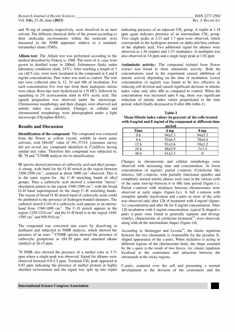
Time 4 mg 8 mg
0 h 94±5.3 94±5.3
6 h 92±6.5 70±4.4
12 h 91±3.6 10±1.2
24 h 88±5.9 2±1.4
48 h 61±3.3 0
Changes in chromosome and cellular morphology were
observed with increasing time and concentration. At lower
concentration (4 mg/ml), partial c-mitosis (Colchicine like
mitosis), full c-mitosis, with partially functional spindles andcompletely normal mitotic phases were seen in the various cells
of the same root-tip between 6 to 48h time period (figure-1.).
Partial c-mitosis with stickiness between chromosomes were
observed at early stages (figure-1a.). A full c-mitosis with
complete spindle inactivation and c-pairs in most of the cells
was observed only after 12h of treatment with 4 mg/ml (figure-
1c) concentration and after 6h for 8 mg/ml concentration. After
12h incubation with 4 mg/ml concentration, typical X-shaped c-
pairs (c-pairs were found to generally separate and diverge
widely), characteristic of colchicine treatment13
, were observed,
along with all the intermediate shapes (figure-1d).
According to Steinegger and Levana14
, the elastic repulsion
between the two chromatids is responsible for the peculiar X-
shaped appearance of the c-pairs. When stickiness is acting in
different regions of the chromosome body, the shape assumed
by the c-pairs is the result of two forces, viz. elastic repulsion
localized at the centromere and attraction between the
chromatids at the sticky regions.
C-pairs, scattered over the cell and presenting a normal
development to the division of the centromere and the
7/29/2019 4.ISCA-RJRS-2012-136 Done
http://slidepdf.com/reader/full/4isca-rjrs-2012-136-done 3/5
Research Journal of Recent Sciences ______________________________________________________________ ISSN 2277-2502
Vol. 1(6), 22-26, June (2012) Res. J. Recent Sci
International Science Congress Association 24
production of tetraploid cells, are regular feature in the material
treated for 12h with 4 mg/ml concentration of the compound
(figure-1e and 1f). After the 24h treatments this normal course
of the c-mitosis was found only in a few cases. Generally new
forces seem to enter into action during c-metaphase, causing
distributed c-mitosis, the peculiarity of which is a partition of
the c-pairs in two polar groups (figure-1i), similar pseudo-
anaphases with chromosomes lacking the customary
polarization and having their arms extending in all directions
(figure-1g), were visible. In addition to the different
distributions of c-pairs described above, interphases with c-pairs
clumped in the centre of the cell (figure-1k) were also observed,
together with the intermediate transitions after 48h of treatment.
Higher concentration of the compound (8 mg/ml) was found to
have more damaging effect on the root tip cells. After 6h c-
mitosis, distributed c-mitosis, multipolar anaphase (figure-2a),
psuedoanaphase, stickeness between chromosome, clumping of
c-metaphase in the center of the cell (figure-2d), misdivision
during c-metaphase and c-anaphase (figure-2e) and lagging
chromosomes (figure-2c) were observed. In some cells largebridges joining the two telophase nuclei appeared (figure-2b).
At increased time period of 12h, breakages in the chromosomes
were visible (figure-2f).
The induction of bridges between chromosomes could be
attributed to chromosome breaks, stickiness and breakage and
reunion of the broken ends. The stickiness prevented the
separation of daughter-chromosomes and thus they remained
connected by bridges15
. Large number of vagrant chromosomes
and c-anaphases indicates that compound 10 acts as a potent
spindle inhibitor. In the present study, a few cells with
fragments of chromosomes were observed indicating the
clastogenic effect of test compound as suggested forcypermethrin compound in A. cepa
16.
After 24h of treatment the toxic symptom appeared in whole
cell (Figure-3). Autolysis in the cytoplasm of the cell was
visible, which continued with increasing time, causing complete
dissolution of the cytoplasm, leaving behind only nucleus
(Figure-3c and 3d). In some cells, nuclear material was found to
be disintegrated with the cellular boundary still intact (Figure-
3b). This disintegration of nuclear material continued with the
increase in time. Within 48h nuclear material was completely
lost with the cellular boundary still maintained forming a ghost
cell (Figure-3e).
Conclusion
For many years, laboratories from all over the world have been
working on finding effective remedy for tumors, up to now the
most severe disease of our civilization9. Among many
therapeutic plants that are objects of interest, the compounds
from P. juliflora seemed particularly important, as shown in our
results. According to the obtained results, one can conclude that
the compound play a role in the antiproliferative and cytotoxic
effect on A. cepa cells and these results could be useful in
correlating with the mammalian system.
Acknowledgments
We acknowledge Birla Institute of Technology and Science for
providing laboratory facilities to conduct the research work. We
are also thankful to Central Drug Research Institute (CDRI),Lucknow, for performing mass, NMR and IR analysis. Funding
for the research work was provided by CSIR (Council for
Scientific and Industrial research).
References
1. De Flora S., Izzotti A., D’Agostini F. and Balansky R.M.,
Multiple points of intervention in the prevention of cancer
and other mutationrelated diseases, Mut. Res., 9, 480 – 481
(2001)
2. Fonrose X., Ausseil F., Soleilhac E., Masson V. and David
P., Parthenolide inhibits tubulin carboxypeptidase activity,Cancer Res., 67, 3371 – 3378 (2007)
3. Iwasaki S., Antimitotic agents; Chemistry and recognition
of tubulin molecule, Med. Res. Rev., 13, 183 – 198 (1993)
4. Mangale S.M., Chonde S.G. and Raut P.D., Use of
Moringa oleifera (drumstick) seed as natural absorbent and
an antimicrobial agent for ground water treatment,
Res.J.Recent Sci., 1(3), 31-40 (2012)
5. Mondal D. and Mondal T., A review on efficacy of
Azadirachta indica A. juss based biopesticides: an Indian
perspective, Res.J.Recent Sci., 1(3), 94-99 (2012)
6. Edelman M.J., Novel cytotoxic agents for nonsmall cell
lung cancer, J. Thorac. Oncol., 1, 752-755 (2006)
7. Latha P.G. and Panikkar K.R., Chemoprotective effect of
Ixora coccinea L. flowers on cisplatin induced toxicity in
mice, Cancer Lett ., 130, 197 – 202 (1998)
8. Andrade L.F., Campos J.M.S. and Davide L.C.,
Cytogenetic alterations induced by SPL (spent potliners) in
meristematic cells of plant bioassays, Ecotoxicol. Environ
Saf., 71, 706–710 (2008)
9. Leme D.M. and Marin-Morales M.A., Chromosome
aberration and micronucleus frequencies in Allium cepa
cells exposed petroleum polluted water – a case study,
Mutat. Res., 650, 80–86 (2008)
10. Havey M.J., Rabinowitch H.D., Currah L., Genome
organization in Allium. In Allium Crop Science, (Eds.).
pp. 59-79. Recent Advances, CABI Publishing, United
Kingdom, (2002)
7/29/2019 4.ISCA-RJRS-2012-136 Done
http://slidepdf.com/reader/full/4isca-rjrs-2012-136-done 4/5
Research Journal of Recent Sciences ______________________________________________________________ ISSN 2277-2502
Vol. 1(6), 22-26, June (2012) Res. J. Recent Sci
International Science Congress Association 25
11. Kura’s M., Nowakowska J., S´ liwin´ ska E., Pilarski R.,
Ilasz R., Tykarska T., Zobel A. and Gulewicz K., Changes
in chromosome structure, Mitotic activity and nuclear
DNA content from cells of Allium test induced by bark
water extract of Uncaria tomentosa (Willd.) DC., J.
Ethnopharmacol., 107, 211–221 (2006)
12. Fiskesjo G., Allium test for screening chemicals;
evaluation of cytological parameters. In Plants for
Environmental Studies, Wang W., Gorsuch J. W., Hughes
J.S. (Eds.) 307-333 CRC, Lewis Publishers, New York,
(1997) 13. Witkus and Berger C.A., Veratrine, a new polyploidy
inducing agent, J. Heredity, 35, 129-133 (1944)
14. Steineggeer E. and Levana A., Constitution and c-mitotic
activity of isocolchicine, Hereditas, 33, 385-396 (1947)
15. Badr A., Ghareeb A. and El-Din H.M., Cytotoxicity of
some pesticides in mitotic cells of V. faba roots, Egyptian
J. App. Sci., 7, 457-468 (1992)
16. Saxena P.N., Chauhan L.K.S. and Gupta S.K., 2005,
Cytogenetic effects of commercial formulation of
cypermethrin in root meristem cells of Allium sativum:
Spectroscopic basis of chromosome damage, Toxicol., 216,
244-252 (2005)
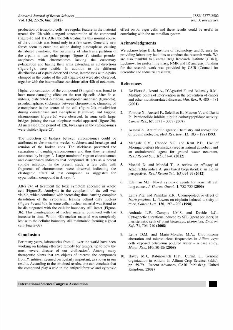
Figure-1
Chromosomal abnormalities in root cells of Allium cepa, seen after treatment of the Compound at concentration of 4 mg/mlwithin 48h - cells after 6h: a) Partial c-mitosis b) Partial c-mitosis sticking of the cell cells after 12h: c) C-mitosis d)
Stickiness between c-mitosis X shaped cells after 24h: e) and f) Tetraploid g) Psuedoanaphase h) and i) Division in
groups cells after 48h: j) and k) clumping in the center. 100x
7/29/2019 4.ISCA-RJRS-2012-136 Done
http://slidepdf.com/reader/full/4isca-rjrs-2012-136-done 5/5
Research Journal of Recent Sciences ______________________________________________________________ ISSN 2277-2502
Vol. 1(6), 22-26, June (2012) Res. J. Recent Sci
International Science Congress Association 26
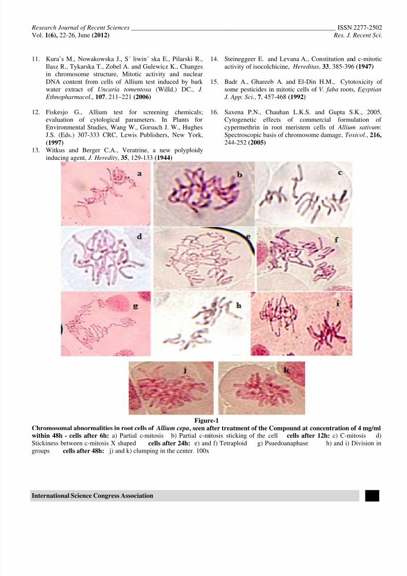
Figure-2Chromosomal abnormalities in root cells of Allium cepa, seen after treatment of the compound at concentration of 8 mg/mlwithin 12h - cells after 6h: a)Multipolar anaphase b) chromosomal bridge c) lagging chromosome d) clumping
of chromosome in the centre e) unequal division with chromosomal bridge cells after 12h: f) framentation of
chromosomes.
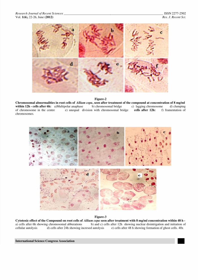
Figure-3
Cytotoxic effect of the Compound on root cells of Allium cepa seen after treatment with 8 mg/ml concentration within 48 h -a) cells after 6h showing chromosomal abberations b) and c) cells after 12h showing nuclear disintrigation and initiation of
cellular autolysis d) cells after 24h showing incresed autolysis e) cells after 48 h showing formation of ghost cells. 40x