41.1: diet provides- 1)chemical energy 2)organic molecules 3)essential nutrients
TRANSCRIPT
41.1: Diet Provides-
1)Chemical Energy2)Organic Molecules3)Essential Nutrients
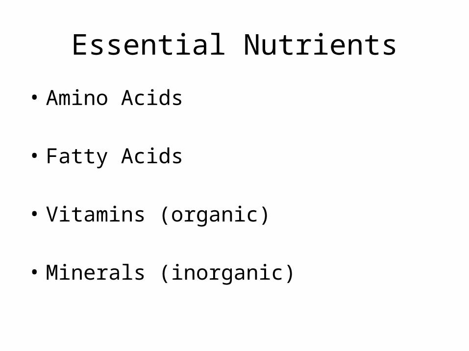
Essential Nutrients
• Amino Acids
• Fatty Acids
• Vitamins (organic)
• Minerals (inorganic)
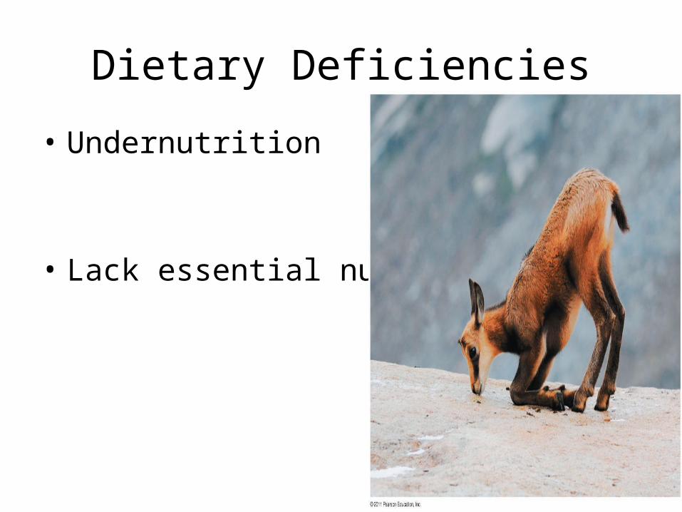
Dietary Deficiencies
• Undernutrition
• Lack essential nutrients
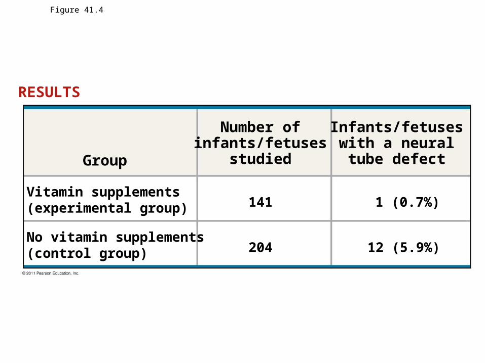
Figure 41.4
RESULTS
Group
Number ofinfants/fetuses
studied
Vitamin supplements(experimental group)
No vitamin supplements(control group)
141
204
1 (0.7%)
12 (5.9%)
Infants/fetuseswith a neuraltube defect
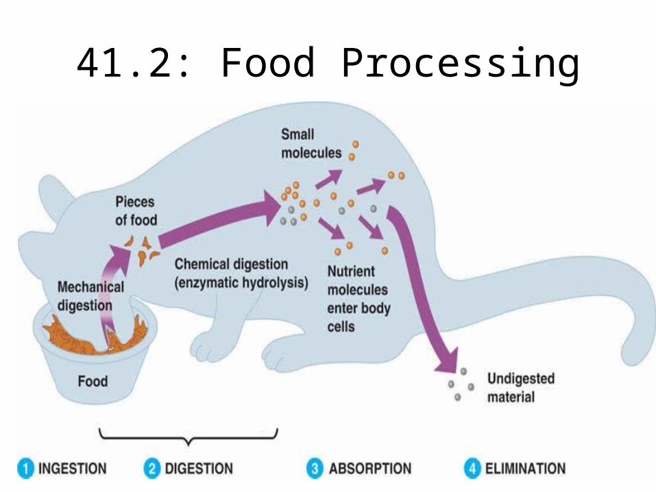
41.2: Food Processing
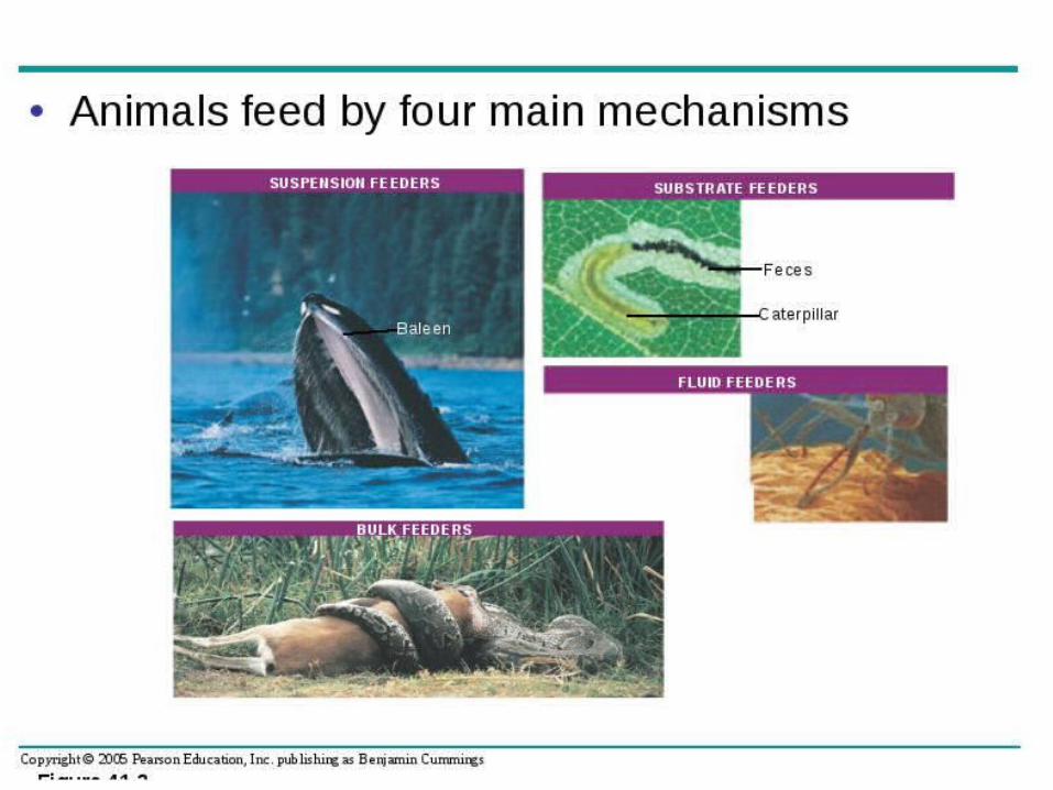
4 Main Animal Feeding Mechanisms
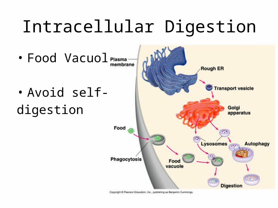
Intracellular Digestion
• Food Vacuoles
• Avoid self-digestion
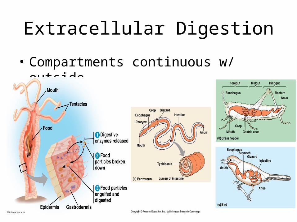
Extracellular Digestion
• Compartments continuous w/ outside
CONCEPT 41.3ORGANS SPECIALIZED FOR SEQUENTIAL STAGES OF FOOD PROCESSING FORM THE MAMMALIAN DIGESTIVE SYSTEM
•Chapter 40•Grace Cunnie

THE HUMAN DIGESTIVE SYSTEM
Figure 41.9: The human digestive system11
- Mammalian digestive system- Alimentary canal- Accessory glands
- Salivary glands- Pancreas- Liver- Gallbladder
TRAVELING DOWN THE ALIMENTARY CANAL
Food is pushed along canal by peristalsisAlternating waves of contraction and relaxation in the smooth muscles lining the canal
Can be helped along by sphinctersRing-like valves that form at some of the junctions between specialized compartments

THE ORAL CAVITY, PHARYNX, AND ESOPHAGUS
- As saliva is released- Amylase
helps break down starch
- Mucus is produced
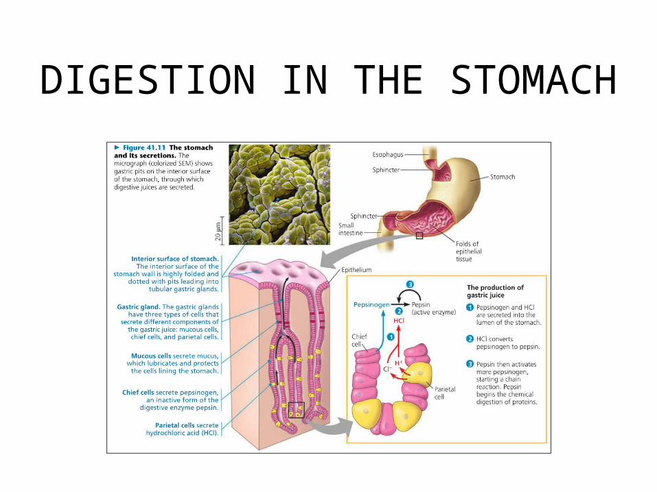
DIGESTION IN THE STOMACH

DIGESTION IN THE SMALL INTESTINE- Duodenum- First 25 cm- Chyme from
stomach mixes with digestive juices
- Pancreas- Aids in digestion,
produces alkaline solution rich with bicarbonate, several enzymes
- Liver- Produces bile
- Gallbladder- Stores bile
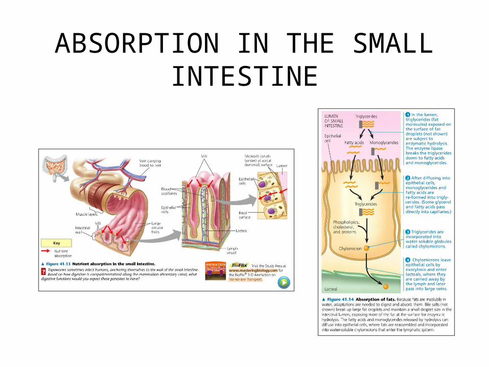
ABSORPTION IN THE SMALL INTESTINE
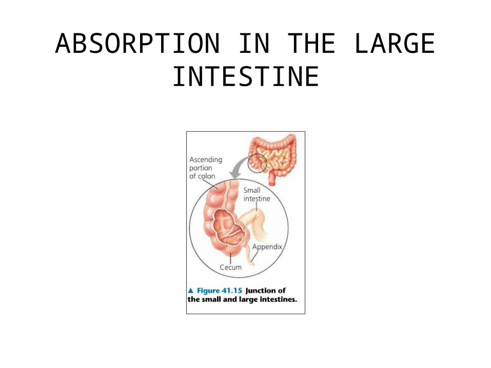
ABSORPTION IN THE LARGE INTESTINE
41.4- EVOLUTIONARY ADAPTATIONS OF VERTEBRATE DIGESTIVE SYSTEMS CORRELATE WITH DIET
Xia Whitaker
Mr. Reis
AP Biology
13 March 2013

DENTAL ADAPTATIONS Dentition- an animals’ assortment of teeth
Structural variation reflects diet
Nonmammalian vertebrates generally have less specialized dentition
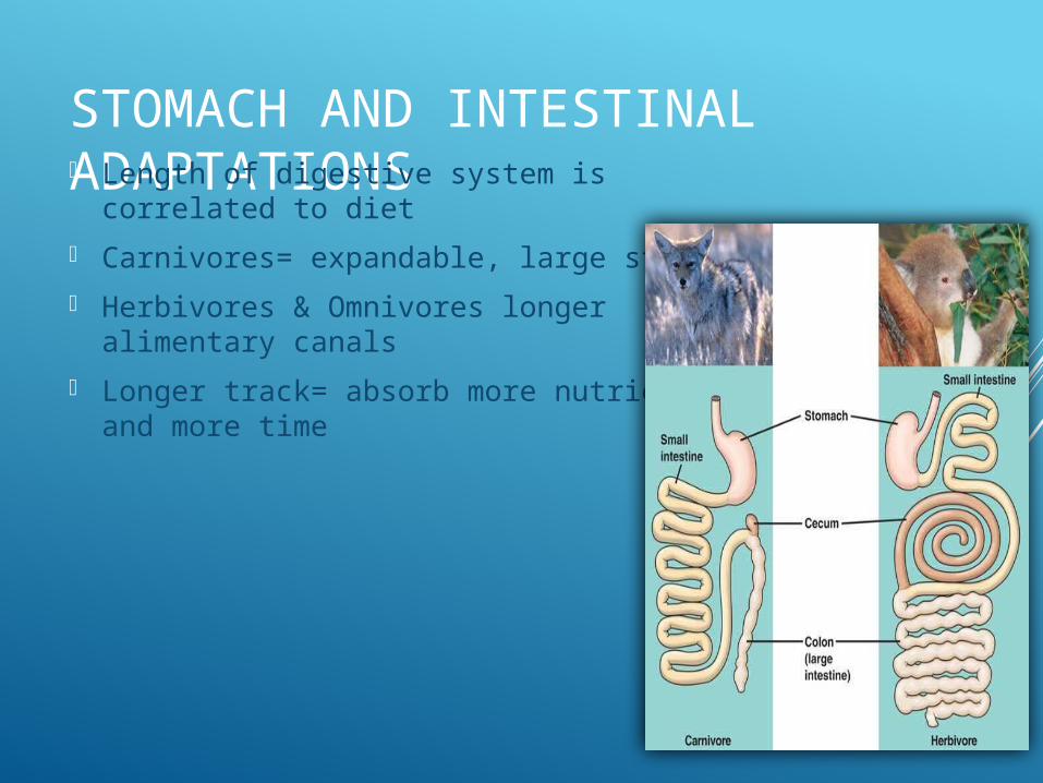
STOMACH AND INTESTINAL ADAPTATIONS Length of digestive system is correlated to
diet
Carnivores= expandable, large stomachs
Herbivores & Omnivores longer alimentary canals
Longer track= absorb more nutrients and more time
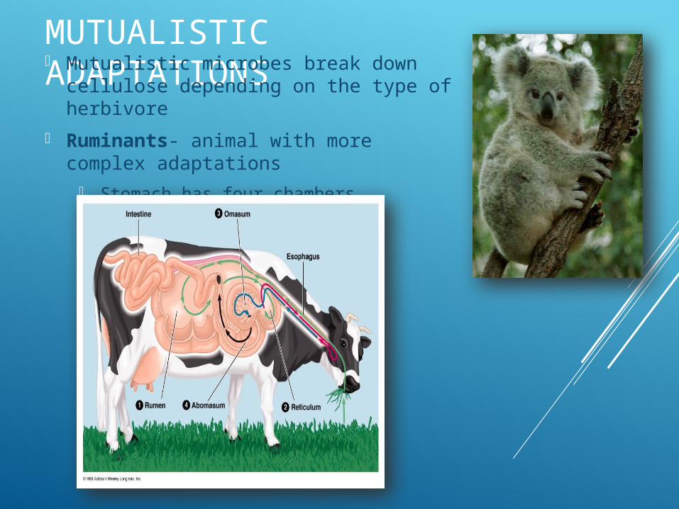
MUTUALISTIC ADAPTATIONS Mutualistic microbes break down
cellulose depending on the type of herbivore
Ruminants- animal with more complex adaptations
Stomach has four chambers
41.5- FEEDBACK CIRCUITS REGULATE DIGESTION, ENERGY STORAGE, AND APPETITE
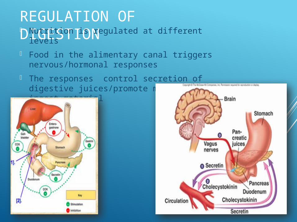
REGULATION OF DIGESTION Nutrition is regulated at different levels
Food in the alimentary canal triggers nervous/hormonal responses
The responses control secretion of digestive juices/promote movement of ingest material
REGULATION OF ENERGY STORAGE When an animal takes in more energy-rich
molecules than needed, it stores excess energy
Vertebrates store excess calories is glycogen (liver/muscle cells) and fat (adipose cells)
Glucose Homeostasis Insulin and glucagon maintain glucose homeostasis
Insulin levels rise= glucose enters liver to synthesize glycogen
Lower glucose level= glucagon stimulates liver to breakdown glycogen (releases glycogen into blood)
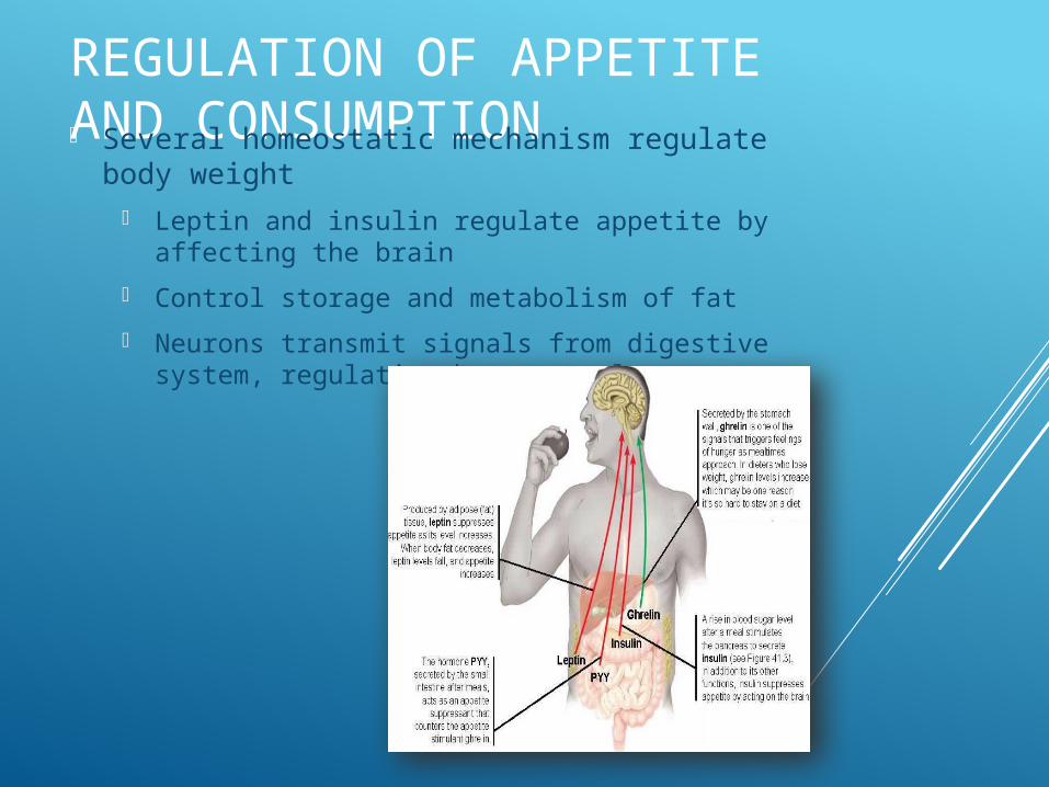
REGULATION OF APPETITE AND CONSUMPTION Several homeostatic mechanism regulate
body weight Leptin and insulin regulate appetite by
affecting the brain
Control storage and metabolism of fat
Neurons transmit signals from digestive system, regulating hormone release
OBESITY AND EVOLUTION Fat hoarding was necessary to our
ancestors
Natural selection may have selected them for their ability to store food
Circulation and Gas Exchange
Chapters 42.1 and 42.2Mike Tillman
Circulatory SystemAnimals with simple body plans – gas exchange
via diffusion Diffusion efficient over small distances Cells exchange materials with surrounding medium
Cells of most animals exchange materials with the environment with a circulatory system
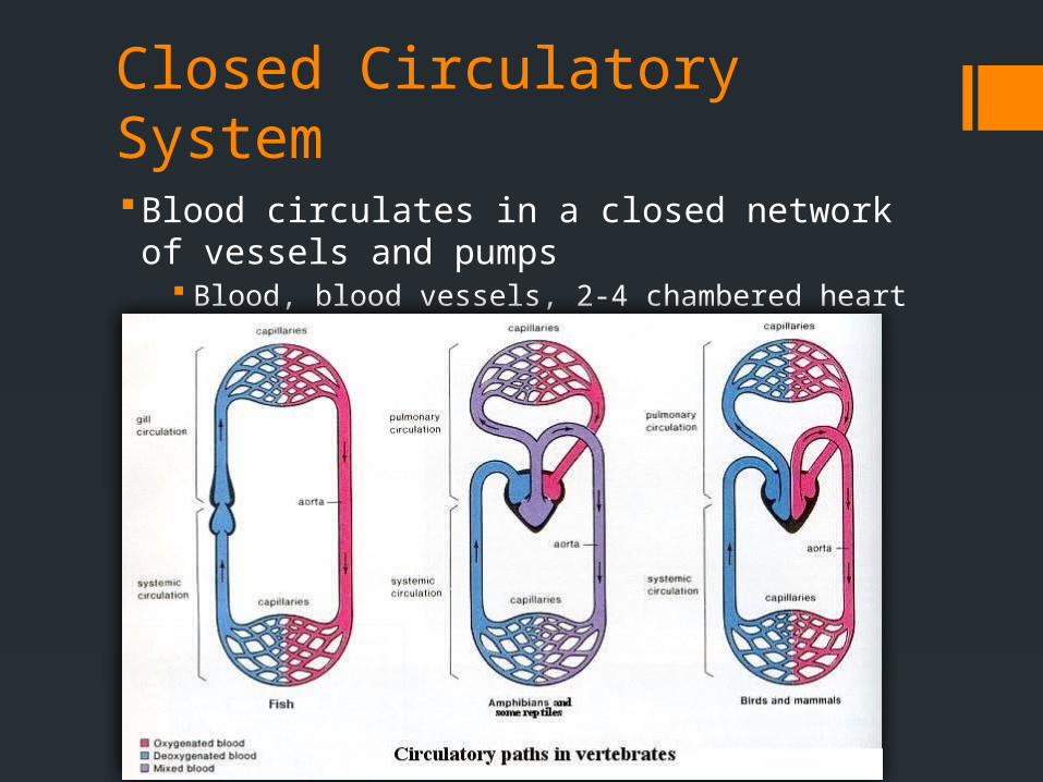
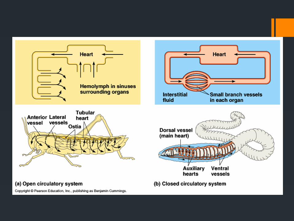
Closed Circulatory SystemBlood circulates in a closed network of vessels and
pumps Blood, blood vessels, 2-4 chambered heart
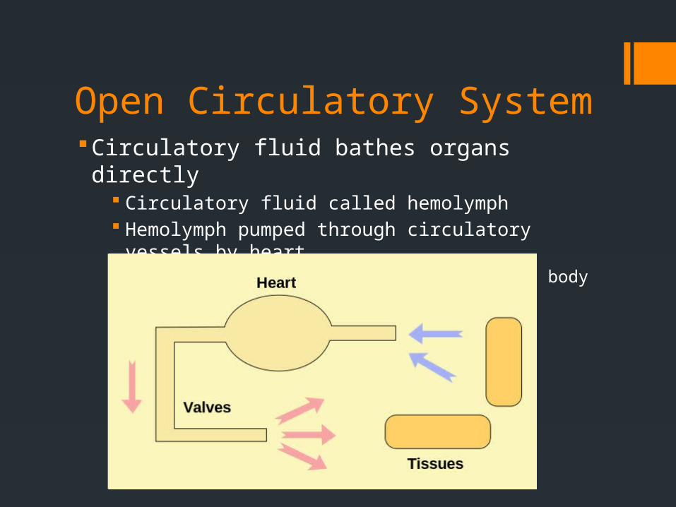
Open Circulatory SystemCirculatory fluid bathes organs directly
Circulatory fluid called hemolymph Hemolymph pumped through circulatory vessels by heart
Chemical exchange between hemolymph and body cells
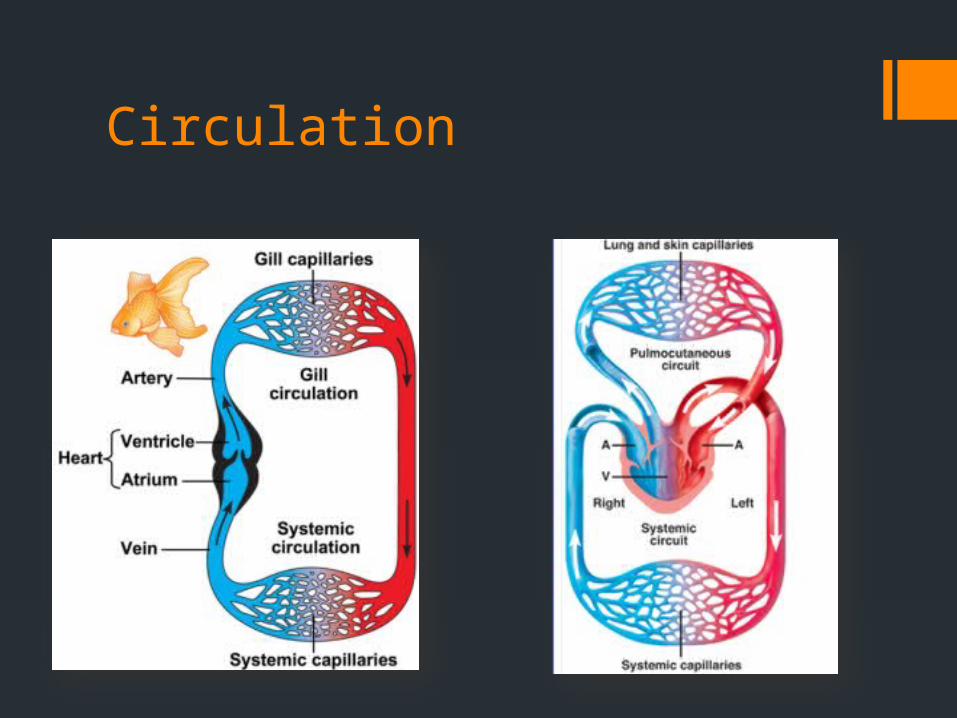
Circulation
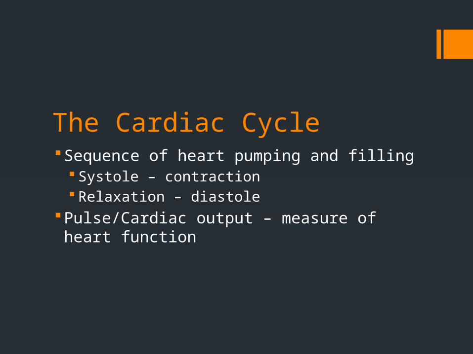
The Cardiac CycleSequence of heart pumping and filling
Systole – contraction Relaxation – diastole
Pulse/Cardiac output – measure of heart function
HeartbeatOriginates at the sinoatrial (SA) node
“pacemaker” Pacemaker ability influenced by
Nervous system Hormones Body temperature
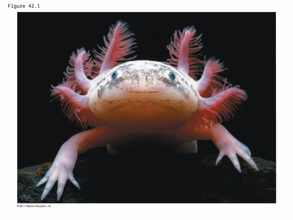
Figure 42.1
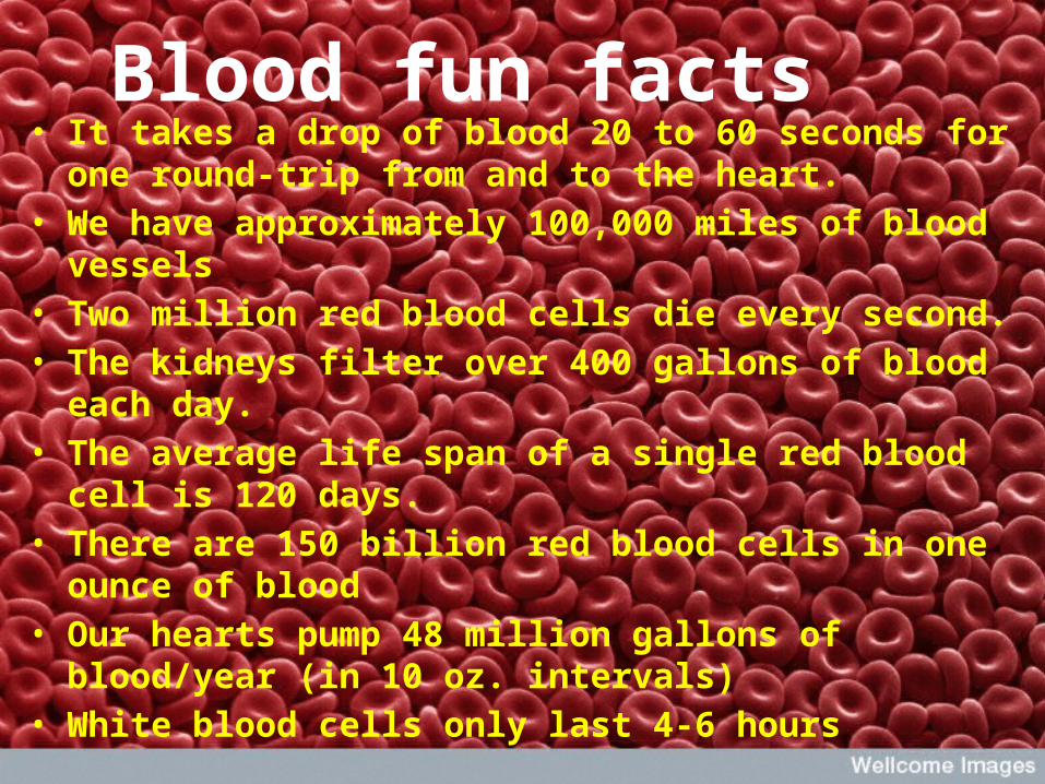
Blood fun facts• It takes a drop of blood 20 to 60 seconds for one round-
trip from and to the heart.• We have approximately 100,000 miles of blood vessels • Two million red blood cells die every second.• The kidneys filter over 400 gallons of blood each day.• The average life span of a single red blood cell is 120
days.• There are 150 billion red blood cells in one ounce of
blood• Our hearts pump 48 million gallons of blood/year (in 10
oz. intervals)• White blood cells only last 4-6 hours
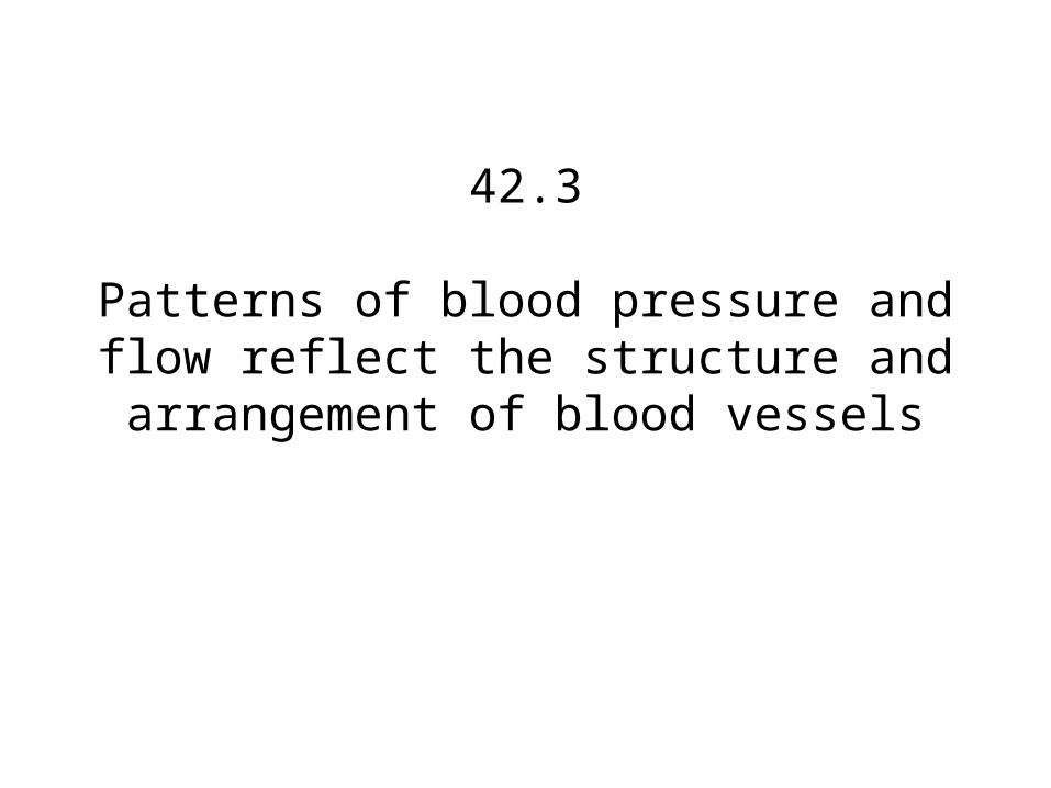
42.3
Patterns of blood pressure and flow reflect the structure and
arrangement of blood vessels

• Endothelium – single layer of flattened epithelial cells
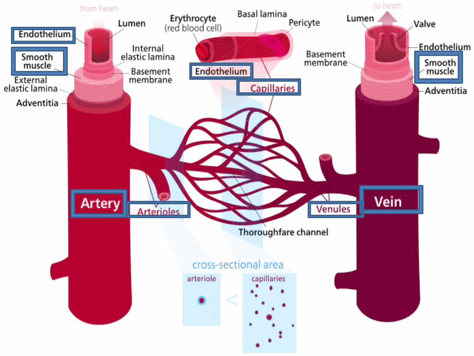
• Capillaries – provides blood to tissues and interstitial fluid in-between artery and vein
• Venules – connects vein to capillary• Arterioles – conects artery to capillary• Smooth Muscle – receives hormones, regulates
artery size
• Artery – carries blood away from heart to organs• Vein – carries blood back toward heart

Blood PressureLumen
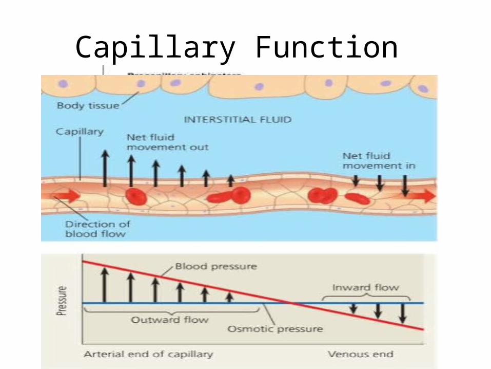
Capillary Function
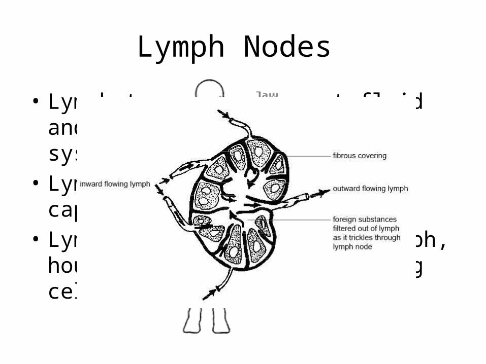
Lymph Nodes
• Lymphatic system – lost fluid and proteins return to blood system
• Lymph – fluid lost by capillaries• Lymph nodes – filter the lymph, house
bacteria/virus fighting cells
Concept 42.4: Blood components function in exchange, transport, and defense
By Ryan Tudino

The composition of mammalian blood
Plasma 55%
Constituent Major functions
Water
Ions (bloodelectrolytes)SodiumPotassiumCalciumMagnesiumChlorideBicarbonate
Solvent forcarrying othersubstances
Osmotic balance,pH buffering,and regulationof membranepermeability
Plasma proteinsOsmotic balance,pH buffering
Albumin
Fibrinogen
Immunoglobulins(antibodies)
Clotting
Defense
Substances transported by blood
NutrientsWaste productsRespiratory gasesHormones
Separatedbloodelements
Basophils
Neutrophils Monocytes
Lymphocytes
Eosinophils
Platelets
Erythrocytes (red blood cells) 5–6 million
250,000–400,000 Bloodclotting
Transportof O2 andsome CO2
Defense andimmunity
FunctionsNumber per L(mm3) of blood
Cell type
Cellular elements 45%
Leukocytes (white blood cells) 5,000–10,000
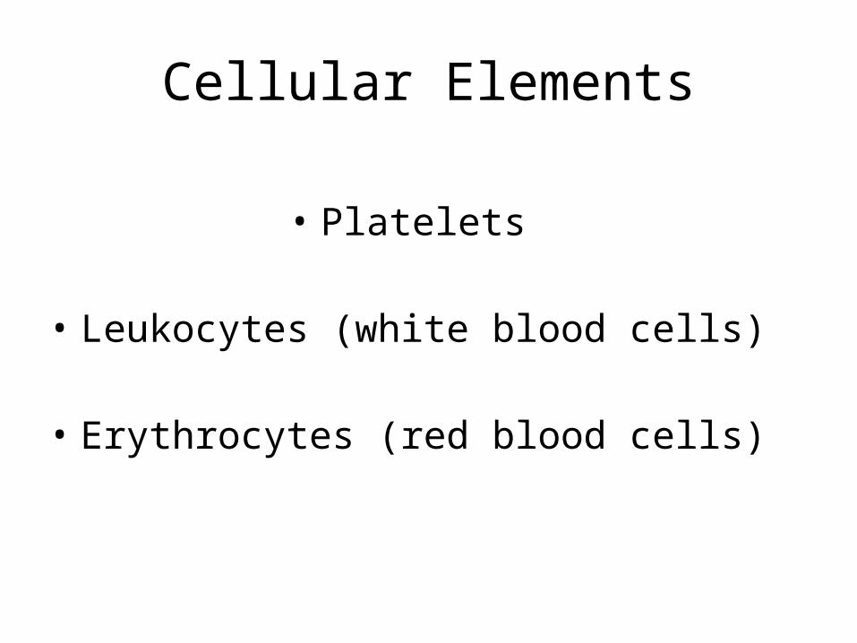
Cellular Elements
• Platelets
• Leukocytes (white blood cells)
• Erythrocytes (red blood cells)

Why is the occasional cut or scrap not life-threatening?
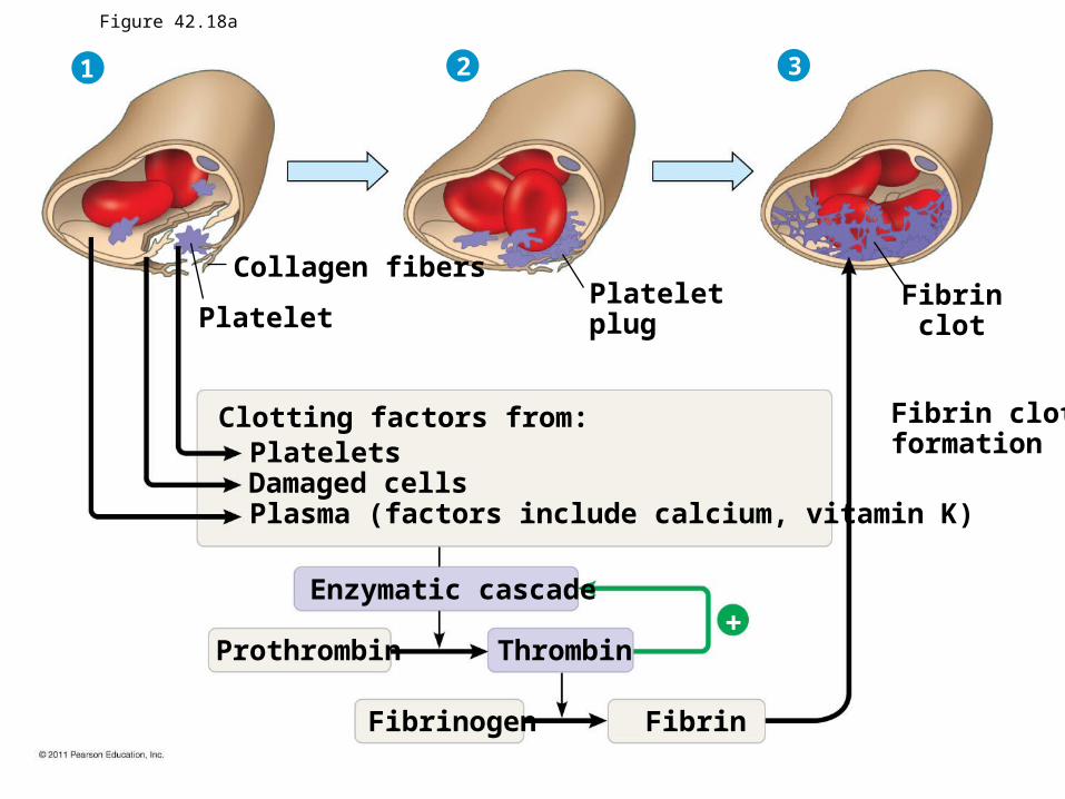
Collagen fibers
1
PlateletPlateletplug
Fibrinclot
Clotting factors from:PlateletsDamaged cellsPlasma (factors include calcium, vitamin K)
Enzymatic cascade
Prothrombin Thrombin
Fibrinogen Fibrin
Fibrin clotformation
2 3
Figure 42.18a
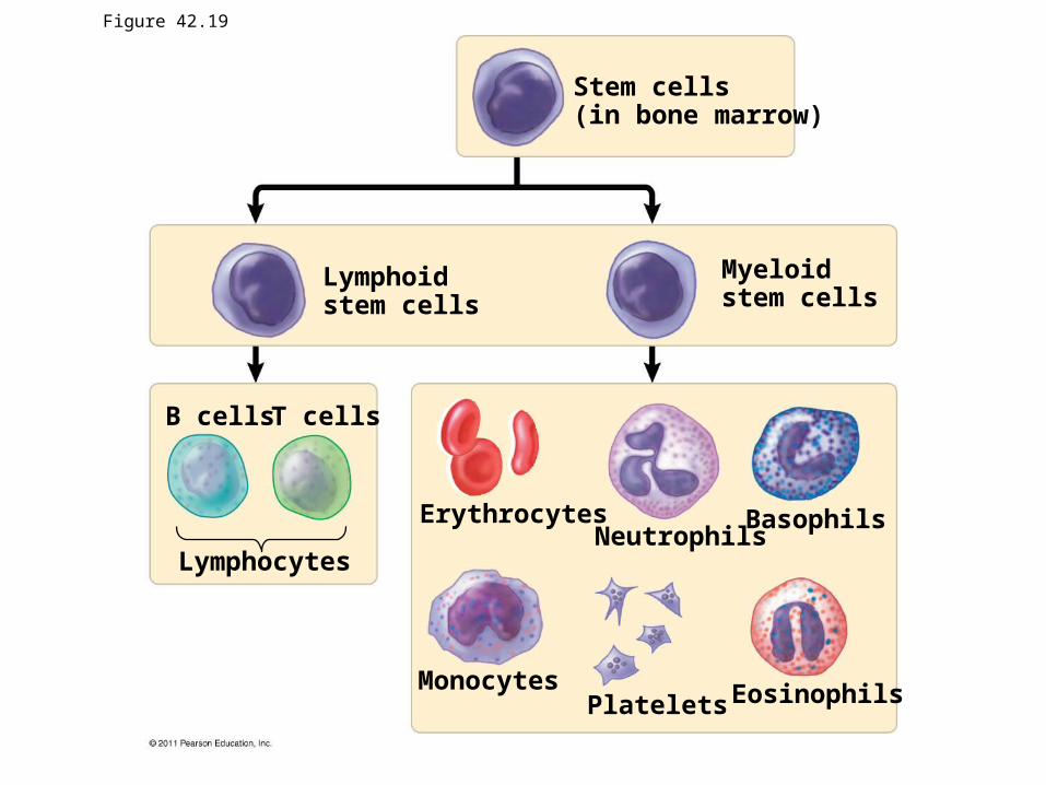
Stem cells(in bone marrow)
Myeloidstem cells
Lymphoidstem cells
B cells T cells
Lymphocytes
ErythrocytesNeutrophils
Basophils
EosinophilsPlateletsMonocytes
Figure 42.19
Cardiovascular Disease
• Low-density lipoprotein (LDL)
• High-density lipoprotein (HDL)– Risk for heart disease increases with a high LDL to HDL
ratio
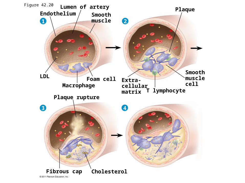
Lumen of arterySmoothmuscle
EndotheliumPlaque
Smoothmusclecell
T lymphocyte
Extra-cellularmatrix
Foam cellMacrophage
Plaque rupture
LDL
CholesterolFibrous cap
1 2
43
Figure 42.20
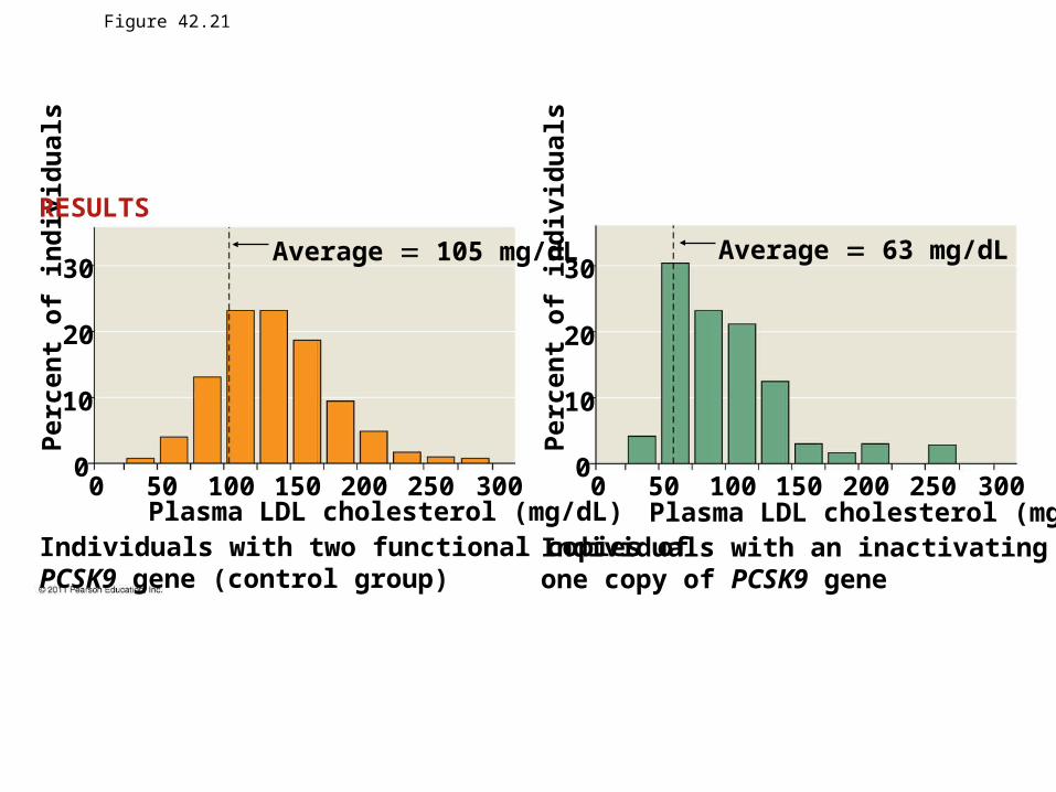
Figure 42.21
Individuals with two functional copies ofPCSK9 gene (control group)
Plasma LDL cholesterol (mg/dL)Individuals with an inactivating mutation inone copy of PCSK9 gene
Plasma LDL cholesterol (mg/dL)
Average 63 mg/dLAverage 105 mg/dL30
20
10
00 50 100 150 200 250 300 0
0
10
20
30
50 100 150 200 250 300
Perc
ent o
f ind
ivid
ualsRESULTS
Perc
ent o
f ind
ivid
uals

42.6Bridget Peterson

42.6 How an Amphibian Breathes
Positive Pressure Breathing: inflation of the lungs through forced air-flow
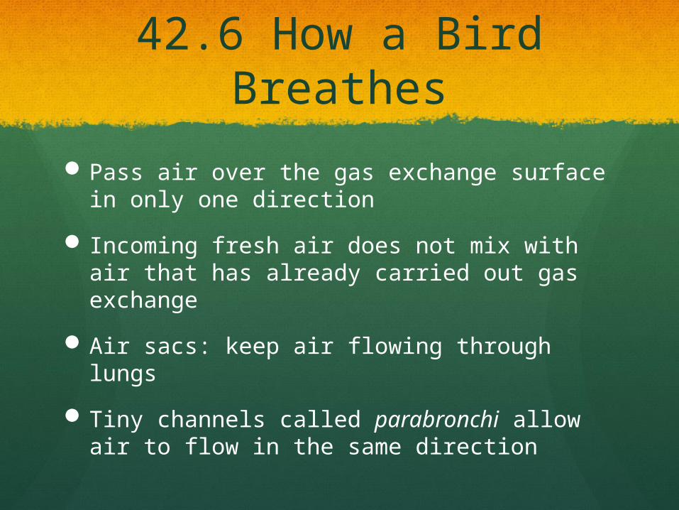
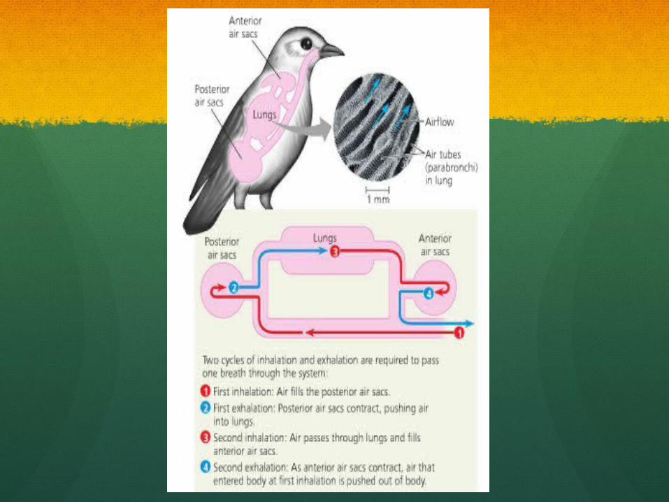
42.6 How a Bird Breathes
Pass air over the gas exchange surface in only one direction
Incoming fresh air does not mix with air that has already carried out gas exchange
Air sacs: keep air flowing through lungs
Tiny channels called parabronchi allow air to flow in the same direction
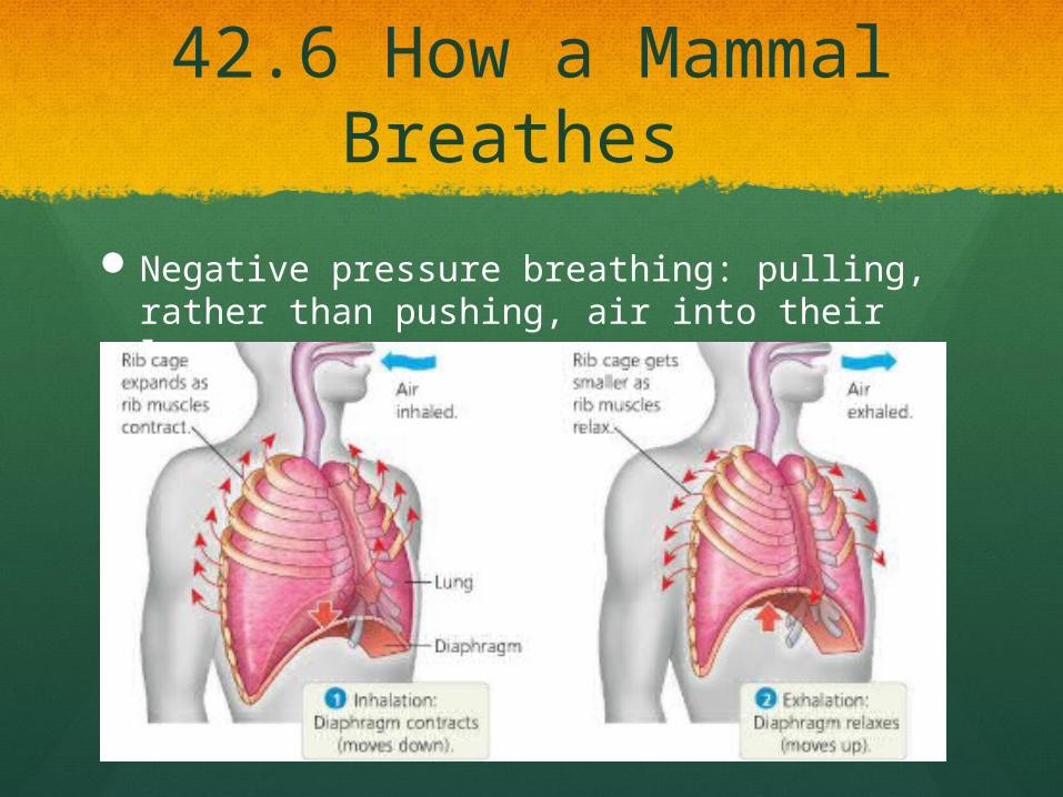
42.6 How a Mammal Breathes
Negative pressure breathing: pulling, rather than pushing, air into their lungs
42.6 How a Mammal Breathes
Inhalation requires work Change air pressure within lungs relative to
pressure of outside atmosphere Air rushes through the nostrils and mouth and
down the breathing tubes to the alveoli
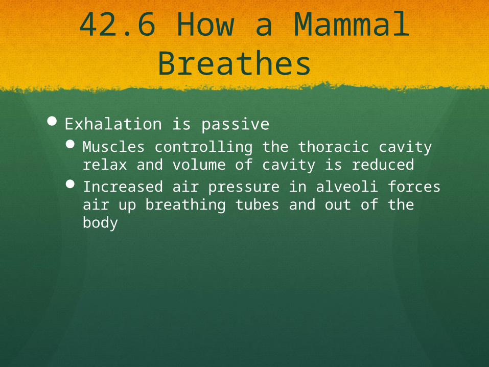
42.6 How a Mammal Breathes
Exhalation is passive Muscles controlling the thoracic cavity relax
and volume of cavity is reduced Increased air pressure in alveoli forces air up
breathing tubes and out of the body

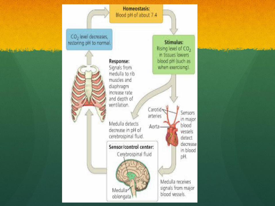
42.6 Control of Breathing
Breathing is regulated to ensure that gas exchange is coordinated with blood circulation and with metabolic demand
Neurons in the medulla oblongata: form a breathing control center When you breath deeply a negative feedback
mechanism prevents the lungs from over expanding

Concept 42.7: Adaptations for gas exchange include pigments that bind and transport gases
• The metabolic demands of many organisms require that the blood transport large quantities of O2 and CO2
© 2011 Pearson Education, Inc.
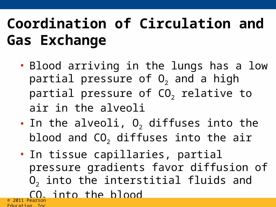
Coordination of Circulation and Gas Exchange
• Blood arriving in the lungs has a low partial pressure of O2 and a high partial pressure of CO2 relative to air in the alveoli
• In the alveoli, O2 diffuses into the blood and CO2 diffuses into the air
• In tissue capillaries, partial pressure gradients favor diffusion of O2 into the interstitial fluids and CO2 into the blood
© 2011 Pearson Education, Inc.
Exhaled air Inhaled air
Pulmonaryarteries
Systemicveins
Systemicarteries
Pulmonaryveins
Alveolarcapillaries
AlveolarspacesAlveolar
epithelialcells
Inhaledair
160
120
80
40
0Heart
8 1
2
3
46
7
CO2 O2
SystemiccapillariesCO2 O2
Body tissue5
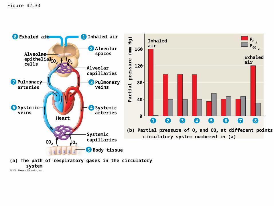
(a) The path of respiratory gases in the circulatory system
(b) Partial pressure of O2 and CO2 at different points in the
circulatory system numbered in (a)
4321 5 6 7
Exhaledair
Pa
rtia
l p
res
su
re (
mm
Hg
)
PO2
PCO 2
8
Figure 42.30
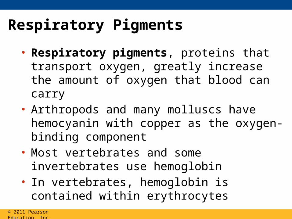
Respiratory Pigments
• Respiratory pigments, proteins that transport oxygen, greatly increase the amount of oxygen that blood can carry
• Arthropods and many molluscs have hemocyanin with copper as the oxygen-binding component
• Most vertebrates and some invertebrates use hemoglobin
• In vertebrates, hemoglobin is contained within erythrocytes
© 2011 Pearson Education, Inc.
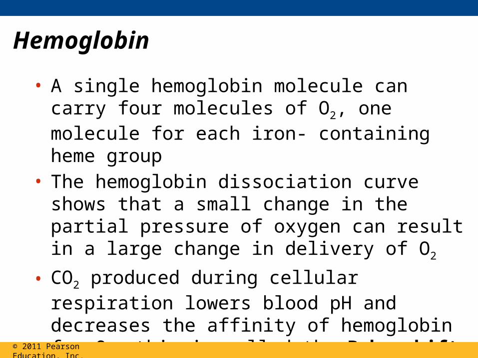
Hemoglobin
• A single hemoglobin molecule can carry four molecules of O2, one molecule for each iron- containing heme group
• The hemoglobin dissociation curve shows that a small change in the partial pressure of oxygen can result in a large change in delivery of O2
• CO2 produced during cellular respiration lowers blood pH and decreases the affinity of hemoglobin for O2; this is called the Bohr shift
© 2011 Pearson Education, Inc.© 2011 Pearson Education, Inc.
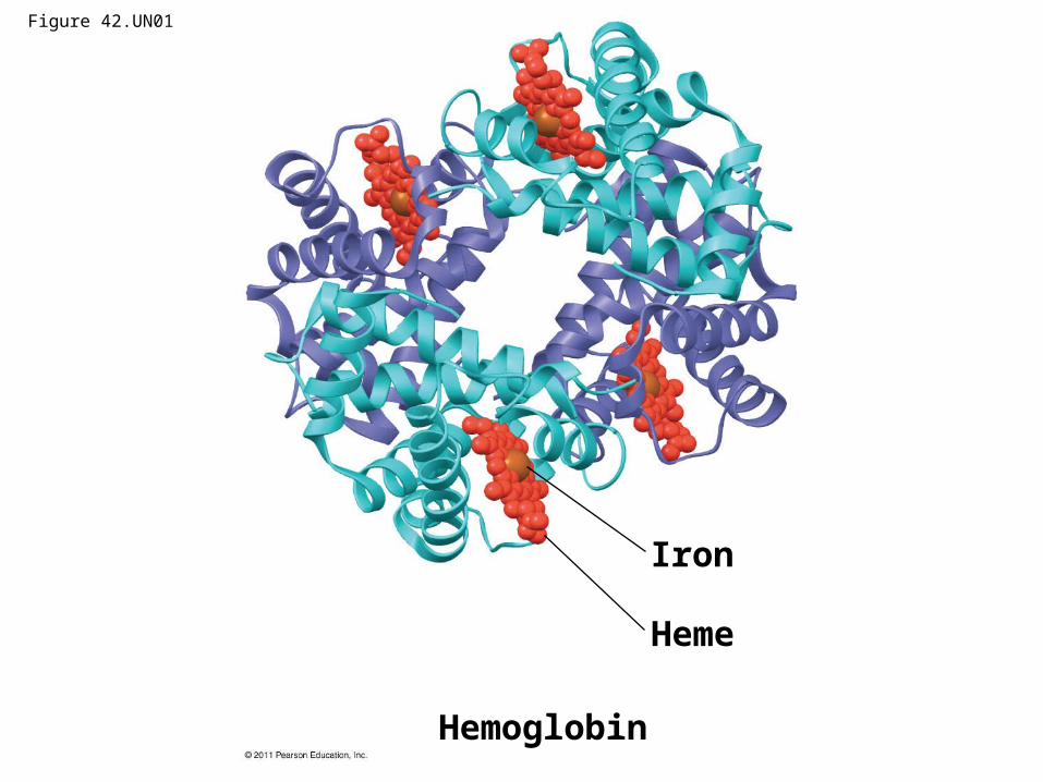
Figure 42.UN01
Iron
Heme
Hemoglobin
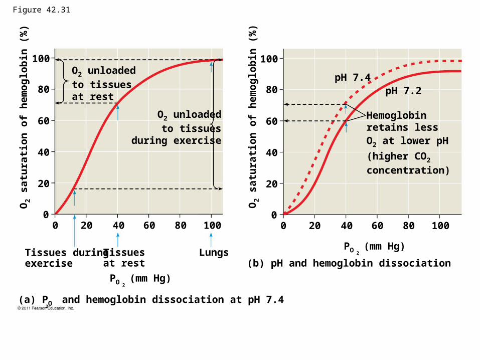
Figure 42.31
2(a) PO and hemoglobin dissociation at pH 7.4
Tissues duringexercise
Tissuesat rest
Lungs
PO (mm Hg)2
(b) pH and hemoglobin dissociation
PO (mm Hg)2
0 20 40 60 80 1000
20
40
60
80
100
0 20 40 60 80 1000
20
40
60
80
100
Hemoglobinretains lessO2 at lower pH
(higher CO2
concentration)
pH 7.2pH 7.4
O2 unloaded
to tissuesduring exercise
O2 s
atu
rati
on
of
he
mo
glo
bin
(%
)
O2 unloaded
to tissuesat rest
O2 s
atu
rati
on
of
he
mo
glo
bin
(%
)
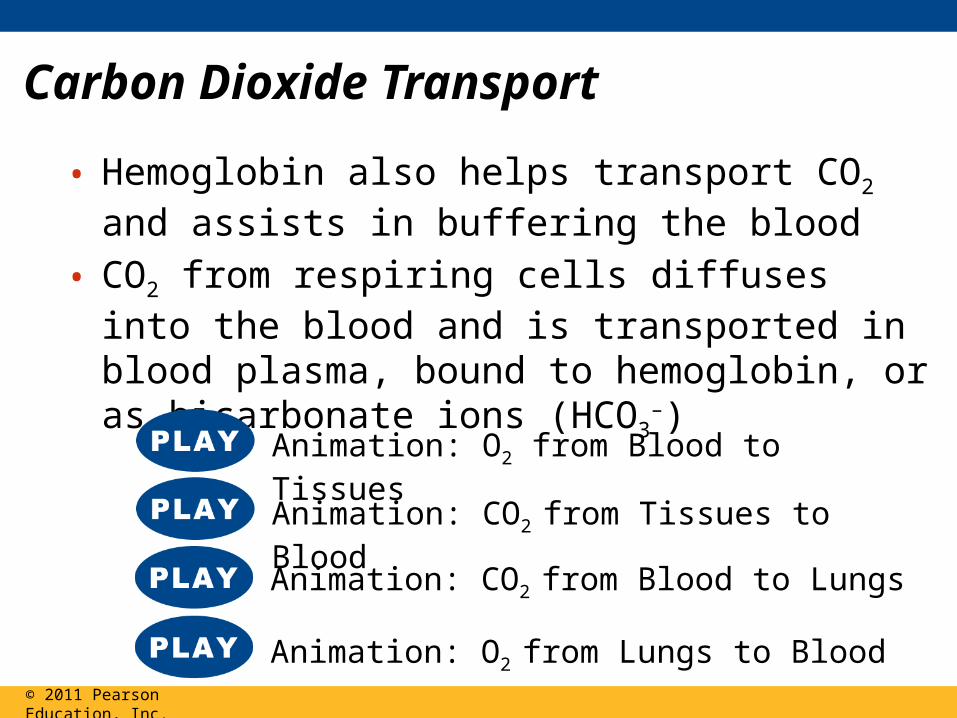
Carbon Dioxide Transport
• Hemoglobin also helps transport CO2 and assists in buffering the blood
• CO2 from respiring cells diffuses into the blood and is transported in blood plasma, bound to hemoglobin, or as bicarbonate ions (HCO3
–)
© 2011 Pearson Education, Inc.
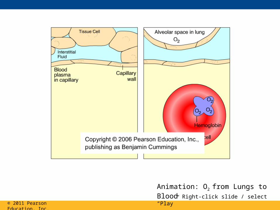
Animation: O2 from Lungs to Blood
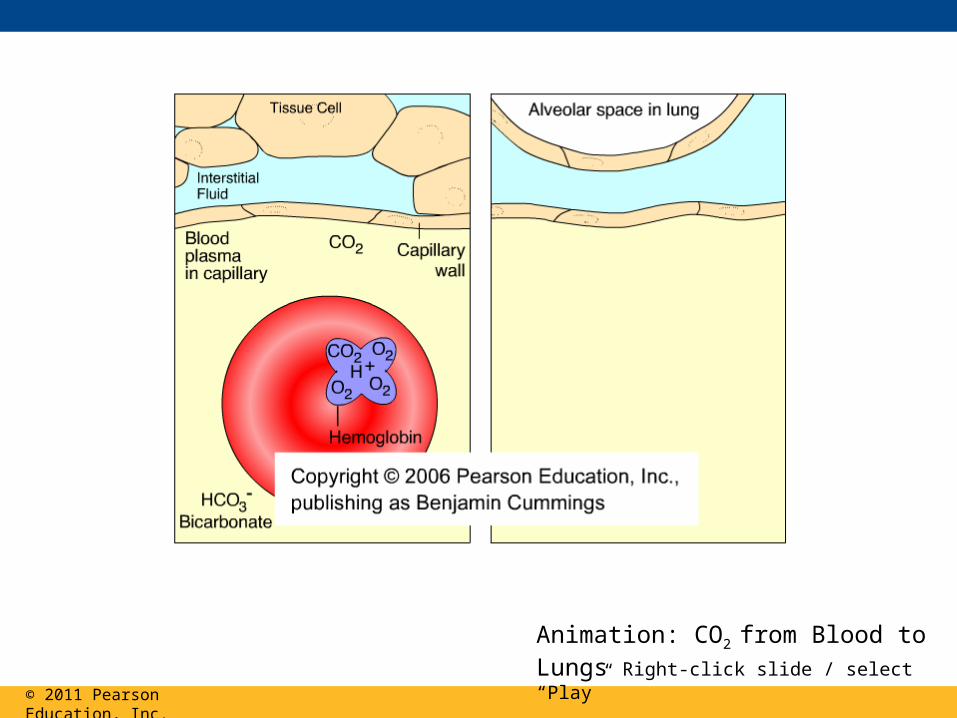
Animation: CO2 from Blood to Lungs
Animation: CO2 from Tissues to Blood
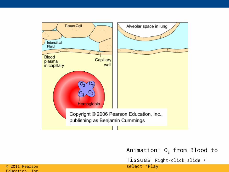
Animation: O2 from Blood to Tissues
© 2011 Pearson Education, Inc.
Animation: O2 from Blood to Tissues Right-click slide / select “Play”

© 2011 Pearson Education, Inc.
Animation: CO2 from Tissues to Blood Right-click slide / select “Play”
© 2011 Pearson Education, Inc.
Animation: CO2 from Blood to Lungs Right-click slide / select “Play”
© 2011 Pearson Education, Inc.
Animation: O2 from Lungs to Blood Right-click slide / select “Play”
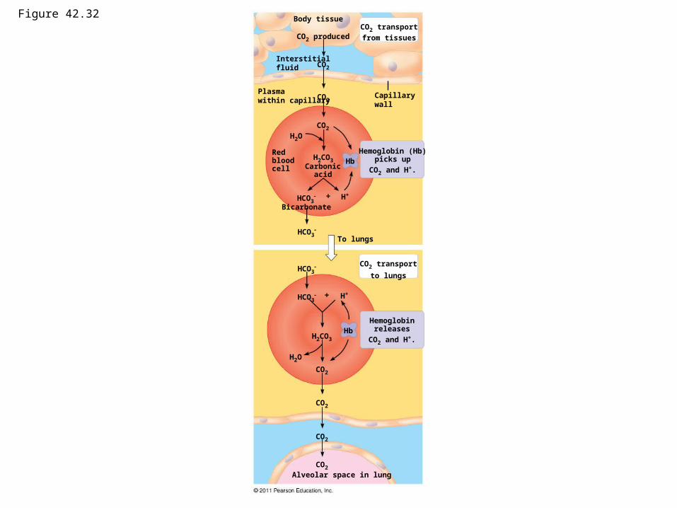
Figure 42.32 Body tissue
Capillarywall
Interstitialfluid
Plasmawithin capillary
CO2 transport
from tissuesCO2 produced
CO2
CO2
CO2
H2O
H2CO3 HbRedbloodcell Carbonic
acid
Hemoglobin (Hb)picks up
CO2 and H+.
H+HCO3
Bicarbonate
HCO3
HCO3
To lungs
CO2 transport
to lungs
HCO3
H2CO3
H2O
CO2
H+
HbHemoglobin
releases
CO2 and H+.
CO2
CO2
CO2
Alveolar space in lung
Respiratory Adaptations of Diving Mammals
• Diving mammals have evolutionary adaptations that allow them to perform extraordinary feats
– For example, Weddell seals in Antarctica can remain underwater for 20 minutes to an hour
– For example, elephant seals can dive to 1,500 m and remain underwater for 2 hours
• These animals have a high blood to body volume ratio
© 2011 Pearson Education, Inc.
• Deep-diving air breathers stockpile O2 and deplete it slowly
• Diving mammals can store oxygen in their muscles in myoglobin proteins
• Diving mammals also conserve oxygen by– Changing their buoyancy to glide passively– Decreasing blood supply to muscles– Deriving ATP in muscles from fermentation once
oxygen is depleted
© 2011 Pearson Education, Inc.