4 hemodynamic disorders, thromboembolic disease, and...
TRANSCRIPT
Hemodynamic Disorders, Thromboembolic Disease, and Shock
Downloaded from: StudentConsult (on 10 January 2011 10:49 AM)Kumar et al: Robbins & Cotran Pathologic Basis of Disease 7E
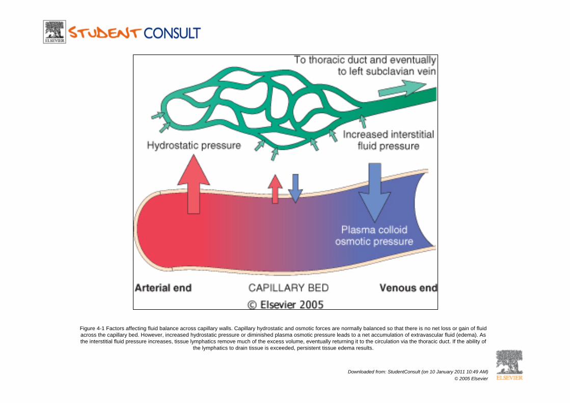
Figure 4-1 Factors affecting fluid balance across capillary walls. Capillary hydrostatic and osmotic forces are normally balanced so that there is no net loss or gain of fluid across the capillary bed. However, increased hydrostatic pressure or diminished plasma osmotic pressure leads to a net accumulation of extravascular fluid (edema). As the interstitial fluid pressure increases, tissue lymphatics remove much of the excess volume, eventually returning it to the circulation via the thoracic duct. If the ability of
the lymphatics to drain tissue is exceeded, persistent tissue edema results.
Downloaded from: StudentConsult (on 10 January 2011 10:49 AM)© 2005 Elsevier
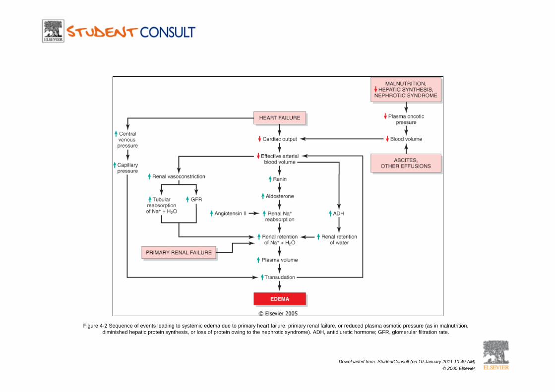
Figure 4-2 Sequence of events leading to systemic edema due to primary heart failure, primary renal failure, or reduced plasma osmotic pressure (as in malnutrition, diminished hepatic protein synthesis, or loss of protein owing to the nephrotic syndrome). ADH, antidiuretic hormone; GFR, glomerular filtration rate.
Downloaded from: StudentConsult (on 10 January 2011 10:49 AM)© 2005 Elsevier
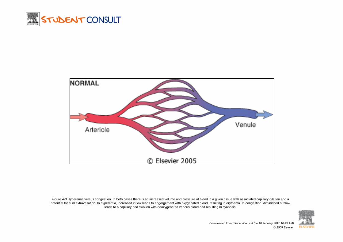
Figure 4-3 Hyperemia versus congestion. In both cases there is an increased volume and pressure of blood in a given tissue with associated capillary dilation and a potential for fluid extravasation. In hyperemia, increased inflow leads to engorgement with oxygenated blood, resulting in erythema. In congestion, diminished outflow
leads to a capillary bed swollen with deoxygenated venous blood and resulting in cyanosis.
Downloaded from: StudentConsult (on 10 January 2011 10:49 AM)© 2005 Elsevier
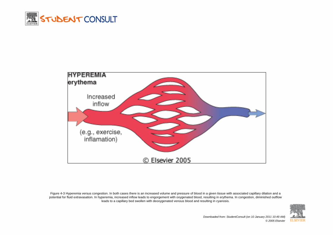
Figure 4-3 Hyperemia versus congestion. In both cases there is an increased volume and pressure of blood in a given tissue with associated capillary dilation and a potential for fluid extravasation. In hyperemia, increased inflow leads to engorgement with oxygenated blood, resulting in erythema. In congestion, diminished outflow
leads to a capillary bed swollen with deoxygenated venous blood and resulting in cyanosis.
Downloaded from: StudentConsult (on 10 January 2011 10:49 AM)© 2005 Elsevier
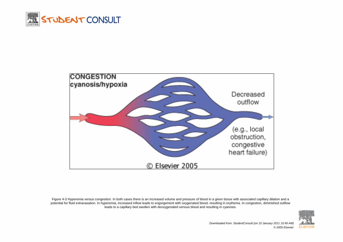
Figure 4-3 Hyperemia versus congestion. In both cases there is an increased volume and pressure of blood in a given tissue with associated capillary dilation and a potential for fluid extravasation. In hyperemia, increased inflow leads to engorgement with oxygenated blood, resulting in erythema. In congestion, diminished outflow
leads to a capillary bed swollen with deoxygenated venous blood and resulting in cyanosis.
Downloaded from: StudentConsult (on 10 January 2011 10:49 AM)© 2005 Elsevier
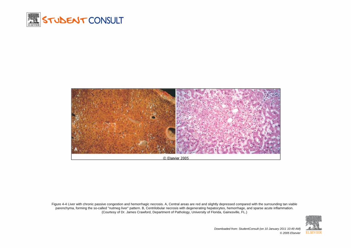
Figure 4-4 Liver with chronic passive congestion and hemorrhagic necrosis. A, Central areas are red and slightly depressed compared with the surrounding tan viable parenchyma, forming the so-called "nutmeg liver" pattern. B, Centrilobular necrosis with degenerating hepatocytes, hemorrhage, and sparse acute inflammation.
(Courtesy of Dr. James Crawford, Department of Pathology, University of Florida, Gainesville, FL.)
Downloaded from: StudentConsult (on 10 January 2011 10:49 AM)© 2005 Elsevier
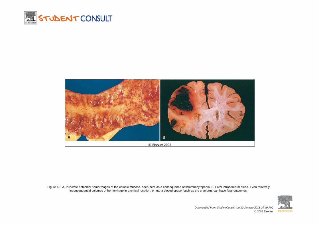
Figure 4-5 A, Punctate petechial hemorrhages of the colonic mucosa, seen here as a consequence of thrombocytopenia. B, Fatal intracerebral bleed. Even relatively inconsequential volumes of hemorrhage in a critical location, or into a closed space (such as the cranium), can have fatal outcomes.
Downloaded from: StudentConsult (on 10 January 2011 10:49 AM)© 2005 Elsevier
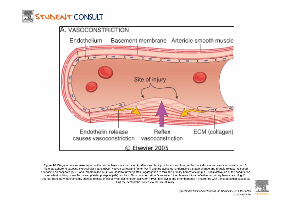
Figure 4-6 Diagrammatic representation of the normal hemostatic process. A, After vascular injury, local neurohumoral factors induce a transient vasoconstriction. B, Platelets adhere to exposed extracellular matrix (ECM) via von Willebrand factor (vWF) and are activated, undergoing a shape change and granule release; released
adenosine diphosphate (ADP) and thromboxane A2 (TxA2) lead to further platelet aggregation to form the primary hemostatic plug. C, Local activation of the coagulation cascade (involving tissue factor and platelet phospholipids) results in fibrin polymerization, "cementing" the platelets into a definitive secondary hemostatic plug. D,
Counter-regulatory mechanisms, such as release of tissue type plasminogen activator (t-PA) (fibrinolytic) and thrombomodulin (interfering with the coagulation cascade), limit the hemostatic process to the site of injury.
Downloaded from: StudentConsult (on 10 January 2011 10:49 AM)© 2005 Elsevier
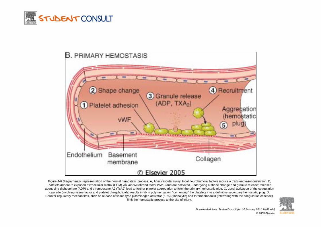
Figure 4-6 Diagrammatic representation of the normal hemostatic process. A, After vascular injury, local neurohumoral factors induce a transient vasoconstriction. B, Platelets adhere to exposed extracellular matrix (ECM) via von Willebrand factor (vWF) and are activated, undergoing a shape change and granule release; released
adenosine diphosphate (ADP) and thromboxane A2 (TxA2) lead to further platelet aggregation to form the primary hemostatic plug. C, Local activation of the coagulation cascade (involving tissue factor and platelet phospholipids) results in fibrin polymerization, "cementing" the platelets into a definitive secondary hemostatic plug. D,
Counter-regulatory mechanisms, such as release of tissue type plasminogen activator (t-PA) (fibrinolytic) and thrombomodulin (interfering with the coagulation cascade), limit the hemostatic process to the site of injury.
Downloaded from: StudentConsult (on 10 January 2011 10:49 AM)© 2005 Elsevier
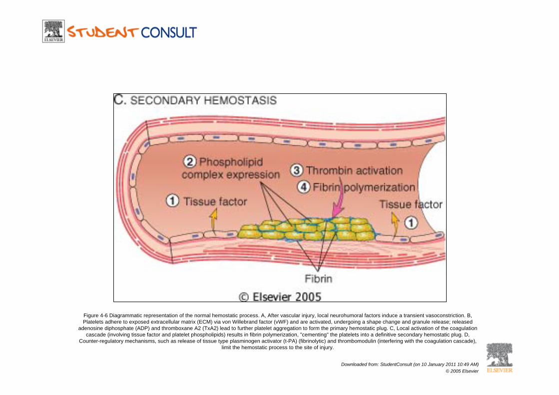
Figure 4-6 Diagrammatic representation of the normal hemostatic process. A, After vascular injury, local neurohumoral factors induce a transient vasoconstriction. B, Platelets adhere to exposed extracellular matrix (ECM) via von Willebrand factor (vWF) and are activated, undergoing a shape change and granule release; released
adenosine diphosphate (ADP) and thromboxane A2 (TxA2) lead to further platelet aggregation to form the primary hemostatic plug. C, Local activation of the coagulation cascade (involving tissue factor and platelet phospholipids) results in fibrin polymerization, "cementing" the platelets into a definitive secondary hemostatic plug. D,
Counter-regulatory mechanisms, such as release of tissue type plasminogen activator (t-PA) (fibrinolytic) and thrombomodulin (interfering with the coagulation cascade), limit the hemostatic process to the site of injury.
Downloaded from: StudentConsult (on 10 January 2011 10:49 AM)© 2005 Elsevier
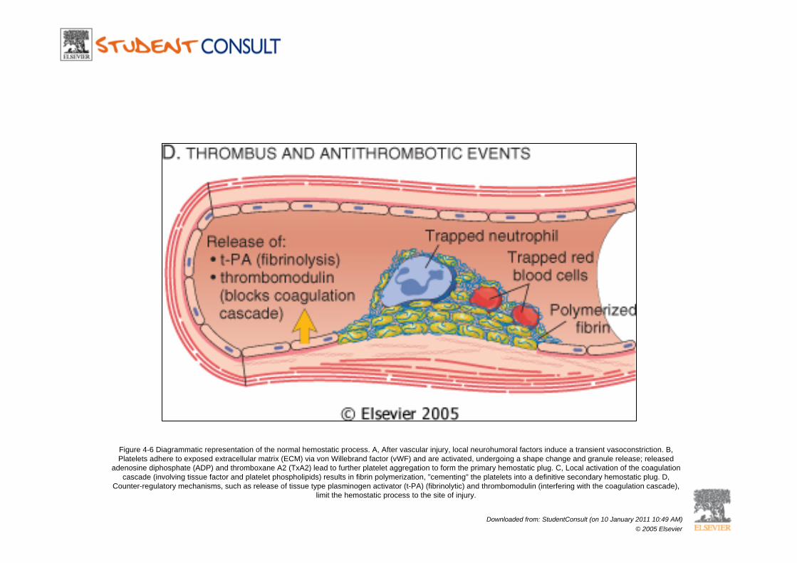
Figure 4-6 Diagrammatic representation of the normal hemostatic process. A, After vascular injury, local neurohumoral factors induce a transient vasoconstriction. B, Platelets adhere to exposed extracellular matrix (ECM) via von Willebrand factor (vWF) and are activated, undergoing a shape change and granule release; released
adenosine diphosphate (ADP) and thromboxane A2 (TxA2) lead to further platelet aggregation to form the primary hemostatic plug. C, Local activation of the coagulation cascade (involving tissue factor and platelet phospholipids) results in fibrin polymerization, "cementing" the platelets into a definitive secondary hemostatic plug. D,
Counter-regulatory mechanisms, such as release of tissue type plasminogen activator (t-PA) (fibrinolytic) and thrombomodulin (interfering with the coagulation cascade), limit the hemostatic process to the site of injury.
Downloaded from: StudentConsult (on 10 January 2011 10:49 AM)© 2005 Elsevier
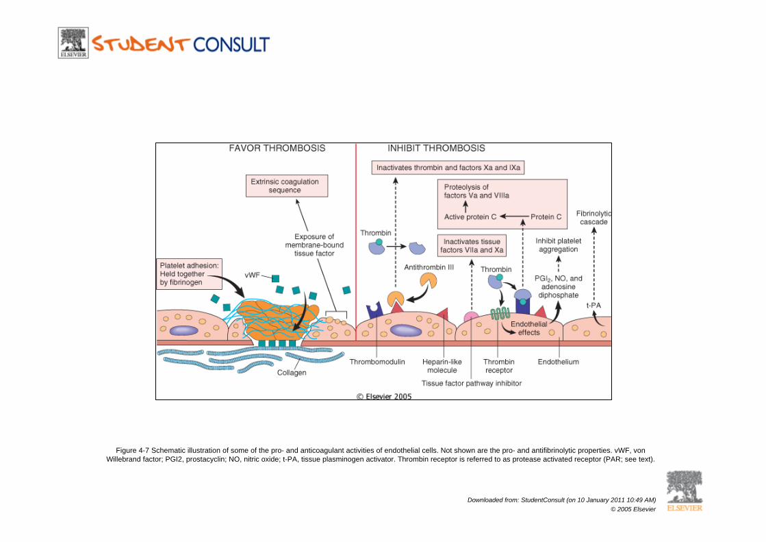
Figure 4-7 Schematic illustration of some of the pro- and anticoagulant activities of endothelial cells. Not shown are the pro- and antifibrinolytic properties. vWF, von Willebrand factor; PGI2, prostacyclin; NO, nitric oxide; t-PA, tissue plasminogen activator. Thrombin receptor is referred to as protease activated receptor (PAR; see text).
Downloaded from: StudentConsult (on 10 January 2011 10:49 AM)© 2005 Elsevier
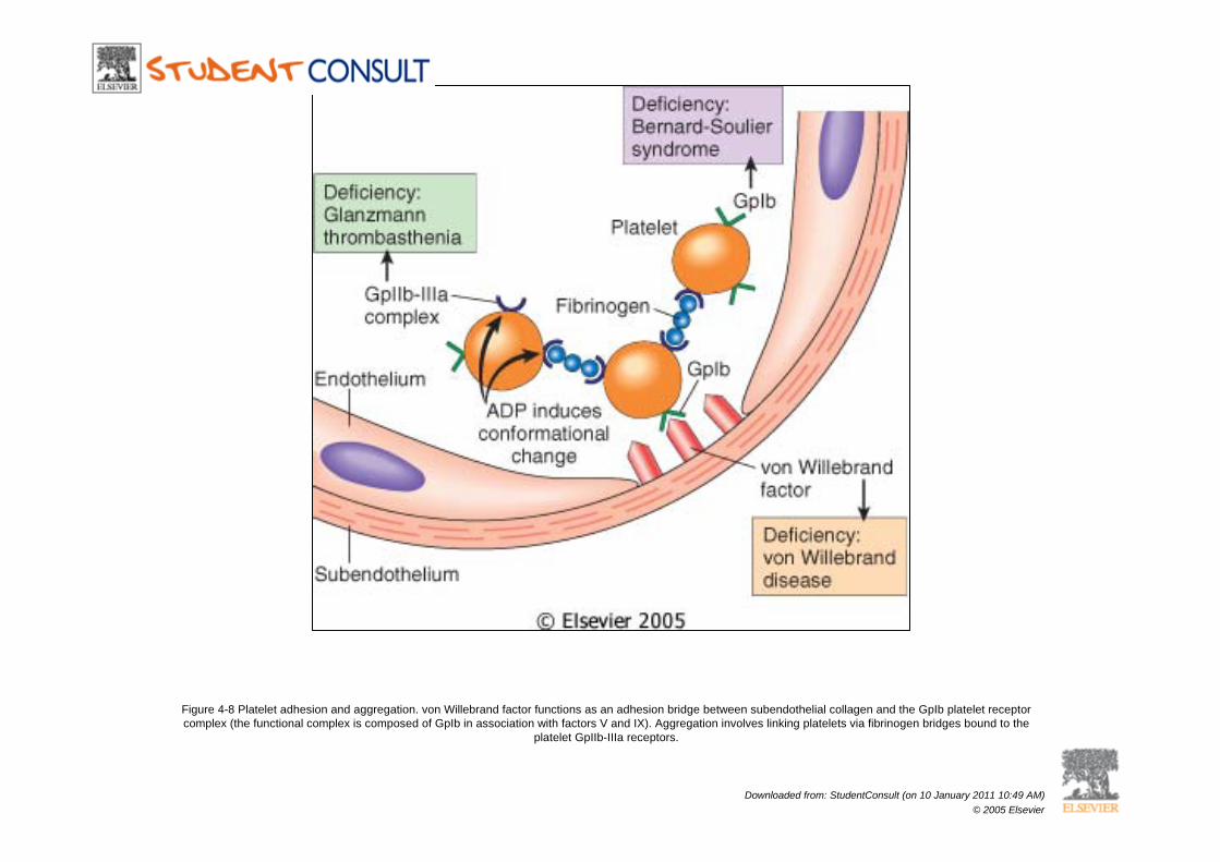
Figure 4-8 Platelet adhesion and aggregation. von Willebrand factor functions as an adhesion bridge between subendothelial collagen and the GpIb platelet receptor complex (the functional complex is composed of GpIb in association with factors V and IX). Aggregation involves linking platelets via fibrinogen bridges bound to the
platelet GpIIb-IIIa receptors.
Downloaded from: StudentConsult (on 10 January 2011 10:49 AM)© 2005 Elsevier
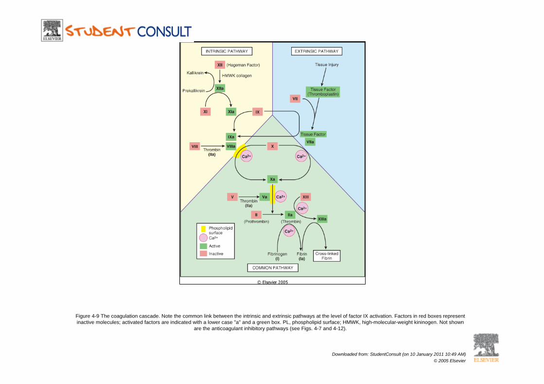
Figure 4-9 The coagulation cascade. Note the common link between the intrinsic and extrinsic pathways at the level of factor IX activation. Factors in red boxes represent inactive molecules; activated factors are indicated with a lower case "a" and a green box. PL, phospholipid surface; HMWK, high-molecular-weight kininogen. Not shown
are the anticoagulant inhibitory pathways (see Figs. 4-7 and 4-12).
Downloaded from: StudentConsult (on 10 January 2011 10:49 AM)© 2005 Elsevier
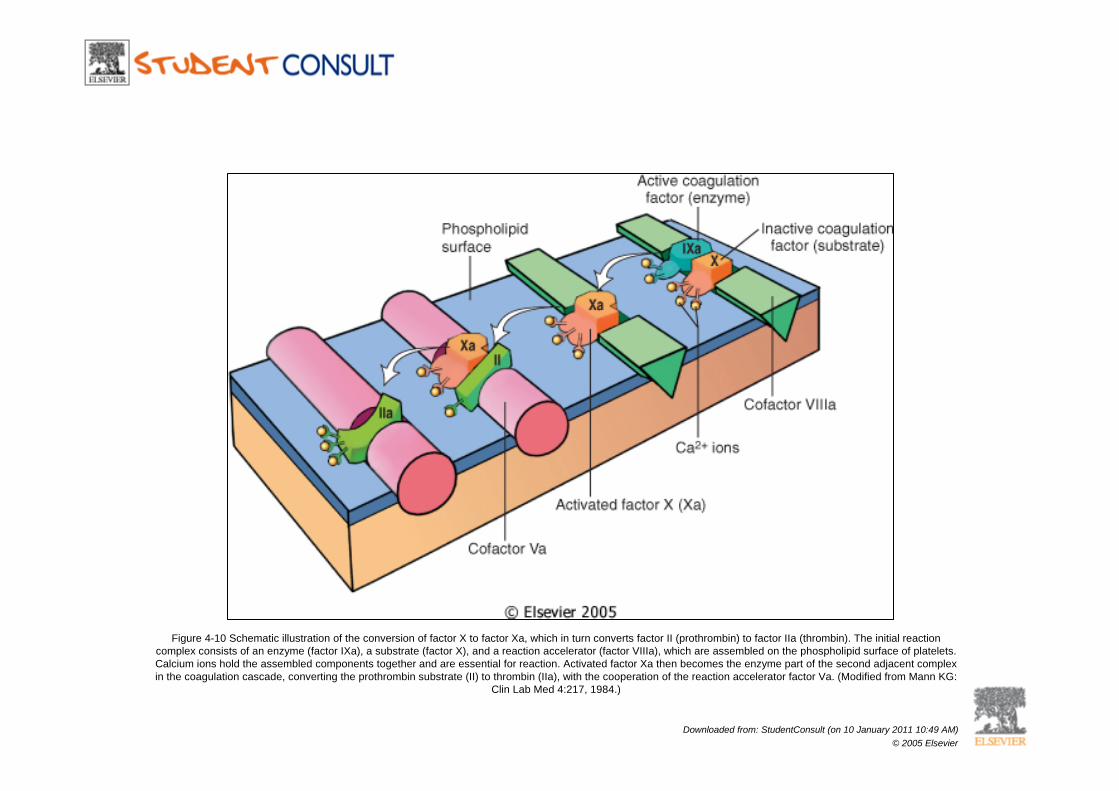
Figure 4-10 Schematic illustration of the conversion of factor X to factor Xa, which in turn converts factor II (prothrombin) to factor IIa (thrombin). The initial reaction complex consists of an enzyme (factor IXa), a substrate (factor X), and a reaction accelerator (factor VIIIa), which are assembled on the phospholipid surface of platelets. Calcium ions hold the assembled components together and are essential for reaction. Activated factor Xa then becomes the enzyme part of the second adjacent complex in the coagulation cascade, converting the prothrombin substrate (II) to thrombin (IIa), with the cooperation of the reaction accelerator factor Va. (Modified from Mann KG:
Clin Lab Med 4:217, 1984.)
Downloaded from: StudentConsult (on 10 January 2011 10:49 AM)© 2005 Elsevier
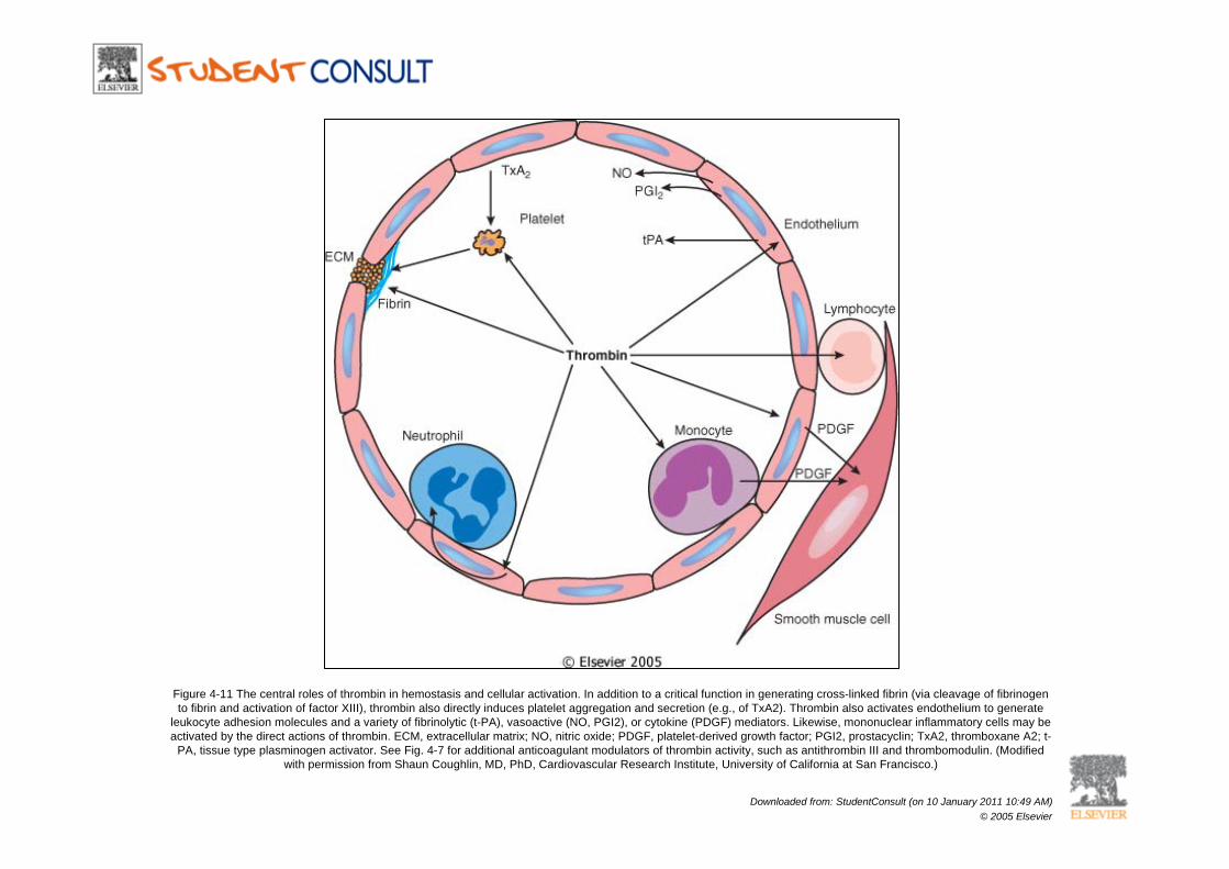
Figure 4-11 The central roles of thrombin in hemostasis and cellular activation. In addition to a critical function in generating cross-linked fibrin (via cleavage of fibrinogen to fibrin and activation of factor XIII), thrombin also directly induces platelet aggregation and secretion (e.g., of TxA2). Thrombin also activates endothelium to generate
leukocyte adhesion molecules and a variety of fibrinolytic (t-PA), vasoactive (NO, PGI2), or cytokine (PDGF) mediators. Likewise, mononuclear inflammatory cells may be activated by the direct actions of thrombin. ECM, extracellular matrix; NO, nitric oxide; PDGF, platelet-derived growth factor; PGI2, prostacyclin; TxA2, thromboxane A2; t-
PA, tissue type plasminogen activator. See Fig. 4-7 for additional anticoagulant modulators of thrombin activity, such as antithrombin III and thrombomodulin. (Modified with permission from Shaun Coughlin, MD, PhD, Cardiovascular Research Institute, University of California at San Francisco.)
Downloaded from: StudentConsult (on 10 January 2011 10:49 AM)© 2005 Elsevier
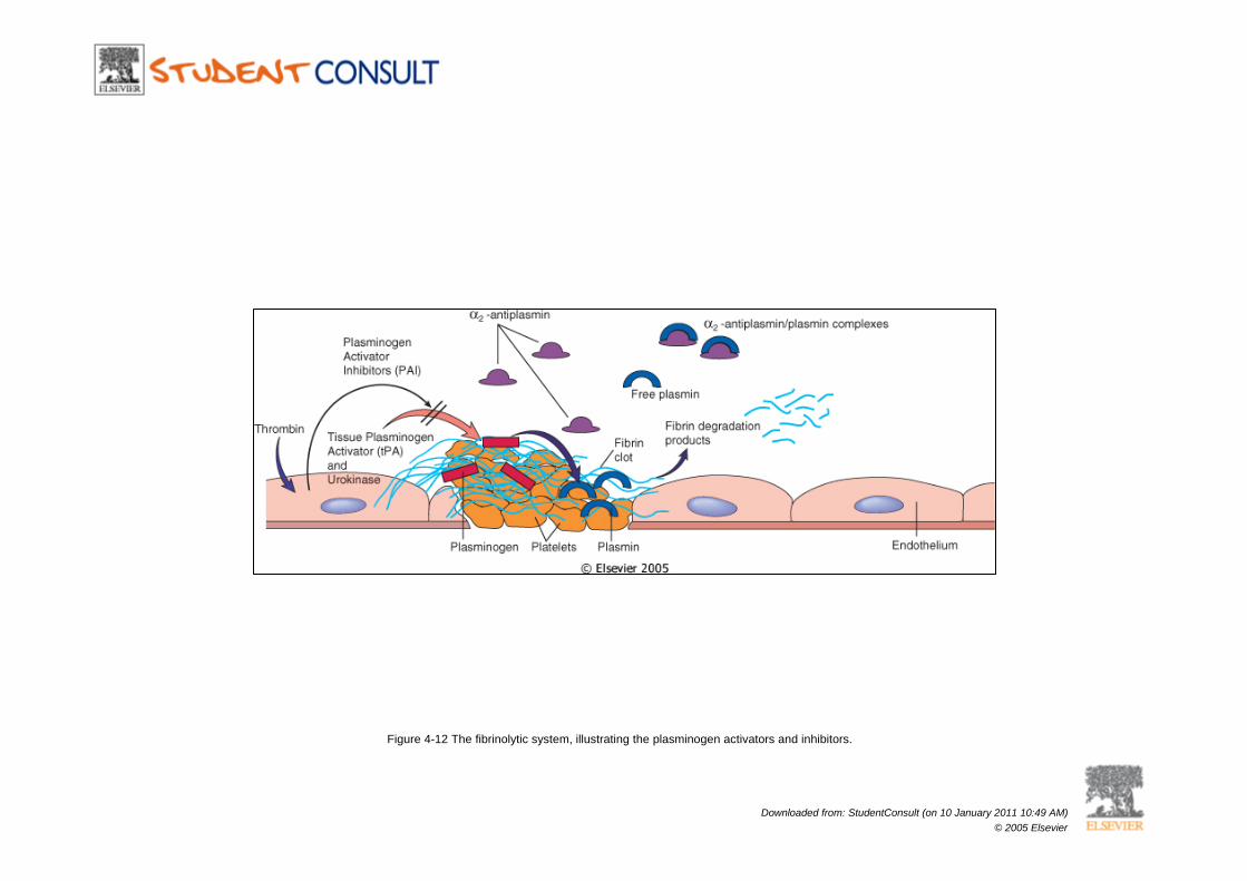
Figure 4-12 The fibrinolytic system, illustrating the plasminogen activators and inhibitors.
Downloaded from: StudentConsult (on 10 January 2011 10:49 AM)© 2005 Elsevier
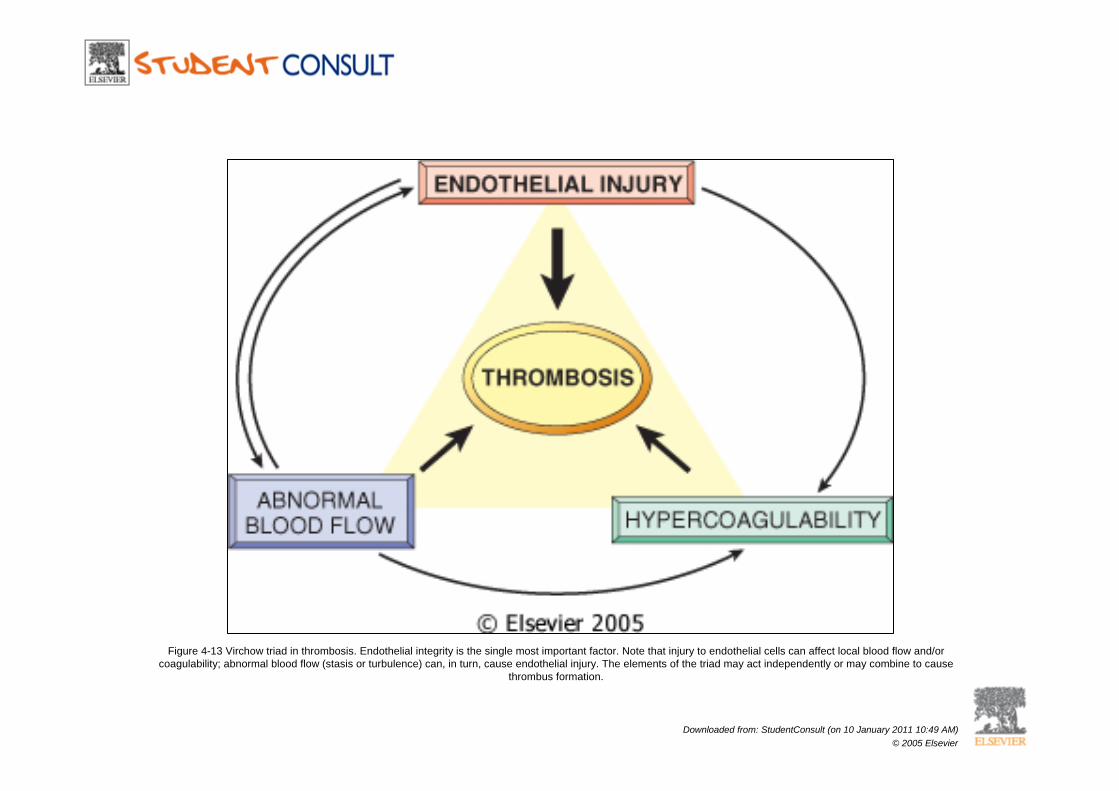
Figure 4-13 Virchow triad in thrombosis. Endothelial integrity is the single most important factor. Note that injury to endothelial cells can affect local blood flow and/or coagulability; abnormal blood flow (stasis or turbulence) can, in turn, cause endothelial injury. The elements of the triad may act independently or may combine to cause
thrombus formation.
Downloaded from: StudentConsult (on 10 January 2011 10:49 AM)© 2005 Elsevier
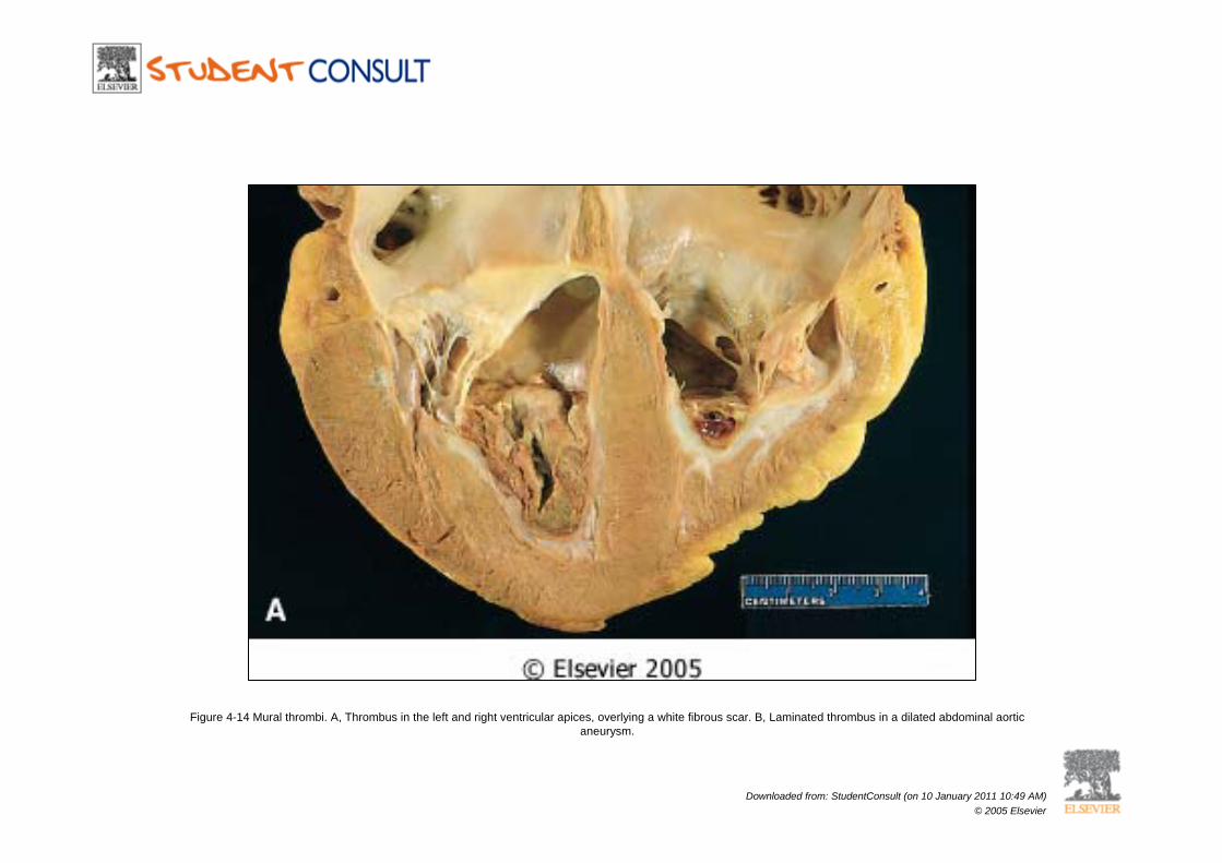
Figure 4-14 Mural thrombi. A, Thrombus in the left and right ventricular apices, overlying a white fibrous scar. B, Laminated thrombus in a dilated abdominal aortic aneurysm.
Downloaded from: StudentConsult (on 10 January 2011 10:49 AM)© 2005 Elsevier
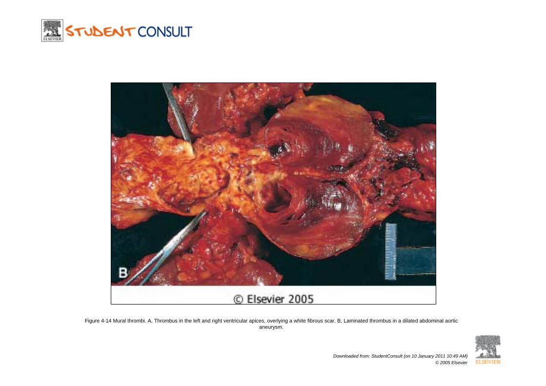
Figure 4-14 Mural thrombi. A, Thrombus in the left and right ventricular apices, overlying a white fibrous scar. B, Laminated thrombus in a dilated abdominal aortic aneurysm.
Downloaded from: StudentConsult (on 10 January 2011 10:49 AM)© 2005 Elsevier
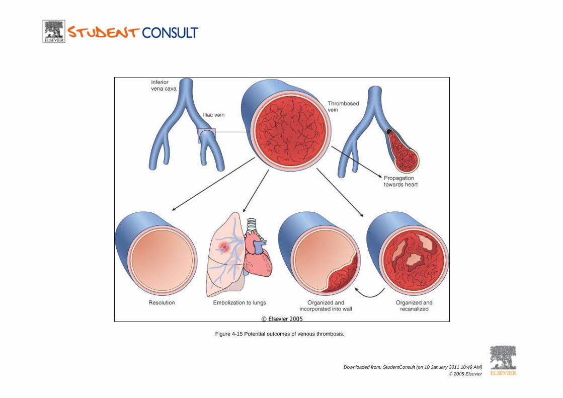
Figure 4-15 Potential outcomes of venous thrombosis.
Downloaded from: StudentConsult (on 10 January 2011 10:49 AM)© 2005 Elsevier
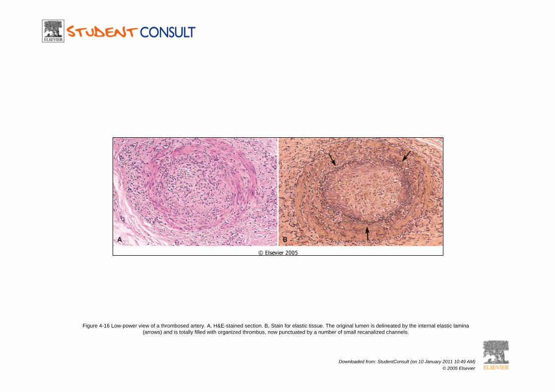
Figure 4-16 Low-power view of a thrombosed artery. A, H&E-stained section. B, Stain for elastic tissue. The original lumen is delineated by the internal elastic lamina (arrows) and is totally filled with organized thrombus, now punctuated by a number of small recanalized channels.
Downloaded from: StudentConsult (on 10 January 2011 10:49 AM)© 2005 Elsevier
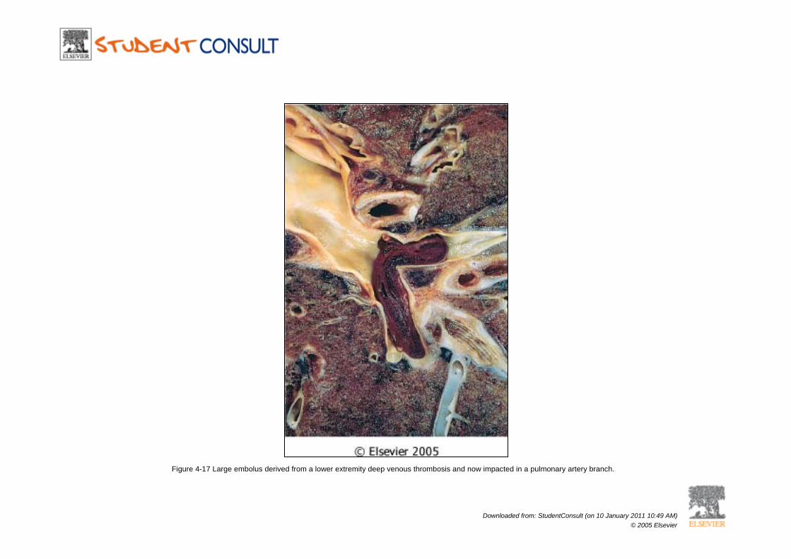
Figure 4-17 Large embolus derived from a lower extremity deep venous thrombosis and now impacted in a pulmonary artery branch.
Downloaded from: StudentConsult (on 10 January 2011 10:49 AM)© 2005 Elsevier
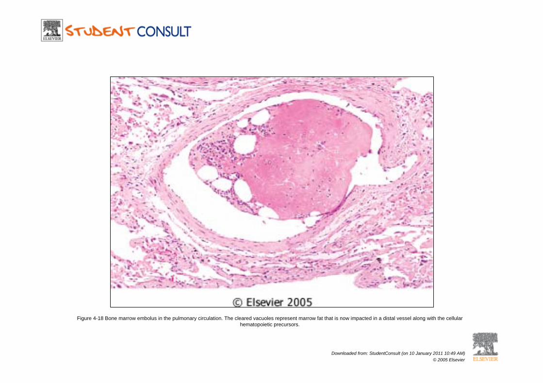
Figure 4-18 Bone marrow embolus in the pulmonary circulation. The cleared vacuoles represent marrow fat that is now impacted in a distal vessel along with the cellular hematopoietic precursors.
Downloaded from: StudentConsult (on 10 January 2011 10:49 AM)© 2005 Elsevier
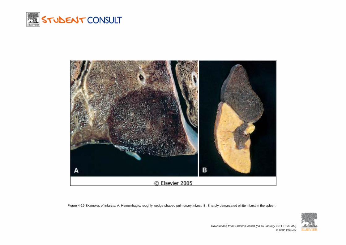
Figure 4-19 Examples of infarcts. A, Hemorrhagic, roughly wedge-shaped pulmonary infarct. B, Sharply demarcated white infarct in the spleen.
Downloaded from: StudentConsult (on 10 January 2011 10:49 AM)© 2005 Elsevier
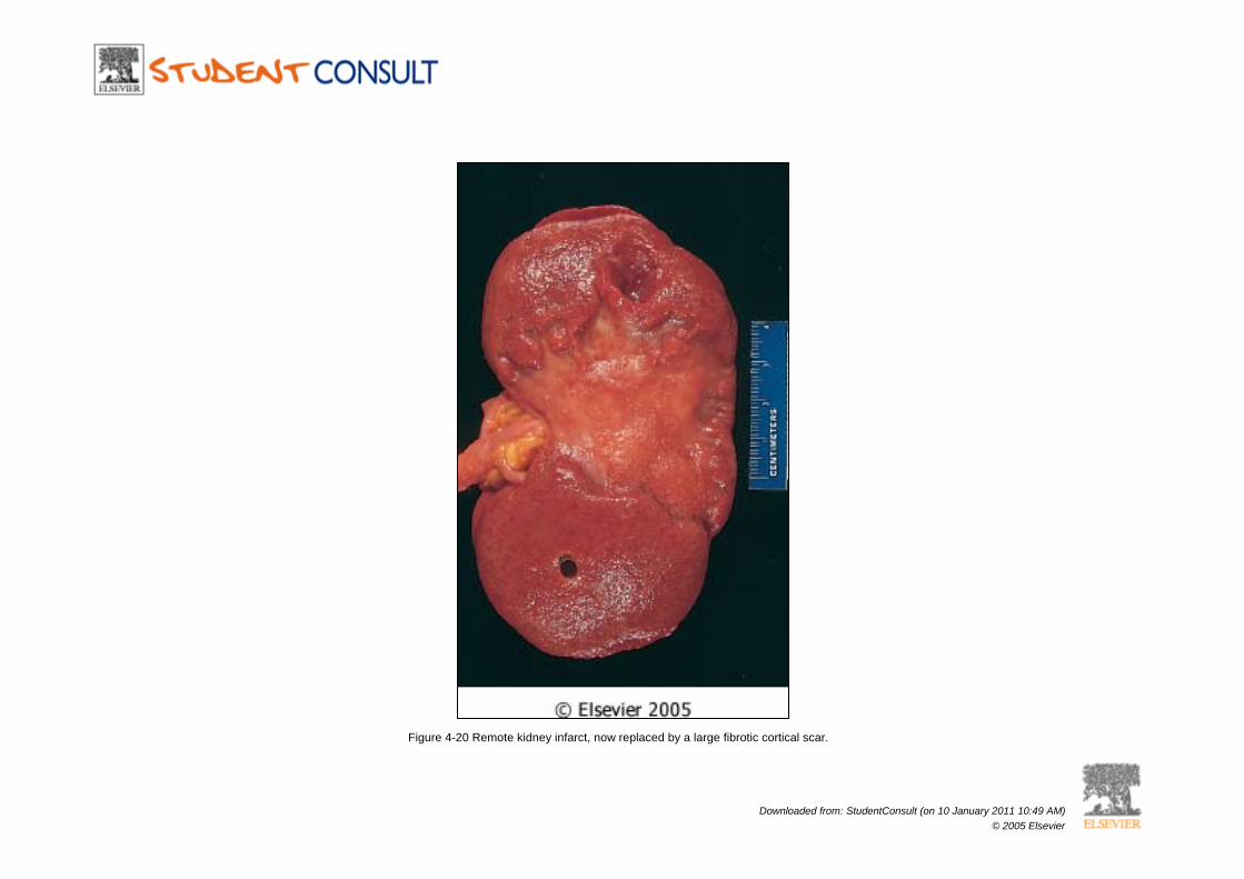
Figure 4-20 Remote kidney infarct, now replaced by a large fibrotic cortical scar.
Downloaded from: StudentConsult (on 10 January 2011 10:49 AM)© 2005 Elsevier
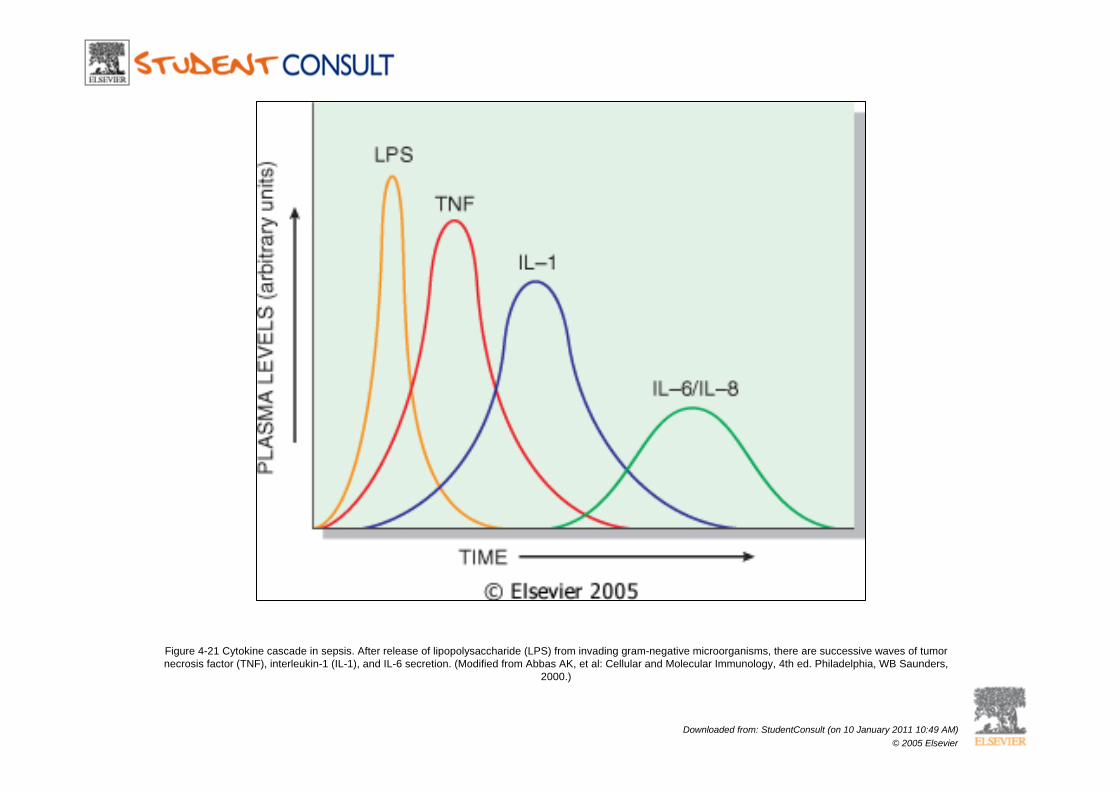
Figure 4-21 Cytokine cascade in sepsis. After release of lipopolysaccharide (LPS) from invading gram-negative microorganisms, there are successive waves of tumor necrosis factor (TNF), interleukin-1 (IL-1), and IL-6 secretion. (Modified from Abbas AK, et al: Cellular and Molecular Immunology, 4th ed. Philadelphia, WB Saunders,
2000.)
Downloaded from: StudentConsult (on 10 January 2011 10:49 AM)© 2005 Elsevier
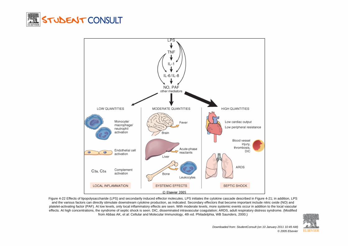
Figure 4-22 Effects of lipopolysaccharide (LPS) and secondarily induced effector molecules. LPS initiates the cytokine cascade described in Figure 4-21; in addition, LPS and the various factors can directly stimulate downstream cytokine production, as indicated. Secondary effectors that become important include nitric oxide (NO) and
platelet-activating factor (PAF). At low levels, only local inflammatory effects are seen. With moderate levels, more systemic events occur in addition to the local vascular effects. At high concentrations, the syndrome of septic shock is seen. DIC, disseminated intravascular coagulation; ARDS, adult respiratory distress syndrome. (Modified
from Abbas AK, et al: Cellular and Molecular Immunology, 4th ed. Philadelphia, WB Saunders, 2000.)
Downloaded from: StudentConsult (on 10 January 2011 10:49 AM)© 2005 Elsevier